

Submitted:
08 July 2026
Posted:
13 July 2026
You are already at the latest version
Preprints on COVID-19 and SARS-CoV-2
Abstract
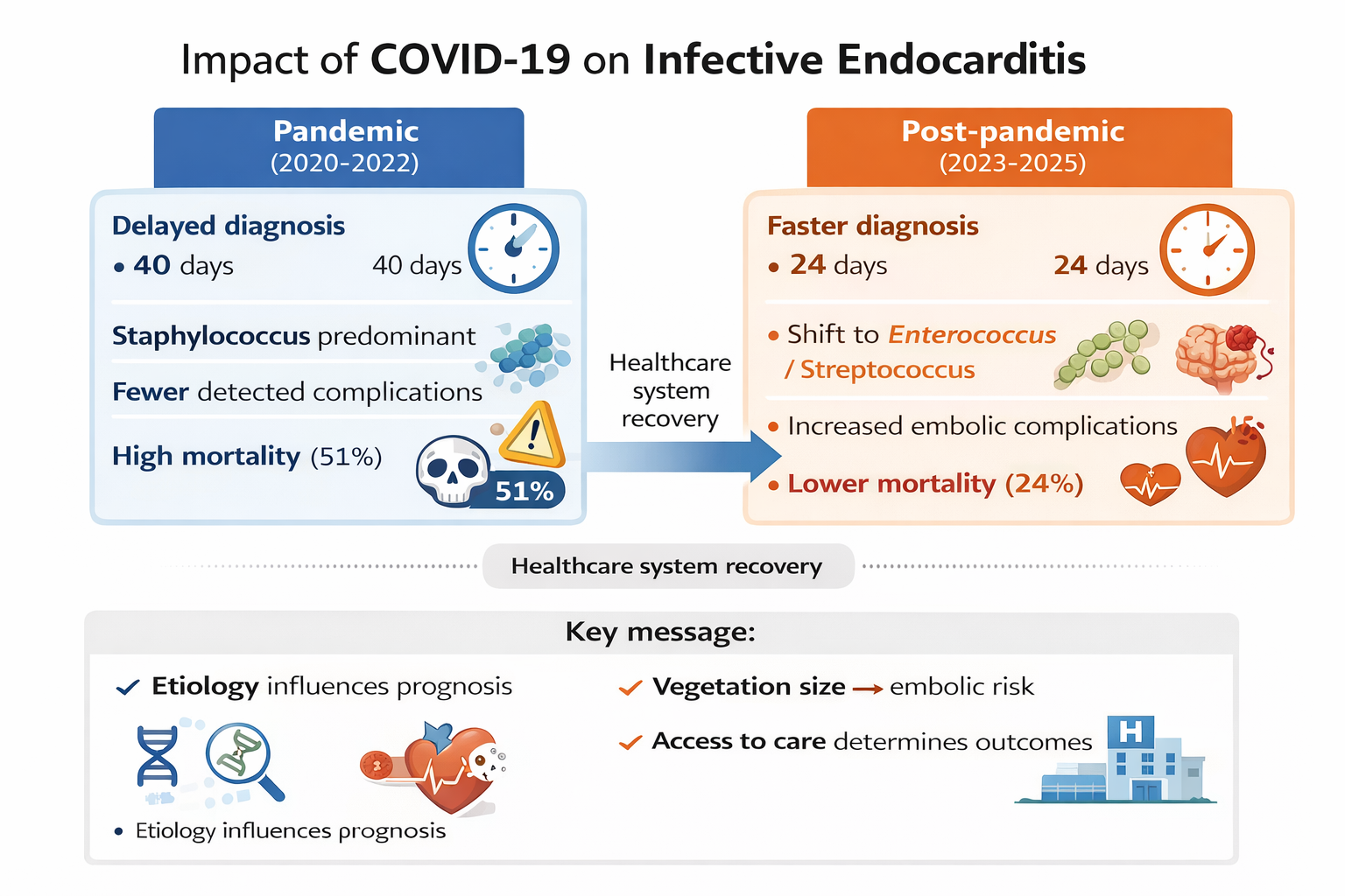
Objectives: To assess the impact of the COVID-19 pandemic on the epidemiology, microbiology, clinical presentation, management, and outcomes of infective endocarditis (IE). Methods: A retrospective cohort study was conducted, including 100 adult patients with definite IE diagnosed between January 2020 and December 2025 at a Romanian tertiary care center. Patients were stratified by year of admission into pandemic (2020–2022) and post-pandemic (2023–2025) groups. Results: Among the 100 patients (47 in the pandemic period and 53 in the post-pandemic period), the time from symptom onset to diagnosis was significantly shorter in the post-pandemic period (median of 24 days versus 40 days; p < 0.01). The microbiological etiology shifted from a predominance of staphylococci during the pandemic to a more balanced distribution of enterococci and streptococci in the post-pandemic period (p = 0.03). In-hospital mortality decreased significantly from 51.1% to 24.5% (p = 0.007), and survival improved from 48.9% to 75.5% (p = 0.007). Embolic events were more frequent in the post-pandemic period (62.3% vs. 38.3%; p = 0.028). Vegetation size was associated with embolic risk (OR: 1.05 per mm; p < 0.01). Conclusions: The COVID-19 pandemic was associated with delayed presentation and worse outcomes in IE. Post-pandemic improvements likely reflect healthcare system adaptation and earlier diagnosis. Microbiological etiology remains a key determinant of prognosis.
Keywords:
infective endocarditis
; COVID-19
; Enterococcus
; Staphylococcus
; embolism
; mortality
1. Introduction
Infective endocarditis (IE) is a severe disease associated with high morbidity and mortality, with an estimated incidence of 3–10 cases per 100,000 population and mortality rates of 20–30% [1]. The epidemiology of IE has evolved substantially over recent decades due to population ageing, increasing comorbidity burden, and greater exposure to healthcare-associated interventions. These factors have contributed to a rising incidence and a shift toward more complex, high-risk patient profiles [2,3].
IE is associated with significant short- and long-term mortality and frequent complications. Outcomes are influenced by patient characteristics, access to specialized care, and, importantly, the causative microorganism. Delayed diagnosis and limited access to multidisciplinary endocarditis teams are associated with worse outcomes, including higher complication rates and delayed or underuse of indicated cardiac surgery [4].
The microbiological profile of IE has also changed. While viridans group streptococci historically predominated, staphylococci—particularly Staphylococcus aureus—are now leading pathogens in many contemporary series, reflecting the increasing burden of healthcare-associated infections [2,5,6]. In addition, enterococci, especially Enterococcus faecalis, are increasingly recognized, particularly in elderly patients and those with prior healthcare exposure. Microbial etiology is a major determinant of disease severity and prognosis [2,7].
Advances in diagnostic techniques and treatment strategies, together with multidisciplinary team approaches, have improved outcomes [8,9,10]. However, the SARS-CoV-2 pandemic placed unprecedented strain on healthcare systems, disrupting diagnostic pathways, delaying presentation, and limiting access to specialized care. Several reports have described reductions in IE diagnoses and cardiac surgical procedures during peak pandemic periods, although the long-term consequences remain unclear [11,12].
Data from Eastern Europe remain limited, particularly regarding the impact of the pandemic on IE. This study aimed to evaluate changes in epidemiology, microbiology, management, and outcomes of IE in a Romanian tertiary care centre during and after the COVID-19 pandemic.
2. Results
A total of 100 patients with definite infective endocarditis were included: 47 during the pandemic and 53 afterward. The baseline characteristics of the two groups were comparable (Table 1), including age (median 68 years in both groups, p = 0.853), sex distribution, and type of valve involvement.
The microbiological distribution differed between the two periods (Figure 1A; p = 0.03). There was a reduction in staphylococcal IE (27.7% vs. 20.8%) and a relative increase in enterococcal (12.8% vs. 17.0%) and streptococcal (14.9% vs. 17.0%) cases post-pandemic. Culture-negative cases were more frequent post-pandemic (Table 1).
Post-pandemic patients had higher rates of complications, including perivalvular abscess (8.9% vs. 26.5%, p = 0.033) and embolic events (37.8% vs. 63.3%, p = 0.023), particularly central nervous system embolism (6.7% vs. 32.7%, p = 0.002). Vegetation size was associated with embolic risk (Figure 1C; odds ratio [OR] 1.05 per mm, p < 0.01).
3. Discussion
This study identified significant changes in the presentation, microbiology, and outcomes of infective endocarditis (IE) during and after the pandemic. These results underscore the impact of healthcare system disruption during the pandemic and the subsequent recovery of diagnostic and management processes.
One notable finding is the substantial reduction in time to diagnosis during the post-pandemic period. This likely reflects improved access to healthcare, restored referral pathways, and increased clinical awareness following the pandemic. During the pandemic, delayed presentation and restricted access to care likely contributed to prolonged symptom duration prior to diagnosis [11]. Although diagnostic timeliness improved post-pandemic, it did not fully translate into reduced disease severity at presentation.
Despite earlier diagnoses, post-pandemic patients experienced higher rates of embolic and local cardiac complications, including perivalvular abscesses and central nervous system embolisms. This may be due to several factors. Differences in pathogen virulence, vegetation characteristics, and host comorbidities may influence the risk of embolization independently of diagnostic timing. Second, more widespread use of advanced cardiac imaging, particularly TEE, after the pandemic may have led to more cases of abscesses being detected. Finally, embolic complications often occur before infective endocarditis is suspected, meaning that earlier diagnosis after hospital presentation may not prevent these events. Therefore, diagnostic delay is just one of several factors determining disease severity. The observed association between vegetation size and embolic risk reinforces vegetation’s role as a key prognostic marker, supporting early surgical consideration in high-risk patients [14,15,16].
We also observed a significant shift in microbiological etiology: a reduction in staphylococcal infections and an increase in enterococcal and streptococcal IE post-pandemic. This pattern is consistent with evolving epidemiological trends and may reflect changes in healthcare exposure, patient demographics, and the resumption of elective procedures [6,15,17,18].
The persistently high proportion of culture-negative cases underscores the ongoing limitations of microbiological diagnostics and the necessity of improved techniques, including molecular methods [8,19].
In-hospital mortality was significantly higher during the pandemic, which underscores the impact of healthcare disruption. Reduced access to specialized care and surgical services likely contributed to worse outcomes. The substantial improvement in survival post-pandemic underscores the importance of timely diagnosis, multidisciplinary management, and access to cardiac surgery [14,20].
These findings suggest that improving diagnostic timeliness alone is insufficient to optimize outcomes. Disease severity at presentation, microbial etiology, and access to specialized care remain key determinants of prognosis [10,18,21,22].
This study has limitations, including its retrospective, single-center design and moderate sample size. Potential changes in referral patterns may have introduced selection bias as well. In addition to the limitations inherent in its retrospective, single-centre design, this study did not systematically evaluate changes over time in the use, timing or frequency of transoesophageal echocardiography (TEE). The increased availability of TEE or its earlier implementation in the post-pandemic period may have improved the detection of vegetations and perivalvular complications, thereby influencing abscess formation and other imaging findings. Furthermore, we did not assess the evolution of blood culture-negative infective endocarditis over the study period, nor did we consider the use of molecular techniques, prolonged culture incubation, serological testing for fastidious organisms or 16S rRNA sequencing. These factors may have impacted the microbiological diagnosis and must be considered when interpreting changes in pathogen distribution between the study periods.
Nevertheless, the study provides valuable data from Eastern Europe, a region that is underrepresented in IE research.
4. Materials and Methods
4.1. Study Design and Setting
This retrospective observational cohort study was conducted at a university hospital providing tertiary care in Bucharest, Romania. The study adhered to the Strengthening the Reporting of Observational Studies in Epidemiology (STROBE) guidelines.
4.2. Study Population
All consecutive adult patients (≥18 years old) diagnosed with definite infective endocarditis (IE) between January 2020 and December 2025 were included. Diagnosis was established according to the modified Duke criteria.
Patients were stratified according to their admission period into the following groups:
Pandemic period: January 2020–December 2022
Post-pandemic period: January 2023–December 2025.
Patients with incomplete medical records or an uncertain diagnosis were excluded.
4.3. Data Collection
Data were extracted from electronic medical records using a standardized form. The following variables were recorded:
- -
- Demographic characteristics (age and sex);
- -
- Comorbidities (including cardiovascular disease, diabetes, chronic kidney disease, and others);
- -
- Microbiological etiology (based on blood cultures and/or intraoperative samples);
- -
- Valve involvement (native or prosthetic) and location;
- -
- Echocardiographic findings, including vegetation size;
- -
- Embolic and other complications (e.g., stroke and systemic embolism);
- -
- Management strategy (including antimicrobial therapy and surgical intervention);
- -
- Clinical outcomes (including in-hospital mortality and one-year survival); and
- -
- Outcomes.
The primary outcome was in-hospital mortality. Secondary outcomes included one-year survival and the occurrence of embolic complications.
4.4. Statistical Analysis
Continuous variables are presented as median and interquartile range (IQR) and were compared using the Mann–Whitney U test. Categorical variables are presented as counts and percentages, and were compared using the chi-squared test or Fisher’s exact test, as appropriate.
Univariable analysis was performed to identify factors associated with in-hospital mortality. Variables with p <0.10 in univariable analysis were entered into a multivariable logistic regression model to identify independent predictors of mortality. Results are reported as odds ratios (ORs) with 95% confidence intervals (CIs). Survival analysis was performed using the Kaplan–Meier method, with comparisons between groups assessed using the log-rank test.A two-sided p-value <0.05 was considered statistically significant.
Statistical analyses were performed using SPSS Statistics version 26.0 (IBM Corp., Armonk, NY, USA). Figures were generated using Python version 3.10, utilizing the Matplotlib and Seaborn libraries for data visualization.
4.5. Ethics
The study was approved by the institutional ethics committee. The requirement for informed consent was waived due to the retrospective nature of the study. All data were anonymized prior to analysis, and the study was conducted in accordance with the Declaration of Helsinki.
5. Conclusions
This single-centre study demonstrated that the post-pandemic period was associated with shorter diagnostic delays, lower in-hospital mortality, and improved survival compared with the pandemic period, alongside a shift in microbiological profile toward Enterococcus species. These findings suggest that healthcare disruptions during COVID-19 contributed to delayed diagnosis and worse outcomes, whereas post-pandemic improvements reflect recovery and adaptation of healthcare systems. Microbiological etiology remains a key determinant of prognosis and should guide clinical management and risk stratification.
Author Contributions
Conceptualization: Adelina Maria Radu, Oana Săndulescu and Valeriu Gheorghiță; Methodology: Adelina Maria Radu; Software: Adelina Maria Radu; Validation: Adelina Maria Radu, Oana Săndulescu and Valeriu Gheorghiță; Formal analysis: Adelina Maria Radu, Oana Săndulescu and Valeriu Gheorghiță; Investigation: Adelina Maria Radu, Irina Florentina Talpoși, Raluca Elena Țoțoiu, Roxana Berbecaru and Ana Dobrin; Resources: Adelina Maria Radu, Irina Florentina Talpoși, Raluca Elena Țoțoiu, Roxana Berbecaru, Ana Dobrin, Violeta Melinte, Cristina Maria Văcăroiu, Tiberiu Sebastian Holban, Matei Cherecheanu Popa, Oana Andreea Popa, Mădălina Elena Iancu, Luminița Tomescu and Amalia Călinoiu.; Data curation: Adelina Maria Radu; Writing -original draft preparation: Adelina Maria Radu; Writing -review and editing: Adelina Maria Radu, Oana Săndulescu and Valeriu Gheorghiță; Visualization: All authors; Supervision: Oana Săndulescu and Valeriu Gheorghiță; Project administration: Adelina Maria Radu, Oana Săndulescu and Valeriu Gheorghiță. All authors have read and agreed to the published version of the manuscript.
Funding
No external funding was received for this study.
Institutional Review Board Statement
The study was conducted in accordance with the Declaration of Helsinki, and approved by the Institutional Ethics Committee of Clinical Emergency Hospital Prof. Dr. Agrippa Ionescu (750262/ 21 Nov 2023).
Informed Consent Statement
The requirement for informed patient consent was waived by the Ethics Committee due to the retrospective observational design of the study, which involved the analysis of anonymized routinely collected clinical data without any intervention or direct patient contact.
Data Availability Statement
During the preparation of this manuscript, the authors used AI-assisted language tools to enhance the clarity and readability of the text. These tools were used solely for editorial purposes and did not contribute to the design of the study, the collection or analysis of the data, the interpretation of the findings, or the drawing of scientific conclusions. The authors reviewed and edited all AI-generated suggestions, and take full responsibility for the content of the manuscript.
Conflicts of Interest
The authors declare no conflicts of interest.
References
- Chen, H.; Zhan, Y.; Zhang, K.; Gao, Y.; Chen, L.; Zhan, J.; Chen, Z.; Zeng, Z. The Global, Regional, and National Burden and Trends of Infective Endocarditis From 1990 to 2019: Results From the Global Burden of Disease Study 2019. Front. Med. 2022, 9, 774224. [Google Scholar] [CrossRef] [PubMed]
- Patel, S.K.; Hassan, S.M.A.; Côté, M.; Leis, B.; Yanagawa, B. Current trends and challenges in infective endocarditis. Curr. Opin. Cardiol. 2024, 40, 75–84. [Google Scholar] [CrossRef] [PubMed]
- Calinoiu, A.; Guluta, E.-C.; Rusu, A.; Minca, A.; Minca, D.; Tomescu, L.; Gheorghita, V.; Minca, D.G.; Negreanu, L. Accessory renal arteries - a source of hypertension: A case report. World J. Clin. Cases 2023, 11, 1506–1512. [Google Scholar] [CrossRef] [PubMed]
- Epidemiological Profile and Mortality of Infective Endocarditis Over the Past Decade: A Systematic Review and Meta-Analysis of 133 Studies - PubMed [Internet]. 30 Mar 2026. Available online: https://pubmed.ncbi.nlm.nih.gov/40015544/ (accessed on 30 Mar 2026).
- Tzoumas, A.; Sagris, M.; Xenos, D.; Ntoumaziou, A.; Kyriakoulis, I.; Kakargias, F.; Liaqat, W.; Nagraj, S.; Patel, R.; Korosoglou, G.; et al. Epidemiological Profile and Mortality of Infective Endocarditis Over the Past Decade: A Systematic Review and Meta-Analysis of 133 Studies. Am. J. Cardiol. 2025, 244, 67–88. [Google Scholar] [CrossRef] [PubMed]
- Raad, I.I.; Hanna, H.A. Intravascular Catheter-Related Infections. Arch. Intern. Med. 2002, 162, 871–878. [Google Scholar] [CrossRef] [PubMed]
- Infective Endocarditis: Background, Pathophysiology, Etiology [Internet]. 30 Mar 2026. Available online: https://emedicine.medscape.com/article/216650-overview (accessed on 30 Mar 2026).
- Burban, A.; Słupik, D.; Reda, A.; Szczerba, E.; Grabowski, M.; Kołodzińska, A. Novel Diagnostic Methods for Infective Endocarditis. Int. J. Mol. Sci. 2024, 25, 1245. [Google Scholar] [CrossRef] [PubMed]
- Fourré, N.; Zimmermann, V.; Senn, L.; Monney, P.; Tzimas, G.; Tagini, F.; Tozzi, P.; Kirsch, M.; Guery, B.; Papadimitriou-Olivgeris, M. Comparison of the 2023 ISCVID and ESC Duke clinical criteria for the diagnosis of infective endocarditis among patients with positive blood cultures for new typical microorganisms. Infection 2025, 53, 1393–1401. [Google Scholar] [CrossRef] [PubMed]
- Lau, L.; Baddour, L.; Hidalgo, N.F.; Brothers, T.D.; Kong, W.K.F.; A Borger, M.; Duval, X.; Tribouilloy, C.; Obadia, J.-F.; Golian, M.; et al. Infective endocarditis: it takes a team. Eur. Hear. J. 2025, 46, 2275–2288. [Google Scholar] [CrossRef] [PubMed]
- Novelli, A.; Ingason, A.B.; Jirka, C.; Callas, P.; Hirashima, F.; Lovoulos, C.; Dauerman, H.L.; Polomsky, M. Impact of the COVID-19 Pandemic on Infective Endocarditis Management and Outcomes: Analysis of a National Clinical Database. Am. J. Cardiol. 2023, 209, 224–231. [Google Scholar] [CrossRef] [PubMed]
- Mincă, A.; I Mincă, D.; Calinoiu, A.L.; Gheorghiță, V.; Popescu, C.C.; Rusu, A.; Cristea, A.M.; Mincă, D.G. Myasthenia Gravis Triggered by a COVID-19 Infection: A Case Report and Literature Review. Cureus 2024, 16, e59538. [Google Scholar] [CrossRef] [PubMed]
- Hayley, B.; Chan, K.L. Infectious CompLications in Infective Endocarditis. In Infective Endocarditis: Epidemiology, Diagnosis, Imaging, Therapy, and Prevention [Internet]; Springer: Cham, 2016; pp. 123–36. [Google Scholar] [CrossRef]
- Bishev, D.; DiCaro, M.V.; Chakraborty, S.; Stanger, G.-T.; Ho, C.; Tak, T. Infective Endocarditis and Complications; Surgical Indications and Management: An Integrative Review. J. Pers. Med. 2026, 16, 103. [Google Scholar] [CrossRef] [PubMed]
- Hutt, E.; Canosa, F.J.M.; Unai, S.; Jaber, W.A. Manifestations of Prosthetic Valve Endocarditis: Lessons From Multimodality Imaging and Pathological Correlation. Circ. Cardiovasc. Imaging 2024, 17, e016435. [Google Scholar] [CrossRef] [PubMed]
- Santos-Patarroyo, S.D.; Quintero-Martinez, J.A.; Lahr, B.D.; Chesdachai, S.; Abu Saleh, O.; Michelena, H.I.; Villarraga, H.R.; DeSimone, D.C.; Baddour, L.M. Correlation Between Blood Culture Time to Positivity and Vegetation Size in Staphylococcus aureus Infective Endocarditis. Antibiotics 2025, 14, 456. [Google Scholar] [CrossRef] [PubMed]
- Rajani, R.; Klein, J.L. Infective endocarditis: A contemporary update. Clin. Med. 2020, 20, 31–35. [Google Scholar] [CrossRef] [PubMed]
- Menon, T. Uncommon pathogens causing infective endocarditis. Indian J. Thorac. Cardiovasc. Surg. 2023, 40, 8–15. [Google Scholar] [CrossRef] [PubMed]
- DeSimone, D.C.; Garrigos, Z.E.; Marx, G.E.; Tattevin, P.; Hasse, B.; McCormick, D.W.; Hannan, M.M.; Zuhlke, L.J.; Radke, C.S.; Baddour, L.M.; et al. Blood Culture–Negative Endocarditis: A Scientific Statement From the American Heart Association. J. Am. Hear. Assoc. 2025, 14, e040218. [Google Scholar] [CrossRef] [PubMed]
- D’alonzo, M.; Di Bacco, L.; Fiore, A.; Baudo, M.; Zanin, F.; Baldelli, C.; Moini, C.; Folliguet, T.; Muneretto, C. The Dilemma of Surgical Timing in Acute Aortic Valve Endocarditis: Does Early Surgery Improve Risks or Prognosis? J. Cardiovasc. Dev. Dis. 2025, 12, 153. [Google Scholar] [CrossRef] [PubMed]
- Arregle, F.; Martel, H.; Philip, M.; Gouriet, F.; Casalta, J.P.; Riberi, A.; Torras, O.; Casalta, A.-C.; Camoin-Jau, L.; Lavagna, F.; et al. Infective endocarditis with neurological complications: Delaying cardiac surgery is associated with worse outcome. Arch. Cardiovasc. Dis. 2021, 114, 527–536. [Google Scholar] [CrossRef] [PubMed]
- Li, M.; Kim, J.B.; Sastry, B.K.S.; Chen, M. Infective endocarditis. Lancet 2024, 404, 377–392. [Google Scholar] [CrossRef] [PubMed]
Figure 1.
Microbiological distribution, outcomes, and diagnostic parameters of infective endocarditis during the pandemic and post-pandemic periods. (A) Etiological distribution by study period (p = 0.03). (B) In-hospital mortality (%), higher during the pandemic (51.1%) compared with the post-pandemic period (24.5%) (p = 0.007). (C) Relationship between vegetation size and embolic events, showing increased embolic risk with larger vegetations (OR 1.05 per mm, p < 0.01). (D) Time to diagnosis (days), shorter in the post-pandemic period (median 24 vs 40 days, p < 0.01). Data are presented as percentages or medians, as appropriate. Abbreviations: IE, infective endocarditis; OR, odds ratio.
Figure 1.
Microbiological distribution, outcomes, and diagnostic parameters of infective endocarditis during the pandemic and post-pandemic periods. (A) Etiological distribution by study period (p = 0.03). (B) In-hospital mortality (%), higher during the pandemic (51.1%) compared with the post-pandemic period (24.5%) (p = 0.007). (C) Relationship between vegetation size and embolic events, showing increased embolic risk with larger vegetations (OR 1.05 per mm, p < 0.01). (D) Time to diagnosis (days), shorter in the post-pandemic period (median 24 vs 40 days, p < 0.01). Data are presented as percentages or medians, as appropriate. Abbreviations: IE, infective endocarditis; OR, odds ratio.
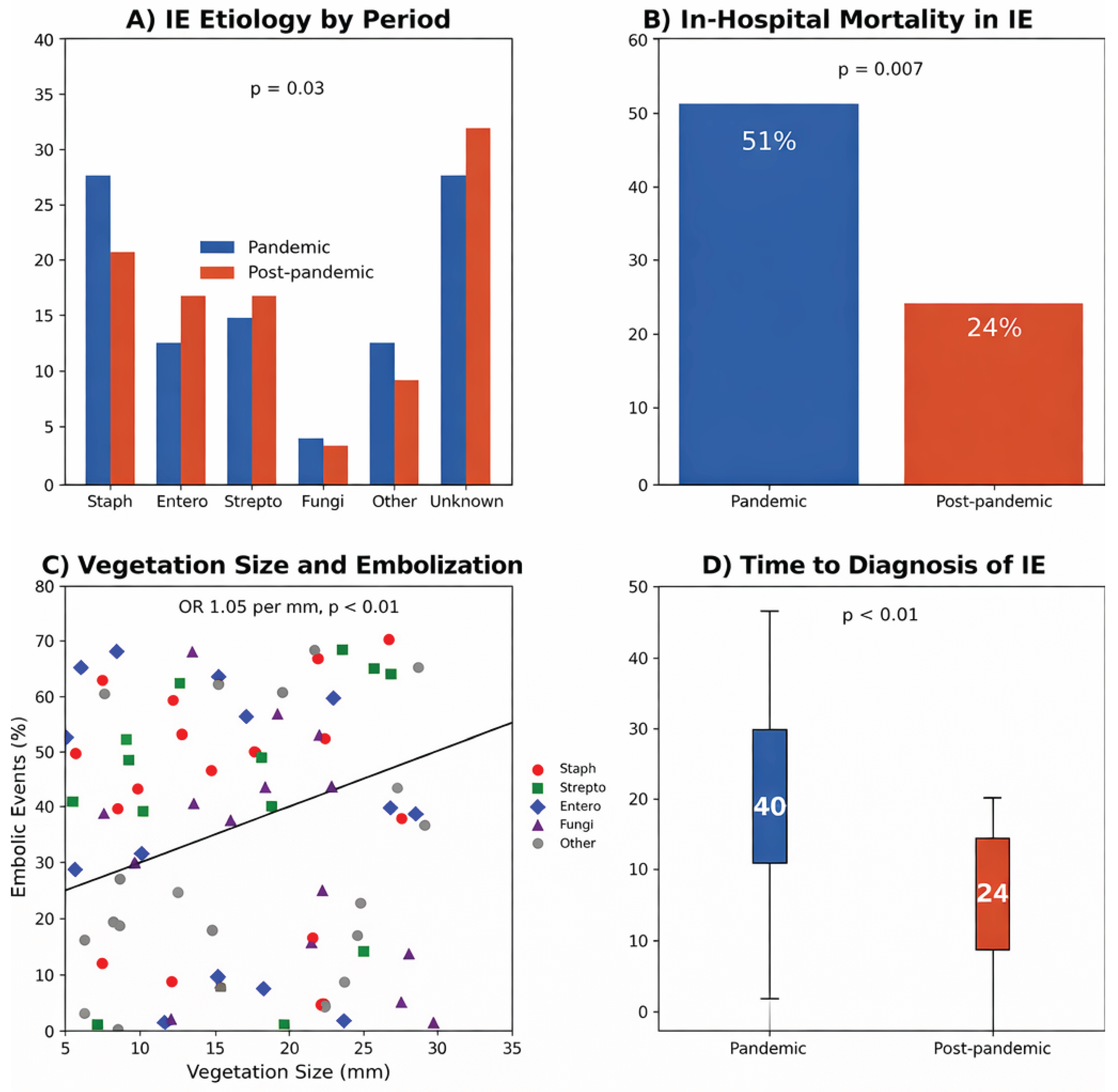

Table 1.
Clinical characteristics, microbiology, complications and management of infective endocarditis during pandemic and post-pandemic periods (n = 47 and n = 53, respectively).
Table 1.
Clinical characteristics, microbiology, complications and management of infective endocarditis during pandemic and post-pandemic periods (n = 47 and n = 53, respectively).
| Variable | Pandemic (n=47) | Post-pandemic (n=53) | p-value |
|---|---|---|---|
| Demographics | |||
| Age, years, median (IQR) | 68 (62–72) | 68 (58–74) | 0.853 |
| Male sex, n (%) | 31 (68.9) | 35 (71.4) | 0.824 |
| Urban residence, n (%) | 19 (42.2) | 24 (49.0) | 0.540 |
| Previous IE, n (%) | 3 (6.7) | 1 (2.0) | 0.346 |
| Clinical presentation | |||
| Sepsis criteria, n (%) | 4 (8.9) | 6 (12.2) | 0.742 |
| Positive blood cultures, n (%) | 27 (60.0) | 32 (65.3) | 0.671 |
| Type of infective endocarditis | |||
| Native valve IE, n (%) | 28 (62.2) | 32 (65.3) | 0.831 |
| Prosthetic valve IE, n (%) | 9 (20.0) | 4 (8.2) | 0.136 |
| Device-related IE, n (%) | 6 (13.3) | 5 (10.2) | 0.753 |
| Type of acquisition | |||
| Community-acquired, n (%) | 26 (57.8) | 36 (73.5) | 0.130 |
| Healthcare-associated, n (%) | 14 (31.1) | 11 (22.4) | 0.361 |
| Mixed acquisition, n (%) | 0 (0.0) | 1 (2.0) | 1.000 |
| Portal of entry | |||
| Cutaneous, n (%) | 6 (13.3) | 11 (22.4) | 0.293 |
| Digestive, n (%) | 4 (8.9) | 9 (18.4) | 0.238 |
| Oral/ENT, n (%) | 4 (8.9) | 10 (20.4) | 0.152 |
| Pulmonary, n (%) | 1 (2.2) | 0 (0.0) | 0.479 |
| Unknown portal, n (%) | 21 (46.7) | 19 (38.8) | 0.532 |
| Aetiology | |||
| Staphylococci, n (%) | 13 (27.7) | 11 (20.8) | |
| Enterococci, n (%) | 6 (12.8) | 9 (17.0) | |
| Streptococci, n (%) | 7 (14.9) | 9 (17.0) | |
| Fungi (Candida), n (%) | 2 (4.3) | 2 (3.8) | |
| Other, n (%) | 6 (12.8) | 5 (9.4) | |
| Unknown (negative BC), n (%) | 13 (27.7) | 17 (32.1) | |
| Echocardiography | |||
| TEE performed, n (%) | 33 (73.3) | 44 (89.8) | 0.059 |
| Local cardiac complications | |||
| Any complication, n (%) | 8 (17.8) | 15 (30.6) | 0.160 |
| Perivalvular abscess, n (%) | 4 (8.9) | 13 (26.5) | 0.033 |
| Valve perforation, n (%) | 3 (6.7) | 1 (2.0) | 0.346 |
| Prosthetic dehiscence, n (%) | 1 (2.2) | 0 (0.0) | 0.479 |
| Prosthetic leak, n (%) | 0 (0.0) | 1 (2.0) | 1.000 |
| Peripheral embolism | |||
| Any embolism, n (%) | 17 (37.8) | 31 (63.3) | 0.023 |
| Central nervous system, n (%) | 3 (6.7) | 16 (32.7) | 0.002 |
| Spleen, n (%) | 6 (13.3) | 9 (18.4) | 0.581 |
| Osteoarticular, n (%) | 3 (6.7) | 9 (18.4) | 0.124 |
| Kidney, n (%) | 2 (4.4) | 9 (18.4) | 0.053 |
| Lung, n (%) | 2 (4.4) | 6 (12.2) | 0.271 |
| Treatment and outcomes | |||
| Time to diagnosis, days, median (IQR) | 40 (30–54) | 24 (13.8–36.3) | <0.01 |
| Antibiotic duration, days, median (IQR) | 42 (35–46) | 42 (30–42) | 0.347 |
| Length of hospital stay, days, median (IQR) | 39 (24–52) | 40 (29–46) | 0.932 |
| Surgery indicated, n (%) | 33 (73.3) | 43 (87.8) | 0.114 |
| Surgery performed, n/N (%) | 26/33 (78.8) | 29/43 (67.4) | 1.000 |
| In-hospital mortality, n (%) | 24 (51.1) | 13 (24.5) | 0.007 |
| Survival, n (%) | 23 (48.9) | 40 (75.5) | 0.007 |
| Recurrence, n (%) | 2 (4.4) | 2 (4.1) | 1.000 |
Data are presented as n (%) or median (IQR). Continuous variables were compared using the Mann–Whitney U test and categorical variables using the chi-squared or Fisher’s exact test. Abbreviations: IE, infective endocarditis; IQR, interquartile range; TEE, transesophageal echocardiography; CNS, central nervous system.
Disclaimer/Publisher’s Note: The statements, opinions and data contained in all publications are solely those of the individual author(s) and contributor(s) and not of MDPI and/or the editor(s). MDPI and/or the editor(s) disclaim responsibility for any injury to people or property resulting from any ideas, methods, instructions or products referred to in the content. |
© 2026 by the authors. Licensee MDPI, Basel, Switzerland. This article is an open access article distributed under the terms and conditions of the Creative Commons Attribution (CC BY) license (http://creativecommons.org/licenses/by/4.0/).
Copyright: This open access article is published under a Creative Commons CC BY 4.0 license, which permit the free download, distribution, and reuse, provided that the author and preprint are cited in any reuse.


