

Submitted:
07 July 2026
Posted:
10 July 2026
You are already at the latest version
Abstract
Chronic pain arises from complex interactions between peripheral and central nervous system structures that process nociceptive information and integrate it with sensory, autonomic, endocrine, emotional, and cognitive functions. Numerous neuroactive substances, including classical neurotransmitters, neuropeptides, hormones, neurotrophic factors, endocannabinoids, and endogenous opioids, participate in these processes. These substances are produced by specific anatomical structures and, in turn, act upon the same or other structures, thereby creating highly interconnected networks of reciprocal modulation. This review examines the actions of individual neuromodulators on pain-related anatomical structures throughout the nervous system, including primary afferents, spinal cord circuits, brainstem nuclei, hypothalamic systems, basal ganglia, limbic structures, and cortical regions. Particular emphasis is placed on the divergence of modulator actions, whereby a single substance influences multiple neural structures and functions, and on the converse principle of convergence, according to which individual structures receive inputs from numerous modulatory systems. The review covers hypothalamic hormones and peptides, monoamines, neuropeptides, neurotrophic factors, ion channels, and major excitatory and inhibitory neurotransmitter systems. The available evidence indicates that chronic pain is associated with widespread alterations in many anatomical and functional structures, although such changes do not necessarily constitute the primary cause of chronic pain. Instead, pain states emerge from dynamic interactions among multiple neuronal populations, signaling molecules, and physiological systems whose contributions vary according to disease state, internal bodily conditions, and environmental influences. By emphasizing both the divergence and convergence of neuromodulatory actions, this review highlights the extraordinary complexity of nociceptive networks and argues that chronic pain cannot be adequately explained by single mechanisms or isolated “key” processes. Rather, chronic pain appears to arise from the collective behavior of highly interconnected and adaptive neural systems.
Keywords:
chronic pain
; pain modulation
; neuromodulators
; nociceptive networks
; neuroplasticity
; neurotransmitters
; neuropeptides
... and by a sleep to say we endThe heart-ache and the thousand natural shocksThat flesh is heir to, 'tis a consummationDevoutly to be wish'd...“(William Shakespeare, Hamlet, 1600-1601)
1. Introduction
Acute pain is a strong stressor that per definitionem lasts no longer than the healing period, usually covering three months. Chronic pain outlasting three months is a complex and persistent condition resulting from sustained nociceptive inputs, maladaptive neuroplastic changes, and neuro-immune interactions (Windhorst and Dibaj 2026).
With the exceptions of a few central pain states of unknown origins, acute and chronic pain states are signaled by group III (Aδ) and IV (C) nociceptive afferents that enter the spinal cord dorsal horn (DH) and higher equivalents from the rear side (Windhorst and Dibaj 2025a). Not only do they meet a complex neuronal network, but also the constituent cells are characterized and influenced by a multitude of modulators as illustrated in Figure 1.
The transition from acute to chronic pain (pain chronification) has been hypothesized to occur in steps. For example, sciatic nerve lesions (and others) can invoke chronic neuropathic pain that is accompanied by persistent, spontaneous activity in primarynociceptive fibers. This activity, which reflects changes in the properties and functional expression of Na+, K+, and Ca2+ ion channels, initiates a further increase in the excitability of second-order sensory neurons in the DH. This change persists for a long time. The origin of the pain thus moves from the peripheral nervous system (PNS) to the central nervous system (CNS). This `centralization´ of pain involves the inappropriate release of peptidergic neuromodulators from primary afferent fibers. Peptides, such as substance P (SP), calcitonin gene-related peptide (CGRP), neuropeptide Y (NPY), nerve growth factor (NGF), and brain-derived neurotrophic factor (BDNF), may promote enduring changes in excitability as a consequence of neurotrophic actions on ion channel expression in the DH (Abdulla et al. 2003).
During pain, numerous types of nociceptors are activated which vary in their signaling pathways. On the basis of their anatomical location, transient receptor potential ion channels (e.g., TRPV1, TRPV2 and TRPM8), Piezo 2, acid-sensing ion channels (ASICs), purinergic (P2X and P2Y), bradykinin (B1 and B2), α-amino-3-hydroxy-5-methylisoxazole-4-propionate (AMPA), N-methyl-D-aspartate (NMDA), metabotropic glutamate (mGlu), neurokinin-1 (NK-1) and CGRP receptors are activated during pain sensitization. Local inhibitory regulation by the activation of 5-HT, cannabinoids, opioids, and adrenergic receptors has shown analgesic properties by modulating the central and peripheral perception of painful stimuli (Khan et al. 2019).
Beyond changes in macroscopic and microscopic structures, i.e., neuronal nuclei and their connections, another dimension of complexity is opened by a plethora of modulators, including neuropeptides, hormones, neurotrophins, and neurotransmitters, which influence neuronal signal transfer and processing, depending on external circumstances and internal body states. Of pathophysiological importance is the dysregulation of modulatory signaling pathways, including, e.g., dopamine (DA), noradrenaline (NA), and serotonin (5-HT), neuropeptides, e.g., SP, CGRP, and neurotrophic factors, e.g., NGF, BDNF, and neurotransmitters, e.g., glutamate (Glu) and γ-amino-butyric acid (GABA), which all modulate central and peripheral sensitization mechanisms (Varrassi et al. 2025).
The following exposition will show that the neuromodulators and neurotransmitters exert divergent effects on several pain-related structures, and consequently many structures receive convergent effects from the former.
2. Divergence of Modulator Effects on Structures
Not only do nociceptive afferents meet a complex neuronal network, but also the constituent cells are characterized and influenced by a multitude of modulators as illustrated in Figure 1. In addition, nociceptor neurons and immune cells interact with each other. Immune cells at peripheral nociceptor terminals and within the spinal cord release mediators that modulate mechanical and thermal sensitivity. Conversely, nociceptive afferents release neuromodulators and neurotansmitters from nerve terminals that modulate vascular and adaptive immune-cell reponses (Pinto-Ribeiro et al. 2017).
Each individual modulator may exert effects on different structures. This is exemplified by some important modulators, e.g., DA, NA, 5-HT, OXT, and ORX, in Figure 2, which also shows several pain- related structures along the neuraxis. Stress including pain responses are mediated through the activation of the hypothalamic-pituitary-adrenal (HPA) axis and the sympathetic nervous system (SNS) (Holsboer and Ising 2021; Kvetnansky et al. 2009; McEwen 2007). The neuro-endocrine response to multiple stresses, including various types of acute and chronic noxious stimuli, are mediated by activation of the HPA axis, which results in an increase in circulating corticosteroids that target multiple organ systems (Morena et al. 2016), as well as substances such OXT, vasopressin (AVP) and ORX.
2.1. Hypothalamus-Controlled Agents
As is well known, nociceptive signals emanating from the spinal cord ascend rostrally via several neuronal tracts to contact an array of supraspinal structures. On the other hand, many descending tracts and connections modulate nociceptive processing (Windhorst and Dibaj 2025a; Figure 2). Again, this heroic attempt of the CNS may be presumed to be prone to failures and mal-adaptations.
Stress including pain responses are mediated through the activation of the HPA axis and the SNS (Holsboer and Ising 2021; Kvetnansky et al. 2009; McEwen 2007). The neuro-endocrine response to multiple stressors, including various types of acute and chronic noxious stimuli, are mediated by activation of the HPA axis, which results in an increase in circulating corticosteroids that target multiple organ systems (Morena et al. 2016), as well as substances such OXT, AVP and ORX. The autonomic response involves the stimulation of sympathetic motor and hormonal outputs via neural circuits originating in HYP pre-autonomic centers, brainstem NA nuclei, adrenal medulla, and results in the release of catecholamines within the brain and circulation.
2.1.1. Corticotropin-Releasing Hormone (CRH)
Stress activation of the HPA axis comprises a slower and more sustained cascade in which CRH and AVP are released from neuro-endocrine cells in the HYP to trigger adreno-corticotropic hormone (ACTH) secretion from the anterior pituitary gland into the systemic circulation, which in turn stimulates glucocorticoid synthesis and secretion from the adrenal cortex. CRH is a 41-amino-acid neuropeptide involved in pain modulation and neuro-endocrine, autonomic and behavioral stress responses. CRH exerts its biological roles through CRH1 and CRH2 receptors, which have different pharmacological profiles and distributions in the CNS and periphery. CRH is an integral part of the HPA axis and is secreted from parvocellular neuro-endocrine neurons of the HYP paraventricular nucleus (HYP PVN) in response to stressors (Neugebauer et al. 2020).
- Corticotropin-releasing Hormone (CRH) in Pain Modulation
CRH elicits strong anti-nociceptive effects at the peripheral, spinal, and brain levels. In the brainstem, CRH acts on the LC to influence NA modulation of pain (Kuner and Kuner 2021). CRH is also expressed in nociceptors and their neighboring components, giving rise to hypotheses for possible pain modulations at this level (Zheng et al. 2020). In primates and rodents, various regions outside the HYP host CRH-containing neurons of varying density, e.g., cerebral cortex, lateral septum, hippocampus (HIPP), bed nucleus of the stria terminalis (BNST), AMY, THAL, HYP, PAG and deep mesencepahlic nucleus, DA system and inter-peduncular nucleus, parabrachial nucleus (PBN), raphé nuclei, LC, nucleus tractus solitarii (NTS), and others (Kelly and Fudge 2018). CRH mRNA and protein expression in the central nucleus of the AMY (CeA) are increased in neuropathic pain models (Neugebauer et al. 2020).
- Corticotropin-releasing Hormone (CRH) in Amygdala (AMY)
The AMY CRH neurons might play a role in the perturbations of descending pain inhibition associated with by neuropathic pain. In un-injured mice, forced swimming increased the tail-flick latency (TFL), a phenomenon called stress-induced analgesia (SIA) but did not change the TFL in mice with neuropathic pain caused by sciatic nerve constriction. Neuropathic pain also increased the expression of CRH in the CeA and ΔfosB in the spinal DH. In mice with neuropathic pain, ablation of AMY projections to the LC on the side of injury but not on the opposite side, completely restored SIA, decreased allodynia and decreased spinal ΔfosB expression. The process of pain chronification is often attributed to abnormal functioning of descending pain inhibition. In fact, the continuous activity of CRH neurons associated with persistent pain leads to impaired SIA, which is a symptom of dysregulation of descending pain inhibition. An over-activation of AMY CRH neurons is very likely an important factor contributing to pain chronification (Andreoli et al. 2017).
2.1.2. Corticosteroids
Many steroids are produced outside of the CNS from the precursor cholesterol, namely in the adrenal cortex: glucocorticoids, mineralocorticoids and androgens, which have far-reachings effects. The glucocorticoid hormones, cortisol in humans and corticosterone in rodents, are considered the classical stress hormones that target all physiological systems, affecting cell metabolic activity, fluid homeostasis, and the cardio-vascular, musculo-skeletal, and enteric and nervous system (Dos-Santos et al. 2023).
- Glucocorticoids
In inflammation, an elevated level of pro-inflammatory cytokines exerts regulatory effects on the HPA axis, which counters inflammation through the secretion of endogenous cortisol, as do exogenous glucocorticoids in patients with chronic pain conditions (Jovanovic et al. 2023). Glucocorticoids exert their effects through glucocorticoids receptors (GRs).
In rats, chronic constriction injury (CCI) of the sciatic nerve evokes sensory dysfunction characteristic of neuropathic pain. Approximately, 30 % of CCI rats show disabilities similar to those identified in clinical evaluation of neuropathic pain patients, which include: altered social behaviors; sleep disturbances; and endocrine dysfunction. CCI increases corticosterone, which through its actions at the GR can trigger cellular adaptation. In nerve-injured rats, GR expression in PAG was quantified using various methods. The PAG of disabled rats had significantly increased expression of GR mRNA and protein. This increased protein expression reflected contrasting patterns of change in GR expression in PAG su-bregions. The dorso-lateral PAG (dlPAG) showed significant increases in the number of GR-immuno-reactive (GR-IR) cells and the caudo-lateral PAB (clPAG) and ventro-lateral PAG (vlPAG) each had significant reductions in the number of GR-IR cells. These regional increases and decreases correlated with the degree of disability, as indicated by the degree of change in social behaviors. This suggests a role for altered PAG, GR-corticosterone interactions and their resultant cellular consequences in the expression of disabilities in a sub-population of nerve-injured rats (Mor and Keay 2013).
- Dexamethasone
Dexamethasone is a GR agonist and is widely used in the therapy of chronic inflammatory diseases for its pain-modulating effects. Male rats with chronic inflammation in the tibio-tarsal join induced by complete Freund´s adjuvant (CFA) received dexamethasone for eight days, which suppressed a local inflammatory response that occurred only in the control animals (injected with saline solution), which demonstrated that the dexamethasone treatment decreased the inflammatory process. Dexamethasone also produced significantly increased latencies in the hot-plate test and withdrawal threshold in the electronic von Frey test. The dexamethasone rat group showed increased concentrations of spinal BDNF compared to the control group. This emphasized the anti-nociceptive as well as anti-inflammatory properties of dexamethasone. Furthermore, dexamethasone treatment increased BDNF concentrations in the spinal cord, which can explain its pain-modulating effects (Laste et al. 2013).
2.1.3. Oxytocin (OXT)
OXT is involved in pain modulation, e.g., of the pain-inhibitory descending pathway (Kawasaki ert al. 2024; red lines in Figure 2). In mammals, OXT is synthetized in the HYP PVN, supra-optic nucleus (SON) and accessory nucleus, and some peripheral locations, and is transported to the anterior cingulate cortex (ACC), HIPP, AMY, nucleus accumbens (NAc), and HYP. Magnocellular OXT neurons of the SON and PVN are large cells that release OXT into the bloodstream via axonal projections to the posterior pituitary. By contrast, parvocellular OXT neurons are smaller cells located exclusively in the PVN, synthesize OXT and transport it via axons projecting to the brainstem and spinal cord (Figure 2), but not the posterior pituitary. A small population of approximately thirty PVN parvocellular OXT neurons attenuates pain perception via two pathways: (i) through coordinated OXT release into the bloodstream from magnocellular OXT neurons leading to the modulation of peripheral nociceptor activity in the dorsal-root ganglion (DRG) and skin, and (ii) by inhibiting sensory neurons in the spinal cord (Hökfelt et al. 2018; Iwasaki et al. 2023; Kuner and Kuner 2021; Lefevre et al. 2021; Takayanagi and Onaka 2021; Tracy et al. 2015; Wang et al. 2022).
Oxytocin receptors (OXTRs) are widely expressed in the CNS and also on peripheral sensory neurons, where they bind and de-sensitize the vanilloid transient receptor potential channel 1 (TRPV1), a prominent sensor of protons (H+), heat, and diverse algogens. Exogenously administered OXT attenuated inflammatory nociceptive hypersensitivity when administered systemically, spinally, or in the brain. Endocannabinoid (eCB) and opioidergic mechanisms are implicated in these actions. Furthermore, activation of OXT-expressing HYP neurons is sufficient to elicit defensive behavior via activation of premotor nuclei in the brainstem (Kuner and Kuner 2021). OXT transmission blocks nociception at the peripheral, spinal, and supraspinal levels through the OXTRs. In fact, a neuronal pathway exists from the PVN to the spinal cord and trigeminal nucleus caudalis (SpVc) (Condés-Lara et al. 2024; Figure 2).
Bottom-up OXT Effects. Neurons in the prefrontal cortex (PFC) can provide top-down regulation of sensory-affective experiences such as pain. In freely behaving rats, potential bottom-up modulation by HYP OXT was examined by in vivo time-lapse endoscopic Ca2+ imaging. OXT selectively enhanced population activity in the pre-limbic PFC in response to nociceptive inputs. This population response resulted from the reduction of evoked GABAergic inhibition and manifested as elevated functional connectivity involving pain-responsive neurons. Direct inputs from OXT-releasing neurons in the HYP PVN were crucial in maintaining this PFC nociceptive response. Activation of the pre-limbic PFC by OXT or direct optogenetic stimulation of the PVN OXT projections reduced acute and chronic pain. This suggests that OXT signaling in the PVN-PFC pathway is an important mechanism to regulate cortical sensory processing (Liu et al. 2023b).
Top-down OXT Effects. In transgenic rats, a subset of PVN parvocellular OXT neurons sends projections preferentially to OXTR-expressing neurons in the vlPAG (Figure 2, red arrow labeled OXT). In vivo, optogenetically-evoked axonal OXT release in the vlPAG as well as chemogenetic activation of OXTR vlPAG neurons resulted in a long-lasting increase of vlPAG neuronal activity. This led to an indirect suppression of sensory neuron activity in the spinal cord and strong analgesia in both female and male rats. Hence, there is a distinct pathway from parvocellular OXT neurons to lateral peri-aqueductal gray (lPAG) to spinal cord (wide dynamic range: WDR cells), which is critical for analgesia in both inflammatory and neuropathic pain models and is able to promote analgesia in both inflammatory and chronic neuropathic pain (Iwasaki et al. 2023; Figure 2).
- Systemic OXT Effects
Neurosecretory cells in the parvocellular part of the PVN synthesize OXT and transport it via axons to remote parts of the CNS. Optogenetic activation of these specific OXT neurons dampened nociception and inflammatory pain, both via direct suppression of nociceptive processing in the spinal cord and via release of OXT into the bloodstream (Kuner and Kuner 2021). In particular situations, systemically circulating levels of OXT may be involved in endogenous control of pain. Indeed, OXT blood concentrations may also modulate nociception, wind-up plasticity and pain responses. This suggests that the PVN may be a part of an integrated homeostatic analgesic system that modulates the transmission of pain in the DH by releasing OXT (Boadas-Vaello et al. 2017).
- OXT Effects in Anterior Cingulate Cortex (ACC)
In the ACC, OXT is released by projections from the HYP PVN (Figure 2). OXT micro-injection into the ACC increases mechanical withdrawal thresholds. Optogenetic stimulation of the PVN-ACC pathway exerts anti-nociceptive effects (Lançon and Séguéla 2023). Moreover, there are two major forms of long-term potentiation (LTP) in the ACC: presynaptic LTP (pre-LTP) and postsynaptic LTP (post-LTP). Both pre-LTP and post-LTP contribute to chronic-pain-related anxiety and behavioral sensitization. In animals with neuropathic pain, micro-injections of OXT into the ACC attenuated nociceptive responses and anxiety-like behavioral responses. Application of OXT selectively blocked the maintenance of pre-LTP but not post-LTP. In animals with neuropathic pain, similar results were obtained by using selective optical stimulation of OXT-containing projecting terminals in the ACC. This demonstrates that OXT acts on central synapses and reduces chronic-pain-induced anxiety by reducing pre-LTP (Li et al. 2021).
OXTR expression changed over time from mainly excitatory Glu neurons at p14 to GABA neurons at p28, where OXT perfusion presynaptically enhanced inhibitory transmission and reduced excitatory transmission. Micro-injections of OXT into the ACC enhanced inhibitory transmission via depolarization of inhibitory neurons and reduced chronic-pain-induced anxiety via the reduction in pre-LTP. OXT also alleviated hyperalgesia. This suggests that the increased facilitation of inhibitory transmission via modulators may have analgesic effects in animal models of pain by balancing out cortical overexcitation (Lee et al. 2022).
- OXT Effects in Basal Ganglia (BG)
The striatum plays a central role in guiding numerous complex behaviors, ranging from motor control to action selection and reward learning. The diverse responsibilities of the striatum are reflected by the complexity of its organization. The striatum is laid out compartmentally, namely in terms of striosome (patch) and matrix compartments, and the engagement of these compartments is uniquely controlled by their afferents, intrinsic properties, and neuromodulation (Prager and Plotkin 2019).
The caudate nucleus of the striatum has been implicated in the anti-nociceptive role of endogenous OXT because direct activation of the SON increased the concentration of OXT in the caudate nucleus (Kuner and Kuner 2021).
- OXT Effects in Amygdala (AMY)
The AMY is a limbic region that plays an important role in pain modulation and emotional-affective behaviors (Neugebauer et al. 2020). Ascending OXT axons arising from parvocellular PVN innervate limbic and cortical brain regions, particularly the CeA, and thereby mediate effects of OXT on emotional processing, including the emotional components of pain (Kuner and Kuner 2021). Evidence suggests mutually opposing functions of OXT and AVP in the CeA through GABAergic INTs. CeA neurons inhibited by OXTR activation were activated by AVP. Stereotaxic administration of AVP into CeA had no effect on mechano-sensitivity (nocifensive reflexes) but increased emotional responses (vocalizations to noxious stimuli) and anxiety-like behavior (elevated plus maze) (Neugebauer et al. 2020).
- Effects of Paraventricular Nucleus (PVN) Stimulation
Intra-plantar injection of carrageenan caused the activation of PVN OXT neurons and led to an elevation of spinally located OXT, producing local synthesis of allopregnanolone (below) which, in turn, increased the γ-aminobutyric acid A receptor (GABAAR)-mediated inhibitory tone. In anesthetized rats, PVN stimulation or intra-thecal OXT reduced or prevented the ability of the spinal LTP to selectively facilitate nociceptive-evoked responses of WDR neurons (Boadas-Vaello et al. 2017).
In rats with neuropathic pain elicited by loose sciatic ligature, stimulation of the anterior part of the PVN increased OXT concentration and produced analgesia states, as measured by von Frey hairs, cold tests, and application of heat to the plantar pad. Differential effects were produced by electrical stimulation of the anterior or posterior PVN regions: electrical stimulation of the anterior part enhanced the OXT concentrations in cerebro-spinal fluid (CSF) and blood plasma, and it increased OXT protein concentrations in the spinal cord tissue. In contrast, stimulation of the posterior PVN part only increased OXT concentrations in CSF (Martínez-Lorenzana et al. 2008).
- OXT Effects in Peri-aqueductal Gray (PAG)
OXT released into the bloodstream from the SON acts in the PAG following noxious stimulation to release β-endorphin and L-ENK and M-ENK, but not Dyn in the PAG, thereby activating opioid-dependent descending inhibition (Kuner and Kuner 2021). In transgenic rats, a subset of OXT neurons project preferentially to OXTR-expressing neurons in the vlPAG. Most of the vlPAG OXTR-expressing cells targeted by OXT projections are GABAergic. Ex vivo stimulation of parvocellular OXT axons in the vlPAG induced local OXT release. In vivo, optogenetically-evoked axonal OXT release in the vlPAG as well as chemogenetic activation of OXTR vlPAG neurons resulted in a long-lasting increase of vlPAG neuronal activity. This led to an indirect suppression of sensory neuron activity in the spinal cord and strong analgesia in both female and male rats (Iwasaki et al. 2023).
- Peripheral or Spinal OXT Effects
There is a direct OXT pathway from the HYP to the spinal cord (Figure 2), which elicits anti-nociception (Rojas-Piloni et al. 2010).
In neuropathic male Wistar rats with spinal nerve ligation (SNL), peripheral or spinal OXT partially restored the nociceptive threshold assessed as tactile allodynia, thermal hyperalgesia and mechanical hyperalgesia for twelve days. Electrophysiological data showed that spinal OXT diminished the neuronal firing of the WDR neurons evoked by peripheral stimulation. This effect was associated with a decline in the activity of primary afferent group III (Aδ)- and group IV (C) fibers. Hence, repeated peripheral or spinal OXT administration attenuates the pain-like behavior in a model of neuropathic pain (González-Hernández et al. 2019).
Intra-thecal injection of OXTR antagonists was effective at reducing withdrawal threshold in both sexes, while having no or minimal effects in animals without surgery. OXT fiber immuno-reactivity was three to five times greater in lumbar than other regions of the spinal cord and was increased more than two-fold in lumbar cord ipsilateral to surgery. Injury was also associated with a 6.5-fold increase in OXTR messenger RNA expression in the L4 DRG ipsilateral to surgery. Hence, the capacity for OXT signaling in the spinal cord increases after surgery and that spinal OXT signaling plays ongoing roles in both sexes in the recovery from mechanical hypersensitivity after surgery with known nerve injury (Severino et al. 2018).
In normal and neuropathic rats, DH neuronal responses to electrical and mechanical stimulation of receptive fields were compared. In Chung rats, SNL at L5 and L6 was used to produce experimental neuropathy. Single-unit activity was recorded at the L4-L5 level from neurons identified as WDR presenting latency responses corresponding to group II (Aβ), III (Aδ) and IV (C) fibers and also exhibiting post-discharge and wind-up. OXT was injected intra-thecally at various doses. Minor effects on responses to electrical stimulation occurred in normal rats, and mechanical responses evoked by von Frey filaments were slightly reduced in normal animals. In neuropathic rats, OXT produced a significant reduction in group IV (C) fiber and post-discharge activities, and higher doses caused a more pronounced reduction in post-discharge and wind-up. The most marked change was the post-discharge reduction at ten and twenty minutes after OXT administration. Mechanical responses were significantly reduced in terms of their discharge rate response in neuropathic rats. The differing results obtained in normal and neuropathic rats indicated that plastic changes occur as a consequence of nerve damage. In neuropathic rats, the mechanisms involving ascending noxious signals to the PVN and descending OXT activities could be altered, thus sensitizing the OXT receptors of the spinal DH cells (Condés-Lara et al. 2005).
In male Wistar rats, in vivo electrophysiological recordings from trigeminal ganglion (TG) WDR cells sensitive to stimulation of the peri-orbital or meningeal region were performed. PVN electrical stimulation diminished the neuronal firing evoked by peri-orbital or meningeal electrical stimulation. Accordingly, neuronal projections existed from the PVN to the WDR cells. This may imply that endogenous OXT transmission inhibits the nociceptive activity of second-order neurons via OXTR activation in CGRPergic (primary afferent fibers) and GABAergic cells (Condés-Lara et al. 2024).
Spinal LTP elicited by noxious stimulation enhances the responsiveness of DH nociceptive neurons to their normal input. Electrophysiological and behavioral consequences of the interactions between LTP and descending OXY anti-nociceptive mechanisms mediated by the PVN were investigated. PVN stimulation or intra-thecal OXY reduced or prevented the ability of spinal LTP to facilitate selectively nociceptive-evoked responses of spinal WDR neurons recorded in anesthetized rats. LTP mediated long-lasting pain hyper-sensitivity that was strongly modulated by endogenous HYP oxytocinergic OXY controls (DeLaTorre et al. 2009).
- OXT Effects on Sensory Afferents
OXT reduces the excitability of primary sensory afferents and produces analgesia, in part through a peripheral mechanism. In female rats, intracellular recordings were performed from L4 DRG neurons, characterized as low-threshold mechano-receptors (LTMRs) or high-threshold mechano-receptors (HTMRs), one week after L5 partial spinal nerve injury or sham control before, during, and after ganglionic perfusion with OXT. Nerve injury de-sensitized and hyperpolarized LTMRs, and sensitized HTMRs without affecting the membrane potential. In nerve-injured rats, OXT depolarized LTMRs towards normal and, in six of 21 cells, resulted in spontaneous action potentials. By contrast, OXT hyperpolarized HTMRs. These effects were reversed after removal of OXT, and OXT had minimal effects in neurons from sham-surgery animals. This shows that neuropathic injury de-sensitizes LTMRs while sensitizing HTMRs and shows rapid and divergent OXT effects on these afferent sub-types towards normal, potentially re-balancing input to the CNS (Boada et al. 2019).
- Oxytocin (OXT) and Opioids
Analgesic effects of both endogenous and exogenous OXT can be blocked by naloxone, an antagonist of opioid receptors. Hence, besides the central OXT effects, the endogenous opioid system could be involved in pain modulation by OXT (Tracy et al. 2015).
2.1.4. Vasopressin (AVP)
OXT and AVP are twin peptides that are synthesized and secreted by the PVN. Both have direct pathways from the HYP to the spinal cord. Both AVP and OXT have anti-nociceptive effects in tail-flick, hot-plate and formalin tests after systemic, peripheral, intra-thecal or intra-cerebro-ventricular (ICV) administration, which is in contrast to their generally opposing roles in the regulation of stress responses, fear and anxiety (Neugebauer et al. 2020). By contrast, it has been argued that, while activation of OXT-containing axons modulates nociceptive neuronal responses in DH neurons, the direct AVP descending projection does not modulate the anti-nociceptive effects mediated by the PVN on DH neurons (Rojas-Piloni et al. 2010).
- Central Vasopressin (AVP)
Outside the HYP, AVP occurs in the BNST and medial AMY. AVP acts through three receptors: V1aR, V1bR, V2R. AVP shows the highest affinity for V1bR and lower affinity at V1aR and OXTR in the CNS. V1aR is abundantly expressed in the brain (Neugebauer et al. 2020). In different pain models in rats, centrally administered cytidine-5'-diphosphate-choline (CDP-choline; citicoline) elicited an analgesic effect (Bagdas et al. 2013). In rats, CDP-choline enhanced central and peripheral AVP levels. Rats were pretreated ICV with the AVP V1 or AVP V2 receptor antagonist 15 minutes before ICV injection of CDP-choline or saline, and pain thresholds were determined. V1R and V2R antagonists blocked the CDP-choline-induced analgesic effect either in acute or neuropathic models. Hence, central AVP receptors are involved in the CDP-choline-elicited analgesic effect (Bagdas et al. 2013).
In rats subjected to unilateral SNL, the chemogenetic activation of AVP neurons significantly attenuated mechanical and thermal hyperalgesia with elevated plasma AVP concentration. These analgesic effects were suppressed by pre-administration with a V1aR antagonist. AVP neurons increased the neuronal activity of 5-HT DRN, NA LC, and inhibitory INTs in the spinal DH. This suggests that the AVP system is up-regulated in neuropathic pain and activates endogenous AVP exerts analgesic effects via the V1aRs (Baba et al. 2022).
- Vasopressin (AVP) Effects in Anterior Cingulate Cortex (ACC)
It has been reported that neuropathic pain is related to the increased activity of Glu pyramidal cells and changed neural oscillations in the ACC. AVP caused pain-alleviating effects when applied to the peripheral system, but the extent to which, and the mechanisms by which, AVP induces analgesic effects in the CNS was unknown. In mice with spared nerve injury (SNI), intra-nasal AVP application inhibited mechanical pain, thermal pain, and spontaneous pain sensitivity. AVP application exclusively reduced the fos expression in the ACC pyramidal cells but not INTs. In vivo electrophysiological recording showed that AVP application not only inhibited the theta oscillation in local field potentials but also reduced the firing rate of pyramidal cells. Hence, in mice with neuropathic pain, AVP induces analgesic effects by inhibiting neural theta oscillations and the discharge of ACC pyramidal cells (Si et al. 2024).
- Vasopressin (AVP) Effects in Amygdala (AMY)
Both magnocellular and parvocellular AVP neurons project to extra-hypothalamic sites such as the AMY, and AVP neurons occur in the medial AMY of rodents but not primates. While AVP neurons in supra-chiasmatic nucleus (SCN) project strongly to the medial AMY, PVN AVP neurons project to CeA and BLA. There is some evidence suggesting mutually opposing functions of AVP and OXT in the CeA through GABAergic INTs. CeA neurons inhibited by OXTR activation were activated by AVP through an action on V1aRs but not V1bRs and V2Rs (Neugebauer et al. 2020).
AVP micro-injection into the CeA suppressed an electromyographic (EMG) nociceptive jaw-opening reflex evoked by electrical stimulation. Stereotaxic AVP administration into the CeA had no effect on mechano-sensitivity (nocifensive reflexes) but increased emotional responses (vocalizations to noxious stimuli) and anxiety-like behavior (elevated plus maze), and these effects were blocked by a selective V1aR antagonist (Neugebauer et al. 2020).
- Vasopressin (AVP) in Spinal Cord
Intra-thecal administration of AVP has effects on pain sensitivity and anti-nociception. AVP produced non-opiate anti-nociception at the highest doses tested and none of the AVP doses appeared to interact with intra-thecal morphine anti-nociception (Watkins et al. 1986).
It has been hypothesized that, in rats, recovery after surgery could be reversed by antagonism of spinal AVP or OXY receptors. Male and female rats underwent partial SNL surgery. Effects of non-selective and selective V1AR antagonists on mechanical hypersensitivity during partial recovery were assessed. Intra-thecal injection of AVP receptor antagonists were effective at reducing withdrawal threshold in both sexes, while having no or minimal effects in animals without surgery. Injury was associated with a two-fold increase in AVP 1a receptor messenger RNA expression in the L4 DRG ipsilateral to surgery (Severino et al. 2018).
- Vasopressin (AVP) in Sensory Afferents
In mice, the use of DNA micro-array for the extraction of candidate genes in the DRG from three nerve-injury mouse models and a sham-operated model (sciatic nerve ligation: SCL) and resection, sural nerve resection, SNI, and sham) elucidated the genes responsible for the neuropathic pain mechanism in the SNI model. Fifty genes were identified which significantly up-regulated in the DRG of the SNI model. Among them, V1a was identified as a candidate SNI-specific gene. Administration of V1a agonists to wild-type SNI mice significantly alleviated neuropathic pain. However, V1a knockout mice did not exhibit higher hypersensitivity to mechanical stimulation than wild-type mice. In addition, V1a knockout mice showed pain behaviors after SNI similar to that of wild-type mice. Hence, V1a in the DRG may partially contribute to the mechanism of neuropathic pain (Yokoyama et al. 2020).
2.1.5. Orexin (ORX)
The ORX peptides are selectively expressed by neurons in the lateral and peri-fornical areas of the HYP, which project widely to a large number of brain areas including the ventral tegmental area (VTA), LC, DRN and PAG (Figure 2) (Kaneko et al. 2024), and are involved in arousal, cardio-vascular control, and endogenous anti-nociceptive control. The ORX peptides, ORX-A and ORX-B, act via ORX receptors 1 and 2 (ORX1Rs and ORX2Rs), and exert anti-nociceptive effects in a number of rodent models, via modulation at supraspinal and spinal levels, with the potency being higher by ORX-A than by ORX-B. Activation of the ORXRs increased the response thresholds to noxious heat and depressed responses to inflammatory and noxious stimuli, such as formalin. Chemogenetic activation of ORX neurons also reduced nociceptive sensitivity. This suggests that ORX neurons are activated as part of the endogenous pain control system (Kuner and Kuner 2021).
Pharmacological treatment with the potent, selective and structurally distinct dual ORX receptor antagonists (ORAs) DORA-12 and DORA-2 significantly reduced pain responses during both phases I and II and significantly reversed hyperalgesia in the mouse formalin pain model and in the rat CFA pain model, respectively. Significant anti-nociceptive effects of DORA-12 in the formalin model also occurred in ORX1R knockout mice, but not ORX2R or ORX1R/ORX2R double knockout mice. Mechanical hyper-sensitivity was significantly reduced with a series of structurally distinct, potent and highly selective ORAs (DORA-2, DORA-12 and DORA-22) in the rat SNL injury model of neuropathic pain. Selective pharmacological targeting of ORX2R with 2-SORA-7 also reduced pain responses in acute inflammatory (CFA) and neuropathic (SNL) rat pain models (McDonald et al. 2016).
- Orexin (ORX) Effects in Hippocampus (HIPP)
Stressful stimuli may reduce nociception (SIA), in which ORX could be involved. ORX neurons project to various HIPP regions, such as the dentate gyrus (DG). In the restraint SIA animal models of chronic pain, the role of ORXRs within the DG region was examined in adult male Wistar rats. Animals were given SB334867 or TCS OX2 29 into the DG region as ORX1R and ORX2R antagonists, respectively, five minutes before exposure to a three-hour restraint stress period. Animals were then subjected to the formalin test (a model of persistent inflammatory pain) to assess pain-related behaviors. The results showed that restraint stress produces an analgesic response during the early and late phases of the formalin test. Intra-DG micro-injection of ORX1R and ORX2R antagonists attenuated the restraint SIA. It was concluded that the ORX system in the DG region might act as a potential endogenous pain control system (Baghani et al. 2024).
- Orexin (ORX) Effects in Nucleus Accumbens (NAc)
It has been suggested that stimulation of ORX2Rs in the brain may suppress chronic pain. In rats, the use of isolated group C-fiber-like neurons revealed that chronic pain was associated with reduced ORX neural activity and that the NAc contained ORX2Rs, which inhibited NAc basal DA efflux. Two types of chronic pain were induced, i.e., inflammatory and neuropathic pain, by performing, respectively, intra-plantar injection of the pro-inflammatory carrageenan into the hindpaws and SCL. Decreased paw-withdrawal threshold (PWT) following carrageenan treatment or SCL, was inhibited by morphine. However, the non-steroidal anti-inflammatory drug meloxicam inhibited these changes in carrageenan-treated rats but not in those with SCL. Neither carrageenan injection nor SCL altered basal NAc DA efflux. In both carrageenan-treated and SCL rats, the ORX1R and ORX2R antagonist MK-4305 and ORX2-receptor antagonist EMPA-induced increase in NAc DA efflux was reduced, as compared to their respective controls. The ORX2R agonist ORX-B counteracted the EMPA-induced increase in DA efflux both in carrageenan-treated and in SCL rats. This suggests that inflammatory and neuropathic pain each lead to decreased stimulation of NAc ORX2Rs by their endogenous agonists, ORX-A and/or ORX-B, thereby inhibiting DA efflux (Kawashima et al. 2025).
Whether discrete neuronal circuits underlie behavioral subsets of chronic pain and comorbid depression was not known. In mice after nerve injury, DA 2 (D2) receptor-expressing medium spiny neurons in the NAc medial shell (mNAcSh) mediated pain hypersensitivity and depression-like behaviors. Two separate neural pathways mediated different symptoms. The Glu inputs from the antero-medial THAL nucleus to mNAcSh D2 neurons that innervated ORX-expressing neurons in the lateral HYP (lHYP) area contributed to pain regulation. In contrast, the lateral septum GABAergic inputs to mNAcSh D2 neurons, which disinhibited the ventral pallidum (BG) glutamatergic neurons mediated depression-like behaviors (Liu et al. 2025).
- Orexin (ORX) Effects in Amygdala (AMY)
The CeA as the main AMY output is involved in the regulation of emotional behaviors including fear and anxiety. ORX is also associated with emotion-related behaviors, such as depression- and anxiety-like behaviors. The CeA receives ORX fibers originating from the lHYP and expresses ORX1Rs. In rats, extracellular recordings of single units were performed in vivo, as well as open field and elevated plus maze tests (EPM). Micro-pressure administration of ORX-A increased the firing rate in 18 out of the 31 CeA neurons, while the other 13 neurons were not excited by ORX-A. The excitatory effects of ORX-A on CeA neurons were mainly mediated by ORX1Rs rather than ORX2Rs. ORX-B did not change the firing activity in all recorded CeA neurons. Selectively blocking ORX1Rs by SB-334867 significantly decreased the spontaneous firing rate in 14 out of the 33 CeA neurons, the remaining 19 neurons not being affected. However, blocking ORX2Rs by TCS-OX2-29 did not change the firing activity. Finally, both open-field test (OPT) and EPM showed that bilateral micro-injection of ORX-A into the CeA induced significantly anxiolytic-like behaviors (Pan et al. 2020).
- Direct Orexinergic (ORX) Hypothalamic-Spinal Cord Pathway
Evidence has accumulated to suggest that there is a direct ORX HYP-spinal cord pathway (Figure 2; left).
In rats with CCI, the effect of ORX-A-mediated lHYP stimulation was examine on thermal hyperalgesia. Rats receiving the acetylcholne (ACh) agonist carbachol into the lHYP demonstrated anti-nociception on both the left CCI and right non-ligated paws. Rats were given carbachol in the lHYP followed by intra-thecal injection of the ORX1R antagonist SB-334867, which blocked lHYP-induced anti-nociception compared with control groups in the left paw, but not in the right paw. This supports the hypothesis that lHYP stimulation produces anti-nociception in rats with thermal hyperalgesia from neuropathic pain via an ORX-A connection between the lHYP and the spinal DH (Wardach et al. 2016).
In male Wistar rats with neuropathic pain, the role of spinal ORXRs was examined in anti-nociceptive responses elicited by the lHYP stimulation Intra- lHYP micro-injection of carbachol was performed five minutes after intra-thecal administration of the ORXR antagonists SB-334867 or TCS OX2 29. Carbachol induced anti-allodynic and anti-thermal hyperalgesic effects in a dose-dependent manner. The anti-allodynic and anti-thermal hyperalgesic effects induced by intra-lHYP injection of carbachol were reversed by intra-thecal administration of SB-334867 or TCS OX2. However, solely intra-thecal administration of both antagonists had no effect. This supports the existence of a neural pathway from the lHYP to the spinal cord, which potentially contributes to the modulation of neuropathic pain (Salehi et al. 2020).
- Orexin (ORX) Effects in Ventral Tegmental Area (VTA)
DA neurons in the VTA are part of the neuronal circuits underlying reward learning and motivation. VTA neurons send dense projections throughout the brain (Figure 2). lHYP ORX neurons project to the VTA and contain both ORX and Dyn peptides in the same dense core vesicles suggesting they may be co-released (Baimel and Borgland 2017).
In the formalin test, the ORX system exerted a pain-modulatory role through a neural pathway from the lHYP to the VTA. Intra-VTA administration of the ORX1R antagonist SB-334867 or the ORX2R antagonist TCS OX2 29 was performed five minutes before intra- lHYP micro-injection of carbachol. This was followed by sub-cutaneous injection of formalin after a five-minute interval. Carbachol attenuated formalin-induced biphasic pain responses, and SB-334867 or TCS OX2 29 administration dose-dependently antagonized the lHYP-induced analgesia during both phases. The contribution of ORX1Rs in mediation of lHYP-induced analgesia was greater than ORX2Rs during the late phase. Hence, the formalin test mimics the conditions encountered in clinical situations. In the formalin test, the pain-modulatory role of the ORX system acts through a neural pathway from the lHYP to the VTA (Ezzatpanah et al. 2016).
- Orexin (ORX) Effects in Locus Coeruleus (LC)
The LC receives dense ORX projections from the lHYP. Activation of ORX1Rs induced eCB synthesis and altered synaptic neurotransmission by retrograde signaling via affecting eCB type-1 (CB1) receptors. In a rat model of inflammatory pain, the interaction of ORX-A and eCBs was examined at the LC level. Pain was induced by formalin injection into the hindpaw. Intra-lHYP micro-injection of ORX-A decreased the nociception score during both phases of formalin test. Moreover, intra- lHYP micro-injection of either an ORX1R antagonist or a CB1 receptor antagonist increased flinches and also the nociception score during phase 1, 2 and the inter-phase of formalin test. The analgesic effect of ORX-A was diminished by prior intra-LC micro-injection of either an ORX1R antagonist or a CB1 receptor antagonist. Hence, activation of ORX1Rs in the LC can induce analgesia, and the blockade of ORX1Rs or CB1 receptors is associated with hyperalgesia during the formalin test. This suggests that CB1 receptors may modulate the analgesic effect of ORX-A (Kargar et al. 2015).
- Orexin (ORX) Effects in Ventro-lateral Peri-aqueductal Gray (vlPAG)
Supraspinally, anti-nociceptive ORX actions have been suggested to be mediated via the vlPAG. The analgesic effects of ORX-A acting at ORXRs in the vlPAG are postulated to involve eCB signaling. Data show that vlPAG neurons, which project to the RVM (Figure 2), receive OXR inputs from the lHYP and express ORX1Rs. In addition, OXR-A elicited the synthesis and release of 2-arachidonyl glycerol (2-AG; derived from arachidonic acid), which inhibited the release of GABA in the vlPAG via presynaptic eCB receptor 1 (CB1R), resulting in net dominance of excitatory vlPAG-RVM projections. This mechanism of ORX release in the vlPAG from incoming lHYP projections has also been postulated to contribute to SIA. The ORX lHYP-vlPAG-RVM-spinal DH pathway is recruited during median nerve stimulation, a therapeutic measure that is long in practice for intractable pain, and mediates analgesia induced by median nerve stimulation in neuropathic mice (Kuner and Kuner 2021).
Pain and itch are antagonistically regulated sensations. Pain suppresses itch, and pain inhibition enhances itch. The lHYP suppresses pain while enhancing itch neural processing. The optical inhibition of neuronal terminals from the lHYP to the lPAG and vlPAG resulted in a decrease in itch-related behavior and an increase in pain-related behavior. The use of an atopic dermatitis model confirmed the involvement of ORX neurons in regulating chronic itch processing (Kaneko et al. 2024).
- Orexin (ORX) Effects in Spinal Cord
In some rat strains, neurons containing ORX-A are located in the posterior HYP (pHYP), and intra-thecal injection of ORX-A produces anti-nociception in a neuropathic pain model. It has been hypothesized that ORX-A from neurons in the pHYP modifies nociception in the spinal DH. In lightly anesthetized female Sprague-Dawley rats with CCI, the ACh agonist carbachol or normal saline was micro-injected into the pHYP. Carbachol-induced pHYP stimulation produced dose-dependent anti-nociception as shown by significantly increased foot withdrawal latencies (FWL) compared to saline controls. The ORX1R antagonist SB-334867 or dimethyl sulfoxide (DMSO) for control, was given intra-thecally following carbachol-induced pHYP stimulation. SB-334867 decreased FWL compared to DMSO controls. This suggests that, in a neuropathic pain model, stimulating the pHYP produces anti-nociception and the anti-nociceptive effect is mediated in part by ORX-1 receptors in the DH (Jeong and Holden 2009)..
In rats with CCI of the sciatic nerve, ORX-A, ORX-B, the vehicle, or ORX-A-antiserum were intra-thecally administered to CCI rats. Paw-withdrawal latency (PWL) was measured from 30 to 300 minutes after injection, which was repeated for two days. ORX-A administration normalized ΔPWL (PWL on the CCI side minus PWL on the control side) and inhibited heat-evoked hyperalgesia in CCI rats, while ORX-A antiserum inhibited the normalization of heat-evoked hyperalgesia caused by ORX-A two-fold. In contrast, ORX-B had no significant effect (Suyama et al. 2004).
In CCI-treated rats with neuropathic pain, ORX2Rs but not ORX1Rs were reduced in the spinal cord. Mechanical withdrawal threshold and thermal withdrawal latency showed that administration of ORX-B ameliorated CCI-evoked neuropathic pain dose-dependently. Moreover, ORX-B suppressed the inflammatory response in the spinal cord tissue. ORX-B administration also attenuated oxidative stress. Hence, ORX-B might have a role in ameliorating CCI-evoked neuropathic pain through the inhibition of microglial activation and inflammatory response (Zhu et al. 2024).
2.1.6. Sex Hormones
The striking difference in female and male pain sensitivity has been assigned to their different GHs. In females, sex hormones include female hormones including estrogens, progesterone, allopregnanolone, and prolactin, and androgens include testosterone and dehydroepiandrosterone (DHEA). They account for sex differences including those in chronic pain sensitivity (Mogil et al. 2024; Vincent and Tracey 2010).
- Sex Differences
The prevalence and severity of chronic pain differ between in females and men. Some common pain conditions, including temporo-mandibular disorders, tension-type and migraine headaches, rheumatoid arthritis, fibromyalgia (FM), and musculo-skeletal pain syndromes, show fairly marked sex-related differences in their occurrence, with the exception of rheumatoid arthritis. The association of musculo-skeletal pain conditions with the reproductive cycle of women is strongly suggestive of a role of the estrogens and/or progesterones. Testosterone has been suggested to protect men from these chronic musculo-skeletal pain conditions (Cairns and Gazerani 2009).
Sex Hormones in Inflammatory Pain. In the formalin model of inflammatory pain, C57BL/6J male and female mice were evaluated for baseline sex differences and the effects of gonadectomy (ovariectomy or castration) within this model. Female mice in the metestrus or diestrus phase had decreased inflammatory pain relative to both male mice and female mice in the proestrus or estrus phase. Ovariectomy resulted in decreased pain, which was restored through treatment with estradiol (E2). Castration similarly reduced pain in male mice. Injection of the G-protein coupled estrogen receptor (GPER) agonist G1 resulted in significant anti-nociception in both female and male mice, in both mice that had received sham surgery or gonadectomy (Barnes et al. 2026).
Sex Hormones in Activity-induced Muscle Pain. In a model of activity-induced muscle pain, female mice developed widespread, more severe, and longer-duration hyperalgesia than did male mice. It was hypothesized that testosterone protects males from developing the female pain phenotype. It was tested whether orchiectomy of males before induction of an activity-induced pain model produced a female phenotype and whether testosterone administration produced a male phenotype in females. Orchiectomy produced longer-lasting, more widespread hyperalgesia, similar to females. Administration of testosterone to females or orchiectomized males produced unilateral, shorter-lasting hyperalgesia. In models of chronic pain, the 5-HT transporter (SERT) was increased in the nucleus raphé magnus (NRM) (Figure 2), and blockade of SERT in the NRM reduced hyperalgesia. Using immuno-histochemistry, a sex difference in SERT was found in the NRM in the activity-induced pain model. Females had greater SERT immuno-reactivity than males. This suggests that testosterone protects against development of widespread, long-lasting muscle pain and that alterations in SERT may underlie the sex differences (Lesnak et al. 2020).
In the activity-induced muscle pain model, females showed widespread pain and increased SERT expression in the RVM whereas males showed localized pain and no changes in SERT expression. It was hypothesized that testosterone modulates 5-HT signaling to enhance analgesia in female mice with widespread pain. In mice, testosterone reduced the enhanced SERT protein expression and increased 5-HT2A receptor mRNA expression in the RVM usually observed in the activity-induced pain model in females, but not males. Inhibition of SERT in the RVM was analgesic in both female and male mice. This analgesia was blocked by co-administration of 5-HT2A antagonist. In female mice, SERT, 5-HT2AR and androgen receptor (AR) mRNA were co-expressed in cells within the RVM. Activation of AR using dihydro-testosterone reduced hyperalgesia in female mice. Hence, AR are expressed in female RVM, activation of these AR reduces nociceptive behaviors, and endogenous testosterone modulates SERT and 5-HT2 receptor expression (Plumb et al. 2025).
Diffuse Noxious Inhibitory Control (DNIC). DNIC is a `pain-inhibits-pain´ phenomenon, demonstrated in humans and animals. DNIC is diminished in many chronic pain states, including neuropathic pain. Prolonged noxious stimuli applied anywhere on the body surface may suppress the effects of noxious stimuli applied at other other sites. The efficiency of DNIC has been suggested to prospectively predict both the likelihood of pain chronification and treatment response. DNIC is triggered by peripheral group I and IV fibers, and involves supraspinal structures such as the PAG, RVM and descending pathways in the dorso-lateral funiculi.
In rats, a capsaicin-induced DNIC behavioral assay and resting-state functional magnetic resonance imaging (rsfMRI) was used to assess the effect of testosterone on pain modulation and related brain networks. Male, female, and orchidectomized male rats had a capsaicin injection into the forepaw to induce DNIC, and mechanical thresholds were monitored on the hindpaw. rsfMRI scans were acquired before and after capsaicin injection to analyze the effects of DNIC on ACC, PAG, and NAc connectivity to the whole brain. The strength of DNIC was higher in males compared to females and orchidectomized males. PAG connectivity with pre-limbic PFC, ACC and insular cortex (IC) was stronger in males compared to females and orchidectomized males, whereas females and GDX males had increased connectivity between the right ACC, HIPP and THAL. Orchidectomized males also showed a stronger connectivity between right ACC and NAc, and right NAc with PRL, ACC, IC and THAL. This suggests that testosterone plays a n important role in reinforcing the endogenous pain inhibitory system, while circuitries related to reward and emotion are more strongly recruited in the absence of testosterone (Da Silva et al. 2018).
- Differential Effects of Gonadal Hormones on Opioid Anti-nociception
Generally, estradiol negatively modulates opioid analgesia via both non-genomic and genomic effects. Testosterone facilitates opioid analgesia mainly through the transcriptional activities of AR. Under normal physiological conditions, estrogen and progestin exist in parallel and have a combined effect. However, progestin alone could promote opioid analgesia by increasing the expression of opioid receptors (Xu et al. 2024).
2.1.6.1. Female Gonadal Hormones
GHs play important roles in not only reproductive behavior and sexual differentiation, but also contribute to thermo-regulation, feeding, memory, neuronal survival, and the perception of somatosensory stimuli. Moreover, in both animals and human subjects, GHs, such as estrogens, exert potential effects on pain transmission. These effects most likely involve multiple neuroanatomical circuits as well as diverse neurochemical systems (Amandusson and Blomqvist 2013).
Thus, ovarian hormones are responsible, at least in part, for the pain-threshold differences between the sexes (Paredes et al. 2019). The influence of sex on nociception and its amelioration has been extensively documented but the underlying biology remains elusive. Multiple cross-validating studies revealed that women are more likely than men to experience chronic pain as well as pain of greater severity and duration. Chronic pain disorders that are vastly more prevalent in women than men include migraine (2:1), irritable bowel syndrome (IBS) (2:1), interstitial cystitis (9:1) and FM (6:1). Sex-dependent differences in nociception are observed across multiple modalities of nociceptive stimuli, e.g., thermal, electrical, pressure. Moreover, women exhibit greater severity to and frequency of visceral pain than do men (Gintzler and Liu 2012).
In female rats, estradiol and progesterone rapidly induce changes in DA signaling within the dorsal striatum and NAc. In ovariectomized females, estradiol rapidly enhances DA release and modulates binding of DA receptors. Progesterone further potentiates the effect of estradiol on DA release. The effects of both estradiol and progesterone depend on time course, with increases in DA release immediately after acute hormone administration followed by later inhibition of DA release. These changes also occur in naturally cycling females (Yoest et al. 2018)
- Estrogens
Estrogens, such as estradiol, are synthesized in the gonads (Figure 1), but also in the brainstem and the spinal cord and locally influence pain processing. Potential cellular sources of local estrogen may be primary afferent neurons and their central targets in the spinal cord and medulla oblongata as well as in the NTS, and may be detected by the expression of aromatase, the enzyme that catalyzes the conversion of testosterone to estradiol. In an aromatase-reporter mouse, immuno-histochemical staining showed that many neurons in DH laminae I and V, in the caudal spinal trigeminal nucleus (SpV) and in the NTS expressed aromatase (Tran et al. 2017).
- Estrogen Receptors
Classical genomic actions of estrogens have long been thought to be responsible for the effects on nociception and opioid anti-nociception but estrogens can also activate estrogen receptors (ERs) that are located in the plasma membrane, the effects of which start in seconds to minutes instead of hours to days. Membrane estrogen receptors (mERs) modulate MOR and κ-opioid receptor (KOR) hetero-dimerization and regulate pro- vs. anti-nociceptive functions of Dyn. Membrane ER activity influences sex differences in pain processes. Sex-dependent differences in nociception occur across multiple modalities of nociceptive stimuli, e.g., thermal, electrical, pressure (Gintzler and Liu 2012).
Estradiol modulates nociception and opioid anti-nociception in various brain regions, including areas involved in nociception, such as several cortical areas, PAG, PBN, raphé nuclei, HYP, and the spinal cord. The α isoform of the estrogen receptors (ERα) is present in DH laminae I, II, VI and VII. ERα co-localizes with ENK in many neurons of the superficial DH lamina. ERα is also co-expressed by Dyn neurons in the lumbar DH, whose numbers, in L6 and primary motor cortex (S1), significantly increase in the hormone-simulated pregnancy concentrations of estrogens and progesterone. Cells expressing the ERβ isoform exist in DH lamina II. ERs are also present in DRG (Gintzler and Liu 2012).
- Estrogen and Progesterone during the Estrous Cycle
During the estrous cycle, animals were more sensitive to pain during the estrus stage than in the diestrus stage, suggesting a role for reproductive hormones, estrogen, and progesterone. In estrogen-primed mice treated with progesterone, the pain threshold was lower two days later and stayed that way for the duration of the testing. A specific progesterone receptor (PR) agonist, segesterone, promoted pain, and mice lacking PR in the brain did not experience lowered pain threshold when treated with progesterone or segesterone. PR activation increased the cold sensitivity but did not affect the heat sensitivity and had a small effect on light sensitivity (Joshi et al. 2024).
- Estrogen in Anterior Cingulate Cortex (ACC)
The rostral ACC (rACC) is an important structure of pain affect. In rats, using formalin-induced conditioned place aversion (F-CPA) which is believed to reflect the pain-related negative emotion, application of an ER inhibitor or inhibitor of aromatase androstatrienedione into the rACC completely blocked F-CPA in either sex. An analogous effect also occurred in ovariectomy rats. In the absence of a noxious formalin stimulus, exogenous estrogen was sufficient to elicit CPA in both sexes by activating the mERs and NMDARs. This suggests that estrogen in the rACC drives affective pain via facilitating NMDA receptor-mediated synaptic transmission (Xiao et al. 2013).
- Estrogen in Thalamus (THAL)
Herpes zoster or shingles results from varicella zoster virus (VZV) infection and often leads to chronic pain that lasts for months after visible symptoms subside. Testosterone often attenuates pain in males. However, there are also ovarian estrogen effects on GABA signaling in the THAL reducing pain. Since aromatase affects pain and is present in the THAL, the hypothesis was tested that testosterone converted to estrogen in the THAL attenuates VZV-induced pain. In male Sprague-Dawley rats, various techniques were used to show that indeed aromatase-derived estradiol interacts with the ER to increase neursonal inhibition in the THAL to attenuate VZV induced pain (Kramer et al. 2018).
- Estrogen in Nucleus Accumbens (NAc)
There is evidence of the interaction between GHs and neuromodulatory systems including opioidergic and Glu systems. In morphine-tolerant rats, the sex differences were examined as to the role of GH on the Glu level in the NAc using in vivo micro-dialysis. After chronic morphine administration, tolerance to anti-nociceptive effects of morphine was significantly greater in male rats. Sex differences in tolerance to morphine disappeared with gonadectomy of animals. There was also a significant sex difference in the Glu level in the NAc of morphine-tolerant rats; ovariectomy of female rats decreased the Glu level significantly, while gonadectomy did not significantly change the Glu level in males. Hence, the excitatory amino-acid release in the NAc may be modulated by an estrogen-sensitive mechanism and play a role in the morphine analgesia and tolerance (Mousavi et al. 2007).
- Estrogen in Amygdala (AMY)
In the AMY, estrogen and/or progesterone may influence anxiety, fear, and pain behaviors. Ovariectomized rats were administered sub-cutaneous or intra-AMY vehicle, estrogen, progesterone, or estrogen + progesterone. Sub-cutaneous estrogen + progesterone or intra-AMY estrogen, progesterone, or estrogen + progesterone increased open-field central entries and open-arm time in the plus-maze compared with vehicle. Sub-cutaneous or intra-AMY estrogen, progesterone, or estrogen + progesterone decreased the time spent freezing post-shock compared with vehicle. Sub-cutaneous or intra-AMY estrogen + progesterone increased the latencies to lick paws compared with vehicle. Thus, estrogen and progesterone may have effects in the AMY to decrease anxiety, fear, and/or pain responses (Frye and Walf 2004).
During pain and analgesia, the activation of the descending pathway from the PAG to the RVM shows sex differences. The AMY projects to and engages the PAG-RVM pathway during persistent inflammatory pain. In male and female rats, the neural projections from the AMY to the PAG were investigated as to whether they are sexually distinct, whether this pathway is activated by inflammatory pain and whether it has receptors for estrogen. Injection of the retrograde tracer fluorogold (FG) into the vlPAG resulted in dense retrograde labeling in both the CeA and medial AMY (MeA). While the number of CeA-vlPAG neurons were comparable between the sexes, there were more MeA-vlPAG neurons in females. Inflammatory pain resulted in greater AMY activation in males. However, females displayed higher fos expression within CeA-vlPAG projection neurons. Females expressed higher ERα in the MeA and CeA and the same was true of the projection neurons. Hence, although the MeA-vlPAG projections were denser in females, inflammatory pain did not significantly activate these projections. By contrast, inflammatory pain resulted in a greater activation of the CeA-vlPAG pathway in females. As females experience a greater number of chronic pain syndromes, the CeA-vlPAG pathway may play a facilitatory (and not inhibitory) role in pain modulation (Cantu et al. 2022).
- Estrogen Effects in Dorsal Raphé Nucleus (DRN)
Menopausal and post-menopausal women, characterized by a significant reduction in ovarian hormones, have a high prevalence of chronic pain with great pain intensity. In mice, decreases in the activity and excitability of GABAergic neurons in the DRN are associated with hyperalgesia induced by ovariectomy. Supplementation with 17β-estradiol, but not progesterone, was sufficient to increase the mechanical pain threshold in ovariectomized mice and the excitability of DRN GABA neurons. Activation of DRN GABA neurons projecting to the lateral PBN (lPBN) was critical for alleviating hyperalgesia in ovariectomized mice. This demonstrates the essential role of DRN GABA neurons and their modulation by estrogen in regulating hyperalgesia induced by ovarian hormone withdrawal (Wu et al. 2025).
- Estrogen Receptors in the Peri-aqueductal Gray (PAG)
Morphine administered systemically or directly into the PAG produced greater analgesia in male compared to female rats, while manipulation of GH alters morphine potency in both sexes. The expression of estrogen receptors (ERα) and AR in the PAG of female rats and within this descending inhibitory pathway in both sexes was investigated using immuno-histochemical techniques to map the distribution of AR and ERα across the rostro-caudal axis of the PAG and to determine whether ERα and/or AR were co-localized on PAG neurons projecting to the RVM in male and female rats. ERα and AR immuno-reactive neurons (ERα-IR, AR-IR) were densely distributed within the caudal PAG of male rats, with the majority localized in the lPAG and vlPAG. Females had significantly fewer AR-IR neurons, while the quantity of ERα was comparable between the sexes. In both sexes, approximately 20-50% of ERα-IR neurons and 25-50% of AR-IR neurons were retrogradely labeled. This indicates the expression of steroid receptors in the PAG and the descending pathway driving pain inhibition in both male and female rats (Loyd and Murphy 2008).
- Estrogens in the Rostral Ventro-medial Medulla (RVM)
It has been hypothesized that estrogens produced locally in the RVM may be involved in the maintenance of chronic visceral hyperalgesia. Besides the circulating estrogens produced mainly by the ovaries, many brain regions are also capable of de novo synthesizing estrogens. Aromatase may be distributed in the RVM, where it may impact on visceral pain. Experiments in adult female rats confirmed the expression of aromatase in the RVM. Thus, estrogens produced locally in the RVM may be involved in the maintenance of chronic visceral hyperalgesia (Gao et al. 2017).
- Estrogen Receptors in Spinal Cord
In normal and ovariectomized rats, 17β-estradiol may play a role in pain signal transduction and transmission. Either systemic or local administration of 17β-estradiol produced a significant rise of mechanical pain threshold. Local administration of 17β-estradiol significantly decreased adenosine 5´-triphosphate (ATP)-induced spontaneous hindpaw withdrawal duration (PWD), which was blocked by an ER antagonist. In CFA-treated rats, systemic application of 17β-estradiol decreased the mechanical pain threshold significantly, but did not change the inflammatory process. The expression of c-fos in lumbo-sacral DH was increased significantly by administration of 17β-estradiol. This suggests that 17β-estradiol plays an anti-hyperalgesic role in physiological pain. However, peripheral 17β-estradiol plays anti-hyperalgesic roles in ATP-induced inflammatory pain. Systemic application of 17β-estradiol plays hyperalgesic roles in CFA-induced chronic pain (Lu et al. 2012).
In the mouse superficial DH, molecularly characterized neurons expressing ERα were explored as to the behavioral consequences of their ablation. ERα-positive neurons were largely excitatory INTs and many co-expressed SP. After viral, caspase-mediated ablation of ERα-expressing cells, there was a significant decrease in the first phase of the formalin test, but in male mice only. ERα-expressing neuron-ablation also reduced pruritogen-induced scratching in both male and female mice. No ablation-related changes in mechanical or heat withdrawal thresholds or in capsaicin-induced nocifensive behavior occurred. In chronic pain models, there was no change in CFA-induced thermal or mechanical hyper-sensitivity, or in partial sciatic nerve injury-induced mechanical allodynia. Hence, ERα labels a sub-population of excitatory INTs that are specifically involved in chemically evoked persistent pain and pruritogen-induced itch (Tran et al. 2020).
- Estrogen Receptors in Nociceptors
ERs are also expressed in the small-sized primary sensory neurons of the DRG. Long-term (28 days) ovariectomy of adult rats induced a profound thermal and mechanical hyperalgesia of the hindpaw and tail compared to ovariectomized animals that were continuously estrogen-treated (ovariectomy+E). Significant changes occurred in the expression of SP and CGRP in the small lumbar DRG neurons, which contained ER. CGRP and SP were differentially regulated by estrogen, with SP showing a significant down-regulation at both the peptide and mRNA levels, while CGRP and its mRNA were increased in the DRG of estrogen-treated animals. After partial sciatic nerve injury, both ovariectomy and ovariectomy+E animals developed significant allodynia within a week of the partial nerve injury, which continued for at least one month. The estrogen-treated animals showed a partial amelioration of the extent of the allodynia at two weeks post-injury. This suggests that estrogen has significant anti-nociceptive actions that can be directly correlated with changes in expression of two peptides in the small nociceptive ERα-expressing neurons of the DRG (Sarajari and Oblinger 2010).
- Progesterone and Allopregnanolone
Progesterone and its reduced derivative allopregnanolone, are implicated in the modulation of both nociceptive and neuropathic pain. Progesterone can bind to the `classic´ PRs that may be directed to the nucleus and act as a ligand-activated transcription factor that regulates the expression of target genes. Two isoforms of PR exist, PRA and PRB, which are products of a single genes, of which PRB is a more potent transactivator of gene expression than PRA. Moreover, two types of membrane proteins unrelated to nuclear steroid receptors, progesterone membrane receptors (mPRs) and progesterone receptor membrane component 1 (PGMRC1) may mediate progesterone actions in the nervous system. The mPRs comprise at least three sub-types, α, β, and γ, which. are expressed in the nervous system. Progesterone also modulates the activity of the nicotinic ACh receptor (nAChR). Progesterone and allopregnanolone are synthesized in the sensory pathways, specifically in oligodendrocytes and neurons of the spinal cord. By inhibiting allopregnanolone production, SP may be involved in the reduction of the inhibitory tone in the spinal cord, facilitating noxious signal transmission (González et al. 2019).
- Progesterone and Allopregnanolone in Neuropathy
In a rat CCI model of neuropathic pain, progesterone and its vehicle were injected intra-peritoneally on days 1-13 after the surgery to study the effect of progesterone on development of neuropathic pain and only on 14th day post-surgery in order to assess its effect on expression of neuropathic pain. The chronic administration of progesterone significantly reduced the behavioral scores of cold- and mechano-allodynia and heat hyperalgesia but a single dose of progesterone did not have any effect on behavioral scores of neuropathic pain. This indicates that the early chronic administration of progesterone prevents the development of neuropathic pain but its acute injection does not change the expression of neuropathic pain (Verdi et al. 2013).
Progesterone and its reduced metabolites can restore biochemical, morphological, and functional variables after peripheral nerve injuries of different etiologies, e.g., physical trauma, diabetes, chemotherapy, among others. By using animal models of diabetic neuropathy, sciatic nerve crush, or others, progesterone and/or allopregnanolone treatment restore the activity of Na+K+-ATPase pump, counteract the injury-induced decrease in the expression of several myelin proteins in the sciatic nerve and the expression of CGRP in the spinal cord (González et al. 2019). The main mechanisms underlying the increased excitability of spinal circuits in the pain pathway after trauma, mediated by the activation of resident glial cells (Dibaj et al. 2024; Windhorst and Dibaj 2023), the subsequent release of pro-inflammatory cytokines and their impact on NMDAR function have been investigated (Ferreyra and González 2023).
- Neurosteroids in Thalamus (THAL)
In the lateral THAL, neurosteroids play an important role in pain modulation. Rats with SNI displayed increased concentrations of neurosteroids (progesterone, allopregnanolone, pregnenolone, deoxycorticosterone, and tetra-hydro-deoxy-corticosterone) in the chronic stage of neuropathic pain (28 days after SNI). In vivo stereotaxic micro-injection of neurosteroids into the lateral THAL alleviated the mechanical allodynia in neuropathic pain. The analgesic effects of neurosteroids were significantly attenuated by the GABAAR antagonist bicuculline. This suggests that elevated neurosteroids in the lateral THAL play a protective role in the chronic stage of neuropathic pain (Zhang et al. 2016a).
- Progesterone in Spinal Cord
In the rat, intra-peritoneal injections of high doses of progesterone produced anesthesia. In rats, spinal GABAAR-dependent activation inhibited the induction of repetitive stimulation-induced spinal-reflex potentiation. Progesterone was capable of producing GABAAR-dependent inhibition of the induction of spinal reflex potentiation by actions through neurosteroid metabolites. The induction of spinal reflex potentiation was attenuated after a short (30 minutes) intra-thecal treatment with the neurosteroids allopregnanolone and 3α,5α-tetrahydrodeoxycorticosterone. Acute intra-thecal administration of the GABAAR antagonist bicuculline reversed the inhibition produced by progesterone and allopregnanolone. This implies that progesterone-mediated effects on GABAAR expression and neural inhibition are regulated by neurosteroids synthesis rather than PR activation (Peng et al. 2009).
- Allopregnanolone in Neuropathic Pain
Allopregnanolone influences the excitability of the CNS by acting as a positive allosteric modulator of GABAARs (Kawano et al. 2011).
- Allopregnanolone in Spinal Cord
Allopregnanolone exerts analgesic, neuroprotective, anti-depressant and anxiolytic effects. This results from allopregnanolone´s ability to modulate GABAA, Gly, L- and T-type Ca2+ channels, without side effects. For example, in rats, allopregnanolone counteracted chemotherapy-evoked neuropathic pain. In the spinal cord of neuropathic rats with peripheral nerve injury, the modulation of allopregnanolone-producing enzyme, 3α-hydroxysteroid oxido-reductase (3α-HSOR) regulated thermal and mechanical pain thresholds. The painful symptoms were exacerbated by intra-thecal injections of a pharmacological inhibitor of 3α-HSOR, which decreased allopregnanolone production in the spinal cord. By contrast, the enhancement of allopregnanolone concentration in the intra-thecal space induced analgesia and suppression of neuropathic symptoms. Moreover, in healthy rat DRG, in vivo knockdown of 3α-HSOR expression increased thermal and mechanical pain perceptions while allopregnanolone evoked a potent anti-nociceptive action. In humans, blood levels of allopregnanolone were inversely associated with low back and chest pain. Moreover, oral administration of allopregnanolone analogs induced anti-nociception (Patte-Mensah et al. 2014).
Allopregnanolone concentrations measured in the spinal cord and brain of rats that underwent SNL were greater than the corresponding in control animals. More importantly, spinal allopregnanolone concentrations in hyperalgesic rats were lower than those in the rats that did not develop hyperalgesia following SNL. By contrast, brain allopregnanolone concentrations were comparable among these groups. No differences in serum allopregnanolone concentrations occurred among the groups. In addition, intra-thecal exogenous administration of allopregnanolone showed the anti-hyperalgesic effects in hyperalgesic rats after SNL. This suggests that changes in spinal allopregnanolone biosynthesis are involved in the pathogenesis of neuropathic pain following peripheral nerve injury (Kawano et al. 2011).
After sciatic nerve ligature, allopregnanolone was able to alleviate thermal and mechanical hyperalgesia by potentiating GABAAR activity and blocking T-type Ca2+ channels, while progesterone prevented allodynic behaviors in male rats subjected to sciatic nerve CCI by preventing the injury-induced increase in the expression of two important players involved in pain generation: the NR-1 sub-unit of NMDAR and the gamma isoform of PKCγ (González et al. 2019).
- Prolactin (PRL)
PRL, also called lactotropin or lactotropic hormone, is a hormone that is produced in the lactotropic cells of the anterior pituitary gland, but also by extra-pituitary tissues, such as mammary gland, decidua, prostate, skin, and possibly the brain, and is mainly responsible for the growth of the breast gland during pregnancy and for the milk production during lactation. Prolactin receptors (PRLRs) are expressed in the pituitary gland, many peripheral tissues, and in contrast to PRL, its receptors have been consistently detected in several brain regions, such as olfactory bulb, cerebral cortex, HYP, HIPP, AMY, and others. The distribution and putative expression of PRL and its receptors (PRLRs) in several neuronal tissues suggest that this hormone has multiple functions in the brain, including psychic functions (Cabrera-Reyes et al. 2017).
- Prolactin (PRL) in Spinal Cord
As a model for pain chronicity in mice, hyperalgesic priming with interleukin-6 (IL-6) priming and PGE2 as a second stimulus were used. Intra-plantar IL-6-induced hypersensitivity was similar in magnitude and duration in both males and females, while both paw and intra-thecal PGE2 hypersensitivity was more persistent in females. This difference in PGE2 response depended on both circulating estrogen and translation regulation signaling in the spinal cord. In males, the duration of hypersensitivity was regulated by testosterone. Since the PRLR is regulated by reproductive hormones and is female-selectively activated in sensory neurons, it was evaluated whether PRLR signaling contributes to hyperalgesic priming. Using ΔPRL, a competitive PRLR antagonist, and a mouse line with ablated PRLR in the Nav1.8 sensory neuronal population, PRLR in sensory neurons is necessary for the development of hyperalgesic priming in female, but not male mice. Overall, sex-specific mechanisms in the initiation and maintenance of chronic pain are regulated by the neuro-endocrine system and, specifically, sensory neuronal PRLR signaling (Paige et al. 2020).
- Prolactin (PRL) Effects on Sensory Neurons
Sensory neurons exhibit sex-dependent responsiveness to PRL. This could contribute to sexual dimorphism in pathological pain conditions. In mice, PRLR long and short isoform mRNAs were expressed at comparable levels in female and male mouse DRG. In PRL reporter mice, percentages of PRLR+ sensory neurons in female and male DRG were also similar. Characterization of PRLR+ DRG neurons using immuno-histochemistry and electrophysiology revealed that PRLR+ DRG neurons are mainly peptidergic nociceptors in females and males. Hence, sensory neuron type-dependent expression of PRLR could explain the unique sex dimorphism in responsiveness of nociceptors to PRL (Patil et al. 2019).
2.1.6.2. Androgens
Androgens are steroid hormones in both sexes. In males, testosterone is the predominant androgen and is produced primarily in testicular Leydig cells. In adult females, testosterone concentrations are about 15-fold lower, and androgen precursors are converted to estrogens by aromatase. In women, precursors are biosynthesized in the adrenal cortex and ovaries and converted into testosterone in the periphery. The more potent androgen is dihydro-testosterone. The classic action of androgens on target organs is mediated through the AR, which regulates nuclear receptor gene transcription. However, the AR complex may also interact directly with membrane proteins or signaling molecules to exert more rapid effects (Elzenaty et al. 2022).
In a mouse model of male-specific pain hypersensitivity in response to pain-conditioning environments (contextual pain hypersensitivity model), elevated free-testosterone led to hyperactivity of Glu neurons in the medial preoptic area (mPOA) through activation of AR signaling, which in turn induced contextual pain hypersensitivity in male mice. Although not occurring in naïve female mice, this pain phenotype could be induced in females via chronic administration of testosterone propionate. Glu mPOA neurons send excitatory inputs to GABAergic neurons in the vlPAG, which are required for contextual pain hypersensitivity. Thus, testosterone/AR signaling enhances the Glu mPOA→vlPAG GABA pathway activity, which drives a male-specific contextual pain hypersensitivity (Zhang et al. 2025).
Selective androgen receptor modulators (SARMs) activate AR. A mouse-model of widespread pain (male and female C57BL/6J mice), was used to test whether daily soluble SARMs or a SARM-loaded micro-particle formulation alleviated muscle hyperalgesia and whether the analgesic effects of the SARM-loaded micro-particle formulation was mediated through ARs by blocking ARs with flutamide pellets. It turned out that SARM-loaded micro-particles, which release drug for a sustained period, alleviate muscle pain (Lesnak et al. 2023).
- Transient Receptor Potential Vanilloid 1 (TRPV1) Agonist-induced Chronic Pain
Clinical studies showed sex differences in response to TRPV1 agonist-induced chronic pain. In rats, the effects of inflammation and GH on TRPV1 expression in TG were investigated. Inflammatory pain was modeled by injecting CFA into the left masseter muscle. TRPV1 mRNA and protein levels in the TG of male and female rats following CFA injection were assessed. CFA-induced changes in TRPV1 mRNA and protein expression in the TG from orchid-ectomized (ODX) male rats and testosterone-replaced ODX rats were examined. Moreover, TRPV1 mRNA levels in the TG from ovariectomized (OVX) female and ODX male rats treated with tamoxifen were assessed. The levels of TRPV1 mRNA and protein in the TG from female rats following CFA injection were significantly higher than in the TG from naïve female rats. CFA-induced inflammatory hyperalgesia did not alter TRPV1 expression in the TG from male rats. The TRPV1 mRNA and protein expression levels in the ODX male TG were significantly up-regulated on day three following the initiation of inflammation. However, CFA-induced inflammatory pain had no significant effect on TRPV1 mRNA or protein expression in testosterone-replaced ODX rats. Also, tamoxifen was unable to inhibit the up-regulation of TRPV1 expression in OVX female and ODX male rats after CFA injection. These data indicate that gender differences in TRPV1 function may be, in part, mediated by sex-dependent TRPV1 expression in sensory ganglia. Testosterone plays a key role in the inhibition of TRPV1 expression in this rat chronic inflammatory pain model (Bai et al. 2018).
- Dehydroepiandrosterone (DHEA)
DHEA is an adrenal and neurosteroid hormone with strong neuro-protective and immuno-modulatory properties. It binds to all high-affinity neurotrophin tyrosine kinase receptors (Trk) and also exerts effects on hyperalgesia. Contrary to DHEA, its synthetic analogue BNN27 cannot be converted to estrogen or androgen, but is a specific agonist of TrkA, the receptor for NGF. It conserves the immuno-modulatory properties of DHEA. In mice, TrkA, the receptor for NGF. In mice, treatment with BNN27 reversed hyperalgesia produced by CFA. The effect of BNN27 involved the inhibition of NGF in the DRG and the increased synthesis of opioid peptides and their receptors in the inflamed paw. There were also alterations in the cytokine concentrations (Poulaki et al. 2021).
DHEA modulates Glu-activated NMDARs and ATP-activated P2X receptors, which control neurobiological activities including nociception. DHEA can be endogenously synthesized in the spinal cord where it appears to be an important factor in regulating nociception. However, DHEA effects on nociceptive mechanisms are complex. Acute DHEA treatment exerted a biphasic effect on nociception, i.e., a rapid pro-nociceptive action and a delayed anti-nociceptive effect. In neuropathic and control rats, chronic DHEA treatment increased basal nociceptive thresholds, suggesting that androgenic metabolites of DHEA exerted analgesic effects while DHEA itself caused a rapid pro-nociceptive action (Patte-Mensah et al. 2010).
- Dehydroepiandrosterone (DHEA) in Spinal Cord
In sciatic-neuropathic and control rats, the role and mechanism of action of DHEA produced by the spinal cord was tested in pain modulation. In neuropathic spinal-cord slices, endogenous DHEA biosynthesis from pregnenolon decreased. Behavioral analysis showed a rapid pro-nociceptive and a delayed anti-nociceptive action of acute DHEA treatment. In neuropathic rats, inhibition of DHEA biosynthesis in the spinal cord by intra-thecally administered ketoconazole (P450c17 inhibitor) induced analgesia. BD1047 (sigma-1 receptor antagonist) blocked the transient pro-nociceptive effect evoked by acute DHEA administration. Chronic DHEA treatment increased and maintained elevated the basal nociceptive thresholds in neuropathic and control rats, suggesting that androgenic metabolites generated from daily administered DHEA exerted analgesic effects while DHEA itself (before being metabolized) induced a rapid pro-nociceptive action. Intra-thecal administration of testosterone (derived from DHEA), caused analgesia in neuropathic rats. Hence, DHEA synthesized in the spinal cord controls pain mechanisms (Kibaly et al. 2008).
2.1.7. Thyroid Hormones (THs)
Many painful conditions appear to be directly and/or indirectly induced, reduced or, in some cases, modulated by hormones, including THs. Pain and thyroid disorders appear to be related, albeit the direction is not quite clear. For example, about half of the patients suffering from thyroid disorders also had chronic pain, mostly musculo-skeletal pain. Conversely, about 20% of women treated for chronic pain had some history of thyroid problems. While thyroid-stimulating hormone (TSH) and triiodothyronine (T3) concentrations were within the normal range, thyroxine (T4) concentrations appeared to be lower in chronic pain patients.
- Behavioral Changes
Of a population of male Sprague-Dawley rats with neuropathic pain following ligation of the sciatic nerve, 20% showed persistently decreased social dominance. The mean plasma T4, free T4 and T3 concentrations decreased significantly post-injury in rats with persistently changed behavior compared to rats with unchanged behavior. There was a correlation between decreased dominance behavior and decrease in both T4 and fT4, but no correlation with TSH (Kilburn-Watt et al. 2010).
In a rat model of inflammatory stress, altered social behavior, representing social disability, persisted in a sub-group following injury. Rats with social disability following injury had significantly decreased peripheral THs, with no increase in TSH. Only rats identified by behavioral change showed changes in HYP gene expression. In whole HYP-extracted RNA, relative expression of mRNA for thyrotropin-releasing hormone (TRH) was significantly down-regulated in disabled rats and deiodinase 3 up-regulated compared to controls. Specifically in the HYP PVN, the numbers of immuno-reactive cells for deiodinase 3-like and TH receptor ß-like proteins were decreased in the sub-group with disability compared to the control group. In rats with behavioral change post-injury, down-regulation of TRH provided an explanation for the failure of the hypothalamo-pituitary-thyroid (HPT) axis to respond to the post-injury decrease in T4. Decreased local expression of deiodinase 3 protein, resulting in a local increase in T3, explains down-regulation of TRH in the TRH neurons. It is possible that, in a sub-group of animals identified behaviorally, a mechanism resulting in HYP down-regulation of the HPT axis persists following inflammatory injury (Kilburn-Watt et al. 2014).
- Corticosterone Effects
Corticosterone plays a role in HPT-axis regulation. Adult male Sprague-Dawley rats were subjected to one daily session of inescapable foot-shock (FS) for 14 days. Repeated exposure to FS led to a significant decrease in serum concentrations of T3 and T4. Plasma corticosterone concentrations were not altered. Despite the decrease in peripheral hormone concentrations, the concentrations of TRH mRNA in HYP PVN were not altered. By contrast, ARC Agouti-related protein (AgRP) mRNA concentrations were significantly increased in the animals exposed to repeated FS. This suggests that repeated exposure to mild-electric foot-shock causes a decrease in peripheral TH concentrations.
- Thyrotropin-releasing Hormone (TRH) in Spinal Cord
Intra-thecal administration of TRH had effects on pain sensitivity and anti-nociception. TRH exerted no marked effect on basal pain sensitivity over the dose range examined. However, a U-shaped dose-response effect on morphine anti-nociception occurred, wherein potent attenuation, moderate attenuation, or enhancement of morphine-induced anti-nociception occurred following the various doses tested. Intra-thecal TRH was not observed to interact with intra-thecal AVP anti-nociception (Watkins et al. 1986).
2.1.8. Hypothalamic Dopamine (DA)
The dorsal posterior HYP contains a DA cluster named A11 cell group, which projects to all levels of the spinal cord and provides the main source of spinal DA (Figure 2, left). The DA D2 receptor in or adjacent to the DA A11 cell group affects the descending modulation of neuropathic hypersensitivity. In rats, experiments indicated that activation of the DA D2 receptors in A11 could selectively suppress neuropathic hyper-sensitivity, due to mechanisms that involve GABAA receptors in the HYP and descending NA pathways acting on spinal α2-adrenoceptors, possibly together with a little contribution of descending 5-HT pathways acting on spinal 5-HT1a receptors (Puopolo 2019; Wei et al. 2009).
The anatomical organization of descending DA pathways from HYP A11 nuclei to the medullary dorsal horn (MDH) was investigated as to their role in trigeminal pain. Immuno-chemistry analysis revealed that A11 is a heterogeneous nucleus that contains at least three neuronal phenotypes, DA, GABA, and α-calcitonin gene-related peptide (α-CGRP) neurons, exhibiting different distribution patterns, with a large proportion of GABA relative to DA neurons. Descending pathways from A11 nuclei to MDH originated mainly from DA neurons and are bilateral. Facial nociceptive stimulation elevated fos-immuno-reactivity in both ipsilateral and contralateral A11 nuclei. Fos immuno-reactivity was not detected in DA or projecting neurons but in GABA neurons. Inactivating A11, using muscimol, or partially lesioning A11 DA neurons, using the neurotoxin 6-hydroxydopamine, inhibited trigeminal pain behavior. Noxious stimuli seem to activate GABAergic neurons within A11 nuclei, which suggests that noxious stimuli inhibit rather than activate descending DA controls. Such inhibition produces an anti-nociceptive effect. Pain-associated inhibition of descending DA controls and the resulting reduced DA concentration within the DH may inhibit the transfer of nociceptive information to higher brain centers through preferential activation of DH D2-like receptors (Abdallah et al. 2015).
In a model of chronic pain called hyperalgesic priming, DA modulation from the HYP A11 nucleus contributes to plasticity. In mice, the hypothesis was tested that the important receptor sub-type mediating this effect is the D5 receptor (D5R). A spinally directed lesion of DA neurons reversed hyperalgesic priming in both sexes and a D1/D5 antagonist transiently inhibited neuropathic pain. Mice lacking D5Rs (DRD5KO mice) were used to show that carrageenan, IL-6, as well as BDNF-induced hyperalgesia and priming were reduced specifically in male mice. These male DRD5KO mice also showed reduced formalin-pain responses and decreased heat pain. c-Fos labeling was used to characterize the sub-types of DH neurons engaged by DA signaling in the hyperalgesic priming model. A mixed D1/D5 agonist given spinally to primed mice activated a subset of neurons in DH laminae III and IV that co-expressed PAX2, a transcription factor for GABAergic INTs. In line with this, the GABAA receptor antagonist gabazine was anti-hyperalgesic in primed mice exposed to spinal administration of a D1/D5 agonist. Hence, the D5R, in males, and the D1R, in females, exert powerful influences over spinal cord circuitry in pathological pain likely via modulation of deep DH GABAergic neurons (Megat et al. 2018).
In rats with neuropathy produced by SNL, the role of the DA D2R in or adjacent to the DA A11 cell group in descending modulation of neuropathic hypersensitivity and the spinal neurotransmitter receptors mediating the modulatory effect were detemined. The rats had a chronic cannula for drug delivery into A11 or a control site in the LC, and a catheter for spinal drug delivery. Hypersensitivity was assessed by a withdrawal response to monofilaments. The DA D2/D3R agonist quinpirole injected into A11 attenuated hypersensitivity, without influencing thermal nociception in the un-injured tail. In the LC, quinpirole failed to influence hypersensitivity. The DA D2R antagonist TL-741,626, the DA D2/D3R antagonist raclopride, and the GABAAR antagonist bicuculline in A11 reversed the anti-hypersensitivity effect of quinpirole. Raclopride or bicuculline alone in A11 had no effects, whereas the GABAAR agonist muscimol alone in A11 suppressed hypersensitivity. Spinal administration of the α2-adrenoceptor antagonist atipamezole or marginally also the 5-HT1AR antagonist WAY-100635, but not raclopride or bicuculline, reduced the anti-hypersensitivity effect induced by quinpirole in A11. Electrical stimulation of A11 produced thermal anti-nociception following intra-thecal administration of saline but not raclopride. All this indicates that activation of the DA D2R in A11 may selectively suppress neuropathic hypersensitivity, due to mechanisms that involve GABAARs in the HYP and descending NA pathways acting on spinal α2-adrenoceptors, possibly together with a slight contribution of descending 5-HT pathways acting on spinal 5-HT1ARs (Wei et al. 2009).
2.2. Brainstem Dopamine (DA)
Much evidence suggests a central role for DA neurotransmission in modulating pain perception and analgesia. Dysregulation in DA signaling may modulate the experience of pain both directly, by enhancing or diminishing the propagation of nociceptive signals, and indirectly, by influencing affective and cognitive processes, which affect the expectation, experience, and interpretation of nociceptive signals. Hypersensitivity to pain and high rates of comorbid chronic pain are common in disorders linked with deficits in DA system function, including Parkinson´s disease (PD) (Jarcho et al. 2012).
The substantia nigra pars compacta (SNc) and VTA lie in the midbrain and contain DA and other (e.g., GABAergic) neurons. Sub-populations project rostrally, others caudally (Figure 2). DA neurons respond to noxious, behavioral events and environmental stimuli. DA neurotransmission has a central role in modulating pain perception (Dibaj et al. 2020, 2021; Schomburg et al. 2011a, 2011b, 2012, 2013, 2015) and natural analgesia within supraspinal regions, including the ACC, IC, THAL and PAG (Wood 2008). The meso-limbic system delivers DA from the VTA to neural structures such as the PFC, ACC, NAc, and AMY. It controls executive, affective, and motivational functions. The DA system modulates the perception of nociceptive information and the affective symptoms of chronic pain. DA agents improve symptoms of pain and promote analgesia. On the other hand, malfunction of meso-limbic DA regions, such as the striatum and the VTA, results in excessive pain medications (Mitsi and Zachariou 2016; Serafini et al. 2020; Yang et al. 2020).
Different DA receptors, particularly D1Rs and D2Rs, exert analgesic effects in different CNS regions, including the BG (striatum and NAc), PAG and spinal cord. These regions are not only involved in pain modulation but also express a high density of DA receptors. D2-like receptors may exert a higher analgesic potency, but D1-like receptors act in different manners across several mechanisms in the mentioned regions. In the striatum, DA anti-nociception is predominantly mediated by D2-like receptors. In the NAc and PAG, both D1- and D2-like receptors are involved as analgesic targets. D2-like receptor agonists can act as adjuvants of MOR agonists to potentiate analgesic effects and provide a better approach to pain relief. In the spinal cord, DA anti-nociception is mainly mediated by D2-like receptors (Wang et al. 2021d).
DA plays a crucial role in descending pain inhibition (Figure 2). Abnormalities in DA neurotransmission have been demonstrated in painful clinical conditions, including FM, burning mouth syndrome and restless legs syndrome. A role for DA has also been suggested in chronic regional pain syndrome and painful diabetic neuropathy (Wood 2008).
- Modulators of Dopamine (DA) Neurons
Acetylcholine (ACh). DA activity closely depends on the brainstem ACh systems, ascending from the pedunculo-pontine nucleus (PPN) and latero–dorsal tegmental nucleus. These excitatory ACh neurons activate nicotinic and muscarinic ACh receptors and are thus in a position to critically influence the activity of DA neurons, and thereby have a critical role in the expression of behavior (Mena-Segovia et al. 2008).
Serotonin (5-HT). DA and 5-HT interact. In rodents, neuro-anatomical data indicated that DA-containing neurons received a prominent innervation from 5-HT originating in the raphé nuclei. This modulation seems to be reciprocal. DA neurons innervated the raphé nuclei and exerted a tonic excitatory effect on them. Electrophysiological data showed that 5-HT can exert complex effects on the electrical activity of midbrain DA neurons mediated by the various receptor sub-types. The main effect seems to be inhibitory and is stronger in the VTA than the SNc. Despite a direct effect of 5-HT by its receptors located on DA cells, 5-HT can modulate their activity indirectly, modifying GABAergic and Glu input to the VTA and SNc (Di Giovanni et al. 2008).
- Ascending Dopaminergic (DA) System
- Dopamine (DA) in Medial Prefrontal Cortex (mPFC)
The PFC exerts important executive functions, but is also a critical area for chronic pain modulation and affective disorders. PFC pain processing depends on its connections to other brain areas: HIPP, THAL, BG, AMY and PAG. The mPFC could serve dual, opposing roles in pain. (i) It mediates anti-nociceptive effects, due to its connections with other cortical areas, and as the main source of cortical afferents to the PAG for modulation of pain. (ii) It could induce pain chronification via its cortico-striatal projection, possibly depending on the level of DA receptor activation (or lack of) in the VTA-NAc reward pathway (Ong et al. 2019).
The PFC receives DA innervation from the VTA (Figure 2). In the rat PFC, high-frequency stimulation of the VTA produced long-lasting suppression of nociceptive responses including the cingulate cortex and pre-limbic PFC. A D2R but not a D1R antagonist impaired the long-lasting suppression evoked by high-frequency stimulation, suggesting that DA may modify PFC nociceptive responses via the D2R (Ong et al. 2019).
DA inputs into the mPFC modulate plasticity. In mice with neuropathic pain, optogenetics revealed that phasic activation of DA inputs from the VTA into the mPFC reduced mechanical hypersensitivity. These mice exhibited a preference for contexts paired with photostimulation of DA terminals in the mPFC. Ca2+ imaging revealed that DA increased the activity of mPFC neurons projecting to the vlPAG (Huang et al. 2020).
- Dopamine (DA) in Anterior Cingulate Cortex (ACC)
The ACC is known for its abnormal activity in chronic pain conditions, its top-down modulation of pain perception, and its contribution to cognitive functions frequently impaired in chronic pain states. Micro-injection of D1R agonists into the ACC was analgesic and D1R signaling was required for pain relief. D1R was also expressed on ACC GABA INTs (Lançon and Séguéla 2023). Close appositions exist between DA fibers and GABAergic INTs in the ACC (Ong et al. 2019).
A genetically defined subset of the medial VTA projection neurons innervated the ACC, and this meso-cortical pathway was hypothesized to underlie salience and tune attention. Data indicated that acute pain induced an inhibition of VTA neurons indirectly via the PBN and the substantia nigra (SN). In chronic pain conditions, the VTA was hypoactive and DA levels were reduced in the ACC three days following CFA injection to evoke inflammatory pain. Human imaging studies have also indicated a reduced activity of the VTA following stimulation in multiple forms of chronic pain (Lançon and Séguéla 2023).
In the ACC, DA D1Rs are expressed on pyramidal neurons in layers 2/3 and L5/6 as well as GABAergic INTs. In pyramidal neurons, D1Rs are co-localized with hyperpolarization-activated cyclic nucleotide-gated cation channels (HCN) and their activation increased the open-channel probability of HCN channels, decreasing the input resistance. Activation of D1R expressed on pyramidal ACC inhibited excitatory postsynaptic currents (EPSCs), AMPAR currents, and caused general inhibition by increasing the rheobase of pyramidal neurons. D1Rs are also expressed on parvalbumin (PV) fast-spiking GABAergic INTs in the ACC. In chronic pain, D1R mRNA was decreased whereas D2R mRNA was increased in the ACC. Correspondingly, D1R-mediated inhibition of AMPAR currents was reduced in inflammatory pain models, and other data indicated that D1R-evoked inhibitory postsynaptic currents (IPSCs) were reduced in high-stress models. All in all, it could be concluded that chronic pain is inducing a hypo-DA state in the ACC. Activation of DA projections in the ACC, or activating DA neurons in the VTA directly, was analgesic in chronic pain models. Activation of D2Rs on INTs decreased IPSCs on pyramidal neurons. D2Rs are also expressed on pyramidal neurons and their activation appears to play an excitatory role but the effect appears less robust than D1R activation In chronic pain conditions, overall D1R activation in the ACC appears to play a critical role in controlling pain perception and pain relief. Micro-injection of D1R agonists in the ACC was analgesic, and D1R signaling in the ACC was required for effective pain relief in neuropathic mice (Lançon and Séguéla 2023).
In mice, the effect of optogenetic activation of ACC D1- and D2-expressing neurons was investigated on trigeminal neuropathic pain induced by CCI of infra-orbital nerve (CCI-ION). ACC DA receptors D1 and D2 were primarily expressed in excitatory neurons. Optogenetic stimulation to specifically activate ACC D1- and D2-expressing neurons was performed. Optogenetic activation of ACC D1-expressing neurons markedly exacerbated CCI-ION-induced trigeminal neuropathic pain in both early and late phases, but optogenetic activation of ACC D2-expressing neurons robustly ameliorated such pain in its late phase. This suggests that ACC DA receptors D1 and D2 play differential roles in the modulation of trigeminal neuropathic pain (Liu et al. 2020).
DA modulates the activity of HCN channels within ACC synapses, producing an inhibitory effect on pyramidal neurons. In neuropathic pain models, the use of DA agonists had analgesic effects believed to be the result of the inhibition of hyperexcitable pyramidal neurons via this DA signaling. Conversely, ACC D1R antagonists blocked the anti-nociceptive effects of gabapentin and lidocaine suggesting a role in chronic pain. There was also a dose-dependent inhibitory effect of DA on AMPAR- and KAR-mediated eEPSCs in the ACC, and that this inhibition was driven by postsynaptic D2Rs. In a rodent model, D2Rs were also involved in the regulation of NMDA-dependent long-term depression (LTD) in the ACC as down-regulation of D2R expression impaired LTD (Lee et al. 2022).
- Dopamine (DA) in Insular Cortex (IC)
The IC is consistently activated in pain studies of normal and chronic pain patients. Functional changes in the IC contribute to chronic pain. The IC plays an important role in both physiological and possibly pathological pain. From all the brain areas involved in pain processing, only the IC and the secondary somatosensory cortex (S2) produce a pain experience when stimulated. IC lesions lead to nociceptive deficits. The IC integrates sensory with emotional and cognitive processes and is involved in aversive motivational salience. Persistent alterations in its circuitry and synaptic physiology might underlie chronic pain and the emergence of comorbidities (Labrakakis 2023).
Inputs/Outputs. The IC has extensive connections with other brain regions. The pIC receives inputs from THAL nuclei, some of which are indirect nociceptive and thermoceptive sensory inputs from spinal cord lamina I neurons. The posterior IC (pIC) receives processed sensory information from S1 and S2. The anterior IC (aIC) receives inputs from medial THAL nuclei. Integration of these with sensory inputs from ventro-posterior THAL, S1 and S2, in combination with inputs from the PFC and AMY reveals a main role of the aIC in the affective and cognitive functions of pain. Both the pIC and aIC receive inputs from the ACC, mid-cingulate (MCC), AMY, mainly from the baso-lateral amygdala (BLA). The IC projects to the S1, mPFC, PAG, RVM and spinal cord and thereby contributes to pain modulaton (Labrakakis 2023).
It has been hypothesized that a VTA/SN-IC-AMY-PAG circuit may participate in pain modulation. The IC receives projections from the VTA and hosts DA D1Rs and D2Rs. This DA system is involved in the modulation of chronic nociception. IC D1Rs and D2Rs have a pro-nociceptive opponent role in the development of neuropathic pain. In a rat modal of neuropathic pain, application of antagonists and agonists of D1Rs and D2Rs showed that D2R activation and D1Rs receptor blockade in the IC both diminished the score and incidence of autotomy due to neuropathic pain and delayed its onset (Lu et al. 2016).
In rats, the injection of the DA re-uptake inhibitor GBR-12935 into the rostral agranular insular cortex (RAIC) had an anti-nociceptive effect and decreased c-Fos expression in the spinal DH. These findings suggest that DARs in the IC are involved in pain relief and that the meso-limbic system plays a regulatory role in the IC (Wang et al. 2021b).
The RAIC receives DA projections from the meso-limbic system, which has been involved in the modulation of nociceptive processes. In a rat neuropathic pain model, the contribution of DA D1Rs and D2Rs in the RAIC were determined as to nociception processing as well as inflammatory articular nociception. Micro-injection of vehicle or substances into the RAIC was performed after the induction of nociception. The groups were treated with: a DA D1R antagonist (SCH-23390), a D1R agonist (SKF-38393), a D2R agonist (TNPA) and a D2R antagonist (spiperone). Chronic nociception, induced by denervation, was measured by the autotomy score in which onset and incidence were also determined. The SCH-23390 and TNPA groups showed a decrease in the autotomy score and a delay on the onset as compared to control. This shows the differential role of DARs within the RAIC in which the activation of D2Rs) or the blockade of D1Rs elicited anti-nociception (Coffeen et al. 2008).
- Dopamine (DA) in Basal Ganglia (BG)
DA modulation of striatal circuit function plays an integral role in shaping behavioral output. Canonically, elevations in DA will promote activity of dSPNs and suppress activity of iSPNs, ultimately favoring disinhibition of the THAL. Although all areas of the striatum receive dense DA innervation, the source of DA fibers varies by region and compartment. The dorsal part of the SNc and the VTA preferentially supply DA to dorsal and ventral striatal regions, respectively. SP modulation of striatal DA release is spatially heterogeneous and compartment-specific. Evoked DA release, and its modulation by cocaine, is also compartment- and region-specific (Prager and Plotkin 2019).
The nigro-striatal DA systems is involved in the modulation of chronic pain. During persistent pain, DA exerts analgesic effects, stimulating the D2Rs in the dorsal striatum and NAc. DA inhibits striatal output via the D2R-expressing medium spiny neurons. DA neuro-transmission in the meso-striatal pathways is compromised in chronic pain states (Ziólkowska 2021). Dysfunction of this connection is at the base of PD.
Striatum. Release of DA in the ventral striatum is normally associated with analgesia. Clinical and human imaging studies suggested that DA may be disrupted in neuropathic pain patients. A C57Bl/6 mouse model of neuropathic pain was used to elucidate the changes in striatal neurotransmitter content and their relationship to evoked pain thresholds. Striatal DA content negatively correlated with mechanical thresholds in sham animals. By contrast, neuropathic-pain animals had reduced DA content that was not correlated with mechanical thresholds. By contrast, NA content was significantly increased and correlated with mechanical thresholds in neuropathic, but not sham, animals. Hence, this shows significant loss of ventral striatal DA in neuropathic pain conditions, and the relationship of ventral striatal catecholamines to pain thresholds is changed in neuropathic pain (Taylor et al. 2014).
Nucleus Accumbens (NAc). Chronic pain conditions are associated with hypo-DA tone in the NAc. In a model of neuropathic pain induced by partial ligation of sciatic nerve, the effects of increasing signaling at DA D1/D2-expressing neurons in the NAc neurons were evaluated. Bilateral micro-injections of either a selective D1-receptor or a selective D2-receptor agonist into the NAc partially reversed nerve injury-induced thermal allodynia. Either optical stimulation of D1-receptor-expressing cells or optical suppression of D2-receptor-expressing cells in both the inner and outer sub-structures of the NAc also transiently, but significantly, restored nerve injury-induced allodynia. Under neuropathic pain-like conditions, specific facilitation of terminals of D1-receptor-expressing NAc neurons projecting to the VTA revealed a feedforward-like anti-nociceptive circuit. Moreover, functional suppression of ACh INTs that negatively and positively control the activity of D1- and D2-receptor-expressing neurons, respectively, also transiently elicited anti-allodynic effects in nerve injured animals. This suggests that activation of D1-receptor-expressing neurons and suppression of D2-receptor-expressing neurons in the NAc may lead to a significant relief of neuropathic pain (Sato et al. 2022).
Subthalamic Nucleus (STN). The STN is a lens-shaped sub-cortical structure located ventrally to the THAL that despite being embryologically derived from the diencephalon, is functionally implicated in the BG circuits. The STN is a frequent target for deep brain stimulation used to alleviate symptoms in movement disorders, such as PD and dystonia. Few studies have specifically addressed the detection of neurotransmitter systems and their receptors within the STN. Neurotransmitters relevant in the STN function of rodents, non-human primates and humans include DA, NA, 5-HT, Glu, and GABA (Emmi et al. 2023).
In a rat model in which daily sub-cutaneous injections of PGE2 into the hindpaw for 14 days induced a long-lasting state of nociceptor sensitization lasting for at least 30 days, it was demonstrated that the increase of DA in the NAc by local administration of a DA re-uptake inhibitor blocked PGE2-induced acute hyperalgesia. This blockade was prevented by a DA D2R antagonist but not changed by a D1R antagonist in the NAc. By contrast, the induction of persistent hyperalgesia was facilitated by continuous infusion of the DA re-uptake inhibitor into the NAc over seven days of PGE2 treatment. This suggests that the chronification of pain involves the plasticity of DA neurotransmission in the NAc, which switches its modulatory role from anti-nociceptive to pro-nociceptive (Vieira Dias et al. 2015).
NAc κ-Opioid Receptors (KORs). Decreased DA activity and increased κ-opioid activity in the meso-limbic system underlie the negative emotional states associated with chronic pain. In rats, the lesion of the DA cells of the VTA prevented the transition from acute to chronic hyperalgesia when performed in pain-free rats, but did not affect the maintenance of chronic hyperalgesia, when performed in chronic pain. As hyperalgesia became chronic, the DA levels in the NAc decreased. The blockade of the κ-opioid receptors (KORs) in the NAc both prevented and reversed the development of chronic hyperalgesia, but did not affect its maintenance. Complementarily, the pharmacological activation of the KORs in the NAc facilitated the transition from acute to chronic hyperalgesia. This suggests that the meso-limbic DA and κ-opioid systems specifically drive the pain chronification process (Vergara et al. 2020).
Brain-derived Neurotrophic Factor (BDNF). Effects of the meso-limbic DA circuit and its BDNF signaling have an important role in mediating neuropathic pain. GABAergic inputs from the lHYP to the VTA [lHYP(GABA)→VTA] may regulate the meso-limbic DA circuit and its BDNF signaling underlying physiological and pathologic pain. In naive male mice, optogenetic manipulation of the lHYP(GABA)→VTA projection bi-directionally regulated pain sensation. In mice with pathological pain induced by CCI of the sciatic nerve and persistent inflammatory pain by CFA, optogenetic inhibition of this projection generated an analgesic effect. In vivo Ca2+/neurotransmitter imaging showed an increased DA neuronal activity, decreased GABAergic neuronal activity in the VTA, and increased DA release in the NAc, in response to optogenetic activation of the lHYP(GABA)→VTA projection. In mice with neuropathic pain, repeated activation of the lHYP(GABA)→VTA projection was sufficient to increase the expression of meso-limbic BDNF protein. Inhibition of this circuit induced a decrease in meso-limbic BDNF expression in CCI mice. This demonstrated that the lHYP(GABA)→VTA projection regulated pain sensation by targeting local GABAergic INTs to disinhibit the meso-limbic DA circuit and regulating accumbal BDNF release (Ma et al. 2023).
Modulation of NMDARs. DA when considered a neuromodulator, alters the way the striatum cells respond to Glu inputs. Within a model of receptor sub-type, DA via activation of D1Rs potentiates responses mediated by activation of NMDARs. DA via activation of D2Rs attenuates responses mediated by activation of non-NMDARs. The mechanisms underlying the D1-NMDAR interactions appear to involve alterations in cell excitability mediated by activation of Ca2+ conductances and/or phosphorylation of NMDARs (Cepeda and Levine 1998).
- Dopamine (DA) in Amygdala (AMY)
Electroacupuncture (EA) can improve the clinical outcomes in neuropathic pain. In male Sprague-Dawley rats subjected to the CCI, the effectiveness of EA on pain and pain-related depressive-like and anxiety-like behaviors was assessed. EA treatment was carried out for 30 minutes once every other day for three weeks. CCI caused mechanical hyperalgesia and depressive and anxiety-like behaviors and neuro-inflammation in the AMY, such as an increased protein level of tumor necrosis factor-α (TNF-α) and interleukin-1ß (IL-1β) and activation of astrocytes. EA treatment significantly improved mechanical allodynia and the emotional dysfunction induced by CCI. The effects of EA were accompanied by decreased expression of TNF-α, IL-1β, and glial fibrillary acid protein (GFAP) in the AMY. Moreover, EA treatment reversed CCI-induced down-regulation of DA concentration, tyrosine hydroxylase expression, and D1Rs and D2Rs. This suggests that EA-ameliorated neuropathic pain may possibly be associated with the DA system to inhibit the neuro-inflammation in the AMY (Zhang et al. 2021).
DA D1R and D2R in the BLA have been implicated in mediating anxiety-related behaviors. In naive mice, injection of the D1R antagonist SCH23390 or the D2R agonist quinpirole into the BLA contributed to anxiety-like behaviors. EA also activated D1R or inhibited D2R in the BLA to alleviate anxiety-like behaviors. In the SNI model, the D1R agonist SKF38393 or the D2R antagonist sulpiride were injected into the BLA. Both activation of D1R and inhibition of D2R could alleviate SNI-induced anxiety-like behaviors, and EA had a similar effect of alleviating anxiety. Neither D1R nor D2R in the BLA affected SNI-induced mechanical allodynia, but EA did (Wu et al. 2022).
- Dysfunctions of Monoamine Transmission in Ventral Tegmental Area (VTA) and Dorsal Raphé Nucleus (DRN)
Dysfunctions in CNS monoamine transmission have been hypothesized to underlie depressive and anxiety disorders in neuropathic pain. In rats subjected to the SNI model of neuropathic pain, in vivo extracellular single-unit recordings were carried out from DA neurons in VTA and from 5-HT neurons in the DRN. Two weeks after peripheral nerve injury, the burst firing of VTA DA cells and the discharge rates of DRN 5-HT neurons were enhanced, when compared with sham-operated animals. This confirms that peripheral neuropathy induces changes in the DA and 5-HT systems that might be the early result of chronic maladaptation to persistent pain (Sagheddu et al. 2015).
- Dopamine (DA) in Hypothalamus (HYP)
It is worth remembering that the dorsal posterior HYP contains a DA cluster named A11 cell group, which projects to all levels of the spinal cord and provides the main source of spinal DA (Sect 2.1.9). Moreover, the HYP PVN and VTA circuits may have roles in the pathogenesis of visceral pain-depression. Neonate male Sprague-Dayley (SD) rats underwent colo-rectal distension (CRD) on postnatal days 8, 10, and 12, and when matured, were tested for adult abdominal withdrawal reflex (AWR) scores to assess visceral hypersensitivity. The forced-swimming test (FST) was employed to evaluate depression-like behaviors. The rats exhibiting visceral pain-depressive behaviors received lidocaine injection in the VTA to explore the relationship between VTA and visceral pain. Intra-VTA micro-injection of lidocaine increased the pain threshold of CRD group. Hence, the VTA plays a functional role in chronic visceral pain and depression, and the HYP CRH-containing neurons in PVN may be implicated in the onset and maintenance of the chronic visceral pain and depression via the activation of DA in the VTA (Ji et al. 2018a).
- Posterior Hypothalamic Nucleus (pHYP) to Ventral Tegmental Area (VTA) Connection
The VTA and pHYP have been independently implicated in pain modulation. In a mouse model of neuropathic pain induced by CCI, a Glu projection was identified from pHYP to VTA DA neurons that modulated pain behavior. In naïve mice, optogenetic activation of the pHYP→VTA pathway induced pain-like hypersensitivity. Conversely, in CCI mice, inhibition of this pathway reduced pain-related behaviors. pHYP activation increased Glu input onto VTA DA neurons, enhancing DA release in the NAc. Fiber photometry confirmed increased activity in this pathway during nociceptive states. This defined a functional pHYP→VTA→NAc circuit that contributes to chronic pain processing. Hyperactivity in this pathway facilitates nociceptive behaviors, while its inhibition exerts analgesic effects (Ma et al. 2026).
- Descending Dopaminergic (DA) System
The transmitters in the PAG-RVM projections include mainly DA, Glu, GABA, endocannabinoids and opioids (Peng et al. 2023).
- Dopamine (DA) in Peri-aqueductal Gray (PAG) and Dorsal Raphé Nucleus (DRN)
DA systems in the vlPAG and DRN have been implicated in anti-nociception and anti-depression effects. To establish a mouse model of pain and depression comorbidity, chronic unpredictable mild stress (CMS) was used to induce thermal hypersensitivity and depression-like behaviors in C57BL/6J (wild-type) or DA transporter promoter mice. Micro-injections of the DA D2R agonist quinpirole up-regulated D2R expression in DRN and reduced depressive behaviors and thermal hypersensitivity with CMS, while DRN injections of the D2R antagonist JNJ-37822681 had the reciprocal effect on D2R expression and behaviors. Using a chemical genetics approach to activate or inhibit DA neurons in vlPAG ameliorated or exacerbated depression-like behaviors and thermal hypersensitivity, respectively, in DA transporter promoter-Cre CMS mice. This demonstrated the role of vlPAG and DRN DA systems in the regulation of pain and depression comorbidity in mice (Liu et al. 2023a).
- Dopamine (DA) in Spinal Cord
DA receptors are expressed in primary nociceptors as well as in different laminae in the DH, suggesting that DA can modulate pain signals by acting at both presynaptic and postsynaptic targets (Puopolo 2019).
In rats subjected to CCI, intra-thecal administration of DA D1R and D2R antagonists inhibited D1-D2R complex formation and ameliorated mechanical and thermal hypersensitivity. The D1-D2R complex was formed in the spinal cord, and the anti-nociceptive effects of D1R and D2R antagonists could be reversed by D1R, D2R, and D1-D2R agonists. D1R, D2R, and D1-D2R complex agonists all increased the intracellular Ca2+ concentration in primary cultured spinal neurons, and this increase could be reversed by D1R, D2R antagonist. In CCI rats, levo-corydalmine (l-CDL) suppressed the formation of the spinal D1-D2R complex to alleviate neuropathic pain and to decrease the intracellular Ca2+ concentration in spinal neurons. Hence, D1R and D2R form a complex and in turn couple with the Gαq protein to increase neuronal excitability in the spinal cords of CCI rats (Bao et al. 2021).
Highly correlated firing of primary afferent inputs and lamina I projection neurons evokes LTP. In male and female adult mice, activation of spinal DA D1/D5Rs created a permissive environment for the production of LTP in spino-parabrachial neurons by promoting non-Hebbian plasticity. Bath application of the mixed D1/D5R agonist SKF82958 unmasked LTP at spike-triggering pairing intervals that normally fail to alter synaptic efficacy. During D1/D5R signaling, action-potential discharge in projection neurons became dispensable for LTP generation, and primary afferent stimulation alone was sufficient to induce strengthening of sensory synapses. This non-Hebbian LTP was blocked by the D1/D5R antagonist SCH 39166 or genetic deletion of D5R, and required activation of metabotropic Glu receptor 5 (mGluR5) and intracellular Ca2+ release but was independent of NMDAR activation. D1/D5R-enabled non-Hebbian plasticity occurred across multiple neuronal sub-populations in the superficial DH but was more prevalent in spino-parabrachial neurons than INTs. This suggests that joint spinal D1/D5R and mGluR5 activation can allow potentiation of sensory synapses onto the output neurons responsible for conveying pain and itch information to the brain (Li et al. 2022a).
The molecular mechanisms of spinal LTP at group IV (C)-fiber synapses are similar to HIPP LTP. Thus, induction of LTP depends on postsynaptic Ca2+ rise resulting from opening of NMDA and voltage-gated Ca2+ channels (VGCCs), and Ca2+ release from intracellular store. Early-phase LTP (<three hours) needs activation of intracellular protein kinase A (PKA), protein kinase C (PKC), Ca2+/calmodulin-dependent protein kinase II (CaMKII), phospholipase C (PLC) and release of NO. Late-phase LTP (>three hours) depends on de novo protein synthesis; Activation of either DA D1 receptors or PKA, and extrogenous BDNF or ATP directly induces late-phase LTP. The striking difference between HIPP LTP and spinal LTP at group IV-fiber synapses is that activation of glial cells and the over-expression of pro-inflammatory cytokines, such as TNF-α and IL-1β, inhibit LTP in HIPP, but promote LTP in spinal DH. The drugs targeting at the neuroinflammatory process may not only attenuate pathological pain but also improve memory in HIPP (Liu and Zhou 2015).
DA can influence NMDA receptor function and regulate Glu-triggered long-term changes in synaptic strength. In spinal cord, regulation of the threshold of synaptic plasticity may determine the proneness to undergo sensitization and hyper-responsiveness to noxious input. Endogenous DA concentrations in the DH were increased by using re-uptake inhibitor GBR 12935. During the so-induced hyper-DA transmission, conditioning low-frequency (1 Hz) stimulation (LFS) to the sciatic nerve induced LTP of group IV (C)-fiber-evoked potentials in DH neurons. The magnitude of LTP was attenuated by blockade of either DA D1-like receptors (D1LRs) with SCH 23390 or NMDAR sub-unit NR2B with antagonist Ro25-6981. This suggests that coincidental endogenous recruitment of D1LRs and NR2B in DH synapses plays a role in regulating afferent-induced nociceptive plasticity (Buesa et al. 2016).
- Dopamine (DA) in Sensory Afferents
DA receptors are expressed in primary nociceptors, suggesting that DA can modulate pain signals by acting at presynaptic sites. Indeed, in the DRG, DA regulates the intrinsic excitability of DRG neurons, the activity of Ca2+ channels, tetrodotoxin (TTX)-sensitive Na+ channels, and TRPV1Rs. Data support both anti-nociceptive effects of DA mediated by D2-like receptors and pro-nociceptive effects mediated by D1-like receptors (Puopolo 2019).
2.3. Brainstem Noradrenaline (NA)
Locus Coeruleus (LC). The LC (A6 cell group) is a compact nucleus of NA neurons. Despite its relatively small size, the LC comprises approximately 1,500 neurons per nucleus in rodents and between 10,000 to 15,000 neurons in humans. These neurons exhibit extensive axonal arborization, serving as the principal source of NA throughout the CNS (Caestecker et al. 2025).
The LC-NA system is a phylogenetically conserved neuromodulatory hub that regulates fundamental brain states and behaviors, including arousal, cognition, emotion, and pain (Hao et al. 2025). The LC-NA system is intensively involved in the modulating pain and stress-related disorders, e.g., major depressive disorder (MDD) and anxiety, and in their comorbidity (Suárez-Pereira et al. 2022).
Subcoeruleus (SubC). The SubC, also known as the A7 NA cell group, is situated adjacent to but extending well beyond the boundaries of the LC. SubC neurons are less densely packed and display a more diffuse distribution (Caestecker et al. 2025).
Afferent Connections. The cellular composition of the LC is heterogeneous. While the connectivity of the LC has been characterized in rodents, direct investigations of LC projections in humans remain limited due to methodological restrictions. LC neurons receive extensive afferent projections from numerous brain regions. Individual LC-NA neurons integrate signals from numerous regions, and neurons projecting to functionally diverse targets generally receive similar afferent inputs. The IC receives inputs from the THAL, frontal and parietal cortices, has reciprocal connections with the PFC and ACC, and projects to the AMY, LC and PAG. The PFC sends the most substantial excitatory input to the LC, with additional Glu projections from the ACC and orbito-frontal cortex (OFC). Among the important afferents are also those from NTS. The LC is also reciprocally connected with the DRN. Afferent projections from regions involved in arousal and stress regulation also converge on the LC (Caestecker et al. 2025; Lu et al. 2016).
Efferent Connections. The LC exerts extensive modulatory influences via widespread axonal projections that innervate nearly the entire CNS (Figure 2). Major targets include the neocortex, HIPP, AMY, THAL, HYP, cerebellum, and spinal cord. Many of these connections are reciprocal. In contrast, certain structures, including the BG [striatum, NAc, globus pallidus (GP), substantia nigra pars reticulata (SNr)], receive little to no direct NA innervation. Evidence indicates a degree of topographical organization within LC efferent projections, with distinct neuronal populations targeting specific CNS regions. Dorsal LC neurons primarily project to forebrain structures, including the HIPP and septum, while ventral LC neurons predominantly innervate the cerebellum and spinal cord (Caestecker et al. 2025).
Functions. The wide distributions of afferent sources and efferent targets reflects the LC´s involvement in a plethora of functions that cannot be reviewed here. In addition to its role in cognitive and autonomic regulations, especially stress responses, the LC is deeply involved in pain modulation and anti-nociception. Excitatory input from the vlPAG plays a crucial role in opioid-induced analgesia, in conjunction with projections from the medial RVM. These inputs disinhibit LC-NA neurons, thereby enhancing NA release in the spinal cord, a key mechanism underlying endogenous analgesic processes (Caestecker et al. 2025). The LC is an important mental stress-responsive nucleus, but as such is also a significant region involved in the modulation of descending pain, mainly through direct spinal-cord projections as well as effects on RVM activity via NA projections. Moreover, stimulation of LC NA neurons increased the release of NA and increased 1-adrenoceptor concentrations of α1-adrenoceptors (NAα1R) in NRM, leading to analgesia. By contrast, LC-NRM projecting NA neurons may induce hyperalgesia by activating NRM NAα1R. RVM ON-cells receive NA inputs from LC and contain NAα1R, which contributed to hyperalgesia during opioid withdrawal, while inhibition of NAα2R-expressed ON-cells by clonidine suppressed DAMGO-mediated analgesia instead of modulating hyperalgesia during opioid withdrawal. OFF cells also receive dense NAergic input from the LC, and mainly expressed NAα1R and some co-expressed NAα2R. This suggest that LC-RVM projections on NA neurons trigger not only anti-nociceptive but also pro-nociceptive effects by modulating RVM ON- and OFF-cells (Peng et al. 2023).
Ascending Noradrenaline (NA) System
At supraspinal levels, the pain-modulatory effect by NA and NA receptors varies depending on many factors such as the supraspinal site, the type of the adrenoceptor, the duration of the pain and pathophysiological condition. In baseline conditions, the NA system may have little effect, but sustained pain is associated with NA feedback inhibition of pain (Pertovaara 2006, 2013).
A net facilitatory role for NA modulation after injury has been demonstrated for ascending NA projections to diverse brain regions, including neocortical regions, THAL, HYP, HIPP, AMY (Kuner and Kuner 2021). In chronic pain, the LC shows increased excitability. LC activity then produces pain facilitation, anxiety, increased aversive memory, and behavioral despair, acting at the PFC, AMY and medulla oblongata. Thus, the activation/de-activation of specific LC projections contribute to different behavioral outcomes in the shift from acute to chronic pain (Suárez-Pereira et al. 2022).
- Noradrenaline (NA) in Prefrontal Cortex (PFC)
The PFC receives dense NA innervation from the LC (Figure 2). The NA innervation of the PFC was significantly increased in nerve-injured animals compared to controls. In the PFC, NA induced persistent firing of excitatory pyramidal neurons. This effect involved presynaptic α1-adrenoceptors that facilitated Glu release. This mechanism enhanced pro-nociceptive processing in the PFC. LC neurons projecting to the PFC are functionally linked to enhanced neuropathic pain, aversion and anxiety. Nociceptive hyper-sensitivity associated with empathy has been linked to enhanced levels of circulating NA and its pro-nociceptive effects on peripheral targets (Kuner and Kuner 2021; Ong et al. 2019).
- Noradrenaline (NA) in Anterior Cingulate Cortex (ACC)
In neuropathic rodents, anatomical evidence indicates NA-fiber sprouting in the ACC. Stimulation of LC projections to the ACC increased Glu transmission and induced sensitization to pain and itch. The synergistic role of α1- and α2-activation leads NA to have a robust excitatory drive on ACC pyramidal and consequently on pain perception. Due to the excitatory effect of NA on pyramidal excitability, activation of PFC-projecting LC-fiber activation caused aversion and exacerbated pain perception whereas micro-injection of an α1-antagonist in the ACC was analgesic. Similarly to the situation in the VTA, ineffective MOR activation by endogenous opioids in ACC-projecting LC neurons may cause a disinhibition in chronic pain states (Lançon and Séguéla 2023).
NA application produced both pre- and postsynaptic potentiation effects in ACC excitatory transmission in vivo and in vitro. Chronic pain was associated with an impairment to descending NA modulation and increased anxio-depressive behaviors. After nerve ligation, clonidine (NA agonist) had analgesic effects on spontaneous pain and could induce conditioned place preference, which was blocked by inhibiting ACC adrenoreceptors (Lee et al. 2022).
Further influences may come from astrocytes and microglia, which play an important role in synapse reinforcement and pruning in cortical circuits, are also modulated by NA in the ACC and play a role in tuning pain-induced aversion. NA β2- and α1-receptors are expressed on astrocytes and their activation induces aversion. Given the ACC exhibits a reduction in GABAergic signaling in neuropathic conditions, NA over-activation of microglia could play a role in cortical circuits (Lançon and Séguéla 2023).
- Noradrenaline (NA) in Insular Cortex (IC)
The IC integrates multi-modal information ranging from sensation to cognitive-affective events to create conscious interoception. The IC has been implicated in participating in both sensory-discriminative and affective-motivational aspects of pain. It has been proposed that sub-regions of the IC are involved in isolated pain networks: the posterior sensory circuit and the anterior emotional network. Due to abundant connections with other brain areas, the IC likely serves as an interface where cross-modal shaping of pain occurs. In chronic pain, however, this mode of emotional awareness and the modulation of pain is disrupted. The IC receives inputs from the THAL, frontal and parietal cortices, has reciprocal connections with the PFC and ACC, and projects to the AMY, LC and PAG (Lu et al. 2016).
- Noradrenaline (NA) in Nucleus Accumbens (NAc)
Repeated opioid treatment can induce side-effects such as nausea, vomiting, drowsiness, respiratory depression, euphoria, dependence, hyperalgesia, and tolerance. NA participates both in the process of opioid dependence and pain modulation in the CNS. In rats having developed morphine dependence, the role of NA was examined on the evoked discharges of pain-excitation neurons (PENs) and pain-inhibition neurons (PINs) in the NAc. Data revealed that NA inhibited the evoked discharges of PENs and attenuated the inhibition of PINs, while phentolamine enhanced the evoked discharges of PENs and facilitated the inhibition of PINs. This indicates that the inhibitory action of NA on pain modulation acts via α-adrenoreceptors in the NAc of morphine-dependent rats (Zhang et al. 2015).
- Noradrenaline (NA) in Amygdala (AMY)
The AMY is an important structure in the expression of emotions, receives direct nociceptive information from the PBN, and is densely innervated by NA brain centers. In addition to the AMY involvement in negative emotions during the perception of pain, this structure is also a target site for many neuromodulators to regulate the perception of pain, among them NA with effects on hypoalgesia and analgesia (Strobel et al. 2014).
The AMY also plays an important role in pain and pain modulation. Animal studies showed that α2-adrenoreceptor activation in the CeA mediated hypoalgesia produced by restraint stress, and that direct application of an α2-agonist in this region produced analgesia. The CeA has dense connections to a descending pain modulatory network, centered in the PAG and the RVM. Whether this circuit mediates the hypoalgesic effects of α2-adrenergic agonist administration into the CeA as well as the contribution of endogenous opioids and cannabinoids was investigated, as well as the possibility that activation of α2-receptors in the CeA produces anti-nociception by recruitment of NA pathways projecting to the spinal cord. Hypoalgesia resulting from bilateral application of the α2-adrenergic agonist clonidine in the CeA was not reversed by chemical inactivation of the RVM or by systemic injections of naloxone (μ-opioid antagonist) or rimonabant (CB1 antagonist). By contrast, spinal α2-receptor blockade (intra-thecal idazoxan) completely prevented the hypoalgesic effect of clonidine in the CeA, and unmasked a small but significant hyperalgesia. Hence, in rats, adrenergic actions in the CeA mediating hypoalgesia require spinal adrenergic neurotransmission but not the PAG-RVM pain modulatory network, or opiate or endocannabinoid systems (Maire et al. 2016).
- Noradrenaline (NA) in Ventral Tegmental Area (VTA)
Evidence suggests that LC NA neurons innervate VTA DA neurons and thereby influence VTA-DA neural activity. Also, electrical stimulation of the LC evoked NA release in the VTA and activated VTA-DA neurons, resulting in DA release in the NAc. In urethane-anesthetized rats, catecholamine release in the NAc and VTA evoked by electrical stimulation of the LC was simultaneously monitored with carbon-fiber micro-electrodes. The electrically evoked DA release in the NAc was regulated by D2Rs and DA transporters as well as α1-adrenergic receptors in the VTA, whereas NA release in the VTA was regulated by α2-adrenergic receptors and NA transporters, not by D2Rs or DA transporters. This suggests that electrical stimulation of LC modulates VTA-DA neurons and DA transmission in the NAc via NA receptors (Park et al. 2017).
- Noradrenaline (NA) in Peri-aqueductal Gray (PAG)
In naive and neuropathic pain mice, GABA neurons in or beside the LC (LC-GABA neurons) responded to noxious stimuli with enhanced activity, and stimulation of these neurons elevated pain thresholds and exerted an anti-depressant-like effect. Conversely, inhibition of the LC-GABA neurons led to hyperalgesia and depression-like behaviors in naive mice and exacerbated existing pain- and depression-like behaviors in mice with neuropathic pain. Although LC-GABA neurons inhibited pain responses in the LC-NA neurons, they modulated pain thresholds and depression-like behaviors in a manner independent of LC-NA neurons. By contrast, the projection from LC-GABA neurons to the vlPAG is enhanced, and stimulation of this projection mimicked that of LC-GABA neurons conferring analgesic- and anti-depressant-like effects. Hence, the LC-GABA to vlPAG-GABA projection was enhanced as a compensatory mechanism in neuropathic pain (Gao et al. 2025).
- Descending Noradrenaline (NA) System
Multiple separate and distinct descending inhibitory systems are capable of modulating spinal nociceptive transmission (Figure 2). In this system, NA is very important for the inhibition of neuropathic pain. Increasing NA in the spinal cord by re-uptake inhibition directly inhibited neuropathic pain through α2-adrenergic receptors. Increasing NA acted on the LC and improved the function of an impaired descending NA inhibitory system. DA and 5-HT may reinforce the NA effects to inhibit neuropathic pain (Obata 2017). Focal electrical stimulation in the LC produced anti-nociception and increased the spinal content of NA metabolites. The inhibition of the nociceptive withdrawal threshold produced by electrical stimulation in the LC/SubC was mediated by postsynaptic α2-adrenoceptors in the lumbar spinal cord. Similarly, electrical or chemical stimulation of the LC/SubC inhibited noxious-evoked DH neuronal activity (Jones 1991).
- Contribution of Other Brainstem Nuclei
Besides the direct input to the spinal cord, the alterations of the NA system also affect the brainstem pain-modulatory system. Interactions among brainstem pathways and their receptors may modulate both pain inhibition and facilitation, however. LC NA neurons and their terminals in the mPFC, dorsal reticular nucleus (DReN), SpV and DH may participate in the development and maintenance of allodynia and hyperalgesia after nerve injury (Taylor and Westlund 2017).
- Noradrenaline (NA) in Nucleus Tractus Solitarii (NTS)
In rodent models of neuropathic pain, the PBN is involved in aversive processes, and chronic pain is associated with amplified activity of PBN neurons. Catecholaminergic input from the caudal nucleus tractus solitarii (cNTS), which integrates interoceptive and exteroceptive signals, caused amplification of PBN activity and their sensory afferents. In anesthetized mice, noxious mechanical and thermal stimuli activate cNTS neurons. These stimuli also produced prolonged NA transients in PBN that far outlast the noxious stimuli. Similar NA transients can be evoked by focal electrical stimulation of cNTS, a region that contains the NA A2 cell group that projects densely on PBN. In vitro, optical stimulation of catecholaminergic cNTS terminals depolarized PBN neurons and caused a prolonged increase the frequency of excitatory synaptic activity. This suggests that A2 neurons of the cNTS generate long-lasting NA transients in PBN, which increase excitability and potentiate responses of PBN neurons to sensory inputs. This reveals a mechanism through which stressors from multiple modalities may potentiate the aversiveness of nociceptive stimuli (Ji et al. 2023).
- Locus Coeruleus (LC) and Dorsal Reticular Nucleus (DReN)
With the progression of traumatic neuropathy, descending NA inhibition gets gradually lost. While descending NA effects exert anti-nociception due to spinal effects, the NA system may induce pro-nociception by directly acting on the LC and on the DReN, a medullary pro-nociceptive area (Figure 2). The LC and A5 NA cell groups project to the DReN. The increased activation of NA LC and A5 neurons in traumatic neuropathic models leads to increased release of NA into DReN, which was proposed to enhance descending facilitation of nociceptive transmission from that medullary area (Tavares et al. 2021).
In the rat SNI model of neuropathic pain, the expression of the phosphorylated cyclic adenosine-monophosphate (cAMP) response element-binding protein (pCREB), a marker of neuronal activation, was evaluated in the LC and group A5 NA neurons. pCREB was studied in NA DReN-projecting neurons. In SNI animals, pCREB expression significantly increased in the LC and A5, and most NA DReN-projecting neurons expressed pCREB. NA concentrations significantly increased on pinprick and acetone stimulation, and clonidine infusion showed decreased α2-mediated inhibitory function. α1-Adrenoreceptor blockade decreased nociceptive behavioral responses in SNI animals. Hence, in chronic pain, brainstem NA activation enhanced descending facilitation from the DReN. This may enhance pain facilitation from the brain, counteracting their analgesic effects at the spinal cord (Martins et al. 2015).
- Noradrenaline (NA) in Spinal Cord
In a rat neuropathic pain model, activation of the LC projections to the PFC produced aversion and increased spontaneous pain behavior, in contrast to activation of the spinally descending LC projection which reduced spontaneous pain behavior, increased withdrawal thresholds and produced a positive affective bias. These differential effects have been suggested to argue for a modular functional LC architecture (Chandler et al. 2019).
Descending NA pathways that directly affect the activity of spino-thalamic tract (STTr) neurons in the spinal DH exert inhibitory modulation of nociceptive processing (Kuner and Kuner 2021). The spinal DH is an important site for the relay and modulation of nociceptive transmission by the α2-adrenergic receptors. Both the α2A- and α2C-adrenergic receptors are expressed in the spinal DH and the DRG neurons. The mRNA of all three α2-adrenergic receptor sub-types is expressed in the human spinal cord and DRG,.but, very little mRNA of the α2B-adrenergic receptors is detected in the rat spinal DH. Capsaicin treatment in neonatal rats or resiniferatoxin treatment in adult rats removes TRPV1-expressing sensory neurons and induces a large decrease in the α2A-, but not the α2C-, adrenergic receptor immuno-reactivity in the DH. This suggests that the α2A-adrenergic receptor is located primarily on the central terminals of primary afferent neurons, while the α2C sub-type is located primarily on the spinal DH neurons. Peripheral nerve injury decreases the α2A-, but not α2C-, adrenergic receptor immuno-reactivity in the rat spinal cord ipsilateral to the injury side (Pan et al. 2007).
In the spinal cord, NA released from descending pathways suppresses pain by inhibitory action on α2A-adrenoceptors on central terminals of primary afferent nociceptors (presynaptic inhibition), by direct α2A-adrenergic action on pain-relay neurons (postsynaptic inhibition), and by α1-adrenoceptor-mediated activation of inhibitory INTs. α2C-adrenoceptors on axon terminals of excitatory INTs of the spinal DH possibly contribute to spinal control of pain. At supraspinal levels, the pain modulatory effect by NA and NA receptors has varied depending on many factors such as the supraspinal site, the type of the adrenoceptor, the duration of the pain and pathophysiological condition. In baseline conditions the NA system may have little effect, but sustained pain is associated with NA feedback inhibition of pain. Following injury or inflammation, the central as well as peripheral NA system is subject to various plastic changes that influence its anti-nociceptive efficacy (Pertovaara 2006, 2013).
LC-spinal NA projections can be activated by local Glu signaling in the LC. In rats with SNL, the local Glu, GABA, and NA influences on Glu release were examined in the LC and NA descending inhibition. Intra-LC injection of the α2-adrenoceptor antagonist idazoxan or a group II mGluR antagonist MTPG increased withdrawal thresholds in SNL animals, and this was reversed by the blockade of AMPARs in the LC or α2-adrenoceptors in the spinal cord, but not in normal animals. Neither blockade of GABAA nor GABAB receptors in the LC affected withdrawal thresholds in normal and SNL animals. Intra-LC infusion of idazoxan increased extracellular Glu in the LC in SNL animals but not in normal animals. Intra-LC infusion of MTPG increased extracellular Glu in the LC in both normal and SNL animals. This suggests that local NA and Glu tonically inhibit Glu release in the LC after peripheral nerve injury, and that this may contribute to reduced descending inhibition in response to noxious input during chronic neuropathic pain (Hayashida et al. 2018).
To assess whether endogenous NA descending inhibition, acting via spinal α2-receptors, is altered after peripheral nerve damage, the effects of spinal administration of a selective α2-adrenoceptor, atipamezole, was investigated on the evoked activity of deep DH neurons in animals with selective SNL compared with a sham-operated group. Intra-thecal administration of atipamezole did not produce any significant effects on the electrically evoked neuronal responses in either animal group, with the exception of a small but significant enhancement of the post-discharge in the sham control group only. Similarly, no significant effects occurred with the heat-evoked neuronal responses in either group. Atipamezole significantly increased the evoked responses of neurons to low-intensity mechanical stimuli in the sham control group but was without effect in the SNL group. Thus, peripheral nerve injury can result in the suppression of NA spinal α2-adrenoceptor-mediated inhibition of spinal DH neuronal activity evoked by low-intensity mechanical stimuli (Rahman et al. 2008).
In rodents, peripheral nerve injury results in hypersensitivity to mechanical and thermal stimuli and, in some models, sensitivity to sympathetic blockade. α2-Adrenergic receptor agonists increase in potency and efficacy after nerve injury in rodents and effectively relieve neuropathic pain in humans who do not get pain relief from opioids. It was examined whether peripheral nerve injury that caused neuropathic pain modulates the NA innervation to the lumbar DH. At two weeks after CCI of the sciatic nerve, a remarkable increase in tyrosine-hydroxylase (TH) and DA beta-hydroxylase (DbetaH) immuno-reactive (IR) axonal terminals occurred in the ipsilateral L4-L6 DH. Consistently, greater numbers of both TH- and DbetaH-IR neurons were detected in the ipsilateral LC. In the lower lumbar and upper sacral DH, numerous TH-IR neurons occurred in the superficial DH (primarily lamina I). CCI of the sciatic nerve did not change the number of these TH-IR cells. This suggests that augmented descending inhibitory NA innervation to the DH could be one of the mechanisms underlying the increased effectiveness in the anti-allodynic effect elicited by α2-adrenergic receptor agonists (Ma and Eisenach 2003).
In Sprague-Dawley rats subjected to CCI, the effect of analgesic anti-depressants was investigated on LC activity. In vivo extracellular recordings of LC revealed that CCI did not modify the basal tonic activity, although its sensory-evoked response to noxious stimuli was significantly altered. Under normal conditions, noxious stimulation evoked an early response, corresponding to the activation of myelinated group A fibers, which was followed by an inhibitory period and a subsequent late capsaicin-sensitive response, consistent with the activation of un-myelinated group IV (C) fibers. CCI provoked an enhanced early excitatory response and the loss of the late response. Anti-depressant administration over seven days (desipramine or duloxetine, delivered by osmotic minipumps) decreased the excitatory firing rate of the early response in the CCI group. It has been proposed that NMDARs and alpha-2-adrenoceptors are involved in the analgesic effect of anti-depressants (Alba-Delgado et al. 2012).
In neuropathic pain mice with sciatic CCI, activation of the LC increased the mechanical and thermal nociceptive thresholds and reduced the firing of WDR neurons. LC activation (daily, seven days) down-regulated TNF-α and IL-1β expression, up-regulated IL-4 and IL-10 expression in the DH, and inhibited microglia and astrocytes activation. The effects of LC activation could be reversed by intra-thecal injection of yohimbine. Immuno-fluorescence of DH showed that NA receptor α2B-AR was highly expressed in microglia in CCI mice (Li et al. 2022b).
It has been suggested that the activity of the NA system descending to the spinal cord is augmented in conditions of nerve injury in an effort to compensate for enhanced nociceptive inputs. Injury is associated with increased synthesis and release of NA along with an enhanced efficacy of spinal α2-adrenergic receptors. Enhanced spinal NA efficiency in injury or inflammation also provides a mechanistic basis for the clinical success of the 5-HT/NA re-uptake inhibitors in diabetic neuropathy, FM and osteoarthritis (Ossipov et al. 2014). Selective activation of NA fibers descending from the LC alleviates neuropathic pain in mice by increasing the release of NA and reducing neuro-inflammation of astrocytes and microglia in the DH (Li et al. 2022b).
Trigeminal neuropathic pain is a constant excruciating facial pain. In a rat model of chronic orofacial neuropathic pain, the role of LC and its relationship to both the medullary DH receiving trigeminal nerve sensory innervation and the mPFC was investigated. The orofacial pain model of trigeminal neuropathy was induced by chronic constrictive injury of the infra-orbital nerve (CCI-ION). Orofacial neuropathic pain was indicated by development of whisker pad mechanical hypersensitivity. Hypersensitivity was alleviated by selective elimination of NA neurons, including LC (A6 cell group), with the neurotoxin anti-DA-β-hydroxylase saporin (anti-DβH-saporin) micro-injected either ICV or into spVc. The GABAA receptor antagonist bicuculline administered directly into LC (week 8) inhibited hypersensitivity. This indicates a valence shift in which increased GABAA signaling ongoing in LC after trigeminal nerve injury paradoxically produces excitatory facilitation of the chronic pain state. Micro-injection of the NA α1-receptor antagonist benoxathian into the mPFC attenuated whisker-pad hypersensitivity, while the NA α2-receptor antagonist idazoxan was ineffective. Thus, GABAA-mediated activation of NA neurons during CCI-ION can facilitate hypersensitivity through NA α1-receptors in the mPFC (Kaushal et al. 2016).
- A7 Cell Group
Electrical lHYP stimulation produced anti-nociception, which was partially blocked by intra-thecal α-adrenergic antagonists. SP-immuno-reactive neurons in the lHYP project near the NA A7 cell group, which effects anti-nociception in the DH. However, while some A7 cells inhibit nociception through the action of α2-adrenoceptors in the spinal DH, other A7 cells increase nociception through the action of α1-adrenoceptors in the spinal DH (Holden and Naleway 2001). It has been proposed that tonic NA drive of A6 (LC) by A7 promotes neuropathic hyper-sensitivity by suppressing descending NA inhibition originating in LC. The activation of inhibitory α2-adrenoceptors within the pontine A7 cell group would then suppress neuropathic hyper-sensitivity by disinhibiting LC and its descending NA pathways acting on spinal α2-adrenoceptors (Wei and Pertovaara 2013).
- Effects of α 2-Adrenergic Receptor Agonists on Ion Channels
α2-Adrenergic receptor agonists can hyperpolarize the neurons in the LC. Although the effect of α2-adrenergic agonists on the ion channels has not been specifically investigated in DRG neurons, these agents can inhibit depolarization-induced Ca2+ influx in these neuron. (Pan et al. 2007).
- Effects of α 2-Adrenergic Receptor Agonists on Synaptic Transmission
Electrophysiological studies in rat spinal cord slices showed that clonidine could inhibit synaptic Glu release from primary afferent nerves to spinal DH neurons. Clonidine also significantly reduced capsaicin-evoked Glu release from the primary afferent nerves. This supports the notion that presynaptic α2-adrenergic receptors play an important role in the regulation of the glutamatergic synaptic input to spinal DH neurons, which could contribute to the analgesic actions produced by α2-adrenergic receptor agonists. The removal of α2A-adrenergic receptors on TRPV1-expressing afferent neurons paradoxically potentiates the anti-nociceptive effect produced by intra-thecal injection of clonidine in rats. This suggests that α2A-adrenergic receptors or other sub-types expressed on non-TRPV1-expressing sensory neurons and spinal DH neurons are more important than such receptors expressed on TRPV1-expressing afferent neurons for the analgesic action of α2-adrenergic receptor agonists (Pan et al. 2007).
2.4. Serotonin (5-HT)
5-HT is synthesized from the essential amino acid tryptophan, and is important neurotransmitter, growth factor and hormone, which mediates a range of physiological functions. 5-HT is primarily expressed in neurons of the brainstem raphé nuclei and a subset of neurons in the enteric nervous system (ENS). Since 5-HT cannot readily cross the blood-brain barrier (BBB), the central and peripheral pools of 5-HT are anatomically separated. Almost all peripheral 5-HT is derived from specialized entero-endocrine cells (entero-chromaffin), located throughout the lining of the gastro-intestinal tract, where it modulates gastro-intestinal motility. Moreover, it regulates peripheral vascular tone, cerebral vascular tone, and platelet function. In the CNS, it serves as an important neurotransmitter and neuromodulator being implicated in pain, anxiety, mood control, and sleep (Jones et al. 2020; Mohammad-Zadeh et al. 2008).
In the CNS, 5-HT is an important neurotransmitter and neuromodulator. Its widespread and diverse receptors underlie the functional complexity of 5-HT. Both chronic pain and anxiety are associated with synaptic plasticity in the ACC, IC and the spinal cord. 5-HT exerts multiple modulations of synaptic transmission and plasticity in the ACC and spinal cord, including activation, inhibition, and biphasic actions (Hao et al. 2023).
Two brainstem nuclei are the major sources of 5-HT, namely the DRN and the NRM. While the DRN projects rostrally, the NRM sends dense 5-HT fibers to the spinal DH, which comprise both descending inhibitory and facilitatory pathways. Both DRN and NRM are targeted by projections from several nuclei in the brainstem and the midbrain, including several catecholaminergic and ACh cell groups. Recently, PGE2 signaling in the RVM area has been associated with inhibition of 5-HT RVM neurons (Kuner and Kuner 2021).
- Serotonin (5-HT) Receptors
The diversity of 5-HTR sub-types explains why 5-HT may induce pro- and anti-nociceptive effects by the activation of different receptors. However, at CNS level, even activation of a particular sub-type may induce variable effects depending on the physiological or pathophysiological animal state. The neuroplastic changes, e.g., after nerve damage, can contribute to change the effects of an individual 5-HT receptor. For example, the 5-HT7R exerts pro-nociceptive effects in healthy rats, but but alleviates hyperalgesia in neuropathic rats (Viguier et al. 2013).
The 5-HT3R occurs in the CNS and PNS and on extra-neuronal locations like lymphocytes, monocytes and fetal tissue. 5-HT3Rs are located predominantly in CNS regions that are involved in the integration of the pain processing, the reward system, anxiety control, and vomiting reflex. Thus, the 5-HT3R occurs in the area postrema, NTS, nucleus caudatus, NAc, AMY, HIPP, cingulate cortex, entorhinal and frontal cortices, and in the DRG. 5-HT3Rs modulate the release of neurotransmitters and neuropeptides like Glu, GABA, ACh, DA, cholecystokinin (CCK), SP, and 5-HT itself. 5-HT3R agonists cause unpleasant effects like nausea and anxiety. Positive 5-HT effects occur in pain syndromes such as chronic neuropathic pain and migraine. These effects seem to be related to SP-mediated inflammation and hyperalgesia (Färber et al. 2004; Faerber et al. 2007).
- Ascending 5-HT Cell Group
As mentioned above, the DRN is the main nucleus sending 5-HT projections rostrally (Figure 2).
- Dorsal Raphé Nucleus (DRN)
The DRN is involved in various physiological functions, such as sleep-awake, feeding, and emotion, as well as a significant role analgesia. The DRN receives inputs from GABAergic neurons in the vlPAG, Glu neurons in the lateral habenula (LHb), and pro-ENK (pENK)-positive neurons in the ventro-lateral geniculate nucleus (vLGN) to regulate pain. DRN 5-HT neurons projecting to the PFC, ACC, CeA, and IC) modulate chronic pain comorbid anxiety and depression. DRN DA neurons projecting to the BNST mediate analgesic effects. DRN Glu neurons project to the VTA. In addition, DRN sends direct or indirect (via the NRM) inputs caudally to inhibit the DH. In mice, the activation of DRN neurons prevents the establishment of neuropathic, chronic pain symptoms. Chemogenetic or optogenetic inhibition of DRN neurons are sufficient to establish pain phenotypes, including long-lasting tactile allodynia (Zhang et al. 2024).
The DRN contains the largest population of 5-HT expressing neurons in the brain. 5-HT DRN neurons receive tonic GABA inhibitory inputs from several brain areas, as well as from INTs within the same nucleus. 5-HT and GABAergic neurons in the DRN can be distinguished by their size, location, pharmacological responses, and electrophysiological properties. GABAergic neurons regulate the excitability of DRN 5-HT neurons and the 5-HT release in different brain areas. Also, it has been shown that GABAergic neurons can synchronize the activity of 5-HT neurons across functions such as sleep or alertness. Moreover, dysregulation of GABA signaling in the DRN has been linked to psychiatric disorders such as anxiety and depression (Hernández-Vázquez et al. 2019).
- Serotonin (5-HT) in Medial Prefrontal Cortex (mPFC)
The PFC exerts important executive functions, but is also a critical area for chronic pain modulation and affective disorders. PFC pain processing depends on its connections to other areas of the cerebral neocortex, HIPP, THAL, BG, AMY and PAG. The mPFC could serve dual, opposing roles in pain. (i) It mediates anti-nociceptive effects, due to its connections with other cortical areas, and as the main source of cortical afferents to the PAG for modulation of pain. (ii) It could induce pain chronification via its cortico-striatal projection, possibly depending on the level of DA receptor activation (or lack of) in the VTA-NAc reward pathway (Ong et al. 2019).
In a rat model of neuropathic pain with SNI, the role of the PFC in the pathogenesis of anxiety associated with chronic pain was examined. The SNI rats showed apparent anxiety-like behaviors in both OFT and EPM eight weeks after surgery. The neural basis for the association between anxiety and chronic pain was explored by local field potentials recorded from the mPFC and ventral HIPP. SNI rats showed significantly greater increases in both theta-frequency power in the mPFC and theta-frequency synchronization between the mPFC and ventral HIPP, when animals were displaying elevated anxiety-like behaviors in avoiding anxiogenic regions in EPM and OFT chamber. There was also a significant elevation of 5-HT transporter expression in the anxious SNI rats. Inhibition of 5-HT transporter effectively alleviated anxiety-like behaviors following sub-chronic (15 days) treatment with systemic citalopram. Moreover, the anxiety-like behaviors in the SNI rats were also suppressed by direct mPFC application of 5-HT. Hence, the plasticity of 5-HT transmission in the mPFC contributes to the promotion of anxiety state associated with neuropathic pain (Sang et al. 2018).
- Serotonin (5-HT) in Anterior Cingulate Cortex (ACC)
5-HT projections to the ACC from the medial DRN modulated the excitation/inhibition balance and pain sensitivity. Pyramidal ACC neurons expressed several 5-HT receptors, whose activation may exert various effects. Selective 5-HT7 agonists inhibit pyramidal cells via opening of HCN channels, and treatment with a 5-HT7 agonist LP-211 alleviated symptoms of chronic pain. The activation of 4-HT4Rs exerted mixed effects on pyramidal excitability. 5-HTRs were also expressed on GABA INTs (Lançon and Séguéla 2023).
5-HT. In a model with CCI and nerve injury, activation of ACC 5-HT7Rs reduced enhanced synaptic integration and rescued impaired dendritic HCN channel function. This suggests that attenuation of injury-induced cell hyperexcitability via up-regulation of 5-HT7Rs may prevent excessive repetitive firing from nociceptive neuron, as well as inhibit the strengthening of pain-related cortical networks (Lee et al. 2022).
- Serotonin (5-HT) in Insular Cortex (IC)
After inflammation induced by carrageenan injection, the changes in the extracellular release of 5-HT and its major metabolite, 5-hydroxyindoleacetic acid (5HIAA), was explored, as well as the role of 5HTRs (5HT1ARs, 5HT2ARs, and 5HT3Rs) in the IC after inflammatory insult. Experiments revealed an increase in the extracellular levels of 5-HT and 5-HIAA during the inflammatory process compared to physiological levels. Moreover, the 5HT1ARs were over-expressed. The 5HT1AR, 5HT2AR, and 5HT3R blockade in the IC had anti-nociceptive effects (Coffeen et al. 2024).
- Serotonin (5-HT) in Basal Ganglia (BG)
In the vertebrate brain, the BG are a highly interconnected group of subcortical nuclei that play an important role not only in movement control but also in some cognitive and behavioral functions. 5-HT CNS pathways are involved in the modulation of the BG and in the pathophysiology of human involuntary movement disorders, e.g., PD and Huntington´s disease (HD). Anatomical evidence demonstrated large 5-HT innervation of the BG. 5-HT fiber terminals make synaptic contacts with DA-containing neurons and GABA-containing neurons in the striatum, GP, STN and SNr. These brain areas contain the highest concentration of 5-HT, with the SNr receiving the greatest input. Moreover, these structures show a high expression of different 5-HT receptor subtypes (Di Giovanni et al. 2006).
Neurophysiological, clinical and behavioral experiments indicated that the BG also process non-noxious and noxious somatosensory signals. It has been suggested that the striatum, GP and SN play several roles in nociception and pain. Many BG neurons responsive to somatosensory stimulation were activated exclusively or differentially by noxious stimulation. Neuro-anatomical studies indicated that the BG are rich in many different neuroactive chemicals that may be involved in the modulation of nociceptive signals. Micro-injection of opiates, DA and GABA into the BG have varied effects on pain behavior. Some patients with BG disease (e.g., PD, HD) have alterations in pain sensation in addition to motor abnormalities. Frequently, these patients have intermittent pain that is difficult to localize (Chudler and Dong 1995).
In monkeys and humans, midbrain raphé neurons emit 5-HT axons that arborize in virtually all BG components, with the SN receiving the densest innervation and the striatum the most heterogeneous one. The striatum appears to be a common termination site for many of 5-HT ascending axons, but the widely distributed 5-HT neuronal system can also act directly upon neurons located within the two major output structures of the BG, namely the internal pallidum and the SNr. This system also has a direct access to neurons of the DA nigro-striatal pathway, a finding that underlines the importance of the 5-HT/DA interactions in the physiopathology of BG (Parent et al. 2011).
In humans, the roles of the brain DA and 5-HT systems in the modulation of pain were investigated in vivo by using positron emission tomography (PET). In healthy subjects, DA D1R and D2R availability particularly in the striatum and 5-HT1AR and 5-HT2AR availabilities in the cortex predicted the subject´s response to tonic experimental pain. High availability of D1Rs and D2Rs or 5-HT2ARs was associated with high pain intensity, whereas high availability of 5-HT1ARs associated with low pain intensity. Chronic neuropathic pain was associated with high striatal D1R and D2R availability, for which low endogenous DA tone is a plausible explanation, although a compensatory increase in striatal D1R and D2R density may also contribute. In contrast, chronic musculo-skeletal pain was associated with low baseline availability of striatal D1Rs and D2Rs. This suggests that DA acting on striatal dopamine D1Rs and D2Rs and 5-HT acting on cortical 5-HT1A and 5-HT2A receptors contribute to top-down pain regulation in humans (Martikainen et al. 2018).
- Serotonin (5-HT) in Amygdala (AMY)
The DRN sends widespread axonal 5-HT connections to the forebrain (Figure 2), establishing a large network including the BLA, CeA and the LHb. In neuropathic pain states following nerve injury, the activity of DRN neurons was enhanced, which was consistent with elevated concentrations of 5-HT measured in micro-dialysis analyses in the mPFC. These findings have been linked to sleep disturbances in chronic pain disorders, which exacerbate pain. 5-HT projections from the DRN to the CeA may play a role in chronic pain. Optogenetic inhibition of DRN 5-HT neurons projecting to somatostatin (STT)-expressing cells in the CeA specifically induced depression-related behaviors in the context of chronic neuropathic pain (CNP) but not in generalized depression in the absence of pain (Kuner and Kuner 2021).
- Serotonin (5-HT) in Thalamus (THAL)
In anesthetized SNL and sham-operated rats, the roles of spinal 5-HT2ARs and 5-HT3Rs were examined in modulating ascending sensory output. In vivo electrophysiology was performed to record from WDR neurons in the ventral postero-lateral THAL. In sham rats, block of spinal 5-HT3Rs with ondansetron revealed tonic facilitation of noxious punctate mechanical stimulation, whereas blocking 5-HT2ARs with ketanserin had minimal effect on neuronal responses to evoked stimuli. In SNL rats, the inhibitory profiles of both drugs were altered. Ondansetron additionally inhibited neuronal responses to lower intensity punctate mechanical stimuli and noxious heat-evoked responses, whereas ketanserin inhibited innocuous and noxious evaporative cooling-evoked responses. Neither drug had any effect on dynamic brush-evoked responses nor on spontaneous firing rates in both sham and SNL rats (Patel and Dickenson 2018).
- Descending 5-HT Cell Groups
In the rat, the NRM sends terminals into regions including the lHYP, parafascicular nucleus, as well as vlPAG, PBN, NTS, and LC, A5 and A7 cell groups. Projections to the PAG demonstrate reciprocity in PAG-NRM connectivity that may modulate the PAG-NRM-spinal cord pathway. But the NRM may contribute to supraspinal modulation of nociception by efferents identified in the PAG, as well as LC, A7, and A5, which have been shown to project to the DH (Sim and Joseph 1992).
- Serotonin (5-HT) in Peri-aqueductal Gray (PAG)
The anti-hyperalgesic effect of the 5-HT7Rs was examined tin the vlPAG, as well as the importance of the interactions between the 5-HT7Rs and P2X3Rs in this effect. In Sprague-Dawley (SD) rats with neuropathic pain induced by CCI of the sciatic nerve, the expression level and distribution of the 5-HT7Rs were evaluated and the mechanical withdrawal threshold (MWT) was measured. Different doses of AS-19, a selective agonist of the 5-HT7R, were administered in the vlPAG of CCI rats. The effects of pre-treatment with the selective 5-HT7R antagonist SB-269970 or the selective P2X3R antagonist A-317491 on the analgesic effect of AS-19 were monitored. The results showed that CCI decreased the MWT values. The injury also increased the protein level of the 5-HT7 receptor in the vlPAG. AS-19 micro-injection significantly elevated the MWT values in a dose-dependent manner, but SB-269970 pre-treatment attenuated the anti-hyperalgesic effect of AS-19. The anti-hyperalgesic effect of the 5-HT7R was partially but significantly blocked by A-317491 pre-treatment. These data indicate that the 5-HT7R in the vlPAG exerts an anti-hyperalgesic effect on rats with neuropathic pain. The 5-HT7Rs and P2X3Rs interact in the vlPAG and exhibit an analgesic action through the enhanced function of the endogenous analgesic system (Li et al. 2014b).
- Serotonin (5-HT) in Rostral Ventro-medial Medulla (RVM)
The RVM is an important modulator of DH nociceptive transmission. The ON- and OFF-cells express many receptors fror neuromodulators and neurotransmitters. For example, ON-cells express endocannabinoid, opioid, Glu, GABA, TRPV1, NK-1 and CCK2 receptors as well as GPER. OFF cells mainly express endocannabinoid, opioid, Glu, GABA, and TRPV1 receptors (Peng et al. 2023). It was demonstrated that, not only in the RVM, but also at spinal level, the administration of 5-HT antagonists could interfere with hyperalgesia in a range of persistent pain states. Moreover, in mice subjected to a painful nerve injury, selective activation of 5-HT neurons in the RVM can exacerbate or induce hyperalgesia, and 5-HT concentrations at the trigeminal DH are tonically increased. Thus, 5-HT outflow from the RVM has both pro-nociceptive and anti-nociceptive effects through an action in the DH, with the former mediated by the 5HT3 receptor. The variety of 5-HT receptor sub-types and distinct anatomical targets in the DH may be important: Medial RVM 5-HT neurons project more densely to the deeper DH laminae (V through VI), and chemogenetic activation of midline 5-HT neurons produced mechanical (but not thermal) hyperalgesia, whereas lateral 5-HT RVM neurons project densely to superficial DH (laminae I and II) and chemogenetic activation of these more lateral neurons leads to thermal (but not mechanical) hyperalgesia. One possible resolution of these apparent discrepancies may be that 5-HT acts as a modulator, enabling the functional effects of both OFF- and ON-cells at various sites under relevant conditions (De Preter and Heinricher 2024).
Spinal facilitation mediated by 5-HT3Rs is believed to dominate in neuropathic pain conditions (Tavares et al. 2021). Depleting 5-HT in the RVM did not alter basal nociceptive sensitivity but decreased nocifensive responses upon prolonged activation of nociceptors by formalin or neuropathic nociceptive hyper-sensitivity. Conversely, directly stimulating 5-HT RVM neurons induced nociceptive hyper-sensitivity in the absence of injury, which persisted up to two weeks upon repetitive stimulation, suggesting a role for increased 5-HT facilitatory tone in chronic pain. 5-HT RVM neurons receive direct excitatory inputs from the S1 cortex, and 5-HT descending facilitation to the spinal cord plays a critical role in nociceptive hyper-sensitivity. Nociceptive neurons located in spinal laminae V-VIII project back to the NRM, but not the DRN, thus establishing a spino-NRM-spinal loop for regulating the strength of nociceptive processing (Kuner and Kuner 2021).
Stimulation of S2 attenuated pain in humans and inflammatory nociception in animals. In an animal model of neuropathy induced by SNL, S2 stimulation-induced anti-nociception and its underlying mechanisms were investigated. Effect of S2 stimulation on heat-evoked limb withdrawal latency was assessed in lightly anesthetized rats that were divided into three groups based on prior surgery and monofilament testing before induction of anesthesia: (i) sham-operated group and (ii) hypersensitive and (iii) non-hypersensitive (mechanically) SNL groups. In a group of hypersensitive SNL animals, a 5-HT1AR agonist was micro-injected into the RVM to assess whether auto-inhibition of 5-HT cell bodies blocks anti-nociception. Additionally, the effects of S2 stimulation on pro-nociceptive ON-cells and anti-nociceptive OFF-cells in the RVM or nociceptive spinal WDR neurons were assessed in anesthetized hypersensitive SNL animals. S2 stimulation induced anti-nociception in hypersensitive but not in nonhypersensitive SNL or sham-operated animals. Antinociception was prevented by a 5-HT1AR agonist in the RVM. Anti-nociception was associated with decreased duration of heat-evoked response in RVM ON-cells. In spinal WDR neurons, heat-evoked discharge was delayed by S2 stimulation, and this anti-nociceptive effect was prevented by blocking spinal 5-HT1ARrs. This indicates that S2 stimulation suppresses nociception in SNL animals if SNL is associated with tactile allodynia-like hypersensitivity. In hypersensitive SNL animals, S2 stimulation induces anti-nociception mediated by medullo-spinal 5-HT pathways acting on the spinal 5-HT1ARs, and partly through reduction of the RVM ON-cell discharge (Sagalajev et al. 2017).
Recently, PGE2 signaling in the RVM area has been associated with inhibition of 5-HT RVM neurons (Kuner and Kuner 2021).
- Serotonin (5-HT) in Dorsal Horn (DH)
Like the NA system, the 5-HT system has a dual spinal action depending on the targeted spinal receptor, with an exacerbated activity of the excitatory 5-HT3Rs in neuropathic pain models (Tavares et al. 2021). The descending 5-HT pain modulation changes during the development of chronic pain. In normal adult rats, descending 5-HT modulation of nociceptive processing in the DH is mainly inhibitory and mediated by 5-HT1aRs, 5-HT1bRs, 5-HT2cRs, 5-HT3Rs and 5-HT4Rs. Upon injury and in neuropathic rats, this modulation becomes facilitatory via activation of the 5-HT1aRs, 5-HT1bRs, and 5-HT3Rs. Neuromodulatory intervention like spinal-cord stimulation restores the inhibitory function and involves 5-HT2Rs, 5-HT3Rs and 5-HT4Rs (Heijmans et al. 2021).
Noxious stimuli can activate RVM 5-HT neurons and accelerate the turnover of DH 5-HT. The relative contribution of spinal 5-HT7Rs and 5-HT3Rs was examined to anti-nociception or hyperalgesia associated with states of enhanced net descending inhibition or facilitation from the RVM. In un-injured rats, RVM micro-injection of morphine produced dose-dependent anti-nociception in the noxious thermal paw-flick test while RVM micro-injection of CCK produced thermal hyperalgesia and tactile allodynia. Spinal administration of the 5-HT7 antagonist SB-269970, but not of the 5-HT3R antagonist ondansetron, blocked the anti-nociceptive effects of RVM morphine. By contrast, hyperalgesia induced by RVM-CCK was blocked by spinal ondansetron, but not by SB-269970. The anti-nociceptive effects of systemic morphine were also blocked by spinal SB-269970 but not ondansetron while hyperalgesia and allodynia resulting from SNL injury were blocked by spinal ondansetron, but not SB-269970. This suggests that descending pain inhibitory or facilitatory pathways from RVM act ultimately in the spinal cord in acute and chronic pain states through activation of 5-HT7Rs and 5-HT3Rs, respectively (Dogrul et al. 2009).
Among receptors mediating 5-HT actions in pain control, the 5-HT7R is particularly interesting because it is expressed by primary afferent fibers and intrinsic GABAergic and opioidergic INTs within the DH. In rats with unilateral constriction injury to the sciatic nerve, acute administration of 5-HT7R agonists (AS-19, MSD-5a, E-55888) strongly reduced mechanical and thermal hyperalgesia. By contrast, mechanical hyper-sensitivity caused by unilateral constriction injury to the infra-orbital nerve was essentially unaffected by these ligands. 5-HT7R activation by the selective agonist E-55888 was associated with a decrease in interleukin-1ß (IL-1ß) mRNA over-expression in ipsilateral L4-L6 DRG and lumbar DH in sciatic nerve constriction. E-55888 also diminished constriction-associated increase in c-fos immuno-labeling in superficial DH laminae and the LC, but increased c-fos immuno-labeling in the NTS and the PBN in both control and constriction rats. When injected intra-thecally, bicuculline, but neither phaclofen nor naloxon, significantly reduced the anti-hyperalgesic effects of 5-HT7R activation. Hence, 5-HT7R-mediated inhibitory control of neuropathic pain is supported by excitation of DH GABA INTs. Moreover, the 5-HT7R activation-induced c-fos increase in the NTS and the PBN suggests that supraspinal mechanisms might also be involved (Viguier et al. 2012).
Spinal LTP requires the activation of a subset of superficial spinal DH neurons expressing the neurokinin-1 receptor (NK-1R) that mediate certain forms of hyperalgesia. These neurons participate in local spinal sensory processing, but are also the origin of a spino-bulbo-spinal loop driving a 5HT3R-mediated descending facilitation of spinal pain processing. Using a saporin-SP conjugate to produce site-specific neuronal ablation, NK-1R expressing cells in the superficial DH were shown to be crucial for the generation of LTP-like changes in neuronal excitability in deep DH neurons, and this was modulated by descending 5HT3R-mediated facilitatory controls. This supports the suggestion that, following peripheral injury, the generation of LTP in DH neurons may be one mechanism whereby acute pain can be transformed into a long-term pain state and is modulated by 5-HT (Rygh et al. 2006).
The analgesic action of NA and 5-HT re-uptake inhibitors (SNRIs) on nociceptive synaptic transmission in the spinal cord is poorly understood. In anesthetized neuropathic rats, produced by L5 SNL and transection, the effects of milnacipran, an SNRI, on group IV (C)-fiber-evoked field potentials (FPs) were investigated on spinal LTP and on the spinal FPs. FPs evoked by electrical stimulation of sciatic afferent nerve fibers were recorded in the spinal DH, and LTP was induced by high-frequency stimulation of the sciatic nerve fibers. Milnacipran produced prolonged inhibition of group IV (C)-fiber-evoked FPs when applied spinally after the establishment of LTP of group IV (C)-fiber-evoked FPs in naïve animals. In the neuropathic pain model, spinal administration of milnacipran clearly reduced the basal group IV (C)-fiber-evoked FPs. These inhibitory effects of milnacipran were blocked by spinal administration of the 5-HTR antagonist methysergide, and the α2-adrenoceptor antagonists.yohimbine or idazoxan. Spinal administration of milnacipran in naïve animals did not affect the basal group IV (C)-fiber-evoked FPs and the induction of spinal LTP. Hence, milnacipran inhibited group IV (C)-fiber-mediated nociceptive synaptic transmission in the spinal DH after the establishment of spinal LTP and in the neuropathic pain model, by activating both spinal 5-HT and NA systems (Ohnami et al. 2012).
- Serotonin (5-HT) Receptors in Primary Sensory Afferents
Activation of any of the peripheral 5-HTRs, i.e., 5-HTR2A, 5-HTR2B, 5-HTR2C, 5-HTR3, 5-HTR4, 5-HTR6, 5-HTR7, which occur in group III (C) sensory afferents, mediate 5-HT-induced pro-nociceptive effects (Viguier et al. 2013).
In mice after genetic or pharmacological disruption of the 5-HT3R, excitatory 5-HT-gated ion channel, was investigated. Tissue injury-induced persistent, but not acute, nociception was significantly reduced after functional elimination of this receptor subtype. In the case of tissue injury, the 5-HT3R activated nociceptors but did not contribute to injury-associated edema. This can be explained by the localization of 5-HT3R transcripts to a previously uncharacterized subset of myelinated and un-myelinated sensory afferents, few of which express the pro-inflammatory SP. There is also evidence that central 5-HT circuits modulate nociceptive transmission via a facilitatory action at spinal 5-HT3Rs. Hence, the activation of both peripheral and central 5-HT3Rs is pro-nociceptive and the contribution of peripheral 5-HT3Rs involves a novel complement of primary afferent nociceptors (Zeitz et al. 2002).
- Modulation of 5-HT by Estrogen
The relationship between 5-HT and disorders such as IBS, FM, migraine, and other types of headache suggest a clear impact of female hormones. Estrogen can modify 5-HT synthesis and metabolism, promoting a general increase in its tonic effects (Paredes et al. 2019).
- Modulation of 5-HT by Morphine
Systemic administration of morphine increased 5-HT in the DH, which attenuated the analgesic effects of morphine on neuropathic pain through spinal 5-HT3Rs. Morphine injected into the PAG produced analgesic effects in normal rats, but not in rats after SNL. In vivo micro-dialysis showed that PAG morphine increased the DH 5-HT concentration in both groups. Intra-thecal injection of the 5-HT3R antagonist ondansetron and the GABAAR antagonist bicuculline attenuated the analgesic effects of PAG morphine in normal rats, but increased the effects in SNL rats. Activation of spinal 5-HT3Rs by 2-methyl-5-HT increased the GABA concentration in both groups. Morphine activated GABA INTs in the DH by activating descending 5-HT neurons. Functional changes in GABAARs from inhibitory to facilitatory through the activation of BDNFRs (TrkB) may contribute to the attenuated efficacy of morphine against neuropathic pain (Hiroki et al. 2022).
2.5. Somatostatin (STT)
STT was originally described as a HYP polypeptide that inhibits the secretion of pituitary growth hormone. It contains a 14-amino-acid disulfide bridge (SST-14) that inhibits the release of growth hormone in the HYP. There is another bioactive form of STT, the 28 amino-acid SST-28. Both forms are primarily produced by neural and secretory cells and are widely distributed in the PNS and CNS (Pan et al. 2007).
STT exerts diverse physiological effects. In the CNS, STT has a role as a neuromodulator and neurotransmitter. STT is expressed in several different species including humans, non-human primates, and rodents. STT most commonly occurs in endocrine cells, but also in the ENS (Gonkowski and Rytel 2019), and in the CNS and PNS. Thus, STT immuno-reactivity is present in some primary sensory neurons and in the trigeminal sensory nucleus (Pan et al. 2007; Rosen and Schulkin 2022).
SST alleviates pain even in cases when opioids fail. Mice are prone to sustained pain and devoid of analgesic effect in the absence of STT-receptor 4 (SSTR4). In brain slices, cultured neurons and HEK-293 cells, SSTR4 and δ-opioid receptor (DOR) exist in a heteromeric complex and function in synergistic manner. SSTR4 and DOR are co-expressed in the spinal cord and cortical/striatal brain regions. Experiments showed a direct intra-membrane interaction between SSTR4 and DOR and provided insights into the molecular mechanism for the anti-nociceptive property of SST in combination with opioids (Somvanshi and Kumar 2014).
- Somatostatin (STT) in Primary Somatosensory Cortex (S1)
In mice with CCI, the use of in vivo two-photon imaging (Dibaj and Schomburg 2017; Nadrigny et al. 2017) revealed that electro-acupuncture systemically modulated the Ca2+ activity of neural circuits in S1, including the suppression of excitatory pyramidal neurons, potentiation of GABAergic STT-positive INTs, and suppression of vasoactive intestinal peptide (VIP)-positive INTs. Electro-acupuncture-mediated alleviation of pain hyper-sensitivity and cortical modulation depended on the activation of CB1R (Wei et al. 2021).
- Somatostatin (STT) in Prefrontal Cortex (PFC)
STT is a highly expressed in the PFC. PFC STT neurons release STT under basal or tonic conditions as well as following activation. Changes in the number or activity of STT cells in the PFC may not only result in altered GABA signaling but also altered STT tone (Brockway and Crowley 2020).
- Somatostatin (STT) in Amygdala (AMY)
The CeA contains two major sub-populations of GABAergic neurons that express STT or protein kinase Cδ (PKCδ). In the formalin-induced pain model in mice, optogenetic activation of PKCδ neurons sufficed to induce mechanical hyperalgesia without changing anxiety-like behavior in naïve mice. Conversely, chemogenetic inhibition of PKCδ neurons significantly reduced the mechanical hyperalgesia in the pain model. By contrast, optogenetic inhibition of STT neurons induced mechanical hyperalgesia in naïve mice. Optogenetic activation of STT neurons slightly reduced the mechanical hyperalgesia in the pain model but did not change the mechanical sensitivity in naïve mice. Instead, it induced anxiety-like behavior. This suggests that the PKCδ+ and STT CeA neurons exert different functions in regulating pain-like and anxiety-like behaviors in mice (Chen et al. 2022).
- Pathway from Nucleus Tractus Solitarii (NTS) to Central Nucleus of Amygdala (CeA)
In a chemotherapy-induced mouse neuropathic pain model, a Glu projection from the NTS to the CeA mediates depression-like behaviors. The projection neurons form excitatory synapses with STT-expressing neurons in the CeA. Silencing the NTS-CeA projection alleviated depressive but not hyper-sensitive behaviors, while activating the projection promotes depressive behaviors. Hence, NTS and its glutamatergic projection to somatostatin-expressing CeA neurons modulate depression-like behaviors comorbid to chronic pain (He et al. 2022).
- Pathways between Central Nucleus of Amygdala (CeA) and Parabrachial Nucleus (PBN)
The PBN mediates both ascending nociceptive signaling and descending pain-modulatory signals in the affective/emotional pain pathway. In a rat model of neuropathic pain, chronic pain was associated with amplified activity of PBN neurons. Similar activity amplification occurrred in mice, and this was related to suppressed inhibition to lPBN neurons from the CeA in animals of either sex. Animals with pain after CCI of the infra-orbital nerve (CCI-pain) displayed higher spontaneous and evoked activity in PBN neurons, and a dramatic increase in after-discharges, responses that far outlast the stimulus, compared with controls. lPBN neurons in CCI-pain animals showed a reduction in inhibitory, GABA inputs. In both rats and mice, the lPBN contains few GABA neurons, and most of its GABA inputs arise from CeA. These CeA GABA neurons express STT, Dyn, and/or CRH. The efficacy of this CeA-lateral PBN pathway is suppressed in chronic pain. Hence, this pathway is critically involved in pain regulation, and in the pathogenesis of chronic pain (Raver et al. 2020). Conversely, there was a shift from plasticity at the PBN→CeA synapse and hyperexcitability of CRH projection neurons in the acute stage towards synaptic plasticity-independent hyperexcitability in non-CRH neurons in the chronic phase. The cell type of these non-CRH neurons were STT and PKCδ neurons. Chemogenetic inhibition of the PBN→CeA pathway mitigated pain-related behaviors in acute, but not chronic, neuropathic pain. Cell-type-specific temporal changes in neuroplasticity provided neurobiological evidence for the clinical observation that chronic pain is not simply the prolonged persistence of acute pain (Kiritoshi et al. 2024).
Prior adverse injuries are precipitating factors considered to transform nociceptors into a primed state for chronic pain. In a mouse model of chronic muscle pain, the cellular and synaptic alterations of the CeA were investigated. In these mice, local infusion of pregabalin into the CeA or chemogenetic inactivation of the STT-expressing CeA (CeA-SST) neurons during the priming phase prevented the chronification of pain. Electrophysiological recordings revealed that the CeA-SST neurons had increased excitatory synaptic drive and enhanced neuronal excitability in the chronic pain states. Either chemogenetic inactivation of the CeA-SST neurons or pharmacological suppression of the nociceptive afferents from the brainstem to the CeA-SST neurons alleviated chronic pain and anxio-depressive symptoms (Lin et al. 2022).
- Somatostatin (STT) in Peri-aqueductal Gray (PAG)
Projections from the PAG to the RVM engage in descending pain modulation. In a mouse model of chemotherapy-induced neuropathic pain, the lateral/ventro-lateral PAG contained STT-expressing Glu neurons that facilitated mechanical and thermal hypersensitivity. These neurons formed direct excitatory connections with neurons in the RVM region. Inhibition of this PAG-RVM projection alleviated mechanical and thermal hypersensitivity associated with neuropathy, whereas its activation enhanced hypersensitivity in the mice. Thus, STT neurons within the PAG-RVM axis are crucial for descending pain facilitation (Zhang et al. 2023).
- Somatostatin (STT) and Substance P (SP) in Spinal Cord
Complete nerve transection reduced the content of SP and STT in the DH and DRG innervating the limb. In rats subjected to partial sciatic nerve transection, withdrawal thresholds determined with Von Frey hairs dropped dramatically in the operated limb. Sciatic hemisection had no effect on the total content of either SP or STT in the dorsal spinal cord and lumbar DRG as measured by radio-immunoassay on post-operative Days 4, 7, or 14. Immuno-histochemistry revealed a marked re-distribution of both peptides associated with partial spinal transection. On the contralateral side or in sham-operated rats, both SP and STT were confined to the superficial DH laminae. By contrast, on the operated side, the content of both peptides was reduced by more than half in the superficial laminae. There was a compensatory increase in content in the deeper laminae where nociceptive peptides do not usually occur. Re-distribution of SP and STT may be due to axonal sprouting, increased peptide expression by INTs, or aberrant expression of nociceptive peptides by neurons normally mediating mechanical sensation. The presence of increased concentrations of nociceptive peptides in regions of the spinal cord that mediate innocuous sensation may contribute to the development of allodynia (Swamydas et al. 2004).
- Somatostatin (STT) in Sensory Afferents
Inflammatory pain might enhance the biosynthesis of STT in primary sensory neurons. In rats, the effects of adjuvant inoculation were examined on the content of immuno-reactive STT, mainly composed of STT-14 and STT-28, in the DRG and the spinal cord. The adjuvant inoculation, which produced long-lasting inflammation and hyperalgesia, increased the content of immuno-reactive STT, especially STT-14, in the DRG at L4-L6 levels with no change in the DH and ventral horn (VH) of the lumbar enlargement. Such an increase was enhanced by an intra-thecal injection of colchicine that inhibited axonal flow of STT. Chronic administration of the anti-inflammatory analgesic Na+ diclofenac abolished an adjuvant-induced increase in the DRG content of immuno-reactive STT. This suggests that the turnover (biosynthesis and axonal flow) of STT in primary sensory neurons is enhanced in the presence of persisting inflammatory pain, and supports the idea that STT-containing primary afferents are involved in the transmission of pain in the spinal DH (Ohno et al. 1990).
2.6. Substance P (SP)
SP and CGRP are neuropeptides that are present in nociceptive afferents (Figure 1) as well as in other structures.
SP is an 11-amino acid neuropeptide that preferentially activates the NK-1 receptor. SP and its receptor NK-1R are widely distributed in both the CNS and PNS, and also occur in extra-nervous structures, e.g., immune cells, liver, lung, placenta, in all body fluids, such as blood, CSF, breast milk, etc. SP is involved in a multitude of processes, such as pain transmission, endocrine and paracrine secretion, vasodilation, and modulation of cell proliferation, contributes to brain homeostasis and to sensory neuronal transmission associated with stress, depression, anxiety, and emesis (Garcia-Recio and Gascón 2015; Muñoz and Coveñas 2014). SP plays a crucial role in pain modulation, with significant implications for MDD, anxiety disorders, and post-traumatic stress disorder (PTSD). Elevated SP concentrations are linked to heightened pain sensitivity. In chronic pain, SP is an important mediator in pain and emotional regulation (Humes et al. 2024).
Diseases like arthritis are associated with mechanisms of spinal sensitization. In a model of acute arthritis in anesthetized rats, electrophysiological recordings from spinal cord neurons with knee input showed that acute spinal sensitization depends on spinal actions of neuropeptides such as neurokinins and CGRP, prostaglandins, pro-inflammatory cytokines, as well as on spinal Glu receptors (AMPA, NMDA, and mGluRs). In several chronic arthritis models, spinal glia activation occurred coincident with behavioral mechanical hyperalgesia. The spinal cytokines TNF, IL-6, interleukin-1β, and others form a functional spinal network characterized by an interaction between neurons and glia cells which is required for spinal sensitization (Schaible et al. 2024).
- Substance P (SP) in Primary Somatosensory Cortex (S1)
SP and its receptor, NK-1R, play an important role in transmission of nociceptive signals. In a mouse model with SNI to induce neuropathic pain, the role of SP/NK-1R system was assessed in the development of hyperalgesia and central sensitization. Hyperalgesia occurred in non-injured body part of SNI mice in tail withdrawal test, as well as hyperexcitability of S1 apical dendrites. The hyperalgesia behavior and hyperactivity of S1 apical dendrites were alleviated by NK1R antagonist L-703606 (Chen et al. 2026).
- Substance P (SP) in Parabrachial Nucleus (PBN) and Central Nucleus of Amygdala (CeA)
In rats with masticatory myofascial pain (MMP), MMP was induced by electrical-stimulated repetitive tetanic eccentric contraction of the masseter muscle for 14 consecutive days. Myofascial trigger points in the masseter muscle were identified by palpable taut bands, increased prevalence of endplate noise, focal hypo-echoic nodules on ultrasound and restricted jaw opening. Chronic tetanic eccentric contraction induced significantly thicker masseter muscle confirmed by hypo-echogenicity, increased prevalence and amplitudes of endplate noise, and limited jaw opening. Immuno-histochemically, the SP-like positive neurons increased significantly in PBN and CeA of the MMP group (Hsieh et al. 2020).
- Substance P (SP) in Basal Ganglia (BG)
SP, which is released by dSPNs, can also be used to distinguish striosomes and matrix as it is more highly expressed in the striosomes of adult rodents. SP is an endogenous ligand for neurokinin-1 (NK-1) receptors, which are present on Glu terminals within the striatum as well as several striatal INT populations. Activity-dependent release of SP by spiny projection neuron (SPN) axon collaterals can potentiate responses to cortical inputs in neighboring SPNs, in a NK-1 receptor-dependent manner. SP modulation of DA release in the striatum is not only compartment-specific but also bi-directional. Specifically, SP enhances striatal DA release in striosomes but not matrix. Moreover, there is a border region between compartments (“peristriosomal boundaries”) where SP decreased DA release (Prager and Plotkin 2019).
- Substance P (SP) in Other CNS Structures
SP is involved in the transition from acute pain to chronic pain.and has roles in complex psychiatric processes. SP actions not only transmit and integrate nociceptive signals but also control their consequences, which are anxiety and depression SP exerts neuromodulatory effects on pain processing and central synaptic transmission in HIPP areas important for social interaction. Rewarding/reinforcing effects of SP develop by modulating the mesencephalic DA system, while their mnemonic effects are mediated via the mesencephalic DA and the basal forebrain (BFB) cholinergic systems. In the projection system from the PAG to the RVM, SP drives eCB-mediated disinhibition, which facilitates the descending pathways to the spinal DH by enhancing Glu receptor-mediated function. (Zieglgänsberger 2019).
- Substance P (SP) in Inflammatory Soup
SP transmits nociceptive signals via primary afferent fibers to spinal and brainstem second-order neurons. SP is a constituent of the inflammatory soup and modulates nociceptors and functional neuronal plasticity in spinal DH neurons as a major relay for nociceptive information. It is thus involved in the transition between acute pain and chronic pain. Long-term nociception following the injection of CFA into, e.g., an animal´s hindpaw induces the expression of NK-1Rs and transforms non-nociceptive neurons into nociceptive neurons. The enhanced spino-fugal output following this alteration of neuronal properties contributes to the development of chronic persisting pain (Zieglgänsberger 2019).
- Substance P (SP) Modulation of Ion Channels
SP can modulate a variety of ion channels resulting in an increase or decrease of neuronal excitability. SP can enhance the NMDA channel function leading to greater pain sensitivity. In the periphery, SP plays an important role in neurogenic inflammation causing extravasation and sensory-neuron sensitization. During inflammatory processes, inflammatory cells and peripheral nerve terminals release SP, which, in turn, modulates a variety of ion channels rendering sensitization of sensory neurons in an autocrine or paracrine manner. In the PNS, SP mainly exists in the small sensory nociceptors. Release of SP can act on NK-1R via differential intracellular mechanisms to potentiate the channel activities of vanilloid transient receptor potential (TRP) channel 1 (TRPV1), Nav1.8, and l- and N-type Ca2+ channels in a subset of small-diameter DRG neurons, thereby resulting in hyperalgesia. SP could also decease the activity of low-threshold K+ channel (kv4) in capsaicin-sensitive DRG neurons and thus sensitize the nociceptors (Chang et al. 2019).
- Substance P (SP) in Nociceptive Afferents
SP is released in the spinal cord from the central terminals of a group of nociceptors and contributes to persistent hyperalgesia. SP activates neurokinin receptors (3 sub-types) which ultimately depolarize the membrane and facilitate the function of AMPA and NMDA receptors. Activation of NK-1Rs also increases the synthesis of prostaglandins whereas activation of NK31Rs increases the synthesis of nitric oxide (NO). Both prostaglandins and NO act as retrograde messengers across synapses and facilitate nociceptive signaling in the spinal cord. Whereas these cellular effects of SP contribute to the development of increased synaptic strength between nociceptors and spinal neurons in the pathway for pain, the different intracellular signaling pathways also activate different transcription factors. The activated transcription factors initiate changes in the expression of genes that contribute to long-term changes in the excitability of spinal and maintain hyperalgesia (Seybold 2009).
- Substance P and Calcitonin Gene-related Peptide α (CGRPα)
SP and CGRPα are extensively co-expressed, and only their simultaneous inhibition could be effective for analgesia. In Calca double knockout (DKO) mice, SP and CGRPα peptides were undetectable throughout the nervous system. Surprisingly, these mice displayed largely intact responses to mechanical, thermal, chemical, and visceral pain stimuli, as well as itch. Moreover, chronic inflammatory pain and neurogenic inflammation were unaffected by loss of the two peptides. In addition, neuropathic pain evoked by nerve injury or chemotherapy treatment was also preserved in peptide-deficient mice. Hence, even in combination, SP and CGRPα are not required for the transmission of acute and chronic pain (MacDonald et al. 2025).
2.7. Calcitonin Gene-Related Peptide (CGRP)
CGRP is a 37-amino-acid peptide and exists in two forms, αCGRP and ßCGRP; in some species, βCGRP is not found. CGRP and its receptors are widely distributed in nociceptive pathways in human PNS and CNS, where CGRP and CGRP receptors are involved in the transmission and modulation of pain information (Hay et al. 2018; Neugebauer et al. 2020; Schou et al. 2017; Yu et al. 2009). CGRP is expressed in and released from a subset of polymodal primary sensory neurons of the TG. Release of CGRP in the spinal DH has been associated with nociceptive transmission, and release from peri-vascular nerve endings causes neurogenic vasodilatation (Benemei et al. 2009).
The peptides have a range of biological activities. Evidence suggests that CGRP exists in non-nerve cells, such as epithelial cells, endothelial cells, endothelial progenitor cells, T and B-lymphocytes, peripheral blood mononuclear cells, and adipocytes (Hu et al. 2016). Epigenetic regulation of the CGRP gene has been linked to anxiety- and depression like behaviors. The CGRP receptor is a GPCR complex (Hay et al. 2018; Neugebauer et al. 2020; Schou et al. 2017; Yu et al. 2009).
- Effects of Sex
In rodent models of pain, CGRP plays a sexually dimorphic role. In hyperalgesic priming induced by activation of IL-6 signaling, intrathecally given CGRP receptor antagonists olcegepant and CGRP8-37 blocked, and reversed hyperalgesic priming only in females. A monoclonal antibody against CGRP, given systemically, blocked priming specifically in female rodents but failed to reverse it. In the SNI model, intrathecally given CGRP antagonists exerted a transient effect on mechanical hypersensitivity in female mice only. Intrathecally applied CGRP caused a long-lasting, dose-dependent mechanical hypersensitivity in female mice but more transient effects in males. This CGRP-induced mechanical hypersensitivity was reversed by olcegepant and the K+-Cl- co-transporter 2 (KCC2) enhancer CLP257, suggesting a role for anionic plasticity in the DH in the pain-promoting effects of CGRP in females. In DH slices, CGRP shifted GABAA reversal potentials to significantly more positive values, but only in female mice. Therefore, CGRP may regulate KCC2 expression and/or activity downstream of CGRP receptors specifically in females. However, KCC2 hypofunction promotes mechanical pain hypersensitivity in both sexes because CLP257 alleviated hyperalgesic priming in male and female mice. Hence, CGRP promotes pain plasticity in female rodents but has a limited impact in males (Paige et al. 2022).
- Pro-inflammatory Effects
CGRP plays an important role in neurogenic inflammation, in which sensory nerves peripherally release mediators that promote inflammation. In this case, CGRP causes vasodilatation and promotes fluid exudation from blood vessels. CGRP might have a pro-inflammatory role in PNS by leading to the release of pro-nociceptive substances and by facilitating central nociceptive transmission and contributing to central sensitization. But the exact mechanisms and involvement of CGRP in nociceptive processing have not been fully elucidated. There is an association between measured CGRP levels and somatic, visceral, neuropathic and inflammatory pain. Increased CGRP levels were reported in plasma, synovial and CSF in subjects with musculo-skeletal pain (Schou et al. 2017).
- Calcitonin Gene-related Peptide (CGRP) in Amygdala (AMY)
CGRP is an important peptide in the afferent nociceptive pathway from the PBN and mediates excitatory drive of CeA neurons. The main if not exclusive source of CGRP in the AMY is the afferent input from CGRP-containing neurons in the lateral pontine PBN. As part of the spino-parabrachio-amygdaloid pain pathway, PBN projects to neurons in the lateral and capsular divisions of the CeA (Neugebauer et al. 2020).
In various pain models, preventing the actions of CGRP in the AMY by pharmacological blockade of CGRP1Rs exerted anti-nociceptive effects. Conversely, intra-AMY injection of CGRP led to pain-related behaviors, such as vocalizations and paw withdrawal in the absence of exogenous noxious stimuli (Kuner and Kuner 2021). Electrophysiological data suggest that AMY CGRP could potentially link nociception to emotional processing. In awake rats, behavioral studies showed that CGRP administered stereotaxically into the CeA increased emotional responses (audible and ultrasonic vocalizations) and induced mechanical hyper-sensitivity (decreased hindlimb withdrawal thresholds). While these studies targeted the right CeA, administration of CGRP into the left CeA had anti-nociceptive effects on thermal and mechanical withdrawal thresholds, which may suggest hemispheric lateralization (Neugebauer et al. 2020).
In anesthetized rats subjected to an arthritis pain model (kaolin/carrageenan-induced knee joint arthritis), single-unit recordings of latero-capsular division of the central amygdala (CeLC) neurons demonstrated that pharmacological blockade of CGRP receptors with selective antagonists (CGRP8–37 and BIBN4096BS) in the AMY inhibited neuronal activity that was increased six hours after induction of the knee-joint arthritis. In brain slices from arthritic rats, CGRP receptor antagonists inhibited synaptic plasticity at the parabrachio-amygdaloid synapse through a PKA-dependent postsynaptic mechanism. CGRP receptor blockade also decreased NMDAR-mediated currents and neuronal excitability. In CGRP knockout mice, potentiation at the parabrachio-amygdaloid synapse in the formalin pain model (six hours post-induction) was significantly attenuated (Neugebauer et al. 2020).
Intra-AMY injection of CGRP led to potentiated excitatory synaptic transmission at PBN-AMY synapses. Excitatory transmission at PBN-AMY synapses was indeed potentiated in rodent models of inflammatory pain as well as neuropathic pain. Similar results were found in the rACC, where CGRP enhanced NMDAR-dependent LTP (Kuner and Kuner 2021).
- Calcitonin Gene-related Peptide (CGRP) in Parabrachial Nucleus (PBN)
In naive rats and rats subjected to chronic SCL, CGRP and CGRP 8-37 were injected into the PBN, and calcitonin receptor-like receptor (CRLR), a main structure of CGRP receptor, was knocked down by lentivirus-coated CRLR siRNA. The hot-plate test and the Randall Selitto Test (RST) were used to determine the latency of the rat hindpaw response. Intra-PBN injection of CGRP induced an anti-nociceptive effect in naive and neuropathic pain rats in a dose-dependent manner; the CGRP-induced anti-nociception was significantly reduced after injection of CGRP8-37. The mRNA and protein levels of CRLR, in PBN decreased significantly and the anti-nociception CGRP-induced was also significantly lower in neuropathic pain rats than that in naive rats. Knockdown CRLR in PBN decreased the expression of Calcitonin-like receptor (CLR) and the antinociception induced by CGRP was observably decreased (Wang et al. 2021a).
In vivo Ca2+ imaging showed that PBN CGRP neurons not only responded broadly to all types of cutaneous and visceral pain- and itch-inducing stimuli, but also to satiety, aversive tastes, novel foods, and fear conditioning, suggesting that the PBN-CeA pathway constitutes a main channel of transmission of information on all actual and perceived threats to the forebrain. The possible role of PBN-CGRP-expressing neurons is that the body learns to avoid harmful situations, which requires learning processes to be established which associate a particular threat with harm. Mouse genetics revealed that the connectivity of CGRP neurons of the PBN with the CeLC was important for the establishment of a threat memory, resulting ultimately in pain avoidance behavior. Optogenetic activation of PBN-CGRP neurons sufficed to induce a threat memory and elicit pain-associated behaviors in the absence of external noxious stimuli (Kuner and Kuner 2021).
- Calcitonin Gene-related Peptide (CGRP) in Hypothalamic Arcuate Nucleus (HYPARC)
The role of CGRP and its receptor was investigated in inflammatory pain modulation in HYP ARC. Intra- HYPARC injection of CGRP induced anti-nociceptive effects in naïve rats and a selective CGRP receptor antagonist could inhibit rats with inflammatory pain, the effect. The CGRP-induced anti-nociception was decreased in rats with inflammatory pain compared to naïve rats. CLR, a main component of CGRP receptor, had decreased expression concentrations in the HYPARC regions of rats with inflammatory pain. The CGRP-induced anti-nociceptive was significantly impaired after reducing CLR expression by intra-HYPARC administration of CLR targeted siRNA. This demonstrated that CGRP might play a crucial role in nociceptive modulation in the HYPARC during inflammatory pain, which was mediated by CGRP receptor in the HYPARC (Luo et al. 2023).
- Calcitonin Gene-related Peptide (CGRP) in Peri-aqueductal Gray (PAG)
In rats, the effects of ICV injection of CGRP on pain behavioral responses and on levels of monoamines in the PAG was investigated during the formalin test. CGRP was injected into the left cerebral ventricle. After twenty minutes, formalin was subcutaneously injected into the right hindpaw. Behavior nociceptive score was recorded up to sixty minutes. During the formalin test, the PAG was subjected to micro-dialysis to determine the concentrations of DA, NA, 5-HT, and others. ICV injection of CGRP led to significant pain reduction in acute, middle and chronic phases of the formalin test. Dialysate concentrations of DA, NA, 5-HT in the PGA area showed an increase in acute phase, middle phase and beginning of the chronic phase of the formalin test (Rahimi et al. 2018).
- Calcitonin Gene-related Peptide (CGRP) in Trigeminal Ganglion (TG)
Electrical stimulation of the motor cortex is effective in reducing trigeminal neuropathic pain. In rats with infra-orbital nerve constriction injury, it was investigated whether optical stimulation of the primary motor cortex (M1) can modulate chronic neuropathic pain. The animals were treated via selective inhibition of CGRP in the TG. In vivo extracellular recordings were obtained from the THAL ventral postero-medial nucleus and viral and α-CGRP expression were investigated in the M1 and TG, respectively. In the trigeminal-neuralgia animals, optogenetic stimulation significantly improved pain behaviors, which improvement was enhanced in the inhibited α-CGRP state than active α-CGRP state. Electrophysiological recordings revealed decreases in abnormal THAL firing during the stimulation-on condition. This suggests that optical M1 stimulation can alleviate pain behaviors and that the transmission of trigeminal pain signals can be modulated via knock-down of α-CGRP and optical M1 stimulation (Islam et al. 2020).
- Calcitonin Gene-related Peptide (CGRP) in Spinal Cord
Spinal CGRP content mainly derives from CGRP release from the central terminals of nociceptors and contributes to the persistent hyperalgesia. Activation of CGRP receptors on terminals of afferent neurons facilitates transmitter release and receptors on spinal neurons increases Glu activation of AMPA receptors. Whereas the cellular effects of CGRP in the spinal cord contribute to the development of increased synaptic strength between nociceptors and spinal nociceptive neurons, the different intracellular signaling pathways also activate different transcription factors. The activated transcription factors initiate changes in the expression of genes that contribute to long-term changes in the excitability of spinal and maintain hyperalgesia (Seybold 2009).
- Calcitonin Gene-related Peptide (CGRP) in Sensory Afferents
Primary sensory neurons are generally considered the only source of DH CGRP, a neuropeptide critical to the transmission of pain messages. Using a tamoxifen-inducible Calca (CreER) transgenic mouse, there was a distinct population of CGRP-expressing excitatory INTs in DH lamina III and SpVc. As under resting conditions, CGRP INTs are under tonic inhibitory control, neither innocuous nor noxious stimulation provoked significant fos expression in these neurons. However, synchronous, electrical non-nociceptive group II (Aβ) primary afferent stimulation of dorsal roots depolarized the CGRP INTs, consistent with their receipt of a VGLUT1 innervation. On the other hand, chemogenetic activation of the neurons produced a mechanical hypersensitivity in response to von Frey stimulation, whereas their caspase-mediated ablation led to mechanical hyposensitivity. After partial peripheral nerve injury, innocuous stimulation (brush) induced significant fos expression in the CGRP INTs. This suggests that CGRP INTs become hyperexcitable and contribute either to ascending circuits originating in deep DH or to the reflex circuits in baseline conditions, but not in the setting of nerve injury (Löken et al. 2021).
CGRP is released at both central and peripheral terminals of nociceptors. In addition to its classical role in nociceptor sensitization, CGRP is an important modulator of pain via its involvement in brain circuits (Kuner and Kuner 2021). CGRP is released in the spinal cord from the central terminals of nociceptors and contributes to persistent hyperalgesia. Activation of CGRP receptors on primary afferent terminals facilitates transmitter release and receptors on spinal neurons and increases Glu activation of AMPARs. Like with SP, these spinal cellular effects of CGRP contribute to the development of increased synaptic strength between nociceptors and spinal neurons in the pain pathway (Seybold 2009). CGRP might thus have a pro-inflammatory role in the PNS by leading to release of pro-nociceptive substances and by facilitating central nociceptive transmission and contributing to central sensitization (Schou et al. 2017).
2.8. Histamine (HIST)
HIST, is an important constituent of the inflammatory soup and plays roles in pain and itch (Windhorst and Dibaj 2025b, 2026).
HIST is released from mast cells, which also release other mediators, primarily 5-HT by de-granulation in response to various stimuli, which sensitize nociceptors and contribute to the development of chronic pain (Kaur et al. 2017).
HIST H3 receptors (H3Rs) occur within the brain, spinal cord, and on specific types of primary sensory neurons. In the skin, H3Rs occur on certain group II (Aβ) fibers as well as on deep dermal, peptidergic group III (Aδ) fibers terminating on deep dermal blood vessels. Activation of H3Rs on the spinal terminals of these sensory fibers reduces noci-ceptive responses to low-intensity mechanical stimuli and inflammatory stimuli such as formalin. H3 agonists also attenuate several types of pain responses, including phase II responses to formalin (Hough and Rice 2011).
- Histamine (HIST) in Pain and Itch
In normal conditions, painful stimuli suppress itch sensation, whereas pain killers often generate itch. In patients with neuropathic pain, HIST primarily induces pain rather than itch, while in patients with atopic dermatitis, bradykinin triggers itch rather than pain. Thus, in chronic itch conditions, repetitive scratching even enhances itch sensation (Li et al. 2021).
- Histamine (HIST) in Neuropathic Pain
In mice with the SNI model of neuropathic pain, 2-pentadecyl-2-oxazoline (PEA-OXA; a plant-derived agent) shows effectiveness against chronic pain and is associated neuropsychiatric disorders. PEA-OXA, besides being an alpha2-adrenergic receptor antagonist, also acts as a modulator at H3Rs. Treatment for 14 days with PEA-OXA after the onset of the symptoms associated with neuropathic pain (i) allodynia was decreased; (ii) affective/cognitive impairment associated with SNI (depression, spatial, and working memories) was counteracted; (iii) LTP in vivo in the lateral entorhinal cortex (EC)-DG was ameliorated, (iv) HIPP Glu, GABA, HIST, DA and NA concentration alterations after peripheral nerve injury were reversed, (v) expression level of the TH positive neurons in the LC were normalized. Thus, a 16-day treatment with PEA-OXA alleviates the sensory, emotional, cognitive, electrophysiological and neurochemical alterations associated with SNI-induced neuropathic pain (Boccella et al. 2021).
- Histamine in Anterior Cingulate Cortex (ACC)
An increase in proton concentration [H+] or decrease in local and global extracellular pH occurs in both physiological and pathological conditions. ASICs, belonging to the ENaC/Deg superfamily, play an important role in signal transduction as proton sensors. ASICs and in particular ASIC1a, which is permeable to Ca2+, are involved in many physiological processes including synaptic plasticity and neuro-degenerative diseases. Activity-dependent LTP is a major type of long-lasting synaptic plasticity in the CNS, associated with learning, memory, development, fear and persistent pain. Neurons in the ACC play crucial roles in pain perception and chronic pain and express ASIC1a channels. During synaptic transmission, acidification of the synaptic cleft, presumably due to the co-release of neurotransmitter and H+ from synaptic vesicles, activates postsynaptic ASIC1a channels in ACC of mice. This generates ASIC1a synaptic currents that add to the Glu EPSCs. Modulators like HIST and corticosterone, acting through ASIC1a, regulate synaptic plasticity, reducing the threshold for LTP induction of Glu EPSCs. This suggests a role for ASIC1a mediating the neuromodulator action of HIST and corticosterone regulating specific forms of synaptic plasticity in the mouse ACC (Gobetto et al. 2021).
- Histamine in Peri-aqueductal Gray (PAG)
In the PAG, the H3 inverse agonist thioperamide released neuronal HIST and mimicked HIST´s biphasic modulatory effects in thermal nociceptive tests (Hough and Rice 2011). In neuropathic pain, HIST exerts effects in the vlPAG. Naloxone was micro-injected alone or in combination with HIST and thioperamide (a HIST H3 receptor antagonist/inverse agonist). Neuropathic pain was induced by the left CCI. Cold allodynia and mechanical hyperalgesia were recorded by acetone evaporation and von Frey filament tests. After micro-injection into the vlPAG, HIST and thioperamide before HIST suppressed cold allodynia and mechanical hyperalgesia. Micro-injection of naloxone into the vlPAG had no effect on cold allodynia and mechanical hyperalgesia. The anti-allodynic and anti-hyperalgesic effects induced by micro-injection of HIST and thioperamide into the vlPAG were inhibited by prior micro-injection of naloxone into the same site. The mentioned agents did not alter locomotor activity. Hence, exogenous (by HIST micro-injection) and endogenous (by thioperamide micro-injection) HIST of the vlPAG might contribute to the descending pain control mechanisms through a naloxone-sensitive mechanism (Salimi et al. 2021).
- Histamine (HIST) in Spinal Cord
In rats with SNL-induced neuropathy, it was studied whether and through which mechanisms spinal administration of HIST attenuated pain behavior. A chronic intra-thecal catheter was inseted for spinal drug delivery. Mechanical hypersensitivity was assessed with monofilaments while radiant heat was used for assessing nociception. Ongoing neuropathic pain and its attenuation by HIST was assessed using conditioned place-preference test. Following spinal administration, HIST at varying doses produced a dose-related mechanical anti-hypersensitivity effect. With prolonged treatment, the anti-hypersensitivity effect of spinal HIST was reduced. In place-preference test, neuropathic animals preferred the chamber paired with HIST. HIST failed to influence heat nociception in neuropathic animals or mechanically induced pain behavior in a group of healthy control rats. The HIST-induced mechanical anti-hypersensitivity effect was prevented by spinal pre-treatment with the H2R antagonist zolantidine, the α1-adrenoceptor antagonist prazosine and the GABAAR antagonst bicuculine, but not by the HIST H1R antagonist pyrilamine, the α2-adrenoceptor antagonist atipamezole, or the DA D2R antagonist raclopride. A-960656, a HIST H3R antagonist alone that presumably increased endogenous HIST concentrations reduced the hypersensitivity. Additionally, HIST prevented central (presumably postsynaptically-induced) facilitation of hypersensitivity induced by NMDA. All this indicates that spinal HIST at the dose range of 0.1-10µg selectively attenuates mechanical hypersensitivity and ongoing pain in neuropathy. The spinal HIST-induced anti-hypersensitivity effect involves HIST H2Rs and GABAARs and (presumably neuropathy-induced) co-activation of spinal α1-adrenoceptors (Wei et al. 2016).
- Histamine (HIST) in Nociceptors
HIST can modulate nociceptor sensitivity by several mechanisms. In the skin, H3Rs occur on certain group II (Aβ) fibers, and on keratinocytes and Merkel cells, as well as on deep dermal, peptidergic group III (Aδ) fibers terminating on deep dermal blood vessels. Activation of H3Rs on the latter in the skin, heart, lung, and dura mater reduces SP and CGRP release, leading to anti-inflammatory (but not anti-nociceptive) actions. By contrast, activation of H3Rs on the spinal terminals of these sensory fibers reduces nociceptive responses to low-intensity mechanical stimuli and inflammatory stimuli such as formalin (Hough and Rice 2011).
- Histamine (HIST) and Glia Cells
Neuropathic pain is also characterized by significant neuro-inflammation, primarily involving CNS-resident non-neuronal cells. In male mice, subjected to CCI of the sciatic nerve, the influence of a novel H3R antagonist/inverse agonist, E-98 was determined on pain symptoms and glia activation. E-98 attenuated nociceptive responses in a dose- and time-dependent manner, and this effect was correlated with reduced microglia and increased astroglia activation. In vitro studies showed a decreased pro-inflammatory IL-6 level in cell cultures. There was a co-localization of H3R with spinal neurons, microglia, and astrocytes and in primary glial cell cultures. Hence, an analgesic effect of E-98 may be partially due to the modulation of glial activation (Degutis et al. 2025).
2.9. Melanocortin (MC)
The central MC system is implicated in homeostatic and/or non-homeostatic processes of food consumption, energy expenditure and feeding behavior. MCs exert multiple physiological effects that include the modulation of immune responses, inflammation processes, and pain transmission. The major effect is exerted by the melanocortin-4 receptor (MC4R) sub-type. The MC system uses the precursor pro-opio-melanocortin (POMC) to produce α-melanocyte-stimulating hormone (α-MSH), the endogenous agonist of melanocortin receptors (MCRs). In the CNS, POMC neurons are localized in the HYP arcuate nucleus (HYP ARC) and the NTS. There are five receptors. MC1R, MC2R and MC5R are mainly found in the periphery, while MC3R and MC4R are particularly abundant in the CNS. In the CNS, the MC4R is widely expressed, predominantly in several HYP areas, in the brainstem and moderately in the limbic system, in particular in the cerebral cortex, EC, lateral septal nucleus, HIPP, striatum, paraventricular nucleus of THAL (PVT), and the spinal cord (Micioni Di Bonaventura et al. 2022).
- Melanocortin (MC) in Amygdala (AMY)
MC4Rs presumably mediate pain-signaling and pain-like behaviors via actions at various nodes in the pain-neural axis. The CeA expresses large quantities of MC4R, and in vivo CeA manipulations alter nociceptive behavior in pain-naïve and in animals with chronic pain. In male and female Wistar rats with chronic inflammatory pain, the hypothesis was tested that MC4Rs in the CeA modulate thermal nociception and mechanical sensitivity, as well as pain avoidance. CFA produced long-lasting hyperalgesia in both sexes, and long-lasting pain avoidance in male Wistar rats. In both male and female Wistar rats treated with CFA, MC4R antagonism in the CeA reduced thermal nociception and mechanical sensitivity. MC4R antagonism in the CeA reduced pain avoidance in male, and that this effect was not due to drug effects on locomotor activity. This suggests that chronic inflammatory pain produces long-lasting increases in pain-like behaviors in adult male and female Wistar rats, and that antagonism of MC4Rs in the CeA reverses those effects (Sharfman et al. 2022).
- Melanocortin (MC) in Descending Pain-modulatory System
In the mouse, MC4Rs exist in the descending pain-modulatory system from the motor cortex via the PAG to the spinal cord, suggesting that MC4R signaling in this pathway may participate in descending pain modulation (Ye et al. 2014). MC4R is also expressed in the RVM. Fluorescence immuno-histochemistry revealed that approximately 10% of the labeled cells co-expressed tyrosine hydroxylase, indicating that they were catecholaminergic, whereas 50%-75% of those co-expressed tryptophan hydroxylase, indicating that they were 5-HT. This supports the hypothesis that MC4R signaling in RVM may modulate the activity of 5-HT sympathetic outflow sensitive to nociceptive signals, and that MC4R signaling in RVM may contribute to the descending modulation of nociceptive transmission (Pan et al. 2013).
- Melanocortin (MC) in Peri-aqueductal Gray (PAG)
The PAG is an important component of descending pain facilitatory system and takes part in spinal nociceptive signals processing. As well, spinal MC4Rs may participate in the regulation of central sensitization and chronic pain condition induced by peripheral nerve injury. In a rat model of CCI, PAG injection of a selective inhibitor of MC4R (HS014), not only significantly reduced the established mechanical allodynia and thermal hyperalgesia, but also delayed the development of pain facilitation. Blockade of MC4R decreased immuno-reactivity of glia cells and protein levels of pro-inflammatory cytokines, and increased protein levels of anti-inflammatory cytokine IL-10 after CCI. This suggests that, after peripheral nerve injury, activation of MC4R in the PAG participates in pain facilitation by regulating the glial activation and inflammatory cytokines secretion (Chu et a. 2021).
- Melanocortin (MC) in Spinal Cord and Sensory Afferents
In naive, sham and neuropathic rats, gene expression of MC system components (receptor, agonist and antagonist) was studied in the spinal cord and DRG by PCR and quantitative real-time PCR. MC4 receptor, POMC and AgRP transcripts occurred in both spinal cord and DRG, whereas MC3 receptor occurred only in the spinal cord. Gene expression analysis was performed in CCI rats showing both tactile allodynia and thermal hyperalgesia. MC4 and POMC transcript were up-regulated in the spinal cord of neuropathic rats, whereas MC3 and AgRP expression were unaffected. This demonstrates the presence of AgRP in the spinal cord and DRG, suggesting that it could play a role in the regulation of MC system activity. Moreover, the up-regulation of POMC and MC4, in parallel with the presence of tactile allodynia and thermal hyperalgesia, supports the involvement of MC system in nociception (Beltramo et al. 2003).
In rats, MC4 receptors may have effects on SNL-induced nociceptive behavior. The intra-thecal injection of synthetic antagonists with different selectivity to the MC4R and of an endogenous antagonist AgRP reduced mechanical allodynia in neuropathic rats, as measured by the von Frey hair test. AgRP was present in both spinal cord and DRG, and its expression was unchanged in neuropathic animals. Hence, MC4R antagonists with different selectivity profiles induced anti-allodynic effects. The expression of AgRP in spinal cord and DRG suggests an endogenous tonic inhibitory control on melanocortin system activity. In pathological conditions, this steady control could be insufficient to cope with an over-activated MC system leading to increase in nociception (Bertorelli et al. 2005).
In rats with a CCI of the sciatic nerve, the involvement of the spinal MC system in neuropathic pain was investigated. The effects of the MCR antagonist SHU9119 and agonist MTII were evaluated. Drugs were continuously infused into the cisterna magna. Anti-nociceptive effects were measured with tests involving temperature or mechanical (von Frey) stimulation. The administration of MTII increased mechanical allodynia, whereas SHU9119 produced a profound cold and mechanical anti-allodynia, altering responses to control levels. The anti-allodynic effects of SHU9119 were very similar to those produced by the α2-adrenergic agonist tizanidine. The effects of SHU9119 and MTII are most likely mediated through the MC4 receptor, because this is the only MCR sub-type present in the spinal cord (Vrinten et al. 2001).
In rats subjected to neuropathic pain induced by CCI of the sciatic nerve, the peripheral anti-nociceptive effects of MC4R antagonists and the expression of MC4Rs in the spinal cord and the DRG were investigated. Injection of the MC4R antagonists SHU9119 and JKC-363 into the ipsilateral paw resulted in a significant and dose-dependent alleviation of mechanical allodynia (assayed by the von Frey test) and thermal hyperalgesia (assayed by the Hargreaves test). Compared to naive control animals, immuno-histochemistry revealed a 40% and 22% increase in MC4R-immuno-reactivity (IR) in the spinal DH ipsilateral to the injury at 3 and 14 days after CCI, respectively. Similarly, in the ipsilateral L4-L5 DRG, a 21.1% enhancement in MC4R-IR was seen three days after CCI, as well as a 40.5% increase 14 days after CCI. Hence, painful neuropathy resulted in the up-regulation of MC4Rs in the spinal and peripheral nociceptive pathways. This up-regulation of MC4Rs promotes the pro-nociceptive action of their endogenous ligands. A block of the MC4Rs results in the antagonism of neuropathic pain (Starowicz et al. 2009).
- Melanocortin (MC) and Opioids
In rats with a sciatic CCI, administration of the MC4R antagonist SHU9119 decreased neuropathic pain symptoms. It was hypothesized that there is a balance between tonic pro-nociceptive effects of the spinal MC system and tonic anti-nociceptive effects of the spinal opioid system. In CCI rats, MC and opioid receptor ligands were administered through a lumbar spinal catheter, and their effects on mechanical allodynia were assessed by von Frey probing. Naloxone dose-dependently increased allodynia, which is in agreement with a tonic anti-nociceptive effect of the opioid system. SHU9119 decreased allodynia, and this effect could be blocked by a low dose of naloxone, which by itself had no effect on withdrawal thresholds. Morphine dose-dependently decreased allodynia. When SHU9119 was given 15 minutes before morphine, there was an additive anti-allodynic effect of both compounds (Vrinten et al. 2003).
MC-receptor and opioid-receptor expression are co-localizated, especially in the spinal DH and in the gray matter surrounding the central canal. In rats, neuropathic pain was induced by CCI of the right sciatic nerve. Tactile allodynia was assessed using von Frey filaments, while thermal hyperalgesia was evaluated in cold-water allodynia test. The MCR antagonist SHU9119 was much more potent than the μ-opioid-receptor (MOR) agonist morphine after their intra-thecal administration in neuropathic rats. SHU9119 alleviated allodynia in a comparable manner to DAMGO, a selective and potent μ-opioid-receptor agonist. Administration of MCR agonist melanotan-II (MTII) increased the sensitivity to tactile and cold stimulation. Moreover, the selective blockade of MOR by cyprodime (CP) enhanced anti-allodynic effect of SHU9119 as well as pro-nociceptive action of MTII, whereas the combined administration of the MOR agonists DAMGO and SHU9119 significantly reduced the analgesic effect of those ligands. DAMGO also reversed the pro-allodynic effect of MCR agonist MTII. Hence, apparently the endogenous opioidergic system acts as a functional antagonist of the MC system, and MOR activity appears to be involved in the modulation of MC system function (Starowicz et al. 2002).
2.10. Neuropeptide Y (NPY)
NPY is a 36-amino acid and a neurotransmitter regulator (e.g., DA and Glu) with multipe functions including pain, circadian rhythms, learning, memory, neurogenesis, neuroprotection and neuropsychiatric conditions such as depression, anxiety, and addiction (Burback et al. 2024). NPY is the most highly concentrated and widely expressed peptide in the mammalian brain, In particular, NPY is very dense in the cortical, limbic and HYP regions, in particular, , HIPP, HYP, AMY, NAc, cortex, PAG, and lower brainstem. NPY is also expressed in the superficial laminae of the spinal DH, where it appears to mediate its anti-nociceptive actions via the Y1 and Y2 receptors (Diaz-delCastillo et al. 2018; Nelson et al. 2024).
Genetic knockdown of NPY or pharmacological inhibition of its receptors demonstrates that NPY signaling tonically inhibits indices of chronic inflammatory and neuropathic pain (Nie and Taylor 2025). Intra-thecal administration of NPY in animal models of neuropathic, inflammatory or postoperative pain has been shown to cause analgesia, even though its exact mechanisms are still unclear. However, NPY has also been implicated in both pro- and anti-nociceptive effects, depending on the brain region (Diaz-delCastillo et al. 2018; Holsboer and Ising 2021). NPY exerts its actions primarily through receptor sub-types Y1, Y2, Y4 and Y5.
- NPY and Pain/Itch
Pain and itch are regulated by a diverse array of neuropeptides and their receptors in superficial DH laminae. NPY is normally expressed on DH neurons but not sensory neurons. By contrast, the Npy2r receptor (Y2) is expressed on the central and peripheral terminals of sensory neurons but not on DH neurons. Neurophysiological slice recordings indicated that Y2-selective agonists inhibits spinal neurotransmitter release from sensory neurons. Behavioral pharmacology, however, indicated that Y2 agonists exert minimal changes in nociception, even after injury. In the normal state, spinally-directed (intra-thecal) administration of BIIE0246 elicited ongoing nociception, hypersensitivity to sensory stimulation, and aversion. Conversely, after nerve injury and during inflammation, intra-thecal BIIE024 reduced not only mechanical and thermal hypersensitivity, but also a measure of the affective dimension of pain (conditioned place preference). It has been proposed that tissue or nerve injury induces a G-protein switch in the action of NPY-Y2 signaling from anti-nociception in the naïve state to the inhibition of mechanical and heat hyperalgesia in the injured state, and then a switch back to anti-nociception (Basu and Taylor 2024).
- Neuropeptide Y (NPY) in Bed Nucleus of the Stria Terminalis (BNST)
The BNST has a role in the negative emotional aspects of pain. BNST NPY and CRH have opposing effects on pain-induced aversion. Whereas intra-BNST infusion of CRH induced aversion, NPY infusion suppressed CRH- and formalin-induced pain aversion (Nelson et al 2024).
- Neuropeptide Y (NPY) in Nucleus Accumbens (NAc)
Direct NPY infusion into the NAc induced a dose-dependent increase in mechanical and thermal withdrawal thresholds, reversed by co-administration of a Y1 antagonist. Direct infusion of NPY Y1, but not Y2, receptor agonists into the NAc dose-dependently reduced inflammation-induced mechanical and thermal hyper-sensitivity (Nelson et al 2024).
- Neuropeptide Y (NPY) in Amygdala (AMY)
In the BLA, opposite effects of NPY and CRH similar to those in BNST occurred in stress-related behaviors (Nelson et al 2024).
- Neuropeptide Y (NPY) in Hypothalamus (HYP)
NPY and its receptors are abundantly expressed in the HYP ARC, which contributes to pain modulation, endogenous opiate release and SIA. Direct NPY infusion into the HYP ARC induced dose-dependent decreases in hindpaw sensitivity to thermal and mechanical stimuli (Nelson et al 2024).
- Neuropeptide Y (NPY) in Parabrachial Nucleus (PBN)
Since the PBN is part of the spino-parabrachio-amygdaloid pathway, it might be amenable to anti-nociceptive influences. Indeed, during hunger states, a subset of HYP ARC neurons release NPY in the PBN and dampen behavioral symptoms of inflammatory pain ((Hökfelt et al. 2018; Nelson et al 2024).
- Neuropeptide Y (NPY) in Peri-aqueductal Gray (PAG)
Applying NPY or an Y1 receptor agonist directly into the PAG dose-dependently increased hindpaw withdrawal responsivenss to mechanical and thermal stimuli, analgesia in the tail-flick test. NPY application into the PAG dose-dependently reduced mechanical and thermal hyper-sensitivity produced by inflammation and nerve injury (Nelson et al 2024).
The PAG is an important brain center for modulating pain, anxiety and fear. It is the main structure implicated in integrated defensive behaviors, one such behavior being tonic immobility (TI), which resembles fear and is able to induce analgesia. In adult male Wistar rats, the micro-injection of [Leu31,Pro34]-NPY into the PAG produced an analgesic effect (increasing the tail-flick latency), overall decreased the tonic-immobility duration, which might represent an important anti-fear effect. Moreover, [Leu31,Pro34]-NPY micro-injected into the PAG enabled a tonic-immobility-induced analgesic effect, as well as a substantial anxiolytic effect. Hence, [Leu31,Pro34]-NPY micro-injected into the PAG produced both analgesic and anxiolytic effects, in a higher magnitude within ventro-lateral area (Vázquez-León et al. 2017).
- Neuropeptide Y (NPY) in Rostral Ventro-medial Medulla (RVM)
ON- and OFF-cells show abundant Y1 expression. In fact, NPY enhanced the activity of ON- and OFF-cells. In non-injured rodents, the application of NPY directly into the RVM dose-dependently decreased the hindpaw sensitivity to thermal and mechanical stimuli, prevented by co-administration of an Y1 receptor antagonist (Nelson et al 2024).
- Neuropeptide Y (NPY) in Spinal Cord
Spinal NPY modulates chronic pain and itch. NPY is expressed in the superficial DH laminae I and II. Y1 is expressed in STT INTs (Y1-INTs) that are densely packed in the DH, particularly in superficial lamina I-II. Selective ablation of spinal Y1 INTs with an NPY-conjugated saporin neurotoxin attenuated the development of peripheral nerve injury-induced mechanical and cold hyper-sensitivity. Conversely, conditional knockdown of NPY expression or intra-thecal administration of Y1 antagonists reinstates hyper-sensitivity in models of chronic latent pain sensitization. Spinal NPY release and the consequent inhibition of pain facilitatory Y1 INTs is an important mechanism of endogenous analgesia (Nelson and Taylor 2021).
Intra-thecal administration of NPY in animal models of neuropathic, inflammatory or postoperative pain has been shown to cause analgesia, even though its exact mechanisms are still unclear (Diaz-delCastillo et al. 2018). In rats, intra-thecal administration of NPY dose-dependently increased noxious hotplate thresholds, reduced mechanical and cold hyper-sensitivity in rats with SNI or CCI, and reduced hindpaw withdrawal latency to heat in CDI mice following pSCL. This was specific to the heat modality, as NPY or Y1-selective agonists did not change baseline mechanical withdrawal thresholds. In naïve rats and in models of nerve injury and acute inflammation, intra-thecal NPY also decreased the nociceptive flexor reflex. Evidence across a variety of injury conditions as far ranging as bone cancer-induced pain indicates that intra-thecal administration of NPY acts through Y1 receptors to dose-dependently reduce behavioral (and spinal molecular) markers of spinal nociceptive transmission, including not just heat but also cold and mechanical hyper-sensitivity (Nelson and Taylor 2021).
Although SCL leading to neuropathic pain down-regulates Y1 receptor expression in the DRG and slightly alters Y1 expression in the DH, other forms of peripheral nerve injury, including dorsal rhizotomy, sciatic nerve transection, or SNI, cause little to no change in Y1 immuno-reactivity in the superficial DH. After peripheral nerve injury, the DH circuitry of mechanical allodynia is thought to involve the propagation of low-threshold input from group II (Aß) fibers. For example, selective pharmacological inhibition of group II (Aß) fibers suppressed mechanical allodynia after chemotherapy, nerve injury, and diabetic neuropathy, while inhibition of group IV fibers did not. In inflammatory pain, Y1 agonists likely prevent nociceptive group III and group IV fiber afferent transmission to DH lamina-I projection neurons. This effect may occur via both pre- and postsynaptic mechanisms. In fact, in the rat spinal-cord slice, whole cell recordings indicated that bath application of NPY inhibited both presynaptic and postsynaptic components of excitatory neurotransmission in the DH. Voltage-clamp recordings revealed that Y2- but not Y1-selective agonists inhibit the frequency but not amplitude of TTX-resistant miniature excitatory postsynaptic currents (mEPSCs). Likewise, Y2- but not Y1-selective antagonists blocked the ability of NPY itself to inhibit the frequency of mEPSCs (Nelson and Taylor 2021).
Endogenous NPY exerts long-lasting spinal inhibitory control of neuropathic pain, but its mechanism of action is complicated by the expression of its receptors at multiple sites in the DH: NPY Y1 receptors (Y1Rs) on post-synaptic neurons and both Y1Rs and Y2Rs at the central terminals of primary afferents. In the rat, Y1R-expressing spinal neurons contain multiple markers of excitatory but not inhibitory INTs superficial DH. To test the imoprtance of this spinal population to the development and/or maintenance of acute and neuropathic pain, Y1R-expressing INTs were selectively ablated with intra-thecal administration of an NPY-conjugated saporin ribosomal neurotoxin that spares the central terminals of primary afferents. NPY-saporin decreased spinal Y1R immuno-reactivity but did not change the primary afferent terminal markers isolectin B4 or CGRP immuno-reactivity. In the SNI model of neuropathic pain, NPY-saporin decreased mechanical and cold hypersensitivity, but disrupted neither normal mechanical or thermal thresholds, motor coordination, nor locomotor activity. Hence, Y1R-expressing excitatory DH INTs neuropathic pain hypersensitivity. This neuronal population also remains sensitive to intra-thecal NPY after nerve injury (Nelson et al. 2019).
- Neuropeptide Y (NPY) in Sensory Afferents
Y1 is located in key sites of pain transmission, including the peptidergic sub-population of primary afferent neurons. Y1 receptors are expressed in the somatic plasmalemma of small, unmyelinated, CGRP-expressing, peptidergic neurons in the DRG. Although SCL leading to neuropathic pain down-regulates Y1 receptor expression in the DRG and slightly alters Y1 expression in the DH (Nelson and Taylor 2021).
- NPY1R Knockout Mice
These mice displayed abnormally enhanced behavioral reflex responses to noxious heat, visceral chemical, and non-noxious mechanical stimuli as compared to genetic controls. They also became more hypersensive to inflammation or peripheral nerve injury. In mice with NPY1R ablated, specifically in spinal and hindbrain neurons (including the medulla, pons, and cerebellum), hyper-sensitivity occurred to von Frey hairs but not to other sensory stimuli including light brush, noxious pinprick, pressure, heat, capsaicin, or chemical pruritogens. This suggests that spinal/hindbrain Y1 contributes to a tonic inhibition of responsiveness to non-noxious punctate mechanical stimulation, while Y1 receptors located elsewhere contribute to the tonic inhibition of responsiveness to dynamic brush, noxious mechanical stimulation, noxious heat, and chemical pain. However, in contrast to the reduced thresholds observed in NPY1R deletion mutant mice, intra-thecal administration of Y1 antagonists did not change von Frey mechanical withdrawal thresholds in naïve or sham-injured mice (Nelson and Taylor 2021).
2.11. Cholecystokinin (CCK)
CCK is a gastrin-like peptide, synthesized as a 115 amino acid pre-pro-hormone and converted into multiple isoforms, and is.released in the gastro-intestinal tract and mammalian brain. It is involved in numerous physiological functions, including nociception, pain modulation, feeding, satiety, gallbladder contraction, temperature regulation, sexual functions, learning, memory, anxiety and panic disorders, and depressive disorders. External CCK application increases ventilation, blood pressure and heart rate (Bowers et al. 2012; Hebb et al. 2005; LaVigne and Alles 2022; Rana et al. 2022). There are two CCK receptors: CCK1R and CCK2R (Bowers et al. 2012; Hebb et al. 2005; LaVigne and Alles 2022; Rana et al. 2022; Roberts 1986). CCK-expressing neurons are widely distributed across the CNS, with high concentrations the cerebral cortex, ACC, HIPP, NAc, AMY, THAL, HYP, SN, PAG, VTA, RVM, and the spinal cord (LaVigne and Alles 2022).
- Cholecystokinin (CCK) Dysregulation
Dysregulation of CCK signaling has been implicated in chronic pain, anxiety, schizophrenia, and obesity (Zhang et al. 2026). Upon injury, CCK or CCK2R concentrations increase in the DRG and spinal cord. CCK1Rs increase DA release and decrease opioid analgesia. CCK2Rs have an effect opposite to that of CCK1Rs on DA release and a similar negative effect on opioid-induced analgesia. There is a significant overlap of CCK2R presence in pathways modulating both sensory and affective components of pain processing (LaVigne and Alles 2022). During chronic pain, CCK is activated in various brain regions, including HIPP, NTS, PAG, RVM and others. In the RVM, CCK is expressed on nerve terminals from the dorso-medial nucleus (DMH) of HYP (Weiwei et al. 2021).
- Cholecystokinin (CCK) in Primary Somatosensory Cortex – Baso-lateral Amygdala Pathway
There is a neural pathway connecting the hindlimb region of the S1 to the basolateral amygdala (BLA) that mediated neuropathic pain-induced depression. In a mouse neuropathic pain model, depressive-like behaviors in the chronic phase were associated with heightened activity in the S1 and BLA. In vivo fiber-photometry Ca2+ imaging revealed that both the S1-BLA-projecting afferents and the BLA-S1-innervating neurons exhibited hyperactivity in neuropathic pain-induced depressive states. Chemogenetic inhibition of the S1→BLA pathway could block neuropathic pain-induced depressive-like behaviors. Moreover, specific knockdown of CCK expression in BLA-S1-innervating neurons alleviated these depressive-like behaviors. Hence, the cortical-AMY circuit S1→BLA drove the transition from chronic pain to depression (Chen et al. 2025).
- Cholecystokinin (CCK) in Nucleus Accumbens (NAc)
In rats, the effects of endogenous CCK were investigated on the tolerance to morphine anti-nociception in the (NAc). Chronic administration of morphine to NAc induced marked tolerance to morphine anti-nociception. Intra-NAc administration of the CCK2 receptor antagonist LY225910 inhibited not only the development but also the expression of chronic morphine-induced anti-nociceptive tolerance. By contrast, in intact rats, intra-NAc injection of LY225910 did not influence the anti-nociception induced by intra-NAc administration of morphine. Hence, endogenous CCK in the NAc of rats plays an important role in morphine-induced anti-nociceptive tolerance (Xiong and Yu 2006).
- Cholecystokinin (CCK) in Hypothalamus (HYP)
While intense or highly arousing stressors suppress pain (SIA), relatively mild or chronic stress can enhance pain (stress-induced hyperalgesia: SIH). The physiological and neuro-endocrine effects of mild stress are mediated by the DMH, which has connections with the RVM. It has been hypothesized that stress engages both the DMH and the RVM to produce hyperalgesia. Direct pharmacological activation of the DMH increased the sensitivity to mechanical stimulation in awake animals, confirming that the DMH can mediate behavioral hyperalgesia. A behavioral model of mild stress also produced mechanical hyperalgesia, which was blocked by inactivation of either the DMH or the RVM. CCK was abundant in the DMH and acted in the RVM to enhance nociception. CCK-expressing neurons in the DMH were the only significant supraspinal source of CCK in the RVM. However, not all neurons projecting from the DMH to the RVM contained CCK, and micro-injection of the CCK2 receptor antagonist YM022 in the RVM did not interfere with SIH, suggesting that transmitters in addition to CCK play a significant role in this connection during acute stress. The DMH, with its role in stress, may also be engaged in a number of chronic or abnormal pain states (Wagner et al. 2013).
- Cholecystokinin (CCK) in Peri-aqueductal Gray (PAG)
Individual differences in the effects of a chronic neuropathic injury on social behaviors characterize both the humans and pre-clinical animal models. Rats with sciatic nerve CCI, showing persistent changes in social interactions during a resident-intruder encounter, had increased CCK-mRNA in neurons of the vlPAG and DRN, as well as increased CCK-8 peptide expression in terminal boutons located in the lPAG and vlPAG. Micro-injecting small volumes of CCK-8 into the PAG of un-injured rats led to changes in their resident-intruder social interactions. Disturbances to social interactions identical to those observed in CCI rats were evoked when injection sites were located in the rostral lateral and vlPAG. It was suggested that CCI-induced changes in CCK expression in these PAG regions contributes to the disruptions to social behaviors experienced by a subset of individuals with neuropathic injury (Keay et al. 2021).
- Cholecystokinin (CCK) in Rostral Ventro-medial Medulla (RVM)
The descending pain facilitation may also be elicited in part by increased activity of CCK in the RVM. Several pro-nociceptive events may follow, such as opioid-induced up-regulation of spinal Dyn concentrations enhancing input from primary afferent nociceptors. This mechanism appears to depend on intact descending pathways from the RVM, since interrupting this pathway abolishes enhanced abnormal pain. Furthermore, extended opioid exposure also can elicit increased CGRP and SP expression in the DRG. Hence, opioids elicit systems-level adaptations resulting in pain due to descending facilitation, up-regulation of spinal Dyn, and enhanced, evoked release of excitatory transmitters from primary afferents (Ossipov et al. 2005).
Complete or partial spinal section at T(8) blocks tactile allodynia but not thermal hyperalgesia following L5/L6 SNL, suggesting the supraspinal integration of allodynia in neuropathic pain. The possibility of mediation of nerve injury-associated pain through tonic activity of descending nociceptive facilitation arising from the RVM was investigated. In SNL rats, lidocaine given bilaterally into the RVM blocked tactile allodynia and thermal hyperalgesia but was inactive in sham-operated rats. Bilateral injection of L365,260 (CCK(B) receptor antagonist) into the RVM also reversed both tactile allodynia and thermal hyperalgesia. Micro-injection of CCK-8 (s) into the RVM of naive rats produced a robust tactile allodynic effect and a more modest hyperalgesia. The anti-nociceptive effect of morphine given into the vlPAG was substantially reduced by SNL. This suggests that changes in supraspinal processing are likely to contribute to the observed poor efficacy of opioids in clinical states of neuropathic pain. Moreover, the activation of descending nociceptive facilitatory pathways may be important in the maintenance of neuropathic pain and appears to depend on CCK release, and may be driven from sustained afferent input from injured nerves to brainstem sites (Kovelowski et al. 2000).
- Cholecystokinin (CCK) in Dorsal Horn (DH)
Chronic primary pain (CPP) occurs in the absence of tissue injury and includes temporo-mandibular disorder (TMD), FM and IBS. CPP is commonly considered a stress-related chronic pain and often presents as wide-spread pain or comorbid pain conditions in different regions of the body. In mice, a 21 day heterotypic stress paradigm was adapted and examined whether chronic stress induced wide-spread hyperalgesia. In female mice, chronic stress induced anxiety- and depression-like behaviors and resulted in long-lasting wide-spread hyperalgesia across several body regions such as the oro-facial area, hindpaw, thigh, upper back and abdomen. The expression of CCK1 receptors was significantly increased in the L4-L5 DH after 14 and 21 day heterotypic stress compared with the control animals. Intra-thecal injection of the CCK1 receptor antagonist CR-1505 blocked pain hypersensitivity in the sub-cervical body including the upper back, thigh, hindpaw and abdomen. This suggests that, after chronic stress, the up-regulation of spinal CCK1 receptors contributes to the central mechanisms underlying the development of wide-spread hyperalgesia (Li et al. 2024).
- Cholecystokinin (CCK) and Opioids
CCK reduces the anti-nociceptive effect of opioids, and CCKR antagonists can potentiate opioid-induced anti-nociception, for which CCK2Rs appear to responsible. These anti-nociceptive effects of CCK2R antagonists appear to occur also when endogenous opioid concentrations increase through inhibition of their degradation. The CCKR interaction with opioid receptors also occurred in CCK2R knockout mice, in which an up-regulation of the endogenous opioid system was apparent (LaVigne and Alles 2022). The concentrations of CCK and CCKRs as well as CCK release exhibit considerable plasticity after nerve injury and inflammation, conditions associated with chronic pain. Such altered CCK release, coupled in some situation with changes in CCKR concentrations, may underlie the clinical phenomenon of varying opioid sensitivity in different clinical pain conditions. In particular, neuropathic pain after injury to the PNS and CNS does not respond well to opioids, which is likely caused by increased activity in the endogenous CCK system (Wiesenfeld-Hallin et al. 2002).
2.12. Galanin (GAL)
GAL has 29 amino acids in animals and 30 in humans. GAL has a number of functions, including nociception, mood regulation, feeding behavior, cardio-vascular and sleep regulation, learning and memory, and some neuro-protective effects in the PNS and on the promotion of neurogenesis (Osório et al. 2017; Kumamoto 2019).
GAL exhibits a strong reaction to nerve injury. In rats, transection of the sciatic nerve caused an >100-fold increase in GAL synthesis (mRNA and peptide concentrations) in the corresponding somata of DRG neurons. Up-regulation could also be detected in the brain after various types of injury and manipulations (Hökfelt et al. 2018).
At least in some areas of the CNS, eBCs are tonically released, and regulate thermal nociceptive thresholds. The tonic release of eBCs under normal conditions is in stark contrast to the opioid system which is engaged following stress or threatening situations. Although eCBs are not as efficacious as opioids in reducing acute pain when administered directly into the PAG or RVM, they appear to have increased efficacy in chronic pain states (Bouchet and Ingram 2020).
- Galanin (GAL) Receptors and Distributions
GAL binds with high affinity to several receptor sub-types designated as GAL1R, GAL2R and GAL3R, which have different characteristics, distributions and region-specific effects. Central GAL1Rs are expressed mainly in the cerebral cortex, HIPP, AMY, THAL, HYP and medulla oblongata. Central GAL2Rs occur in the piriform cortex, DG, AMY, and HYP nuclei including the mamillary bodies. GAL3Rs occur predominantly in the HYP, but also in cortex, HIPP and AMY (Hökfelt et al. 2018; Kormos and Gaszner 2013; Millón et al. 2017; Rana et al. 2022).
GAL peptides and agonists exert analgesic actions by acting on multiple peripheral and central avenues. In the brain, inhibitory actions occur in the AMY, NAc, and also in the ACC. However, GAL has also been suggested to exert pro-nociceptive actions in the HYP DMH, which projects to the medullary DReN as well as the nucleus raphé magnus (NRM) 5-HT located within the RVM, both of which can enhance spinal nociceptive processing via descending 5-HT facilitation (below). In contrast, neurotensin (NT) peptides largely exert anti-nociceptive actions by recruiting descending NA pathways by acting on type 1 and type 2 NT receptors in the RVM (Kuner and Kuner 2021).
- Galanin (GAL) in Anterior Cingulate Cortex (ACC)
GAL and GAL2R play a role in nociceptive modulation in ACC of normal rats and rats with mononeuropathy. Intra-ACC GAL injection increased hindpaw withdrawal latencies (HWLs) in response to thermal and mechanical stimulations in both normal rats and rats with mononeuropathy. The increased HWLs were attenuated by intra-ACC injection of GAL2R antagonist M871, indicating an involvement of GAL receptor 2 in nociceptive modulation in ACC. The GAL-induced HWL was higher in rats with mononeuropathy than that in normal rats. This indicated that GAL induced anti-nociception in ACC in both normal rats and rats with mononeuropathy (Zhang et al. 2017b).
- Galanin (GAL) in Nucleus Accumbens (NAc)
In the NAc, GAL plays an anti-nociceptive effect via binding to GalRs. In rats with CCI of the sciatic nerve, the involvement of GalR2 in GAL-induced anti-nociceptive effect in NAc was investigated. The HWL to thermal stimulation and the hindpaw withdrawal threshold (HWT) to mechanical stimulation were measured as the indicators of pain threshold. 14 and 28 days after CCI, the expression of GalR2 was up-regulated in bilateral NAc of rats, and intra-NAc injection of GalR2 antagonist M871 reversed GAL-induced increases in HWL and HWT of CCI rats. Moreover, intra-NAc injection of GalR2 agonist M1145 induced increases in HWL and HWT at day 14 and day 28 after CCI, which could also be reversed by M871. M1145-induced anti-nociceptive effects in NAc of CCI rats was stronger than that in intact rats. This implies that the GalR2 is activated in the NAc from day 14 to day 28 after CCI and GalR2 is involved in the GAL-induced anti-nociceptive effect in NAc of CCI rats (Dong et al. 2021).
In the NAc of rats with neuropathic pain due to ligation of the left sciatic nerve, the anti-nociception induced by GalR1 was investigated via PKA signaling pathway. The expression of phospho-PKA (p-PKA) in the NAc was significantly up-regulated on the 14th and 28th day after ligation of sciatic nerve, and p-PKA expression was down-regulated by intra-NAc injection of GalR1 agonist M617, but the GalR1 antagonist M35 did not have an effect. M35 in the NAc also blocked the M617-induced increase in the HWLs of rats with mononeuropathy, but M35 alone had no effect on HWLs, and PKA inhibitor H-89 attenuated the M617-induced an increase in the HWLs. This suggested that GalR1 induced anti-nociception via inhibiting PKA activation (Zhang et al. 2019a).
- Galanin (GAL) in Amygdala (AMY)
In normal rats and rats with neuropathy, HWLs to thermal and mechanical stimulations were increased in a dose-dependent manner after intra-CeA GAL injection. The increased HWLs were significantly attenuated by intra-CeA injection of GAL receptor antagonist M40, Intra-CeA administration of the GalR 1 agonist M 617 induced increases in HWLs in normal rats, suggesting that GalR 1 may be involved in GAL-induce anti-nociception in CeA. This indicates that GAL induced anti-nociception in CeA in normal rats and rats with neuropathy, and there is an up-regulation of GalR1 expression in rats with neuropathy.
- Galanin (GAL) in Hypothalamus (HYP)
Activation of the DMH by GAL induces behavioral hyperalgesia. Since DMH neurons do not project directly to the spinal cord, the medullary DReN, a pro-nociceptive region projecting to the spinal DH and/or the 5-HT raphé-spinal pathway acting on the spinal 5HT3R could relay descending nociceptive facilitation induced by GAL in the DMH. In mono-arthritic (ARTH) and control (SHAM) animals, heat-evoked PWL and activity of DH neurons were assessed after pharmacological manipulations of the DMH, DeRN and spinal cord. GAL in the DMH and Glu in the DeRN led to behavioral hyperalgesia in both SHAM and mono-arthritic animals, which was accompanied particularly by an increase in heat-evoked responses of WDR neurons, a group of nociceptive DH neurons. Facilitation of pain behavior induced by GAL in the DMH was reversed by lidocaine in the DeRN and by a 5HT3R antagonist, ondansetron, in the spinal cord. But the hyperalgesia induced by Glu in the DeRN was not blocked by spinal ondansetron. Moreover, in mono-arthritic but not SHAM animals, PWL was increased after lidocaine in the DReN and ondansetron in the spinal cord. Hence, GAL in the DMH activates two independent descending facilitatory pathways: (i) one relays in the DReN and (ii) the other one involves 5-HT neurons acting on spinal 5HT3Rs. In experimental mono-arthritic, the tonic pain-facilitatory action is increased in both of these descending pathways (Amorim et al. 2015).
Four weeks after peripheral nerve injury, the ipsilateral mechanical threshold in the SNI was significantly lower than that in the sham group. In the SNI group, the number of GAL-immunoreactive neurons per section in the HYP ARC were significantly higher than that in the sham group. This suggests that the GAL-ergic neurons in the ARC may be involved in the functional modulation of descending pain modulation system following peripheral nerve injury (Imbe et al. 2004).
- Galanin (GAL) in Rostral Ventro-medial Medulla (RVM)
In mice, an ensemble of neurons in the RVM was identified that regulates mechanical nociception. Among these, forced activation or silencing of excitatory RVM BDNF projection neurons respectively mimicked or completely reversed morphine-induced mechanical anti-nociception, via a BDNF/TrkB-dependent mechanism and activation of inhibitory spinal GAL-positive neurons (Fatt et al. 2024).
- Galanin (GAL) in Dorsal Horn (DH)
One of the GAL functions most often suggested is pain modulation, based on its preferential distribution in the dorsal spinal cord. GAL is expressed in a small proportion of intact small-diameter sensory neurons in the DRG and in the afferent terminals of the superficial DH lamina. Spinally applied GAL produces a biphasic, dose-dependent effect on spinal nociception through activation of GAL1R (inhibitory) or GAL2R (excitatory) receptors. GAL also appears to have an endogenous inhibitory role, particularly after peripheral nerve injury when the synthesis of GAL is increased in sensory neurons (Xu et al. 2010). GAL shows a differential role in pain, depending on the pain state, site of action, and concentration. Normally, GAL can modulate nociceptive processing through both a pro- and anti-nociceptive action, in a dose-dependent manner (Fonseca-Rodrigues et al. 2022).
In naive rats, mechanically evoked responses in group IV (C)-fiber nociceptors sensitized after close intra-arterial infusion of GAL or GAL2-11 (a GAL receptor-2/3 agonist) confirming that GAL modulated nociception via activation of GALR2. In contrast, the same dose and route of administration of GAL, but not GAL2-11, inhibited responses evoked by acetone and menthol cooling, demonstrating that this inhibitory mechanism was not mediated by activation of GALR2. In the rat model of neuropathic pain elicited by partial saphenous nerve ligation injury and the CFA model of inflammation, close intra-arterial infusion of GAL, but not GAL2-11, reduced cooling-evoked nociceptor activity and cooling allodynia in both paradigms, whereas GAL and GAL2-11 both decreased mechanical activation thresholds (Hulse et al. 2012).
- Galanin (GAL) Effects on Wide Dynamic Range (WDR) Neurons
In rats with nerve ligation, a model of neurogenic pain, the effect of GAL on WDR neuron discharge frequency was investigated by extracellular recording methods. Seven to 14 days after SCL, varying doses of GAL were administered directly on the dorsal surface of the L3-L5 spinal cord. GAL inhibited the activity of WDR neurons dose-dependently, the effect was more pronounced in sciatic nerve ligated rats than intact rats. When galantide, a GAL antagonist, was administered on the dorsal surface of the L3-L5 spinal cord, the WDR neuron discharge frequency increased significantly. Hence, GAL plays an important role in the modulation of presumed nociception in mono-neuropathy (Xu et al. 2000).
2.13. Neurotensin (NT)
NT is an endogenous neuropeptide consisting of 13 amino acids. The effects of NT are thought to be mediated by both neurotensin receptor sub-type 1 (NTR1) and neurotensin receptor sub-type 2 (NTR2). NT and its receptors are widely distributed in the CNS pain structures (Feng et al. 2015). NT is involved in the control of various physiological activities in both the CNS and in the periphery. Its biological effects are mediated by four receptor types. Exogenously administered NT exerts different behavioral effects, including potent anti-nociception, most likely via NTR2. NT anti-nociceptive effects are distinct from opioid analgesia (Kleczkowska and Lipkowski 2013; Wang et al. 2014).
The NT-NTR2 system is primarily localized in structures that constitute the descending pain control pathway, such as the PAG, the RVM, and the spinal DH. There is evidence that NTR2-immuno-reactive (IR) neurons in the RVM receive NT-IR projections originating from the PAG; express NT, 5-HT, or both; and send projections that terminate in DH laminae I and II. This suggests that NTR2 may contribute to pain control by binding to NT in the PAG-RVM-DH pathway (Wang et al. 2014).
- Neurotensin Source
A large population of NT-expressing neurons had been characterized a in the lateral hypothalamic area (LHANT neurons) which project to brain regions that participate in descending control of pain processing. It was hypothesized that LHANT neurons are an endogenous NT source and activating them would alleviate pain dependent on NT signaling via NTRs. In mice, this idea was tested by various methods. In naïve mice, activating LHANT neurons had no effect on thermal pain and mechanical responses. By contrast, mechanical hypersensitivity induced by both SNI and CFA was comp2015). letely reversed by CNO-stimulation of LHANT neurons. Moreover, LHANT neurons alleviated chronic pain in an NTR-dependent manner. Hence, LHANT neurons may serve as an endogenous NT source that modulates central pain processing (Khan et al. 2024).
- Pain Modulation
NT and its receptors are widely distributed in the pain-related structures in CNS. NT might therefore modulate pain in structures of pain pathways, such as PAG, RVM and spinal cord. In fact, intra-thecal NT application or direct NT injection into PAG or RVM or ICV NT injection showed analgesic effects. NT exerted its anti-nociceptive effects in both acute pain and chronic pain models. In pathological pain, formalin injection induced inflammatory pain and sciatic nerve constriction induced neuropathic pain, NT also shows anti-nociceptive effects. The effects exist in somatic pain as well as visceral pain induced by noxious color-ectal distension (CRD) or writhing test. NT also plays an important role in SIA (Feng et al. 2015).
However, NT can produce a profound analgesia or enhance pain responses, depending on the circumstances. This may be due to a dose-dependent recruitment of distinct populations of pain modulatory neurons. NT knockout mice display deficits in both basal nociceptive responses and SIA (Dobner 2006).
NTR1 and NTR2 are involved in mediating the naloxone-insensitive anti-nociceptive effects of NT in different analgesic tests including hotplate, tail-flick, and tonic pain. In rats, neuropathy was induced by CCI, and the development of mechanical allodynia and thermal hyperalgesia on the ipsi- and contralateral hindpaws was tested 3, 7, 14, 21, and 28 days post-surgery. CCI-operated rats exhibited significant increases in thermal and mechanical hyper-sensitivities over a 28-day testing period. Spinal injection of NT to CCI rats alleviated the behavioral responses to radiant heat and mechanical stimuli. Intra-thecal administration of NTS1-selective agonists also produced potent anti-allodynic and anti-hyperalgesic effects in nerve-injured rats. Heat hyperalgesia and tactile allodynia produced by CCI of the sciatic nerve were fully reversed by the NTS1 agonist. This confirms that the NTS1 receptor sub-type is involved in pain modulation (Guillemette et al. 2012).
- Neurotensin (NT) in Spinal Cord
Central administration of NT induces anti-nociceptive responses both spinally and supraspinally. NTR2´s play an important role in modulating the activity of spinal neurons, but NTR1´s have also been implicated in NT´s analgesic effects in acute spinal pain paradigms. In formalin-induced tonic pain in rats, immuno-blotting and immuno-histochemical approaches demonstrated that NTR1´s were present in small- and medium-sized DRG cells and localized in the superficial layers of the spinal DH. NTR1-agonists dose-dependently attenuated the formalin-induced behaviors and strongly suppressed pain-evoked c-fos expression in the superficial, nucleus proprius and neck regions of the spinal DH (Roussy et al. 2008). Activation of NTR2´s also produced analgesia in the persistent inflammatory pain model of formalin, indicating that.both NTR1´s and NTR2´s are involved in tonic pain inhibition and these two NT receptors modulate the pain-induced behavioral responses by acting on distinct spinal and/or supraspinal neural circuits (Roussy et al. 2009).
- Neurotensin (NT) in Nocicptors
The GPCR NT type 2 (NTS2) emerged as an attractive target for treating transitory pain states. NTS2 receptors are largely localized to primary afferent fibers and superficial DH. After sciatic nerve constriction, changes occurred in the time course of the gene expression profile of NT, NTS1, and NTS2 over a 28-day period. The effects were determined of central delivery of selective-NTS2 agonists to CCI-treated rats on both mechanical allodynia (evoked withdrawal responses) and weight-bearing deficits (discomfort and quality-of-life proxies). In CCI-treated rats, the NTS2 analogs JMV431, levocabastine, and β-lactotensin were all effective in reducing ongoing tactile allodynia. Likewise, amitriptyline, pregabalin, and morphine significantly attenuated CCI-induced mechanical hypersensitivity (Tétreault et al. 2013).
NTS2 receptors were associated with ascending nociceptive pathways, both at the level of the DRG and of the spinal DH. Spinally administered NTS2-selective agonists induced dose-dependent anti-nociceptive responses in the acute tail-flick test. Furthermore, spinally applied NT and NT69L agonists, which bind to both NTS1 and NTS2 receptors, significantly reduced pain-evoked responses during the inflammatory phase of the formalin test. Pre-treatment with the NTS2-selective analogs JMV-431 and levocabastine was effective in inhibiting the aversive behaviors induced by formalin. Activation of spinal NTS2 receptors reduced formalin-induced c-fos expression in DH neurons. While non-selective drugs suppressed pain-related behaviors activity in both part of phase 2, intra-thecal injection of NTS2-selective agonists was only efficient in reducing pain during the late phase 2 (Roussy et al. 2009).
2.14. Neuropeptide S (NPS)
The AMY and NPS play an important role in the pain modulation. NPS is an endogenous anxiolytic that protects against chronic pain through interacting with its NPS receptor (NPSR). In the brain, the main sources of NPS are a few clusters of NPS-producing neurons in the brainstem. NPS binds to a GPCR stimulates mobilization of intracellular Ca2+ as well as activation of protein kinases. In synaptic circuits within the AMY, NPS increased the release of Glu, especially in synaptic contacts to a subset of GABAergic INTs (Pape et al. 2010). NPS modulates several central functions including arousal, wakefulness, food intake, alcohol and drug addiction, social behavior, locomotor activity, memory processes, fear and anxiety. NPS and its receptor NPSR are mainly expressed in the brain (Neugebauer et al. 2020).
- Neuropeptide S (NPS) in Anterior Cingulate Cortex (ACC)
In rodents, the neuropeptide S/neuropeptide S receptor (NPS/NPSR) system is involved in the regulation of anxiety. Chronic inflammation can induce anxiety. EA has a beneficial effect on chronic inflammatory pain and pain-related anxiety. An inflammatory pain model was we used to investigate the role of the NPS/NPSR system in the ACC in the analgesic and anti-anxiety effects of EA. In an inflammatory pain model, the paw withdrawal thresholds (PWTs) were decreased, pain-related anxiety-like behaviors were induced, and the ipsilateral protein expression of NPS and NPSR was decreased in the ACC. EA stimulation increased the PWTs, reduced pain-related anxiety-like behavior, and enhanced the ipsilateral protein expression of NPS and NPSR in the ACC. NPS micro-injection increased the PWTs and decreased pain-related anxiety-like behaviors. Moreover, an NPSR inhibitor combined with EA reversed the effect of EA on the PWTs and pain-related anxiety-like behaviors. This suggests that EA suppresses pain and pain-related anxiety-like behavior of chronic inflammation in rats by increasing the expression of the NPS/NPSR system in the ACC (Du et al. 2020).
- Neuropeptide S (NPS) in Amygdala (AMY)
The anxiolytic and fear-extinction effects of NPS have been supposed to be associated with an action in the AMY because of the effects of direct NPS injections into the AMY, which was also identified as an important site of action for inhibitory NPS effects on pain-like behaviors.
In rodents suffering from peripheral nerve injury, persistent nociception induced anxiety-like behavior. Brain expression and release of NPS was diminished in rodents with coexisting nociceptive and anxiety-like behaviors. ICV administration of exogenous NPS concurrently improved both nociceptive and anxiety-like behaviors. At the cellular level, NPS enhanced intra-AMY inhibitory transmission by increasing presynaptic GABA release from INTs. This indicates that the interaction between nociceptive and anxiety-like behaviors in rodents may be regulated by the altered NPS-mediated intra-AMY GABAergic inhibition. The data suggest that enhancing the brain NPS function may be a new strategy to manage comorbid pain and anxiety (Zhang et al. 2014).
In rats subjected to the kaolin/carrageenan-induced knee-joint arthritis pain, but not under normal conditions, administration of NPS into the intercalated cells (ITC), but not CeA, decreased emotionalneuropeptide s responses (vocalizations to noxious stimuli) and anxiety-like behavior (elevated plus maze). In the same model, electrophysiological recordings showed that NPS administered nasally or stereotaxically into the ITC area inhibited the activity of CeA neurons. In neuropathic rats (SNL model), aministration of NPS into the BLA attenuated mechanical and thermal hyper-sensitivity. In brain slices from arthritic rats, NPS increased the feedforward inhibition of CeA neurons but this effect involved a direct action on ITC cells based on the analysis of mEPSC. In mice, ICV administration of NPS exerted anti-nociceptive effects in the tail-flick, hot-plate and both phases of the formalin tests (Neugebauer et al. 2020).
After SNL in rats, the intra-AMY micro-infusion of NPS attenuated symptoms of neuropathic pain and suppressed the response of spinal microglia and astrocytes. In the AMY, SNL resulted in a strong decline in the NPS concentration and the density of NPS-immuno-positive cells. SNL rats randomly received chronic bilateral micro-injections of NPS or saline into the AMY via cannulas on days 3, 6, 9, 12, 15 and 18 post-surgery. Chronic treatment with NPS increased the thermal withdrawal latency (TWL) and MWT on days 11-21 post-SNL. NPS also significantly attenuated microglia and astrocytes. Hence, NPS has a protective role in the AMY against the development of NP, possibly attributing to its anti-inflammatory activity and inhibition of spinal microglia and astrocytes (Yang et al. 2016).
In the rat´s AMY, NPS inhibited the HCN channel current (Ih) through activation of NPSR. The characters of the recorded Ih suggested a major role for HCN1 activity in this process. Inhibition of Ih by NPS stimulated the glutamatergic drive onto fast spiking intra-AMY GABAergic INTs, which in turn facilitated GABA release onto pyramidal-like neurons. Moreover, the HCN1 expression was increased in the AMY of rats with peripheral nerve injury and intra-AMY administration of the HCN channel inhibitor ZD7288 attenuated the rats´ nociceptive behavior. This suggests that NPS-mediated modulation of intra-AMY HCN channel activities may be an important central inhibitory mechanism for regulation of chronic pain (Zhang et al. 2016b).
2.15. Melatonin
Melatonin (N-acetyl-5-methoxytryptamine) is a neurohormone secreted by the epiphysis (pineal gland) and extra-pineal structures. It exerts several functions including chronobiotic, anti-oxidant, oncostatic, immune modulating, normo-thermal and anxiolytic functions. Melatonin affects the cardio-vascular system and gastro-intestinal tract, participates in reproduction and metabolism, and body mass regulation (Danilov and Kurganova 2016). Melatonin is also involved in the control of circadian rhythms and associated physiological responses such as sleep, anxiety, and pain. Sleep disturbances are commonly associated with neuropathic pain because pain intensity of neuropathic pain conditions is often worse at night. It has been suggested that the pineal hormone melatonin may reduce pain in clinical settings. However, a double-blind, placebo-controlled, randomized, crossover trial in 31 patients revealed no statistically significant differences between placebo and melatonin for any secondary outcomes. Overall, the results of this trial do not provide any evidence to suggest promise for melatonin as an effective treatment for neuropathic pain (Gilron et al. 2025). By contrast, treatment with melatonin has been argued to improve conditions such as migraine, FM, IBS, chronic back pain, rheumatoid arthritis, pre-operative anxiety, and post-operative pain (Danilov and Kurganova 2016; Yang and Chang 2019). In vlPAG, melatonin acts primarily through melatonin 2 (MT2) receptors, inhibiting ON-cell activities and facilitating OFF-cells, leading to analgesia. In vlPAG, melatonin acts primarily through melatonin 2 (MT2) receptors, inhibiting ON-cell activities and facilitating OFF-cells, leading to analgesia (Peng et al. 2023).
- Melatonin Effects on Inflammatory Pain
In a rat model of oro-facial inflammatory pain, melatonin pretreatment significantly attenuated mechanical allodynia in both the acute and chronic phases Melatonin also decreased the formalin-evoked elevated neuronal nitric oxide synthase (nNOS) mRNA and protein levels in the TG and SpVc neurons in the acute and chronic phases. This suggests that nNOS may play an active role in both peripheral and central processing of nociceptive information following oro-facial inflammatory pain induction (Xie et al. 2020).
- Melatonin in Descending Pain Control
Melatonin displays analgesic properties in several animal paradigms of acute, chronic, inflammatory and neuropathic pain. These effects are mediated by MT2 receptors since they are blocked by selective MT2 antagonists. In different pain paradigms, UCM924 and UCM765, two selective MT2 receptor partial agonists, produce analgesic effects with higher potency than melatonin, thus confirming the involvement of MT2 receptors in pain. Although their analgesic mechanism of action is not yet completely elucidated, they act on anti-nociceptive descending pathways by stimulating MT2 receptors on glutamatergic neurons of the vlPAG, which in turn activate OFF cells and inhibit ON cells of the RVM (Posa et al. 2018).
- Melatonin Effects on Irritable Bowel Syndrome (IBS)
In a rat model of visceral hypersensitivity induced by neonatal colonic inflammation (NCI), TTX-resistant (TTX-R) Na+ channels in colon-specific DRG neurons were sensitized. Rats exhibited visceral hypersensitivity after NCI treatment. Intra-thecal application of melatonin significantly increased the threshold of colo-rectal distention in NCI rats in a dose-dependent manner, but had no role in the control group. Whole-cell patch clamp recording showed that melatonin remarkably decreased the excitability and the density of TTX-R Na+ channel in DRG neurons from NCI rats. The expression of MT2 receptor at the protein level was markedly lower in NCI rats. These data suggest that sensitization of Na+ channels of colon DRG neurons in NCI rats is most likely mediated by MT2 receptors (Lv et al. 2023).
- Melatonin Effects on Itch
In mice, melatonin attenuated acute and chronic itch, possibly via melatonin receptors, and its anti-oxidant, and anti-inflammatory effects (Zhang et al. 2022).
2.16. Endocannabinoids (eCBs)
Extracts of the Cannabis sativa plant have been used as analgesics for centuries. Most commonly, cannabis is claimed to relieve chronic pain, stimulate appetite, and acts as anti-emetic, with the underlying mechanisms being little known. Among more than 450 constituents in cannabis, the most abundant cannabinoids are Δ9-tetrahydrocannabinol (THC) and cannabidiol (CBD) (Louis-Gray et al. 2022). eCBs are classified by their source: herbal, endogenous (eBCs, produced by animal cells) or synthetic. eCBs regulate multiple physiological and pathological conditions, e.g. regulation of food intake, immuno-modulation, inflammation, analgesia, cancer, addictive behavior, epilepsy and others (Guindon and Hohmann 2009).
Within the nervous system, the components of the endocannabinoid system (ECS) comprise the eBC receptors 1 and 2 (CB1R and CB2R), their endogenous ligands anandamide (AEA) and 2-AG (derived from arachidonic acid). The best characterized eBCs to date are AEA and 2-AG. eCBs, AEA and 2-AG also exert cannabino-mimetic effects through the CB1Rs and CB2Rs, which are located on presynaptic membranes in the CNS and in peripheral tissues, respectively. The eCBs are synthesized on demand and are eliminated rapidly after their usage by hydrolyzing enzymes. eCBs also bind to certain subsets of TRP channels (Bouchet and Ingram 2020; Finn et al. 2021; Guindon and Hohmann 2009; Hillard 2014, 2015; Hillard et al. 2016; Huang et al. 2016; Louis-Gray et al. 2022; Micale and Drago 2018; Milligan et al. 2019; Morena et al. 2016; Tasker et al. 2015; Woodhams et al. 2017).
CB1Rs are distributed widely in the brain and participate in the regulation of memory, cognition, and other functions, such as analgesia. Similar to the opioid antagonist, micro-injection of the CB1R antagonist AM-251 into the IC can also inhibit the analgesic effect of NSAIDs in rats with inflammatory pain. In rats with neuropathic pain induced by sciatic nerve ligature, the injection of salvinorin A, an agonist of the KOR and CB1R, into the IC had a significant analgesic effect, which could be blocked by both KOR and CB1R antagonists. Hence, the activation of CB1Rs (endocannabinoids or micro-injection of endocannabinoid receptor agonist) in the IC can produce an analgesic effect in neuropathic pain.(Wang et al. 2021b).
At least in some areas of the CNS, eBCs are tonically released, and regulate thermal nociceptive thresholds. Although eCBs are not as efficacious as opioids in reducing acute pain when administered directly into the PAG or RVM, they appear to have increased efficacy in chronic pain states (Bouchet and Ingram 2020). As to pain processing, an important distinction between peripheral and central CB1R action is that within the CNS, CB1 receptors may modulate descending pain information as well as regulate the affective or emotional components of pain. Direct administration of CB1R agonists on the THAL, PAG, RVM, and DRN produce anti-nociceptive effects (Milligan et al. 2019).
Marijuana and its cannabinoid constituents have profound effects on anterior pituitary hormone secretion. Exposure to Δ9-tetrahydrocannabinol inhibits the release of gonadotropin, PRL, growth hormone and TSH, and stimulates the release of corticotropin. Cannabinoid exposure could thus have strong effects on the function of the reproductive system, lactation, metabolism, and on the endocrine stress axis (Murphy et al. 1998).
- eCB Receptor Distributions
The above components are expressed almost ubiquitously throughout nociceptive pathways, in the periphery, the spinal DH and in supraspinal pain-associated regions of the brain. Within the rat brain, CB1Rs are present on many neuronal sub-types. Very high densities of CB1Rs exist in the PFC, ACC, HIPP, SN and cerebellum. Moderate to low densities occur in the BFB, M1, BNST, AMY, NAc, THAL, HYP PVN and brainstem regions such as the PAG and LC. CB1Rs are also present in the spinal DH, specifically on INTs and axon terminals of descending inputs and peripheral afferents. CB1Rs are epressed on Glu, GABA, 5-HT, NA, and DA terminals, but the predominant effects of eCB signaling occur at Glu and GABA synapses. Sympathetic nerves express CB1R (Bouchet and Ingram 2020; Finn et al. 2021; Hillard 2014, 2015; Hillard et al. 2016; Huang et al. 2016; Micale and Drago 2018; Morena et al. 2016; Tasker et al. 2015; Woodhams et al. 2017).
Peripheral tissue inflammation increases the ratio of CB1-positive to CB1-negative DRG neurons, primarily in group IV (C)-fiber nociceptors. Nerve injury increases CB1 receptor mRNA and proteins in DRG neurons. In rats subjected to rhizotomy, about 50% of CB1 receptors are lost in the DH. This indicates that most CB1 receptors are located on non-TRPV1-expressing primary afferent neurons and their central terminals in the spinal cord. CB1 receptors are also expressed in numerous astrocytes in laminae I and II of the DH. Nerve injury induces the up-regulation of spinal CB1 receptors primarily within the ipsilateral superficial DH (Pan et al. 2007).
- Endocannabinoid (eCB) Actions
eCBs suppress behavioral responses to noxious stimulation and suppress nociceptive processing through activation of eCB CB1 and CB2 receptor sub-types. eCBs are present in nociceptive transduction pathways.
eCBs regulate multiple physiological and pathological conditions, e.g., food intake, immuno-modulation, inflammation, analgesia, cancer, addictive behavior, epilepsy and others. In the CNS, they serve as synaptic circuit breakers. AEA and 2-AG, released under physiological conditions, have roles of in modulating nociceptive responding at different levels of the neuraxis (Guindon ad Hohmann 2009).
After injury, neural and non-neural cells release arachidonic-acid derivatives known as eCBs. eCBs regulate neural conduction of nociceptive signals by attenuating sensitization and inflammation via the activation of CB1 and CB2. eCBs inhibit the release of presynaptic neurotransmitters and neuropeptides, modulate postsynaptic neuronal excitability, activate descending inhibitory pathways, and reduce neuro-inflammatory signaling. Thus eCB receptors modulate neuro-immune interactions and inflammatory hyperalgesia (Yang and Chang 2019).
- Endocannabinoid (eCB) Pain Modulation
eCBs suppress behavioral responses to noxious stimulation and nociceptive processing through activation of eCB CB1R and CB2R sub-types (Guindon and Hohmann 2009).
It has been argued that eCB receptors are involved in the supraspinal modulation of pain. Intra-cerebral micro-injections of eCB ligands or positive modulators have proved to be analgesic in different pain models, whereas eCB receptor antagonists or antisense nucleotides towards CB1Rs have facilitated pain. eCBs produce centrally mediated analgesia by activating a descending pathway which includes the PAG and its projection to downstream RVM neurons, which in turn send inhibitory projections to the spinal DH. Supraspinal regulation of eCBs exerts effects on GABA and Glu release, which inhibit and enhance the anti-nociceptive descending pathway, respectively. eCBs receptor activation expressed on presynaptic GABA terminals reduce the probability of neurotransmitter release. eCBs seem to increase Glu release (maybe as consequence of GABA decrease) and to require Glu receptor activation to induce anti-nociception. Consequently, the outcome is behavioral analgesia, which is reproduced in several pain conditions, from acute to chronic pain models such as inflammatory and neuropathic pain (Palazzo et al. 2010).
- Endocannabinoid (eCB) Effects on Descending Pain Modulation
Evidence for the involvement of eBC receptors in the supraspinal modulation of pain has been adduced by intra-cerebral micro-injections of eBC ligands or positive modulators, which proved to be analgesic in different pain models, whereas eBC receptor antagonists facilitated pain. eBCs supraspinally regulate GABA and Glu release which inhibit and enhance the anti-nociceptive descending pathway, respectively. CB activation expressed on presynaptic GABAergic terminals reduces the probability of neurotransmitter release, thus dis-inhibiting the PAG-RVM-DH anti-nociceptive pathway. eBCs seem to increase Glu release (maybe as consequence of GABA decrease) and to require Glu receptor activation to induce anti-nociception. The outcome is behavioral analgesia, which is reproduced in several pain conditions, from acute to chronic pain models such as inflammatory and neuropathic pain (Palazzo et al. 2010). eCB receptors in the descending pain-modulatory pathway are plastic as their expression and function changes in response to various manipulations, including persistent inflammation. In the rat RVM following injections of CFA into the hindpaw and the development of persistent inflammation lasting 5–7 days, CB1R-mediated inhibition of GABA release decreased. CB2Rs in the RVM were up-regulated in persistent inflammation, and CB2R agonists inhibited presynaptic GABA release in the RVM of CFA-treated rats but not naïve rats (Bouchet and Ingram 2020).
The eCB signaling machinery operates in a synapse-specific manner. Analgesic activity in chronic pain states may be mediated via CB1R action in the spinal cord, brainstem, peripheral sensory neurons, or immune cells, while the psychotropic effects are mediated via brain endocannabinoid type 1 CB1Rs. One of the main roles of the eCB system is the regulation of GABA and/or Glu release. The peripheral eCB system also modulates nociception (Milligan et al. 2019).
- Endocannabinoids (eCBs) in Prefrontal Cortex (PFC)
The pre-limbic division of the mPFC is putatively implicated in chronic pain and depression. In Wistar rats subjected to CCI of sciatic nerve, the activity of pre-limbic cortex neurons were investigated. The rats were subjected to the FST and mechanical allodynia (by von Frey test). The blockade of synapses with cobalt chloride (CoCl2) decreased the time spent immobile during the FST but did not alter mechanical allodynia. CBD (at 15, 30 and 60 nmol) in the pre-limbic cortex also decreased the frequency and duration of immobility; however, only the dose of 30 nmol of CBD attenuated mechanical allodynia in rats with chronic neuropathic pain. The CB1 receptor antagonist AM251 and the 5-HT1A receptor antagonist WAY-100635 in the pre-limbic cortex attenuated the anti-depressive and analgesic effect caused by CBD but did not alter the immobility and the mechanical allodynia when administered alone. This shows that the pre-limbic cortex is part of the neural substrate underlying the comorbidity between neuropathic pain and depression. Also, the previous blockade of CB1Rs and 5-HT1aRs in the pre-limbic cortex attenuated the anti-depressive and analgesics effect of the CBD (Malvestio et al. 2021).
- Endocannabinoids (eCBs) in Anterior Cingulate Cortex (ACC)
In chronic pain, neurons in the ACC are hyperactive, while the mechanism by which CB1Rs in the ACC are involved in EA-mediated analgesic mechanisms remains to be elucidated. The potential central mechanism of EA analgesia was investigated. During chronic inflammatory pain, the hyperactivity of ACC pyramidal neurons was associated with impairment of the eCB system. EA at the Zusanli acupoint (ST36) could reduce the hyperactivity of pyramidal neurons and exerted analgesic effects by increasing the eCB ligands AEA, 2-AG and CB1R. CB1R in the ACC was one of the necessary conditions for the EA-mediated analgesia effect, which may be related to the negative regulation of the NMDAR by the activation of CB1R down-regulating NR1 sub-units of NMDAR (NR1). This suggests that the ACC eCB system plays an important role in acupuncture analgesia and provides evidence for a central mechanism of EA-mediated analgesia (Wu et al. 2024).
- Endocannabinoids (eCBs) in Nucleus Accumbens (NAc)
The NAc is as a major site of action of eCBs in persistent pain conditions. In rats, the effect was investigated of CBD micro-injection into the NAc on the modulation of nociception induced by formalin injection into the rat´s paw. One week after surgery, intra-accumbal administration of CBD attenuated the nociceptive responses during the early and late phases of the formalin test in a dose-dependent manner. However, the anti-nociceptive effect of CBD was significantly higher in the late phase of the formalin test than that in the early phase (Razavi et al. 2021).
The mouse chronic social-defeat stress (CSDS) model exhibits chronic pain and depressive-like behavior. It was hypothesized that mGluR5 expressed in the NAc normalizes the depressive-like behaviors and pain following CSDS. CSDS induced both pain and social avoidance, and the concentration of mGluR5 decreased in susceptible mice. Over-expression of mGluR5 in the NAc shell and core prevented the development of depressive-like behaviors and pain in susceptible mice, respectively. Conversely, depression-like behaviors and pain were exacerbated in mice with mGluR5 knockdown in the NAc shell and core, respectively, compared to control mice subjected to three days of social-defeat stress. Moreover, the mGluR5 agonist (RS)-2-chloro-5-hydroxyphenylglycine (CHPG) reversed the reduction in the level of the eCB 2-AG in the NAc of susceptible mice, an effect that was blocked by an mGluR5 antagonist. The injection of CHPG into the NAc shell and core normalized depressive-like behaviors and pain, respectively, and these effects were inhibited by the CB1R antagonist AM251. Hence, mGluR5-mediated eCB production in the NAc relieves stress-induced depressive-like behaviors and pain (Xu et al. 2021).
- Endocannabinoids (eCBs) in Amygdala (AMY)
Utilizing a conditioned place preference paradigm in male rats, the CB1R agonist arachidonylcyclopropylamide (ACPA) increased place preference in a dose-dependent manner. ACPA was administered to directly to the CeA, providing strong evidence that CB1R plays a role in affective processing and can modulate behavior. The CB1R antagonist AM251 reversed these effects and even induced conditioned place aversion when injected directly into the AMY (Milligan et al. 2019).
- Endocannabinoids (eCBs) in Peri-aqueductal Gray (PAG)-Rostral Ventro-medial Medulla (RVM)-Spinal Cord Axis
In animal models of neuropathic pain, the activation of the CB1R by synthetic agonists and pharmacological elevation of eCB concentrations suppress hyperalgesia and allodynia. Following CCI of the sciatic nerve in the rat, the concentrations of AEA and 2-AG were determined in three brain areas involved in nociception, i.e. the DRN, PAG and RVM, as well as in the spinal cord. After three days from CCI, AEA or 2-AG concentrations were significantly enhanced only in the spinal cord or PAG, respectively. After seven days from CCI, when thermal hyperalgesia and mechanical allodynia were maximal, a strong (1.3-3-fold) increase of both AEA and 2-AG concentrations occurred in the PAG, RVM and spinal cord. At this time point, AEA, but not 2-AG, concentrations were also enhanced in the DRN. This indicates that AEA and 2-AG, operating at both supraspinal and spinal levels, are up-regulated during CCI of the sciatic nerve, possibly to inhibit pain (Petrosino et al. 2007).
The PAG synthesizes eCBs, which in turn are released into the RVM. The PAG-to-RVM projections mediate eCB-induced analgesia and contribute to SIA. Micro-injection of eCB receptor agonists into the RVM decreased the firing of ON-cells while increasing ongoing OFF-cell activities, thus increasing the rat TFL. CB1R is expressed in approximately one-third of PAG neurons and is co-expressed with MOR. Activation of PAG CB1R decreased GABA release and activated mGlu5R, leading to the inhibition of ON-cells and disinhibition of OFF-cells, ultimately resulting in SIA and analgesia in both normal and neuropathic pain situations. CB2R agonists inhibited presynaptic GABA release and ON-cell activities in the RVM in CFA-treated but not in naïve rats. eCBs activated opioid-insensitive SIA predominantly through the CB1R rather than the CB2R, and inhibiting eCB hydrolysis in RVM can enhance SIA (Peng et al. 2023).
- Endocannabinoids (eCBs) in Peri-aqueductal Gray (PAG)
The PAG is likely a critical component of the ECS since it is densely saturated with CB1 receptors. In response to noxious stimuli, this area releases endogenous AEA. After intra-dermal formalin injection in rats, electrical stimulation of the PAG had analgesic effects that were associated with increased AEA release in the PAG. These analgesic effects were attenuated by injection of the CB1R antagonist S141716 into the PAG. Injections of WIN 55-212-2 directly into the dlPAG inhibited nociceptive responses to noxious heat. After formalin injection, early-phase algesia in the hindpaw was not affected by HU-210 injection directly into the dorsal PAG (dPAG), but late-phase algesia was significantly reduced, suggesting an important role of eCBs in the PAG in chronic pain inhibition. PAG administration of HU210 significantly attenuated formalin-evoked increases in c-fos expression in the caudal lateral PAG (Milligan et al. 2019).
- Endocannabinoids (eCBs) in Rostral Ventro-medial Medulla (RVM)
The RVM is a major contributor to the affective pain pathway. Just as stimulation of the PAG caused analgesia, stimulation of the RVM inhibited the firing of nociceptive cells within the DH, specifically in laminae I, II, and V. Sub-cutaneous injection of formalin modified the activity of RVM neurons via CB1R activity in the PAG. Injections of WIN55,212-2 inhibited the RVM activity change and at higher doses increased TFL. In rats, injection of HU-210 and WIN 55-212-2 into the RVM strongly increased TFL. Co-administration of S141716 blocked these nociceptive effects, suggesting specific CB1R modulation of this analgesic effect. After the induction of a noxious stimulus, eCBs affect ON-cells and OFF-cells, inhibiting ON-cells and exciting OFF-cells. WIN 55-212-2 reduced firing of ON-cell bursts related to tail-flick activity and increased firing of OFF-cells in the brainstem rostral ventral medial nucleus (Milligan et al. 2019).
At least in some areas of the CNS, eBCs are tonically released, and regulate thermal nociceptive thresholds. Although eCBs are not as efficacious as opioids in reducing acute pain when administered directly into the PAG or RVM, they appear to have increased efficacy in chronic pain states (Bouchet and Ingram 2020). As to pain processing, an important distinction between peripheral and central CB1R action is that within the CNS, CB1 receptors may modulate descending pain information as well as regulating the affective or emotional components of pain. Direct administration of CB1R agonists on the THAL, PAG, RVM, and DRN produce anti-nociceptive effects (Milligan et al. 2019).
Following CCI of the sciatic nerve, the concentrations of both AEA and 2-AG strongly increased in RVM after seven days (when thermal hyperalgesia and mechanical allodynia were maximal). Hence, AEA and 2-AG were up-regulated during CCI of the sciatic nerve. OFF cells displayed a measurable increase in their activity after local infusion of a CB1 agonist in RVM. On the other hand, CCI caused a decrease in the expression of CB1 receptors in RVM neurons. These neural plasticity changes after CCI made the compensatory increase of eCBs ineffective for relieving neuropathic pain, as there is a simultaneous reduction in the CB1 expression from OFF cells. This suggests that neuropathic pain and inflammatory pain cause molecular neural plasticity that reduces effects from inhibitory descending pathway from RVM, while promoting the facilitating descending pathway from the RVM (Boadas-Vaello et al. 2017).
- Endocannabinoids (eCBs) in Spinal Cord
It has been proposed that eCBs are produced after stimulation of Glu nociceptive, small- and medium-diameter group IV (C)-fibers and activate CB1 receptors expressed on inhibitory INTs within the DH. A small population of astrocytes in the spinal cord express CB1Rs and activation of these receptors on astrocytes leads to transient Ca2+ currents that stimulate the production of 2-AG. CB1 receptors expressed in the DH may be responsible for mediating the effects of chronic neuropathic pain (Milligan et al. 2019).
CB1R-like immuno-reactivity exists in the superficial DH (laminae I and II), and lamina X, and the dorso-lateral funiculus. In rats subjected to rhizotomy, about 50% of DH CB1Rs were lost. This indicates that most CB1Rs receptors are located on non-TRPV1-expressing primary afferent neurons and their central terminals in the spinal cord. Nerve injury induced the up-regulation of spinal CB1Rs primarily within the ipsilateral superficial DH. In normal and inflammatory pain models, the CB1R agonist ACEA inhibited group IV (C)-fiber-evoked responses in DH neurons. Spinal administration of the non-selective eCB receptor agonist HU210 reduced the group IV (C)-fiber-mediated response of DH neurons only in spinal nerve-injured rats. However, in both sham-operated and nerve-injured rats, for reducing group III (Aδ)-fiber-evoked responses, HU210 was effective. In rat models of inflammatory and neuropathic pain, intra-plantar injection of the CB2R agonist JWH-133 significantly inhibited evoked responses of DH neurons. Spinal application of JWH-133 also attenuated responses of DH neurons to mechanical stimuli in neuropathic, but not sham-operated, rats. Application of other CB2R agonists, such as L768242 and AM1241, reduced capsaicin-mediated CGRP release in rat spinal cord slices. CB1Rs are also expressed in numerous astrocytes in DH laminae I and II. (Pan et al. 2007).
- Endocannabinoids (eCBs) in Sensory Afferents
Indole and indene compounds were developed with high affinity for CB1R but are unable to cross the BBB. These agonists target the PNS and do not engage the CNS. Intra-peritoneal and oral administration of these drugs suppressed mechanical allodynia induced by sciatic nerve entrapment. In animal models, CB1R modulation occurred in nociceptors in the DRG. CB1Rs are typically expressed on the terminals of sensory afferent fibers and are found on over 75% of nociceptive neurons in the DRG. These neurons send CB1Rs out to the peripheral nerve terminals in response to noxious stimuli, further suggesting a mediating role of endogenous eCBs and their receptors on nociception. Intra-thecal injection of AEA inhibited group III (Aδ) and group IV (C) fiber neuronal responses to inflammatory pain. This also occurred with intra-thecal HU-210, an exogenous eCB ligand, and with WIN 55-212-2, another CB1R agonist, showing inhibition of hyper-excitability of group IV (C) afferents after repeated firing to inflammatory pain. Co-administration of selective CB1R antagonist S141716 (i.t.) blocked the analgesic effects of AEA and HU-210. This suggests a specific role of CB1R in anti-nociception and acute analgesia in the PNS. In several rodent models, analgesic effects of local administration of eCBs occurred, also with the endogenous CB1R ligand AEA. Mice with sensory-neuron CB1R knockout had significantly increased mechanical allodynia compared to wildtype after intra-planar CFA, capsaicin, and formalin injections. Data supporting the idea that eCB modulation occurs in the PNS have not been replicated in humans (Milligan et al. 2019).
CB1R mRNA and proteins are highly expressed in a subpopulation of rat DRG neurons, especially in medium- and large-sized neurons. CB1R mRNA is also expressed in trigeminal ganglion neurons, mainly those of medium and large diameter. Peripheral tissue inflammation increased the ratio of CB1-positive to CB1-negative DRG neurons, primarily in nociceptive group IV (C) fibers. Nerve injury increased CB1 receptor mRNA and proteins in the DRG neurons (Pan et al. 2007).
There seems to be little or no CB2R-like immuno-reactivity in normal DRG neurons and in spinal cord tissue. Sciatic nerve injury, but not tissue inflammation, induced CB2R mRNA expression in the ipsilateral DH of the spinal cord. Up-regulation of CB2R mRNA and proteins in the DRG and spinal cord also occurred in animals subjected to SNL or saphenous nerve ligation (Pan et al. 2007).
- Endocannabinoid (eCB) Effects on Ion Channels
eCBs either directly or indirectly modulate ion-channel function. TRPV1 is an ion channel responsible for mediating several modalities of pain. Activation of TRPV1 in sensory neurons mediates nociception in the ascending pain pathway, while activation of TRPV1 in the central descending pain pathway, which involves the PAG and RVM, mediates anti-nociception. Activation of TRPV1 can also cause the release of CGRP and other neuropeptides and neurotransmitters from the peripheral and central nerve terminals (Louis-Gray et al. 2022).
CB1R receptor activation inhibited N-type, P/Q-type, and R-type VGCCs in cultured neurons and cell lines. Stimulation of CB1Rs seems to solely inhibit N-type VGCCs in rat large DRG neurons. The CB1R agonists CP55,940 and ACEA inhibited the increase in the intracellular Ca2+ concentration evoked by depolarization or capsaicin, and this effect was limited primarily to medium- and large-sized DRG neurons. The anti-nociceptive effect of WIN55,212-2 was reduced in GIRK2 knockout mice, suggesting that the analgesic action of eCBR agonists is at least partially dependent on their effect on G protein-gated inwardly rectifying K+ (GIRK) channels (Pan et al. 2007).
- Plasticity
eCBRs in the descending pain modulatory pathway are plastic as their expression and function changes in response to various manipulations, including persistent inflammation. In the rat RVM following injections of CFA into the hindpaw and the development of persistent inflammation lasting 5–7 days, CB1 receptor-mediated inhibition of GABA release decreased. CB2 receptors in the RVM were up-regulated in persistent inflammation, and CB2R agonists inhibited presynaptic GABA release in the RVM of CFA-treated rats but not naïve rats (Bouchet and Ingram 2020).
- Epigenetic Mechanisms
There are indications that the ECS regulates the nociceptive threshold, thus raising the possibility that the hypoactivity/inactivation of the ECS produces/prolongs chronic pain and hyperalgesia. Epigenetic mechanisms underlying the increased expression of pro-nociceptive and the decreased expression of anti-nociceptive genes (e.g., genes encoding for α2δ-1, NMDAR, K+ channels, pannexin-1, opioid, and eBC receptors) or those expressed in glia and macrophages encoding for inflammatory cytokines and chemokines in response to nerve injury [e.g., IL-1β or IL-6]. Epigenetic changes affect gene expression without any effect on DNA nucleotide sequence (Secondulfo et al. 2024).
2.17. Endogenous Opioids
Endogenous opioids are molecules that are produced in the brain and circulate widely throughout all organ systems. They are powerful analgesic substances against strong chronic pain and therefore of great benefits, if it were not for their side-effects.
- Opioid Types
Endogenous opioids, including four major families of endogenous opioid ligands: β-endorphins, ENKs, Dyns, and nociceptin/orphanin, are substances with effects similar to those of opiate drugs (opioids). The receptors include four seven-transmembrane G protein-coupled receptors: μ, κ, δ and ε. Opioid receptors differ in terms of their distribution and affinity to ligands (Bagley and Ingram 2020; Bowers et al. 2012; Corder et al. 2018; Ferdousi and Finn 2018; Hebb et al. 2005; Kumamoto 2019; Neugebauer et al. 2020; Pan et al. 2007; Shenoy and Lui 2021).
- Opioid Distribution
Opioid receptors are found throughout the brain and spinal cord in networks relevant to the modulation of pain. In the periphery, opioid receptors are expressed in the lungs, heart, kidney, small intestine, and pancreas, can modulate organ function, inflammation, as well as multiple homeostatic processes. Opioid receptors also exist in neuro-endocrine (adrenals, pituitary), immune (leukocytes), and ectodermal cells, where they can modulate nociception and inflammation (Higginbotham et al. 2022).
In the context of pain, endogenous opioids are expressed throughout the PNS and CNS, especially along the pain pathways, including DRG and DH neurons, but differ in terms of their distribution and affinity to ligands (Bowers et al. 2012; Corder et al. 2018; Hebb et al. 2005; Neugebauer et al. 2020; Shenoy and Lui 2021). Opioid receptors are ideally situated among, and connected with, somatosensory neurons of DRG and second-order neurons of the spinal DH. Local release of endogenous opioids or acute application of exogenous opioids at injury sites can suppress DRG activity to reduce nociceptive signaling and pain perception. ENK and Dyn inhibit the release of excitatory neurotransmitters from afferent sensory terminals and reduce neuronal excitability, resulting in decreased pain sensation. Similarly, top-down regulation by opioid receptor systems within the PAG and RVM can exert descending modulatory control over nociceptive signal transduction. The level of top-down control over anti-nociceptive responses can also be influenced by opioid receptor systems in other brain regions involved in cognition, affect, sensation, and motivation (Higginbotham et al. 2022).
- General Functions
Opioids exert a plethora of functions, including pain relief (analgesia), modulation of respiration, cardio-vascular, gastro-intestinal, endocrine, autonomic and immune functions, as well as adaptation to environmental and psychic stressors, stress resilience, euphoria induction, perception of reward, learning and memory, and drug abuse (Ferdousi and Finn 2018; Hebb et al. 2005; Neugebauer et al. 2020; Shenoy and Lui 2021).
The endogenous opioid system is crucial for maintaining homeostasis, and alterations in its activity are largely state-dependent. This system is also highly integrated with other biological systems involved in stress regulation, mood, and reward such as the eCB, 5-HT, OXT, AVP, and DA systems and the HPA axis. This extensive crosstalk contributes to the adaptive nature of the opioid system and its ability to acutely respond to noxious stimuli. Chronic perturbations to opioid systems render them vulnerable to dysfunction and debilitating consequences. Local release of endogenous opioids or acute application of exogenous opioids at injury sites can suppress DRG activity to reduce nociceptive signaling and pain perception. Similarly, top-down regulation by opioid receptor systems within the PAG and RVM can exert descending modulatory control over nociceptive signal transduction. The level of top-down control over anti-nociceptive responses can also be influenced by opioid receptor systems in other brain regions involved in cognition, affect, sensation, and motivation (Higginbotham et al. 2022).
As a part of the body´s defense machinery, the immune system is heavily regulated by endogenous opioid peptides. Many types of immune cells, including macrophages, dendritic cells, neutrophils, and lymphocytes are influenced by endogenous opioids, which affect cell activation, differentiation, proliferation, apoptosis, phagocytosis, and cytokine production. Conversely, immune cells synthesize and secrete endogenous opioid peptides and participate in peripheral analgesia (Du 2024).
Changes in the brain´s capacity to respond to endogenous or exogenous opioids are related to decreased opioid receptor expression, which may emphasize the lack of efficacy of opioids in chronic pain. Reduced opioid receptor availability, reflecting decreased receptor expression, contributes to the development of chronic pain (Yang and Chang 2019).
- Opioid Analgesia
Although opioid analgesia attenuates the sensory aspects of pain, a major component of the analgesic response involves a blunting of the negative affective component of pain. In animals and humans, many stressors, including those that are non-noxious, produce an analgesia that is cross-tolerant with morphine and is antagonized by naloxone. The diminution of pain could be considered a broader function to counter stress. The ENKs, which signal through the MOR and DOR, have roles in analgesia and SIA, emotional behaviors, anxiety and depression (Valentino and van Bockstaele 2015).
Some opioid targets may be components of homeostatic systems tending to reduce the effects of opioids. `Anti-opioid´ properties have been attributed to various peptides, especially CCK, neuropeptide FF (NPFF) and melanocyte-inhibiting factor (MIF)-related peptides. Paradoxically, some opioid peptides themselves exert anti-opioid effects. These peptides can oppose some of the acute effects of opioids, and a hyperactivation of anti-opioid peptidergic neurons due to the chronic administration of opioids may be involved in the development of opioid tolerance and/or dependence. In fact, CCK, NPFF and the MIF family of peptides can act as opioid-like as well as anti-opioid peptides. Opioid modulating peptides act through the activation of their own receptors. For example, CCK appears to exert its anti-opioid actions mainly through the activation of CCK-B receptors, whereas its opioid-like effects appear to result from the stimulation of CCK-A receptors (Cesselin 1995).
- Endogenous Opioids in Anterior Cingulate Cortex (ACC)
The efficiency of descending pain modulation is diminished in patients with chronic pain. A hypothesisis is that the efficiency of pain modulation is controlled by cortical opioid circuits. – Descending control of nociception was diminished in the ipsilateral, but not contralateral, hindpaw of rats with SNL. Bilateral administration of morphine in the ACC had no effect in sham-operated rats but restored diminished descending control of nociception without altering hyper-sensitivity in rats with neuropathic pain. Bilateral ACC micro-injection of KOR antagonists also re-established descending control of nociception in rats with neuropathic pain without altering hyper-sensitivity and with no effect in sham-operated rats. Conversely, bilateral injection of a KOR agonist into the ACC of naive rats inhibited descending control of nociception without altering withdrawal thresholds. Hence, ACC KOR activation diminishes descending control of nociception both in naive animals and as an adaptive response to chronic pain, likely by enhancing net descending facilitation. Descending control of nociception can be restored by activation of MORs in the ACC but also by KOR antagonists (Navratilova et al. 2024).
- Endogenous Opioids in Thalamus (THAL)
Under chronic pain conditions, changes in the mRNA expression of neurotransmitter receptors occur in various areas of the CNS. In control and mono-arthritic rats, in situ hybridization for DORs mRNA was performed in brain sections with two, four, seven and 14 days of inflammation, bilaterally in the ventro-basal (VB), posterior (Po), centro-medial/centro-lateral (CM/CL) and reticular (Rt) nuclei of the THAL, and in the DReN, lateral reticular nucleus (LReN), and parvocellular reticular nucleus (PCReN). Control animals exhibited weak mRNA expression in the VB, Po and CM/CL, as well as in PCReN, while moderate grain densities occurred in the Rt, DReN and LReN. In mono-arthritic rats, DOR mRNA expression was significantly decreased (22%) in the Rt contralateral to the affected joint at both seven and 14 days of inflammation, as compared to controls. A bilateral reduction (35%) was also observed in the DReN at 14 days, while a contralateral increase occurred in the PCReN at seven days (+39%). No significant changes occurred in the other regions analyzed. Thus, there are changes in the DOR mRNA expression during the development of chronic inflammatory pain, in THAL and brainstem nuclei implicated in pain processing mechanisms (Neto et al. 2008).
In mice subjected to a CCI to the sciatic nerve, the changes in the pain-related behavior were studied on days 1, 14, and 28 following the CCI. In parallel, the changes of MOR, DOP and KOR receptors, pENK and pro-dynorphin (PDYN) mRNA oncentrations were investigated, as well as guanosin-5´-O-(3-thio)triphosphat (GTPγS) (a badly or not hydrolyable analogon of the natural cellular energy carrier GTP) binding of opioid receptors on the ipsi- and contralateral parts of the spinal cord and THAL on the 14th day following CCI, as on this day the greatest manifestation of pain-related behavior occurred. On ipsilateral spinal cord, a decrease in MOR/DOR/KOR and an increase in PDYN/pENK mRNA expression occurred. In the THAL, MOR/DOR/KOR expression decreased contralaterally. On the ipsilateral side, there were no changes in PDYN/pENK or DOR/KOR expression, but MOR mRNA decreased. The spinal GTPγS binding of MOR/DOR/KOR ligands decreased on the ipsilateral side, but the effect was less pronounced for DOR ligands. In the THAL, a decrease occurred on the contralateral side for all opioid receptor ligands, especially for DOP ligand (Rojewska et al. 2018).
The role of MORs in inflammatory pain processing mechanisms within the VB THAL is not well understood. In rats, the effect of modulating MOR activity upon nociception was investigated by stereotaxically injecting specific ligands into the VB. Nociceptive behavior was evaluated in two established animal models of inflammatory pain, by using the formalin (acute and tonic pain) and the ankle-bend (chronic mono-arthritic pain) tests. Control (saline intra-VB injection) formalin-injected rats showed acute and tonic pain-related behaviors. By contrast, intra-THAL administration of the MOR-specific agonist DAMGO induced a statistically significant decrease of all tonic phase pain-related behaviors assessed until 30-35 minutes after formalin hindpaw injection. In mono-arthritic rats, there was a noticeable anti-nociceptive effect with approximately 40 minutes of duration, as denoted by the reduced ankle-bend scores observed after DAMGO injection. The data showed that DAMGO-induced MOR activation in the VB has an anti-nociceptive effect in the formalin test as well as in chronic pain in mono-arthritic rats (Pozza et al. 2010).
In mice, the effect of chronic nerve injury was examined on MORs and receptor-mediated G-protein activity within the supraspinal brain regions involved in pain processing. CCI reduced PWL, which was maximal at ten days post-injury. In CCI mice, DAMGO-stimulated [(35)S]GTPγS binding was reduced in the THAL and PAG with no change in the rACC. This suggests that CCI induced a region-specific adaptation of MOR-mediated G-protein activity, with apparent desensitization of the MOR in the THAL and PAG (Hoot et al. 2011).
- Endogenous Opioids (eCBs) in Basal Ganglia (BG)
The BG contain numerous heterogeneous, diverse and small-molecule transmitters, neuroactive peptides and proteins.
The striatum has receptors for DA, tachykinins including SP, STT, NPY, CCK, opioids, ACh, Glu, GABA and benzodiazepines. The GP contains richly concentrated endocannabinoids, opioids, SP, NT, ORX STT and pituitary adenylate cyclase-activating polypeptides. Neuropeptides and endocannabinoids exert excitatory or inhibitory effects in the GP mainly by modulating DA, Glu, and GABA neurotransmission, as well as many ionic mechanisms. In the SN, identified receptors include DA, 5-HT, STT, NT, SP, opioids, Gly, GABA, and opioids. 5-HT. In PD, striatal DA D2 receptors are elevated in PD patients that have not received L-3,4-dihydroxyphenylalanine (L-DOPA) therapy. Muscarinic binding to AChRs appears to correlate with DA receptors. DORs are increased in the caudate and μ-opiate binding is reduced in the striatum. In the SN of PD patients, there is reduced binding of STT, NT, μ-opiate and κ-opiates, benzodiazepine and Gly and GABA. HD shows reduced binding of DA, CCK, ACh, Glu, GABA and benzodiazepines. Both the SN and GP show increased binding of GABA. In the SN, Gly binding is increased (Chen et al. 2020; Guttman 1987; Parent et al. 1995; Ugedo and De Deurwaerdère 2021).
The striatum expresses MORs, DORs and KORs. Unlike KORs, which are expressed homogenously throughout the striatum, MORs and DORs are enriched in striosomes and exo-patch neurons. Despite the striosomal-expression pattern of MORs and DORs, their endogenous ligand, ENKs, is positioned to modulate synaptic transmission throughout the striatum. Presynaptically expressed MORs inhibit Glu excitatory synaptic transmission to SPNs similarly in both striosome and matrix compartments. Thalamo-striatal inputs are the targets of MORs, while cortico-striatal inputs are attenuated by DORs. Though opioid receptor-mediated attenuation of excitatory inputs to SPNs may be similar across compartments, attenuation of inhibitory inputs is not. Pharmacological activation of presynaptic MORs selectively attenuates GABA release and resulting postsynaptic inhibitory currents within striosomes, but not matrix. This preferential action of opioid receptor activation on inhibitory inputs has been extended to include exo-patches. Genetic and optical techniques (pro-dynorphin-EGFP mice to visualize striosomes, crossed with pathway-specific cre lines to target channelrhodopsin to dSPNs or iSPNs) were used to dissect the circuits involved. While both dSPNs and iSPNs within striosomes express MORs, striosomal iSPNs contain functional DORs. Activation of DORs within striosomes attenuates iSPN-mediated collateral inhibition of dSPNs, promoting the disinhibition of striosome-associated targets such as the SNc. The involvement of DORs (rather than MORs) in this phenomenon is at odds with earlier reports, but may reflect limitations in pharmacological tools or a developmental shift in receptor expression or function. Disinhibition of striosomal output may also be achieved in opioid receptor-independent ways. For example, cannabinoid-1 receptors (CB1Rs), which attenuate presynaptic Glu and GABA release in much the same way as opioid receptors do, are preferentially expressed in SPN axon collaterals within striosomes in the dorso-lateral striatum. As presynaptic CB1Rs are important determinants of eCB-mediated LTD within the striatum, it is tempting to speculate that the propensity for LTD at inhibitory SPN-SPN collateral connections or extrastriatal SPN axonal targets is augmented in striosomes (Prager and Plotkin 2019).
Capsaicin-induced Analgesia. It has been hypothesized that pain chronification and ongoing chronic pain reduce the activity and induce plastic changes in an endogenous analgesia circuit, the ascending nociceptive control. This form of endogenous analgesia, referred to as capsaicin-induced analgesia, depends on NAc MOR mechanisms. Rats received daily sub-cutaneous PGE2 injections into the hindpaw for 14 days. The nociceptor hypersensitivity was measured by the shortening of the time interval for the animal to respond to a mechanical stimulation of the hindpaw. The duration of capsaicin-induced analgesia was significantly reduced during the induction and maintenance period of persistent mechanical hyperalgesia. Intra-NAc injection of the MOR-selective antagonist CTOP (Cys(2),Tyr(3),Orn(5),Pen(7)amide) ten minutes before the sub-cutaneous injection of capsaicin into the rat´s forepaw blocked capsaicin-induced analgesia. This indicates that pain chronification and chronic pain reduce the duration of capsaicin-induced analgesia, without affecting its dependence on NAc MOR mechanisms (Miranda et al. 2015).
Opioids can modify long-term plasticity of cortico-striatal synapses. The role of opioid receptors in striatal LTD has been characterized, but their effect on LTP remains unknown. In particular, direct-pathway SPNs characterized by DA D1Rs co-release the opioid neuropeptide Dyn, which acts at presynaptic KORs on DA afferents and can negatively regulate DA release. The interaction of co-released Dyn and KORs on striatal LTP was investigated by optogenetically facilitating the release of endogenous Dyn from D1R-SPNs in brain slices while using whole-cell patch recording to measure changes in the synaptic response of SPNs following theta-burst stimulation (TBS) of cortical afferents. TBS evoked cortico-striatal LTP, and optogenetic activation of D1R-SPNs during induction impaired LTP. Optogenetic activation of D1R-SPNs reduced stimulation-evoked DA release, and bath application of a KOR antagonist fully rescued both LTP induction and DA release during optogenetic activation of D1R-SPNs. This suggests that an increase in Dyn is responsible for reduced TBS LTP (Hawes et al. 2017).
Substances of Abuse. Activity of substances of abuse such as opioids in the NAc has been implicated in pain modulation. Since NAc nicotinic ACh receptors are important in nicotine addiction and because nicotinic activity can interact with opioid action, the contribution was investigated of NAc nicotinic receptors to opioid-mediated analgesia/anti-nociception. In the rat, the response of the nociceptive jaw-opening reflex to opioids was studied, both before and during chronic nicotine exposure. In nicotine-naive rats, intra-NAc injection of the nicotinic receptor antagonist mecamylamine blocked anti-nociception produced by either systemic morphine, intra-NAc co-administration of a MOR and a DOR agonist, or noxious stimulation (i.e., sub-dermal capsaicin in the hindpaw). Intra-NAc mecamylamine alone had no effect. The anti-nociceptive effect of either morphine or noxious stimulation was unchanged during nicotine tolerance. But intra-NAc mecamylamine lost its ability to block anti-nociception produced by either treatment. Intra-NAc mecamylamine by itself precipitated significant hyperalgesia in nicotine-tolerant rats which could be suppressed by noxious stimulation as well as by morphine. This indicates that NAc nicotinic receptors play a role in both opioid- and noxious stimulus-induced anti-nociception in nicotine-naive rats. This role was attenuated in the nicotine-dependent state. The suppression of withdrawal hyperalgesia by noxious stimulation suggests that pain can ameliorate the symptoms of withdrawal (Schmidt et al. 2001).
Morphine-induced Anti-nociceptive Tolerance. In rats, the effects of endogenous CCK were investigated on the tolerance to morphine anti-nociception in the NAc. Chronic administration of morphine to NAc induced marked tolerance to morphine anti-nociception. Intra-NAc administration of the CCK2 receptor antagonist LY225910 inhibited not only the development but also the expression of chronic morphine-induced anti-nociceptive tolerance. By contrast, in intact rats, intra-NAc injection of LY225910 did not influence the anti-nociception induced by intra-NAc administration of morphine. Hence, endogenous CCK in the NAc of rats plays an important role in morphine-induced anti-nociceptive tolerance (Xiong and Yu 2006).
Neuro-inflammation. There is evidence that MORs, expressed in the meso-cortico-limbic system (MCLS), are involved in neuro-inflammatory events. It has been proposed that MOR activation within the MCLS activates and triggers the local release of pro-inflammatory cytokines and that this pattern of activation is impacted by the presence of systemic inflammatory pain. In a rat model of inflammatory pain, in-vivo microdialysis was used coupled with flow cytometry to measure cytokines release in the NAc and immuno-fluorescence of IBA1 in areas of the MCLS. The application of DAMGO locally in the NAc triggered the release of interleukins IL-1α, IL-1β, and IL-6 pro-inflammatory cytokines. MOR activation in the VTA modified the levels of IBA1-positive cells in the VTA, PFC, NAc and AMY in a dose-dependent way, without impacting mechanical nociception. MOR blockade in the VTA prevented DAMGO-induced effects. Systemic inflammatory pain altered the IBA1 immuno-staining derived from MOR activation in the MCLS (Cuitavi et al. 2023).
Expression of Genes. In the mouse NAc, the influence of CCI of the sciatic nerve was assessed on the expression of genes coding for DA and opioid receptors as well as opioid pro-peptides. Bilateral increases occurred in mRNA levels of the DA D1 and D2 receptors (the latter accompanied by elevated protein level), opioid pro-peptides pro-ENK and pro-Dyn, as well as DORs and KORs, but not MORs in the NAc at seven to 14 days after CCI. This shows that CCI-induced neuropathic pain is accompanied by a major transcriptional dysregulation of molecules involved in DA and opioidergic signaling in the striatum/NAc. Possible functional consequences of these changes include opposite effects of up-regulated ENK/DOR signaling vs. Dyn/KOR signaling, with the former most likely having an analgesic effect and the latter exacerbating pain and contributing to pain-related negative emotional states (Wawrczak-Bargiela et al. 2020).
- Endogenous Opioids in Amygdala (AMY)
Chronic pain is associated with neuroplastic changes in the AMY that may promote hyper-responsiveness to mechanical and thermal stimuli (allodynia and hyperalgesia) and/or enhance emotional and affective consequences of pain (Navratilova et al. 2019).
Opioid neuropeptides L-ENK, M-ENK and Dyn can be synthetized and released locally by neurons within the AMY nuclei. These endogenous peptides can also be released from terminals of neurons located in brain regions that innervate the AMY. Three opioid receptors (MOR, DOR and KOR) and their peptide ligands (β-endorphin, ENK, Dyn) have complex and partially opposing effects on AMY function. ENKs likely activate the MORs and DORs in the AMY and promote physiological effects. In the BLA, opioid analgesics would be expected to inhibit neuronal activity because of hyper-activity of BLA neurons in pain conditions. Brain-slice electrophysiology showed that MOR activation hyperpolarized non-pyramidal neurons in the lateral AMY (LA). A KOR agonist increased inhibitory synaptic transmission in BLA pyramidal cells from adolescent rats (post-natal day 30–45), but had no effect in adult rats (postnatal day >60). Conversely, the KOR agonist increased Glu transmission at the adult but not adolescent age. The observed diversity of physiological responses to opioids in distinct populations of BLA neurons may be necessary for encoding of a wide range of behavioral outcomes. ITC cells modulated the flow of information within AMY micro-circuits, could regulate CeA output neurons, and were driven by direct or indirect (prefrontal) cortical influences (Neugebauer et al. 2020).
Micro-injection of Morphine into the CeA of animals with experimental neuropathic pain can modulate the affective, but not sensory, dimensions of ongoing pain. A MOR agonist in the right or left CeA had no effect on nerve-injury-induced allodynia or mechanical hyperalgesia in the neuropathic pain condition. Some studies report inhibition of the rat tail-flick response following CeA micro-injection of morphine or β-endorphin. Following micro-injection into the CeA, morphine and β-endorphin were significantly more effective in the jump test than in the tail-flick test, suggesting preferential effects in modulating affective components of acute pain. In humans, KOR agonists are both aversive and analgesic, possibly reflecting actions in supraspinal and spinal circuits, respectively. Data suggest that the negative affective, but not sensory, aspects of experimental ongoing pain are due to KOR signaling in the CeA and other brain regions (Neugebauer et al. 2020).
DOR1s in the CeA. The roles of DORs in anxiety symptoms at different stages of pain are unclear. Mice with inflammatory pain, in the fourth hour following CFA injection, displayed significant anxiety-like behavior, which disappeared on the seventh day. Activation of DOR 1 (DOR1) in the CeA inhibited both the anxiolytic excitatory input from the BLA and the anxiogenic excitatory input from the PBN. By contrast, activation of DOR 2 (DOR2) did not affect CeA excitatory synaptic transmission in normal and four-hour CFA mice but inhibited the excitatory projection from the PBN rather than the BLA in seven-day CFA mice. Moreover, the function of both DOR1 and DOR2 was down-regulated to the point of not being detectable in the CeA of mice at the 21st day following CFA injection. This suggests that functional switching of DOR1 and DOR2 is associated with anxiety states at different stages of pain via modulating the activity of specific pathways (BLA-CeA and PBN-CeA) (Zhou et al. 2021a).
ENK Neurons in the CeA modulate the activity of the AMY projection neurons, and likely changes in ENK signaling cause the heightened anxiety accompanying chronic pain. In transgenic mice, chemogenetics were used to investigate the effects of acute and continuous activation of the AMY ENK neurons on persistent pain and anxiodepressive-like behavior in mice. ENK-cre mice were injected bilaterally into the CeA, while neuropathic pain was induced by sciatic nerve constriction. A single injection of DREADD's ligand CNO decreased the anxiety-like behavior in both, un-injured mice and in mice with neuropathic pain and produced robust analgesia that lasted for 24 hours. The activation of ENK neurons by the DREADD ligand led to increased c-fos expression in PKCδ INTs of the CeA and in non-5-HT neurons in the vlPAG. This suggests an important role of the CeA ENK neurons in the control of both nociception and emotion. Activation of ENK neurons resulted in sustained analgesia accompanied by anxiolysis and anti-depressant effects. Very likely, these effects of CeA ENK neurons are result of the activation of vlPAG (Paretkar and Dimitrov 2019).
Anxiety. In mice, it was investigated whether chronic pain could induce anxiogenic effects and changes in the opioidergic function in the AMY. Either injection of CFA or neuropathic pain induced by sciatic nerve ligation produced a significant anxiogenic effect at four weeks after the injection or surgery. Under these conditions, the selective MOR agonist DAMGO- and the selective DOR agonist (SNC80)-stimulated [35S]GTPγS binding in AMY membranes was significantly suppressed by CFA injection or nerve ligation. CFA injection was associated with a significant increase in the KOR agonist (ICI199,441)-stimulated [35S]GTPγS binding in AMY membranes. The ICV administration and micro-injection of a selective MOR antagonist, a selective DOR antagonist, and the endogenous KOR ligand Dyn A caused a significant anxiogenic effect in mice. Thermal hyperalgesia induced by sciatic nerve ligation was reversed at eight weeks after surgery. In the light-dark test, the time spent in the lit compartment was not changed at eight weeks after surgery. Hence, chronic pain has an anxiogenic effect in mice. This phenomenon may be associated with changes in opioidergic function in the AMY (Narita et al. 2006).
Synaptic Transmission. In male C57BL/6JOlaHsd mice, the effect was investigated of KOR activation on synaptic transmission and synaptic plasticity in the AMY. Electrophysiological in vitro experiments were carried out in brain slice. The effect of the KOR agonist U50,488H and the selective KOR antagonist nor-BNI on field potential (FP) amplitude and the induction of LTP in the BLA was examined. High-frequency stimulation (HFS) of afferents in the BLA with two trains of 100 pulses at 50 Hz increased the FP amplitudes to 119+/-2% in the BLA. U50,488H decreased synaptic transmission and blocked the induction of LTP (U50,488H). The effect on synaptic transmission and on LTP was completely reversed or prevented by application of nor-BNI, which itself had no effect on synaptic transmission or the induction of LTP. Hence, KOR activation decreases synaptic transmission and inhibits the induction of LTP in the BLA of the mouse. This may be associated with the effects of KOR agonists in chronic pain and pain memory formation (Huge et al. 2009).
Neuroplasticity in the CeA plays an important role in the modulation of pain and its aversive component. The AMY Dyn/KOR system is critical for averse-affective behaviors in pain conditions. In mice, chemogenetic manipulations of AMY KOR-expressing neurons were used to analyze the behavioral consequences in a chronic neuropathic pain model. In sham control mice, the chemogenetic inhibition of KOR neurons expressing hM4Di with a selective DREADD actuator (deschloroclozapine, DCZ) significantly decreased inhibitory transmission, resulting in a shift of inhibition/excitation balance to promote excitation and induce pain behaviors. In neuropathic mice, the chemogenetic activation of KOR neurons expressing hM3Dq with DCZ significantly increased inhibitory transmission, decreased excitability, and decreased neuropathic pain behaviors. This suggests that AMY KOR neurons modulate pain behaviors by exerting an inhibitory tone on downstream CeA neurons. Thus, activation of these INTs or blockade of inhibitory KOR signaling in these neurons could restore control of AMY output and mitigate pain (Ji et al. 2024).
In rodent models of functional pain, stress promotes Dyn-mediated signaling at the KOR in the AMY and mechanical hypersensitivity. The hypothesis was tested that KOR circuits in the CeA undergo neuroplasticity in chronic neuropathic pain resulting in increased sensory and affective pain responses. After SNL injury in rats, pretreatment with a long-acting KOR antagonist, nor-binaltorphimine (nor-BNI), sub-cutaneously or through micro-injection into the right CeA, prevented conditioned place preference to intravenous gabapentin, suggesting that nor-BNI eliminated the aversiveness of ongoing pain. By contrast, systemic or intra-CeA administration of nor-BNI had no effect on tactile allodynia in SNL animals. Whole-cell patch-clamp electrophysiology revealed that nor-BNI decreased synaptically evoked spiking of CeA neurons in brain slices from SNL but not sham rats. This effect was mediated through increased IPSCs, suggesting tonic disinhibition of CeA output neurons due to increased KOR activity as a possible mechanism promoting ongoing aversive aspects of neuropathic pain. Interestingly, this mechanism is not involved in SNL-induced mechanical allodynia (Navratilova et al. 2019).
Knockout Mice. In knockout mice deficient in opioid-encoding genes, the involvement of Dyns in the affective component of pain has been shown. Increased release of Dyn together with increased KOR signaling occurred in the meso-limbic circuit and AMY, the up-regulation of this system being responsible for mediating the aversiveness/unpleasantness of neuropathic pain (Tavares et al. 2021).
DNIC. In the rat L5/L6 SNL model of chronic pain, DNIC was evaluated using both behavioral and electrophysiological outcomes. For behavior, nociceptive thresholds were determined using response to noxious paw pressure on both hindpaws as the test stimulus before, and after, injection of a conditioning stimulus of capsaicin into the left forepaw. The spike firing of spinal WDR neuronal activity was evaluated before and during noxious ear pinch, while stimulating the ipsilateral paw with von Frey hairs of increased bending force. In both assays, the DNIC response was significantly diminished in the ipsilateral (ie, injured) paw of SNL animals. However, behavioral loss of DNIC did not occur on the contralateral (I.e, un-injured) paw. Systemic application of a KOR antagonist, nor-binaltorphimine, did not ameliorate SNL-induced hyperalgesia but reversed loss of the behavioral DNIC response. Micro-injection of nor-binaltorphimine into the right CeA of SNL rats did not affect baseline thresholds but restored DNIC both behaviorally and electrophysiologically. This suggests that net enhanced descending facilitations may be mediated by KOR signaling from the right CeA to promote diminished DNIC after neuropathy (Phelps et al. 2019).
- Endogenous Opioids in Hypothalamus (HYP)
The endogenous opioid peptide systems influence the gonadotrophin-releasing hormone (GnRH) and OXT neurons, and the secretion of all the anterior pituitary hormones. These effects are likely mediated, at least in part, in the HYP. Thus, opioids exert inhibitory effects on the secretion from the median eminence of DA and STT, and this site of action probably accounts for at least some of the stimulatory effects of exogenous opioids on plasma growth hormone and PRL concentrations. For the GnRH neurons, the influence of endogenous opioid neurons, possibly the arcuate ß-endorphin system, appears to be mediated indirectly by inhibiting release of excitatory or facilitatory monoamines. This opioid-adrenergic interaction itself appears to be central in the regulation of GnRH secretion and mediation of the feedback effects of gonadal steroids in the brain. The steroids may act directly on both adrenergic and opioid neurons, altering monoamine metabolism and release, which may, in turn, regulate numbers of adrenergic receptors perhaps located on the GnRH neurons. Regulation of the opioid-adrenergic input may not only acutely affect the secretory output of the GnRH neurons but also influence synthesis or processing of GnRH itself and its degradation by HYP peptidases. OXT neurons display further levels of interaction with endogenous opioid peptides. The anatomical organization of the OXT neurons demonstrates the action of opioids close to the secretory terminals to uncouple the generation of electrical activity from release of peptide. Both the OXT and the neighboring AVP neurons themselves synthesize process and package opioid peptides (Bicknell 1985).
- Endogenous Opioid Effects on Descending Pain Modulation
Endogenous opioids play an important role in descending pain control. Endogenous opioids [e.g., ENKs, β-endorphins, and Dyns)] are involved in this control through the activation of MOR, DOR, KOR, and nociceptin opioid receptor (NOP) receptors. The PAG and RVM are major sites of supraspinal MOR analgesia. The MOR activates the PAG-RVM descending pathway via suppression of the inhibitory influence of local GABA INTs. The administration of opioids in the RVM produces anti-nociception through direct inhibition of pro-nociceptive MOR-expressing ON-cells and indirect activation (i.e., disinhibition) of anti-nociceptive OFF-cells. PAG-RVM neurons co-expressing GABA functionally correspond to OFF-cells and directly project onto nociceptor terminals in the DH to inhibit nociceptive transmission. Other GABA RVM neurons express MORs and project to pre-ENK DH INTs, the transmission of nociceptive information. The activation of ON-cells plays a role in the maintenance of neuropathic pain (Tavares et al. 2021).
Optogenetic stimulation of RVM fibers together with whole-cell patch-clamp recordings from DH neurons in spinal-cord slices were used to demonstrate that both GABA and Gly neurotransmission was employed and that DH targeted neurons had diverse morphological and electrical properties, consistent with both inhibitory and excitatory INTs. The KOR agonist U69593 presynaptically suppressed most RVM-DH synapses. By contrast, the MOR agonist DAMGO acted both pre- and postsynaptically at a subset of synapses, and the DOR agonist deltorphin II had little effect (Otsu and Aubrey 2022).
- Endogenous Opioids in Locus Coeruleus (LC)
Opioids also have effects on the LC, where MORs are highly expressed. Opioid receptors in the LC are implicated in pain modulation, stress responses, and opioid drug effects. Opioids produce anti-nociception partly by enhancing the descending NA inhibition. In the extreme, opioids inhibit LC neurons. After chronic treatment with morphine, LC neurons undergo de-sensitization, however (Tavares et al. 2021).
- Endogenous Opioid Effects in Peri-aqueductal Gray (PAG)
An important area for opioid action is the vlPAG. vlPAG projects to the RVM, which projects on to the spinal cord to modulate processing of incoming nociceptive afferents. Stimulation within either the PAG or RVM results in analgesia (Bagley and Ingram 2020). Opioid receptors are expressed on a subset of vlPAG neurons, as well as on both GABA and Glu presynaptic terminals that impinge on vlPAG neurons. Micro-injection of opioids into the vlPAG produced analgesia and micro-injection of the opioid receptor antagonist naloxone blocked stimulation-mediated analgesia. Endogenous opioid effects within the vlPAG are complex and likely depend on specific neuronal circuits activated by acute and chronic pain stimuli (McPherson and Ingram 2022).
The vlPAG is an important structure for the development of opioid tolerance. An increased activity of `anti-opioids´ like CCK has been proposed as a possible mechanism for opioid tolerance. In male rats, opioid tolerance was induced by repeated micro-injections of morphine (MOR) into the PAG. Micro-injection of MOR into the PAG caused anti-nociception as quantified with the tail-flick and the hot-plate tests. When MOR micro-injection was repeated twice daily, the anti-nociceptive effect disappeared within two days (tolerance). However, if each MOR micro-injection was preceded (within 15 minutes) by a micro-injection of the non-selective CCK receptor antagonist proglumide (PRO) into the same PAG site, the micro-injections of MOR always produced anti-nociception and did not induce tolerance. If PRO micro-injections were stopped, subsequent MOR micro-injections induced tolerance. In MOR-tolerant rats, a single PRO micro-injection into the same PAG site was enough to restore the anti-nociceptive effect of MOR. On the other hand, if CCK was micro-injected into the PAG, then MOR micro-injection administered 15 minutes later into the same PAG site did not elicit anti-nociception. Hence, CCK has anti-opioid activity in the PAG and tolerance to MOR in the PAG can be prevented or reversed if CCK receptors are blocked with PRO (Tortorici et al. 2003).
- Endogenous Opioids in Rostral Ventro-medial Medulla (RVM)
Following nerve injury, CNP is associated with reduced descending tonic inhibitory and increased facilitatory drive from the RVM (Mills et al. 2021).
The selective disruption of RVM cells expressing MORs prior to nerve injury can protect against the development of pain behaviors including hypersensitivity to somatosensory stimuli (Mills et al. 2021). In rats demonstrating allodynia due to nerve injury, blockade of RVM activity with lidocaine reversed both evoked hyper-sensitivity and produced conditioned place preference, revealing the presence of ongoing pain. In SNL rats that were not demonstrating evoked hyper-sensitivity (i.e., presumably `pain-free´), RVM lidocaine precipitated allodynia and produced CPA. Moreover, selective inhibition of pain-inhibitory RVM neurons with the KOR agonist U69593 or spinal administration of the α2-adrenergic antagonist yohimbine also unmasked signs of enhanced pain in asymptomatic nerve-injured rats. Electrophysiological studies suggested that these `pain-free´ injured rats had a reduced functioning of RVM ON-cells and enhanced function of RVM OFF-cells. In rats with tibial-nerve transection, descending NA inhibition delayed the expression and extent of enhanced pain. Blockade of spinal α2-adrenergic receptors accelerated the onset of behavioral sensitization as well as the onset of contralateral allodynia and enhanced c-fos expression in the DH. Hence, an imbalance between pain inhibition and facilitation could lead to enhanced abnormal pain (Ossipov et al. 2014).
One consequence of activation of RVM CCK2 receptors may be enhanced spinal nociceptive transmission. In vivo micro-dialysis was used to demonstrate that levels of RVM CCK increased by approximately two-fold after ligation of L5/L6 spinal nerves (SNL). In naïve rats, micro-injection of CCK into the RVM elicited hypersensitivity to tactile stimulation of the hindpaw. Morever, RVM CCK elicited a time-related increase in PGE2 measured in CSF from the lumbar spinal cord. The peak increase in spinal PGE2 was approximately five-fold and was observed at approximately 80 minutes post-RVM CCK, a time coincident with maximal RVM CCK-induced mechanical hypersensitivity. Spinal administration of naproxen, a non-selective COX-inhibitor, significantly attenuated RVM CCK-induced hindpaw tactile hypersensitivity. RVM-CCK also resulted in a two-fold increase in spinal 5-hydroxyindoleacetic acid (5-HIAA), a 5-HT metabolite, as compared with controls, and mechanical hypersensitivity that was attenuated by spinal application of ondansetron, a 5-HT3R antagonist. This suggests that chronic nerve injury can result in activation of descending facilitatory mechanisms that may promote hyperalgesia via ultimate release of PGE2 and 5-HT in the spinal cord (Marshall et al. 2012).
- Endogenous Opioids in Rostral Ventro-medial Medulla (RVM) and Anterior Cingulate Cortex (ACC)
In rodents, opioid actions within the ACC produced selective modulation of affective qualities of neuropathic pain but whether such effects may occur in other areas of the ACC is not known. Morphine was micro-injected into three regions of the ACC or into the RVM, and pain behaviors in naive, sham, or SNL rats were evaluated. In naive animals, the tail-flick response was inhibited by morphine injections into the RVM, but not the ACC. ACC morphine did not affect tactile allodynia (the von Frey test) or mechanical (Randall-Selitto) or thermal (Hargreaves) hyperalgesia in SNL rats. In contrast, RVM morphine reduced tactile allodynia and produced both anti-hyperalgesic and analgesic effects against mechanical and thermal stimuli as well as conditioned place preference selectively in nerve-injured rats. Within the RVM, opioids inhibited nociceptive transmission reflected in both withdrawal thresholds and affective pain behaviors. Activation of MORs within specific rostral ACC circuits, however, selectively modulated affective dimensions of ongoing pain without altering withdrawal behaviors. These data suggest that RVM and ACC opioid circuits differentially modulate sensory and affective qualities of pain, allowing for optimal behaviors that promote escape and survival (Gomtsian et al. 2018).
- Endogenous Opioid Effects in Dorsal Reticular Nucleus (DReN)
During neuropathic pain, the opioidergic modulation of brainstem pain control areas is altered, with the release of enhanced local opioids along with reduced expression and de-sensitization of MORs. In the DReN, the development of neuropathic pain is associated with increased concentrations of ENKs and de-sensitization of MORs, which may enhance descending facilitation from the DReN and impact the efficacy of exogenous opioids (Tavares et al. 2021).
- Endogenous Opioid Effects in Dorsal Horn (DH)
In the spinal DH, MOR, DOR and KOR as well as ORL1-opioid receptor mRNA and proteins are expressed predominantly in the superficial laminae (laminae I and II), where nociceptive group III (Aδ)-fiber- and group IV (C) of primary afferents terminate and where Met-ENK immuno-reactive-fibers are distributed. The expression level of MORs, DORs and KORs is influenced by different pain conditions. For example, nerve injury reduced MOR expression in the spinal DH, and the analgesic effect of MOR agonists was reduced in chronic pain caused by nerve ligation injury. In a rat model of diabetic neuropathy, functional MORs, but not the total number of opioid receptors, are attenuated in the spinal DH. Chronic morphine treatment also enhances the surface expression of DORs and may potentiate the efficacy of the DOR agonists (Pan et al. 2007).
Spinal Dyn concentrations were elevated during development of chronic pain and sustained during persistent chronic pain. In mice, knockout of the Dyn gene prevented the development of chronic pain, but acute nociception was unaffected. Intra-thecal administration of opioid and non-opioid Dyn peptides initiated allodynia through a non-opioid receptor mechanism. Anti-Dyn antibodies administered by the intra-thecal route attenuated chronic pain. Thus, Dyn appears to facilitate the development of persistent pain states (Podvin et al. 2016).
Recurrent exposure to intermittent electrical foot-shock (30 minutes, twice daily) for seven days caused an increase in immuno-reactive (IR) Dyn and IR-alpha-neo-endorphin in lumbar and cervical (but not thoracic) spinal cord as measured 16 hour following the final session. At this time the level of IR-Met-ENK-Arg6-Gly7-Leu8 (MEAGL) was also increased at the lumbar level. An acute foot-shock depleted spinal cord Dyn in chronically stressed but not in naive rats. No alterations in levels of IR-Dyn or IR-MEAGL occurred in discrete brain tissues. In contrast to the brain, where no effects occurred, the concentrations of ß-endorphin increased in both lobes of the pituitary. This change, however, was not accompanied by an alteration in concentrations of ß-endorphin in plasma. This shows that chronic foot-shock stress selectively influences particular pools of opioid peptides, predominantly those derived from pENK B in the spinal cord and from POMC in the anterior pituitary. Hence, alterations in the spinal cord may reflect enhanced activity of the pENK B system in response to chronic nociceptive stimulation (Przewlocki et al. 1987).
- Endogenous Opioid Effects on Synaptic Transmission
Although the acute action of opioids on Ca2+ and K+ channels typically reduces neurotransmission within seconds to minutes, chronic (hours to days) or abruptly interrupted opioid signaling can also facilitate excitatory synaptic plasticity. For example, withdrawal of exogenous opioids can elicit LTP of synaptic transmission between primary afferent DRG nociceptors and second-order spinal cord neurons. This form of spinal LTP is considered a major substrate for opioid-induced hyperalgesia (OIH), a paradoxical decrease in pain threshold following opioid administration, and might also contribute to analgesic tolerance (Corder et al. 2018).
Both the MOR and DOR agonists produced dose-dependent inhibition of group IV (C)-fiber-evoked firing activity of DH neurons in response to noxious stimuli. Activation of MORs and DORs profoundly inhibited Glu release from the primary afferent terminals to DH neurons. Some of the analgesic actions of MORs may be due to modulation of the descending pathways to reduce nociceptive transmission in the DH. For example, activation of presynaptic MORs primarily attenuated GABA synaptic input in the AMY. Opioids also reduced synaptic GABA release to spinally projecting neurons in the PAG and RVM. Furthermore, through presynaptic inhibition of GABA release, activation of DORs may disinhibit spinally projecting NA neurons in the LC (Pan et al. 2007).
- Endogenous Opioid Effects in Primary Sensory Afferents
Opioid receptors are expressed in sensory neurons and interact there with endogenous and exogenous opioid ligands. Dense expression of MOR, DOR and KOR and ORL1-opioid receptor mRNA and proteins was detected present in DRG neurons. In contrast to the DH, KORs were up-regulated in the DRG neurons of mice following nerve injury. In addition, DORs seemed to be located mostly in the cytoplasm, but chronic inflammatory pain can enhance the surface availability of DORs in rat DRG neurons (Pan et al. 2007).
In adult rat lumbar DRG, the distribution of mRNA for the peptide CCK was examined following unilateral section of the sciatic nerve. As well, the effect was determined of systemic application of the selective CCK type B receptor antagonist, CI 988, applied alone or in combination with intra-thecal morphine, on the self-mutilating behavior of rats (autotomy) after axotomy, a sign of neuropathic pain and/or dysesthesia. After sciatic nerve section, there was a dramatic increase in the number of CCK-synthesizing neurons in DRG. Moreover, the autotomy behavior of rats was significantly inhibited by chronic intra-thecal administration of morphine in conjunction with sub-cutaneous injection of CI 988. Neither intra-thecal morphine nor sub-cutaneous CI 988 alone produced a comparable effect on autotomy. This suggests that, after nerve injury, up-regulation of the mRNA for CCK in primary afferents may be related to the clinical phenomenon of opioid insensitivity (Xu et al. 1993).
Inflammation of peripheral tissues leads to increased synthesis and axonal transport of opioid receptors in DRG neurons. This leads to opioid receptor up-regulation and enhanced G protein coupling at peripheral sensory nerve terminals. These events depend on neuronal electrical activity, and on the production of pro-inflammatory cytokines and NGF within the inflamed tissue. Together with the disruption of the peri-neurial barrier, these factors lead to an enhanced analgesic efficacy of peripherally active opioids. The major local source of endogenous opioid ligands (e.g. ß-endorphin) is leukocytes. These cells contain and up-regulate signal-sequence-encoding messenger RNA of the ß-endorphin precursor pro-opio-melanocortin. Opioid-containing immune cells extravasate using adhesion molecules and chemokines to accumulate in inflamed tissues. Upon stressful stimuli or in response to releasing agents such as CRH, cytokines, chemokines, and catecholamines, leukocytes secrete opioids. Depending on the cell type, this release depends on extracellular Ca2+ or on inositol-triphosphate receptor triggered release of Ca2+ from the endoplasmic reticulum. Opioid peptides then activate peripheral opioid receptors and produce analgesia by inhibiting the excitability of sensory nerves and/or the release of pro-inflammatory neuropeptides (Stein and Zöllner 2009).
- Endogenous Opioid and Endocannabinoid Synergistic Interactions
It is worth knowing whether MOR agonists and CB2 agonists act synergistically to inhibit chronic pain while reducing unwanted side-effects including reward liability. In rodent models of acute and chronic inflammatory, post-operative and neuropathic pain, it was determined if analgesic synergy exists between the MOR agonist morphine and the selective CB2 agonist, JWH015, and if the MOR-CB2 agonist combination decreased morphine-induced conditioned place preference and slowing of gastro-intestinal transit. Co-administration of morphine with JWH015 synergistically inhibited preclinical inflammatory, post-operative and neuropathic-pain in a dose- and time-dependent manner; no synergy occurred for nociceptive pain. Opioid-induced side-effects of impaired gastro-intestinal transit and conditioned place preference were significantly reduced in the presence of JWH015. Hence, MOR+CB2 agonism results in a significant synergistic inhibition of preclinical pain while significantly reducing opioid-induced unwanted side-effects (Grenald et al. 2017).
- Endogenous Opioid Effects on Ion Channels
The MOR, DOR and KOR as well as ORL1-opioid receptor agonists inhibit neuronal activity through (i) inhibition of VGCCs in the DRG neurons, and (ii) suppression of neuronal excitability through activation of GIRK channels in the postsynaptic neurons in the spinal cord (Pan et al. 2007).
All four opioid receptors inhibited in N-, P/Q-, and L-type voltage-gated Ca2+ channels, via the Gβγ sub-unit inhibition of the channel. This decreased the presynaptic Ca2+-dependent neurotransmitter release. In DRG neurons, N-type Ca2+ channels along with opioid receptors can be co-internalized following prolonged agonist exposure, which may further reduce neurotransmitter release and the transmission of pain signals to the CNS. Postsynaptically, opioids also cause a Gβγ-mediated activation of G protein-gated inwardly rectifying K+ (GIRK) channels. Mutant mice lacking GIRK channels, or expressing dysfunctional channels, show reduced opioid anti-nociception. Although the acute action of opioids on Ca2+ and K+ channels typically reduced neurotransmission within seconds to minutes, chronic (hours to days) or abruptly interrupted opioid signaling could also facilitate excitatory synaptic plasticity. For example, withdrawal of exogenous opioids can elicit LTP of synaptic transmission between primary afferent DRG nociceptors and second-order spinal cord neurons. This form of spinal LTP is considered a major substrate for OIH, a paradoxical decrease in pain threshold following opioid administration, and might contribute to analgesic tolerance. The mechanisms underlying OIH and analgesic tolerance have not been fully resolved, but they require presynaptic MORs in nociceptors and involve the activation of microglia and molecules (Corder et al. 2018).
- Epigenetic Mechanisms
In a rat model of CCI of the sciatic nerve, the trafficking of MORs in the NRM to pain-modulating neuronal synapses depended on the epigenetic up-regulation of NGF by histone-deacetylase (HDAC) inhibitors. DNA-methyltransferase (DNMT) inhibitors increased the expression of Oprm1, the gene encoding for MOR, and MOR antagonism by naloxone exacerbated mechanical hyper-sensitivity induced by incision. Also, non-coding RNAs played a role in the epigenetic modulation of the healthy brain, pain, and drug addiction. DRG neurons express many opioid receptors, whose peripheral activation is responsible for a great part of the analgesic effect of exogenous opioid (Secondulfo et al. 2024).
2.18. Changes in Neurotrophic Factors
Neurotrophic factors are a large and heterogeneous group of molecules supporting the growth, survival, and differentiation of developing and mature neurons. However, besides their `classical´ trophic activity, neurotrophins also act as neuronal modulators, affecting synaptic plasticity and neuronal endeavor. Several neurotrophins are also modulators of nociception and players in the maladaptive changes leading to chronic pain under pathological conditions (Ferrini et al. 2021; Pezet and McMahon 2006). Neurotrophins, particularly NGF and BDNF, play essential roles in pain production and sensitization, which has so far mainly been investigated in somatic pain. However, neurotrophins also exert important roles in visceral pain and visceral sensitization (López-Pérez et al. 2018).
2.18.1. Nerve Growth Factor (NGF)
NGF belongs to a family of small glycoproteins that also include neurotrophin 3 (NT-3), neurotrophin 4/5 (NT-4/5) and BNDF (below). It is crucial for survival of nociceptive neurons during development, but also plays an important role in nociceptive functions in adults and in the development and modulation of persistent pain (Boyce and Mendell 2014; Yang and Chang 2019). NGF is active in both peripheral and central sensitization and has complex multi-functional roles in the modulation of nociceptive processing through effects on the release of inflammatory mediators, nociceptive ion channel/receptor activity, nociceptive gene expression, and local neuronal sprouting effects (Barker et al. 2020; Finnerup et al. 2021; Lewin 1995; Mizumura and Murase 2015; Nicol and Vasko 2007; Pezet and McMahon 2006).
The binding of NGF to its TrkA receptors on peripheral nociceptors triggers rapid sensitization of the nociceptive response. NGF receptor activation and downstream signaling alters nociception through direct sensitization of nociceptors at the site of injury and changes in gene expression in the DGR that collectively increase nociceptive signaling from the periphery to the CNS. NGF has a multi-functional role in nociceptive processing, although the precise signaling pathways downstream of NGF receptor activation that mediate nociception are complex and not completely understood. NGF influences the modulation of nociception through effects on the release of inflammatory mediators, nociceptive ion channel/receptor activity, nociceptive gene expression, and local neuronal sprouting (Barker et al. 2020). While acute effects of NGF are considered to be mediated peripherally, by sensitization of nociceptors to heat and mechanical stimulation, long-lasting sensitizing effects are mediated both by changed expression of neuropeptides and ion channels [Na+ channels, ASICs, TRPV1] in primary afferents and by spinal NMDARs (Mizumura and Murase 2015).
NGF signaling also induces pain by promoting the sprouting of nociceptive fibers in peripheral tissues, which causes hyper-innervation. Subjecting an animal to an anti-NGF antibody led to failure of high-threshold mechano-receptors to survive. Beginning about two days after birth, withdrawing NGF has little effect on nociceptor survival. However, during this same postnatal period, these cells begin to show sensitization of the response to noxious heat or capsaicin after application of NGF (Boyce and Mendell 2014; Yang and Chang 2019). NGF plays a central role in initiating and sustaining heat and mechanical hyperalgesia following inflammation (Lewin and Nykjaer 2014).
- Nerve Growth Factor (NGF) in Skeletal Muscle
Muscle NGF could be involved in chronic muscle pain. Injection of NGF into human muscles resulted in a delayed onset of muscle hyperalgesia, but did not elicit spontaneous pain. This hyperalgesia was abolished by a systemic block of NMDARs, indicating a role for Glu in the effects of NGF. In rats, approximately 60% of nociceptive group IV muscle afferents were excited by NGF, and NGF sensitized nociceptors to peripherally applied noxious stimuli. NGF also produced central sensitization of DH neurons. A prior injection of NGF (one day before) resulted in a much stronger effect from a second NGF injection with significant increases in the number of neurons responding with action potentials, increased frequency of firing, and enhanced response to noxious stimuli. Thus, NGF-induced input changes the responsiveness of DH neurons to electrical stimulation within minutes, inducing sensitization to both noxious and innocuous intensities of mechanical stimulation (Sluka and Clauw 2016).
- Nerve Growth Factor (NGF) in Spinal Cord and Nociceptors
The chronicity of neuropathic pain is believed to be related to many neurochemical changes in the DRG and spinal cord, including a reduction in the retrograde transport of NGF. In an animal model of neuropathic pain, the ability of chronic intra-thecal infusion of NGF was determined to reverse neuropathic pain symptoms and to restore morphine´s effectiveness. Seven days after sciatic nerve constriction, NGF was administered to the spinal cord by continuous infusion via osmotic pumps attached to chronically implanted intra-thecal catheters. In neuropathic rats, spinal infusion of NGF did not affect the expression of tactile allodynia or thermal (hot) hyperalgesia, although it significantly increased cold-water responses on day 14. In neuropathic rats, following infusion of vehicle, intra-thecal morphine was ineffective in altering somatosensory thresholds. In contrast, in neuropathic rats chronically infused with intra-thecal NGF, morphine substantially attenuated the neuropathy-induced warm and cold hyperalgesia, as well as tactile allodynia. Moreover, intra-thecal morphine-induced anti-nociception was augmented by a CCK antagonist in animals chronically infused with intra-thecal antibodies directed against NGF. It has been hypothesized that NGF is critical in maintaining neurochemical homeostasis in the spinal nociceptive neurons (Cahill et al. 2003).
Antibodies against NGF (anti-NGF) are a potent analgesic treatment for numerous conditions. In a rat model of sciatic CCI, the effectiveness of anti-NGF in reducing chronic pain was investigated by local administration. NGF and SP in the DRG and spinal cord were evaluated. Using c-fos, neuronal activation was also measured in the vlPAG and ACC. At 14 days after CCI, anti-NGF significantly and dose-dependently improved the mechanical threshold, thermal withdrawal latency, and cold sensitivity, lasting for five hours. NGF up-regulation in the DRG and spinal cord after CCI was decreased by anti-NGF, while SP was increased only in the DRG. Anti-NGF induced a significant reduction of neuronal activation in the ACC, but not in the vlPAG. This suggests that anti-NGF improves chronic neuropathic pain, acting directly on peripheral sensitization and indirectly on central sensitization (Da Silva et al. 2019).
In a rat model of inflammatory chronic pain (adjuvant-induced arthritis: AIA), modifications in immuno-reactivity (IR) for the high-affinity NGF receptor, TrkA, were studied at spinal levels. Arthritic rats showed a specific increase in the number of TrkA-IR profiles in laminae V-VI at lumbar levels L3 and L4. Tract tracing using FluoroGold injections into the THAL VB and into the brainstem showed that these increased TrkA-IR profiles are spino-reticular neurons. Dual labeling with CGRP or SP showed that TrkA-IR neurons were mainly located in projection fields of small- to medium-sized primary afferent fibers, which convey nociceptive inputs. This suggests that TrkA-containing neurons of the spinal DH participate in the first central relay of transmission of nociceptive information to supraspinal centers. Enhanced numbers of TrkA-IR neurons during AIA strongly support the hypothesis of a participation of NGF in adaptive mechanisms of central nociceptive pathways observed in chronic pain states (Pezet et al. 1999).
The sensitization elicited by NGF in adult nociceptive neurons in the DRG results from an increase in the current produced by TRPV1 receptor activation. Exposure of adult rats to systemic NGF results in robust mechanical and thermal hyperalgesia. Thermal hyperalgesia has a strong peripheral component while mechanical hyperalgesia induced by exposure to NGF is mediated largely centrally (Boyce and Mendell 2014). Mast cells are considered important components in the action of NGF, because prior degranulation abolishes the early NGF-induced component of hyperalgesia. Substances degranulated by mast cells include 5-HT, HIST, and NGF (Shu and Mendell 1999).
2.18.2. Brain-Derived Neurotrophic Factor (BDNF)
BDNF belongs to a family of small glycoproteins that also include NGF, neurotrophin 3 (NT-3), and neurotrophin 4/5 (NT-4/5). BDNF plays important roles in neuronal differentiation, development, survival, neuro-protection, neuro-degeneration, synaptic plasticity, and the control of mood disorders. It is crucial in the transformation of synaptic activity into long-term synaptic memories, influencing dendritic spines and, at least in the HIPP, adult neurogenesis (Colucci-D´Amato et al. 2020; Phillips 2017). Mature BDNF binding to its high-affinity receptor, TrkB receptor, increases cell survival and differentiation, dendritic spine complexity, LTP, and the re-sculptering of networks. Deployment of TrkB receptors significantly increases at synaptic sites following neuronal activity (Phillips 2017; Pitsillou et al. 2020).
- Distribution
BDNF is widely distributed in the PNS and CNS. In the CNS, BDNF concentration is highest in the cortex, HIPP, AMY, BFB, dorsal vagal complex, hindbrain, and midbrain. Several brain regions retrogradely transport BDNF from their projection areas. Thus, in rodents, raphé nuclei do not contain BDNF mRNA, but 5-HT neurons in these nuclei retrogradely transport BDNF from the frontal cortex, occipital cortex, EC, and AMY (their projection areas) to their cell bodies. NA neurons of the LC retrogradely transport BDNF from the frontal and EC (Colucci-D´Amato et al. 2020; Phillips 2017). However, differently from other neurotrophic factors, BDNF, can also be anterogradely transported to targets, which explains its messenger role in the modulation of synaptic activity. Thus, central neurons in the cerebral cortex, HIPP, PBN, and LC synthesize and transport BDNF anterogradely. As to peripheral neurons, peptidergic small- to medium-sized dark neurons in DRGs produce and transport BDNF to their central terminals in the spinal DH (Merighi 2024).
- Synthesis and Binding
BDNF is synthetized in both the CNS and PNS by neurons under physiological conditions and by astrocytes following injury, inflammation, or administration of anti-depressants. In the brain, neurons are considered a significant cellular source of BDNF, and synthesis occurs in regions that participate in emotional and cognitive function, e.g., HIPP and frontal, parietal, and entorhinal areas (Philipps 2017). Noxious stimuli can trigger the production and release of BDNF by these cells and/or up-regulate BDNF synthesis and release. Thus, BDNF is also synthesized by and released from central terminals of nociceptive afferents and increases the excitability of DH neurons. It is markedly up-regulated in inflammatory conditions in an NGF-dependent fashion, and may play a role as a sensitizing modulator in inflammatory pain states by acting on postsynaptic TrkB receptors (Merighi et al. 2008; Pezet and McMahon 2006).
In rats, tt was examined whether chronic pain parenchymal administration of the BDNF, neurotrophin-3 (NT-3) or NGF could prevent the severe degenerative loss of 5-HT axons normally caused by the selective 5-HT neurotoxin p-chloroamphetamine (PCA). The neurotrophins or the control substances (cytochrome c or PBS vehicle) were continuously infused into the rat fronto-parietal cortex using an osmotic minipump. One week later, rats were sub-cutaneously administered PCA or vehicle, and the 5-HT innervation was evaluated after two more weeks of neurotrophin infusion. As revealed with 5-HT immuno-cytochemistry, BDNF infusions into the neocortex of intact (non-PCA-lesioned) rats caused a substantial increase in 5-HT axon density in a three mm diameter region surrounding the cannula tip. In PCA-lesioned rats, intra-cortical infusions of BDNF completely prevented the severe neurotoxin-induced loss of 5-HT axons near the infusion cannula. BDNF attenuated the PCA-induced loss of 5-HT and 5-HIAA contents and 3H-5-HT uptake near the infusion cannula. Thus, BDNF can promote the sprouting of mature, un-injured 5-HT axons and dramatically enhance the survival or sprouting of 5-HT axons normally damaged by the 5-HT neurotoxin PCA (Mamounas et a. 1995).
- Brain-derived Neurotrophic Factor (BDNF) in Pain Modulation
Importantly, BDNF is a modulator of synaptic transmission in the mature CNS (Ferrini et al. 2021). It is involved in the induction of a form of synaptic plasticity, leading to an increase in the responsiveness of peripheral nociceptors to nociceptive stimuli. Inflammatory processes are often accompanied by an increased release of BDNF as part of a complex network of signaling cascades. Lesions of the peripheral nerves leading to neuropathic pain may be accompanied by an up-regulation of BDNF and the activation of the BDNF/TrkB signaling cascade (Merighi 2024). The precise role of BDNF in pain transmission is still somewhat controversial, though, because evidence has been presented of pro-nociceptive as well as anti-nociceptive and anti-inflammatory activities (Cappoli et al. 2020).
- Brain-derived Neurotrophic Factor (BDNF) in Prefrontal Cortical (PFC)
In rats, the effects of short- and long-term administration of melatonin on central BDNF concentrations were evaluated in acute and chronic inflammatory pain. In experiment 1, all rats were injected with CFA to induce inflammation and were randomly allocated to receiving melatonin or vehicle. Injections were administered one hour after CFA and once daily for two more days (for a total of three days of melatonin administration). In experiment 2, fifteen days after CFA injection, the rats were treated with melatonin or vehicle for eight days. BDNF expression was studied in the PFC, spinal cord, and brainstem. In the first experiment, the BDNF concentrations of the melatonin group were reduced in the PFC and increased in the spinal cord. In experiment 2, BDNF concentrations were similar in both groups for all structures. The PFC presented higher BDNF concentrations than other structures. Hence, the high spinal cord BDNF concentrations and the low PFC BDNF concentrations in rats with acute CFA-induced inflammation following short-term melatonin administration may be related to the pain-modulating and neuro-protective effects of this protein (Laste et al. 2015).
- Brain-derived Neurotrophic Factor (BDNF) in Hippocampus (HIPP)
Through modulation of neuronal differentiation, BDNF influences 5-HT and DA neurotransmission, acting as a paracrine and autocrine factor on both presynaptic and postsynaptic target sites. It is crucial in the transformation of synaptic activity into long-term synaptic memories, influencing dendritic spines and, at least in the HIPP, adult neurogenesis (Colucci-D´Amato et al. 2020; Phillips 2017).
- Brain-derived Neurotrophic Factor (BDNF) in Nucleus Accumbens (NAc)
In a mouse model of neuropathic pain caused by CCI of the sciatic nerve, projection-specific in vitro recordings from brain slices and in vivo recordings from anesthetized animals were obtained to measure firing of DA neurons in the VTA. The role of the VTA to NAc circuitry in nociceptive regulation was assessed using optogenetic and pharmacological manipulations. c-Fos expression in and firing of contralateral VTA-NAc DA neurons were elevated in CCI mice, and optogenetic inhibition of these neurons reversed CCI-induced thermal hyperalgesia. CCI increased the expression of BDNF protein but not messenger RNA in the contralateral NAc. This increase was reversed by pharmacological inhibition of VTA DA neuron activity, which induced an anti-nociceptive effect that was neutralized by injecting exogenous BDNF into the NAc. Inhibition of BDNF synthesis in the VTA with anisomycin or selective knockdown of BDNF in the VTA-NAc pathway was anti-nociceptive in CCI mice (Zhang et al. 2017a).
- Brain-derived Neurotrophic Factor (BDNF) in Amygdala (AMY)
The CeA contains a high concentration of BDNF in terminals, originating from the pontine PBN. Since the spino-parabrachio-amygdaloid neural pathway conveys nociceptive information, a possible involvement of BDNF in supraspinal pain-related processes might occur. In adult floxed-BDNF mice, localized deletion of BDNF in the PBN was achieved using local bilateral injections of adeno-associated viruses. Basal thresholds of thermal and mechanical nociceptive responses were not altered by BDNF loss and no behavioral deficit occurred in anxiety and motor tests. However, BDNF-deleted animals displayed a major decrease in the analgesic effect of morphine. In control mice, intra-CeA injections of the BDNF scavenger TrkB-Fc also decreased morphine-induced analgesia. Finally, the number of c-fos immuno-reactive nuclei after acute morphine injection was decreased by 45% in the extended AMY of BDNF-deleted animals. The absence of BDNF in the PBN thus altered the parabrachio-amygdaloid pathway. Hence, BDNF produced in the PBN modulates the functions of the parabrachio-amygdaloid pathway in opiate analgesia (Sarhan et al. 2013).
- Brain-derived Neurotrophic Factor (BDNF) in Rostral Ventral-medial Medulla (RVM)
As mentioned above, there is an ensemble of excitatory BDNF RVM neurons whose activation or silencing mimicked or completely reversed morphine-induced mechanical anti-nociception, respectively, via activation of inhibitory spinal GAL-positive neurons (Fatt et al. 2024).
- Brain-derived Neurotrophic Factor (BDNF) in Dorsal Horn (DH)
From peptidergic primary sensory neurons, BDNF is anterogradely transported to lamina I and outer lamina II of the DH via primary group IV (C) afferents. BDNF acts both pre- and post-synaptically through its preferred receptor TrkB. BDNF/TrkB activation in the superficial DH plays a pro-nociceptive role by increasing local network excitability. BDNF can positively or negatively modulate fast synaptic transmission. In the DH, BDNF is up-regulated and can promote neuropathic pain by activating glial cells, reducing inhibitory functions and enhancing excitement after nociceptive stimulation (Zhou et al. 2021b). Multiple sensitizing actions of microglial-derived BDNF include changes in the Cl- equilibrium potential, decreased excitatory synaptic drive to inhibitory neurons, complex changes in GABA or glycinergic synaptic transmission, and increases in excitatory synaptic drive to excitatory neurons. BDNF effects are confined to changes in synaptic transmission since there is little change in the passive or active properties of neurons in the superficial DH (Ferrini et al. 2021; Smith 2014).
Ligands for the TrkB receptor for BDNF and neurotrophin-4/5 acutely sensitize nociceptive afferents and elicit hyperalgesia (Shu and Mendell 1999). An additional property of BDNF is that its expression in nociceptor somata is up-regulated by exposure to NGF in the periphery as part of the response to peripheral inflammation. Enhanced expression of BDNF in the DRG results in increased concentrations of BDNF in the superficial DH. This rapidly enhances the synaptic action of nociceptive afferents on cells of DH lamina II via a mechanism involving NMDARs. NMDARs in these cells are not subject to Mg2+ block in the adult unlike those in motoneurons (MNs), presumably because their subunit composition differs (Boyce and Mendell 2014).
- Brain-derived Neurotrophic Factor (BDNF) in Dorsal Horn (DH) and Nociceptors
BDNF and its receptor occur particularly in DRGs and in spinal DH neurons. BDNF is expressed in small- to-medium-size primary sensory neurons, yet in distinct populations. BDNF immuno-reactive fibers are mainly localized in laminae I-II outer (Ferrini et al. 2021). BDNF is expressed on nociceptors, non-nociceptor sensory neurons, and non-neuronal cells in the periphery, and is a potential contributor to induction and persistence of pain after spinal cord injury (SCI) (Jang and Garraway 2024). Specifically, BDNF is synthesized by and released from central terminals of nociceptive afferents and increases the excitability of DH neurons. It is markedly up-regulated in inflammatory conditions in an NGF-dependent fashion, and may play a role as a sensitizing modulator in inflammatory pain states by acting on postsynaptic TrkB receptors (Merighi et al. 2008; Pezet and McMahon 2006). This sensitization process can take place at the level of peripheral nerve endings (peripheral sensitization), but also at central neurons (central sensitization). Inflammation-induced BDNF release can have both neuro-protective and detrimental effects, depending on the context and duration of the inflammation. Similarly, lesions of the peripheral nerves leading to neuropathic pain may be accompanied by an up-regulation of BDNF and the activation of the BDNF/TrkB signaling cascade (Merighi 2024).
- Brain-derived Neurotrophic Factor (BDNF) in Spinal Cord Injury (SCI)
Maladaptive plasticity leading to pain, including neuropathic pain after SCI, also exists at peripheral sites, such as the DRG. DRGs play important roles in nociceptive processing underlying inflammatory and neuropathic pain. Nociceptor hyperexcitability is critical to increased pain states. Influential agents are Glu and GluRs, voltage-gated Na+ channels, and BDNF. BDNF-TrkB expression on nociceptors, non-nociceptor sensory neurons and non-neuronal cells are important in the periphery as a potential contributors to the induction and persistence of pain after SCI (Jang and Garraway 2024).
2.18.3. Glia Cell Line-Derived Neurotrophic Factor (GDNF)
GDNF and its family of ligands (GFLs) have several functions in the nervous system. As a survival factor for DA neurons, GDNF was used in clinical trials for PD. GFLs control nociception and their intervention in inflammatory and neuropathic pain (Merighi 2016).
While the role of BDNF and GNDF in the physiology of pain transmission is still debated, it is, on the other hand, well established that both neurotrophic factors significantly contribute to the maladaptive changes occurring in pathological conditions. GDNF displays both pro-nociceptive and anti-nociceptive effects in the sensory system, depending on the site of action and on the type of pain. While GDNF principally acts as an anti-nociceptive modulator in neuropathic pain, pro-nociceptive effects often occur at the peripheral level and in experimental models of inflammatory pain (Ferrini et al. 2021).
Like BDNF, GDNF and its receptors have been consistently described in nociceptive pathways, and particularly in DRGs and in spinal DH neurons. GDNF is mainly expressed by small-to-medium-sized neurons in DRGs, although in a distinct and smaller population. In mouse DRGs, GDNF is localized in a specific sub-group of peptidergic neurons containing CGRP and STT. The GDNF is anterogradely transported to the spinal DH, as suggested by immuno-histochemical studies that localized the protein in the peptidergic afferent fibers within lamina I and lamina II outer (Iio). Once released, GDNF activates the Ret/GFRα1 complex, which is mainly expressed by non-peptidergic afferent terminals ending in inner lamina II. GDNF, acting on pre-synaptic Ret/GFRα1, reduces the Glu release associated with the activation of non-peptidergic fibers, thus limiting their associated excitatory drive. GDNF displays both pro-nociceptive and anti-nociceptive functions in the sensory system, depending on the site of action and on the type of pain. While in neuropathic pain, including nerve-injury and diabetic neuropathy, GDNF acts as an anti-nociceptive modulator, pro-nociceptive effects occur at the peripheral level and in experimental models of inflammatory pain. In mouse DRGs, GDNF is localized in a specific sub-group of peptidergic neurons containing CGRP and STT. When in mice the GDNF receptor Ret was ablated from sensory neurons by the conditional deletion of Ret in Nav1.8 expressing nociceptors, the mutants expressed increased sensitivity to cold and increased formalin-induced pain, thus demonstrating a constitutive inhibitory role of Ret signaling in modulating nociception. This is consistent with findings ex vivo, demonstrating that GDNF, acting on the GFRα1 receptor complex, constitutively constrains the excitatory drive induced by afferent fiber activation upon spinal DH neurons (Ferrini et al. 2021).
2.19. Changes in Ion Channels
The precise patterns of ion-channel changes can vary widely in inflammatory pain or in various etiologies of neuropathic pain, and are influenced by several other factors including genetic mutations (Finnerup et al. 2021).
- Voltage-gated Ion Channels
Voltage-gated ion channels altered by inflammatory or neuropathic pain include Na+ (Nav) channels, K+ (Kv) channels, Ca2+ (Cav) channels, Ca2+-dependent K+ channels and HCN channels (Baron 2006; Bennett et al. 2019; Carbone 2009; Costigan et al. 2009; Dib-Hajj and Waxman 2019; Finnerup et al. 2021; Levinson 2009; Mathie and Veale 2009; Rogers et al. 2006; Tsantoulas and McMahon 2014).
- Voltage-gated Sodium (Na+) Channels
Voltage-gated Na+ channels are important determinants of sensory neuron excitability. They are essential for the initial transduction of sensory stimuli, the electrogenesis of action potentials, and neurotransmitter release from sensory neuron terminals. Nav1.1, Nav1.6, Nav1.7, Nav1.8, and Nav1.9 are all expressed by adult sensory neurons. In chronic pain states, changes in the expression of voltage-gated Na+ channels, as well as post-translational modifications, contribute to the sensitization of sensory neurons. Moreover, gene variants in Nav1.7, Nav1.8, and Nav1.9 have been associated with human Mendelian pain disorders and more recently with common pain disorders such as small-fiber neuropathy (Bennett et al. 2019).
The array of Na+ channels changes properties, leading to spontaneous discharge and higher than normal firing rates in response to stimuli. For example, one responsible ion channel is the Nav1.3 channel that is over-expressed in DH neurons, as well in ventro-posterior lateral (VPL) THAL neurons following spinal injury (Waxman and Hains 2006).
- Potassium (K+) Channels
K+ channels are important co-determinants of neuronal activity throughout the nervous system. Opening of these channels facilitates a hyperpolarizing K+ efflux across the plasma membrane that counteracts inward ion conductance. Depending on the biophysical profile and sub-cellular localization in sensory neurons, K+ channel conduction is thought to inhibit peripheral excitability, by counteracting action-potential initiation at peripheral terminals, reducing conduction fidelity or limiting neurotransmitter release at central terminals. In chronic pain states, K+ channels could brake the spontaneous activity developing in the DRG somata or other ectopic loci (e.g., neuroma). In the CNS, K+ channels opening could conceptually entail enhanced noiception (Tsantoulas and McMahon 2014).
A large variety of K+ channels may be altered leading to increased excitability (Smith 2020). Changes in intrinsic plasticity in the superficial DH involves phosphorylation of Kv4.2. This would reduce IA currents, leading to an increase in excitability. Firing patterns associated with IA currents are largely restricted to excitatory INTs in lamina II. Increasing transmission in excitatory INTs could enhance activation of lamina I projection cells through polysynaptic pathways, contributing to the hyperalgesia arising in inflammatory pain states (Todd 2010).
In the rat, Kcns1 was one of the first K+ channels to be associated with neuronal hyperexcitability and mechanical sensitivity as well as pain intensity and risk of developing chronic pain in humans. In mice, Kcns1 is predominantly expressed in the cell body and axons of myelinated sensory neurons positive for neurofilament-200, including group III (Aδ)-fiber nociceptors and low-threshold group II (Aβ) mechano-receptors. In the spinal cord, Kcns1 was detected in DH laminae III to V where most sensory group A fibers terminate, as well as large VH MNs. In transgenic mice, the gene was deleted in all sensory neurons but retained in the CNS. Kcns1 ablation resulted in a modest increase in basal mechanical pain, with no change in thermal pain processing. After neuropathic injury, Kcns1-KO mice exhibited exaggerated mechanical pain responses and hypersensitivity to noxious and innocuous cold, consistent with increased group A-fiber activity. Kcns1 deletion also improved locomotor performance in the rotarod test, indicative of augmented proprioceptive signaling (Tsantoulas et al. 2018).
- Acid-sensing Ion Channels (ASICs)
Pain-inducing conditions, such as inflammation, may lower the tissue pH. pH-sensitive receptors exists on nociceptive neurons, peripherally and throughout the CNS. In the mouse, ASIC1 is enriched in the PAG, AMY, NAc, caudate and putamen, habenula (Hb), BNST, cerebral cortex and cerebellum. For example, in the spinal cord, ASIC1A and ASIC2A concentrations were increased by peripheral inflammation. All over, ASICs may contribute to pain processing (Wemmie et al. 2013).
- Hyperpolarization-activated Cyclic Nucleotide (HCN)-modulated Ion Channels
The HCN2 isoform acts like a `pacemaker for pain', in that its activity in nociceptive neurons is critical for the maintenance of electrical activity and for the sensation of chronic pain in pathological pain states. In various animal models of inflammatory or neuropathic pain, genetic deletion or pharmacological blockade of HCN2 in sensory neurons provides robust pain relief without any effect on normal sensation of acute pain (Tsantoulas et al. 2016).
2.20. Changes in Neurotransmitters
Just like volage-gated ion channels are vulnerable, so are ligand-gated channels.
2.20.1. Adenosine and Adenosine 5´-triphosphate (ATP)
Extracellular adenosine and ATP and related agents influence pain transmission at peripheral and spinal sites. ATP produces pain by enhancing neuron excitability through the activation of purinergic ion channels, the P2X3 and P2X2/3 receptors. In response to persistent stimulation, ATP also activates CNS glial cells (Inoue and Tsuda 2021; Jarvis 2010).
2.20.1.1. Adenosine
Adenosine is a purine nucleoside, responsible for the regulation of multiple physiological and pathological cellular and tissue functions. In the CNS, adenosine regulates neuronal and non-neuronal cellular function (e.g. microglia) by actions on extracellular adenosine receptors A1Rs, A2ARs, A2BR and A3Rs. Extracellular levels of adenosine are regulated by synthesis, metabolism, release and uptake of adenosine. Adenosine also regulates pain transmission in the periphery and the spinal cord, and a number of agents can alter the extracellular availability of adenosine and subsequently modulate pain transmission, particularly by activation of A1Rs (Sawynok and Liu 2003). Most of the anti-nociceptive effects of adenosine depend upon A1AR activation located at peripheral, spinal, and supraspinal sites. The role of A2AAR and A2BAR is more controversial since their activation has both pro- and anti-nociceptive effects. A3AR agonists developing to candidates against neuropathic pain (Vincenzi et al. 2020).
Endogenous adenosine contributes to anti-nociception by several pharmacological agents, herbal remedies, acupuncture, transcutaneous electrical nerve stimulation, exercise, joint mobilization, and water immersion via spinal and/or peripheral effects. All adenosine receptors have effects on spinal glial cells in regulating nociception, and there are gender differences in the involvement of such cells in chronic neuropathic pain. A1Rs have anti-nociceptive effects in various preclinical pain models. A2AR agonists show some peripheral pro-nociceptive effects, but also act on immune cells to suppress inflammation and on spinal glia to suppress pain signaling. A2BR agonists exhibit peripheral pro-inflammatory effects on immune cells, but also spinal anti-nociceptive effects similar to A2AR agonists. A3Rs produce anti-nociception in several preclinical neuropathic pain models (Sawynok 2016).
In the periphery, adenosine is released from both neuronal and non-neuronal sources. Neuronal release from capsaicin-sensitive afferents is induced by Glu and by neurogenic inflammation (capsaicin, low concentration of formalin). Following nerve injury, there is an alteration in capsaicin-sensitive adenosine release, as spinal release now is less responsive to opioids, while peripheral release is less responsive to inhibitors of metabolism. Following inflammation, adenosine is released from a variety of cell types in addition to neurons (e.g. endothelial cells, neutrophils, mast cells, fibroblasts). ATP is released both peripherally and spinally following inflammation or injury, and may be converted to adenosine by ecto-5´-nucleotidase contributing an additional source of adenosine. Release of adenosine from both peripheral and spinal compartments has inhibitory effects on pain transmission (Sawynok and Liu 2003).
- Adenosine in Hippocampus (HIPP)
Glial activation and dysregulation of ATP/adenosine are involved in the neuropathology of several neuropsychiatric illnesses. In a mouse model resembling trigeminal neuralgia (TN), chronic cheek pain elevated ventral HIPP (vHIPP) extracellular ATP/adenosine, through a mechanism that involved synergistic effects of astrocytes and microglia. Trigeminal neuralgia was associated with robust activation of astrocytes and microglia in the vHIPP CA1 area (vCA1). Genetic or pharmacological inhibition of astrocytes completely attenuated trigeminal neuralgia-induced extracellular ATP/adenosine elevation and anxio-depressive-like behaviors. Moreover, inhibiting microglia significantly suppressed the increase in extracellular adenosine and anxio-depressive-like behaviors. Blockade of the A2AR alleviated trigeminal neuralgia-induced anxio-depressive-like behaviors. Moreover, interleukin-17A (IL-17A), a pro-inflammatory cytokine probably released by activated microglia, markedly increased intracellular Ca2+ in vCA1 astrocytes and triggered ATP/adenosine release. Astrocytic metabolic inhibitors attenuated IL-17A-induced increases in extracellular ATP and adenosine,. All this suggests that activation of astrocytes and microglia in the vCA1 increases extracellular adenosine, which leads to pain-related anxio-depression via A2AR activation (Lv et al. 2024).
- Adenosine in Spinal Cord
In the spinal cord, adenosine A receptor activation produced anti-nociceptive effects in acute nociceptive, inflammatory and neuropathic pain tests. Anti-nociception was effected by the inhibition of intrinsic neurons by an increased K+ conductance and PSI of sensory nerve terminals, which inhibits the release of SP and perhaps Glu (Sawynok 1998).
In addition, adenosine availability/release was enhanced by depolarization [K+, capsaicin, SP, NMDA], by inhibition of metabolism or uptake, and by receptor-operated mechanisms (opioids, 5-HT, NA). Release can be capsaicin-sensitive, Ca2+-dependent and involve G-proteins, and this suggests that within group IV (C)-fibers, Ca2+-dependent intracellular processes regulate production and release of adenosine (Sawynok and Liu 2003).
In intact anesthetized rats, the effects of intra-thecal selective adenosine receptor agonists were investigated on acute and more persistent evoked responses of DH nociceptive neurons. Sub-cutaneous formalin was used to produce a more prolonged nociceptive response initiated by peripheral inflammation. The effects of an A1R agonist and a non-selective agonist as well as an A2AR agonist were gauged on the group II (Aß)-fiber, group III (Aδ) and group IV (C) fiber post-discharge and wind-up responses produced by peripheral trans-cutaneous stimulation. Both A1R agonists produced inhibitions of the group group IV (C) evoked responses, wind-up and post-discharge of the neurons with no significant effects on the group II (Aß) responses. By contrast, the group III (Aδ)-evoked responses were facilitated over the same time course and dose-range as the inhibitions. In marked contrast to these agonists, the A2AR agonist produced only weak non-specific inhibitions. The formalin response was markedly inhibited by the selective A1R agonist with both the acute first phase and more prolonged second phase being dose-dependently inhibited. This agonist was considerably more potent on the formalin response than on the other neuronal measures. This suggests a role of AlRs in the modulation of both acute and inflammatory nociception in the spinal cord (Reeve and Dickenson 1995).
- Adenosine in Sensory Afferents
At peripheral nerve terminals in rodents, A1R activation produced anti-nociception. A3R activation produced pain behaviors due to the release of HIST and 5-HT from mast cells and subsequent actions on the sensory nerve terminal (Sawynok 1998).
2.20.1.2. Adenosine 5'-triphosphate (ATP)
ATP occurs in the inflammatory soup because it is released from injured tissue cells (Dibaj et al. 2010; Windhorst and Dibaj 2026). ATP acts as a co-transmitter in most, if not all, nerves in both the CNS and PNS. ATP also acts as a short-term signaling molecule in neurotransmission, neuromodulation, and neurosecretion. In addition, it has potent, long-term (trophic) roles in cell proliferation, differentiation, and death in development and regeneration. Receptors to purines and pyrimidines have been cloned and characterized: P1 adenosine receptors (with four sub-types), P2X ionotropic nucleotide receptors (seven sub-types) and P2Y metabotropic nucleotide receptors (eight sub-types). ATP is released from different cell types by mechanical deformation, whereafter, it is rapidly broken down by ectonucleotidases. Purinergic receptors are widely distributed on many different non-neuronal cell types as well as neurons (Burnstock 2020).
In addition to playing roles in many disease, purinergic mechano-sensory transduction is involved in cutaneous, musculo-skeletal and visceral nociception, executed by receptor sub-types in neuropathic and inflammatory pain. Activation of homomeric P2X3 receptors contributes to acute nociception, and activation of heteromeric P2X2/3 receptors appears to modulate longer-lasting nociceptive sensitivity associated with nerve injury or chronic inflammation. In neuropathic pain, activation of P2X4, P2X7, and P2Y12 receptors on microglia may serve to maintain nociceptive sensitivity through complex neural-glial cell interactions and antagonists to these receptors reduce neuropathic pain (Burnstock 2016).
Release of ATP from injured tissue or sympathetic efferents has sensitizing effects on sensory neurons in the periphery, and presynaptic vesicular release of ATP from the central terminals can increase Glu release, thereby potentiating down-stream central sensitization mechanisms, as happens in many chronic pain conditions. The purinergic receptors on sensory nerves primarily responsible for ATP signaling are P2X3 and P2X2/3. Selective knockdown experiments, or inhibition with small molecules, demonstrate P2X3-containing receptors are important targets to modulate nociceptive signals (Krajewski 2020).
In response to noxious stimuli, great amounts of ATP leave the damaged cells according to their concentration gradient and activate P2X7Rs localized at peripheral and central immune cells. P2X7Rs are involved in immune functions and particularly the initiation of macrophage/microglial and astrocytic secretion of cytokines, chemokines, prostaglandins, proteases, reactive oxygen, and nitrogen species as well as the excitotoxic Glu/ATP. P2X7R-bearing microglia and astrocyte-like cells play roles in chronic pain (Ren and Illes 2022).
In inflammatory pain in rats and humans, P2XR-mediated nociceptive responses were enhanced and ATP concentrations in inflamed tissue were increased. P2X3R-mediated nociceptice responses were increased by prostaglandin E2. P2XR antagonists reduced hyperalgesia and mechanical allodynia in rats with peripheral inflammation induced by CFA. In response to peripheral nerve injury, spinal microglia are activated and the epression of genes is altered including those that code for neurotransmitter receptors such as P2X4Rs. P2X7Rs expressed in glia also contribute to neuropathic pain. P2X7R-knockout mice showed reduced hypersensitivy after peripheral nerve injury (Inoue and Tsuda 2021).
2.20.2. Acetylcholine (ACh)
ACh plays roles in the CNS, PNS, ANS and at the neuromuscular junction. The widely distributed expression of AChRs in different human organs suggests actions in other biological processes in addition to synaptic transmission.
- Acetylcholine Receptors
In the CNS, ACh acts as a neurotransmitter and neuromodulator upon release from groups of ACh projection and INTs in both the brain and spinal cord. Two primary types of receptors respond to ACh. Neuronal nAChRs are ligand-gated cation channels, which are widely expressed in the CNS (Naser and Kuner 2018). The muscarinic Ach Receptors (mAChRs) are widely expressed throughout the CNS and PNS. In the rat DRG, there is a high level of expression of M2 mRNA, and much lower levels of M3 and M4 mRNA were also detected. All three of these sub-types are preferentially localized in medium- and small-sized DRG neurons. These findings suggest the possible involvement of the M2, M3, and M4 sub-types in the modulation of nociceptive transduction (Pan et al. 2007).
Non-ionic signaling mechanisms via nAChRs have been demonstrated in immune cells. The signaling pathways where nAChRs are expressed can be activated by endogenous ligands other than the canonical agonists ACh and choline. A subset of nAChRs containing α7, α9, and/or α10 sub-units are involved in the modulation of pain and inflammation via the cholinergic anti-inflammatory pathway (Hone and McIntosh 2023).
Centrally administered muscarinic agonists caused analgesic effects via spinal and supraspinal mechanisms. The analgesia induced by ACh agonists was comparable to that observed with morphine. Knockout mice for muscarinic receptor sub-types used in common analgesia tests (e.g. hot-plate test or tail-flick) demonstrated the role of M2 and M4 receptor sub-types in mediating the analgesic effects of muscarinic agonists both at the spinal cord and brain level. Although the pre-synaptic M3 receptor also appears to be involved in the nociception induced by formalin, the M2 receptor seems to predominate as the mediator of the analgesic response due to its high expression levels in the spinal cord (De Angelis and Tata 2016).
M2 and M4 receptors can suppress the transmission of noxious stimuli at the peripheral level. Peripheral nociceptors, isolated from the skin saphenous nerve, can be influenced by selective activation of M2 receptors, which reduce the responsiveness of peripheral nociceptors to various noxious stimuli and inhibits CGRP release (De Angelis and Tata 2016).
- Populations of Acetylcholine (ACh) Projection Neurons
There are four rostro-caudally distinct, yet partially overlapping, populations of ACh projection neurons: Ch1-Ch4. The Ch1 and Ch2 groups project to the HIPP formation, whereas Ch3 innervates the olfactory cortex. The Ch4 population largely corresponds to the `nucleus basalis of Meynert´ (NBM) and projects into higher cortical areas. The BFB neurons receive a wide variety of inputs from almost all brain areas. Thus, input from diverse pain-related areas as the IC, CeA, and midbrain areas such as the PAG or RVM, could also mediate neuroplastic changes in BFB projections. In turn, cortical and sub-cortical brain regions receive ACh inputs from BFB projection neurons (Naser and Kuner 2018).
Whereas local ACh neurons form dense networks in a few regions, such as the striatum, the remaining parts of the brain are widely modulated by ACh projections extending to almost all cortical regions, including those implicated in pain processing. A broad activation of the BFB ACh centers leads to a large overall increase in cortical excitation (Kuner and Kuner 2021). ACh inputs to the neocortex, dorsal HIPP and BLA are critical for neuronal function and synaptic plasticity in these brain regions. Synaptic plasticity in the neocortex, dorsal HIPP (dHIPP) and ventral HIPP (vHIPP), and BLA has been implicated in fear and extinction memory (Knox 2016). Acetylcholine (ACh) receptors (nAChRs) are distributed in the pain transmission pathways, including PNS and CNS (Toma et al. 2020).
- Pain Perception and Modulation
Systemic administration of ACh drugs strongly implicate ACh modulation in analgesia. There may be an effect unspecific for pain in that ACh stimulation broadly enhances sensory processing in nearly all sensory cortices. These would not only directly facilitate processing of noxious stimuli and promote saliency detection in S1, but also contribute indirectly by enhancing attention via modulation of PFC circuits. In the naïve rat ACC, pharmacological mAChR stimulation exerted an analgesic effect by virtue of increasing the frequency and amplitude of GABAA-mediated IPSCs. Bi-directionally modulating the activity of medial-septal ACh neurons showed that their inhibition suppressed pain affect in a model of inflammatory pain, whereas chemogenetic activation of medial septal ACh neurons elicited anti-nociceptive effects. Pro-nociceptive modulation may arise from ACh neurons in latero-dorsal tegmental area, which project to the DA neurons of the VTA. These ACh inputs enhanced the responsivity of VTA DA neurons to aversive stimuli. There are central ACh contributions to opioid-induced analgesia as well as to endogenous control of pain, particularly in the AMY circuits as well as the vlPAG and the RVM in the descending inhibitory pathways, supporting a role in anti-nociception (Kuner and Kuner 2021; Naser and Kuner 2018).
ACh profoundly modifies the perception of pain. In rodents and humans, directly activating ACh receptors or extending the action of endogenous ACh via pharmacological blockade of ACh-esterase (AChE) reduced pain. Conversely, inhibition of mAChRs induced nociceptive hyper-sensitivity. ACh modulation influences some of the important regions in nociceptive processing and pain, such as the S1, mPFC, IC, ACC, and descending modulatory systems (Naser and Kuner 2018).
It has been suggested that a nerve-injury-induced loss of ACh tone underlies the analgesic effects of exogenously administered ACh agonists in neuropathic pain. In rodents and humans, directly activating mAChRs reduced pain, and conversely, inhibition of spinal mAChRs induced nociceptive hyper-sensitivity. nAChRs have also been implicated in spinal modulation of pain. Intra-thecally administered AChE inhibitors, such as neostigmine, reduced inflammatory hyper-sensitivity, which is sensitive to muscarinic antagonists (Naser and Kuner 2018).
In rats subjected to inflammatory pain induced by carrageenan, intra-plantar application of the specific muscarinic M1 receptor antagonist telenzepine (TEL) caused a dose-dependent reduction in the nociceptive threshold induced by carrageenan, as did the nAChR antagonist mecamylamine. In the presence of PGE2, TEL and mecamylamine did not reduce the nociceptive threshold, suggesting that this hyperalgesic agent did not induce the release of endogenous ACh. This suggest that muscarinic M1 and nicotinic receptors participate in the modulation of endogenous cholinergic inflammatory pain at the peripheral level (Motta et al. 2011).
- Acetylcholine (ACh) in Anterior Cingulate Cortex (ACC)
ACh inputs to the ACC derive from the BFB. The ACC expresses both nAChRs and mAChRs. nAChRs are ACh-gated cation channels and expressed on both pyramidal and GABA neurons. mAChRs are expressed on pyramidal neurons and GABA INTs. Due to the expression of various ACh receptors and different ACh inputs, the effect of ACh on ACC neurons can excite or inhibit neurons dependent on the situation. Moreover, DA and NA modulate mAChR-induced persistent firing (Lançon and Séguéla 2023).
- Acetylcholine (ACh) in Striatum
ACh acts through nicotinic and muscarinic AChRs and has been thought to be central for the potent ACh regulation of BG activity and motor behaviors. ACh activation of mAChRs has multiple actions to oppose DA release, signaling and related motor behaviors, which has led to the idea that a delicate balance of DA and mAChR signaling in the striatum is critical for maintaining normal motor function. Consistent with this, mAChR antagonists are efficacious in reducing motor symptoms in diseases where DA release or signaling is diminished, such as in PD and dystonia. The M4 sub-type plays a central role in regulating DA signaling and release in the BG (Moehle and Conn 2019).
ACh INTs are the main source of ACh in the striatum. ACh signaling modulates myriad aspects of striatal circuit function (including cellular excitability, synaptic transmission and plasticity, DA release and circuit responses to salient cues). ACh INTs excite several classes of striatal GABA INTs, via postsynaptic nAChRs, which in turn send GABA projections back to ACh INTs, forming an inhibitory feedback loop. In addition to sending axon collaterals back to ACh INTs, several populations of striatal INTs also send GABA projections to SPNs, allowing CINs to modulate SPN activity in a mult-synaptic nAChR- and GABA receptor-dependent manner. Repeated in vivo administration of D-amphetamine, which induces behavioral stereotypies and preferential cFos induction in striosomes relative to reduced activation in matrix, abolishes the ability of ACh INTs to disrupt SPN firing. Various data suggest that a precise balance of striatal ACh signaling may be required to shape striosome-linked behaviors and prevent pathological stereotypy (Prager and Plotkin 2019).
The high level of ACH in the striatum derives from the ACh INTs, which provide an extensive local innervation that suggests they may be an important modulator of striatal micro-circuits. Roles have been proposed for ACh INTs in normal striatal physiology and in neurological disorders such as PD, HD, and Tourette´s pathologies and dystonia (Abudukeyoumu et al. 2019).
The caudate putamen (CPu) may be involved in nociceptive modulation, recruiting some neurotransmitters, including ACh. In Wistar rats, the effects were investigated of the AChR agonists ACh itself and pilocarpine and the muscarinic AChR antagonist atropine on the pain-induced response of pain-related neurons in the CPu . Trains of electrical impulses applied to the sciatic nerve of rat were used as the noxious stimulus. The electrical activities of pain-excited neurons (PENs) or pain-inhibited neurons (PINs) in the CPu were recorded by a glass micro-electrode. An intra-CPu injection ACh or pilocarpine decreased and increased the pain-induced discharge frequency in the PENs and PINs, respectively. Intra-CPu administration of atropine produced the opposite effects. This suggests that ACh may play an analgesic role by affecting the electric activities of PENs and PINs, and the muscarinic pathway may be involved in the modulation of pain perception in the CPu (Li et al. 2014a).
- Acetylcholine (ACh) in Amygdala (AMY)
In the BLA, the facilitation of synaptic transmission and increase in excitability associated with fear memory was modulated by ACh inputs, leading to a slowed extinction of learned fear. Via its reciprocal connections with the descending pain-modulatory system, the AMY can induce acute contextual analgesia in states of fear or stress. In guinea pigs, ACh stimulation by carbachol injection into the CeA induced anti-nociception in an mAChR-dependent and opioid receptor-dependent manner, that was reversed by blockade of the pathway connecting the CeA to the vlPAG (Naser and Kuner 2018).
- Cholineric Pedunculo-pontine Tegmental Nucleus (PPT)
The afferent connections of the PPT were examined by retrograde tracing. Afferents to the PPT originate in the DRN, central superior raphé nucleus, PAG, and NTS (Steininger et al. 1992).
- Acetylcholine (ACh) in Ventro-lateral Peri-aqueductal Gray (vlPAG)
Cholinergic signaling occurs in the vlPAG. Biosensor assays revealed that pain states decreased ACh release in vlPAG. Activation of ACh projections from the pedunculo-pontine tegmentum to vlPAG relieved pain, even in opioid-tolerant conditions, through α7-nAChR. Activating α7-nAChRs with agonists or stimulating endogenous ACh inhibited vlPAG neuronal activity through Ca2+-dependent signaling. In vivo 2-photon imaging revealed that chronic pain induced aberrant excitability of vlPAG neuronal ensembles and that α7-nAChR-mediated inhibition of these cells relieved pain, even after opioid tolerance. Pain relief through these ACh mechanisms was not associated with tolerance, reward, or withdrawal symptoms (Sullere et al. 2023).
- Acetylcholine (ACh) in Spinal Cord
ACh exerts actions in the spinal DH (Kumamoto 2019). mAChRs are concentrated in the DH superficial layers, where secondary nociceptive neurons lie, which reflect innervation primarily from ACh neurons with cell bodies deep in the DH neck. Spinal injection of ACh agonists resulted in analgesia, which primarily reflected mAChR activation. Analgesia occurred in animal models of acute noxious stimulation and of chronic hypersensitivity pain. Although no ACh agonists have been tested for safety in humans, the cholinesterase inhibitor neostigmine has undergone such testing, and produced analgesia to experimental, acute post-operative, and chronic pain (Eisenach 1999).
- Acetylcholine (ACh) in Nociceptors
The rat DRG exhibits a high level of expression of M2 mRNA, and much lower levels of M3 and M4 mRNA were also detected. All three of these sub-types are preferentially localized in medium- and small-sized DRG neurons. These findings suggest the possible involvement of the M2, M3, and M4 sub-types in the modulation of nociceptive transduction (Pan et al. 2007).Although the presynaptic M3 receptor also appears to be involved in the nociception induced by formalin, the M2 receptor seems to predominate as the mediator of the analgesic response due to its high expression levels in the spinal cord (De Angelis and Tata 2016).
M2 and M4 receptors can suppress the transmission of noxious stimuli at the peripheral level. Data obtained on peripheral nociceptors, isolated from the skin saphenous nerve preparation, demonstrated that selective activation of M2 receptors reduced the responsiveness of peripheral nociceptors to various noxious stimuli and inhibited CGRP release. This was confirmed in animal models in which the orofacial formalin test was used (De Angelis and Tata 2016)
- Acetylcholine (ACh) and GABA
A multitude of neurotransmitter systems contributes to the fine-tuning of the local circuitry, of which ACh and GABA signaling are emerging as relevant components of affective pain processing within the PFC (Kummer et al. 2020). A portion of inputs from the BLA to mPFC terminate on GABA INTs, allowing for feedforward inhibition of mPFC output through modulation of mPFC projection neurons. The ITC send GABA projections to CeA projection neurons, allowing for feedforward inhibitory control of AMY output by the mPFC (Thompson and Neugebauer 2019).
2.20.3. Glutamate Receptors (GluRs)
Glu is the most abundant excitatory neurotransmitter in the brain. Nociceptive sensory afferents release Glu, which activates postsynaptic GluRs on spinal cord DH neurons. GluRs are divided into ionotropic (AMPA, kainate and NMDA) and metabotropic sub-types. They are also expressed on presynaptic terminals, where they regulate neurotransmitter release. Presynaptic GluRs thus play crucial roles in nociceptive synaptic transmission and plasticity. Application of Glu, or agonists selective for one of the several types of GluR, to the spinal cord or periphery induces nociceptive behaviors. Inhibition of Glu release, or of GluRs, in the spinal cord or periphery attenuates both acute and chronic pain in animal models (Bardoni 2013; Mazzitelli et al. 2018; Pereira and Goudet 2019; Xie et al. 2023b; Yang and Chang 2019). In the brain, Glu actions appear to be complex. In some brain areas, activation of Glu receptors seems to be pro-nociceptive (e.g., in THAL), while in other brain areas, it seems to be anti-nociceptive (e.g., PAG, ventro-lateral medulla). Glu interacts with the opioid system, and intra-thecal or systemic co-administration of GluR antagonists with opioids may enhance analgesia while reducing the development of opioid tolerance and dependence (Fundytus 2001).
Glu dysfunction has been implicated in the pathophysiology of multiple conditions including chronic pain, epilepsy, PTSD, and pre-menstrual dysphoric disorder. Current evidence suggests that sex hormones can directly modulate Glu neurotransmission, with specific protective effects against excitotoxicity noted for estrogens. An effect of monosodium Glu consumption on sex hormone concentrations suggests a possible bi-directional effect. Thus, there appears to be a role for sex hormones, and specifically for estrogens, in the modulation of Glu neurotransmission (Goyette et al. 2023).
- Parvalbumin-positive (PV) Glutamatergic Neurons in Lateral Hypothalamus (lHYP)
In the mouse lHYP, parvalbumin-positive Glu neurons responded to acute thermal stimuli and a persistent inflammatory irritant. Their chemogenetic modulation altered both pain-related behavioral adaptations and the unpleasantness of a noxious stimulus. In two models of persistent pain, optogenetic activation of these PV neurons or their vlPAG axonal projections attenuated nociception. Neuro–anatomical tracing revealed that these neurons preferentially targeted Glu over GABA neurons in the vlPAG. PV neuron activation evoked additive to synergistic anti-nociceptive interactions with morphine and restored morphine anti-nociception following the development of morphine tolerance. Hence, PV neurons as a lHYP cell type are involved in nociception (Siemian et al. 2021).
The transient expression of vesicular glutamate transporter-3 (VGluT3) by a discrete population of neurons in the deep DH is required for mechanical pain, and activation of the cells in the adult conveys mechanical hyper-sensitivity. The cells receive direct low-threshold input. c-fos location revealed that the circuit extends dorsally to nociceptive lamina I projection neurons, and includes lamina II CR neurons, which also convey mechanical allodynia. Using inflammatory and neuropathic pain models, it was shown that multiple microcircuits in the DH encode this form of pain (Peirs et al. 2015).
2.20.3.1. AMPA Receptors (AMPARs)
AMPARs are Glu-gated ion channels that mediate the majority of fast excitatory synaptic transmission throughout the brain. Changes in the properties and postsynaptic abundance of AMPARs are important mechanisms in synaptic plasticity, such as LTP and LTD of synaptic transmission. Despite their extremely diverse etiology, pathogenesis and symptoms, multiple neuro-degenerative, neuro-developmental and neuro-psychiatric disorders exhibit brain region-specific and AMPAR sub-unit-specific aberrations. These include abnormally enhanced or reduced AMPAR-mediated synaptic transmission or plasticity. Bi-directional reversal of these changes by targeting AMPAR sub-units or trafficking ameliorates chronic pain, drug-seeking behavior, epileptic seizures, or cognitive deficits (Zhang and Bramham 2020).
- Trafficking of AMPA Receptors (AMPARs)
The activity-dependent trafficking of AMPARs mediates synaptic strength and plasticity, while the perturbed trafficking of the receptors of different sub-unit compositions has been linked to memory impairment and to causing neuropathology. In the spinal cord, nociceptive-induced changes in AMPAR trafficking determined the central sensitization of the DH. Changes in AMPAR sub-unit composition compromised the balance between synaptic excitation and inhibition, rendering INTs hyperexcitable to afferent inputs, and promoting Ca2+ influx into the DH neurons, thereby amplifying neuronal hyperexcitability. Hence, the DH circuits become over-excitable and carry out aberrant sensory processing, which causes an increase in pain sensation in central sensory pathways, giving rise to chronic pain syndrome (Kopach and Voitenko 2021).
- Involvement of AMPARs in Nociceptive Processing
AMPARs are involved in synaptic transmission and plasticity. AMPARs modulate nociceptive processing in the spinal DH. In various pain states, intra-thecal AMPAR antagonists exerted anti-nociception. In male Sprague-Dawley rats, intra-thecal perampanel, which is a selective, non-competitive inhibitor of the AMPARs, attenuated neuropathic pain induced by a CCI to the sciatic nerve. In CCI rats, spinally applied perampanel inhibited mechanical and cold hyperalgesia dose-dependently. In normal rats, perampanel remarkably suppressed the early- and late-phase responses in the formalin test, and it weakly affected motor performance for a short period at the highest dose. This suggests that perampanel exerts anti-nociceptive actions on neuropathic and persistent inflammatory pain in the spinal cord (Hara et al. 2020; Yang and Chang 2019).
In the glabrous skin of the rat hindpaw, activation of AMPARs and KARs resulted in mechanical allodynia and mechanical hyperalgesia, while peripheral application of antagonists, specific for Ca2+-permeable AMPARs, alleviated inflammatory pain. Specific deletion of the GluA1 sub-unit in nociceptors decreased sensitization in inflammatory pain models (injection of CFA into the hindpaw or knee arthritis), suggesting that both peripheral and central AMPARs could exert a pro-nociceptive action (Bardoni 2013).
The potentiation of AMPAR-mediated responses through protein phosphorylation is thought to be a key mechanism for postsynaptic LTP. Silent synapses may exist in mature adult cortex, and they can be recruited by LTP-inducing protocols, as well as chemical-induced LTP. In pain-related cortical regions, silent synapses may not only contribute to cortical excitation after peripheral injury, but also the recruitment of new cortical circuits. It has thus been proposed that silent synapses and modification of functional AMPARs and NMDARs may play important roles in chronic pain (Zhuo 2024).
Pyramidal cells in the deep layers of the ACC send direct descending projections to the spinal DH (laminae I-III). After peripheral nerve injury, these projection cells were activated, and postsynaptic excitatory responses of these descending projecting neurons were significantly enhanced. Newly recruited AMPARs contributed to the potentiated synaptic transmission of cingulate neurons. Direct top-down projection systems provide rapid and profound modulation of spinal sensory transmission, including painful information (Chen et al. 2014; Zhuo 2024).
- AMPA and Endocannabinoids (eCBs)
In acute and chronic pain models, perampanel was given orally either in acute or repeated administration. Pain responses were assessed using models of nociceptive sensitivity, inflammatory and visceral pain, and mechanical allodynia and hyperalgesia induced by CCI. Perampanel significantly reduced pain perception in all behavioral tests as well as CCI-induced mechanical allodynia and hyperalgesia in acute regimen. This effect also occurred after repeated treatment. The anti-nociceptive, anti-allodynic and anti-hyperalgesic effects of perampanel were attenuated when a CB1R antagonist was applied before erampanel treatment, suggesting the involvement of the eCB system. Moreover, perampanel significantly increased CB1R expression and reduced inflammatory cytokines, i.e. TNF-α, IL-1β, and IL-6, in the spinal cord (De Caro et al. 2021).
- AMPA and GABA Interactions
In rats suffering from neuropathic pain after CCI, thermal hyperalgesia and mechanical allodynia were attenuated by the short-acting benzodiazepine midazolam administered intra-thecally once daily for seven post-operative days. CCI-induced up-regulation of AMPARs within the DH was significantly reduced by the intra-thecal midazolam treatment. These effects on neuropathic pain behaviors and AMPAR expression were prevented by co-administration of midazolam with the GABAAR antagonist bicuculline, whereas intra-thecal treatment with bicuculline alone in naive rats induced the up-regulation of spinal AMPAR expression and nociceptive responses, indicating a tonic regulatory effect from endogenous GABA activity on the AMPAR expression and spinal nociceptive processing. This indicates that modulation of spinal AMPA receptor expression and function by GABA activity may serve as a mechanism contributing to the spinal nociceptive processing (Lim et al. 2006).
2.20.3.2. Kainate Receptors (KARs)
KARs consist of tetrameric combinations of five sub-units, forming two groups according to their low (GluK1 or GluR5, GluK2 or GluR6, GluK3 or GluR7), or high (GluK4 or KA1 and GluK5 or KA2) binding affinity. GluK1 is the predominant KAR sub-unit expressed by DRG neurons (Bardoni 2013).
KARs are expressed in nociceptive pathways, including the cerebral cortex, THAL, spinal cord, and DRG. Functional KARs are located postsynaptically, where they mediate a portion of excitatory synaptic transmission, or are located presynaptically, where they modulate excitatory or inhibitory neurotransmission (Wu et al. 2007).
KARs are involved in neurophysiological activity and play an important role in both health and disease, including conditions such as neuropathic pain, migraine, anxiety, schizophrenia, and epilepsy (Chałupnik and Szymańska 2023). In a number of animal models of chronic pain, pharmacological inhibition or genetic ablation of KAR activity reduced pain behaviors (Bhangoo and Swanson 2013).
KARs modulate pain transmission, suggesting the involvement of DRG KARs in pain sensitization. Intra-peritoneal administration of a selective GluK1 antagonist significantly attenuated the late phase of formalin-induced paw-licking behavior. Anti-allodynic and anti-hyperalgesic effects of selective GLUK1 antagonists have also been described in the capsaicin and carrageenan models in rat. Accordingly, nociceptive responses to capsaicin or formalin are significantly reduced in mice lacking GLUK1, but not GluK2, sub-units (Bardoni 2013).
2.20.3.3. NMDA Receptors (NMDARs)
The brain network dealing with nociception and pain might contribute to the transition from acute pain to chronic pain via neuroplastic changes. The mechanisms underlying neuroplasticity can occur all along the neuraxis. A main mechanism is the activation of the NMDARs for Glu. This activation increases synaptic efficiency and causes Ca2+ influx and increased excitability. Where there are NMDARs, long-term changes in synaptic plasticity are not far, it might be presumed. But there are also non-NMDA mechanisms. Synaptic plasticity, besides relying on further substances such as NA-β ligand, CGRP, BDNF, and SP (Cui et al. 2023), can also occur through non-NMDA mechainisms (e.g., Johnston et al. 1992).
NMDARs are hetero-tetrameric complexes generally composed of two NR1 and two NR2 subunits (NR2A, NR2B, NR2C and NR2D) (Jing et al. 2022).
NMDARs function as plasma membrane ionic channels and partipate in very tightly controlled cellular processes activating neurogenic and inflammatory pathways. The NR1 sub-unit (new terminology: GluN1) is required for many neuronal and non-neuronal cell functions, including plasticity, survival, and differentiation. Physiological levels of Glu agonists and NMDAR activation are required for normal neuronal functions such as neuronal development, learning, and memory. When GluR agonists are present in excess, binding to NMDARs produces neuronal/CNS/PNS long LTP, conditions of acute pain, ongoing severe intractable pain, and potential excitotoxicity and pathology. The GluN1 sub-unit composition and specifically nuclear GluN1 has major physiologic potential in tissue and/or sub-nuclear functioning assignments. The shift of the GluN1 sub-unit from a surface cell membrane to nuclear localization assigns the GluN1 promoter immediate early gene behavior with access to nuclear and potentially nucleolar functions (McNearney and Westlund 2023).
- NMDA Receptors (NMDARs)
NMDARs are also present on presynaptic endings of primary afferent terminals. In neuropathic but not inflammatory pain conditions, NMDAR activity is increased and can potentiate Glu release from primary afferent endings and thus enhance synaptic plasticity (Deng et al. 2019).
- NMDA in Anterior Cingulate Cortex (ACC)
In the ACC, both presynaptic enhancement of neurotransmitter release (pre-LTP) and postsynaptic receptor up-regulation and structural modifications (post-LTP) contribute to synaptic plasticity. The activation of postsynaptic ligand-gated voltage-dependent NMDA or NMDARs are the main driver of postsynaptic LTP. In a CCI model, a spike-timing-dependent form of ACC LTD was strongly impaired which continued after recovery and suggested that the loss or reduction in LTD persists in chronic pain. In rat chronic inflammatory models, BDNF can produce ACC LTP. In models of chronic pain, insufficient LTD associated with the dysregulated over-activation of pain-related cortical networks may explain the persistence of pain, and imbalances in the excitation/inhibition balance could be rescued by up-regulating factors involved in LTD such as mGluRs, GABA transmission (Lee et al. 2022).
- NMDA in Insular Cortex (IC)
Neurons in the IC are activated by acute and chronic pain, and inhibition of neuronal activity in the IC has analgesic effects. In a mouse model in which peripheral nerve injury led to the development of neuropathic pain, the IC showed changes in synaptic plasticity, which were associated with a long-term increase in the amount of synaptic NMDARs, but not that of extrasynaptic NMDARs. Injecting NMDAR or GluN2B-specific antagonists into the IC reduced behavioral responses to normally non-noxious stimuli. This suggests that activity-dependent plasticity takes place in the IC after nerve injury (Qiu et al. 2013).
- NMDA in Thalamus
The effect of the NMDA-glycine site antagonist GV196771A was examined on responses to noxious stimuli both in normal rats and during peripheral mono-neuropathy induced by CCI of the sciatic nerve. In one series of experiments, activity of nociceptive neurons in the THAL VPL nucleus was recorded in response to pressure stimuli to the contralateral hindpaw. Intravenous injection (iv) of the glycine antagonist had no effect on these cells in normal rats. When tested in rats with CCI induced 2-3 weeks previously, however, GV196771A blocked responses to noxious stimulation in a dose-dependent and reversible manner. Morphine and the NMDA-channel blocker MK801 suppressed noxious stimulus-evoked activity of VPL neurons in both normal and CCI-treated rats. MK801 also decreased the responses of non-nociceptive neurons to brush stimulation in both sets of animals, in contrast to the glycine antagonist, which did not alter the responses of these cells. GV196771A injected orally, reduced the hyperalgesic response in the treated rats but did not change the withdrawal latency in normal rats. This suggests that block of the NMDA receptor decreases nociceptive transmission in the THAL and can modulate hyperalgesic states (Bordi and Quartaroli 2000).
- NMDA in Nucleus Accumbens (Nac)
NMDARs dysfunction in the NAc participates in regulating many neurological and psychiatric disorders such as chronic pain, drug addiction, and depression. In chronic pain, the function of NR2C/2D subunits in the NAc remains unknown. In mice, SNL induced a persistent sensory abnormity and depressive-like behavior. Whole-cell patch clamp recording on NAc medium spiny neurons (MSNs) showed that the amplitude of NMDAR-mediated EPSCs was significantly increased when membrane potential held at -40 to 0 mV after 14 days of SNL operation. Selective inhibition of NR2C/2D-containing NMDARs with PPDA caused a larger decrease on peak amplitude of NMDAR-EPSCs in SNL than that in sham-operated mice. Applying the selective potentiator of NR2C/2D CIQ markedly enhanced the evoked NMDAR-EPSCs in SNL-operated mice, but no change in sham-operated mice. Intra-NAc injection of PPDA significantly attenuated SNL-induced mechanical allodynia and depressive-like behavior. This shows that the functional change of NR2C/2D subunits-containing NMDARs in the NAc might contribute to the sensory and affective components in neuropathic pain (Jing et al. 2022).
DA and Glu inputs converge on NAc and affect neuropathic pain. In rats submitted to CCI and the SNI, the effects of daily systemic administrations of the NMDA non-competitive receptor antagonist dizocilpine (MK-801), or of the DA D1 and D2 receptor agonist apomorphine (APO), were tested on neuropathic manifestations. Six groups of rats were subjected to CCI or SNI neuropathy and 5-7 days later received daily intra-peritoneal (ip) injections of saline, MK-801, or APO for two weeks. Tactile and cold allodynia were assessed using von Frey hairs or acetone drops, respectively. Heat hyperalgesia was assessed by the paw withdrawal test. Tests were performed before administering the daily injections. Another four groups of rats were subjected to SNI surgery, and then had their NAc (contralateral to the lesioned paw) perfused for two weeks with MK-801, saline, APO+ascorbic acid, or ascorbic acid alone using mini-osmotic pumps. Systemic daily injections of MK-801 and APO markedly attenuated the neuropathic manifestations in the CCI and SNI models with a minimal effect on cold allodynia. The same results occurred in the SNI model with chronic perfusion of NAC. This suggests that daily systemic administration of DA agonists and NMDA antagonists can attenuate neuropathic pain manifestations and that the NAc is involved in the modulation of neuropathic-like behaviors (Sarkis et al. 2011).
- NMDA in Amygdala (AMY)
The CeA is often called the `nociceptive amygdala´. In a mouse model of neuropathic pain, the mechanical nociceptive thresholds were monitored, and modulators of the Glu/GABA transmission in the BLA were infused via chronically-implanted cannulas. The NMDAR antagonist MK-801 exerted a potent anti-allodynic effect, whereas a transient allodynia was induced after perfusion of the GABAAR antagonist bicuculline. Potentiating GABAAR function using diazepam or etifoxine (a non-benzodiazepine anxiolytic) fully but transiently alleviated mechanical allodynia. The anti-allodynic effect of etifoxine disappeared in animals that were incapable of producing 3α-steroids. Diazepam had a similar effect but of shorter duration. As indicated by patch-clamp recordings of BLA neurons, these effects were mediated by a potentiation of GABAAR-mediated synaptic transmission. Together with a presynaptic elevation of the frequency of mIPSCs, the duration and amplitude of GABAA mIPSCs were also increased (postsynaptic effect). The analgesic contribution of endogenous neurosteroids seemed to be exclusively postsynaptic. Hence, the BLA and the local inhibitory/excitatory neuronal network activity isimportant while setting the mechanical nociceptive threshold (Zeitler et al. 2016).
Chronic pain induces plastic changes of the NMDAR functions in the brain including the AMY. d-Serine is synthesized endogenously by serine racemase and modulates NMDAR-mediated synaptic transmission as a co-agonist of Gly binding site. To clarify the functional roles of endogenous d-serine in chronic pain-induced plasticity of NMDAR mediated synaptic transmission, the NMDAR-mediated EPSC of neurons in the CeLC were investigated using brain slices from serine racemase knockout (SR-KO) mice with chronic pain induced by monoarthritis. The decay time of NMDAR-mediated EPSC was significantly elongated by monoarthritis in wild type (WT) mice, but not in SR-KO mice. The d-serine application-induced increase of NMDAR-mediated EPSC was significantly facilitated by monoarthritis in WT mice, but not in SR-KO mice. This suggests that endogenous d-serine facilitates chronic pain-induced plastic changes of NMDAR mediated synaptic transmission in CeLC (Maekawa et al. 2012).
In brain slices from arthritic rats, CGRP receptor antagonists inhibited synaptic plasticity at the parabrachio-AMY synapse. CGRP receptor blockade also decreased NMDAR-mediated currents and neuronal excitability, without effects in brain slices from normal animals. Importantly, in CGRP knockout mice, potentiation at the parabrachio-AMY synapse in the formalin pain model (six hours post-induction) was significantly attenuate (Neugebauer et al. 2020)
- NMDA in Ventral Tegmental Area (VTA)
The VTA was shown to mediate visceral pain in mice. Visceral pain stimulation increased c-Fos expression and Ca2+ activity of Glu VTA neurons, and optogenetic modulation of Glu VTA neurons altered visceral pain. In particular, the up-regulation of NMDAR 2A (NR2A) subunits within the VTA resulted in visceral pain in mice. Administration of a selective NR2A inhibitor decreased the number of visceral pain-induced c-Fos-positive neurons and attenuated visceral pain. Pharmacology combined with chemogenetics demonstrated that Glu VTA neurons regulated visceral pain behaviors based on NR2A. Thuís demonstrates that the up-regulation of NR2A in Glu VTA neurons plays a critical role in visceral pain (Li et al. 2025).
- NMDA in Peri-aqueductal Gray (PAG)
Nerve injury alters the properties of GluRs in PAG neurons, resulting in reduced activity and malfunction of descending pain inhibition. In the normal state, the PAG showed consistent mGluR1/5 and Ca2+ activity to regulate the descending pain modulatory signal. In rats, SNL induced mechanical allodynia and decreased mGluR1/5 activity of PAG neurons. A single injection of mGluR5 inverse agonist 2-methyl-6-(phenylethynyl) pyridine (MPEP) or mGluR1 inverse agonist BAY 36-7620 in naïve animals resulted in mechanical allodynia and decreased the intrinsic excitability of vlPAG neurons. In mice, persistent inflammatory pain induced by injection of CFA into the hindpaw, caused up-regulation of NR2B-containing NMDARs, while NR2A-containing NMDARs were not altered. Whole-cell patch-clamp recordings revealed that NMDAR-mediated mEPSC amplitudes increased in the PAG. In the rat vlPAG, SNL resulted in up-regulation of the NR1 and NR2B sub-units. Up-regulation of NMDARs after nerve injury might lead to the hypofunction of AMPARs through sub-cellular redistribution. Neurons in brain slices from neuropathic rats had decreased EPSC frequency and amplitude (Bak et al. 2021).
In neuropathic rats induced by L5/L6 SNL, neuroplastic changes were investigated in the vlPAG of midbrain slices ivia electrophysiological and neurochemical approaches. Significant mechanical hypersensitivity was induced in rats two days after SNL lasting for >14 days. Compared with the sham-operated group, vlPAG slices from neuropathic rats three and ten days after SNL displayed smaller EPSCs with prolonged latency, less frequent and smaller miniature EPSCs, higher paired-pulse ratio of EPSCs, smaller AMPAR-mediated EPSCs, smaller AMPA currents, greater NMDAR-mediated EPSCs, greater NMDA currents, lower AMPAR-mediated/NMDAR-mediated ratios, and up-regulation of the NR1 and NR2B subunits, but not the NR2A, GluR1, or GluR2 subunits, of GluRs. There were no significant differences between day three and day ten neuropathic groups. This suggests that SNL leads to hypo-Glu neurotransmission in the vlPAG resulting from both presynaptic and postsynaptic mechanisms. Up-regulation of NMDARs might contribute to hypofunction of AMPARs via subcellular redistribution. Long-term hypo-Glu function in the vlPAG may lead to persistent reduction of descending pain inhibition, resulting in chronic neuropathic pain (Ho et al. 2013).
- NMDA in Rostral Ventro-medial Medulla (RVM)
Repeated injections of acidic saline into the gastrocnemius muscle induce both muscle and cutaneous hyper-sensitivity. Micro-injection of local anesthetic into either the RVM or the nucleus reticularis gigantocellularis (NRG) reverses this muscle and cutaneous hyper-sensitivity. NMDARs in the RVM play a role in mediating visceral and inflammatory hyper-sensitivity, but the role of NMDA receptors in the NRG or in non-inflammatory muscle pain is unclear. It was investigated how NMDARs in the RVM and NRG were involved in muscle and cutaneous hyper-sensitivity induced by repeated intramuscular injections of acidic saline. Repeated intramuscular injections of acidic saline, five days apart, resulted in a bilateral decrease in the withdrawal thresholds of the paw- and muscle in all groups 24 hours after the second injection. Micro-injection of NMDAR antagonists into the RVM reversed both the muscle and cutaneous hyper-sensitivity. However, micro-injection of NMDAR antagonists into the NRG only reversed cutaneous but not muscle hypers-ensitivity. This suggests that NMDARs in supraspinal facilitatory sites maintain non-inflammatory muscle pain. But NMDARs in the RVM mediate both muscle and cutaneous hyper-sensitivity, whereas those in the NRG mediate only cutaneous hyper-sensitivity after muscle insult (Da Silva et al. 2010).
NMDA function is accompanied and supported by cytokines. In the rat, an early and transient reaction of microglia and prolonged reaction of astrocytes occurred after (CCI) of infra-orbital nerve in the RVM. There were prolonged elevations of cytokines TNF-α and interleukin-1ß (IL-1ß) after CCI, which were expressed in RVM astrocytes at 14 days after injury. Intra-RVM injection of microglial and astrocytic inhibitors attenuated mechanical hyperalgesia and allodynia at 3 and 14 days after CCI, respectively. TNFR1 and IL-1R, receptors for TNF-α and IL-1ß, respectively, were expressed primarily in RVM neurons exhibiting immuno-reactivity to the NMDAR sub-unit NR1. CCI increased TNFR1 and IL-1R concentrations and NR1 phosphorylation in the RVM. Neutralization of endogenous TNF-α and IL-1ß in the significantly reduced CCI-induced behavioral hyper-sensitivity and attenuated NR1 phosphorylation. Finally, intra-RVM administration of recombinant TNF-α or IL-1ß up-regulated NR1 phosphorylation and caused a reversible and NMDAR-dependent allodynia in normal rats, further suggesting that TNF-α and IL-1ß couple glial hyperactivation with NMDAR function (Wei et al. 2008).
- NMDA in Dorsal Horn (DH)
Of particular interest are NMDARs expressed in the superficial DH. In the DH, NMDARs undergo potentiation and increases in the trafficking of receptors to the synapse, both of which contribute to increases in excitability and plastic increases in nociceptive output from the DH to the brain. In addition to postsynaptic NMDARs, presynaptic NMDARs can undergo similar plastic changes. Specific NMDAR sub-units in presynaptic membranes of nociceptive afferents and local DH INTs modulate various pain modalities and play roles in pathological pain states (Dedek and Hildebrand 2022).
LTP at group IV (C) fiber synapses in the DH is induced by high-frequency stimulation (HFS) of the peripheral nerve and is considered a synaptic model of pathological pain. In fact, spinal LTP and nerve injury-induced neuropathic pain share many common mechanisms. In particular, both depend on NMDARs. LTP-inducible stimulation triggered chronic pain lasting for more than 35 days and increased the number of CGRP terminals in the DH. The behavioral and morphological changes could be prevented by blocking NMDARs, ablating spinal microglia, or conditionally deleting microglial BDNF. HFS-induced spinal LTP, microglial activation, and up-regulation of BDNF were inhibited by antibodies against colony-stimulating factor 1 (CSF-1). Hence, microglial CSF1 and BDNF signaling are indispensable for spinal LTP and chronic pain. The microglia-dependent transition of synaptic potentiation to structural alterations in pain pathways may underlie pain chronicity (Zhou et al. 2019).
NMDARs contribute to the initiation of long-term plasticity such as LTP. NMDA and AMPA receptors are both involved in the development of chronic pain and depression (Yang and Chang 2019). In the spinal cord, LTP is an important form of synaptic plasticity and a unique form of central sensitization in chronic pain. The main mechanism is the activation of the NMDARs for Glu. NMDAR channels are normally blocked by Mg2+ ions and activated because the blockade is abolished by membrane depolarization caused by nociceptive afferent input. This activation increases synaptic efficiency and causes Ca2+ influx, thus activating intracellular signaling pathways, and ultimately initiating and maintaining central sensitization. This process also relies on NA-β ligand, CGRP, BDNF, SP and Glu neurotransmitters which together activate various intracellular signaling pathways within the DH neurons (Cui et al. 2023; Ji et al. 2018b; Yang and Chang 2019).
LTP of excitatory postsynaptic potentials (EPSPs) in rat DH neurons of laminae I-III can be evoked by brief, high-rate tetanization of dorsal-root fibers in the group III to group IV range. LTP is also seen in WDR cells in the rat DH after high-frequency stimulation of the sciatic nerve (Svendsen et al. 1997) and probably contributes to sensitization of STTr neurons (Willis 2002). Inhibiting or reducing NMDAR-dependent LTP produced analgesic effects in animal models of chronic pain (Li et al. 2019).
LTP at group IV (C)-fiber synapses in spinal DH is considered a synaptic model of pathological pain, since the spinal LTP is only induced by noxious electrical and natural stimuli but not by innoxious ones and LTP-inducible stimulation is capable of leading to lasting behavioral signs of pathological pain in human and in animals (Liu and Zhou 2015).
Postsynaptic NMDARs at spinal synapses are required for postsynaptic LTP and chronic pain. How presynaptic NMDARs (PreNMDARs) in spinal nociceptor terminals control presynaptic plasticity and pain hyper-sensitivity was less clear. PreNMDARs in spinal nociceptor terminals modulated synaptic transmission in a nociceptive tone-dependent manner. PreNMDARs depressed presynaptic transmission in the basal state, while paradoxically causing presynaptic potentiation upon injury. This state-dependent modulation depends on Ca2+ influx via PreNMDARs. Small-conductance Ca2+-activated K+ (SK) channels were responsible for PreNMDARs-mediated synaptic depression. Tissue inflammation induced PreNMDARs-PKG-I-dependent BDNF secretion from spinal nociceptor terminals, leading to SK channels down-regulation, which in turn converted presynaptic depression into potentiation (Xie et al. 2022).
Spinal nociceptive transmission receives biphasic modulation from supraspinal structures. The ACC facilitated spinal excitatory synaptic transmission and nociceptive reflexes. ACC stimulation caused enhancement of group IV (C)-fiber-evoked field potentials in the DH. The enhancement lasted for more than a few hours. Spinal application of NMDAR antagonist D-AP5 abolished this enhancement, suggesting that the activation of the NMDAR is required. Furthermore, spinal application of methysergide, a 5-HT receptor antagonist, also blocked the ACC-induced spinal LTP. Hence, ACC stimulation can produce heterosynaptic form of LTP at the DH, and this novel form of LTP may contribute to top-down long-term facilitation in chronic pain conditions (Chen et al. 2018).
Chronic neuropathic pain is associated with aberrant NMDAR activity in the DH. In male and female mice, SNI or chemotherapy with paclitaxel similarly increased the NMDAR-mediated mEPSC frequency and dorsal root-evoked EPSCs in excitatory DH neurons expressing vesicular Glu transporter-2 (VgluT2). By contrast, neither paclitaxel nor SNI had any effect on mEPSCs or evoked EPSCs in inhibitory GABA cells expressing vesicular GABA transporter VGAT. Chemotherapy and traumatic nerve injury preferentially enhanced the NMDAR activity at primary afferent-excitatory neuron synapses but had no effect on primary afferent input to spinal inhibitory INTs. NMDARs in primary sensory neurons were essential for chemotherapy-induced chronic pain, whereas nerve trauma caused pain hypersensitivity predominantly via postsynaptic NMDARs in spinal excitatory INTs. Thus, presynaptic and postsynaptic NMDARs at primary afferent to excitatory INT synapses are differentially engaged in chemotherapy- and nerve injury-induced chronic pain and could be targeted respectively for treating these painful conditions (Huang et al. 2023b).
- Wind-up
One aspect of central sensitizationis is the wind-up in animals and temporal summation of pain in humans. Wind-up appears as progressively increasing activity in DH cells following repetitive activation of primary group IV (C) afferents. In humans, temporal summation of repeated painful stimuli has been regarded as a psychophysical correlate of wind-up. Both wind-up and temporal summation appear to depend on NMDAR activation. Clinical trials in patients with chronic pain suggest that the NMDARs might a new target for modulation of abnormal temporal summation of pain (Eide 2000).
The NMDAR antagonist AP5 caused a stronger inhibition of wind-up in single WDR neurons after carrageenan inflammation compared with control neurons without inflammation in the receptive field. This indicates that even a short period (2.5 hours) of inflammation induced changes in the function of NMDA receptors. Sseparate control experiments with a few wind-up-inducing stimulus trains and little nociceptive input prior to baseline recordings, evoked responses that were reduced by the drug, but the wind-up was significantly increased. A wind-up increase after NMDA receptor antagonism has been reported before. Thus, other mechanisms than NMDA receptor stimulation may be more important for the wind-up in non-sensitized DH neurons. As for LTP, it seems that NMDAR antagonists have an increased effect after sensitization. Thus, sensitized and non-sensitized DH neurons may respond differently to an NMDAR-active drug (Svendsen et al. 1999).
Opening of peripheral receptors by intra-plantar NMDA injection caused an increase of c-fos expression in superficial DH. Local cutaneous administration of NMDAR antagonists significantly inhibited phase 2 but not phase 1 response to sub-cutaneous formalin. In adult rats, intra-thecal injection of the NMDAR antagonist D-APV prevented the development of analgesic tolerance and hyperalgesia induced by chronic morphine (Bardoni 2013).
- Interactions between NMDARs and Glucocorticoid Receptors (GRs)
The expression and function of spinal NMDARs after peripheral nerve injury were modulated by central GRs. In rats, CCI induced a time-dependent up-regulation of NR1 and NR2 sub-units of the NMDAR within the DH ipsilateral to the CCI. The up-regulation of NMDARs was significantly diminished by intra-thecal administration (twice daily for post-operative days 1-6) of either the GR antagonist RU38486 or an antisense oligonucleotide against GRs. Moreover, this CCI-induced expression of NMDARs was significantly attenuated in rats receiving intra-thecal treatment with an IL-6 antiserum and in mice with PKCγ knock-out. Because IL-6 and PKCγ mediated the up-regulation of central GRs after CCI, this suggests that IL-6 and PKCγ served as cellular mediators contributing to the GR-mediated expression of NMDARs after CCI. Functionally, nociceptive behaviors induced by CCI and NMDAR activation were reversed by a single intrat-hecal administration of the GR antagonist RU38486. Conversely, a single intra-thecal injection with the non-competitive NMDAR antagonist MK-801 reversed neuropathic pain behaviors exacerbated by the GR agonist dexamethasone in CCI rats. Hence, interactions between central GRs and NMDARs through genomic and non-genomic regulation may be an important mechanism critical to neuropathic pain behaviors in rats (Wang et al. 2005).
2.20.3.4. Metabotropic Glutamate Receptors (mGluRs)
G-protein coupled mGluRs are widely expressed in the PNS and CNS and mediate neuronal excitability and synaptic transmission. mGluRs play important modulatory roles in nociception and pain behavior, including peripheral and central sensitization (Bleakman et al. 2006; Goudet et al. 2009; Neugebauer 2002).
mGluRs have diverse neuromodulatory actions of Glu at the levels of synaptic plasticity, neuronal excitability, and gene transcription (Fabian et al. 2023). Eight mGluRs (mGluR1-mGluR8) have been cloned and are classified into three groups based on similarities in their amino-acid sequences, their linkage to second messenger systems, and their pharmacology. Group I mGluRs (mGluRs 1 and 5) generally increase neuronal firing and synaptic transmission. In contrast, stimulation of group II mGluRs (mGluRs 2 and 3) and group III mGluRs (mGluRs 4, 6, 7, and 8) generally reduces neuronal excitability and synaptic transmission. Thus, group I mGluR antagonists and group II and III mGluR agonists generally produce anti-nociceptive effects (Pan et al. 2007). Group II mGluR2 and mGluR3 are expressed in peripheral, spinal and supraspinal elements of pain-related neural processing and mainly presynaptically. They typically inhibit the release of neurotransmitters, including Glu and GABA. Group II mGluRs are linked to pain modulation. In pre-clinical models of acute and chronic pain, group II mGluR agonists have anti-nociceptive/analgesic effects (Mazzitelli et al. 2018).
mGlu8 is localized to the presynaptic active zone of neurotransmitter release and is among the mGlu sub-types with high affinity for Glu. mGlu8 inhibits Glu release to maintain homeostasis of Glu transmission. mGlu8 receptors are expressed in limbic brain regions and play a pivotal role in modulating motivation, emotion, cognition, and motor functions. The clinical relevance of abnormal mGlu8 activity is increasing. The use of mGlu8 selective agents and knockout mice have unveiled the linkage of mGlu8 receptors to multiple neuropsychiatric and neurological disorders, including chronic pain, anxiety, epilepsy, PD, and drug addiction. Expression and function of mGlu8 receptors in some limbic structures undergo long-lasting adaptive changes in animal models of these disorders, which may contribute to the re-modeling of Glu transmission critical for the pathogenesis and symptomatology of brain illnesses (Mao et al. 2023).
- Metabotropic Glutamate Receptors (mGluRs) in Striatum
The dorsal striatum controls voluntary movement, but also exerts pain inhibition. It connects to the descending pain modulatory system and in particular to the RVM through the medullary dorsal reticular nucleus. Diseases of the BG, such as PD, are often associated with pain and hyperactivation of the excitatory transmission. Counteracting Glu hyperactivation may be achieved by the activation of group III mGluRs, which are located on presynaptic terminals and inhibit neurotransmitter release. In the dorsal striatum, mGluR7 and mGluR8 are located at Glu cortico-striatal terminals, and their stimulation inhibits pain in pathological conditions such as neuropathic pain. Selective ligands for each group III mGluR, in particular positive and negative allosteric modulators, have allowed to elucidate the role of each sub-type. The neuroprotective potential of group III mGluRs in pathological conditions, such as those characterized by elevated Glu, has been shown.
- Metabotropic Glutamate Receptors (mGluRs) in Amygdala (AMY)
Despite the ineffective function of CeA under normal conditions, AMY-mediated hyperalgesia in pain-related disorders occurs in CeA through the interactions with mGluR1/5, since CeA contains many nociceptive neurons, and under conditions of chronic pain, the excitability of CeA increased. CeA exerts anti-nociceptive effects by acting on the mGluR8. Under carrageenan-triggered inflammatory pain conditions, intra-CeA micro-injections of mGluR8 agonists increased OFF-cell activities while decreasing ON-cell activities in the RVM, thus creating anti-nociceptive effects. Hence, the AMY-RVM pathway, particularly CeA-rvm projections, modulate pain through acting at mGluRs (Peng et al. 2023).
- Metabotropic Glutamate Receptors (mGluRs) in Peri-aqueductal Gray (PAG)
In male Wistar rats with chronic central neuropathic pain (spinal cord contusion at T6-T8), the modulatory effect of the mGluR8 agonist DCPG was assessed. Three weeks after spinal cord injury, DCPG, siRNA and normal saline were administered into vlPAG region. The paw-withdrawal response to acetone (cold allodynia) was assessed through acetone test. In addition, the effects of DCPG on the RVM OFF-cell activity were evaluated with immuno-histochemistry. mGluR8 expressions were also measured. Treatment with DCPG increased pain thresholds in the acetone test. Immuno-histochemistry showed that DCPG increased the suppressive function of RVM OFF-cells. Hence, activation of mGluR8 in PAG is capable to improve pain thresholds via modulation of RVM OFF-cell activity (Hosseini et al. 2020).
- Metabotropic Glutamate Receptors (mGluRs) in Dorsal Horn (DH)
Group II mGluRs (mGluR2/3) are present in DRG cells and at their terminals in the superficial DH. Two sub-types of group III mGluRs, mGluR4 and mGluR7, are located in the DH, particularly laminae I and II. mGluR4 and mGluR7 mRNA also occurs in the DRG. Peripheral nerve injury reduced group II and III mGluRs in the spinal DH, which may contribute to an increase in excitatory Glu input to DH neurons and central sensitization in CNP. In primates, group II mGluR and group III mGluR agonists could reverse the sensitization of DH neurons induced by intra-dermal capsaicin injection. Group II and III mGluR agonists have effects on ion channels and synaptic transmission (Pan et al. 2007).
Intra-thecal administration of the group III mGluR agonist L-AP4 reduced nociceptive responses induced by formalin injection and attenuated allodynia in rats subjected to SNL. Application of L-AP4 on spinal-cord slices had a greater inhibitory effect on the amplitude of monosynaptic and polysynaptic EPSCs in nerve-injured rats than in control animals (Bardoni 2013).
2.20.4. Inhibitory Neurotransmitters
In DH nociceptive processing, synaptic inhibition plays a crucial role. At this site and in corresponding brainstem areas, various processes triggered by peripheral inflammation or nerve damage compromise synaptic inhibition. Among these processes are a compromised electro-chemical gradient of Cl- ions, alterations in the excitatory drive to inhibitory DH neurons, and altered responsiveness of inhibitory neurotransmitter receptors. While the first two mechanisms are triggered by peripheral nerve damage and affect both GABA and Gly inhibition, peripheral inflammation has a specific impact on the function of DH GlyRs. The hyper-sensitivity associated with inflammatory and neuropathic pain can be reproduced by blocking inhibition at the spinal level. In this process, Gly and GABAA receptors together with the associated molecules are involved in transmitter handling and Cl- regulation (Prescott 2015; Zeilhofer et al. 2021).
In the spinal cord, GABA and Gly control the flow of sensory information mainly by regulating the excitability of DH neurons. A presynaptic action of GABA has also been proposed as an important modulatory mechanism of transmitter release from sensory afferent terminals. By inhibiting the release of Glu from these terminals, activation of presynaptic GABARs could play a role in nociceptive and tactile sensory coding, while changes in their expression or function could be involved in pathological pain conditions, such as allodynia (Bardoni et al. 2013).
Spinal neurons that use Gly and/or GABA (Gly/GABA neurons) were identified by combining in situ hybridization and immuno-histochemistry for c-fos. This procedure was used with acute pain models induced by the injection of capsaicin or formalin, and chronic pain models using CFA chronic inflammation and the SNI model (neuropathic pain). In all models Gly/GABA neurons were activated as indicated by their expression of c-fos. The pattern of Gly/GABA neuronal activation was different for every model, both anatomically and quantitatively. However, the averaged percentage of activated neurons that were Gly/GABA in the chronic phase (≥20h survival, 46%) was significantly higher than in the acute phase (≤2h survival, 34%). In addition, the total numbers of activated Gly/GABA neurons were similar in both phases, showing that the activation of non-Gly/GABA (presumed excitatory) neurons in the chronic phase decreased. Morphine application equally decreased the total number of activated neurons and activated Gly/GABA neurons. This showed that morphine did not specifically activate Gly/GABA neurons to achieve nociceptive inhibition (Hossaini et al. 2010).
2.20.4.1. Glycine (Gly)
Gly is an amino acid with amazingly numerous effects in genetic stability, gastric ulcer, neuronal death protection, decrease in lung injuries, decrease in necrosis, hemorrhagic shock, endotoxic shock, sepsis protection. GlyRs occur in the CNS and PNS, in amacrine cells and in renal medulla and cortex. Gly mediates fast inhibitory neurotransmission and is involved in functions such as motor coordination [control of motoneuron (MN) activity and muscle tone], regulation of the respiratory rhythm and pain processing (Mizzi and Blundell 2025; Zeilhofer et al. 2021).
A high density of GlyRs is present in both the spinal VH and DH, in various nuclei of the brainstem, including the SpV, and the cerebellum. The mouse brain shows the high-density innervation by GlyRs in the brainstem with particularly dense expression in the medulla oblongata and pons, and generally weaker expression in the midbrain and parts of the THAL. Dense innervation also occurs in the cerebellar cortex and the inferior colliculus,. The cerebral cortex and HIPP are nearly devoid of Gly innervation. In the spinal cord, Gly innervation is very widespread, with slightly less expression observed in the most dorsal laminae I and II (Zeilhofer et al. 2021).
Gly activates a plasma membrane Cl- channel that is selectively blocked by strychnine. This distinguishes inhibitory GlyRs not only from GABARs but also from excitatory NMDARs, which also possess a Gly binding site. At these excitatory receptors, Gly functions as endogenous co-agonists and is required, together with the principal excitatory neurotransmitter L-Glu, for full channel activation. The sub-unit composition of strychnine-sensitive GlyRs shows considerably less heterogeneity than that of GABAARs. Unlike GABAARs, the repertoire of sub-units that GlyRs can draw from is limited to four α sub-units, designated α1-α4, and one β sub-unit. Evidence suggests that the β sub-units also participate in the formation of the Gly binding site and that GlyRs are composed of two α and three β sub-units. Most GlyRs in the DH substantia gelatinosa contain both α1 and α3 sub-units. Evidence shows that GlyR potentiators will reduce pathological hyperalgesia. The α3 sub-type of GlyRs, which is highly enriched in the superficial DH layers, seems to play an important role (Zeilhofer et al. 2021).
- Changes during Neuropathic Pain
An interesting hypothesis as to a potential mechanism for neuropathic pain following nerve damage is as follows (Coull et al. 2003). Injury to peripheral nerves induces a depolarizing shift in the Cl- equilibrium potential of DH neurons. This shift originates from a reduction in the expression of the K+-Cl- co-transporter 2 (KCC2) which is necessary to maintain a low intracellular Cl- concentration. Consequently, GABA and Gly inputs become less inhibitory and may even become excitatory, i.e. induce depolarizations sufficiently large to trigger action potentials. Since the initial injury involves peripheral nerve fibers and the changes in Cl- concentration occur in central neurons, this hypothesis depends on the action of one or more diffusible messengers released in the DH. A messenger was identified as the activation of DH microglia and the release of BDNF. The initial trans-synaptic trigger is likely to be the release of cytokine CCL2 from damaged nerve fibers into the DH, which then activates resident microglia. ATP acting primarily on P2X4 receptors might trigger the release of BDNF from activated microglia cells. BDNF then binds to its TrkB receptors on DH neurons leading to the down-regulation of KCC2. In vivo electrophysiology has since shown that peripheral nerve damage, the transplant of ATP-activated microglia, and pharmacological disruption of the transmembrane Cl- gradient lead to similar phenotypical switches in lamina I projection neurons. Under physiological conditions, the projection neurons are not spontaneously active, but respond to noxious stimuli. All three of the beforementioned manipulations induce spontaneous activity, increase firing responses to noxious stimulation and transform nociceptive-specific neurons into WDR neurons (Zeilhofer et al. 2012, 2021).
- Changes during Inflammation
Diminished synaptic inhibition in the DH also occurs in response to peripheral inflammation. In response to peripheral inflammation, PGE2 is not only produced in the periphery at the site of inflammation, but also in the CNS, especially in the spinal DH. Critical roles are played by α3 GlyRs in inflammation and PGE2-mediated central sensitization. PGE2 reduces Gly synaptic transmission in superficial DH neurons through a postsynaptic mechanism involving the activation of EP2 receptors, production of cAMP, and subsequent activation of PKA. This inhibitory effect was lost in mice that lack a specific GlyR sub-type defined by the inclusion of the α3 subunit in the pentameric receptor complex (GlyR α3). The expression of this subunit in the spinal cord is largely confined to layer II of the DH (Zeilhofer et al. 2021). PGE2 specifically reduced strychnine-sensitive Gly inhibition in the superficial DH (Zeilhofer et al. 2012).
GlyRα3-deficient mice behaved normally in tests of baseline nociception (noxious heat or punctate mechanical stimulation with von Frey filaments). This absence of a pro-nociceptive phenotype may be due unaltered baseline Gly neurotransmission in GlyR α3-deficient mice and may hint at a compensatory up-regulation of other GlyR sub-units. When the mice were subjected to peripheral inflammation, the development of hyperalgesia was strongly altered. In wild-type mice, sub-cutaneous injection of CFA induced thermal and mechanical hyperalgesia, which last for several days to weeks depending on the amount injected. GlyR α3-deficient mice showed strongly reduced thermal and mechanical hyperalgesia, especially during the later phases of inflammation (Zeilhofer et al. 2021).
Under healthy conditions, relay pathways for noxious stimuli and innocuous tactile stimuli are mainly separated. But there are several cross-connections involving inhibitory INTs, which usually silence these connections. These different pathways are differentially recruited in inflammatory of neuropathic pain states. Several of these neuron types express GlyRs (as well as GABAARs) on their surface. In inflammation of neuropathy, these GlyRs become activated by input from non-nociceptive tactile fibers (Zeilhofer et al. 2021).
2.20.4.2. GABA Receptors
The inhibitory transmitters GABA and Gly play an important role in modulating spinal pain transmission, both in normal and in pathological situations. There are three types of GABA receptors. In mammals, GABAARs are widely distributed throughout the nervous system and peripheral tissues. GABAB receptors occur in the olfactory bulb, neocortex, HIPP, THAL and cerebellum. GABAC receptors occur mainly in the retina, but are also distributed in the spinal cord, THAL, pituitary gland, and intestine. The rapid inhibition by the GABA is mediated by GABAARs (Huang et al. 2023a).
GABA activates three pharmacologically distinct types of receptors: ionotropic GABAARs and GABACRs, and G protein-coupled GABABRs. In the brain, GABABR binding sites are present in the THAL, AMY, PAG, PBN, and medullary raphé nuclei. GABABRs are also present in primary afferent neurons and the spinal cord. GABABR immuno-reactivities are distributed in the DH laminae I-III and in DRG neurons (Pan et al. 2007).
GABA INTs play an important role in shaping various brain functions, including learning and memory, anxiety, sleep, and cognition. GABA neurons modulate neuronal activity locally in diverse cortical and sub-cortical areas involved in sensory and affective components of pain, generate γ oscillatory rhythms, and participate in fine-tuning the balance between descending facilitation and descending inhibition of nociception via the PAG-RVM-spinal cord axis (Kuner and Kuner 2021). Chemogenetic or optogenetic depolarization of GABAergic neurons in the DRG in vivo reduced acute and chronic peripherally induced nociception (Du et al. 2017).
- Extrasynaptic GABAA Inhibition
Both synaptic and extrasynaptic GABAA inhibition is present in neurons that process nociceptive information. In the spinal cord, extrasynaptic α5GABAARs are expressed in DH laminae I-II, sensory neurons, and MNs. In various models of chronic pain, blockade of the extrasynaptic α5GABAARs reduced mechanical allodynia and restored the associated loss of rate-dependent depression of the Hoffmann reflex (H-reflex). In healthy animals, extrasynaptic α5GABAAR blockade induced both allodynia and hyperalgesia. This receptor may thus have an anti-nociceptive and pro-nociceptive role in healthy and chronic pain-affected animals, respectively (Delgado-Lezama et al. 2021).
- Short-range GABA Effects
A majority of GABA INTs modulates local activity in diverse cortical and sub-cortical areas involved in sensory and affective components of pain, and thus participate in fine-tuning the balance between descending facilitation and descending inhibition of nociception via the PAG-RVM-spinal cord axis. For example, local GABA circuits in higher brain centers, such as the S1 and the PFC, may modulate the RVM activity so that, in addition to descending NA and 5-HT pathways, descending GABA pathways can be employed to fine-tune the processing of incoming nociceptive information in the spinal cord (Kuner and Kuner 2021).
PSI effects are mediated via inhibitory GABA or Gly INTs contacting the terminals of sensory afferents (Figure 1). PSI is modulated by many sensory and spinally desending inputs. A classic role is played by GABA INTs in regulating nociceptive signal strength and separating nociception from touch signals. Presynaptic GABARs located on sensory afferent terminals are involved in gating both tactile and noxious stimuli in the DH (Bardoni 2013; Bardoni 2019; Quevedo 2009). Stimulation of GABARs leads to inhibition of voltage-gated Ca2+ channel activity and inhibits the release of other neurotransmitters, such as Glu, SP, and CGRP. In neuropathic and chronic inflammatory pain conditions, GABA inhibitory control is decreased, leading to increased excitation and central sensitization (Yang and Chang 2019).
- Long-range GABA Effects
Long-range GABA neurons may couple brain regions that are functionally related. Long-range GABA inhibition may play a major role in pain-related networks and participate in fine-tuning the balance between descending facilitation and descending inhibition of nociception via the PAG-RVM-spinal cord axis. Long-range GABA projections typically connect with GABA neurons in target areas, thereby leading to disinhibition rather than the classical inhibitory modulation associated with local GABA circuits. For example, GABA neurons in the RVM traverse long distance to the spinal cord. GABA neurons of the RVM connect to DH ENK/GABA INTs. These INTs gate synaptic transmission between primary afferents and spinal neurons, resulting in PSI (Kuner and Kuner 2021).
- GABA Changes in Neuropathic Pain
Disruption of the balance between excitatory and inhibitory CNS neurons is a crucial factor in the genesis of hyperalgesia or allodynia occurring with neuropathic pain. GABA inhibitory changes that occur in the INTs along ascending nociceptive and descending modulatory pathways are thought to generate neuronal plasticity, such as synaptic plasticity or functional plasticity of the related genes or proteins that is the foundation of persistent neuropathic pain. The primary plasticity observed in neuropathic pain includes homo- and heterosynaptic GABA synaptic plasticity, decreased synthesis of GABA, down-expression of Glu acid decarboxylase and GABA transporter, abnormal expression of Na+-K+-2Cl- co-transporter isoform 1 (NKCC1) or KCC2, and disturbed function of GABARs. Possible mechanisms associated with GABA plasticity include central sensitization and GABA interneuron (INT) apoptosis, and the epigenetic etiologies of GABA plasticity (Li et al. 2019).
- Deficits in GABA Inhibition
Such deficits could arise due to reduced GABA content, caused e.g. by a selective loss of inhibitory DH neurons, but this issue remains controversial. In allodynic rats after spinal cord contusion injury, the effect of bicuculline on the activity and receptive field sizes of inhibitory INTs was reduced. Whether or not a microglia-induced disturbance of Cl- homeostasis contributed to non-neuropathic pain remains unclear. The phenotypes of different mouse mutations may provide some hints. Mice lacking the CCL2 receptor showed a dramatic decrease in nociceptive responses after peripheral nerve damage and in the formalin test but only a relatively minor phenotype and no microglia activation after intra-plantar injection of CFA. Intra-thecal injection of the microglial activation inhibitor fluocitrate partially reversed zymosan A-induced mechanical hyper-sensitivity but has the effect in CFA treated rats. Furthermore, while expression of KCC2 was down-regulated in the DH after peripheral nerve damage, was up-regulated, together with the chloride importer NKCC1, in the superficial DH during arthritis. The net effect of up-regulation of both chloride transporters, which transport chloride in opposite directions, is not known and difficult to predict. However, the switch from analgesic to hyperalgesic action of intra-thecally administered gabazine may indicate its functional relevance to pain. At a first glance, the idea of GABA acting as an excitatory transmitter in neuropathic pain states seems to be at odds with the observation that facilitation of GABAARs by benzodiazepines, under these conditions, produced anti-hyperalgesic effects. There are several possible explanations for this apparent paradox. (i) the GABAARs relevant for anti-hyperalgesia may reside at the terminals of primary nociceptors which do not express KCC2, (ii) the main mechanism behind the anti-hyperalgesic action may involve an increase in shunting conductance which would be retained despite changes in the chloride gradient, (iii) in the majority of DH neurons, the reversal potential of chloride may become less negative but still remain hyperpolarizing after induction of neuropathy, (iv) spinal output neurons may retain low intracellular Cl- concentrations even in the presence of neuropathy (Zeilhofer et al. 2012).
Whether the loss of GABA inhibitory INTs in the spinal superficial DH contributes to reduced GABA tone and neuropathic pain following SCI was explored. In mice, a moderate contusion injury to T11 resulted in the development of mechanical hyperalgesia and thermal hyperalgesia below the level of the lesion that were alleviated by intra-peritoneal administration of the GABA transporter antagonist tiagabine. Six weeks following SCI, a decreased number of GFP(+) neurons were observed in the DH of SCI animals relative to sham mice. Tissue from a mouse two weeks post-SCI expressed activated caspase-3, indicative of apoptosis, co-localized to some GFP(+) GABA neurons. Glu decarboxylase (GAD)65 and GAD67 immuno-histochemical staining was reduced in the DH of SCI animals. Western blots showed reduced immuno-reactivity for GAD67, as well as GABA transporter (GAT)1. Reversal of post-SCI neuropathic pain by tiagabine suggested that reduced GABA tone might contribute to hyperalgesia symptoms. SCI reduced the number of GFP(+) inhibitory neurons, and some GABA GFP(+) neurons underwent cell death at a time point consistent with the development of neuropathic pain following SCI. Reductions in both GAD65 and GAD67 and GAT1 immuno-reactivity also indicated the loss of GABAergic inhibition and the associated spinal INTs (Meisner et al. 2010).
- GABAA Receptors (GABAA Rs)
GABAARs are composed of a repertoire of 19 sub-units. GABAARs are the most diverse family of neurotransmitter receptors in the mammalian nervous system. The majority of these receptors contain two α subunits, two β subunits and one γ subunit. They are activated by GABA released from presynaptic terminals. They mediate phasic inhibition. In the brain, most GABAAR isoforms are composed of α1, β2, and γ2 sub-units. In the spinal cord, α2 and α3 are more abundant than α1 sub-units, and β2 is replaced in the majority of spinal GABAARs by β3. The physiological activator GABA binds to an interface formed by the α and β sub-units, which occurs twice in a typical GABAAR. In addition to the physiological activator GABA, many GABAARs bind endogenous neuromodulators, such as neurosteroids and modulatory drugs, including benzodiazepines, barbiturates, alcohols, and anesthetics. A subset of GABAARs, which possess the δ or ε sub-unit instead of the γ sub-unit, are benzodiazepine-insensitive and are exclusively located at extrasynaptic sites. They typically exhibit a higher affinity for GABA than γ2 sub-unit containing receptors and mediate tonic inhibitory currents. These channels exhibit a highly restricted distribution within the CNS. The δ sub-unit is most abundant in the cerebellum but is also found in several forebrain areas including the DG, the neostriatum, and certain cortical layers. The ε sub-unit is found in the HYP, the spinal cord, and several hindbrain areas. Bicuculline is the most commonly used GABAAR antagonist. It blocks all ionotropic GABA receptors, with the exception of those containing ρ su-bunits, but also inhibits certain K+ channels. Gabazine is another GABAAR antagonist, which elicits preferential block of synaptic GABAAR. In the CNS, GABAARs may exert functions beyond inhibitory neurotransmission. Such additional processes include adult HIPP neurogenesis, which is impaired in mice carrying deficits in γ2 subunit containing GABAARs. Evidence for adult neurogenesis in the spinal cord is lacking. Functional GABAARs are also expressed by spinal astrocytes. Astrocytes indirectly participate in sensory processing and contribute to the generation of chronic pain states (Zeilhofer et al. 2012).
- Extrasynaptic GABAA Inhibition
Both synaptic and extrasynaptic GABAA inhibition is present in neurons that process nociceptive information. In the spinal cord, extrasynaptic α5GABAARs are expressed in DH laminae I-II, sensory neurons, and MNs. In various models of chronic pain, blockade of the extrasynaptic α5GABAARs reduced mechanical allodynia and restored the associated loss of rate-dependent depression of the Hoffmann reflex (H-reflex). In healthy animals, extrasynaptic α5GABAAR blockade induced both allodynia and hyperalgesia. This receptor may thus have an anti-nociceptive and pro-nociceptive role in healthy and chronic pain-affected animals, respectively (Delgado-Lezama et al. 2021).
In neuropathic pain with allodynia resulting from ischemic injury of the spinal cord, GABA content of the DH is decreased, and GABAergic presynaptic inhibition is impeded (Levine et al. 1993; Wiesenfeld-Hallin et al. 1997). Presynaptic inhibition is also reduced in inflammatory pain (Guo and Hu 2014). Inhibitory modulation of spinal nociceptive signal transmission exerted by descending pathways may also be disrupted via a number of mechanisms (Costigan et al. 2009; Sandkühler 2009). Still, the precise situation appears not quite settled (Todd 2015).
- GABAA in Somatosensory Cortex
To test whether cortical outputs influenced the nociceptive behaviors, rat models of noxious thermal-induced acute pain, formalin-induced acute and CFA-evoked chronic inflammatory pain were used. Intra-cortical micro-injection of GABAA agonist muscimol significantly reduced the first and second phase behaviors in formalin tests and elevated the nociceptive thresholds in the thermal stimulus-elicited acute pain, suggesting a facilitatory influence of S1 on the acute pain sensation. By contrast, micro-injection of GABAAR antagonist bicuculline reduced the thermal hyperalgesia of the CFA-inflamed hindpaws, indicating an inhibitory effect of S1 output in the chronic pain state. The opposite modulatory effects in acute and chronic pain states suggest that there exists a functional switch for the S1 cortex at different stages of pain disease, which is of great significance for the biological adaptation (Wang et al. 2009).
- Cortex-Thalamus Connections
The S1 plays an essential role in the sensory-discriminative aspect of pain perception. However, its role in the descending modulation of pain remains unclear, despite abundant fibers projecting back to THAL nuclei and the influence of cortical modulation from S1 on the THAL nociceptive relay neurons. Thus, little is known about how the cortical outputs modulate the nociceptive behaviors resulting from tissue injury or evoked by painful stimulation. To test whether the cortical outputs influenced the nociceptive behaviors, rat models of noxious thermal-induced acute pain, formalin-induced acute and CFA-evoked chronic inflammatory pain were used. Intra-cortical micro-injection of GABAAR agonist muscimol significantly reduced the first and second phase behaviors in formalin tests and elevated the nociceptive thresholds in the thermal stimulus-elicited acute pain, suggesting a facilitatory influence of S1 on the acute pain sensation. By contrast, micro-injection of GABAAR antagonist bicuculline reduced the thermal hyperalgesia of the CFA-inflamed hindpaws, indicating an inhibitory effect of S1 output in the chronic pain state. The opposite modulatory effects in acute and chronic pain states suggest that there exists a functional switch for the S1 cortex at different stages of pain disease, which is of great significance for the biological adaptation (Wang et al. 2009).
- GABAA Receptors (GABAARs) in Central Nucleus of Amygdala (CeA)
Pain and itch may in part share common pathways, and GABAARs in the CeA are involved in pain modulation. In rats, bilateral intra-CeA micro-injection of the selective GABAAR agonist muscimol hydrochloride, but not the selective GABAAR antagonist bicuculline or vehicle, showed significant analgesic effects, reflected by an increase in TFL and a decrease in ipsilateral forelimb wipes evoked by mustard oil. Rats subjected to intra-CeA infusion of bicuculline showed a significantly greater number of scratching bouts and time in acute and chronic pruritus animal models than control rats. Conversely, intra-CeA infusion of muscimol in animal models dramatically decreased the number of scratching bouts and time compared with control rats. This has been taken to indicate that the GABAAR-mediated inhibitory system in the CeA is involved in itch modulation as it is in pain control.
- GABAA Receptors (GABAARs) in Peri-aqueductal Gray (PAG)
The vlPAG collaborates with the DRN in pain regulation and emotional response. However, the roles of vlPAG and DRN GABA neurons in regulating nociception and anxiety are contradictory and poorly understood. In naïve mice, pharmacogenetic co-activation of vlPAG and DRN GABA (vlPAG-DRGABA+) neurons enhanced sensitivity to mechanical stimulation and promoted anxiety-like behavior. In mice with inflammatory pain, simultaneous inhibition of vlPAG-DRGABA+ neurons showed adaptive anti-nociception and anti-anxiety effects. vlPAGGABA+ and DRGABA+ neurons exhibited opposing effects on the sensitivity to mechanical stimulation in both naïve state and inflammatory pain. In contrast to the role of vlPAGGABA+ neurons in pain processing, chemogenetic inhibition and chronic ablation of DRGABA+ neurons remarkably promoted nociception while selectively activating DRGABA+ neurons ameliorated inflammatory pain. Optogenetic technology showed that the pro-nociceptive effect arising from DRGABA+ neuronal inhibition was reversed by the systemic administration of morphine (Xie et al. 2023a).
- GABA in Rostral Ventro-medial Medulla (RVM)
The vlPAG has direct GABA projections to RVM ON- and OFF-cells. By micro-injecting the GABAAR antagonist bicuculline into vlPAG, the spontaneous activity of OFF-cells was increased while that of ON-cells was decreased, resulting in pain inhibition. It has suggested that GABA plays a major role in analgesia induced by eCBs and opioids through the PAG-RVM pathway (Peng et al. 2023).
- GABAA in Spinal Cord
GABA and Gly underlie fast inhibitory neurotransmission in different CNS areas and are very important in spinal sensory processing. Under healthy conditions, they limit the excitability of spinal terminals of primary sensory nerve fibers and of intrinsic DH neurons through pre- and postsynaptic mechanisms, and thereby facilitate the spatial and temporal discrimination of sensory stimuli. Loss of fast inhibition not only reduces the fidelity of normal sensory processing but also provokes symptoms very much reminiscent of pathological and chronic pain syndromes (Zeilhofer et al. 2012).
Spinal GABARs and AMPARs have been implicated in the mechanisms of neuropathic pain after nerve injury. In rats subjected to peripheral nerve injury, intra-thecal midazolam attenuated the expression and function of spinal AMPARs through activation of spinal GABAARs. In rats, CCI induced thermal hyperalgesia and mechanical allodynia, which were attenuated by the short-acting benzodiazepine midazolam administered intra-thecally once daily for seven post-operative days. CCI-induced up-regulation of AMPARs within the spinal DH was significantly reduced by the intra-thecal midazolam. The inhibitory effects of midazolam on neuropathic pain behaviors and AMPAR expression were prevented by co-administration of midazolam with the GABAAR antagonist bicuculline, whereas intra-thecal treatment with bicuculline alone in naive rats induced the up-regulation of spinal AMPAR expression and nociceptive responses, indicating a tonic regulatory effect from endogenous GABAergic activity on the AMPAR expression and spinal nociceptive processing (Lim et al. 2006).
Neuropathic pain is, in part, the result of peripheral sensitization leading to a long-lasting increases in synaptic plasticity in the spinal DH. Activation of GABA-mediated inhibitory inputs from sensory neurons could be beneficial in the alleviation of neuropathic pain. In rats with crush injury to the sciatic nerve, long-lasting down-regulation in GABA tone or sensitivity in DRG neurons occurred. Direct application of the GABAAR agonists muscimol and gaboxadol, to L5 DRG immediately after injury induced dose-dependent alleviation, whereas the GABAAR antagonists bicuculline and picrotoxin worsened neuropathic pain. The pain-alleviating effects of muscimol and gaboxadol were blocked by bicuculline. Muscimol, applied at the time of injury, caused complete and long-lasting abolishment of neuropathic pain development. When muscimol was applied after neuropathic pain had already developed, its pain-alleviating effect, although significant, was short-lived (Naik et al. 2008).
- Extrasynaptic GABAARs in Dorsal Horn (DH)
Tonic inhibition mediated by extrasynaptic receptors plays a crucial role in the processing of nociceptive signals. In the DH, α5- and α6-containing receptors shaped inhibitory tone in pain circuits, with their expression and function were modulated by sex, inflammation, and injury. In rodents, chronic pain states were associated with up-regulation of α5-containing receptors in the DH, correlating with increased excitability and mechanical allodynia. Pharmacological inhibition of α5-GABAARs reversed these pain behaviors, suggesting a pro-nociceptive role for α5 under certain conditions. By contrast, α6-containing receptors appear to suppress nociceptive transmission, promoting analgesia. This divergence highlights the significance of context-specific receptor composition in determining ensemble output. Thus, tonic inhibition in nociceptive pathways emerges not as a static gain-control mechanism but as a dynamic process, tuned by receptor subtype, developmental stage, hormonal state, and inflammatory signals (Treviño et al. 2025)
- Spinal Presynaptic Inhibition by GABA Neurons
As shown in several animal models of chronic pain, both pre- and postsynaptic inhibitions mediated by GABA inhibitory neurons become less efficient, throwing the DH into a state of hyperexcitability. A loss of inhibition mediated by GABA occurs in both inflammatory and neuropathic pain. PAD is subjected to complex modifications in chronic pain conditions. In inflammatory pain, a switch from presynaptic inhibition to excitation occurred. The amplitude of PAD was increased in both nociceptive and non-nociceptive fibers after tissue injury, and this could be sufficient to generate action potential firing at primary afferent terminals and DH reflexes. Orthodromically propagated action potentials may enhance Glu and peptide release from central nociceptive terminals, causing excitation of spinal second order neurons. Action potentials propagated antidromically to the periphery are involved in neurogenic inflammation in experimental arthritis or after intra-dermal injection of capsaicin. Changes in chloride homeostasis and GABAAR activity in DRG neurons could be involved in the increase of PAD amplitude observed after inflammation. Following nerve injury, PAD amplitude was generally reduced and presynaptic inhibition was diminished. Several factors seem to contribute to PAD modifications in neuropathic pain: (i) decrease of GABA synthesis, due to the reduced expression of the GABA synthetizing enzyme GAD65 in DH inhibitory INTs; (ii) increase of NKCC1 expression and activity, causing a depolarizing shift of Cl-; and (iii) reduction of GABAA conductance. A decreased activity of inhibitory circuits in DH has been correlated to the development of mechanical allodynia in both inflammatory and neuropathic pain. Alterations of PSI on low-threshold group A fibers could play an important role in the genesis of mechanical allodynia. A reduction of GABA-mediated inhibition on these fibers would cause the increase of Glu release onto excitatory INTs, activating a relay circuit that conveys the tactile input to nociceptive projecting neurons (Comitato and Bardoni 2021).
Besides in PSI, GABA neurons are important in organizing the operation of the complex DH network. They may contain one or more of the following agents: Gly, ACh, NPY, ENK, nitric oxide synthase (NOS) or PV. Parvalbumin-immuno-reactivity was restricted to those GABA-immuno-reactive neurons that also showed Gly-immuno-reactivity and was not co-localized with NPY-immuno-reactivity or NADPH-diaphorase activity. NADPH diaphorase activity was a reliable marker for NOS. Neurons that possess GABA- but not Gly-immuno-reactivity may contain NPY, ENK, ACh or NADPH diaphorase, and all ACh neurons appear to contain NADPH diaphorase. Hence, several phenotypically distinct groups of GABA-immuno-reactive neurons can be identified in DH laminae I-III, and these groups may represent different functional types of inhibitory neuron (Laing et al. 1994).
In the DH, the interactions of NMDARs and GABA synaptic transmission are important. In adult mice DH neurons, tight-seal whole-cell recordings showed that, in a subset of neurons recorded in lamina II, NMDAR activation facilitated spontaneous and miniature GABA synaptic transmission with a target specificity on GABA INTs. In contrast, NMDA reduced the mean amplitude of evoked GABA IPSCs. These results show that NMDAr modulate GABAergic transmission by a presynaptic mechanism of action (Leonardon et al. 2022).
- GABAA Receptors (GABAARs) in Sensory Afferents
Sensory neurons expressed major proteins necessary for GABA synthesis and release, and sensory neurons release GABA in response to depolarization. In vivo, focal infusion of GABA or GABA re-uptake inhibitor to sensory ganglia dramatically reduced acute peripherally induced nociception and alleviated neuropathic and inflammatory pain. Focal application of GABAR antagonists to sensory ganglia triggered or exacerbated peripherally induced nociception. Chemogenetic or optogenetic depolarization of GABA neurons in the DRG in vivo reduced acute and chronic peripherally induced nociception. Mechanistically, GABA depolarized the majority of sensory neuron somata, yet produced a net inhibitory effect on the nociceptive transmission due to the filtering effect at nociceptive fiber T-junctions (Du et al. 2017).
- GABAB Receptors (GABAB Rs)
GABABR agonists exert effects on ion channels and synaptic transmission. The function of presynaptic GABABRs on the primary afferent terminals, but not on GABA and Gly INTs, was significantly reduced in diabetic neuropathy, consistent with the analgesic effect of baclofen is reduced in diabetic rats (Pan et al. 2007).
- GABAB Receptors (GABABRs) in Thalamus (THAL)
The THAL VB participates in the transmission and modulation of noxious information. GABABRs in the VB might also be involved in the modulation of neuronal activity in response to chronic noxious input. In rats, the role of VB GABABRs in acute inflammatory pain, the formalin test of nociception was investigated. GABAB was stereotaxically injected into the VB contralateral to the formalin-injected paw, with saline (controls), baclofen, the specific GABABR agonist baclofen, or the GABABR antagonist CGP35348. Control animals exhibited phase 1 (acute pain) and phase 2 (tonic pain) nociception-related activities. A higher dose of baclofen induced a significant decrease of all pain-related behaviors in both phases of the test but no effects on motor function, while a lower dose could not reduce the total pain-related activities. Injection of CGP35348 prior to baclofen reduced the anti-nociceptive effect caused by baclofen during phase 2 in the paw-jerks and in total pain-related activities. CGP35348 alone had anti-nociceptive effects in both phases, though less pronounced than baclofen. This demonstrates that both the blockade and the activation of GABABRs in the VB induce anti-nociception in acute and tonic pain (Soares Potes et al. 2006).
- GABAB Receptors (GABABRs) in Sensory Afferents and Spinal Cord
In animal models of acute pain, the GABABR agonist baclofen produced an anti-nociceptive effect upon intra-thecal administration. GABABR agonists exerted effects on ion channels and synaptic transmission. In cultured newborn rat DRG neurons, activation of GABABRs reduced high voltage-gated Ca2+ channel activity. In the rat small-diameter TG neurons, baclofen inhibited neuronal excitability through potentiation of voltage-dependent K+ currents. Activation of presynaptic GABABRs likely contributed to the anti-nociceptive effect of baclofen by inhibiting the release of Glu and neuropeptides from primary afferents. In rat spinal cord slices, baclofen dose-dependently decreased the Glu release from primary afferent terminals. Baclofen had a greater effect on group IV (C)-fiber- than on group III (Aδ)-fiber-evoked Glu release, suggesting that GABABRs may be preferentially expressed on group IV fibers as opposed to group III-fiber afferent terminals. GABABRs are also involved in the inhibitory effect of ACh on spinal Glu synaptic transmission. Blockade of GABABRs attenuated the inhibition of ascending DH neurons produced by mAChR agonists and the AChE inhibitor neostigmine. Thus, activation of GABABRs contributed to the anti-nociceptive effect of intra-thecally administered mAChR agonists or neostigmine. Increased GABA release after activation of mAChRs could spill over sufficiently to activate presynaptic GABABRs on the neighboring Glu terminals to indirectly inhibit Glu release. In addition to reducing Glu synaptic transmission, GABABR activation attenuated GABA and Gly release in the spinal DH. In the spinal DH neurons, baclofen activated GIRK channels, which are important in maintaining the resting membrane potential duration in lamina II neurons (Pan et al. 2007).
3. Concluding Remarks
“Quae medicamenta non sanant, ferrum sanat; quae ferrum non sanat, ignis sanat; quae vero ignis non sanat, insanabilia reputari oportet““What medicaments don´t cure, iron cures; what iron doesn´t cure, fire cures; what firedoesn´t cure, must be considered incurable“(Hippocrates, ca. 460-370 BC)
“Can we conquer pain?“ (Scholz and Woolf 2002), in particular chronic pain? The chances to completely defeat and abolish chronic pain are low, for several reasons residing in different levels of complexity. There are many reasons for this complexity. First, at a sub-cellular level exists a network of structures and biochemical processes, which have not even been treated here. Second, the various macroscopic structures of cell assemblies (sensory afferents, central nuclei, cerebral areas etc.) have themselves a complicated internal structure with diverse cell types of different morphologies, functions and internal interactions. Third, in addition to nociceptive inputs, neuronal nodes in this huge network receive multifarious inputs of multiple origins and send multiple outputs to other nodes and thus are interconnected into complicated super-networks. In brief, they are multi-functional. Fourth, a plethora of neuromodulators, including neurotransmitters, hormones, neuropeptides and neurotrophins, influences signal transfer and processing, depending on external circumstances and internal body states, adding another dimension. Fifth, many of these substances can exert multiple, in part opposite actions, due to diverse ligand receptors. Sixth, and most importantly, in chronic pain states, all these interactions and influential factors can change, depending on a plentitude of disease states.
Chronic pain is associated with changes in many anatomical structures, but does not cause these changes (Windhorst and Dibaj 2026). These changes are additionally modulated by a huge plethora of neuromodulators and neurotransmitters. It has become commonplace for researchers who work on particular structures and processes in reduced preparations to talk of `key´ elements. How many `keys´ are there? And what door does any individual key open? Too many to assume that an individual process could have a decisive influence. Rather, it is the stupendous manifold of interactions that determines an output.
This complexity is beyond human grasp. “...our ignorance may result from the lack of an adequate theoretical framework in the light of which to order and interpret the relevant facts, a problem that has in more or less degree retarded progress in fields as diverse as particle physics, pure mathematics, the study of cancer, and the functioning of the central nervous system.” (J. Kendrew 1977; “Introduction”. In: Duncan R, Weston-Smith M (eds) The encyclopedia of ignorance. Pergamon, Oxford, p 206). Is there progress “Towards a theory of chronic pain” (Apkarian et al. 2009). Not in sight and probably impossible. And: what is a theory?
“A few months ago Werner Heisenberg and Wolfgang Pauli believed they had made an essential step forward in the direction of a theory of elementary particles. Pauli happened to be passing through New York, and was prevailed upon to give a lecture explaining the new ideas to an audience, which included Niels Bohr. Pauli spoke for an hour, and then there was a general discussion during which he was critized rather sharply by the younger generation. Finally Bohr was called on to make a speech summing up the argument. `We are all agreed,´ he said, `that your theory is crazy. The question, which divides us, is whether it is crazy enough to have a chance of being correct. My own feeling is that it is not crazy enough. …When the great innovation appears, it will almost certainly be in a muddled, incomplete and confusing form. To the discoverer himself it will be only half-understood; to everybody else it will be a mystery. For any speculation which does not at first glance look crazy, there is no hope” (Dyson FJ (1958) Innovation in physics. Sci Am 199:74-82). Are our concepts of nociceptive network function crazy enough to have a chance of being correct? As difficult as it is to capture the particle zoo by a coherent theory, it is to do so with the mosaic of data in chronic pain.
Are we to give up? Of course not. We have an ethical obligation to continue our efforts, additionally pushed by the more general ambition to understand the world. We will never quite succeed to reach a final comprehension. However, we have no choice.
Future progress in understanding chronic pain will require approaches that move beyond the investigation of isolated structures, transmitters, or signaling pathways. Simultaneous analyses of multiple interacting neural systems, integrating molecular, cellular, anatomical, physiological, behavioral, and clinical data, are needed to identify the principles governing nociceptive networks. Advances in large-scale recording techniques, neuroimaging, connectomics, computational modeling, and artificial intelligence may help to uncover patterns of interaction that remain inaccessible to reductionistic approaches. Longitudinal studies are particularly required to distinguish changes that precede the development of chronic pain from those that merely accompany or follow established pain states. Equally important is the characterization of individual variability, since genetic background, sex, age, endocrine status, environmental influences, and coexisting diseases may profoundly alter neuromodulatory interactions. Future therapies may therefore need to target network states rather than single molecules or structures (Safavi-Abbasi et al. 2025).
Whether a comprehensive theory of chronic pain can ultimately be achieved remains uncertain. Nevertheless, the continuing accumulation and integration of experimental and clinical knowledge may progressively reveal organizational principles underlying the extraordinary complexity of nociceptive systems and thereby open new avenues for diagnosis and treatment.
Abbreviations
2-AG: 2-arachidonyl glycerol (derived from arachidonic acid); 5-HT: 5-hydroxytryptamine (serotonine); 5-HT3R: 5-hydroxytryptamine 3 receptor; A1Rs, A2ARs, A2BR and A3Rs: adenosine receptors; Aß: group II (nerve fiber); ACC: anterior cingulate cortex; ACh: acetylcholine; AChE: acetylcholine esterase; ACTH: adreno-corticotropic hormone; Aδ: group III (nerve fiber); AEA: anandamide; AgRP: agouti-related protein; aIC: anterior insular cortex; α-MSH: α-melanocyte-stimulating hormone; AMPA: α-amino-3-hydroxy-5-methyl-4-isoxazole-propionic acid; AMPAR: α-amino-3-hydroxy-5-methyl-4-isoxazole propionic acid receptor; AMY: amygdala; AR: androgen receptor; ASIC: acid-sensing ion channel; ASIC1A: acid-sensing ion channel 1A; ASIC2A: acid-sensing ion channel 2A; ATP: adenosine 5´-triphosphate; AVP: (arginine-)vasopressin; AWR: abdominal withdrawal reflex; BBB: blood-brain barrier; BDNF: brain-derived neurotrophic factor; BFB: basal forebrain; BG: basal ganglia; BLA: baso-lateral amygdala; BNST: bed nucleus of the stria terminalis; cAMP: cyclic adenosine-monophosphate; CB1R: endocannabinoid receptor 1; CB2R: endocannabinoid receptor 2; CBD: cannabidiol; CC: cingulate cortex; CCI: chronic constriction injury; CCK: cholecystokinin; CeA: central nucleus of the amygdala; CeLC: latero-capsular division of the central amygdala; CM/CL: centro-medial/centro-lateral nuclei (of the thalamus); CFA: complete Freund´s adjuvant; CGRP: calcitonin gene-related peptide; clPAG: caudo-lateral peri-aqueductal gray; CLR: Calcitonin-like receptor; CMS: chronic unpredictable mild stress; CNP: chronic neuropathic pain; cNTS: caudal nucleus of the tractus solitarii; CNS: central nervous system; CPA: conditioned place aversion; CPP: chronic primary pain; CR: calretinin; CRH: corticotropin-releasing hormone; CRH1: corticotropin-releasing hormone receptor 1; CRH2: corticotropin-releasing hormone receptor 2; CRLR: calcitonin receptor-like receptor; CSDS: chronic social-defeat stress; CSF: cerebro-spinal fluid; D2R: dopamine D2 receptor; D3R: dopamine D3 receptor; D5R: dopamine D5 receptor; DA: dopamine; DAMGO: D-Ala(2), N-Me-Phe(4), Gly(5)-ol]-enkephalin acetate; DG: dentate gyrus; DH: dorsal horn; DHEA: dehydroepiandrosterone; dHIPP: dorsal HIPP; dPAG: dorsal peri-aqueductal gray; dlPAG: dorso-lateral peri-aqueductal gray; DMH: dorso-medial nucleus (of hypothalamus); DNIC: diffuse noxious inhibitory control; DOR: δ-opioid receptor; DReN: dorsal reticular nucleus; DRG: dorsal-root ganglion; DRN: dorsal raphé nucleus; Dyn: dynorphin; E2: estradiol; EA: electroacupuncture; EC: entorhinal cortex; eCB: endocannabinoid; ECS :endocannabinoid system; ENK: enkephalin; ENS: enteric nervous system; EPM: elevated-plus maze test; EPSC: excitatory postsynaptic current; ER: estrogen receptor; ERα: estrogen receptor α; Erβ: estrogen receptor ß; F-CPA: formalin-induced conditioned place aversion; FM: fibromyalgia; FST: forced-swimming test; FWL: foot withdrawal latency; GABA: γ-aminobutyric acid; GABAA: γ-aminobutyric acid A; GABAAR: γ-aminobutyric acid A receptor; GFAP: glial fibrillary acid protein; GAL: Galanin; GAL1R: galanin 1 (inhibitory) receptor; GAL2R: galanin 2 (excitatory) receptor; GAL3R: galanin 3 receptor; GH: gonadal hormone; GIRK: G protein-gated inwardly rectifying K+ channel; Glu: glutamate; GluR: glutamate receptor; Gly: glycine, glycinergic; GlyR: glycine receptor; GnRH: gonadotrophin-releasing hormone; GP: globus pallidus; GPCR: G-protein-coupled receptor; GPER: G-protein coupled estrogen receptor; GR: glucocorticoid receptor; GTO: Golgi tendon organ; GTPγS: guanosin-5´-O-(3-thio)triphosphate; H3R: histamine H3 receptor; Hb: habenula; HCN: hyperpolarization-activated cyclic nucleotide-gated cation channel; HD: Huntington´s disease; HIPP: hippocampus; HPA: hypothalamic-pituitary-adrenal (axis); 5HT1AR: 5-hydroxytryptamine 1A receptor; 5HT3: 5-hydroxytryptamine 3; 5HT3R: 5-hydroxytryptamine 3 receptor; HTMR: high-threshold mechano-receptor; HYP: hypothalamus; HYP ARC: hypothalamic arcuate nucleus; HYP PVN: hypothalamic paraventricular nucleus; HPT: hypothalamo-pituitary-thyroid (axis); HWL: hindpaw withdrawal latency; IBS: irritable bowel syndrome; IC: insular cortex; ICV: intra-cerebro-ventricular; IL-1α: interleukin-1α; IL-1β: interleukin-1ß; IL-4: interleukin-4; IL-6: interleukin-6; IL-10: interleukin-10; IL-17A: interleukin-17A; INT: interneuron; IPSC: inhibitory postsynaptic current; ITC: intercalated cell in amygdala; KAR: kainate receptor; KCC2: K+-Cl- co-transporter 2; KOR: κ-opioid receptor; LA: lateral amygdala; LC: locus coeruleus; L-DOPA: L-3,4-dihydroxyphenylalanine; LHb: lateral habenula; lHYP: lateral hypothalamus; lPAG: lateral peri-aqueductal gray; lPBN: lateral parabrachial nucleus; LReN: lateral reticular nucleus; LTD: long-term depression; LTMR: low-threshold mechano-receptor; LTP: long-term potentiation; M1: primary motor cortex; mAChR: muscarinic acetylcholine receptor; MC1R: melanocortin-1 receptor; MC2R: melanocortin-2 receptor; MC3R: melanocortin-3 receptor; MC4R: melanocortin-4 receptor; MC5R: melanocortin-5 receptor; MCC: mid-cingulate cortex; MCLS: meso-cortico-limbic system; MCR: melanocortin receptor; MDD: major depression disorder; MDH: medullary dorsal horn; MeA: medial amygdala; mEPSC: miniature excitatory synaptic current; mER: membrane estrogen receptor; mGlu: metabotropic glutamate; mGluR: metabotropic glutamate receptor; mGluR3: metabotropic glutamate receptor 3; mGluR5: metabotropic glutamate receptor 5; MMP: masticatory myofascial pain; MN: motoneuron; MOR: μ-opioid receptor; mPFC: medial prefrontal cortex; mPOA: medial preoptic area; mPR: progesterone membrane receptor; MS: muscle spindle; MSN: medium spiny neuron; MWT: mechanical withdrawal threshold; NA: noradrenaline; NAα1R: α1-adrenoceptors; NAc: nucleus Accumbens; nAChR: nicotinic acetylcholine receptor; NBM: nucleus basalis of Meynert; NGF: nerve growth factor; NK-1: neurokinin-1; NK-1R: neurokinin-1 receptor, substance P receptor; NKCC1: Na+-K+-2Cl- co-transporter isoform 1; NMDA: N-methyl-D-aspartate; NMDAR: N-methyl-D-aspartate receptor; nNOS: neuronal nitric oxide synthase; NO: nitric oxide; NOS: nitric oxide synthase; NPS: neuropeptide S; NPSR: neuropeptide S receptor; NPY: neuropeptide Y; NRG: nucleus reticularis gigantocellularis; NRM: nucleus raphé magnus; NT: Neurotensin; NTR1: neurotensin receptor sub-type 1; NTR2: neurotensin receptor sub-type 2; NTS: nucleus tractus solitarii; ODX: orchid-ectomized; OFC: orbito-frontal cortex; OFT: open-field test; OIH: opioid-induced hyperalgesia; ORL1: opioid receptor-like 1; ORX: orexin; ORXR: orexin receptor; ORX1R: ORX 1 receptor; ORX2R: ORX2 receptor; OVX: ovariectomized; OXT: oxytocin; OXTR: oxytocin receptor; P2X2/3: purinergic receptor; P2X3: purinergic receptor; PAG: peri-aqueductal gray; PBN: parabrachial nucleus; pCREB: phosphorylated cAMP response element-binding protein; PCReN: parvocellular reticular nucleus; PD: Parkinon´s disease; PDYN: pro-dynorphin; pENK: pro-enkephalin; PET: positron emission tomography; PFC: prefrontal cortex; PGE2: prostaglandin E2; PGMRC1: progesterone receptor membrane component 1; pHYP: posterior hypothalamic (nucleus); PINs: pain-inhibition neurons; PKA: protein kinase A; PKC: protein kinase C; PKCγ: protein kinase Cγ; PKCδ: protein kinase Cδ; PNS: peripheral nervous system; Po: posterior nucleus (of the thalamus); POA: preoptic area; POMC: pro-opio-melanocortin; p-PKA: phospho-PKA; PPN: pedunculo-pontine nucleus; PR: progesterone receptor; PRL: prolactin; PRLR: prolactin receptor; PSI: presynaptic inhibition; PTSD: post-traumatic stress disorder; PV: parvalbumin; PVN: paraventricular nucleus (of hypothalamus); PVT: paraventricular nucleus of thalamus; PWD: hindpaw withdrawal duration; PWL: paw-withdrawal latency; PWT: paw-withdrawal threshold; rACC: rostral anterior cingulate cortex; RAIC: rostral agranular insular cortex; Rt: reticular nucleus (of the thalamus); RVM: rostral ventro-medial medulla; rsfMRI: resting-state functional magnetic resonance imaging; S1: primary somatosensory cortex; S2: secondary somatosensory cortex; SARM: selective androgen receptor modulator; SCI: spinal cord injury; SCL: sciatic nerve ligation; SCN: suprachiasmatic nucleus; SERT: serotonin transporter; SIA: stress-induced analgesia; SIH: stress-induced hyperalgesia; SN: substantia nigra; SNc: substantia nigra pars compacta; SNI: spared nerve injury; SNL: spinal nerve ligation; SNr: substantia nigra pars reticularis; SNS: sympathetic nervous system; SON: supra-optic nucleus; SP: substance P; SPN: spiny projection neuron; SpV: spinal trigeminal nucleus; SpVc: spinal trigeminal nucleus caudalis; STN: subthalamic nucleus; STT: somatostatin; SSTR4: somatostatin receptor 4; STTr: spino-thalamic tract; SubC: subcoeruleus; T3: triiodothyronine; T4: thytoxine; TFL: tail-flick latency; TG: trigeminal ganglion; TH: thyroid hormone; THAL: thalamus; THC: Δ9-tetrahydrocannabinol; TNF-α: tumor necrosis factor-α; TRH: thyrotropin-releasing hormone; Trk: neurotrophin tyrosine kinase; TrkA: receptor for nerve growth factor (NGF); TrkB: tropomyosine receptor kinase B for brain-derived nerve growth factor (BDNF); TMD: temporo-mandibular disorder; TRP: transient receptor potential; TRPV1: vanilloid transient receptor potential (TRP) channel 1; TRPV2: vanilloid transient receptor potential (TRP) channel 2; TRPM8: transient receptor potential cation channel subfamily M member 8; TSH: thyroid-stimulating hormone; TTX: tetrodotoxin; VB: ventro-basal complex (of the thalamus); VGAT: vesicular GABA transporter; VGCC: voltage-gated Ca2+ channel; VGluT2: vesicular glutamate transporter-2; VGluT3: vesicular glutamate transporter-3; VH: ventral horn; vHIPP: ventral hippocampus; VIP: vasoactive intestinal peptide; vLGN: ventral lateral geniculate nucleus; vlPAG: ventro-lateral peri-aqueductal gray; VPL: ventro-posterior lateral nucleus of thalamus (THAL); VTA: ventral tegmental area; VZV: varicella zoster virus; WDR: wide dynamic-range (neuron).
Funding
This research received no external funding.
Acknowledgments
UW is grateful to his wife Siggi for occasional help and much patience.
Conflicts of Interest
The authors declare no conflict of interest.
References
- Abdallah, K.; Monconduit, L.; Artola, A.; Luccarini, P.; Dallel, R. GABAAergic inhibition or dopamine denervation of the A11 hypothalamic nucleus induces trigeminal analgesia. Pain 2015, 156, 644–655. [Google Scholar] [CrossRef] [PubMed]
- Abdulla, F.A.; Moran, T.D.; Balasubramanyan, S.; Smith, P.A. Effects and consequences of nerve injury on the electrical properties of sensory neurons. Can. J. Physiol. Pharmacol. 2003, 81, 663–682. [Google Scholar] [CrossRef] [PubMed]
- Abudukeyoumu, N.; Hernadez-Flores, T.; Garcia-Munoz, M.; Arbuthnott, G.W. Cholinergic modulation of striatal microcircuits. Eur. J. Neurosci. 2019, 49, 604–622. [Google Scholar] [CrossRef] [PubMed]
- Alba-Delgado, C.; Mico, J.A.; Sánchez-Blázquez, P.; Berrocoso, E. Analgesic antidepressants promote the responsiveness of locus coeruleus neurons to noxious stimulation: Implications for neuropathic pain. Pain 2012, 153, 1438–1449. [Google Scholar] [CrossRef] [PubMed]
- Amorim, A.; Viisanae, H.; Wei, H.; Almeida, A.; Pertovaara, A.; Pinto-Ribeiro, F. Galanin-mediated behavioural hyperalgesia from the dorsomedial nucleus of the hypothalamus involves two independent descending pronociceptive pathways. PLoS ONE 2015, 10, e0142919. [Google Scholar] [CrossRef] [PubMed]
- Amandusson, A.; Blomqvist, A. Estrogenic influences in pain processing. Front Neuroendocrinol. 2013, 34, 329–349. [Google Scholar] [CrossRef] [PubMed]
- Andreoli, M.; Marketkar, T.; Dimitrov, E. Contribution of amygdala CRF neurons to chronic pain. Exp. Neurol. 2017, 298, 1–12. [Google Scholar] [CrossRef] [PubMed]
- Apkarian, A.V.; Baliki, M.N.; Geha, P.Y. Towards a theory of chronic pain. Prog. Neurobiol. 2009, 87, 81–97. [Google Scholar] [CrossRef] [PubMed]
- Baba, K.; Kawasaki, M.; Nishimura, H.; Suzuki, H.; Matsuura, T.; Ikeda, N.; Fujitani, T.; Yamanaka, Y.; Tsukamoto, M.; Ohnishi, H.; Yoshimura, M.; Maruyama, T.; Sanada, K.; Sonoda, S.; Nishimura, K.; Tanaka, K.; Onaka, T.; Ueta, Y.; Sakai, A. Upregulation of the hypothalamo-neurohypophysial system and activation of vasopressin neurones attenuates hyperalgesia in a neuropathic pain model rat. Sci. Rep. 2022, 12, 13046. [Google Scholar] [CrossRef] [PubMed]
- Bagdas, D.; Yucel-Ozboluk, H.; Orhan, F.; Kanat, O.; Isbil-Buyukcoskun, N.; Gurun, M.S. Role of central arginine vasopressin receptors in the analgesic effect of CDP-choline on acute and neuropathic pain. Neuroreport 2013, 24, 941–946. [Google Scholar] [CrossRef] [PubMed]
- Baghani, M.; Bolouri-Roudsari, A.; Askari, R.; Haghparast, A. Orexin receptors in the hippocampal dentate gyrus modulated the restraint stress-induced analgesia in the animal model of chronic pain. Behav. Brain Res. 2024, 459, 114772. [Google Scholar] [CrossRef] [PubMed]
- Bagley, E.E.; Ingram, S.L. Endogenous opioid peptides in the descending pain modulatory circuit. Neuropharmacology 2020, 173, 108131. [Google Scholar] [CrossRef] [PubMed]
- Bai, X.; Zhang, X.; Zhou, Q. Effect of testosterone on TRPV1 expression in a model of orofacial myositis pain in the rat. J. Mol. Neurosci. 2018, 64, 93–101. [Google Scholar] [CrossRef] [PubMed]
- Baimel, C.; Borgland, S.L. Hypocretin/orexin and plastic adaptations associated with drug abuse. Curr. Top. Behav. Neurosci. 2017, 33, 283–304. [Google Scholar] [CrossRef] [PubMed]
- Bak, M.S.; Park, H.; Kim, S.K. Neural plasticity in the brain during neuropathic pain. Biomedicines 2021, 9, 624. [Google Scholar] [CrossRef] [PubMed]
- Bao, Y.-N.; Dai, W.-L.; Fan, J.-F.; Ma, B.; Li, S.-S.; Zhao, W.-L.; Yu, B.-Y.; Liu, J.-H. The dopamine D1-D2DR complex in the rat spinal cord promotes neuropathic pain by increasing neuronal excitability after chronic constriction injury. Exp. Mol. Med. 2021, 53, 235–249. [Google Scholar] [CrossRef] [PubMed]
- Bardoni, R. Role of presynaptic glutamate receptors in pain transmission at the spinal cord level. Curr. Neuropharmacol. 2013, 11, 477–483. [Google Scholar] [CrossRef] [PubMed]
- Bardoni, R. Serotonergic modulation of nociceptive circuits in spinal cord dorsal horn. Curr. Neuropharmacol. 2019, 17, 1133–1145. [Google Scholar] [CrossRef] [PubMed]
- Bardoni, R.; Takazawa, T.; Tong, C.-K.; Choudhury, P.; Scherrer, G.; Macdermott, A.B. Pre- and postsynaptic inhibitory control in the spinal cord dorsal horn. Ann. N Y Acad. Sci. 2013, 1279, 90–96. [Google Scholar] [CrossRef] [PubMed]
- Barker, P.A.; Mantyh, P.; Arendt-Nielsen, L.; Viktrup, L.; Tive, L. Nerve growth factor signaling and its contribution to pain. J. Pain Res. 2020, 13, 1223–1241. [Google Scholar] [CrossRef] [PubMed]
- Barnes, R.C.; Alanis, A.; Quick, H.; Guindon, J. Impact of estrous cycle, gonadectomy (ovariectomy or castration), and selective G-protein estrogen receptor agonism on inflammatory pain in wild-type mice. Mol. Pain 2026, 22, 17448069261421801. [Google Scholar] [CrossRef] [PubMed]
- Baron, R. Mechanisms of disease: Neuropathic pain—A clinical perspective. Nat. Clin. Pract. Neurol. 2006, 2, 95–106. [Google Scholar] [CrossRef] [PubMed]
- Basu, P.; Taylor, B.K. Neuropeptide Y Y2 receptors in acute and chronic pain and itch. Neuropeptides 2024, 108, 102478. [Google Scholar] [CrossRef] [PubMed]
- Beltramo, M.; Campanella, M.; Tarozzo, G.; Fredduzzi, S.; Corradini, L.; Forlani, A.; Bertorelli, R.; Reggiani, A. Gene expression profiling of melanocortin system in neuropathic rats supports a role in nociception. Brain Res. Mol. Brain Res. 2003, 118, 111–118. [Google Scholar] [CrossRef] [PubMed]
- Benemei, S.; Nicoletti, P.; Capone, J.G.; Geppetti, P. CGRP receptors in the control of pain and inflammation. Curr. Opin. Pharmacol. 2009, 9, 9–14. [Google Scholar] [CrossRef] [PubMed]
- Bennett, D.L.; Clark, A.J.; Huang, J.; Waxman, S.G.; Dib-Hajj, S.D. The role of voltage-gated sodium channels in pain signaling. Physiol. Rev. 2019, 99, 1079–1151. [Google Scholar] [CrossRef] [PubMed]
- Bertorelli, R.; Freduzzi, S.; Tarozzo, G.; Campanella, M.; Grundy, R.; Beltramo, M.; Reggiani, A. Endogenous and exogenous melanocortin antagonists induce anti-allodynic effects in a model of rat neuropathic pain. Behav. Brain Res. 2005, 157, 55–62. [Google Scholar] [CrossRef] [PubMed]
- Bhangoo, S.K.; Swanson, G.T. Kainate receptor signaling in pain pathways. Mol. Pharmacol. 2013, 83, 307–315. [Google Scholar] [CrossRef] [PubMed]
- Bicknell, R.J. Endogenous opioid peptides and hypothalamic neuroendocrine neurones. J. Endocrinol. 1985, 107, 437–446. [Google Scholar] [CrossRef] [PubMed]
- Bleakman, D.; Alt, A.; Nisenbaum, E.S. Glutamate receptors and pain. Sem. Cell Devel Biol. 2006, 17, 592–604. [Google Scholar] [CrossRef]
- Boada, M.D.; Gutierrez, S.; Eisenach, J.C. Peripheral oxytocin restores light touch and nociceptor sensory afferents towards normal after nerve injury. Pain 2019, 160, 1146–1155. [Google Scholar] [CrossRef] [PubMed]
- Boadas-Vaello, P.; Homs, J.; Reina, F.; Carrera, A.; Verdú, E. Neuroplasticity of supraspinal structures associated with pathological pain. Anat. Rec. 2017, 300, 1481–1501. [Google Scholar] [CrossRef]
- Boccella, S.; Guida, F.; Iannotta, M.; Iannotti, F.A.; Infantino, R.; Ricciardi, F.; Cristiano, C.; Vitale, R.M.; Amodeo, P.; Marabese, I.; Belardo, C.; de Novellis, V.; Paino, S.; Palazzo, E.; Calignano, A.; Di Marzo, V.; Maione, S.; Luongo, L. 2-Pentadecyl-2-oxazoline ameliorates memory impairment and depression-like behaviour in neuropathic mice: Possible role of adrenergic alpha2- and H3 histamine autoreceptors. Mol. Brain 2021, 14, 28. [Google Scholar] [CrossRef] [PubMed]
- Bordi, F.; Quartaroli, M. Modulation of nociceptive transmission by NMDA/glycine site receptor in the ventroposterolateral nucleus of the thalamus. Pain 2000, 84, 213–224. [Google Scholar] [CrossRef] [PubMed]
- Bouchet, C.A.; Ingram, S.L. Cannabinoids in the descending pain modulatory circuit: Role in inflammation. Pharmacol. Ther. 2020, 209, 107495. [Google Scholar] [CrossRef] [PubMed]
- Bowers, M.E.; Choi, D.C.; Ressler, K.J. Neuropeptide regulation of fear and anxiety: Implications of cholecystokinin, endogenous opioids, and neuropeptide Y. Physiol. Behav. 2012, 107, 699–710. [Google Scholar] [CrossRef] [PubMed]
- Boyce, V.S.; Mendell, L.M. Neurotrophins and spinal circuit function. Front Neural Circuits 2014, 8, 59. [Google Scholar] [CrossRef] [PubMed]
- Brockway, D.F.; Crowley, N.A. Turning the tides on neuropsychiatric diseases: The role of peptides in the prefrontal cortex. Front Behav. Neurosci. 2020, 14, 588400. [Google Scholar] [CrossRef] [PubMed]
- Buesa, I.; Aira, Z.; Azkue, J.J. Regulation of nociceptive plasticity threshold and DARPP-32 phosphorylation in spinal dorsal horn neurons by convergent dopamine and glutamate inputs. PLoS ONE 2016, 11, e0162416. [Google Scholar] [CrossRef] [PubMed]
- Burback, L.; Brémault-Phillips, S.; Nijdam, M.J.; McFarlane, A.; Vermetten, E. Treatment of posttraumatic stress disorder: A state-of-the-art review. Curr. Neuropharmacol. 2024, 22, 557–635. [Google Scholar] [CrossRef] [PubMed]
- Burnstock, G. Purinergic mechanisms and pain. Adv. Pharmacol. 2016, 75, 91–137. [Google Scholar] [CrossRef] [PubMed]
- Burnstock, G. Introduction to purinergic signaling. Methods Mol. Biol. 2020, 2041, 1–15. [Google Scholar] [CrossRef] [PubMed]
- Cabrera-Reyes, E.A.; Limón-Morales, O.; Rivero-Segura, N.A.; Camacho-Arroyo, I.; Cerbón, M. Prolactin function and putative expression in the brain. Endocrine 2017, 57, 199–213. [Google Scholar] [CrossRef] [PubMed]
- Caestecker, S.; Lescrauwaet, E.; Boon, P.; Carrette, E.; Raedt, R.; Vonck, K. The locus coeruleus-noradrenergic system in the healthy and diseased brain: A narrative review. Eur. J. Neurol. 2025, 32, e70337. [Google Scholar] [CrossRef] [PubMed]
- Cahill, C.M.; Dray, A.; Coderre, T.J. Intrathecal nerve growth factor restores opioid effectiveness in an animal model of neuropathic pain. Neuropharmacology 2003, 45, 543–552. [Google Scholar] [CrossRef] [PubMed]
- Cairns, B.E.; Gazerani, P. Sex-related differences in pain. Maturitas 2009, 63, 292–296. [Google Scholar] [CrossRef] [PubMed]
- Cantu, D.J.; Kaur, S.; Murphy, A.Z.; Averitt, D.L. Sex differences in the amygdaloid projections to the ventrolateral periaqueductal gray and their activation during inflammatory pain in the rat. J. Chem. Neuroanat. 2022, 124, 102123. [Google Scholar] [CrossRef] [PubMed]
- Cappoli, N.; Tabolacci, E.; Aceto, P.; Dello Russo, C. The emerging role of the BDNF-TrkB signaling pathway in the modulation of pain perception. J. Neuroimmunol. 2020, 349, 577406. [Google Scholar] [CrossRef] [PubMed]
- Carbone, E. Calcium channels—An overview. In Encyclopedia of neuroscience; Binder, M.D., Hirokawa, N., Windhorst, U., Eds.; Springer-Verlag: Berlin Heidelberg, 2009; pp. 545–550. [Google Scholar]
- Cepeda, C.; Levine, M.S. Dopamine and N-methyl-D-aspartate receptor interactions in the neostriatum. Dev. Neurosci. 1998, 20, 1–18. [Google Scholar] [CrossRef] [PubMed]
- Cesselin, F. Opioid and anti-opioid peptides. Fundam. Clin. Pharmacol. 1995, 9, 409–433. [Google Scholar] [CrossRef] [PubMed]
- Chałupnik, P.; Szymańska, E. Kainate receptor antagonists: Recent advances and therapeutic perspective. Int. J. Mol. Sci. 2023, 24, 1908. [Google Scholar] [CrossRef] [PubMed]
- Chandler, D.J.; Jensen, P.; McCall, J.G.; Pickering, A.E.; Schwarz, L.A.; Totah, N.K. Redefining NA neuromodulation of behavior: Impacts of a modular locus coeruleus architecture. J. Neurosci. 2019, 39, 8239–8249. [Google Scholar] [CrossRef] [PubMed]
- Chang, C.-T.; Jiang, B.-Y.; Chen, C.-C. Ion channels involved in substance P-mediated nociception and antinociception. Int. J. Mol. Sci. 2019, 20, 1596. [Google Scholar] [CrossRef] [PubMed]
- Chen, G.; Luo, M.; Chen, W.; Zhang, Y.; Gu, Z.; Xu, M.; Zhang, Y.; Bian, J. The primary somatosensory sensory cortex-basolateral amygdala pathway contributes to comorbid depression in spared nerve injury-induced neuropathic pain. Sci. Rep. 2025, 15, 13678. [Google Scholar] [CrossRef] [PubMed]
- Chen, J.; Lai, Y.; Li, W. Involvement of substance P/NK1 receptor system in central sensitization in chronic pain. Neurosci. Lett. 2026, 871, 138464. [Google Scholar] [CrossRef] [PubMed]
- Chen, Q.-Y.; Chen, T.; Zhou, L.-J.; Liu, X.-G.; Zhou, M. Heterosynaptic long-term potentiation from the anterior cingulate cortex to spinal cord in adult rats. Mol. Pain 2018, 14, 1744806918798406. [Google Scholar] [CrossRef] [PubMed]
- Chen, T.; Koga, K.; Descalzi, G.; Qiu, S.; Wang, J.; Zhang, L.-S.; Zhang, Z.-J.; He, X.-B.; Qin, X.; Xu, F.-Q.; Hu, J.; Wei, F.; Huganir, R.L.; Li, Y.-Q.; Zhuo, M. Postsynaptic potentiation of corticospinal projecting neurons in the anterior cingulate cortex after nerve injury. Mol. Pain 2014, 10, 33. [Google Scholar] [CrossRef] [PubMed]
- Chen, W.-H.; Lieb, C.-C.; Chen, C.-C. Neuronal basis for pain-like and anxiety-like behaviors in the central nucleus of the amygdala. Pain 2022, 163, e463–e475. [Google Scholar] [CrossRef] [PubMed]
- Chen, X.-Y.; Xue, Y.; Chen, H.; Chen, L. The globus pallidus as a target for neuropeptides and endocannabinoids participating in central activities. Peptides 2020, 124, 170210. [Google Scholar] [CrossRef] [PubMed]
- Chu, H.; Sun, J.; Xu, Z.; Niu, Z.; Xu, M. Effect of periaqueductal gray melanocortin 4 receptor in pain facilitation and glial activation in rat model of chronic constriction injury. Neurol. Res. 2021, 34, 871–888. [Google Scholar] [CrossRef]
- Chudler, E.H.; Dong, W.K. The role of the basal ganglia in nociception and pain. Pain 1995, 60, 3–38. [Google Scholar] [CrossRef] [PubMed]
- Coffeen, U.; Ramírez-Rodríguez, G.B.; Simón-Arceo, K.; Mercado, F.; Almanza, A.; Jaimes, O.; Parra-Vitela, D.; Vázquez-Barreto, M.; Pellicerr, F. The role of the insular cortex and serotonergic system in the modulation of kong-lasting nociception. Cells 2024, 13, 1718. [Google Scholar] [CrossRef] [PubMed]
- Coffeen, U.; López-Avila, A.; Ortega-Legaspi, M.; del Angel, R.; López-Muñoz, F.J.; Pellicer, F. Dopamine receptors in the anterior insular cortex modulate long-term nociception in the rat. Eur. J. Pain 2008, 12, 535–543. [Google Scholar] [CrossRef] [PubMed]
- Colucci-D’Amato, L.; Speranza, L.; Volpicelli, F. Neurotrophic factor BDNF, Physiological functions and therapeutic potential in depression, neurodegeneration and brain cancer. Int. J. Mol. Sci. 2020, 21, 7777. [Google Scholar] [CrossRef] [PubMed]
- Comitato, A.; Bardoni, R. Presynaptic inhibition of pain and touch in the spinal cord: From receptors to circuits. Int. J. Mol. Sci. 2021, 22, 414. [Google Scholar] [CrossRef] [PubMed]
- Condés-Lara, M.; Maie, I.A.S.; Dickenson, A.H. Oxytocin actions on afferent evoked spinal cord neuronal activities in neuropathic but not in normal rats. Brain Res. 2005, 1045, 124–133. [Google Scholar] [CrossRef] [PubMed]
- Condés-Lara, M.; Martínez-Lorenzana, G.; Espinosa de Los Monteros-Zúñiga, A.; López-Córdoba, G.; Córdova-Quiroga, A.; Flores-Bojórquez, S.A.; González-Hernández, A. Hypothalamic paraventricular stimulation inhibits nociceptive wide dynamic range trigeminocervical complex cells via oxytocinergic transmission. J. Nmartínez-Lorenzana G. Eurosci 2024, 44, e1501232024. [Google Scholar] [CrossRef]
- Corder, G.; Castro, D.C.; Bruchas, M.R.; Scherrer, G. Endogenous and exogenous opioids in pain. Annu Rev. Neurosci. 2018, 41, 453–473. [Google Scholar] [CrossRef] [PubMed]
- Costigan, M.; Scholz, J.; Woolf, C.J. Neuropathic pain: A maladaptive response of the nervous system to damage. Annu Rev. Neurosci. 2009, 32, 1–32. [Google Scholar] [CrossRef] [PubMed]
- Coull, J.A.M.; Boudreau, D.; Bachand, K.; Prescott, S.A.; Nault, F.; Sík, A.; De Koninck, P.; De Koninck, Y. Trans-synaptic shift in anion gradient in spinal lamina I neurons as a mechanism of neuropathic pain. Nature 2003, 424, 938–942. [Google Scholar] [CrossRef] [PubMed]
- Cui, C.-X.; Liu, H.-Y.; Yue, N.; Du, Y.-R.; Che, L.-M.; Yu, J.-S. Research progress on the mechanism of chronic neuropathic pain. IBRO Neurosci. Rep. 2023, 14, 80–85. [Google Scholar] [CrossRef] [PubMed]
- Cuitavi, J.; Andrés-Herrera, P.; Meseguer, D.; Campos-Jurado, Y.; Lorente, J.D.; Caruana, H.; Hipólito, L. Focal mu-opioid receptor activation promotes neuroinflammation and microglial activation in the mesocorticolimbic system: Alterations induced by inflammatory pain. Glia 2023, 71, 1906–1920. [Google Scholar] [CrossRef] [PubMed]
- Danilov, A.; Kurganova, J. Melatonin in chronic pain syndromes. Pain Ther. 2016, 5, 1–17. [Google Scholar] [CrossRef] [PubMed]
- Da Silva, J.T.; Evangelista, B.G.; Venega, R.A.G.; Seminowicz, D.A.; Chacur, M. Anti-NGF treatment can reduce chronic neuropathic pain by changing peripheral mediators and brain activity in rats. Behav. Pharmacol. 2019, 30, 79–88. [Google Scholar] [CrossRef] [PubMed]
- Da Silva, J.T.; Zhang, Y.; Asgar, J.; Ro, J.Y.; Seminowicz, D.A. Diffuse noxious inhibitory controls and brain networks are modulated in a testosterone-dependent manner in Sprague Dawley rats. Behav. Brain Res. 2018, 349, 91–97. [Google Scholar] [CrossRef] [PubMed]
- Da Silva, L.F.; Desantana, J.M.; Sluka, K.A. Activation of NMDA receptors in the brainstem, rostral ventromedial medulla, and nucleus reticularis gigantocellularis mediates mechanical hyperalgesia produced by repeated intramuscular injections of acidic saline in rats. J. Pain 2010, 11, 378–387. [Google Scholar] [CrossRef] [PubMed]
- De Angelis, F.; Tata, A.M. Analgesic effects mediated by muscarinic receptors: Mechanisms and pharmacological approaches. Cent. Nerv. Syst. Agents Med. Chem. 2016, 16, 218–226. [Google Scholar] [CrossRef] [PubMed]
- De Caro, C.; Cristiano, C.; Avagliano, C.; Cuozzo, M.; La Rana, G.; Aviello, G.; De Sarro, G.; Calignano, A.; Russo, E.; Russo, R. Analgesic and anti-inflammatory effects of perampanel in acute and chronic pain models in mice: Interaction with the cannabinergic system. Front Pharmacol. 2021, 11, 620221. [Google Scholar] [CrossRef] [PubMed]
- Dedek, A.; Hildebrand, M.E. Advances and barriers in understanding presynaptic N-methyl- D-aspartate receptors in spinal pain processing. Front Mol. Neurosci. 2022, 15, 864502. [Google Scholar] [CrossRef] [PubMed]
- Degutis, M.; ŁażewskŁa, D.; Barut, J.; Bialoń, M.; Latacz, G.; Szczepańska, K.; Pietruś, W.; Werner, T.; Karcz, T.; Stark, H.; Kreiner, G.; Kieć-Kononowicz, K.; Starowicz, K.; Popiolek-Barczyk, K. Histamine H3 receptor blockade alleviates neuropathic pain through the regulation of glial cells activation. BioMed Pharmacother. 2025, 183, 117850. [Google Scholar] [CrossRef] [PubMed]
- DeLaTorre, S.; Rojas-Piloni, G.; Martínez-Lorenzana, G.; Rodríguez-Jimémez, J.; Villanueva, L.; Condés-Lara, M. Paraventricular oxytocinergic hypothalamic prevention or interruption of long-term potentiation in dorsal horn nociceptive neurons: Electrophysiological and behavioral evidence. Pain 2009, 144, 320–328. [Google Scholar] [CrossRef] [PubMed]
- Delgado-Lezama, R.; Bravo-Hernández, M.; Franco-Enzástiga, Ú.; De la Luz-Cuellar, Y.E.; Alvarado-Cervantes, N.S.; Raya-Tafolla, G.; Martínez-Zaldivar, L.A.; Vargas-Parada, A.; Rodríguez-Palma, E.J.; Vidal-Cantú, G.C.; Guzmán-Priego, C.G.; Torres-López, J.E.; Murbarián, J.; Felix, R.; Granados-Soto, V. The role of spinal cord extrasynaptic α5 GABAA receptors in chronic pain. Physiol. Rep. 2021, 9, e14984. [Google Scholar] [CrossRef] [PubMed]
- Deng, M.; Chen, S.R.; Pan, H.L. Presynaptic NMDA receptors control nociceptive transmission at the spinal cord level in neuropathic pain. Cell Mol. Life Sci. 2019, 76, 1889–1899. [Google Scholar] [CrossRef] [PubMed]
- De Preter, C.C.; Heinricher, M.M. The “In’s and Out’s” of descending pain modulation from the rostral ventromedial medulla (RVM). Trends Neurosci. 2024, 47, 447–460. [Google Scholar] [CrossRef] [PubMed]
- Diaz-delCastillo, M.; Woldbye, D.P.D.; Heegard, A.M. Neuropeptide Y and its involvement in chronic pain. Neuroscience 2018, 387, 162–169. [Google Scholar] [CrossRef] [PubMed]
- Dibaj, P.; Brockmann, K.; Gärtner, J. Dopamine-mediated yawning-fatigue syndrome with specific recurrent initiation and responsiveness to opioids. JAMA Neurol. 2020, 77, 254. [Google Scholar] [CrossRef] [PubMed]
- Dibaj, P.; Nadrigny, F.; Steffens, H.; Scheller, A.; Hirrlinger, J.; Schomburg, E.D.; Neusch, C.; Kirchhoff, F. NO mediates microglial response to acute spinal cord injury under ATP control in vivo. Glia 2010, 58, 1133–1144. [Google Scholar] [CrossRef] [PubMed]
- Dibaj, P.; Safavi-Abbasi, S.; Asadollahi, E. In vivo spectrally unmixed multi-photon imaging of longitudinal axon-glia changes in injured spinal white matter. Neurosci. Lett. 2024, 841, 137959. [Google Scholar] [CrossRef] [PubMed]
- Dibaj, P.; Schomburg, E.D. In vivo recording of nerve conduction velocity of spinal CNS fibers in the mouse. Physiol. Res. 2017, 66, 545–548. [Google Scholar] [CrossRef] [PubMed]
- Dibaj, P.; Seeger, D.; Gärtner, J.; Petzke, F. Follow-up of a case of dopamine-mediated yawning-fatigue-syndrome responsive to opioids, successful desensitization via graded activity treatment. Neurol. Int. 2021, 13, 79–84. [Google Scholar] [CrossRef] [PubMed]
- Dib-Hajj, S.D.; Waxman, S.G. Sodium channels in human pain disorders: Genetics and pharmacogenomics. Annu Rev. Neurosci. 2019, 42, 87–106. [Google Scholar] [CrossRef] [PubMed]
- Di Giovanni, G.; Di Matteo, V.; Pierucci, M.; Benigno, A.; Esposito, E. Serotonin involvement in the basal ganglia pathophysiology: Could the 5-HT2C receptor be a new target for therapeutic strategies? Curr. Med. Chem. 2006, 13, 3069–3081. [Google Scholar] [CrossRef] [PubMed]
- Di Giovanni, G.; Di Matteo, V.; Pierucci, M.; Esposito, E. Serotonin-dopamine interaction: Electrophysiological evidence. Prog. Brain Res. 2008, 172, 45–71. [Google Scholar] [CrossRef] [PubMed]
- Dobner, P.R. Neurotensin and pain modulation. Peptides 2006, 27, 2405–2414. [Google Scholar] [CrossRef] [PubMed]
- Dogrul, A.; Ossipov, M.H.; Porreca, F. Differential mediation of descending pain facilitation and inhibition by spinal 5HT-3 and 5HT-7 receptors. Brain Res. 2009, 1280, 52–59. [Google Scholar] [CrossRef] [PubMed]
- Dong, Y.; Li, C.-Y.; Zhang, X.-M.; Liu, Y.-N.; Yang, S.; Li, M.-N.; Xu, S.-L. The activation of galanin receptor 2 plays an antinociceptive effect in of rats with neuropathic pain. J. Physiol. Sci. 2021, 71, 6. [Google Scholar] [CrossRef] [PubMed]
- Dos-Santos, C.R.; Sweeten, B.L.W.; Stelly, C.E.; Tasker, J.G. The neuroendocrine impact of acute stress on synaptic plasticity. Endocrinology 2023, 164, bqad149. [Google Scholar] [CrossRef] [PubMed]
- Du, J.; Fang, J.; Xu, Z.; Xiang, X.; Wang, S.; Sun, H.; Shao, X.; Jiang, Y.; Liu, B.; Fang, J. Electroacupuncture suppresses the pain and pain-related anxiety of chronic inflammation in rats by increasing the expression of the NPS/NPSR system in the ACC. Brain Res. 2020, 1733, 146719. [Google Scholar] [CrossRef] [PubMed]
- Du, W. Interactions between endogenous opioids and the immune system. Adv. Neurobiol. 2024, 35, 27–43. [Google Scholar] [CrossRef] [PubMed]
- Du, X.; Hao, H.; Yang, Y.; Huang, S.; Wang, C.; Gigout, S.; Ramli, R.; Li, X.; Jaworska, E.; Edwards, I.; HDeuchars, J.; Yanagawa, Y.; Qi, J.; Guan, B.; Jaffe, D.B.; Zhang, H.; Gamper, N. Local GABAergic signaling within sensory ganglia controls peripheral nociceptive transmission. J. Clin. Invest 2017, 127, 1741–1756. [Google Scholar] [CrossRef] [PubMed]
- Eide, P.K. Wind-up and the NMDA receptor complex from a clinical perspective. Eur. J. Pain 2000, 4, 5–15. [Google Scholar] [CrossRef] [PubMed]
- Eisenach, J.C. Muscarinic-mediated analgesia. Life Sci. 1999, 64, 549–554. [Google Scholar] [CrossRef] [PubMed]
- Elzenaty, R.N.; Du Toit, T.; Flück, C.E. Basics of androgen synthesis and action. Best Pract. Res. Clin. Endocrinol. Metab. 2022, 36, 101665. [Google Scholar] [CrossRef]
- Emmi, A.; Campagnolo, M.; Stocco, E.; Carechio, M.; Macchi, V.; Antonini, A.; De Caro, R.; Porzionato, A. Neurotransmitter and receptor systems in the subthalamic nucleus. Brain Struct. Funct. 2023, 228, 1595–1617. [Google Scholar] [CrossRef] [PubMed]
- Ezzatpanah, S.; Babapour, V.; Haghparast, A. Differential contribution of orexin receptors within the ventral tegmental area to modulation of persistent inflammatory pain. Eur. J. Pain 2016, 20, 1090–1101. [Google Scholar] [CrossRef] [PubMed]
- Fabian, C.B.; Seney, M.L.; Joffe, M.E. Sex differences and hormonal regulation of metabotropic glutamate receptor synaptic plasticity. Int. Rev. Neurobiol. 2023, 168, 311–347. [Google Scholar] [CrossRef] [PubMed]
- Faerber, L.; Drechsler, S.; Ladenburger, S.; Gschaidmeier, H.; Fischer, W. The neuronal 5-HT3 receptor network after 20 years of research—Evolving concepts in management of pain and inflammation. Eur. J. Pharmacol. 2007, 560, 1–8. [Google Scholar] [CrossRef] [PubMed]
- Färber, L.; Haus, U.; Späth, M.; Drechsler, S. Physiology and pathophysiology of the 5-HT3 receptor. Scand. J. Rheumatol. Suppl. 2004, 119, 2–8. [Google Scholar] [CrossRef] [PubMed]
- Fatt, M.P.; Zhang, M.-D.; Kupari, J.; Altinkök, M.; Yang, Y.; Hu, Y.; Svenningsson, P.; Ernfors, P. Morphine-responsive neurons that regulate mechanical antinociception. Science 2024, 385, eado6593. [Google Scholar] [CrossRef] [PubMed]
- Feng, Y.-P.; Wang, J.; Dong, Y.-L.; Wang, Y.-Y.; Li, Y.-Q. The roles of neurotensin and its analogues in pain. Curr. Pharm. Des. 2015, 21, 840–848. [Google Scholar] [CrossRef] [PubMed]
- Ferdousi, M.; Finn, D.P. Stress-induced modulation of pain: Role of the endogenous opioid system. Prog. Brain Res. 2018, 239, 121–177. [Google Scholar] [CrossRef] [PubMed]
- Ferrini, F.; Salio, C.; Boggio, E.M.; Merighi, A. Interplay of BDNF and GDNF in the mature spinal somatosensory system and its potential therapeutic relevance. Curr. Neuropharmacol. 2021, 19, 1225–1245. [Google Scholar] [CrossRef] [PubMed]
- Ferreyra, S.; González, S. Therapeutic potential of progesterone in spinal cord injury-induced neuropathic pain: At the crossroads between neuroinflammation and N-methyl-D-aspartate receptor. J. Neuroendocrinol. 2023, 35, e13181. [Google Scholar] [CrossRef] [PubMed]
- Finn, D.P.; Haroutounian, S.; Hohmann, A.G.; Krane, E.; Soliman, N.; Rice, A.S. Cannabinoids, the endocannabinoid system, and pain: A review of preclinical studies. Pain 2021, 162, S5–S25. [Google Scholar] [CrossRef] [PubMed]
- Finnerup, N.B.; Kuner, R.; Jensen, T.S. Neuropathic pain: From mechanisms to treatment. Physiol. Rev. 2021, 101, 259–301. [Google Scholar] [CrossRef] [PubMed]
- Fonseca-Rodrigues, D.; Almeida, A.; Pinto-Ribeiro, F. A new gal in town: A systematic review of the role of galanin and its receptors in experimental pain. Cells 2022, 11, 839. [Google Scholar] [CrossRef] [PubMed]
- Frye, C.A.; Walf, A.A. Estrogen and/or progesterone administered systemically or to the amygdala can have anxiety-, fear-, and pain-reducing effects in ovariectomized rats. Behav. Neurosci. 2004, 118, 306–313. [Google Scholar] [CrossRef] [PubMed]
- Fundytus, M.E. Glutamate receptors and nociception: Implications for the drug treatment of pain. CNS Drugs 2001, 15, 29–58. [Google Scholar] [CrossRef] [PubMed]
- Gao, P.; Ding, X.-W.; Dong, L.; Luo, P.; Zhang, G.-H.; Rong, W.-F. Expression of aromatase in the rostral ventromedial medulla and its role in the regulation of visceral pain. CNS Neurosci. Ther. 2017, 23, 980–989. [Google Scholar] [CrossRef] [PubMed]
- Gao, Y.; Zhang, X.; Liu, X.-J.; Sun, Y.-L.; Yin, C.; Tang, D.-L.; Xiao, C.; Zhou, C. The locus coeruleus-periaqueductal gray GABAergic projection regulates comorbid pain and depression. Adv. Sci. 2025, 12, 2503739. [Google Scholar] [CrossRef]
- Garcia-Recio, S.; Gascón, P. Biological and pharmacological aspects of the NK1-receptor. BioMed Res. Int. 2015, 2015, 495704. [Google Scholar] [CrossRef] [PubMed]
- Gilron, I.; Elkerdawy, H.; Tu, D.; Holden, R.R.; Moulin, D.E.; Duggan, S.; Milev, R. Melatonin for neuropathic pain: A double-blind, placebo-controlled, randomized, crossover trial. Pain 2025, 166, 2541–2549. [Google Scholar] [CrossRef] [PubMed]
- Gintzler, A.R.; Liu, N.-J. Importance of sex to pain and its amelioration; relevance of spinal estrogens and its membrane receptors. Front Neuroendocrinol. 2012, 33, 412–424. [Google Scholar] [CrossRef] [PubMed]
- Gobetto, M.N.; González-Inchauspe, C.; Uchitel, O.D. Histamine and corticosterone modulate acid sensing ion channels (ASICs) dependent long-term potentiation at the mouse anterior cingulate cortex. Neuroscience 2021, 460, 145–160. [Google Scholar] [CrossRef] [PubMed]
- Gomtsian, L.; Bannister, K.; Eyde, N.; Robles, D.; Dickenson, A.H.; Porreca, K.; Navratilova, E. Morphine effects within the rodent anterior cingulate cortex and rostral ventromedial medulla reveal separable modulation of affective and sensory qualities of acute or chronic pain. Pain 2018, 159, 2512–2521. [Google Scholar] [CrossRef] [PubMed]
- Gonkowski, S.; Rytel, L. Somatostatin as an active substance in the mammalian enteric nervous system. Int. J. Mol. Sci. 2019, 20, 4461. [Google Scholar] [CrossRef] [PubMed]
- González, S.L.; Meyer, L.; Raggio, M.C.; Taleb, O.; Coronel, M.F.; Patte-Mensah, C.; Mensah-Nyagan, A.G. Allopregnanolone and progesterone in experimental neuropathic pain: Former and new insights with a translational perspective. Cell Mol. Neurobiol. 2019, 39, 523–537. [Google Scholar] [CrossRef] [PubMed]
- González-Hernández, A.; Espinosa De Los Montero-Zuñiga, A.; Martínez-Lorenzana, G.; Cordés-Lara, M. Recurrent antinociception induced by intrathecal or peripheral oxytocin in a neuropathic pain rat model. Exp. Brain Res. 2019, 237, 2995–3010. [Google Scholar] [CrossRef] [PubMed]
- Goudet, C.; Magnaghi, V.; Landry, M.; Nagy, F.; Gereau, R.W., 4th; Pin, J.P. Metabotropic receptors for glutamate and GABA in pain. Brain Res. Rev. 2009, 60, 43–56. [Google Scholar] [CrossRef] [PubMed]
- Goyette, M.J.; Murray, S.L.; Saldanha, C.J.; Holton, K. Sex hormones, neurosteroids, and glutamatergic neurotransmission: A review of the literature. Neuroendocrinology 2023, 113, 905–914. [Google Scholar] [CrossRef] [PubMed]
- Grenald, S.A.; Young, M.A.; Wang, Y.; Ossipov, M.H.; Ibrahim, M.M.; Largent-Milnes, T.M.; Vanderah, T.W. Synergistic attenuation of chronic pain using mu opioid and cannabinoid receptor 2 agonists. Neuropharmacology 2017, 116, 59–70. [Google Scholar] [CrossRef] [PubMed]
- Guillemette, A.; Dansereau, M.A.; Beaudet, N.; Richelson, E.; Sarret, P. Intrathecal administration of NTS1 agonists reverses nociceptive behaviors in a rat model of neuropathic pain. Eur. J. Pain 2012, 16, 473–484. [Google Scholar] [CrossRef] [PubMed]
- Guindon, J.; Hohmann, A.G. The endocannabinoid system and pain. CNS Neurol. Disord. Drug Targets 2009, 8, 403–421. [Google Scholar] [CrossRef] [PubMed]
- Guo, D.; Hu, J. Spinal presynaptic inhibition in pain control. Neuroscience 2014, 283, 95–106. [Google Scholar] [CrossRef] [PubMed]
- Guttman, M. Receptors in the basal ganglia. Can. J. Neurol. Sci. 1987, 14, 395–401. [Google Scholar] [CrossRef] [PubMed]
- Hao, M.; Li, F.; Duan, J.-W.; Han, M.-H. Neural circuit connections and functions of locus coeruleus-norepinephrine system. Int. J. Mol. Sci. 2025, 26, 11163. [Google Scholar] [CrossRef] [PubMed]
- Hao, S.; Shi, W.; Liu, W.; Chen, Q.-J.; Zhuo, M. Multiple modulatory roles of serotonin in chronic pain and injury-related anxiety. Front Synaptic Neurosci. 2023, 15, 1122381. [Google Scholar] [CrossRef] [PubMed]
- Hara, K.; Haranishi, Y.; Terada, T. Intrathecally administered perampanel alleviates neuropathic and inflammatory pain in rats. Eur. J. Pharmacol. 2020, 872, 172949. [Google Scholar] [CrossRef] [PubMed]
- Hawes, S.L.; Salinas, A.G.; Lovinger, D.M.; Blackwell, K.T. Long-term plasticity of corticostriatal synapses is modulated by pathway-specific co-release of opioids through κ-opioid receptors. J. Physiol. 2017, 595, 5637–5652. [Google Scholar] [CrossRef] [PubMed]
- Hay, D.L.; Garelja, M.L.; Poyner, D.R.; Walker, C.S. 2018. Update on the pharmacology of calcitonin/CGRP family of peptides: IUPHAR Review 25. Br J Pharmacol, 175, 3–17. Hayashida K-I, Kimuram M, Eisenach JC. 2018. Blockade of α2-adrenergic or metabotropic glutamate receptors induces glutamate release in the locus coeruleus to activate descending inhibition in rats with chronic neuropathic hypersensitivity. Neurosci Lett 676, 41-45.
- He, S.; Huang, X.; Zheng, J.; Zhang, Y.; Ruan, X. An NTS-CeA projection modulates depression-like behaviors in a mouse model of chronic pain. Neurobiol. Dis. 2022, 174, 105893. [Google Scholar] [CrossRef] [PubMed]
- Hebb, A.L.O.; Poulin, J.-F.; Roach, S.P.; Zacharko, R.M.; Drolet, G. Cholecystokinin and endogenous opioid peptides: Interactive influence on pain, cognition, and emotion. Prog. Neuropsychopharmacol. Biol. Psychiatry 2005, 29, 1225–1238. [Google Scholar] [CrossRef] [PubMed]
- Heijmans, L.; Mons, M.R.; Joosten, E.A. A systematic review on descending serotonergic projections and modulation of spinal nociception in chronic neuropathic pain and after spinal cord stimulation. Mol. Pain 2021, 17, 17448069211043965. [Google Scholar] [CrossRef] [PubMed]
- Hernández-Vázquez, F.; Garduño, J.; Hernández-López, S. GABAergic modulation of serotonergic neurons in the dorsal raphe nucleus. Rev. Neurosci. 2019, 30, 289–303. [Google Scholar] [CrossRef] [PubMed]
- Higginbotham, J.A.; Markovic, T.; Massaly, N.; Morón, J.A. Endogenous opioid systems alterations in pain and opioid use disorder. Front Syst. Neurosci. 2022, 6, 1014768. [Google Scholar] [CrossRef]
- Hillard, C.J. Stress regulates endocannabinoid-CB1 receptor signaling. Semin Immunol. 2014, 26, 380–388. [Google Scholar] [CrossRef] [PubMed]
- Hillard, C.J. Endocannabinoids and the endocrine system in health and disease. Handb. Exp. Pharmacol. 2015, 231, 317–339. [Google Scholar] [CrossRef] [PubMed]
- Hillard, C.J.; Beatka, M.; Sarvaideo, J. Endocannabinoid signaling and the hypothalamic-pituitary-adrenal axis. Compr. Physiol. 2016, 7, 1–15. [Google Scholar] [CrossRef] [PubMed]
- Hiroki, T.; Suto, T.; Ohta, J.; Saito, S.; Obata, H. Spinal γ-aminobutyric acid interneuron plasticity is involved in the reduced analgesic effects of morphine on neuropathic pain. J. Pain 2022, 23, 547–557. [Google Scholar] [CrossRef] [PubMed]
- Ho, Y.-C.; Cheng, J.-K.; Chiou, L.-C. Hypofunction of glutamatergic neurotransmission in the periaqueductal gray contributes to nerve-injury-induced neuropathic pain. J. Neurosci. 2013, 33, 7825–7836. [Google Scholar] [CrossRef] [PubMed]
- Holden, J.E.; Naleway, E. Microinjection of carbachol in the lateral hypothalamus produces opposing actions on nociception mediated by alpha(1)- and alpha(2)-adrenoceptors. Brain Res. 2001, 911, 27–36. [Google Scholar] [CrossRef] [PubMed]
- Hökfelt, T.; Barde, S.; Xu, Z.-Q.D.; Kuteeva, E.; Rüegg, J.; Le Maitre, E.; Risling, M.; Kehr, J.; Ihnatko, R.; Theodorsson, E.; Palkovits, M.; Deakin, W.; Bagdy, G.; Juhasz, G.; Prud’homme, H.J.; Mechawar, N.; Diaz-Heijtz, R.; Ögren, S.O. Neuropeptide and small transmitter coexistence: Fundamental studies and relevance to mental illness. Front Neural Circuits 2018, 12, 106. [Google Scholar] [CrossRef] [PubMed]
- Holsboer, F.; Ising, M. Hypothalamic stress systems in mood disorders. Handb. Clin. Neurol. 2021, 182, 33–48. [Google Scholar] [CrossRef] [PubMed]
- Hone, A.J.; McIntosh, J.M. Nicotinic acetylcholine receptors: Therapeutic targets for novel ligands to treat pain and inflammation. Pharmacol. Res. 2023, 190, 106715. [Google Scholar] [CrossRef] [PubMed]
- Hoot, M.R.; Sim-Selley, L.J.; Selley, D.E.; Scoggins, K.L.; Dewey, W.L. Chronic neuropathic pain in mice reduces μ-opioid receptor-mediated G-protein activity in the thalamus. Brain Res. 2011, 1406, 1–7. [Google Scholar] [CrossRef] [PubMed]
- Hossaini, M.; Duraku, L.S.; Saraç, Ç.; Jongen, J.L.M.; Holstege, J.C. Differential distribution of activated spinal neurons containing glycine and/or GABA and expressing c-fos in acute and chronic pain models. Pain 2010, 151, 356–365. [Google Scholar] [CrossRef] [PubMed]
- Hosseini, M.; Parviz, M.; Shabanzadeh, A.P.; Zamani, E.; Mohseni-Moghaddam, P.; Gholami, L.; Mehrabadi, S. The inhibiting role of periaqueductal gray metabotropic glutamate receptor subtype 8 in a rat model of central neuropathic pain. Neurol. Res. 2020, 42, 515–521. [Google Scholar] [CrossRef] [PubMed]
- Hough, L.B.; Rice, F.L. H3 receptors and pain modulation: Peripheral, spinal, and brain interactions. J. Pharmacol. Exp. Ther. 2011, 336, 30–37. [Google Scholar] [CrossRef] [PubMed]
- Hsieh, Y.-L.; Wu, B.-T.; Yang, C.-C. Increased substance P-like immunoreactivities in parabrachial and amygdaloid nuclei in a rat model with masticatory myofascial pain. Exp. Brain Res. 2020, 238, 2845–2855. [Google Scholar] [CrossRef] [PubMed]
- Hu, R.; Li, Y.-J.; Li, X.-H. An overview of non-neural sources of calcitonin gene-related peptide. Curr. Med. Chem. 2016, 23, 763–773. [Google Scholar] [CrossRef] [PubMed]
- Huang, J.; Xu, F.; Yang, L.; Tuolihong, L.; Wang, X.; Du, Z.; Zhang, Y.; Yin, X.; Li, Y.; Lu, K.; Wang, W. Involvement of the GABAergic system in PTSD and its therapeutic significance. Front Mol. Neurosci. 2023, 16, 1052288. [Google Scholar] [CrossRef] [PubMed]
- Huang, S.; Zhang, Z.; Gambeta, E.; Xu, S.C.; Thomas, C.; Godfrey, N.; Chen, L.; M’Dahoma, S.; Borgland, S.L.; Zamponi, G.W. Dopamine inputs from the ventral tegmental area into the medial prefrontal cortex modulate neuropathic pain-associated behaviors in mice. Cell Rep. 2020, 31, 107812. [Google Scholar] [CrossRef] [PubMed]
- Huang, W.-J.; Chen, W.-W.; Zhang, X. Endocannabinoid system: Role in depression, reward and pain control (Review). Mol. Med. Rep. 2016, 14, 2899–2903. [Google Scholar] [CrossRef] [PubMed]
- Huang, Y.; Chen, H.; Jin, D.; Chen, S.-R.; Pan, H.-L. MDA receptors at primary afferent-excitatory neuron synapses differentially sustain chemotherapy- and nerve trauma-induced chronic pain. J. Neurosci. 2023, 43, 3933–3948. [Google Scholar] [CrossRef] [PubMed]
- Huge, V.; Rammes, G.; Beyer, A.; Zieglgänsberger, W.; Azad, S.C. Activation of kappa opioid receptors decreases synaptic transmission and inhibits long-term potentiation in the basolateral amygdala of the mouse. Eur. J. Pain 2009, 13, 124–129. [Google Scholar] [CrossRef] [PubMed]
- Hulse, R.P.; Donaldson, L.F.; Wynick, D. Differential roles of galanin on mechanical and cooling responses at the primary afferent nociceptor. Mol. Pain 2012, 8, 41. [Google Scholar] [CrossRef] [PubMed]
- Humes, C.; Sic, A.; Knezevic, N.N. Substance P’s impact on chronic pain and psychiatric conditions—A narrative review. Int. J. Mol. Sci. 2024, 25, 5905. [Google Scholar] [CrossRef] [PubMed]
- Imbe, H.; Abe, T.; Okamoto, K.; Sato, M.; Ito, H.; Kumabe, S.; Senba, E. Increase of galanin-like immunoreactivity in rat hypothalamic arcuate neurons after peripheral nerve injury. Neurosci. Lett. 2004, 368, 102–106. [Google Scholar] [CrossRef] [PubMed]
- Inoue, K.; Tsuda, M. Nociceptive signaling mediated by P2X3, P2X4 and P2X7 receptors. Biochem Pharmacol. 2021, 187, 114309. [Google Scholar] [CrossRef] [PubMed]
- Islam, J.; Kc, E.; Oh, B.H.; Kim, S.; Hyun, S.-H.; Park, Y.S. Optogenetic stimulation of the motor cortex alleviates neuropathic pain in rats of infraorbital nerve injury with/without CGRP knock-down. J. Headache Pain 2020, 21, 106. [Google Scholar] [CrossRef] [PubMed]
- Iwasaki, M.; Lefevre, A.; Althammer, F.; Claus Creusot, E.; Ƚapieś, O.; Petitjean, H.; Hilfiger, L.; Kerspern, D.; Melchior, M.; Küppers, S.; Krabichler, Q.; Patwell, R.; Kania, A.; Gruber, T.; Kirchner, M.K.; Wimmer, M.; Fröhlich, H.; Dötsch, L.; Schimmer, J.; Herpertz, S.C.; Ditzen, B.; Schaaf, C.P.; Schönig, K.; Bartsch, D.; Gugula, A.; Trenk, A.; Blsiak, A.; Stern, J.E.; Darbon, P.; Grinevich, V.; Charlet, A. An analgesic pathway from parvocellular oxytocin neurons to the periaqueductal gray in rats. Nat. Commun. 2023, 14, 1066. [Google Scholar] [CrossRef] [PubMed]
- Jang, K.; Garraway, S.M. A review of dorsal root ganglia and primary sensory neuron plasticity mediating inflammatory and chronic neuropathic pain. Neurobiol. Pain 2024, 15, 100151. [Google Scholar] [CrossRef] [PubMed]
- Jarcho, J.M.; Mayer, E.A.; Jiang, Z.K.; Feier, N.A.; London, E.D. Pain, affective symptoms, and cognitive deficits in patients with cerebral dopamine dysfunction. Pain 2012, 153, 744–754. [Google Scholar] [CrossRef] [PubMed]
- Jarvis, M.F. The neural-glial purinergic receptor ensemble in chronic pain states. Trends Neurosci. 2010, 33, 48–57. [Google Scholar] [CrossRef] [PubMed]
- Jeong, Y.; Holden, J.E. The role of spinal orexin-1 receptors in posterior hypothalamic modulation of neuropathic pain. Neuroscience 2009, 159, 1414–1421. [Google Scholar] [CrossRef] [PubMed]
- Ji, G.; Presto, P.; Kiritoshi, T.; Chen, Y.; Navratilova, E.; Porreca, F.; Neugebauer, V. Chemogenetic manipulation of amygdala kappa opioid receptor neurons modulates amygdala neuronal activity and neuropathic pain behaviors. Cells 2024, 13, 705. [Google Scholar] [CrossRef] [PubMed]
- Ji, N.-N.; Kang, J.; Hua, R.; Zhang, Y.-M. Involvement of dopamine system in the regulation of the brain corticotropin-releasing hormone in paraventricular nucleus in a rat model of chronic visceral pain. Neurol. Res. 2018, 40, 650–657. [Google Scholar] [CrossRef] [PubMed]
- Ji, R.-R.; Nackley, A.; Huh, Y.; Terrando, N.; Maixner, W. Neuroinflammation and central sensitization in chronic and widespread pain. Anesthesiology 2018, 29, 343–366. [Google Scholar] [CrossRef]
- Ji, Y.; Onwukwe, C.; Smith, J.; Laub, H.; Posa, L.; Keller, A.; Masri, R.; Cramer, N. Noradrenergic input from nucleus of the solitary tract regulates parabrachial activity in mice. eNeuro 2023, 10, ENEURO.0412-22.2023. [Google Scholar] [CrossRef]
- Jing, P.-B.; Chen, X.-H.; Lu, H.-J.; Gao, Y.-J.; Wu, X.-B. Enhanced function of NR2C/2D-containing NMDA receptor in the nucleus accumbens contributes to peripheral nerve injury-induced neuropathic pain and depression in mice. Mol. Pain 2022, 18, 17448069211053255. [Google Scholar] [CrossRef] [PubMed]
- Johnston, D.; Williams, S.; Jaffe, D.; Gray, R. NMDA-receptor-independent long-term potentiation. Annu Rev. Physiol. 1992, 54, 489–505. [Google Scholar] [CrossRef] [PubMed]
- Jones, L.A.; Sun, E.W.; Martin, A.M.; Keating, D.J. The ever-changing roles of serotonin. Int. J. Biochem Cell Biol. 2020, 125, 105776. [Google Scholar] [CrossRef] [PubMed]
- Jones, S.L. Descending noradrenergic influences on pain. Prog. Brain Res. 1991, 88, 381–394. [Google Scholar] [CrossRef] [PubMed]
- Joshi, S.; Williamson, J.; Moosa, S.; Kapur, J. Progesterone receptor activation regulates sensory sensitivity and migraine susceptibility. J. Pain 2024, 25, 642–658. [Google Scholar] [CrossRef] [PubMed]
- Jovanovic, F.; Jovanovic, V.; Knezevic, N.N. Glucocorticoid hormones as modulators of the kynurenine pathway in chronic pain conditions. Cells 2023, 12, 1178. [Google Scholar] [CrossRef] [PubMed]
- Kaneko, T.; Oura, A.; Imai, Y.; Kusumoto-Yoshida, I.; Kanekura, T.; Okuno, H.; Kuwaki, T.; Kashiwadani, H. Orexin neurons play contrasting roles in itch and pain neural processing via projecting to the periaqueductal gray. Commun. Biol. 2024, 7, 290. [Google Scholar] [CrossRef] [PubMed]
- Kargar, H.M.-P.; Azizi, H.; Mirnajafi-Zadeh, J.; Reza, M.A.; Semnanian, S. Microinjection of orexin-A into the rat locus coeruleus nucleus induces analgesia via cannabinoid type-1 receptors. Brain Res. 2015, 1624, 424–432. [Google Scholar] [CrossRef]
- Kaur, G.; Singh, N.; Jaggi, A.S. Mast cells in neuropathic pain: An increasing spectrum of their involvement in pathophysiology. Rev. Neurosci. 2017, 28, 759–766. [Google Scholar] [CrossRef] [PubMed]
- Kaushal, R.; Taylor, B.K.; Jamal, A.B.; Zhang, L.; Ma, F.; Donahue, R.; Westlud, K.N. GABA-A receptor activity in the noradrenergic locus coeruleus drives trigeminal neuropathic pain in the rat; contribution of NAα1 receptors in the medial prefrontal cortex. Neuroscience 2016, 334, 148–159. [Google Scholar] [CrossRef] [PubMed]
- Kawano, T.; Soga, T.; Chi, H.; Eguchi, S.; Yamazaki, F.; Kumagai, N.; Yokoyama, M. Role of the neurosteroid allopregnanolone in the hyperalgesic behavior induced by painful nerve injury in rats. J. Anesth. 2011, 25, 942–945. [Google Scholar] [CrossRef] [PubMed]
- Kawasaki, M.; Sakai, A.; Ueta, Y. Pain modulation by oxytocin. Peptides 2024, 179, 171263. [Google Scholar] [CrossRef] [PubMed]
- Kawashima, H.; Waddington, J.L.; Saigusa, T. Reduced accumbal dopamine efflux via orexin OX2 receptors in chronic pain models. Eur. J. Neurosci. 2025, 62, e70192. [Google Scholar] [CrossRef] [PubMed]
- Keay, K.A.; Argueta, M.A.; Zafir, D.N.; Wyllie, P.M.; Michael, G.J.; Boorman, D.C. Evidence that increased cholecystokinin (CCK) in the periaqueductal gray (PAG) facilitates changes in Resident-Intruder social interactions triggered by peripheral nerve injury. J. Neurochem 2021, 158, 1151–1171. [Google Scholar] [CrossRef] [PubMed]
- Kelly, E.A.; Fudge, J.L. The neuroanatomic complexity of the CRF and DA systems and their interface: What we still don’t know. Neurosci. Biobehav Rev. 2018, 90, 247–259. [Google Scholar] [CrossRef] [PubMed]
- Khan, A.; Khan, S.; Kim, Y.S. Insight into pain modulation: Nociceptors sensitization and therapeutic targets. Curr. Drug Targets 2019, 20, 775–788. [Google Scholar] [CrossRef] [PubMed]
- Khan, R.; Lee, B.; Inyang, K.; Bemis, H.; Bugescu, R.; Laumet, G.; Leinninger, G. Neurotensin-expressing lateral hypothalamic neurons alleviate neuropathic and inflammatory pain via neurotensin receptor signaling. Neurobiol. Pain 2024, 16, 100172. [Google Scholar] [CrossRef] [PubMed]
- Kibaly, C.; Meyer, L.; Patte-Mensah, C.; Mensah-Nyagan, A.G. Biochemical and functional evidence for the control of pain mechanisms by dehydroepiandrosterone endogenously synthesized in the spinal cord. FASEB J. 2008, 22, 93–104. [Google Scholar] [CrossRef] [PubMed]
- Kilburn-Watt, E.; Banati, R.B.; Keay, K.A. Altered thyroid hormones and behavioural change in a sub-population of rats following chronic constriction injury. J. Neuroendocrinol. 2010, 22, 960–970. [Google Scholar] [CrossRef] [PubMed]
- Kilburn-Watt, E.; Banati, R.B.; Keay, K.A. Rats with altered behaviour following nerve injury show evidence of centrally altered thyroid regulation. Brain Res. Bull. 2014, 107, 110–118. [Google Scholar] [CrossRef] [PubMed]
- Kiritoshi, T.; Yakhnitsa, V.; Singh, S.; Wilson, T.D.; Chaudhry, S.; Neugebauer, B.; Torres-Rodriguez, J.M.; Lin, J.L.; Carrasquillo, Y.; Neugebauer, V. Cells and circuits for amygdala neuroplasticity in the transition to chronic pain. Cell Rep. 2024, 43, 114669. [Google Scholar] [CrossRef] [PubMed]
- Kleczkowska, P.; Lipkowski, A.W. 2013. Neurotensin and neurotensin receptors: Characteristic, structure-activity relationship and pain modulation—A review, Eur J Pharmacol dobner 716, 54-60. Knox, D. 2016. The role of basal forebrain cholinergic neurons in fear and extinction memory. Neurobiol Learn Mem 133, 39-52.
- Kopach, O.; Voitenko, N. Spinal AMPA receptors: Amenable players in central sensitization for chronic pain therapy? Channels 2021, 15, 284–297. [Google Scholar] [CrossRef] [PubMed]
- Kormos, V.; Gaszner, B. Role of neuropeptides in anxiety, stress, and depression: From animals to humans. Neuropeptides 2013, 47, 401–419. [Google Scholar] [CrossRef] [PubMed]
- Kovelowski, C.J.; Ossipov, M.H.; Sun, H.; Lai, J.; Malan Jt, T.P.; Porreca, F. Supraspinal cholecystokinin may drive tonic descending facilitation mechanisms to maintain neuropathic pain in the rat. Pain 2000, 87, 265–273. [Google Scholar] [CrossRef] [PubMed]
- Krajewski, J.L. P2X3-containing receptors as targets for the treatment of chronic pain. Neurotherapeutics 2020, 17, 826–838. [Google Scholar] [CrossRef] [PubMed]
- Kramer, P.R.; Rao, M.; Stinson, C.; Bellinger, L.L.; Kinchington, P.R.; Yee, M.B. Aromatase derived estradiol within the thalamus modulates Pain induced by varicella zoster virus. Front Integr. Neurosci. 2018, 12, 46. [Google Scholar] [CrossRef] [PubMed]
- Kumamoto, E. Cellular mechanisms for antinociception produced by oxytocin and orexins in the rat spinal lamina II—Comparison with those of other endogenous pain modulators. Pharmaceuticals 2019, 12, 136. [Google Scholar] [CrossRef] [PubMed]
- Kummer, K.K.; Mitrić, M.; Kalpachidou, T.; Kress, M. The medial prefrontal cortex as a central hub for mental comorbidities associated with chronic pain. Int. J. Mol. Sci. 2020, 21, 3440. [Google Scholar] [CrossRef] [PubMed]
- Kuner, R.; Kuner, T. Cellular circuits in the brain and their modulation in acute and chronic pain. Physiol. Rev. 2021, 101, 213–258. [Google Scholar] [CrossRef] [PubMed]
- Kvetnansky, R.; Sabban, E.L.; Palkovits, M. Catecholaminergic systems in stress: Structural and molecular genetic approaches. Physiol. Rev. 2009, 89, 535–606. [Google Scholar] [CrossRef] [PubMed]
- Labrakakis, C. The role of the insular cortex in pain. Int. J. Mol. Sci. 2023, 24, 5736. [Google Scholar] [CrossRef] [PubMed]
- Laing, I.; Todd, A.J.; Heizmann, C.W.; Schmidt, H.H. Subpopulations of GABAergic neurons in laminae I-III of rat spinal dorsal horn defined by coexistence with classical transmitters, peptides, nitric oxide synthase or parvalbumin. Neuroscience 1994, 61, 123–132. [Google Scholar] [CrossRef] [PubMed]
- Lançon, K.; Séguéla, P. Dysregulated neuromodulation in the anterior cingulate cortex in chronic pain. Front Pharmacol. 2023, 14, 1289218. [Google Scholar] [CrossRef] [PubMed]
- Laste, G.; Ripoll Rozisky, J.; Cristina de Macedo, I.; Souza Dos Santos, V.; Custódio de Souza, I.C.; Caumo, W.; Torres, I.L.S. Spinal cord brain-derived neurotrophic factor levels increase after dexamethasone treatment in male rats with chronic inflammation. Neuroimmunomodulation 2013, 20, 119–125. [Google Scholar] [CrossRef] [PubMed]
- Laste, G.; Ripoll Rozisky, J.; Caumo, W.; Lucena da Silva Torres, I. Short- but not long-term melatonin administration reduces central levels of brain-derived neurotrophic factor in rats with inflammatory pain. Neuroimmunomodulation 2015, 22, 358–364. [Google Scholar] [CrossRef] [PubMed]
- LaVigne, J.E.; Alles, S.R.A. CCK2 receptors in chronic pain. Neurobiol. Pain 2022, 11, 100092. [Google Scholar] [CrossRef] [PubMed]
- Lee, J.-H.A.; Chen, Q.; Zhuo, M. Synaptic plasticity in the pain-related cingulate and insular cortex. Biomedicines 2022, 10, 2745. [Google Scholar] [CrossRef] [PubMed]
- Lefevre, A.; Benusiglio, D.; Tang, Y.; Krabicher, Q.; Charlet, A.; Grinevich, V. Oxytocinergic feedback circuitries: An anatomical basis for neuromodulation of social behaviors. Front Neural Circuits 2021, 15, 688234. [Google Scholar] [CrossRef] [PubMed]
- Leonardon, B.; Cathenaut, L.; Vial-Markiewicz, L.; Hugel, S.; Schlichter, R.; Inquimbert, P. Modulation of GABAergic Synaptic Transmission by NMDA Receptors in the Dorsal Horn of the Spinal Cord. Front Mol. Neurosci. 2022, 15, 903087. [Google Scholar] [CrossRef] [PubMed]
- Lesnak, J.B.; Inoue, S.; Lima, L.; Rasmussen, L.; Sluka, K.A. Testosterone protects against the development of widespread muscle pain in mice. Pain 2020, 161, 2898–2908. [Google Scholar] [CrossRef] [PubMed]
- Lesnak, J.B.; Nakhla, D.S.; Plumb, A.N.; McMillan, A.; Saha, S.; Gupta, N.; Xu, Y.; Phruttiwanichakun, P.; Rsamussen, L.; Meyerholz, D.K.; Salem, A.K.; Sluka, K.A. Selective androgen receptor modulator microparticle formulation reverses muscle hyperalgesia in a mouse model of widespread muscle pain. Pain 2023, 164, 1512–1523. [Google Scholar] [CrossRef] [PubMed]
- Levine, J.D.; Fields, H.L.; Basbaum, A.I. Peptides and the primary afferent nociceptor. J. Neurosci. 1993, 13, 2273–2286. [Google Scholar] [CrossRef] [PubMed]
- Levinson, S.R. 2009. Sodium channels. In: Binder MD, Hirokawa N, Windhorst U (eds) Encyclopedia of neuroscience. Springer-Verlag, Berlin Heidelberg, pp 3759-3766.
- Lewin, G.R. Neurotrophic factors and pain. Semin. Neurosci. 1995, 7, 227–232. [Google Scholar] [CrossRef]
- Lewin, G.R.; Nykjaer, A. Pro-neurotrophins, sortilin, and nociception. Eur. J. Neurosci. 2014, 39, 363–374. [Google Scholar] [CrossRef] [PubMed]
- Li, C.-M.; Zhang, D.-M.; Yang, C.-X.; Ma, X.; Gao, H.-R.; Zhang, D.; Xu, M.-Y. Acetylcholine plays an antinociceptive role by modulating pain-induced discharges of pain-related neurons in the caudate putamen of rats. Neuroreport 2014, 25, 164–170. [Google Scholar] [CrossRef] [PubMed]
- Li, J.; Price, T.J.; Baccei, M.L. D1/D5 dopamine receptors and mGluR5 jointly enable non-Hebbian long-term potentiation at sensory synapses onto lamina I spinoparabrachial neurons. J. Neurosci. 2022, 42, 350–361. [Google Scholar] [CrossRef] [PubMed]
- Li, J.; Wei, Y.; Zhou, J.; Zou, H.; Ma, L.; Liu, C.; Xiao, Z.; Liu, X.; Tan, X.; Yu, T.; Cao, S. Activation of locus coeruleus-spinal cord noradrenergic neurons alleviates neuropathic pain in mice via reducing neuroinflammation from astrocytes and microglia in spinal dorsal horn. J. Neuroinflammation 2022, 19, 123. [Google Scholar] [CrossRef] [PubMed]
- Li, J.-H.; Zhao, S.-J.; Guo, Y.; Chen, F.; Traub, R.J.; Wei, F.; Cao, D.-Y. Chronic stress induces wide-spread hyperalgesia: The involvement of spinal CCK1 receptors. Neuropharmacology 2024, 258, 110067. [Google Scholar] [CrossRef] [PubMed]
- Li, M.-G.; Qu, S.-T.; Yu, Y.; Xu, Z.; Zhang, F.-C.; Li, Y.-C.; Gao, R.; Xu, G.-Y. Upregulation of NR2A in glutamatergic VTA neurons contributes to chronic visceral pain in male mice. Neurosci. Bull. 2025, 41, 2113–2126. [Google Scholar] [CrossRef] [PubMed]
- Li, S.-F.; Zhang, Y.-Y.; Li, Y.-Y.; Wen, S.; Xiao, Z. Antihyperalgesic effect of 5-HT7 receptor activation on the midbrain periaqueductal gray in a rat model of neuropathic pain. Pharmacol. Biochem Behav. 2014, 127, 49–55. [Google Scholar] [CrossRef] [PubMed]
- Li, X.-H.; Matsuura, T.; Liu, R.-H.; Xue, M.; Zhuo, M. Calcitonin gene-related peptide potentiated the excitatory transmission and network propagation in the anterior cingulate cortex of adult mice. Mol. Pain 2019, 15, 1744806919832718. [Google Scholar] [CrossRef] [PubMed]
- Li, X.-H.; Matsuura, T.; Xue, M.; Chen, Q.Y.; Liu, R.-H.; Lu, J.-S.; Shi, W.; Fan, K.; Zhou, Z.; Miao, Z.; Yang, J.; Wei, S.; Wei, F.; Chen, T.; Zhuo, M. Oxytocin in the anterior cingulate cortex attenuates neuropathic pain and emotional anxiety by inhibiting presynaptic long-term potentiation. Cell Rep. 2021, 36, 109411. [Google Scholar] [CrossRef] [PubMed]
- Lim, J.; Lim, G.; Sung, B.; Wang, S.; Mao, J. Intrathecal midazolam regulates spinal AMPA receptor expression and function after nerve injury in rats. Brain Res. 2006, 1123, 80–88. [Google Scholar] [CrossRef] [PubMed]
- Lin, Y.-L.; Yang, Z.-S.; Wong, W.-Y.; Lin, S.-C.; Wang, S.-J.; Chen, S.-P.; Cheng, J.-K.; Lu, H.; Lien, C.-C. Cellular mechanisms underlying central sensitization in a mouse model of chronic muscle pain. Elife 2022, 11, e78610. [Google Scholar] [CrossRef] [PubMed]
- Liu, D.; Xu, F.-X.; Yu, Z.; Huang, X.-J.; Zhu, Y.-B.; Wang, L.-J.; Wu, C.-W.; Zhang, X.; Li, J. Distinct nucleus accumbens neural pathways underlie separate behavioral features of chronic pain and comorbid depression. J. Clin. Invest 2025, 135, e191270. [Google Scholar] [CrossRef] [PubMed]
- Liu, S.; Shu, H.; Crawford, J.; Ma, Y.; Li, C.; Tao, F. Optogenetic activation of dopamine receptor D1 and D2 neurons in anterior cingulate cortex differentially modulates trigeminal neuropathic pain. Mol. Neurobiol. 2020, 57, 4060–4068. [Google Scholar] [CrossRef] [PubMed]
- Liu, X.; He, J.; Jiang, W.; Wen, S.; Xiao, Z. The roles of periaqueductal gray and dorsal raphe nucleus dopaminergic systems in the mechanisms of thermal hypersensitivity and depression in mice. J. Pain 2023, 24, 1213–1228. [Google Scholar] [CrossRef] [PubMed]
- Liu, X.-G.; Zhou, L.-J. Long-term potentiation at spinal C-fiber synapses: A target for pathological pain. Curr. Pharm. Des. 2015, 21, 895–905. [Google Scholar] [CrossRef] [PubMed]
- Liu, Y.; Li, A.; Bair-Marshall, C.; Xu, H.; Jee, H.J.; Zhu, E.; Sun, M.; Zhang, Q.; Lefevre, A.; Chen, Z.S.; Grinevich, V.; Froemke, R.C.; Wang, J. Oxytocin promotes prefrontal population activity via the PVN-PFC pathway to regulate pain. Neuron 2023, 111, 1795–1811.e7. [Google Scholar] [CrossRef] [PubMed]
- Löken, L.S.; Braz, J.M.; Etlin, A.; Sadeghi, M.; Bernstein, M.; Jewell, M.; Steyert, M.; Kuhn, J.; Hamel, K.; Llewellyn-Smith, I.J.; Basbaum, A. Contribution of dorsal horn CGRP-expressing interneurons to mechanical sensitivity. Elife 2021, 10, e59751. [Google Scholar] [CrossRef] [PubMed]
- López-Pérez, A.E.; Nurgali, K.; Abalo, R. Painful neurotrophins and their role in visceral pain. Behav. Pharmacol. 2018, 29, 120–139. [Google Scholar] [CrossRef] [PubMed]
- Louis-Gray, K.; Tupal, S.; Premkuma, L.S. TRPV1: A common denominator mediating antinociceptive and antiemetic effects of cannabinoids. Int. J. Mol. Sci. 2022, 23, 10016. [Google Scholar] [CrossRef] [PubMed]
- Loyd, D.R.; Murphy, A.Z. Androgen and estrogen (alpha) receptor localization on periaqueductal gray neurons projecting to the rostral ventromedial medulla in the male and female rat. J. Chem. Neuroanat. 2008, 36, 216–226. [Google Scholar] [CrossRef] [PubMed]
- Lu, C.; Yang, T.; Zhao, H.; Zhang, M.; Meng, F.; Fu, H.; Xie, Y.; Xu, H. Insular cortex is critical for the perception, modulation, and chronification of pain. Neurosci. Bull. 2016, 32, 191–201. [Google Scholar] [CrossRef] [PubMed]
- Lu, Y.; Li, Z.; Li, H.-J.; Du, D.; Wang, L.-P.; Yu, L.-H.; Burnstock, G.; Chen, A.; Ma, B. A comparative study of the effect of 17β-estradiol and estriol on peripheral pain behavior in rats. Steroids 2012, 77, 241–249. [Google Scholar] [CrossRef] [PubMed]
- Luo, L.; Qi, W.; Zhang, Y.; Wang, J.; Guo, J.; Wang, M.; Wang, H.-B.; Yu, L.-C. Calcitonin gene-related peptide and its receptor plays important role in nociceptive regulation in the arcuate nucleus of hypothalamus of rats with inflammatory pain. Behav. Brain Res. 2023, 443, 114351. [Google Scholar] [CrossRef] [PubMed]
- Lv, M.-D.; Wei, Y.-X.; Chen, J.-P.; Cao, M.-Y.; Wang, Q.-L.; Hu, S. Melatonin attenuated chronic visceral pain by reducing Nav1.8 expression and nociceptive neuronal sensitization. Mol. Pain 2023, 19, 17448069231170072. [Google Scholar] [CrossRef] [PubMed]
- Lv, X.-J.; Lv, S.-S.; Wang, G.-H.; Chang, Y.; Cai, Y.-Q.; Liu, H.-Z.; Xu, G.-Z.; Xu, W.-D.; Zhang, Y.-Q. Glia-derived adenosine in the ventral hippocampus drives pain-related anxiodepression in a mouse model resembling trigeminal neuralgia. Brain Behav. Immun. 2024, 117, 224–241. [Google Scholar] [CrossRef] [PubMed]
- Ma, W.; Eisenach, J.C. Chronic constriction injury of sciatic nerve induces the up-regulation of descending inhibitory noradrenergic innervation to the lumbar dorsal horn of mice. Brain Res. 2003, 970, 110–118. [Google Scholar] [CrossRef] [PubMed]
- Ma, Y.; Liu, H.; Zhao, W.; Li, Q.; Gao, P.; Han, X.; Chaudhury, D.; Wang, F.; Liu, H.; Yu, W.; Zhang, S. A posterior hypothalamic to midbrain circuit orchestrating nociceptive behaviours in male mice. Br. J. Pharmacol. 2026, 183, 1427–1444. [Google Scholar] [CrossRef] [PubMed]
- Ma, Y.; Zhao, W.; Chen, D.; Zhou, D.; Gao, Y.; Bian, Y.; Xu, Y.; Xia, S.-H.; Fang, T.; Yang, J.-X.; Song, L.; Liu, H.; Ding, H.-L.; Zhang, H.; Cao, J.-L. Disinhibition of mesolimbic dopamine circuit by the lateral hypothalamus regulates pain sensation. J. Neurosci. 2023, 43, 4525–4540. [Google Scholar] [CrossRef] [PubMed]
- MacDonald, D.I.; Jayabalan, M.; Seaman, J.T.; Balaji, R.; Nickolls, A.R.; Chesler, A.T. Pain persists in mice lacking both Substance P and CGRPα signaling. Elife 2025, 13, RP93754. [Google Scholar] [CrossRef] [PubMed]
- Maekawa, M.; Wakamatsu, S.; Huse, N.; Konno, R.; Hori, Y. Functional roles of endogenous D-serine in the chronic pain-induced plasticity of NMDAR-mediated synaptic transmission in the central amygdala of mice. Neurosci. Lett. 2012, 520, 57–61. [Google Scholar] [CrossRef] [PubMed]
- Maire, J.J.; Close, L.N.; Heinricher, M.M.; Selden, N.R. Distinct pathways for norepinephrine- and opioid-triggered antinociception from the amygdala. Eur. J. Pain 2016, 20, 206–214. [Google Scholar] [CrossRef] [PubMed]
- Malvestio, R.B.; Medeiros, P.; Negrini-Ferrari, S.E.; Oliveira-Silva, M.; Medeiros, A.C.; Padovan, C.M.; Luongo, L.; Maione, S.; Coimbra, N.C.; de Freitas, R.L. Cannabidiol in the prelimbic cortex modulates the comorbid condition between the chronic neuropathic pain and depression-like behaviour in rats: The role of medial prefrontal cortex 5-HT1A and CB1 receptors. Brain Res. Bull. 2021, 174, 323–338. [Google Scholar] [CrossRef] [PubMed]
- Mamounas, L.A.; Blue, M.E.; Siuciak, J.A.; Altar, C.A. Brain-derived neurotrophic factor promotes the survival and sprouting of serotonergic axons in rat brain. J. Neurosci. 1995, 15, 7929–7939. [Google Scholar] [CrossRef] [PubMed]
- Mao, L.-M.; Mathur, N.; Shah, K.; Wang, J.Q. Roles of metabotropic glutamate receptor 8 in neuropsychiatric and neurological disorders. Int. Rev. Neurobiol. 2023, 168, 349–366. [Google Scholar] [CrossRef] [PubMed]
- Martikainen, I.K.; Hagelberg NJääskeläinen, S.K.; Hietala, J.; Pertovaara, A. Dopaminergic and serotonergic mechanisms in the modulation of pain: In vivo studies in human brain. Eur. J. Pharmacol. 2018, 834, 337–345. [Google Scholar] [CrossRef] [PubMed]
- Martins, I.; Carvalho, P.; de Vries, M.G.; Teixeira-Pinto, A.; Wilson, S.P.; Westerink, B.H.C.; Tavares, I. Increased noradrenergic neurotransmission to a pain facilitatory area of the brain is implicated in facilitation of chronic pain. Anesthesiology 2015, 123, 642–653. [Google Scholar] [CrossRef] [PubMed]
- Marshall, T.M.; Herman, D.S.; Largent-Milnes, T.M.; Badghisi, H.; Zuber, K.; Holt, S.C.; Lai, J.; Porreca, F.; Vanderah, T.W. Activation of descending pain-facilitatory pathways from the rostral ventromedial medulla by cholecystokinin elicits release of prostaglandin-E2 in the spinal cord. Pain 2012, 153, 86–94. [Google Scholar] [CrossRef] [PubMed]
- Martínez-Lorenzana, G.; Espinosa-López, L.; Carranza, M.; Aramburo, C.; Paz-Tres, C.; Rojas-Piloni, G.; Condés-Lara, M. PVN electrical stimulation prolongs withdrawal latencies and releases oxytocin in cerebrospinal fluid, plasma, and spinal cord tissue in intact and neuropathic rats. Pain 2008, 140, 265–273. [Google Scholar] [CrossRef] [PubMed]
- Mathie, A.; Veale, E.L. 2009. Neuronal potassium channels. In: Binder MD, Hirokawa N, Windhorst U (eds) Encyclopedia of neuroscience. Springer-Verlag, Berlin Heidelberg, pp 2792-2797.
- Mazzitelli, M.; Palazzo, E.; Maione, S.; Neugebauer, V. Group II metabotropic glutamate receptors: Role in pain mechanisms and pain modulation. Front Mol. Neurosci. 2018, 11, 383. [Google Scholar] [CrossRef] [PubMed]
- McDonald, T.; Liang, H.A.; Sanoja, R.; Gotter, A.L.; Kuduk, S.D.; Coleman, P.J.; Smith, K.M.; Winrow, C.J.; Renger, J.J. Pharmacological evaluation of orexin receptor antagonists in preclinical animal models of pain. J. Neurogenet. 2016, 30, 32–41. [Google Scholar] [CrossRef] [PubMed]
- McEwen, B.S. Physiology and neurobiology of stress and adaptation: Central role of the brain. Physiol. Rev. 2007, 87, 873–904. [Google Scholar] [CrossRef] [PubMed]
- McNearney, T.A.; Westlund, K.N. Pluripotential GluN1 (NMDA NR1): Functional significance in cellular nuclei in pain/nociception. Int. J. Mol. Sci. 2023, 24, 13196. [Google Scholar] [CrossRef] [PubMed]
- McPherson, K.B.; Ingram, S.L. Cellular and circuit diversity determines the impact of endogenous opioids in the descending pain modulatory pathway. Front Syst. Neurosci. 2022, 16, 963812. [Google Scholar] [CrossRef] [PubMed]
- Megat, S.; Shiers, S.; Moy, J.K.; Barragan-Iglesias, P.; Pradhan, G.; Seal, R.P.; Dussor, G.; Price, T.J. Critical role for dopamine D5 receptors in pain chronicity in male mice. J. Neurosci. 2018, 38, 379–397. [Google Scholar] [CrossRef] [PubMed]
- Meisner, J.G.; Marsh, A.D.; Marsh, D.R. Loss of GABAergic interneurons in laminae I-III of the spinal cord dorsal horn contributes to reduced GABAergic tone and neuropathic pain after spinal cord injury. J. Neurotrauma 2010, 27, 729–737. [Google Scholar] [CrossRef] [PubMed]
- Mena-Segovia, J.; Winn, P.; Bolam, J.P. Cholinergic modulation of midbrain dopaminergic systems. Brain Res. Rev. 2008, 58, 265–271. [Google Scholar] [CrossRef] [PubMed]
- Merighi, A. Targeting the glial-derived neurotrophic factor and related molecules for controlling normal and pathologic pain. Expert Opin. Ther. Targets 2016, 20, 193–208. [Google Scholar] [CrossRef] [PubMed]
- Merighi, A. Brain-derived neurotrophic factor, nociception, and pain. Biomolecules 2024, 14, 539. [Google Scholar] [CrossRef] [PubMed]
- Merighi, A.; Salio, C.; Ghirri, A.; Lossi, L.; Ferrini, F. BDNF as a pain modulator. Prog. Neurobiol. 2008, 85, 297–317. [Google Scholar] [CrossRef] [PubMed]
- Micale, V.; Drago, F. Endocannabinoid system, stress and HPA axis. Eur. J. Pharmacol. 2018, 834, 230–239. [Google Scholar] [CrossRef] [PubMed]
- Micioni Di Bonaventura, E.; Botticelli, L.; Del Bello, F.; Giorgioni, G.; Piergentili, A.; Quaglia, W.; Romano, A.; Gaetani, S.; Micioni Di Bonaventura, M.V.; Cifani, C. Investigating the role of the central melanocortin system in stress and stress-related disorders. Pharmacol. Res. 2022, 185, 106521. [Google Scholar] [CrossRef] [PubMed]
- Milligan, A.L.; Szabo-Pardi, T.A.; Burton, M.D. Cannabinoid Receptor type 1 and its role as an analgesic: An opioid alternative? J. Dual Diagn. 2019, 16, 106–119. [Google Scholar] [CrossRef] [PubMed]
- Millón, C.; Flores-Burgess, A.; Narváez, M.; Borroto-Escuela, D.O.; Gago, B.; Santin, L.; Castilla-Ortega, E.; Narváez, J.A.; Fuxe, K.; Díaz-Cabiale, Z. The neuropeptides Galanin and Galanin(1-15) in depression-like behaviours. Neuropeptides 2017, 64, 39–45. [Google Scholar] [CrossRef] [PubMed]
- Mills, E.P.; Keay, K.A.; Henderson, L.A. Brainstem pain-modulation circuitry and its plasticity in neuropathic pain: Insights from human brain imaging investigations. Front Pain Res. 2021, 2, 705345. [Google Scholar] [CrossRef]
- Miranda, J.; Lamana, S.M.S.; Dias, E.V.; Athie, M.; Parada, C.A.; Tambeli, C.H. Effect of pain chronification and chronic pain on an endogenous pain modulation circuit in rats. Neuroscience 2015, 286, 37–44. [Google Scholar] [CrossRef] [PubMed]
- Mitsi, V.; Zachariou, V. Modulation of pain, nociception, and analgesia by the brain reward center. Neuroscience 2016, 338, 81–92. [Google Scholar] [CrossRef] [PubMed]
- Mizumura, K.; Murase, S. Role of nerve growth factor in pain. Handb. Exp. Pharmacol. 2015, 227, 57–77. [Google Scholar] [CrossRef] [PubMed]
- Mizzi, N.; Blundell, R. Glycine receptors: Structure, function, and therapeutic implications. Mol. Asp. Med. 2025, 103, 101360. [Google Scholar] [CrossRef]
- Moehle, M.S.; Conn, P.J. Roles of the M4 acetylcholine receptor in the basal ganglia and the treatment of movement disorders. Mov. Disord. 2019, 34, 1089–1099. [Google Scholar] [CrossRef] [PubMed]
- Mogil, J.S.; Parisien, M.; Esfahani, S.J.; Diatchenko, L. Sex differences in mechanisms of pain hypersensitivity. Neurosci. Biobehav Rev. 2024, 163, 105749. [Google Scholar] [CrossRef] [PubMed]
- Mohammad-Zadeh, L.F.; Moses, L.; Gwaltney-Brant, S.M. Serotonin: A review. J. Vet. Pharmacol. Ther. 2008, 31, 187–199. [Google Scholar] [CrossRef] [PubMed]
- Mor, D.; Keay, K.A. Differential regulation of glucocorticoid receptor expression in distinct columns of periaqueductal grey in rats with behavioural disability following nerve injury. Cell Mol. Neurobiol. 2013, 33, 953–963. [Google Scholar] [CrossRef] [PubMed]
- Morena, M.; Patel, S.; Bains, J.S.; Hill, M.N. Neurobiological interactions between stress and the endocannabinoid system. Neuropsychopharmacology 2016, 41, 80–102. [Google Scholar] [CrossRef] [PubMed]
- Motta, P.G.; Perez, A.C.; Alves, D.P.; Duarte, I.D.G. Modulation of peripheral inflammatory pain thresholds by M(1) and nicotinic receptor antagonists. Pharmacology 2011, 88, 309–315. [Google Scholar] [CrossRef] [PubMed]
- Mousavi, Z.; Shafaghi, B.; Kobarfard, F.; Jorjani, M. Sex differences and role of gonadal hormones on glutamate level in the nucleus accumbens in morphine tolerant rats: A microdialysis study. Eur. J. Pharmacol. 2007, 554, 145–149. [Google Scholar] [CrossRef] [PubMed]
- Muñoz, M.; Coveñas, R. Involvement of substance P and the NK-1 receptor in human pathology. Amino Acids 2014, 46, 1727–1750. [Google Scholar] [CrossRef] [PubMed]
- Murphy, L.L.; Muñoz, R.M.; Adrian, B.A.; Villanúa, M.A. Function of cannabinoid receptors in the neuroendocrine regulation of hormone secretion. Neurobiol. Dis. 1998, 5, 432–446. [Google Scholar] [CrossRef] [PubMed]
- Nadrigny, F.; Le Meur, K.; Schomburg, E.D.; Safavi-Abbasi, S.; Dibaj, P. Two-photon laser-scanning microscopy for single and repetitive imaging of dorsal and lateral spinal white matter in vivo. Physiol. Res. 2017, 66, 531–537. [Google Scholar] [CrossRef] [PubMed]
- Naik, A.K.; Pathirathna, S.; Jevtovic-Todorovic, V. GABAA receptor modulation in dorsal root ganglia in vivo affects chronic pain after nerve injury. Neuroscience 2008, 154, 1539–1553. [Google Scholar] [CrossRef] [PubMed]
- Narita, M.; Kaneko, C.; Miyoshi, K.; Nagumo, Y.; Kuzumaki, N.; Nakajima, K.; Nanjo, K.; Matsuzawa, K.; Yamazaki, M.; Suzuki, T. Chronic pain induces anxiety with concomitant changes in opioidergic function in the amygdala. Neuropsychopharmacology 2006, 31, 739–750. [Google Scholar] [CrossRef] [PubMed]
- Naser, P.V.; Kuner, R. Molecular, cellular and circuit basis of cholinergic modulation of pain. Neuroscience 2018, 387, 135–148. [Google Scholar] [CrossRef] [PubMed]
- Navratilova, E.; Ji, G.; Phelps, C.; Qu, C.; Hein, M.; Yakhnitsa, V.; Neugebauer, V.; Porreca, F. Kappa opioid signaling in the central nucleus of the amygdala promotes disinhibition and aversiveness of chronic neuropathic pain. Pain 2019, 160, 824–832. [Google Scholar] [CrossRef] [PubMed]
- Navratilova, E.; Qu, C.; Ji, G.; Neugebauer, V.; Guerrero, M.; Rosen, H.; Roberts, E.; Porreca, F. Opposing effects on descending control of nociception by µ and κ opioid receptors in the anterior cingulate cortex. Anesthesiology 2024, 40, 272–283. [Google Scholar] [CrossRef]
- Nelson, T.S.; Allen, H.A.; Khanna, R. Neuropeptide Y and pain: Insights from brain research. ACS Pharmacol. Transl. Sci. 2024, 7, 3718–3728. [Google Scholar] [CrossRef] [PubMed]
- Nelson, T.S.; Fu, W.; Donahue, R.R.; Corder, G.F.; Hökfelt, T.; Wiley, R.G.; Taylor, B.K. Facilitation of neuropathic pain by the NPY Y1 receptor-expressing subpopulation of excitatory interneurons in the dorsal horn. Sci. Rep. 2019, 9, 7248. [Google Scholar] [CrossRef] [PubMed]
- Nelson, T.S.; Taylor, B.K. Targeting spinal neuropeptide Y1 receptor-expressing interneurons to alleviate chronic pain and itch. Prog. Neurobiol. 2021, 196, 101894. [Google Scholar] [CrossRef] [PubMed]
- Neto, F.L.; Carvalhosa, A.R.; Ferreira-Gomes, J.; Reguenga, C.; Castro-Lopes, J.M. Delta opioid receptor mRNA expression is changed in the thalamus and brainstem of monoarthritic rats. J. Chem. Neuroanat. 2008, 36, 122–127. [Google Scholar] [CrossRef] [PubMed]
- Neubert, M.J.; Kincaid, W.; Heinricher, M.M. Nociceptive facilitating neurons in the rostral ventromedial medulla. Pain 2004, 110, 158–165. [Google Scholar] [CrossRef] [PubMed]
- Neugebauer, V. Metabotropic glutamate receptors—Important modulators of nociception and pain behavior. Pain 2002, 98, 1–8. [Google Scholar] [CrossRef] [PubMed]
- Neugebauer, V.; Mazzitelli, M.; Cragg, B.; Ji, G.; Navratilova, E.; Porreca, F. Amygdala, neuropeptides, and chronic pain-related affective behaviors. Neuropharmacology 2020, 170, 108052. [Google Scholar] [CrossRef] [PubMed]
- Nicol, G.D.; Vasko, M.R. Unraveling the story of NGF-mediated sensitization of nociceptive sensory neurons: ON or OFF the Trks? Mol. Interv. 2007, 7, 26–41. [Google Scholar] [CrossRef] [PubMed]
- Nie, A.A.; Taylor, B.K. The pharmacotherapeutic potential of neuropeptide Y for chronic pain. J. Intern Med. 2025, 298, 280–296. [Google Scholar] [CrossRef] [PubMed]
- Obata, H. Analgesic mechanisms of antidepressants for neuropathic pain. Int. J. Mol. Sci. 2017, 18, 2483. [Google Scholar] [CrossRef] [PubMed]
- Ohnami, S.; Kato, A.; Ogawa, K.; Shinohara, S.; Ono, H.; Tanabe, M. Effects of milnacipran, a 5-HT and noradrenaline reuptake inhibitor, on C-fibre-evoked field potentials in spinal long-term potentiation and neuropathic pain. Br. J. Pharmacol. 2012, 167, 537–547. [Google Scholar] [CrossRef] [PubMed]
- Ohno, H.; Kuraishi, Y.; Nanayama, T.; Minami, M.; Kawamura, M.; Satoh, M. Somatostatin is increased in the dorsal root ganglia of adjuvant-inflamed rat. Neurosci. Res. 1990, 8, 179–188. [Google Scholar] [CrossRef] [PubMed]
- Ong, W.-Y.; Stohler, C.S.; Herr, D.R. Role of the prefrontal cortex in pain processing. Mol. Neurobiol. 2019, 56, 1137–1166. [Google Scholar] [CrossRef] [PubMed]
- Osório, C.; Probert, T.; Jones, E.; Young, A.H.; Robbins, I. Adapting to stress: Understanding the neurobiology of resilience. Behav. Med. 2017, 43, 307–322. [Google Scholar] [CrossRef] [PubMed]
- Ossipov, M.H.; Morimura, K.; Porreca, F. Descending pain modulation and chronification of pain. Curr. Opin. Support Palliat. Care 2014, 8, 143–151. [Google Scholar] [CrossRef] [PubMed]
- Otsu, Y.; Aubrey, K.R. Kappa opioids inhibit the GABA/glycine terminals of rostral ventromedial medulla projections in the superficial dorsal horn of the spinal cord. J. Physiol. 2022, 600, 4187–4205. [Google Scholar] [CrossRef] [PubMed]
- Paige, C.; Barba-Escobedo, P.A.; Mecklenburg, J.; Patil, M.; Goffin, V.; Grattan, D.R.; Dussor, G.; Akopian, A.N.; Price, T.J. Neuroendocrine mechanisms governing sex differences in hyperalgesic priming involve prolactin receptor sensory neuron signaling. J. Neurosci. 2020, 40, 7080–7090. [Google Scholar] [CrossRef] [PubMed]
- Paige, C.; Plasencia-Fernandez, I.; Kume, M.; Papalampropoulou-Tsiridou, M.; Lorenzo, L.-E.; David, E.T.; He, L.; Mejia, G.L.; Driskill, C.; Ferrini, F.; Feldhaus, A.L.; Garcia-Martinez, L.F.; Akopian, A.N.; De Koninck, Y.; Dussor, G.; Price, T.J. A female-specific role for calcitonin gene-related peptide (CGRP) in rodent pain models. J. Neurosci. 2022, 42, 1930–1944. [Google Scholar] [CrossRef] [PubMed]
- Palazzo, E.; Guida, F.; Migliozzi, N.; Gatta, L.; Marabese, I.; Luongo, L.; Rossi, C.; de Novellis, V.; Fernández-Sánchez, E.; Soukopova, M.; Zafra, F.; Maione, S. Intraperiaqueductal gray glycine and D-serine exert dual effects on rostral ventromedial medulla ON- and OFF-cell activity and thermoceptive threshold in the rat. J. Neurophysiol. 2009, 102, 3169–3179. [Google Scholar] [CrossRef] [PubMed]
- Palazzo, E.; Luongo, L.; de Novellis, V.; Rossi, F.; Maione, S. The role of cannabinoid receptors in the descending modulation of pain. Pharmaceuticals 2010, 3, 2661–2673. [Google Scholar] [CrossRef] [PubMed]
- Pan, H.-L.; Wu, Z.-Z.; Zhou, H.-Y.; Chen, S.-R.; Zhang, H.-M.; Li, D.-P. Modulation of pain transmission by G-protein-coupled receptors. Pharmacol. Ther. 2007, 117, 141–161. [Google Scholar] [CrossRef] [PubMed]
- Pan, X.-C.; Song, Y.-T.; Liu, C.; Xiang, H.-B.; Lu, C.-J. Melanocortin-4 receptor expression in the rostral ventromedial medulla involved in modulation of nociception in transgenic mice. J. Huazhong Univ. Sci. Technol. Med. Sci. 2013, 33, 195–198. [Google Scholar] [CrossRef]
- Pan, Y.-P.; Liu, C.; Liu, M.-F.; Wang, Y.; Bian, K.; Xue, Y.; Chen, L. Involvement of orexin-A in the regulation of neuronal activity and emotional behaviors in central amygdala in rats. Neuropeptides 2020, 80, 102019. [Google Scholar] [CrossRef] [PubMed]
- Pape, H.-C.; Jüngling, K.; Seidenbecher, T.; Lesting, J.; Reinscheid, R.K. Neuropeptide S: A transmitter system in the brain regulating fear and anxiety. Neuropharmacology 2010, 58, 29–34. [Google Scholar] [CrossRef] [PubMed]
- Parent, A.; Côté, P.Y.; Lavoie, B. Chemical anatomy of primate basal ganglia. Prog. Neurobiol. 1995, 46, 131–197. [Google Scholar] [CrossRef] [PubMed]
- Parent, M.; Wallman, M.-J.; Gagnon, D.; Parent, A. Serotonin innervation of basal ganglia in monkeys and humans. J. Chem. Neuroanat. 2011, 41, 256–265. [Google Scholar] [CrossRef] [PubMed]
- Paredes, S.; Cantillo, S.; Candido, K.D.; Knezevic, N.N. An association of serotonin with pain disorders and its modulation by estrogens. Int. J. Mol. Sci. 2019, 20, 5729. [Google Scholar] [CrossRef] [PubMed]
- Paretkar, T.; Dimitrov, E. Activation of enkephalinergic (Enk) interneurons in the central amygdala (CeA) buffers the behavioral effects of persistent pain. Neurobiol. Dis. 2019, 124, 364–372. [Google Scholar] [CrossRef] [PubMed]
- Park, J.W.; Bhimani, R.V.; Park, J. Noradrenergic modulation of dopamine transmission evoked by electrical stimulation of the locus coeruleus in the rat brain. ACS Chem. Neurosci. 2017, 8, 1913–1924. [Google Scholar] [CrossRef] [PubMed]
- Patel, R.; Dickenson, A.H. Modality selective roles of pro-nociceptive spinal 5-HT2A and 5-HT3 receptors in normal and neuropathic states. Neuropharmacology 2018, 143, 29–37. [Google Scholar] [CrossRef] [PubMed]
- Patil, M.; Hovhannisyan, A.H.; Wangzhou, A.; Mecklenburg, J.; Koek, W.; Goffin, V.; Grattan, D.; Boehm, U.; Dussor, G.; Price, T.J.; Akopian, A.N. Prolactin receptor expression in mouse dorsal root ganglia neuronal subtypes is sex-dependent. J. Neuroendocrinol. 2019, 31, e12759. [Google Scholar] [CrossRef] [PubMed]
- Patte-Mensah, C.; Meyer, L.; Kibaly, C.; Mensah-Nyagan, A.G. Regulatory effect of dehydroepiandrosterone on spinal cord nociceptive function. Front Biosci. (Elite Ed) 2010, 2, 1528–1537. [Google Scholar] [CrossRef] [PubMed]
- Patte-Mensah, C.; Meyer, L.; Taleb, O.; Mensah-Nyagan, A.G. Potential role of allopregnanolone for a safe and effective therapy of neuropathic pain. Prog. Neurobiol. 2014, 113, 70–78. [Google Scholar] [CrossRef] [PubMed]
- Peirs, C.; Williams, S.-P.G.; Zhao, X.; Walsh, C.E.; Gedeon, J.Y.; Cagle, N.E.; Goldring, A.C.; Hioki, H.; Liu, Z.; Marell, P.S.; Seal, R.P. Dorsal horn circuits for persistent mechanical pain. Neuron 2015, 87, 797–812. [Google Scholar] [CrossRef] [PubMed]
- Peng, B.; Jiao, Y.; Zhang, Y.; Li, S.; Chen, S.; Xu, S.; Gao, P.; Fan, Y.; Yu, W. Bulbospinal nociceptive ON and OFF cells related neural circuits and transmitters. Front Pharmacol. 2023, 14, 1159753. [Google Scholar] [CrossRef] [PubMed]
- Peng, H.-Y.; Chen, G.-D.; Lee, S.-D.; Lai, C.-Y.; Chiu, C.-H.; Cheng, Y.-L.; Chang, Y.-S.; Hsieh, M.-C.; Tung, K.-C.; Lin, T.-B. Neuroactive steroids inhibit spinal reflex potentiation by selectively enhancing specific spinal GABA(A) receptor subtypes. Pain 2009, 143, 12–20. [Google Scholar] [CrossRef] [PubMed]
- Pereira, V.; Goudet, C. Emerging trends in pain modulation by metabotropic glutamate receptors. Front Mol. Neurosci. 2019, 11, 464. [Google Scholar] [CrossRef] [PubMed]
- Pertovaara, A. NA pain modulation. Prog. Neurobiol. 2006, 80, 53–83. [Google Scholar] [CrossRef] [PubMed]
- Pertovaara, A. The NA pain regulation system: A potential target for pain therapy. Eur. J. Pharmacol. 2013, 716, 2–7. [Google Scholar] [CrossRef] [PubMed]
- Petrosino, S.; Palazzo, E.; de Novellis, V.; Bisogno, T.; Rossi, F.; Maione, S.; Di Marzo, V. Changes in spinal and supraspinal endocannabinoid levels in neuropathic rats. Neuropharmacology 2007, 52, 415–422. [Google Scholar] [CrossRef] [PubMed]
- Pezet, S.; McMahon, S.B. Neurotrophins: Mediators and modulators of pain. Annu Rev. Neurosci. 2006, 29, 507–538. [Google Scholar] [CrossRef] [PubMed]
- Pezet, S.; Onténiente, B.; Grannec, G.; Calvino, B. Chronic pain is associated with increased TrkA immunoreactivity in spinoreticular neurons. J. Neurosci. 1999, 19, 5482–5492. [Google Scholar] [CrossRef] [PubMed]
- Phelps, C.E.; Navratilova, E.; Dickenson, A.H.; Porreca, F.; Bannister, K. Kappa opioid signaling in the right central amygdala causes hind paw specific loss of diffuse noxious inhibitory controls in experimental neuropathic pain. Pain 2019, 160, 1614–1621. [Google Scholar] [CrossRef] [PubMed]
- Phillips, C. Brain-derived neurotrophic factor, depression, and physical activity: Making the Neuroplastic Connection. Neural Plast. 2017, 2017, 7260130. [Google Scholar] [CrossRef] [PubMed]
- Pinto-Ribeiro, F.A.; Verri, W.A., Jr.; Chiu, I.M. Nociceptor sensor neuron-mmune interactions in pain and inflammation. Trends Immunol. 2017, 38, 5–19. [Google Scholar] [CrossRef]
- Pitsillou, E.; Bresnehan, S.M.; Kagarakis, E.A.; Wijoyo, S.J.; Liang, J.; Hung, A.; Karagiannis, T.C. The cellular and molecular basis of major depressive disorder: Towards a unified model for understanding clinical depression. Mol. Biol. Rep. 2020, 47, 753–770. [Google Scholar] [CrossRef] [PubMed]
- Plumb, A.N.; Lesnak, J.B.; Rasmussen, L.; Sluka, K.A. Female specific interactions of serotonin and testosterone in the rostral ventromedial medulla after activity-induced muscle pain. J. Pain 2025, 26, 104723. [Google Scholar] [CrossRef] [PubMed]
- Podvin, S.; Yaksh, T.; Hook, V. The emerging role of spinal dynorphin in chronic pain: A therapeutic perspective. Annu Rev. Pharmacol. Toxicol. 2016, 56, 511–533. [Google Scholar] [CrossRef] [PubMed]
- Posa, L.; De Gregorio, D.; Gobbi, G.; Comai, S. Targeting Melatonin MT2 Receptors: A Novel Pharmacological Avenue for Inflammatory and Neuropathic Pain. Curr. Med. Chem. 2018, 25, 3866–3882. [Google Scholar] [CrossRef] [PubMed]
- Poulaki, S.; Rassouli, O.; Liapakis, G.; Gravanis, A.; Venihaki, M. Analgesic and anti-inflammatory effects of the synthetic neurosteroid analogue BNN27 during CFA-induced hyperalgesia. Biomedicines 2021, 9, 1185. [Google Scholar] [CrossRef] [PubMed]
- Pozza, D.H.; Soares Potes, C.; Araújo Barroso, P.A.; Azevedo, L.; Castro-Lopes, J.M.; Neto, F.L. Nociceptive behaviour upon modulation of mu-opioid receptors in the ventrobasal complex of the thalamus of rats. Pain 2010, 148, 492–502. [Google Scholar] [CrossRef] [PubMed]
- Prager, E.M.; Plotkin, J.L. Compartmental function and modulation of the striatum. J. Neurosci. Res. 2019, 97, 1503–1514. [Google Scholar] [CrossRef] [PubMed]
- Prescott, S.A. Synaptic inhibition and disinhibition in the spinal dorsal horn. Prog. Mol. Biol. Transl. Sci. 2015, 131, 359–383. [Google Scholar] [CrossRef] [PubMed]
- Przewlocki, R.; Lasoń, W.; Höllt, V.; Silberring, J.; Herz, A. The influence of chronic stress on multiple opioid peptide systems in the rat: Pronounced effects upon dynorphin in spinal cord. Brain Res. 1987, 413, 213–219. [Google Scholar] [CrossRef] [PubMed]
- Puopolo, M. The hypothalamic-spinal dopaminergic system: A target for pain modulation. Neural Regen. Res. 2019, 14, 925–930. [Google Scholar] [CrossRef] [PubMed]
- Quevedo, J.N. 2009. Presynaptic inhibition. In: Binder MD, Hirokawa N, Windhorst U (eds) Encyclopedia of neuroscience. Springer-Verlag, Berlin Heidelberg, pp 3266-3270.
- Qiu, S.; Chen, T.; Koga, K.; Guo, Y.-Y.; Xu, H.; Song, Q.; Wang, J.-J.; Descalzi, G.; Kaang, B.-K.; Luo, J.-H.; Zhuo, M.; Zhao, M.-G. An increase in synaptic NMDA receptors in the insular cortex contributes to neuropathic pain. Sci. Signal 2013, 6, ra34. [Google Scholar] [CrossRef] [PubMed]
- Rahimi, K.; Sajedianfard, J.; Owji, A.A. The effect of intracerebroventricular injection of CGRP on pain behavioral responses and monoamines concentrations in the periaqueductal gray area in rat. Iran. J. Basic Med. Sci. 2018, 21, 395–399. [Google Scholar] [CrossRef] [PubMed]
- Rahman, W.; D’Mello, R.; Dickenson, A.H. Peripheral nerve injury-induced changes in spinal alpha(2)-adrenoceptor-mediated modulation of mechanically evoked dorsal horn neuronal responses. J. Pain 2008, 9, 350–359. [Google Scholar] [CrossRef] [PubMed]
- Rana, T.; Behl, T.; Sehgal, A.; Singh, S.; Sharma, N.; Abdeen, A.; Ibrahim, S.F.; Mani, V.; Iqbal, M.S.; Bhatia, S.; Abdel Daim, M.M.; Bungau, S. Exploring the role of neuropeptides in depression and anxiety. Prog. Neuropsychopharmacol. Biol. Psychiatry 2022, 114, 110478. [Google Scholar] [CrossRef] [PubMed]
- Raver, C.; Uddin, O.; Li, Y.; Li, Y.; Cramer, N.; Jenne, C.; Morales, M.; Masri, R.; Keller, A. An amygdalo-parabrachial pathway regulates pain perception and chronic pain. J. Neurosci. 2020, 40, 3424–3442. [Google Scholar] [CrossRef] [PubMed]
- Razavi, Y.; Rashvand, M.; Sharifi, A.; Haghparast, A.; Keyhanfar, F.; Haghparast, A. Cannabidiol microinjection into the nucleus accumbens attenuated nociceptive behaviors in an animal model of tonic pain. Neurosci. Lett. 2021, 762, 136141. [Google Scholar] [CrossRef] [PubMed]
- Reeve, A.J.; Dickenson, A.H. The roles of spinal adenosine receptors in the control of acute and more persistent nociceptive responses of dorsal horn neurones in the anaesthetized rat. Br. J. Pharmacol. 1995, 116, 2221–2228. [Google Scholar] [CrossRef] [PubMed]
- Ren, W.-J.; Illes, P. Involvement of P2X7 receptors in chronic pain disorders. Purinergic Signal 2022, 18, 83–92. [Google Scholar] [CrossRef] [PubMed]
- Rogers, M.; Tang, L.; Madge, D.J.; Stevens, E.B. The role of sodium channels in neuropathic pain. Semin Cell Dev. Biol. 2006, 17, 571–581. [Google Scholar] [CrossRef] [PubMed]
- Rojas-Piloni, G.; Mejía-Rodríguez, R.; Martínez-Lorenzana, G.; Condés-Lara, M. Oxytocin, but not vassopressin, modulates nociceptive responses in dorsal horn neurons. Neurosci. Lett. 2010, 476, 32–35. [Google Scholar] [CrossRef] [PubMed]
- Rojewska, E.; Wawrzczak-Bargiela, A.; Szucs, E.; Benyhe, S.; Starnowska, J.; Mika, J.; Przewlocki, R.; Przewlocka, B. Alterations in the activity of spinal and thalamic opioid systems in a mice neuropathic pain model. Neuroscience 2018, 390, 293–302. [Google Scholar] [CrossRef] [PubMed]
- Rosen, J.B.; Schulkin, J. Hyperexcitability: From normal fear to pathological anxiety and trauma. Front Syst. Neurosci. 2022, 16, 727054. [Google Scholar] [CrossRef] [PubMed]
- Roussy, G.; Dansereau, M.-A.; Doré-Savard, L.; Belleville, K.; Beaudet, N.; Richelson, E.; Sarret, P. Spinal NTS1 receptors regulate nociceptive signaling in a rat formalin tonic pain model. J. Neurochem 2008, 05, 1100–1114. [Google Scholar] [CrossRef]
- Roussy, G.; Dansereau, M.-A.; Baudissson, S.; Ezzoubaa, F.; Belleville, K.; Beaudet, N.; Martinez, J.; Richelson, E.; Sarret, P. Evidence for a role of NTS2 receptors in the modulation of tonic pain sensitivity. Mol. Pain 2009, 5, 38. [Google Scholar] [CrossRef] [PubMed]
- Rygh, L.J.; Suzuki, R.; Rahman, W.; Wong, Y.; Vonsy, J.L.; Sandhu, H.; Webber, M.; Hunt, S.; Dickenson, A.H. Local and descending circuits regulate long-term potentiation and zif268 expression in spinal neurons. Eur. J. Neurosci. 2006, 24, 761–772. [Google Scholar] [CrossRef] [PubMed]
- Safavi-Abbasi, S.; Venezia, E.; Sughrue, M.; Dibaj, P. Effectiveness of mindfulness mediationon the quality of life, pain intensity, mobility and physical function in adults with chronic low back pain: A systemic review and meta-analysis. Discov. Psychol. 2025, 5, 183. [Google Scholar] [CrossRef]
- Sagalajev, B.; Viisanen, H.; Wei, H.; Pertovaara, A. Descending antinociception induced by secondary somatosensory cortex stimulation in experimental neuropathy: Role of the medullospinal serotonergic pathway. J. Neurophysiol. 2017, 117, 1200–1214. [Google Scholar] [CrossRef] [PubMed]
- Sagheddu, C.; Aroni, S.; De Felice, M.; Lecca, S.; Luchicchi, A.; Melis, M.; Muntoni, A.L.; Romano, R.; Palazzo, E.; Guida, F.; Maione, S.; Pistis, M. Enhanced serotonin and mesolimbic dopamine transmissions in a rat model of neuropathic pain. Neuropharmacology 2015, 97, 383–393. [Google Scholar] [CrossRef] [PubMed]
- Salehi, S.; Kashfi, K.; Manaheji, H.; Haghparast, A. Chemical stimulation of the lateral hypothalamus induces antiallodynic and anti-thermal hyperalgesic effects in animal model of neuropathic pain: Involvement of orexin receptors in the spinal cord. Brain Res. 2020, 1732, 146674. [Google Scholar] [CrossRef] [PubMed]
- Salimi, S.; Tamaddonfard, E.; Soltanalinejad-Taghiabad, F. Ventrolateral periaqueductal gray exogenous and endogenous histamine attenuates sciatic nerve chronic constriction injury-induced neuropathic pain through opioid receptors. Vet. Res. Forum 2021, 12, 429–436. [Google Scholar] [CrossRef] [PubMed]
- Sandkühler, J. Models and mechanisms of hyperalgesia and allodynia. Physiol. Rev. 2009, 89, 707–758. [Google Scholar] [CrossRef] [PubMed]
- Sang, K.; Bao, C.; Xin, Y.; Hu, S.; Gao, X.; Wang, Y.; Bodner, M.; Zhou, Y.-D.; Dong, N.-W. Plastic change of prefrontal cortex mediates anxiety-like behaviors associated with chronic pain in neuropathic rats. Mol. Pain 2018, 14, 1744806918783931. [Google Scholar] [CrossRef] [PubMed]
- Sarajari, S.; Oblinger, M.M. Estrogen effects on pain sensitivity and neuropeptide expression in rat sensory neurons. Exp. Neurol. 2010, 224, 163–169. [Google Scholar] [CrossRef] [PubMed]
- Sarhan, M.; Pawlowski, S.A.; Barthas, F.; Yalcin, I.; Kaufling, J.; Dardente, H.; Zachariou, V.; Dileone, R.J.; Barrot, M.; Veinante, P. BDNF parabrachio-amygdaloid pathway in morphine-induced analgesia. Int. J. Neuropsychopharmacol. 2013, 16, 1649–1660. [Google Scholar] [CrossRef] [PubMed]
- Sarkis, R.; Saadé, N.; Atweh, S.; Jabbur, S.; Al-Amin, H. Chronic dizocilpine or apomorphine and development of neuropathy in two rat models I: Behavioral effects and role of nucleus accumbens. Exp. Neurol. 2011, 228, 19–29. [Google Scholar] [CrossRef] [PubMed]
- Sato, D.M.; Hamada, Y.; Mori, T.; Tanaka, K.; Tamura, H.; Yamanaka, A.; Matsui, R.; Watanabe, D.; Suda, Y.; Senba, E.; Watanabe, M.; Navratilova, E.; Porreca, F.; Kuzumaki, N.M. Relief of neuropathic pain by cell-specific manipulation of nucleus accumbens dopamine D1- and D2-receptor-expressing neurons. Mol. Brain 2022, 15, 10. [Google Scholar] [CrossRef] [PubMed]
- Sawynok, J. Adenosine receptor activation and nociception. Eur. J. Pharmacol. 1998, 347, 1–11. [Google Scholar] [CrossRef] [PubMed]
- Sawynok, J. Adenosine receptor targets for pain. Neuroscience 2016, 338, 1–18. [Google Scholar] [CrossRef] [PubMed]
- Sawynok, J.; Liu, X.J. Adenosine in the spinal cord and periphery: Release and regulation of pain. Prog. Neurobiol. 2003, 69, 313–340. [Google Scholar] [CrossRef] [PubMed]
- Schaible, H.-G.; König, C.; Ebersberger, A. Spinal pain processing in arthritis: Neuron and glia (inter)actions. J. Neurochem 2024, 168, 3644–3662. [Google Scholar] [CrossRef] [PubMed]
- Schmidt, B.L.; Tambeli, C.H.; Gear, R.W.; Levine, J.D. Nicotine withdrawal hyperalgesia and opioid-mediated analgesia depend on nicotine receptors in nucleus accumbens. Neuroscience 2001, 106, 129–136. [Google Scholar] [CrossRef] [PubMed]
- Scholz, J.; Woolf, C.J. Can we conquer pain? Nat. Neurosci. Suppl. 2002, 5, 1062–1067. [Google Scholar] [CrossRef]
- Schomburg, E.D.; Dibaj, P.; Steffens, H. Differentiation between Aδ and C fibre evoked nociceptive reflexes by TTX resistance and opioid sensitivity in the cat. Neurosci. Res. 2011, 69, 241–245. [Google Scholar] [CrossRef] [PubMed]
- Schomburg, E.D.; Dibaj, P.; Steffens, H. Role of L-DOPA in spinal nociceptive reflex activity: Higher sensitivity of Aδ versus C fibre-evoked nociceptive reflexes to L-DOPA. Physiol. Res. 2011, 60, 701–703. [Google Scholar] [CrossRef] [PubMed]
- Schomburg, E.D.; Kalezic, I.; Dibaj, P.; Steffens, H. Reflex transmission to lumbar α-motoneurones in the mouse similar and different to those in the cat. Neurosci. Res. 2013, 76, 133–140. [Google Scholar] [CrossRef] [PubMed]
- Schomburg, E.D.; Steffens, H.; Dibaj, P.; Sears, T.A. Major contribution of Aδ-fibres to increased reflex transmission in the feline spinal cord during acute muscle inflammation. Neurosci. Res. 2012, 72, 155–162. [Google Scholar] [CrossRef] [PubMed]
- Schomburg, E.D.; Steffens, H.; Pilyavskii, A.I.; Maisky, V.A.; Brück, W.; Dibaj, P.; Sears, T.A. Long lasting activity of nociceptive muscular afferents facilitates bilateral flexion pattern in the feline spinal cord. Neurosci. Res. 2015, 95, 51–58. [Google Scholar] [CrossRef] [PubMed]
- Schou, W.S.; Ashina, S.; Amin, F.M.; Goadsby, P.J.; Ashina, M. Calcitonin gene-related peptide and pain: A systematic review. J. Headache Pain 2017, 18, 34. [Google Scholar] [CrossRef] [PubMed]
- Secondulfo, C.; Mazzeo, F.; Pastorino, G.M.G.; Vicidomini, A.; Meccariello, R.; Operto, F.F. Opioid and cannabinoid systems in pain: Emerging molecular mechanisms and use in clinical practice, health, and fitness. Int. J. Mol. Sci. 2024, 25, 9407. [Google Scholar] [CrossRef] [PubMed]
- Serafini, R.A.; Pryce, K.D.; Zachariou, V. The mesolimbic dopamine system in chronic pain and associated affective comorbidities. Biol. Psychiatry 2020, 87, 64–73. [Google Scholar] [CrossRef] [PubMed]
- Severino, A.L.; Chen, R.; Hayashida, K.; Aschenbrenner, C.A.; Sun, H.; Peters, C.M.; Gutierrez, S.; Pan, B.; Eisenach, J.C. Plasticity and function of spinal oxytocin and vasopress in signaling during recovery from surgery with nerve injury. Anesthesiology 2018, 129, 544–556. [Google Scholar] [CrossRef] [PubMed]
- Seybold, V.S. The role of peptides in central sensitization. Handb. Exp. Pharmacol. 2009, 451–491. [Google Scholar] [CrossRef] [PubMed]
- Sharfman, N.M.; Kelley, L.K.; Secci, M.E.; Gilpin, N.W. Melanocortin-4 receptor signaling in the central amygdala mediates chronic inflammatory pain effects on nociception. Neuropharmacology 2022, 210, 109032. [Google Scholar] [CrossRef] [PubMed]
- Shenoy, S.S.; Lui, F. 2021. Biochemistry, Endogenous opioids. In: StatPearls [Internet]. Treasure Island (FL): StatPearls Publishing; 2021 Jul 22.
- Shu, X.Q.; Mendell, L.M. Neurotrophins and hyperalgesia. Proc. Natl. Acad. Sci. USA 1999, 96, 7693–7696. [Google Scholar] [CrossRef] [PubMed]
- Si, H.-X.; Liu, X.-D.; Sun, C.-Y.; Yang, S.-M.; Zhao, H.; Yan, X.-X.; Chen, T.; Wang, P. Arginine vasopressin induces analgesic effects and inhibits pyramidal cells in the anterior cingulate cortex in spared nerve injured mice. Am. J. Physiol. Endocrinol. Metab. 2024, 327, E700–E710. [Google Scholar] [CrossRef] [PubMed]
- Siemian, J.N.; Arenivar, M.A.; Sarsfield, S.; Borja, C.B.; Erbaugh, L.J.; Eagle, A.L.; Robison, A.J.; Leinninger, G.; Aponte, Y. An excitatory lateral hypothalamic circuit orchestrating pain behaviors in mice. Elife 2021, 10, e66446. [Google Scholar] [CrossRef] [PubMed]
- Sim, L.J.; Joseph, S.A. Efferent projections of the nucleus raphe magnus. Brain Res. Bull. 1992, 28, 679–682. [Google Scholar] [CrossRef] [PubMed]
- Sluka, K.A.; Clauw, D.J. Neurobiology of fibromyalgia and chronic widespread pain. Neuroscience 2016, 338, 114–129. [Google Scholar] [CrossRef] [PubMed]
- Smith, P.A. BDNF: No gain without pain? Neuroscience 2014, 283, 107–123. [Google Scholar] [CrossRef] [PubMed]
- Smith, P.A. KC channels in primary afferents and their role in nerve injury-induced pain. Front Cell Neurosci. 2020, 14, 566418. [Google Scholar] [CrossRef]
- Soares Potes, C.; Neto, F.L.; Castro-Lopes, J.M. Inhibition of pain behavior by GABA(B) receptors in the thalamic ventrobasal complex: Effect on normal rats subjected to the formalin test of nociception. Brain Res. 2006, 1115, 37–47. [Google Scholar] [CrossRef]
- Somvanshi, R.; Kumar, U. δ-opioid receptor and somatostatin receptor-4 heterodimerization: Possible implications in modulation of pain associated signaling. PLoS ONE 2014, 9, e85193. [Google Scholar] [CrossRef] [PubMed]
- Starowicz, K.; Mousa, S.A.; Obara, I.; Chocyk, A.; Przewlocki, R.; Wedzony, K.; Machelska, H.; Przewlocka, B. Peripheral antinociceptive effects of MC4 receptor antagonists in a rat model of neuropathic pain—A biochemical and behavioral study. Pharmacol. Rep. 2009, 61, 1086–1095. [Google Scholar] [CrossRef] [PubMed]
- Starowicz, K.; Przewlocki, R.; Gispen, W.H.; Przewlocka, B. Modulation of melanocortin-induced changes in spinal nociception by mu-opioid receptor agonist and antagonist in neuropathic rats. Neuroreport 2002, 13, 2447–24523. [Google Scholar] [CrossRef] [PubMed]
- Stein, C.; Zöllner, C. Opioids and sensory nerves. Handb Exp Pharmacol 2009, 495–518. [Google Scholar] [CrossRef] [PubMed]
- Steininger, T.L.; Rye, D.B.; Wainer, B.H. Afferent projections to the cholinergic pedunculopontine tegmental nucleus and adjacent midbrain extrapyramidal area in the albino rat. I. Retrograde tracing studies. J. Comp. Neurol. 1992, 321, 515–543. [Google Scholar] [CrossRef] [PubMed]
- Strobel, C.; Hunt, S.; Sullivan, R.; Sun, J.Y.; Sah, P. Emotional regulation of pain: The role of noradrenaline in the amygdala. Sci. China Life Sci. 2014, 57, 384–390. [Google Scholar] [CrossRef] [PubMed]
- Suárez-Pereira, I.; Llorca-Torralba, M.; Bravo, L.; Camarena-Delgado, C.; Soriano-Mas, C.; Berroscoso, E. The role of the locus coeruleus in pain and associated stress-related disorders. Biol. Psychiatry 2022, 91, 786–797. [Google Scholar] [CrossRef] [PubMed]
- Sullere, S.; Kunczt, A.; McGehee, D.S. A cholinergic circuit that relieves pain despite opioid tolerance. Neuron 2023, 111, 3414–3434.e15. [Google Scholar] [CrossRef] [PubMed]
- Suyama, H.; Kawamoto, M.; Shiraishi, S.; Gaus, S.; Kajiyama, S.; Yuge, O. Analgesic effect of intrathecal administration of orexin on neuropathic pain in rats. In Vivo 2004, 18, 119–123. [Google Scholar] [PubMed]
- Svendsen, F.; Rygh, L.J.; Hole, K.; Tjølsen, A. Dorsal horn NMDA receptor function is changed after peripheral inflammation. Pain 1999, 83, 517–523. [Google Scholar] [CrossRef] [PubMed]
- Svendsen, F.; Tjølsen, A.; Hole, K. LTP of spinal Aβ and C-fibre evoked responses after electrical sciatic nerve stimulation. NeuroReport 1997, 8, 3427–3430. [Google Scholar] [CrossRef] [PubMed]
- Swamydas, M.; Skoff, A.M.; Adler, J.E. Partial sciatic nerve transection causes redistribution of pain-related peptides and lowers withdrawal threshold. Exp. Neurol. 2004, 188, 444–451. [Google Scholar] [CrossRef] [PubMed]
- Takayanagi, Y.; Onaka, T. Roles of oxytocin in stress responses, allostasis and resilience. Int. J. Mol. Sci. 2023, 23, 150. [Google Scholar] [CrossRef]
- Tasker, J.G.; Chen, C.; Fisher, M.O.; Fu, X.; Rainville, J.R.; Weiss, G.L. Endocannabinoid regulation of neuroendocrine systems. Int. Rev. Neurobiol. 2015, 125, 163–201. [Google Scholar] [CrossRef] [PubMed]
- Tavares, I.; Costa-Pereira, J.T.; Martins, I. Monoaminergic and opioidergic modulation of brainstem circuits: New insights into the clinical challenges of pain treatment? Front Pain Res. 2021, 2, 696515. [Google Scholar] [CrossRef]
- Taylor, A.M.W.; Murphy, N.P.; Evans, C.J.; Cahill, C.M. Correlation between ventral striatal catecholamine content and nociceptive thresholds in neuropathic mice. J. Pain 2014, 15, 878–885. [Google Scholar] [CrossRef] [PubMed]
- Taylor, B.K.; Westlund, K.N. The NA locus coeruleus as a chronic pain generator. J. Neurosci. Res. 2017, 95, 1336–1346. [Google Scholar] [CrossRef] [PubMed]
- Tétreault, P.; Beaudet, N.; Perron, A.; Belleville, K.; René, A.; Cavelier, F.; Martinez, J.; Stroh, T.; Jacobi, A.M.; Rose, S.D.; Behlke, M.A.; Sarret, P. Spinal NTS2 receptor activation reverses signs of neuropathic pain. FASEB J. 2013, 27, 3741–3752. [Google Scholar] [CrossRef] [PubMed]
- Thompson, J.M.; Neugebauer, V. Cortico-limbic pain mechanisms. Neurosci. Lett. 2019, 702, 15–23. [Google Scholar] [CrossRef] [PubMed]
- Todd, A.J. Neuronal circuitry for pain processing in the dorsal horn. Nat. Rev. Neurosci. 2010, 11, 823–836. [Google Scholar] [CrossRef] [PubMed]
- Todd, A.J. Plasticity of inhibition in the spinal cord. Handb. Exp. Pharmacol. 2015, 227, 171–190. [Google Scholar] [CrossRef] [PubMed]
- Toma, W.; Ulker, E.; Alqasem, M.; AlShahari, S.D.; McIntosh, J.M.; Damaj, M.I. Behavioral and molecular basis of cholinergic modulation of pain: Focus on nicotinic acetylcholine receptors. Curr. Top. Behav. Neurosci. 2020, 45, 153–166. [Google Scholar] [CrossRef] [PubMed]
- Tortorici, V.; Nogueira, L.; Salas, R.; Vanegas, H. Involvement of local cholecystokinin in the tolerance induced by morphine microinjections into the periaqueductal gray of rats. Pain 2003, 102, 9–16. [Google Scholar] [CrossRef] [PubMed]
- Tracy, L.M.; Georgiou-Karistianis, N.; Gibson, S.J.; Giummara, M.J. Oxytocin and the modulation of pain experience: Implications for chronic pain management. Neurosci. Biobehav Rev. 2015, 55, 53–67. [Google Scholar] [CrossRef] [PubMed]
- Tran, M.; Braz, J.M.; Hamel, K.; Kuhn, J.; Todd, A.J.; Basbaum, A.I. Ablation of spinal cord estrogen receptor α-expressing interneurons reduces chemically induced modalities of pain and itch. J. Comp. Neurol. 2020, 528, 1629–1643. [Google Scholar] [CrossRef] [PubMed]
- Tran, M.; Kuhn, J.A.; Bráz, J.; Basbaum, A.I. Neuronal aromatase expression in pain processing regions of the medullary and spinal cord dorsal horn. J. Comp. Neurol. 2017, 525, 3414–3428. [Google Scholar] [CrossRef] [PubMed]
- Treviño, M.; Guerra-Crespo, M.; Padilla-Godinez, F.J.; Soto-Rojas, L.O.; Manjarrez, E.; Ortega-Robles, E.; Rodríguez-de Ita, J.; Arias-Carrión, O. Decoding the structural and functional diversity of GABAA receptors: From ensemble logic to therapeutic opportunities. Front Pharmacol. 2025, 16, 1697905. [Google Scholar] [CrossRef] [PubMed]
- Tsantoulas, C.; Denk, F.; Signore, M.; Nassar, M.A.; Futai, K.; McMahon, S.B. Mice lacking Kcns1 in peripheral neurons show increased basal and neuropathic pain sensitivity. Pain 2018, 159, 1641–1651. [Google Scholar] [CrossRef] [PubMed]
- Tsantoulas, C.; McMahon, S.B. Opening paths to novel analgesics: The role of potassium channels in chronic pain. Trends Neurosci. 2014, 37, 146–158. [Google Scholar] [CrossRef] [PubMed]
- Tsantoulas, C.; Mooney, E.R.; McNaughton, P.A. HCN2 ion channels: Basic science opens up possibilities for therapeutic intervention in neuropathic pain. Biochem J. 2016, 473, 2717–2736. [Google Scholar] [CrossRef] [PubMed]
- Ugedo, L.; De Deurwaerdère, P. Serotonergic control of the glutamatergic neurons of the subthalamic nucleus. Prog. Brain Res. 2021, 261, 423–462. [Google Scholar] [CrossRef] [PubMed]
- Valentino, R.J.; Van Bockstaele, E. Endogenous opioids: The downside of opposing stress. Neurobiol. Stress 2015, 1, 23–32. [Google Scholar] [CrossRef] [PubMed]
- Varrassi, G.; Leoni, M.L.G.; Fari, G.; Abdulhasan Al-Alwany, A.; Al-Sharie, S.; Fornasari, D. Neuromodulatory signaling in chronic pain patients: A narrative review. Cells 2025, 14, 1320. [Google Scholar] [CrossRef] [PubMed]
- Vergara, F.; Fantin Sardi, N.; Pescador, A.C.; Oliveira Guaita, G.; Jark Stern, C.A.; Geremias Chichorro, J.; Fischer, L. Contribution of mesolimbic dopamine and kappa opioid systems to the transition from acute to chronic pain. Neuropharmacology 2020, 178, 108226. [Google Scholar] [CrossRef] [PubMed]
- Verdi, J.; Jafari-Sabet, M.; Mokhtari, R.; Mesdaghinia, A.; Banafshe, H.R. The effect of progesterone on expression and development of neuropathic pain in a rat model of peripheral neuropathy. Eur. J. Pharmacol. 2013, 699, 207–212. [Google Scholar] [CrossRef] [PubMed]
- Vieira Dias, E.; Sartori, C.R.; Ramos Marião, P.; Schwambach Vieira, A.; Camargo, L.C.; Athie, M.C.P.; Pagliusi, M.O.; Herrera Tambeli, C.; Parada, C.A. Nucleus accumbens dopaminergic neurotransmission switches its modulatory action in chronification of inflammatory hyperalgesia. Eur. J. Neurosci. 2015, 42, 2380–2389. [Google Scholar] [CrossRef]
- Viguier, F.; Michot, B.; Hamon, M.; Bourgoin, S. Multiple roles of serotonin in pain control mechanisms—Implications of 5-HT7 and other 5-HT receptor types. Eur. J. Pharmacol. 2013, 716, 8–16. [Google Scholar] [CrossRef] [PubMed]
- Viguier, F.; Michot, B.; Kayser, V.; Bernard, J.-F.; Vela, J.-M.; Hamon, M.; Bourgoin, S. GABA, but not opioids, mediates the anti-hyperalgesic effects of 5-HT7 receptor activation in rats suffering from neuropathic pain. Neuropharmacology 2012, 63, 1093–1106. [Google Scholar] [CrossRef] [PubMed]
- Vincent, K.; Tracey, I. Sex hormones and pain: The evidence from functional imaging. Curr. Pain Headache Rep. 2010, 14, 396–403. [Google Scholar] [CrossRef] [PubMed]
- Vincenzi, F.; Pasquini, S.; Borea, P.A.; Varani, K. Targeting adenosine receptors: A potential pharmacological avenue for acute and chronic Pain. Int. J. Mol. Sci. 2020, 21, 8710. [Google Scholar] [CrossRef] [PubMed]
- Vrinten, D.H.; Adan, R.A.; Groen, G.J.; Gispen, W.H. Chronic blockade of melanocortin receptors alleviates allodynia in rats with neuropathic pain. Anesth. Analg. 2001, 93, 1572–1577. [Google Scholar] [CrossRef] [PubMed]
- Vrinten, D.H.; Gispen, W.H.; Kalkman, C.J.; Adan, R.A. Interaction between the spinal melanocortin and opioid systems in a rat model of neuropathic pain. Anesthesiology 2003, 99, 449–454. [Google Scholar] [CrossRef] [PubMed]
- Wagner, K.M.; Roeder, Z.; Desrochers, K.; Buhler, A.V.; Heinricher, M.M.; Cleary, D.R. The dorsomedial hypothalamus mediates stress-induced hyperalgesia and is the source of the pronociceptive peptide cholecystokinin in the rostral ventromedial medulla. Neuroscience 2013, 238, 29–38. [Google Scholar] [CrossRef] [PubMed]
- Wang, J.; Zhang, H.; Feng, Y.-P.; Meng, H.; Wu, L.-P.; Wang, W.; Li, H.; Zhang, T.; Zhang, J.-S.; Li, Y.-Q. Morphological evidence for a neurotensinergic periaqueductal gray-rostral ventromedial medulla-spinal dorsal horn descending pathway in rat. Front Neuroanat. 2014, 9, 112. [Google Scholar] [CrossRef]
- Wang, L.-L.; Wang, H.-B.; Fu, F.-H.; Yu, L.-C. Role of calcitonin gene-related peptide in pain regulation in the parabrachial nucleus of naive rats and rats with neuropathic pain. Toxicol. Appl. Pharmacol. 2021, 14, 115428. [Google Scholar] [CrossRef]
- Wang, N.; Zhang, Y.-H.; Wang, J.-Y.; Luo, F. Current understanding of the involvement of the insular cortex in neuropathic pain: A narrative review. Int. J. Mol. Sci. 2021, 22, 2648. [Google Scholar] [CrossRef] [PubMed]
- Wang, N.; Wang, J.-Y.; Luo, F. Corticofugal outputs facilitate acute, but inhibit chronic pain in rats. Pain 2009, 142, 108–115. [Google Scholar] [CrossRef] [PubMed]
- Wang, P.; Wang, S.C.; Liu, X.; Jia, S.; Wang, X.; Li, T.; Yu, J.; Parpura, V.; Wang, Y.-F. Neural functions of hypothalamic oxytocin and its regulation. ASN Neuro 2022, 14, 17590914221100706. [Google Scholar] [CrossRef] [PubMed]
- Wang, Q.; Cao, F.; Wu, Y. Orexinergic system in neurodegenerative diseases. Front Aging Neurosci. 2021, 13, 713201. [Google Scholar] [CrossRef] [PubMed]
- Wang, S.; Lim, G.; Zeng, Q.; Sung, B.; Yang, L.; Mao, J. Central glucocorticoid receptors modulate the expression and function of spinal NMDA receptors after peripheral nerve injury. J. Neurosci. 2005, 25, 488–495. [Google Scholar] [CrossRef] [PubMed]
- Wang, X.-Q.; Mokhtari, T.; Zeng, Y.-X.; Yue, L.-P.; Hu, L. The distinct functions of dopaminergic receptors on pain modulation: A narrative review. Neural Plast. 2021, 2021, 6682275. [Google Scholar] [CrossRef] [PubMed]
- Wardach, J.; Wagner, M.; Jeong, Y.; Holden, J.E. Lateral hypothalamic stimulation reduces hyperalgesia through spinally descending orexin-A neurons in neuropathic pain. West J. Nurs. Res. 2016, 38, 292–307. [Google Scholar] [CrossRef] [PubMed]
- Watkins, L.R.; Suberg, S.N.; Thurston, C.L.; Culhane, E.S. Role of spinal cord neuropeptides in pain sensitivity and analgesia: Thyrotropin releasing hormone and vasopressin. Brain Res. 1986, 362, 308–317. [Google Scholar] [CrossRef] [PubMed]
- Wawrczak-Bargiela, A.; Ziólkowska, B.; Piotrowska, A.; Starnowska-Sokół, J.; Rojewska, E.; Mika, J.; Przewłocka, B.; Przewłocki, R. Neuropathic pain dysregulates gene expression of the forebrain opioid and dopamine systems. Neurotox. Res. 2020, 37, 800–814. [Google Scholar] [CrossRef]
- Waxman, S.G.; Hains, B.C. Fire and phantoms after spinal cord injury: Na+ channels and central pain. Trends Neurosci. 2006, 29, 207–215. [Google Scholar] [CrossRef] [PubMed]
- Wei, F.; Guo, W.; Zou, S.; Ren, K.; Dubner, R. Supraspinal glial-neuronal interactions contribute to descending pain facilitation. J. Neurosci. 2008, 28, 10482–10495. [Google Scholar] [CrossRef] [PubMed]
- Wei, H.; Pertovaara, A. Regulation of neuropathic hypersensitivity by α(2)-adrenoceptors in the pontine A7 cell group. Basic Clin. Pharmacol. Toxicol. 2013, 112, 90–95. [Google Scholar] [CrossRef] [PubMed]
- Wei, H.; Viisanen, H.; Pertovaara, A. Descending modulation of neuropathic hypersensitivity by dopamine D2 receptors in or adjacent to the hypothalamic A11 cell group. Pharmacol. Res. 2009, 59, 355–363. [Google Scholar] [CrossRef] [PubMed]
- Wei, H.; Viisanen, H.; You, H.-J.; Pertovaara, A. Spinal histamine in attenuation of mechanical hypersensitivity in the spinal nerve ligation-induced model of experimental neuropathy. Eur. J. Pharmacol. 2016, 772, 1–10. [Google Scholar] [CrossRef] [PubMed]
- Wei, H.-H.; Chen, Z.-K.; Chen, P.-P.; Xiang, Z.; Qu, W.-M.; Li, R.-X.; Zhou, G.-M.; Huang, Z.-L. Presynaptic inputs to vasopressin neurons in the hypothalamic supraoptic nucleus and paraventricular nucleus in mice. Exp. Neurol. 2021, 343, 113784. [Google Scholar] [CrossRef] [PubMed]
- Weiwei, Y.; WenDi, F.; Mengru, C.; Tuo, Y.; Chen, G. The cellular mechanism by which the rostral ventromedial medulla acts on the spinal cord during chronic pain. Rev. Neurosci. 2021, 32, 545–558. [Google Scholar] [CrossRef] [PubMed]
- Wemmie, J.A.; Taugher, R.J.; Kreple, C.J. Acid-sensing ion channels in pain and disease. Nat. RevNeurosci 2013, 14, 461–471. [Google Scholar] [CrossRef]
- Wiesenfeld-Hallin, Z.; Aldskogius, H.; Grant, G.; Hao, J.X.; Hökfelt, T.; Xu, X.-J. Central inhibitory dysfunctions: Mechanisms and clinical implications. Behav. Brain Sci. 1997, 20, 420–425. [Google Scholar] [CrossRef] [PubMed]
- Wiesenfeld-Hallin, Z.; Xu, X.-J.; Hökfelt, T. The role of spinal cholecystokinin in chronic pain states. Pharmacol. Toxicol. 2002, 91, 398–403. [Google Scholar] [CrossRef] [PubMed]
- Willis, W.D. Long-term potentiation in spinothalamic neurons. Brain Res. Rev. 2002, 40, 202–214. [Google Scholar] [CrossRef] [PubMed]
- Windhorst, U.; Dibaj, P. Plastic spinal motor circuits in health and disease. J. Integr. Neurosci. 2023, 22, 167. [Google Scholar] [CrossRef] [PubMed]
- Windhorst, U.; Dibaj, P. (2025a) Nociception and acute pain: Ascending functional structure and descending modulation. Preprints 202505.0743.v1.
- Windhorst, U.; Dibaj, P. (2025b) Nociception and acute pain: Neurotransmitters and neuromodulators. Preprints 202508.0487.v1.
- Windhorst U, Dibaj P (2026) Chronic pain: alterations of functional structures. Preprints 202602.0112.v13.
- Wood, P.B. Role of central dopamine in pain and analgesia. Expert Rev. Neurother. 2008, 8, 781–797. [Google Scholar] [CrossRef]
- Woodhams, S.G.; Chapman, V.; Finn, D.P.; Hohmann, A.G.; Neugebauer, V. The cannabinoid system and pain. Neuropharmacol 2017, 124, 105–120. [Google Scholar] [CrossRef]
- Wu, J.; Hua, L.; Liu, W.; Yang, X.; Tang, X.; Yuan, S.; Zhou, S.; Ye, Q.; Cui, S.; Wu, Z.; Lai, L.; Tang, C.; Wang, L.; Yi, W.; Yao, L.; Xu, N. Electroacupuncture exerts analgesic effects by restoring hyperactivity via cannabinoid type 1 receptors in the anterior cingulate cortex in chronic inflammatory pain. Mol. Neurobiol. 2024, 61, 2949–2963. [Google Scholar] [CrossRef] [PubMed]
- Wu, H.; Xie, L.; Chen, Q.; Xu, F.; Dai, A.; Ma, X.; Xie, S.; Li, H.; Zhu, F.; Jiao, C.; Sun, L.; Xu, Q.; Zhou, Y.; Shen, Y.; Chen, X. Activation of GABAergic neurons in the dorsal raphe nucleus alleviates hyperalgesia induced by ovarian hormone withdrawal. Pain 2025, 166, 759–772. [Google Scholar] [CrossRef] [PubMed]
- Wu, L.-J.; Ko, S.W.; Zhuo, M. Kainate receptors and pain: From dorsal root ganglion to the anterior cingulate cortex. Curr. Pharm. Des. 2007, 13, 1597–1605. [Google Scholar] [CrossRef] [PubMed]
- Wu, M.; Chen, Y.; Shen, Z.; Zhu, Y.; Xiao, S.; Zhu, X.; Wu, Z.; Liu, J.; Xu, C.; Yao, P.; Xu, W.; Liang, Y.; Liu, J.; Du, J.; He, X.; Liu, B.; Jin, X.; Fang, J.; Shao, X. Electroacupuncture alleviates anxiety-like behaviors induced by chronic neuropathic pain via regulating different dopamine receptors of the basolateral amygdala. Mol. Neurobiol. 2022, 59, 5299–5311. [Google Scholar] [CrossRef] [PubMed]
- Xiao, X.; Yang, Y.; Zhang, Y.; Zhang, X.M.; Zhao, Z.Q.; Zhang, Y.Q. Estrogen in the anterior cingulate cortex contributes to pain-related aversion. Cereb. Cortex 2013, 23, 2090–2203. [Google Scholar] [CrossRef]
- Xie, L.; Wu, H.; Chen, Q.; Xu, F.; Li, H.; Xu, Q.; Jiao, C.; Sun, L.; Ullah, R.; Chen, X. Divergent modulation of pain and anxiety by GABAergic neurons in the ventrolateral periaqueductal gray and dorsal raphe. Neuropsychopharmacology 2023, 48, 1509–1519. [Google Scholar] [CrossRef] [PubMed]
- Xie, R.-G.; Chu, W.-G.; Liu, D.-L.; Wang, X.; Ma, S.-B.; Wang, F.; Wang, F.-D.; Lin, Z.; Wu, W.-B.; Lu, N.; Liu, Y.-Y.; Han, W.-J.; Zhang, H.; Bai, Z.-T.; Hu, S.-J.; Tao, H.-R.; Kuner, T.; Zhang, X.; Kuner, R.; Wu, S.-X.; Luo, C. Presynaptic NMDARs on spinal nociceptor terminals state-dependently modulate synaptic transmission and pain. Nat. Commun. 2022, 13, 728. [Google Scholar] [CrossRef] [PubMed]
- Xie, R.-G.; Xu, G.-Y.; Wu, S.-X.; Luo, C. Presynaptic glutamate receptors in nociception. Pharmacol. Ther. 2023, 251, 108539. [Google Scholar] [CrossRef] [PubMed]
- Xie, S.-S.; Fan, W.-G.; Liu, Q.; Li, J.-Z.; Zheng, M.-M.; He, H.-W.; Huang, F. Involvement of nNOS in the antinociceptive activity of melatonin in inflammatory pain at the level of sensory neurons. Eur. Rev. Med. Pharmacol. Sci. 2020, 24, 7399–7411. [Google Scholar] [CrossRef] [PubMed]
- Xiong, W.; Yu, L.-C. Involvement of endogenous cholecystokinin in tolerance to morphine antinociception in the nucleus accumbens of rats. Behav. Brain Res. 2006, 173, 116–121. [Google Scholar] [CrossRef] [PubMed]
- Xu, H.-T.; Xi, X.-Y.; Zhou, S.; Xie, Y.-Y.; Cui, Z.-S.; Zhang, B.-B.; Xie, S.-T.; Li, H.-Z.; Zhang, Q.-P.; Pan, Y.; Zhang, X.-Y.; Zhu, J.-N. Histaminergic innervation of the ventral anterior thalamic nucleus alleviates motor deficits in a 6-OHDA-induced rat model of Parkinson’s Disease. Neurosci. Bull. 2025, 41, 551–568. [Google Scholar] [CrossRef] [PubMed]
- Xu, S.; Zhang, Y.; Lundeberg, T.; Yu, L. Effects of galanin on wide-dynamic range neuron activity in the spinal dorsal horn of rats with sciatic nerve ligation. Regul. Pept. 2000, 95, 19–23. [Google Scholar] [CrossRef] [PubMed]
- Xu, Q.; Jin, L.; Wang, L.Y.; Tang, Y.Y.; Wu, H.; Chen, Q.; Sun, L.H. The role of gonadal hormones in regulating opioid antinociception. Ann. Med. 2024, 56, 2329259. [Google Scholar] [CrossRef] [PubMed]
- Xu, X.; Wu, K.; Ma, X.; Wang, W.; Wang, H.; Huang, M.; Luo, L.; Su, C.; Yuan, T.; Shi, H.; Han, J.; Wang, A.; Xu, T. mGluR5-mediated eCB signaling in the nucleus accumbens controls vulnerability to depressive-like behaviors and pain after chronic social defeat stress. Mol. Neurobiol. 2021, 58, 4944–4958. [Google Scholar] [CrossRef] [PubMed]
- Xu, X.-J.; Hökfelt, T.; Wiesenfeld-Hallin, Z. Galanin and spinal pain mechanisms: Past, present, and future. Exp. Suppl. 2010, 102, 39–50. [Google Scholar] [CrossRef] [PubMed]
- Xu, X.J.; Puke, M.J.; Verge, V.M.; Wiesenfeld-Hallin, Z.; Hughes, J.; Hökfelt, T. Up-regulation of cholecystokinin in primary sensory neurons is associated with morphine insensitivity in experimental neuropathic pain in the rat. Neurosci. Lett. 1993, 152, 129–132. [Google Scholar] [CrossRef] [PubMed]
- Yang, F.; Peng, L.; Luo, J.; Yi, H.; Hu, X. Intra-amygdala microinfusion of neuropeptide S attenuates neuropathic pain and suppresses the response of spinal microglia and astrocytes after spinal nerve ligation in rats. Peptides 2016, 82, 26–34. [Google Scholar] [CrossRef] [PubMed]
- Yang, S.; Boudier-Revéret, M.; Choo, Y.J.; Chang, M.C. Association between chronic pain and alterations in the mesolimbic dopaminergic system. Brain Sci. 2020, 10, 701. [Google Scholar] [CrossRef] [PubMed]
- Yang, S.; Chang, M.C. Chronic pain: Structural and functional changes in brain structures and associated negative affective states. Int. J. Mol. Sci. 2019, 20, 3130. [Google Scholar] [CrossRef] [PubMed]
- Ye, D.-W.; Liu, C.; Liu, T.-T.; Tian, X.-B.; Xiang, H.-B. Motor cortex-periaqueductal gray-spinal cord neuronal circuitry may involve in modulation of nociception: A virally mediated transsynaptic tracing study in spinally transected transgenic mouse model. PLoS ONE 2014, 9, e89486. [Google Scholar] [CrossRef] [PubMed]
- Yoest, K.E.; Quigley, J.A.; Becker, J.B. Rapid effects of ovarian hormones in dorsal striatum and nucleus accumbens. Horm. Behav. 2018, 104, 119–129. [Google Scholar] [CrossRef] [PubMed]
- Yokoyama, H.; Hirai, T.; Nagata, T.; Enomoto, M.; Kaburagi, H.; Leiyo, L.; Motoyoshi, T.; Yoshii, T.; Okawa, A.; Yokota, T. DNA microarray analysis of differential gene expression in the dorsal root ganglia of four different neuropathic pain mouse models. J. Pain Res. 2020, 13, 3031–3043. [Google Scholar] [CrossRef] [PubMed]
- Yu, L.-C.; Hou, J.-F.; Fu, F.-H.; Zhang, Y.-X. Roles of calcitonin gene-related peptide and its receptors in pain-related behavioral responses in the central nervous system. Neurosci. Biobehav Rev. 2009, 33, 1185–1191. [Google Scholar] [CrossRef] [PubMed]
- Zeilhofer, H.U.; Wildner, H.; Yévenes, G.E. Fast synaptic inhibition in spinal sensory processing and pain control. Physiol. Rev. 2012, 92, 193–235. [Google Scholar] [CrossRef] [PubMed]
- Zeilhofer, H.U.; Werynska, K.; Gingras, J.; Yévenes, G.E. Glycine receptors in spinal nociceptive control—An update. Biomolecules 2021, 11, 846. [Google Scholar] [CrossRef] [PubMed]
- Zeitler, A.; Kamoun, N.; Goyon, S.; Wahis, J.; Charlet, A.; Poisbeau, P.; Darbon, P. Favouring inhibitory synaptic drive mediated by GABA(A) receptors in the basolateral nucleus of the amygdala efficiently reduces pain symptoms in neuropathic mice. Eur. J. Neurosci. 2016, 43, 1082–1088. [Google Scholar] [CrossRef] [PubMed]
- Zeitz, K.P.; Guy, N.; Malmberg, A.B.; Dirajlal, S.; Martin, W.J.; Sun, L.; Bonhaus, D.W.; Stucky, C.L.; Julius, D.; Basbaum, A.I. The 5-HT3 subtype of serotonin receptor contributes to nociceptive processing via a novel subset of myelinated and unmyelinated nociceptors. J. Neurosci. 2002, 22, 1010–1019. [Google Scholar] [CrossRef] [PubMed]
- Zhang, H.; Bramham, C.R. Bidirectional dysregulation of AMPA receptor-mediated synaptic transmission and plasticity in brain disorders. Front Synaptic Neurosci. 2020, 12, 26. [Google Scholar] [CrossRef] [PubMed]
- Zhang, H.; Li, L.; Zhang, X.; Ru, G.; Zang, W. Role of the dorsal raphe nucleus in pain processing. Brain Sci. 2024, 14, 982. [Google Scholar] [CrossRef] [PubMed]
- Zhang, H.; Qian, Y.-L.; Li, C.; Liu, D.; Wang, L.; Wang, X.-Y.; Liu, M.-J.; Liu, H.; Zhang, S.; Guo, X.-Y.; Yang, J.-X.; Ding, H.-L.; Koo, J.W.; Mouzon, E.; Deisseroth, K.; Nestler, E.J.; Zachariou, V.; Han, M.-H.; Cao, J.-L. Brain-derived neurotrophic factor in the mesolimbic reward circuitry mediates nociception in chronic neuropathic pain. Biol. Psychiatry 2017, 82, 608–618. [Google Scholar] [CrossRef] [PubMed]
- Zhang, L.; Zhang, J.-T.; Huang, Y.; Zhou, G.-K.; Zhou, Y.; Yang, J.-P.; Liu, T. Melatonin attenuates acute and chronic itch in mice: The antioxidant and anti-inflammatory effects of melatonin receptors. Ann. Transl. Med. 2022, 10, 972. [Google Scholar] [CrossRef] [PubMed]
- Zhang, M.; Liu, J.; Zhou, M.-M.; Wu, H.; Hou, Y.; Li, Y.-F.; Yin, Y.; Zheng, L.; Liu, F.-Y.; Yi, M.; Wan, Y. Elevated neurosteroids in the lateral thalamus relieve neuropathic pain in rats with spared nerve injury. Neurosci. Bull. 2016, 32, 311–322. [Google Scholar] [CrossRef] [PubMed]
- Zhang, M.; Ni, Z.; Ma, J.; Liu, A.; Liu, Y.; Lou, Q.; Dong, W.-Y.; Zhang, Z.; Li, J.; Cao, P. A neural circuit for sex-dependent conditioned pain hypersensitivity in mice. Nat. Commun. 2025, 16, 3639. [Google Scholar] [CrossRef] [PubMed]
- Zhang, M.-L.; Wang, H.-B.; Fu, F.-H.; Yu, L.-C. Involvement of galanin and galanin receptor 2 in nociceptive modulation in anterior cingulate cortex of normal rats and rats with mononeuropathy. Sci. Rep. 2017, 5, 45930. [Google Scholar] [CrossRef]
- Zhang, S.; Jin, X.; You, Z.; Wang, S.; Lim, G.; Yang, J.; McCabe, M.; Li, N.; Marota, J.; Chen, L.; Mao, J. Persistent nociception induces anxiety-like behavior in rodents: Role of endogenous neuropeptide S. Pain 2014, 155, 1504–1515. [Google Scholar] [CrossRef] [PubMed]
- Zhang, S.; You, Z.; Wang, S.; Yang, J.; Yang, L.; Sun, Y.; Mi, W.; Yang, L.; McCabe, M.F.; Shen, S.; Chen, L.; Mao, J. Neuropeptide S modulates the amygdaloidal HCN activities (Ih) in rats: Implication in chronic pain. Neuropharmacology 2016, 105, 420–433. [Google Scholar] [CrossRef] [PubMed]
- Zhang, X.-H.; Feng, C.-C.; Pei, L.-J.; Zhang, Y.-N.; Chen, L.; Wei, X.-Q.; Zhou, J.; Yong, K.; Eang, K. Electroacupuncture attenuates neuropathic pain and comorbid negative behavior: The involvement of the dopamine system in the amygdala. Front Neurosci. 2021, 15, 657507. [Google Scholar] [CrossRef] [PubMed]
- Zhang, Y.; Gao, Y.; Li, C.-J.; Dong, W.; Dong, Y.; Li, M.-N.; Liu, Y.-N.; Xu, S.-L. Galanin receptor 1 plays an antinociceptive effect via inhibiting PKA activation in the nucleus accumbens of rats with neuropathic pain. Physiol. Res. 2019, 68, 511–518. [Google Scholar] [CrossRef] [PubMed]
- Zhang, Y.; Huang, X.; Xin, W.-J.; He, S.; Deng, J.; Ruan, X. Somatostatin neurons from periaqueductal gray to medulla facilitate neuropathic pain in male mice. J. Pain 2023, 24, 1020–1029. [Google Scholar] [CrossRef] [PubMed]
- Zhang, Y.; Qu, H.; Zhou, Y.; Wang, Y.; Zhang, D.; Yang, X.; Yang, C.X.; Xu, M.Y. The involvement of norepinephrine in pain modulation in the nucleus accumbens of morphine-dependent rats. Neurosci. Lett. 2015, 585, 6–11. [Google Scholar] [CrossRef] [PubMed]
- Zhang, Z.; Geng, S.; Hu, S.; Liu, X.; Xu, T.; Liu, A.; Xie, W.; Mu, M. Cholecystokinin neurons in the central nervous system: Functional diversity, circuit mechanisms, and translational perspectives. Prog. Neuropsychopharmacol. Biol. Psychiatry 2026, 145, 111654. [Google Scholar] [CrossRef] [PubMed]
- Zhang, Z.; Zhang, S.; Fu, P.; Zhang, Z.; Lin, K.; Ko, J.K.-S.; Yung, K.K.-L. Roles of glutamate receptors in Parkinson’s Disease. Int. J. Mol. Sci. 2019, 20, 4391. [Google Scholar] [CrossRef] [PubMed]
- Zheng, H.; Lim, J.Y.; Seong, J.Y.; Hwang, S.W. The role of corticotropin-releasing hormone at peripheral nociceptors: Implications for pain modulation. Biomedicines 2020, 8, 623. [Google Scholar] [CrossRef] [PubMed]
- Zhou, L.-J.; Peng, J.; Xu, Y.-N.; Zeng, W.-J.; Zhang, J.; Wei, X.; Mai, C.-L.; Lin, Z.-J.; Liu, Y.; Murugan, M.; Eyo, U.B.; Umpierre, A.D.; Xin, W.-J.; Chen, T.; Li, M.; Wang, H.; Richardson, J.R.; Tan, Z.; Liu, X.-G.; Wu, L.-J. Microglia are indispensable for synaptic plasticity in the spinal dorsal horn and chronic pain. Cell Rep. 2019, 27, 3844–3859.e6. [Google Scholar] [CrossRef] [PubMed]
- Zhou, W.; Li, Y.; Meng, X.; Liu, A.; Mao, Y.; Zhu, X.; Meng, Q.; Jin, Y.; Zhang, Z.; Tao, W. Switching of delta opioid receptor subtypes in central amygdala microcircuits is associated with anxiety states in pain. J. Biol. Chem. 2021, 296, 100277. [Google Scholar] [CrossRef] [PubMed]
- Zhou, W.; Xie, Z.; Li, C.; Xing, Z.; Xie, S.; Li, M.; Yao, J. Driving effect of BDNF in the spinal dorsal horn on neuropathic pain. Neurosci. Lett. 2021, 756, 135965. [Google Scholar] [CrossRef] [PubMed]
- Zhuo, M. Long-term plasticity of NMDA GluN2B (NR2B) receptor in anterior cingulate cortical synapses. Mol. Pain 2024, 20, 17448069241230258. [Google Scholar] [CrossRef] [PubMed]
- Zhu, Z.; Chen, G.; He, J.; Xu, Y. The protective effects of orexin B in neuropathic pain by suppressing inflammatory response. Neuropeptides 2024, 108, 102458. [Google Scholar] [CrossRef] [PubMed]
- Zieglgänsberger, W. Substance P and pain chronicity. Cell Tissue Res. 2019, 375, 227–241. [Google Scholar] [CrossRef] [PubMed]
- Ziólkowska, B. The role of mesostriatal dopamine system and corticostriatal glutamatergic transmission in chronic pain. Brain Sci. 2021, 11, 1311. [Google Scholar] [CrossRef] [PubMed]
Figure 1.
Simplified scheme of nociceptive spinal inputs by group III (Aδ) and IV (C) afferents, presynaptic inhibition (PSI) and subsequent nociceptive networks (right) in the spinal DH. Green ellipses and lines symbolize excitatory Glu neurons, with varying co-transmitters. Red ellipses and lines symbolize inhibitory neurons, expressing varying co-transmitters. The increased excitation of group III (Aδ) and IV (C) afferents during chronic pain are symbolized by thickened green lines and vertical arrows to the left. Connections symbolized by lines are not necessarily monosynaptic. Central cells (CCs; yellow) can be either excitatory (Glu, fast-spiking) or inhibitory (GABAergic, tonically firing; not shown; Zeilhofer et al. 2012). Many neuromodulators are carried into the spinal cord by external inputs, descending and afferent. Nociceptive inputs enter the DH via myelinated group III (Aδ) and un-myelinated group IV (C) afferents. Mechano-sensitive inputs are conveyed in part by group III (Aδ) and by group II (Aß) afferents from cutaneous and muscle sources. Polysynaptic interactions between these modalities exist, but are normally supposed to be silenced by glycinergic (Gly) and GABAergic inhibitory interneurons (INTs) and only unmasked during pathological, e.g., inflammatory and neuropathic pain, conditions (Zeilhofer et al. 2021). Abbreviations: BDNF: brain-derived neurotrophic factor; CC: central cell [CCs in Lam IIi (yellow) can be GABAergic and tonic or Glu and transient, each with different connections (Zeilhofer et al. 2012)]; CCK: cholecystokinin; CGRP: calcitonin gene-related peptide; CR: calretinin; DA: dopamine; Dyn: dynorphin; ENK: enkephalin; GABA: γ-amino-butyric acid; GAL: galanin; Glu: glutamate; Gly: glycine; GTO: Golgi tendon organ; 5-HT: serotonin; IC: islet cell; IIim VC: IIm vertical cell; LVC: large vertical cell (stalked cell); MS: muscle spindle; NA: noradrenaline; NGF: nerve growth factor; NK-1: neurokinin-1; ORX: orexin; OXT: oxytocin; PKCγ: protein kinase C gamma; PN: projection neuron; PV: parvalbumin; SP: substance P; STT: somatostatin; SVC: small vertical cell; WDR: wide dynamic range neuron (may contain ENK) (Data from papers cited in the text).
Figure 1.
Simplified scheme of nociceptive spinal inputs by group III (Aδ) and IV (C) afferents, presynaptic inhibition (PSI) and subsequent nociceptive networks (right) in the spinal DH. Green ellipses and lines symbolize excitatory Glu neurons, with varying co-transmitters. Red ellipses and lines symbolize inhibitory neurons, expressing varying co-transmitters. The increased excitation of group III (Aδ) and IV (C) afferents during chronic pain are symbolized by thickened green lines and vertical arrows to the left. Connections symbolized by lines are not necessarily monosynaptic. Central cells (CCs; yellow) can be either excitatory (Glu, fast-spiking) or inhibitory (GABAergic, tonically firing; not shown; Zeilhofer et al. 2012). Many neuromodulators are carried into the spinal cord by external inputs, descending and afferent. Nociceptive inputs enter the DH via myelinated group III (Aδ) and un-myelinated group IV (C) afferents. Mechano-sensitive inputs are conveyed in part by group III (Aδ) and by group II (Aß) afferents from cutaneous and muscle sources. Polysynaptic interactions between these modalities exist, but are normally supposed to be silenced by glycinergic (Gly) and GABAergic inhibitory interneurons (INTs) and only unmasked during pathological, e.g., inflammatory and neuropathic pain, conditions (Zeilhofer et al. 2021). Abbreviations: BDNF: brain-derived neurotrophic factor; CC: central cell [CCs in Lam IIi (yellow) can be GABAergic and tonic or Glu and transient, each with different connections (Zeilhofer et al. 2012)]; CCK: cholecystokinin; CGRP: calcitonin gene-related peptide; CR: calretinin; DA: dopamine; Dyn: dynorphin; ENK: enkephalin; GABA: γ-amino-butyric acid; GAL: galanin; Glu: glutamate; Gly: glycine; GTO: Golgi tendon organ; 5-HT: serotonin; IC: islet cell; IIim VC: IIm vertical cell; LVC: large vertical cell (stalked cell); MS: muscle spindle; NA: noradrenaline; NGF: nerve growth factor; NK-1: neurokinin-1; ORX: orexin; OXT: oxytocin; PKCγ: protein kinase C gamma; PN: projection neuron; PV: parvalbumin; SP: substance P; STT: somatostatin; SVC: small vertical cell; WDR: wide dynamic range neuron (may contain ENK) (Data from papers cited in the text).
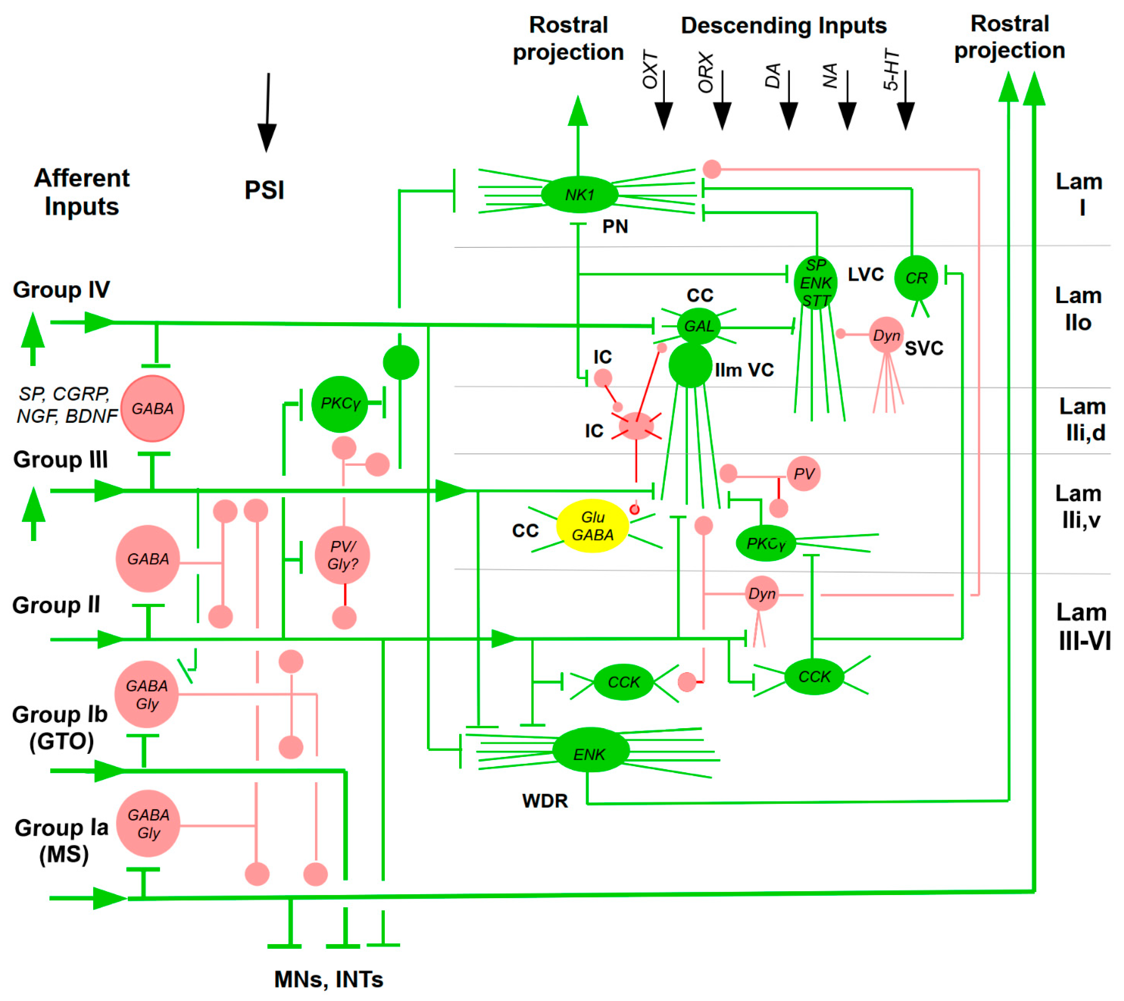
Figure 2.
Simplified and rarefied scheme of locations of some nuclei, brain structures and connections involved in the modulation of pain. The sections are not scaled. Some structures (e.g., raphé nuclei) distribute quite far rostro-caudally and may occur in two cross-sections, which is not shown for graphical reasons. Connections symbolized by arrows may be excitatory (green) or inhibitory (red) or undefined. DA connections are light blue; NA dark blue; 5-HT magenta. OXT and ORX connections are red because of their anti-nociceptive effects. The supraspinal connections are incomplete for graphical reasons. The thick orange line symbolizes mixed effect of the PAG on rostral ventro-medial medulla (RVM). Double-colored connections symbolize the change of their effects from inhibitory (red) to excitatory (green). Dashed black lines indicate humorous effects from the hypothalamus (HYP) to the thyroid gland (light green) secreting thyroid hormones (THs) or gonads (dark red) secreting gonadal hormones (GHs). – The descending pain modulation is mediated by three main systems (Tavares et al. 2021). In animal models of neuropathic pain, a dual action of NA modulation was demonstrated. Besides the anti-nociception due to spinal effects, the NA system may induce pro-nociception by directly acting on brainstem pain modulatory circuits, the locus coeruleus (LC) and medullary dorsal reticular nucleus (DReN). The 5-HT system also has a dual action depending on the targeted spinal receptor, with an exacerbated activity of the excitatory 5-hydroxytryptamine 3 receptor (5-HT3R) in neuropathic pain models. Opioids are involved in the modulation of descending modulatory circuits. In neuropathic pain, the opioidergic modulation of brainstem pain control areas is altered, with the release of enhanced local opioids along with reduced expression and de-sensitization of μ-opioid receptors (MORs). In the DReN, the installation of neuropathic pain increases the levels of enkephalins (ENKs) and induces de-sensitization of MORs, which may enhance descending facilitation from the DReNt and impact the efficacy of exogenous opioids (Tavares et al. 2021). Furthermore, various other neuromodulators and neurotransmitters in the descending system co-determine the output. For example, direct application of prostaglandin E2 (PGE2) within the ventro-lateral PAG (vlPAG) produces hyperalgesia, which activated RVM ON-cells and suppressed the firing of OFF-cells. In a mouse model of chemotherapy-induced neuropathic pain. Somatostatin (STT)-expressing Glu neurons in the lateral/ventro-lateral PAG facilitated mechanical and thermal hypersensitivity (Zhang et al. 2023). There is evidence that neurotensin (NT) type 2 receptors (NTR2)-immuno-reactive neurons in the RVM receive NT projections originating from the PAG; express NT, 5-HT, or both; and send projections that terminate in DH laminae I and II (Wang et al. 2014). NT in the RVM activates ON-cells at low-doses (Neubert et al. 2004). Glycine (Gly) may have a dual role in producing hyperalgesia or analgesia by stimulating the Gly receptors (GlyRs) or the NMDA receptors (NMDARs) within the vlPAG, respectively. Consistently RVM ON and OFF cells displayed opposite firing patterns to the stimulation of the vlPAG NMDAR Gly site and GlyR activation; a tonic role of these receptors within the vlPAG-RVM anti-nociceptive descending pathway (Palazzo et al. 2009). Abbreviations: AMY: amygdala; BG: basal ganglia; CC: cingulate cortex; CMED: caudal medulla; CVLM: caudal ventro-lateral medulla; dlPFC: dorso-lateral prefrontal cortex; DH: dorsal horn; DReN: dorsal reticular nucleus; DRN: dorsal raphé nucleus; HYP: hypothalamus; IC: insular cortex; LC: locus coeruleus; MES: mesencephalon; mPFC: medial prefrontal cortex; NRM: nucleus raphé magnus; NTS: nucleus tractus solitarii; PAG: peri-aqueductal gray; PBN: parabrachial nucleus; PROS: prosencephalon; RMED: rostral medulla; RVM: rostral ventro-medial medulla; SC: spinal cord; SN: substantia nigra; SNc: substantia nigra pars compacta; THAL: thalamus; VTA: ventral tegmental area (Data from papers cited in the text).
Figure 2.
Simplified and rarefied scheme of locations of some nuclei, brain structures and connections involved in the modulation of pain. The sections are not scaled. Some structures (e.g., raphé nuclei) distribute quite far rostro-caudally and may occur in two cross-sections, which is not shown for graphical reasons. Connections symbolized by arrows may be excitatory (green) or inhibitory (red) or undefined. DA connections are light blue; NA dark blue; 5-HT magenta. OXT and ORX connections are red because of their anti-nociceptive effects. The supraspinal connections are incomplete for graphical reasons. The thick orange line symbolizes mixed effect of the PAG on rostral ventro-medial medulla (RVM). Double-colored connections symbolize the change of their effects from inhibitory (red) to excitatory (green). Dashed black lines indicate humorous effects from the hypothalamus (HYP) to the thyroid gland (light green) secreting thyroid hormones (THs) or gonads (dark red) secreting gonadal hormones (GHs). – The descending pain modulation is mediated by three main systems (Tavares et al. 2021). In animal models of neuropathic pain, a dual action of NA modulation was demonstrated. Besides the anti-nociception due to spinal effects, the NA system may induce pro-nociception by directly acting on brainstem pain modulatory circuits, the locus coeruleus (LC) and medullary dorsal reticular nucleus (DReN). The 5-HT system also has a dual action depending on the targeted spinal receptor, with an exacerbated activity of the excitatory 5-hydroxytryptamine 3 receptor (5-HT3R) in neuropathic pain models. Opioids are involved in the modulation of descending modulatory circuits. In neuropathic pain, the opioidergic modulation of brainstem pain control areas is altered, with the release of enhanced local opioids along with reduced expression and de-sensitization of μ-opioid receptors (MORs). In the DReN, the installation of neuropathic pain increases the levels of enkephalins (ENKs) and induces de-sensitization of MORs, which may enhance descending facilitation from the DReNt and impact the efficacy of exogenous opioids (Tavares et al. 2021). Furthermore, various other neuromodulators and neurotransmitters in the descending system co-determine the output. For example, direct application of prostaglandin E2 (PGE2) within the ventro-lateral PAG (vlPAG) produces hyperalgesia, which activated RVM ON-cells and suppressed the firing of OFF-cells. In a mouse model of chemotherapy-induced neuropathic pain. Somatostatin (STT)-expressing Glu neurons in the lateral/ventro-lateral PAG facilitated mechanical and thermal hypersensitivity (Zhang et al. 2023). There is evidence that neurotensin (NT) type 2 receptors (NTR2)-immuno-reactive neurons in the RVM receive NT projections originating from the PAG; express NT, 5-HT, or both; and send projections that terminate in DH laminae I and II (Wang et al. 2014). NT in the RVM activates ON-cells at low-doses (Neubert et al. 2004). Glycine (Gly) may have a dual role in producing hyperalgesia or analgesia by stimulating the Gly receptors (GlyRs) or the NMDA receptors (NMDARs) within the vlPAG, respectively. Consistently RVM ON and OFF cells displayed opposite firing patterns to the stimulation of the vlPAG NMDAR Gly site and GlyR activation; a tonic role of these receptors within the vlPAG-RVM anti-nociceptive descending pathway (Palazzo et al. 2009). Abbreviations: AMY: amygdala; BG: basal ganglia; CC: cingulate cortex; CMED: caudal medulla; CVLM: caudal ventro-lateral medulla; dlPFC: dorso-lateral prefrontal cortex; DH: dorsal horn; DReN: dorsal reticular nucleus; DRN: dorsal raphé nucleus; HYP: hypothalamus; IC: insular cortex; LC: locus coeruleus; MES: mesencephalon; mPFC: medial prefrontal cortex; NRM: nucleus raphé magnus; NTS: nucleus tractus solitarii; PAG: peri-aqueductal gray; PBN: parabrachial nucleus; PROS: prosencephalon; RMED: rostral medulla; RVM: rostral ventro-medial medulla; SC: spinal cord; SN: substantia nigra; SNc: substantia nigra pars compacta; THAL: thalamus; VTA: ventral tegmental area (Data from papers cited in the text).
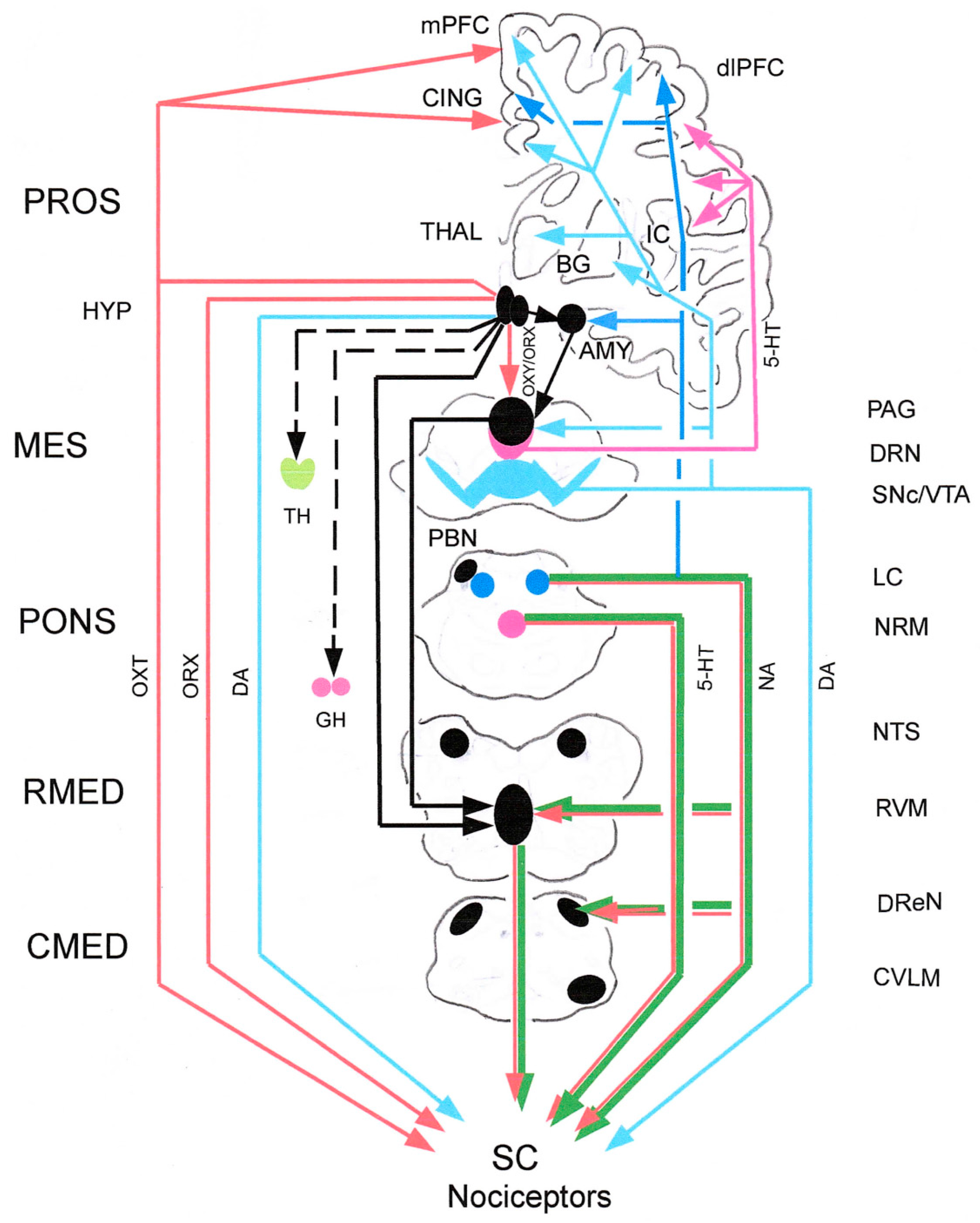
Disclaimer/Publisher’s Note: The statements, opinions and data contained in all publications are solely those of the individual author(s) and contributor(s) and not of MDPI and/or the editor(s). MDPI and/or the editor(s) disclaim responsibility for any injury to people or property resulting from any ideas, methods, instructions or products referred to in the content. |
© 2026 by the authors. Licensee MDPI, Basel, Switzerland. This article is an open access article distributed under the terms and conditions of the Creative Commons Attribution (CC BY) license (http://creativecommons.org/licenses/by/4.0/).
Copyright: This open access article is published under a Creative Commons CC BY 4.0 license, which permit the free download, distribution, and reuse, provided that the author and preprint are cited in any reuse.


