Preprint
Review

This version is not peer-reviewed.

Mechanotherapy for the Non-Surgical Treatment of Urinary Incontinence: Mechanisms and Clinical Evidence

Submitted:

08 July 2026

Posted:

10 July 2026

You are already at the latest version

Abstract

Background: Urinary incontinence (UI) is very common, affecting 421 million women and men worldwide. There are three types of UI: stress, urge and mixed. UI is associated with poor quality of life. There are numerous non-surgical treatments available. This paper reviews the mechanisms and clinical evidence for mechanotherapy gleaned from peer-reviewed literature. Mechanisms: The human body responds to various forms mechanical stimuli that direct cellular tissue forces, resulting in molecular responses that facilitate growth, remodeling and repair. Mechanotransduction is the basis for mechanotherapy, characterized by complex chemical signaling and neural signaling. Two modalities of mechanotherapy have been investigated in published studies. Mechanical Vibration: Vibration caused by oscillatory motion stimulates stretch sensitive mechanoreceptors in the pelvic floor muscle (PFM) spindles, resulting in afferent nerve impulses to the sacral plexus and centers in the brain. Resulting efferent signals cause motor unit activation. Types of vibration delivery include intra-vaginal, whole-body, and local transcutaneous. Acoustical Wave Stimulation: Acoustic energy generated by piezoelectric crystals modify molecules as the waves are transmitted from one molecule to another. This type of mechanotherapy includes four stages: propagation, physical effect, mechanotransduction and biological effect. Most published research studies utilized low intensity extracorporeal shock waves (LiESWT) in their protocols. Conclusion: Mechanotherapy provides powerful physiologic stimulation that has been shown in many studies to improve clinical symptoms of UI and improve QoL. It is a promising, emerging, non-invasive option for inclusion in the clinical armamentarium for treating patients with all forms of UI.

Keywords: 
;  ;  ;  ;  ;  ;  ;  

Introduction

Continence and urination are the result of coordination of smooth and striated muscles of the lower urinary tract and pelvic floor that occurs with regulation between the central and peripheral nervous system; it involves multiple levels both voluntary and involuntary control as explained below. Urinary incontinence (UI) is the involuntary loss of urine, more commonly observed in women. There are three main types of UI: (1) stress urinary incontinence (SUI), characterized as the sudden involuntary loss of urine during activities such as coughing, sneezing, exercise and bending/lifting; (2) urgency urinary incontinence (UUI) is distinguished by an involuntary loss of urine with a sudden, uncontrollable urge of needing to urinate; and (3) mixed urinary incontinence (MUI) which includes features of both SUI and UUI [1]. Urinary incontinence is strongly associated with poor quality of life (QoL) as shown in many peer-reviewed publications that used multiple, validated questionnaires. These studies reported decreases in various domains of QoL (e.g., physical wellbeing, psychological and emotional health, social relations, etc [2,3,4,5,6]. UI is also associated with social withdrawal, depression, earlier retirement and loss of independent living in older patients [7]. Women reporting MUI symptoms describe more severe and bothersome incontinence and poorer QoL [6]. Given the high negative impact on QoL, the burden of urinary incontinence not surprisingly has a high global prevalence, despite likely being underdiagnosed. As of 2018, the global prevalence of UI was approximately 421 million [10]. For adult women in the United States, 61.8% (78,297,094 ) had UI, partitioned as SUI 22.0%, UUI 22.0%, MUI 31.3%, Unspecified 9.2%) [11]. Risk factors for UI include parity, obesity, chronic cough, depression, poor health, lower urinary tract symptoms, previous hysterectomy, and stroke; UI is more common in the elderly, but affects women of all ages [8,9,11]. Women with SUI typically delay seeking medical help for over a decade; this longer duration is associated with increased severity and greater need for continence pads [12].
Numerous non-surgical treatments are available to address UI, including pelvic floor muscle training, behavioral therapy, bladder training, caffeine avoidance and weight loss; pharmacological therapies are used in UUI [13,14]. Among these, mechanotherapy has emerged as a promising clinical approach, either as sole treatment or as an adjunct to other therapies. This paper discusses the mechanisms of action and clinical evidence of various forms of mechanotherapy gleaned from the peer-reviewed literature.

Mechanisms of Action

The human body responds to mechanical stimuli such as vibration, touch, stretch, and pressure via mechanoreceptors. Mechanoreceptor sensor cells detect changes in their environment and respond rapidly to the mechanical stimuli interacting with them. As illustrated in Figure 1, mechanical stimuli direct cellular activities to induce tissue adaptation. These mechanical forces initiate a chain response involving tissue level forces and cellular forces that result in molecular responses, that in turn create a feedback loop via the extracellular matrix (ECM) [13]. This process facilitates tissue-level adaptations including growth, remodeling, and repair that alters tissue mass, structure, and quality [15].
Tensegrity, or “tensional integrity,” is a structural principle where stability is maintained by a balance between continuous tension (muscles / fascia) and discontinuous compression (bones / extracellular matrix) [16]. For example, cells and tissues can respond rapidly to mechanical stresses transmitted over cell surfaces via connections that physically couple the cytoskeleton of cells to the extracellular matrix, as well as other cells [16].
Cells and tissues are under physiological tension even in the absence of external loading through the self-stabilizing system of tensegrity. As such, tensegrity is the basis for cells’ ability to respond immediately to mechanical stresses transmitted across cell receptors that physically couple the cytoskeleton to the extracellular matrix [16].
Mechanotransduction
Mechanotherapy is the intentional clinical triggering of mechanotransduction.
Mechanotransduction is the basis for mechanotherapy. It is the molecular mechanism by which cells sense and convert mechanical signals into cellular response to a variety of mechanical stresses [17]. Mechanotransduction requires a mechanical signal to be transmitted to tissues and cells. This process may be altered by changes in cell mechanics, variations in the extracellular matrix, or by deregulation of the molecular mechanisms by which cells sense mechanical signals and convert them into chemical or electrical responses [18]. Mechanotransduction is mediated by extracellular matrix molecules, transmembrane integrin receptors, cytoskeletal structures and associated signaling molecules, all of which are potential targets for therapeutic applications [16]. Mechanical loads are transmitted across structural elements that are physically interconnected. Mechanotransduction is controlled by both complex chemical and neural signaling pathways, as discussed below.
Chemical Signaling
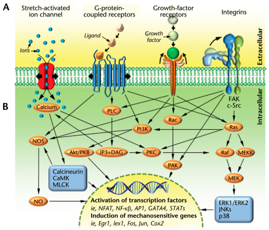
Once a cell has detected a local mechanical stimulus, the cell converts the stimulus into a biochemical response known as biochemical coupling. Multiple signaling pathways are used by the cell to initiate a biochemical signaling response. As illustrated in Figure 2, musculoskeletal cells sense incoming mechanical signals using a diverse group of transmembrane mechanosensitive proteins (mechanosensors), including stretch activated
ion channels, cell-membrane spanning G-protein-coupled receptors, growth-factor
receptors, and integrins [15]. In response, muscle cells convert the mechanical signals into biochemical signals (‘biochemical coupling’) that triggers a cascade of multifarious intracellular pathways culminating in activation of transcription factors and induction of mechanosensitive genes [15].
Neural Signaling and Modulation
Broadly speaking, the nervous system is composed of two parts. The Central Nervous System (CNS), that includes the brain and the spinal cord, and the Peripheral Nervous System (PNS), that involves neural structure outside of the CNS. The PNS is composed two divisions: (1) a somatic portion that controls voluntary responses and (2) the autonomic portion that controls involuntary responses [19]. Normally, these portions of the nervous system operate in equilibrium. Mechanotherapy engages the autonomic portion of the PNS.
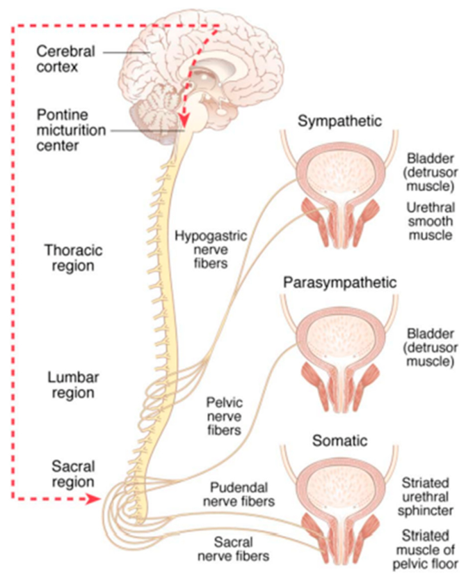
In addition to the cellular mechanisms described above, mechanotherapy also modulates nerve signaling via the stimulation of mechanoreceptors located in muscles, tendons and ligaments that activate the proprioceptive sensory system within the PNS [20]. This nerve signaling enhances afferent and efferent neural communication involving somatic peripheral nerves, the sympathetic and parasympathetic reflexive spinal sacral plexus pathways, and the higher conscious control centers of the brain (Figure 3) [21]. The cumulative effect of improved neuromuscular facilitation and motor learning is development and retention of muscle memory [22,23]

Clinical Evidence

Several modes of mechanotherapy have been used to treat urinary incontinence, either as sole therapy or as adjuncts to other treatments. These include several types of vibration and approaches using shockwave / acoustical energy.
MECHANICAL VIBRATION
Vibration is a mechanical stimulus characterized by an oscillatory motion. The biomechanical variables that determine its intensity are the frequency and amplitude. The extent of the oscillatory motion determines the amplitude (peak to peak displacement, in mm) of the vibration. The repetition rate of the cycles of oscillation determines the frequency of the vibration (measured in Hz) [24]. Vibration stimulates specific mechanoreceptors in muscle spindles in the pelvic floor that are sensitive to stretching. This stimulation triggers an involuntary muscle contraction known as the tonic vibration reflex (TVR) [24,30].
Muscle–tendon vibration generates a strong proprioceptive inflow, primarily driven by Ia afferent nerve fibers, but also involving group II, Ib and cutaneous afferents [25]. These afferent impulses travel through spinal sacral neurons to thalamus and cortical structures in the brain. Resultant efferent signals cause motor unit activation, leading to increased motor unit recruitment of previously inactive muscle fibers [25]. Sub-contractile vibratory stimulation may enhance voluntary muscle contraction using the same neural pathways [26].

Intravaginal Mechanical Vibration

Application of intravaginal vibration provides direct mechano-stimulation of the pelvic floor muscles (PFM). The authors are aware of only one commercial device that provides intravaginal mechanotherapy.
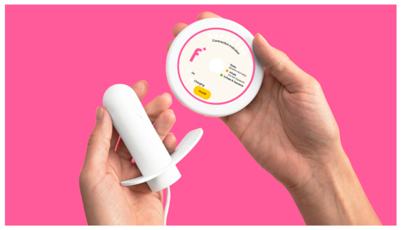
The Flyte® System by Pelvital provides two-step transvaginal mechanotherapy: 1) pre-stretch / pre-loading of the PFM; 2) the superimposition of mechanical pulses at a very specific, low frequency tuned to the PFM, that are superimposed on voluntarily contracted and relaxed PFM (Kegel exercises). The system consists of an intravaginal wand and control unit (Figure 4). At-home treatment sessions are five minutes a day, for a standard treatment protocol of six weeks. It is the only home-based transvaginal mechanotherapy treatment known to the authors.
Flyte System’s intravaginal mechanotherapy has been shown in two published clinical trials in women with stress urinary incontinence with outcomes comparable to surgical outcomes [19]. A study at Arctic University in Norway assessed this mechanotherapy treatment in 60 women with Stress Urinary Incontinence (SUI), characterized as between 5-50g of leakage on the 1-hour stress test, and who had been referred for surgery after having failed supervised PFMT. The study found 44 (73.3%) and 49 (81.7%) of women were defined as continent after 4 and 6 weeks of treatment, respectively. All participants who completed the 2-year follow up interviews had not undergone surgery for SUI since the mechanotherapy treatment, with 77.0% reporting continued continence [27].
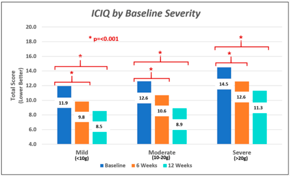
Nakib and co-authors studied the Flyte System’s intravaginal mechanotherapy treatment in 119 women with mild (10-20 g of leakage), moderate (21-70 g of leakage), and severe SUI (>70 g of leakage), with a range of 10.3g to 533.9 g, as measured by the 24-hour pad weight test. The researchers found that the use of mechanical stimulation superimposed on Kegel contractions improved SUI in as little as two weeks (37%), with 80% having responded to treatment in 6 weeks, and 91% in 12 weeks. Using the 24-hour pad weight test, 71% (81/119) of subjects were substantially improved or continent (defined as <10g) within 12 weeks (p=<0.001), with a median pad weight at 12 weeks of 5.3 grams. In addition, Quality of Life (QoL) using the International Consultation on Incontinence Questionnaire - Short Form (ICIQ-SF) also showed highly significant improvements (p=<0.001) in all severity groups (Figure 4) [28].
Figure 4. ICIQ Scores by Baseline Severity Level (Reprinted with permission of the author).
Preprints 222313 g005
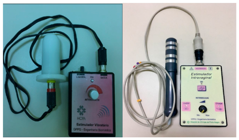
Rodriques, et al., reported results from a randomized clinical trial comparing intravaginal vibratory (IVVS) versus intravaginal electrical stimulation (IEVS ) in women who were unable to contract their PFM, 19% of which had incontinence at baseline. Both devices were custom made with adjustable intensity output (and neither study device is commercially available). Treatment consisted of six 20- minute sessions held once a week. PFM evaluation consisted of bidigital vaginal palpation and the first four items of the New PERFECT scale: P (power), E (endurance), R (repetitions), and F (fast contractions). Measures of PFM performance were made by the Modified Oxford Scale (graded from 0 to 5). Power measures PFM strength by the Modified Oxford Scale (graded from 0 to 5). Endurance measures the duration of a maximum voluntary contraction until its strength is reduced by 50% or more. Repetitions assess the number of repetitions of the previous contraction, and Fast assesses the number of fast contractions performed in 10s. While both groups were noted to have improvements at the end of treatment, PFM contraction strength in the IVVS group improved significantly more than the IVES group (p = 0.026). The authors conclude that “IVVS was significantly superior to IVES in improving pelvic floor muscle strength, is easier to use, may be done at home, and is less uncomfortable.” [29]

Whole Body Vibration

Numerous articles have demonstrated that whole body vibration (WBV) successfully activates the pelvic floor muscles of their subjects, including those with weakened pelvic floor muscles [31,32,33]. WBV devices currently available on the market deliver vibration to the whole body by means of oscillating plates using two different systems: (a) reciprocating vertical displacements on the left and right side of a fulcrum; (b) the whole plate oscillating uniformly up and down [24]. WBV indirectly stimulates the PFM, typically utilizing commercially available vibration plates. It has been reported that WBV: (1) improves the PFMs strength and QOL in individuals with urinary incontinence; (2) does not cause (PFM) fatigue in nulliparous continent women; (3) leads to higher (PFM) activation in subjects with weakened (PFM) and (4) achieves higher pelvic floor (PF) activation than maximum voluntary contraction alone [34]. Several studies have demonstrated the efficacy of WBV for improving UI symptoms, as summarized below.
Farzinmehr and colleagues conducted a randomized study in 43 women with SUI that compared WBV training (Power Plate, USA) and PFMT three time per week over four weeks. Muscle strength was assessed based on the Modified Oxford Scale by digital palpation by a physical therapist trained in pelvic floor muscle examination. Severity of incontinence was appraised using a visual analog scale (VAS) from zero to ten. Quality of life was evaluated using the incontinence quality of life (I-QOL) questionnaire. At the end of treatment both groups experienced significant improvement in all three metrics (p=0.0001) that were sustained at three months. However, there were no difference between groups [35].
Tantaway and co-authors evaluated the effects of WBV in a randomized trial of male patients post-surgical prostatectomy with SUI. Group 1 included 30 patients who performed pelvic floor muscle training and whole-body vibration training; Group 2 included 31 patients who performed pelvic floor muscle training alone. The interventions in both groups were conducted three times per week for 4 weeks. I-VAS score, ICIQ-UI-SF score, and 24-hour Pad test result all showed significant differences at each assessment (p=0.001) in both groups. However, compared to Group 2, significantly greater benefits were noted in Group 1 in all measured parameters at 4 weeks and at all subsequent follow-up periods [36].

Local Transcutaneous Vibration

Cutaneous mechanoreceptors include Merkel disks and Meissner corpuscles located in the superficial layers and Pacinian corpuscles found in deeper layers of the skin and fascia between the muscles and periosteum. Merkel disks respond to low vibratory frequencies (5 – 15 Hz), Meissner corpuscles to medium frequencies (20-50 Hz) and Pacinian corpuscles to high frequencies (60-400 Hz) [37].
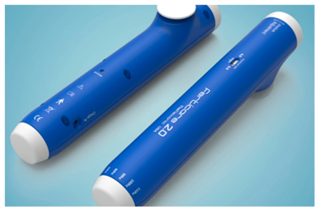
A few studies evaluated the efficacy of local transcutaneous vibration on urinary incontinence. Sonksen, et al., assessed the efficacy of perineal transcutaneous mechanical nerve stimulation (TMNS) in 33 women with SUI using the commercially available FERTI CARE® therapeutic device (Figure 6). All subjects had tried other treatments including pelvic floor muscle training (100%), behavioral intervention (100%) and peripheral electrical stimulation (3 patients), which had failed. Perineal TMNS was performed weekly by placing the device on the center of the perineum for 10 seconds followed by a pause of 10 seconds, for a total of 10 cycles per session. Subjects were instructed to perform standard PFMT after completing the TMNS program. After 6 weeks of TMNS, a statistically significant reduction (p=<0.001) was found in the number of incontinence episodes and the number of pads used as recorded in the voiding diary. Of the 33 subjects, 24 (73%) were cured and 29 (88%) were cured or the condition improved. The benefits remained at three months in the majority of women [38].
Figure 5. FERTI CARE® Therapeutic Device (figure from company webpage).
Preprints 222313 g007
Fode and associates examined the effects of penile vibratory stimulation (PVS) also using the FERTI CARE® vibration device in the treatment of post-prostatectomy urinary incontinence during a 12 week trial in 31 men. A 24-hr pad test and a 72-hr voiding diary were collected at baseline. Participants were then randomized to receive PVS for the first 6 weeks (Group 1) or for the full 12 weeks (Group 2). The difference in the change on the pad test between the groups did not reach statistical significance at 6 weeks (p=0.13), whereas the number of incontinence episodes between groups approached statistical significance (p =0.052). However, there was a median reduction of 33 g (p =0.021) on the pad test and a median reduction in daily incontinence episodes by one (p =0.023) in group 1 at 6 weeks. At 12 weeks, group 2 had a median decrease on the 24-hr pad test of 8 g (p=0.10) and no change in incontinence episodes. A pooled analysis showed a reduction in pad test of 13.5 g (p=0.004) after PVS. Of the patients who completed PVS treatment, 60% were either very satisfied or partly satisfied (60%). Side effects were observed in 15% of men, most frequently reported as discomfort, with one case of mild bleeding on the glans of the penis. Two patients experienced stimulation pain that caused them to withdraw from the study. Five patients voluntarily reported improvement in erectile function [39].
ACOUSTICAL WAVE STIMULATION
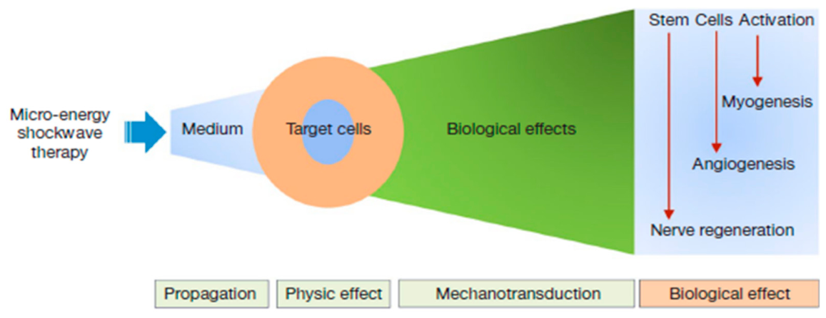
Another form of PFM stimulation utilizes acoustic energy, typically generated using piezoelectric crystals, that interacts with body tissues [40]. The acoustic energy moves through the tissue in longitudinal waves according to the frequency and amplitude of the waves. This energy slightly modifies molecules and passes from one molecule to another. Molecules along the transmission wave oscillate in the same direction of the wave [40]. As illustrated in Figure 5, the mechanisms for acoustical therapy have been postulated to include four stages: propagation, physical effect, mechanotransduction, and biological effect [43]. Depth of energy penetration is inversely proportional to frequency. Lower frequencies produce lower resolution and greater depth in the body [40]. in addition, there is evidence that the physical properties of the wave form can be optimized based on the type of urinary incontinence, with low frequencies (10–20 Hz) for urge incontinence and higher frequencies (50 Hz) for stress incontinence [41,42]. Most published UI research studies utilize low intensity extracorporeal shock waves (LiESWT) in their protocols.
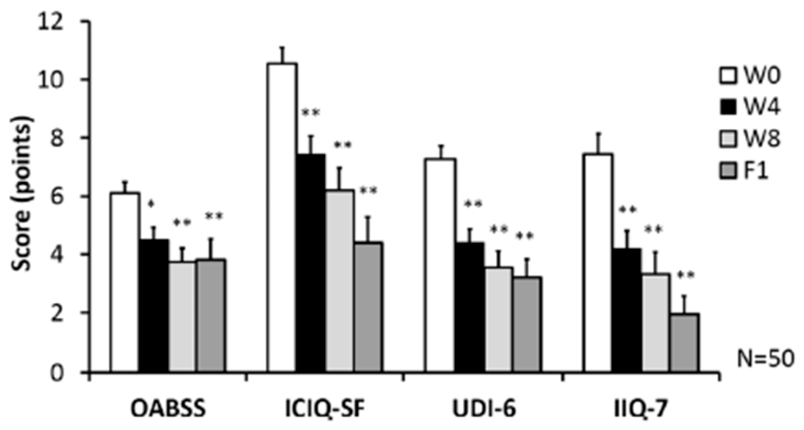
Long and colleagues evaluated the efficacy of LiESWT in 50 women with SUI, assessed at baseline, four weeks, eight weeks and one month. Involuntary urine leakage was assessed by the pad test (type unspecified), which demonstrated significant reductions at all follow-up periods (p < 0.01). In addition, quality of life was assessed using the OABSS, ICIQ-SF, UDI-6 and the IIQ-7 validated questionnaires. All questionnaires demonstrated significant improvement at all follow-up times, as shown in Figure 7. Of note, while the study population consisted of women with SUI, significant improvements in OAB symptoms were also exhibited [44].
Lin and co-authors performed a multicenter, single-blind, sham-controlled, randomized trial that assessed LiESWT in 60 women with SUI. Treatment was provided once weekly for four and eight week periods. LiESWT significantly decreased urine leakage from 7.93±1.32 at baseline to 2.43±0.40 g (p < 0.001) at four weeks that was sustained through six months. In addition, 64.4% of participants had moderate to better improvement (>50%) after 4 weeks of LiESWT, with the proportion of improvement increasing to 68.8%, 77.8%, 82.3%, and 84.5% at week eight, one, three and six months. The 3-day urinary diary revealed that eight weeks of LiESWT significantly improved OAB symptoms in these SUI participants. In addition, four weeks of active treatment significantly improved all questionnaire scores (ICIQ-SF (p = 0.034), UDI-6 (p = 0.040), and IIQ-7 (p = 0.048) compared to the sham group; the OABSS change was not significant (p = 0.520) [45].
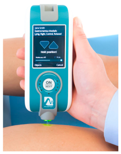
Barassi, et al., investigated whether an integrated rehabilitation protocol including neuromuscular manual therapy and focused mechanical acoustic therapy targeting superficial PFM can significantly reduce pelvic floor dysfunctions in 62 women affected by stress, urge, or mixed urinary incontinence. Treatments were performed twice a week for four weeks. Mechanical characteristics (muscle tone, rigidity, muscle plasticity) of the PFM were measured using the MyotonPRO® handheld device (Figure 8). All evaluated parameters demonstrated significant improvement (p=<0.001). In addition, significant improvements were noted in PFDI-20 and PFIQ-7 questionnaire results showed a significant reduction of scores (p=<0.001) [46].
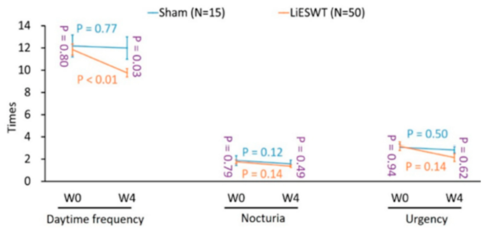
Lu and co-authors examined the therapeutic effect and duration of low intensity LiESWT on overactive bladder (OAB) symptoms in a prospective, randomized, single-blind clinical trial in 65 patients. Subjects were randomly divided into active LiESWT once a week for eight weeks, or identical sham LiESWT without energy transmission. Subject were followed for 4, 8 weeks and one, three and six months. Compared to baseline there was no meaningful difference in the daytime frequency (p = 0.77), nocturia (p = 0.12) and urgency (p = 0.50) after 4 weeks of treatment in the sham group. However, in the active LiESWT group, LiESWT meaningfully decreased daytime frequency (p < 0.01), nocturia (p < 0.01) and urgency symptoms (p < 0.05 ) as illustrated in Figure 9. Uroflowmetry measurements, including voided urine volume, Qmax and PVR were noticeably improved in the active treatment group compared to the sham group. Active LiESWT patients demonstrated significantly (p<0.01) improved OABSS, UDI-6 and IIQ-7, and the ICIQ-SF (p < 0.05) compared to no meaningful change in the sham group [47].
In a retrospective, observational case-controlled study, Paolucci and associates assessed the effects of focal LiESWT in 65 patients with MUI. Subjects were randomized to active therapy using the Vibration Sound System® for four weeks versus no therapy; both groups received home-based postural ergonomic instructions to reinforce the pelvic floor. PFM muscle parameters were evaluated using the MyotonPRO® device. The treatment group demonstrated significant improvement (p=<0.05, p=<0.001) in all muscle indices. Improvement in incontinence symptoms and quality of life as evaluated with PDFI-20 (p=<0.001) and PFIQ-7(p=<0.00) scores were noted [48].
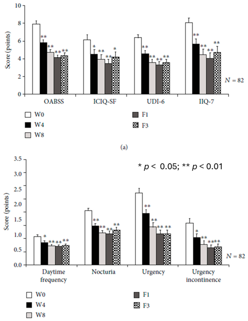
Lee, et al., assessed the therapeutic effects of LiESWT on OAB applied to the suprapubic area using a commercially available instrument (DUOLITH SD1 T-TOP, AG) in 82 women assessed at baseline, four and eight weeks, one month and three months. As demonstrated in Figure 10, significant improvements were observed in Quality of Life using multiple questionnaires (ICIQ-SF, UDI-6 and IIQ-7), as well as OAB symptoms (OABSS). In addition, the 3-day diary showed increases in bladder capacity, daytime frequency (p=<0.01), nocturia (p=<0.01), and urgency (p=<0.05, p=<0.01) at the follow-up timepoints. Furthermore, uroflowmetry was noted to improve voided urine volume (p=<0.05), maximum flow rate (p=<0.05) and post-void residual volume (p=<0.05) [49].

Discussion and Conclusion

Urinary incontinence affects a large number of both women and men. It is estimated that one in seven adult women will undergo surgery for SUI during their lifetime [50]. Healthcare practitioners need to include assessment for UI into their practices, particularly in patients with elevated risk factors. Early detection and treatment of UI can enhance the quality of life and lessen the impact of associated comorbidities [51].
Early detection and treatment of UI can enhance the quality of life and lessen the impact of associated comorbidities, such as recurring UTIs or social isolation brought on by shame.
To reduce the need for surgery, additional and more effective non-invasive therapies, especially those that are home based and easy to access, are needed. Mechanotherapy provides powerful physiologic stimulation that has been shown by many studies to improve clinical symptoms of UI and improve QoL, as summarized in this paper. Mechanotherapy can be used as a stand-alone treatment, or be used as an adjunct with other therapies, e.g., pelvic floor muscle training. It is a promising, emerging non-invasive option for inclusion in the clinical armamentarium for treating patients with all forms of UI.

References

  1. Qaseem A, Dallas P, Forciea MA, Starkey M, Denberg TD, Shekelle P, Clinical Guidelines Committee of the American College of Physicians. Nonsurgical management of urinary incontinence in women: a clinical practice guideline from the American College of Physicians. Annals of internal medicine. 2014 Sep 16;161(6):429-40.
  2. Bartoli S, Aguzzi G, Tarricone R. Impact on quality of life of urinary incontinence and overactive bladder: a systematic literature review. Urology. 2010 Mar 1;75(3):491-500.
  3. Pizzol D, Demurtas J, Celotto S, Maggi S, Smith L, Angiolelli G, Trott M, Yang L, Veronese N. Urinary incontinence and quality of life: a systematic review and meta-analysis. Aging clinical and experimental research. 2021 Jan;33(1):25-35.Milsom I, Gyhagen M. The prevalence of urinary incontinence. Climacteric. 2019 May 4;22(3):217-22.
  4. Kwon BE, Kim GY, Son YJ, Roh YS, You MA. Quality of life of women with urinary incontinence: a systematic literature review. International neurourology journal. 2010 Oct 31;14(3):133.
  5. Pizzol D, Demurtas J, Celotto S, Maggi S, Smith L, Angiolelli G, Trott M, Yang L, Veronese N. Urinary incontinence and quality of life: a systematic review and meta-analysis. Aging clinical and experimental research. 2021 Jan;33(1):25-35.
  6. Minassian VA, Devore E, Hagan K, Grodstein F. Severity of urinary incontinence and effect on quality of life in women by incontinence type. Obstetrics & Gynecology. 2013 May 1;121(5):1083-90.
  7. Didyk N. Urinary incontinence in aging: what to know when you can’t wait to go. https://betterhealthwhileaging.net/urinary-incontinence-in-aging/#:~:text=Incontinence%20is%20more%20than%20inconvenient,and%20loss%20of%20independent%20function.
  8. Minassian VA, Stewart WF, Wood GC. Urinary incontinence in women: variation in prevalence estimates and risk factors. Obstetrics & Gynecology. 2008 Feb 1;111(2 Part 1):324-31.
  9. Minassian VA, Drutz HP, Al-Badr A. Urinary incontinence as a worldwide problem. International Journal of Gynecology & Obstetrics. 2003 Sep 1;82(3):327-38.
  10. Milsom I, Gyhagen M. The prevalence of urinary incontinence. Climacteric. 2019 May 4;22(3):217-22.
  11. Patel UJ, Godecker AL, Giles DL, Brown HW. Updated Prevalence of Urinary Incontinence in Women: 2015-2018 National Population-Based Survey Data. Female Pelvic Med Reconstr Surg. 2022 Apr 1;28(4):181-187. Epub 2022 Jan 12. PMID: 35030139. [CrossRef]
  12. Grzybowska ME, Wydra D, Smutek J. Analysis of the usage of continence pads and help-seeking behavior of women with stress urinary incontinence in Poland. BMC Womens Health. 2015 Sep 30;15:80. [CrossRef]
  13. Syan R, Brucker BM. Guideline of guidelines: urinary incontinence. BJU international. 2016 Jan;117(1):20-33.
  14. Sussman RD, Syan R, Brucker BM. Guideline of guidelines: urinary incontinence in women. BJU international. 2020 May 1;125(5).
  15. Thompson WR, Scott A, Loghmani MT, Ward SR, Warden SJ. Understanding mechanobiology: physical therapists as a force in mechanotherapy and musculoskeletal regenerative rehabilitation. Physical therapy. 2016 Apr 1;96(4):560-9.
  16. Ingber DE. Tensegrity: the architectural basis of cellular mechanotransduction. Annual review of physiology. 1997 Mar;59(1):575-99.
  17. Dunn SL, Olmedo ML. Mechanotransduction: Relevance to physical therapist practice—Understanding our ability to affect genetic expression through mechanical forces. Physical therapy. 2016 May 1;96(5):712-21.
  18. Ingber D. Mechanobiology and diseases of mechanotransduction. Annals of medicine. 2003 Jan 1;35(8):564-77.
  19. Fulker A, Kovela S, Mianulli M. Transvaginal mechanotherapy for stress urinary incontinence: Principles, cellular and neuromuscular mechanisms of action, and comparison to current gold standard treatments. Continence Reports. 2025 Mar 1;13:100077.
  20. Riemann BL, Myers JB, Lephart SM. Sensorimotor system measurement techniques. Journal of athletic training. 2002 Jan;37(1):85.
  21. de Groat WC. Neural Control and Neuromodulation Lower Urinary Traction Function. National Institute of Health Common Fund. Available: https://commonfund.nih.gov/sites/default/files/SPARCdeGroat-UPitt-Sess1.pdf. (Accessed 15 May 2026).
  22. Asavasopon S, Rana M, Kirages DJ, Yani MS, Fisher BE, Hwang DH, Lohman EB, Berk LS, Kutch JJ. Cortical activation associated with muscle synergies of the human male pelvic floor. J Neurosci. 2014 Oct 8;34(41):13811-8. PMID: 25297107; PMCID: PMC4188976. [CrossRef]
  23. Sønksen J, Ohl DA, Bonde B, Læssøe L, McGuire EJ. Transcutaneous mechanical nerve stimulation using perineal vibration: a novel method for the treatment of female stress urinary incontinence. The Journal of urology. 2007 Nov 1;178(5):2025-8.
  24. Cardinale MA, Wakeling J. Whole body vibration exercise: are vibrations good for you?. British journal of sports medicine. 2005 Sep 1;39(9):585-9.
  25. Amiez N, Beaulieu LD, Paizis C, Lapole T, Rienzo FD. Muscle tendon vibration revisited: Why tonic vibration reflex and kinaesthetic illusions matter. The Journal of Physiology. 2026 Apr;604(8):3231-52.
  26. Kalaoglu E, Bucak OF, Kokce M, Ozkan M, Cetin M, Atasoy M, Ayture L, Karacan I. Whole body vibration activates the tonic vibration reflex during voluntary contraction. Journal of Physical Therapy Science. 2023;35(6):408-13.
  27. Nilsen I, Rebolledo G, Acharya G, Leivseth G. Mechanical oscillations superimposed on the pelvic floor muscles during Kegel exercises reduce urine leakage in women suffering from stress urinary incontinence: A prospective cohort study with a 2-year follow up. Acta Obstet Gynecol Scand. 2018 Oct;97(10):1185-1191. Epub 2018 Aug 2. PMID: 29923602. [CrossRef]
  28. Nakib N, Sutherland S, Hallman K, Mianulli M, R Boulware D. Randomized trial of mechanotherapy for the treatment of stress urinary incontinence in women. Ther Adv Urol. 2024 Feb 6;16:17562872241228023. PMID: 38328552; PMCID: PMC10848796. [CrossRef]
  29. Rodrigues MP, Barbosa LJ, Paiva LL, Mallmann S, Sanches PR, Ferreira CF, Ramos JG. Effect of intravaginal vibratory versus electric stimulation on the pelvic floor muscles: a randomized clinical trial. European journal of obstetrics & gynecology and reproductive biology: X. 2019 Jul 1;3:100022.
  30. Pollock RD, Woledge RC, Martin FC, Newham DJ. Effects of whole body vibration on motor unit recruitment and threshold. J Appl Physiol (1985). 2012 Feb;112(3):388-95. Epub 2011 Nov 17. PMID: 22096119; PMCID: PMC3289430. [CrossRef]
  31. Lauper M, Kuhn A, Gerber R, Luginbühl H, Radlinger L. Pelvic floor stimulation: what are the good vibrations?. Neurourology and Urodynamics: Official Journal of the International Continence Society. 2009 Jun;28(5):405-10.Stania.
  32. Stania M, Chmielewska D, Kwaśna K, Smykla A, Taradaj J, Juras G. Bioelectrical activity of the pelvic floor muscles during synchronous whole-body vibration–a randomized controlled study. BMC urology. 2015 Oct 24;15(1):107.
  33. Luginbuehl H, Lehmann C, Gerber R, Kuhn A, Hilfiker R, Baeyens JP, Radlinger L. Continuous versus intermittent stochastic resonance whole body vibration and its effect on pelvic floor muscle activity. Neurourology and urodynamics. 2012 Jun;31(5):683-7.
  34. de Oliveira Guedes-Aguiar E, de Sá-Caputo DD, Moreira-Marconi E, de Macêdo Uchôa SM, de Barros PZ, Valentin EK, Bergmann A, Taiar R, Bernardo-Filho M. Effect of whole-body vibration exercise in the pelvic floor muscles of healthy and unhealthy individuals: a narrative review. Translational Andrology and Urology. 2019 Aug;8(4):395.
  35. Farzinmehr A, Moezy A, Koohpayehzadeh J, Kashanian M. A Comparative Study of Whole Body Vibration Training and Pelvic Floor Muscle Training on Women’s Stress Urinary Incontinence: Three- Month Follow- Up. J Family Reprod Health. 2015 Nov;9(4):147-54. PMID: 27047560; PMCID: PMC4818376.
  36. Tantawy SA, Elgohary HM, Abdelbasset WK, Kamel DM. Effect of 4 weeks of whole-body vibration training in treating stress urinary incontinence after prostate cancer surgery: a randomised controlled trial. Physiotherapy. 2019 Sep 1;105(3):338-45.
  37. Poenaru D, Cinteza D, Petrusca I, Cioc L, Dumitrascu D. Local Application of Vibration in Motor Rehabilitation - Scientific and Practical Considerations. Maedica (Bucur). 2016 Sep;11(3):227-231. PMID: 28694858; PMCID: PMC5486165.
  38. Sønksen J, Ohl DA, Bonde B, Læssøe L, McGuire EJ. Transcutaneous mechanical nerve stimulation using perineal vibration: a novel method for the treatment of female stress urinary incontinence. The Journal of urology. 2007 Nov 1;178(5):2025-8.
  39. Fode M, Sønksen J. Penile vibratory stimulation in the treatment of post-prostatectomy incontinence: A randomized pilot study. Neurourology and Urodynamics. 2015 Feb;34(2):117-22.
  40. Heredia J AH, Tapia Q K, Cabrera L MD, Barrientos C G, Ambrosio L RC, Moreno M M. Non-invasive Ultrasonic Stimulator Applied in the Treatment of Urinary Incontinence in Women. InLatin American Conference on Biomedical Engineering 2019 Oct 1 (pp. 543-547). Cham: Springer International Publishing.
  41. Susset J, Galea G, Manbeck K, Susset A. A predictive score index for the outcome of associated biofeedback and vaginal electrical stimulation in the treatment of female incontinence. The Journal of urology. 1995 May;153(5):1461-6.
  42. Yamanishi T, Yasuda K. Electrical stimulation for stress incontinence. International Urogynecology Journal. 1998 Sep;9(5):281-90.
  43. Lin G, Lue TF. Microenergy shockwave therapies for female stress urinary incontinence. Translational Andrology and Urology. 2024 Aug 31;13(8):1687-94.
  44. Long CY, Lin KL, Lee YC, Chuang SM, Lu JH, Wu BN, Chueh KS, Ker CR, Shen MC, Juan YS. Therapeutic effects of Low intensity extracorporeal low energy shock wave therapy (LiESWT) on stress urinary incontinence. Scientific reports. 2020 Apr 2;10(1):5818.
  45. Lin KL, Chueh KS, Lu JH, Chuang SM, Wu BN, Lee YC, Wu YH, Shen MC, Sun TW, Long CY, Juan YS. Low intensity extracorporeal shock wave therapy as a novel treatment for stress urinary incontinence: a randomized-controlled clinical study. Medicina. 2021 Sep 8;57(9):947.
  46. Barassi G, Bellomo RG, Frondaroli F, Frondaroli S, Santarelli A, Di Felice PA, Supplizi M, Palermo T, Saggini R. Integrated rehabilitation approach with manual and mechanic-acoustic vibration therapies for urinary incontinence. InAdvancements and Innovations in Health Sciences 2019 Aug 30 (pp. 41-50). Cham: Springer International Publishing.
  47. Lu JH, Chueh KS, Chuang SM, Wu YH, Lin KL, Long CY, Lee YC, Shen MC, Sun TW, Juan YS. Low intensity extracorporeal shock wave therapy as a potential treatment for overactive bladder syndrome. Biology. 2021 Jun 16;10(6):540.Chueh.
  48. Paolucci T, Bellomo RG, Pezzi L, Frondaroli F, Frondaroli S, Santarelli A, Barbato C, Porreca A, Saggini R. A novel rehabilitative protocol in the treatment of mixed urinary incontinence in women: the effects of focused mechano-acoustic vibration. BioResearch Open Access. 2019 Dec 20;8(1):biores-2019.Lee.
  49. Lee YC, Chuang SM, Lin KL, Chen WC, Lu JH, Chueh KS, Shen MC, Liu LW, Long CY, Juan YS. Low-intensity extracorporeal shock wave therapy ameliorates the overactive bladder: a prospective pilot study. BioMed Research International. 2020;2020(1):9175676.
  50. Wu JM, Matthews CA, Conover MM, Pate V, Jonsson Funk M. Lifetime risk of stress urinary incontinence or pelvic organ prolapse surgery. Obstet Gynecol. 014;123(6):1201–6. [CrossRef]
  51. Allafi A, Al-Johani A, Babukur R M, et al. (October 11, 2024) The Link Between Menopause and Urinary Incontinence: A Systematic Review. Cureus 16(10): e71260. [CrossRef]
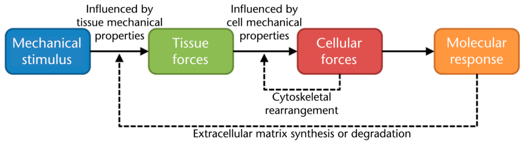
Figure 1. Mechanical stimuli and resultant tissue. cellular and molecular pathways. (Reprinted from Thompson, et al., with permission of Oxford University press and the American Physical Therapy Association).
Preprints 222313 g001
Figure 2. Intracellular mechanosensitive signaling pathways (Reprinted from Thompson, et al., with permission of Oxford University press and the American Physical Therapy Association.).
Preprints 222313 g002
Figure 3. Pelvic Floor Neural Pathways (Reprinted from DeGroat, with permission from the author).
Preprints 222313 g003
Figure 4. The Flyte System (Figure from manufacturer’s website).
Preprints 222313 g004
Figure 5. Custom-made intravaginal vibration (left) and electrical stimulation (right) devices (figure used under Creative Commons license [creativecommons.org/licenses/by-nc-nd/4.0/].
Preprints 222313 g006
Figure 6. Postulated mechanisms of acoustical therapy. (Reprinted from Lin, et al. [43] under Creative Commons license [creativecommons.org/licenses/by-nc-nd/4.0/]).
Preprints 222313 g008
Figure 7. Changes in Quality of Life Outcomes (Reprinted from Long, et al. [44] under Creative Commons license [creativecommons.org/licenses/by-nc-nd/4.0/]).
Preprints 222313 g009
Figure 8. The MyotonPRO Device (figure from manufacturer’s website).
Preprints 222313 g010
Figure 9. Changes in OAB symptoms. Reprinted from Lu, et al. [47] under Creative Commons license [creativecommons.org/licenses/by-nc-nd/4.0/].
Preprints 222313 g011
Figure 10. Quality of Life and OAB Symptoms Reprinted from Lee, et al. [49] under Creative Commons license [creativecommons.org/licenses/by-nc-nd/4.0/].
Preprints 222313 g012
Disclaimer/Publisher’s Note: The statements, opinions and data contained in all publications are solely those of the individual author(s) and contributor(s) and not of MDPI and/or the editor(s). MDPI and/or the editor(s) disclaim responsibility for any injury to people or property resulting from any ideas, methods, instructions or products referred to in the content.
Copyright: This open access article is published under a Creative Commons CC BY 4.0 license, which permit the free download, distribution, and reuse, provided that the author and preprint are cited in any reuse.