

Submitted:
07 July 2026
Posted:
09 July 2026
You are already at the latest version
Abstract
Poor aqueous solubility remains a major barrier to the development, repurposing, and improved delivery of many clinically important small-molecule drugs. This problem is especially important for Biopharmaceutics Classification System (BCS) Class II drugs, in which intestinal permeability may be acceptable but dissolution and aqueous formulation compatibility limit absorption, formulation flexibility, and translational testing. Current formulation-enabling strategies, including solid dispersions, nanosuspensions, nanocrystals, lipid-based systems, cyclodextrin complexes, self-emulsifying systems, and polymer- or surfactant-stabilized particles, can improve drug dissolution and exposure, but these approaches often require drug-specific formulation optimization, additional carriers, multi-step processing, or prolonged development. In this study, we evaluated Facilitated Self-Assembling Technology (FAST) as a rapid carrier-free aqueous nanoformulation platform for eleven poorly water-soluble small-molecule drugs and one inorganic dental material. Ten selected compounds were representative BCS Class II drugs: celecoxib, itraconazole, rifampin, sorafenib, spironolactone, carvedilol, ezetimibe, lurasidone, rivaroxaban, and fenofibrate. Propofol was included as an additional clinically important hydrophobic drug to introduce the concept that FAST may enable “me-better” formulation opportunities for approved drugs with route-specific delivery limitations. Omyadent HA100 hydroxyapatite was included as a proof-of-concept inorganic oral-care material because hydroxyapatite is widely used as a biomimetic dental mineral and nanoscale hydroxyapatite has been investigated for enamel/dentin remineralization and dentin tubule occlusion; the HA100 experiment was designed to evaluate whether FAST could generate nanoscale mineral clusters under mild aqueous conditions. Using FAST, the selected drugs were converted into aqueous nanoscale dispersions and characterized by dynamic light scattering (DLS)/ZetaView analysis. In the initial ten-drug dataset, all formulations were generated and characterized within a same-day workflow, with particle sizes primarily in the nanoscale range and negative zeta potentials. Propofol, itraconazole, and rifampin were evaluated using the same analytical scheme, including particle size, particle concentration, zeta potential, visual dispersion stability, and TEM imaging. Omyadent HA100 was evaluated by DLS/ZetaView and TEM as an inorganic proof-of-concept material. TEM imaging of itraconazole revealed mixed morphology, including rod- or needle-like crystalline/nanocrystalline structures and rounded nanoscale particles, while rifampin showed heterogeneous rounded/oval particles and irregular nanoscale clusters in a clear brown/amber suspension. Together, these studies test whether FAST can rapidly generate carrier-free aqueous nanoformulations across chemically diverse poorly soluble drugs and a nanoscale hydroxyapatite material. The current study does not claim improved dissolution, pharmacokinetics, bioavailability, or therapeutic efficacy for each drug. Instead, it establishes a formulation-feasibility framework. If further studies confirm improved dissolution, stability, drug loading, route-specific delivery, pharmacokinetics, and safety, FAST may provide a rapid self-assembly platform for early formulation screening, drug repurposing, AI-assisted drug discovery, and me-better development of approved drugs with formulation-limited clinical performance.
Keywords:
nanoparticles
; nanoformulations
; BCS Class II drugs
; hydroxyapatite
; propofol
; facilitated self-assembling technology (FAST)
1. Introduction
Poor aqueous solubility is one of the most common formulation barriers in small-molecule drug development. Many drug candidates and approved drugs have high lipophilicity and limited water compatibility, creating challenges in dissolution, oral absorption, parenteral formulation, topical delivery, biological testing, and dose optimization [1,2,3]. The Biopharmaceutics Classification System (BCS) provides a useful framework for understanding oral drug absorption by classifying drugs according to solubility and permeability. BCS Class II drugs are characterized by low solubility and high permeability; therefore, absorption may be limited primarily by dissolution rather than membrane transport [2,3]. For these molecules, improving aqueous dispersion and dissolution can be critical for improving drug exposure, reducing variability, and enabling new formulation strategies.
A wide range of formulation strategies has been developed to address poor solubility, including particle-size reduction, wet milling, high-pressure homogenization, nanocrystals, nanosuspensions, amorphous solid dispersions, lipid-based formulations, self-emulsifying systems, cyclodextrin complexes, cocrystals, and polymeric or surfactant-stabilized systems [1,4]. These technologies have provided important clinical and translational value, and several poorly soluble drugs have benefited from micronized, nanosized, or lipid-based formulation approaches. However, these strategies often require drug-specific excipient selection, stabilizers, carrier matrices, high-energy processing, solvent removal, scale-up optimization, and extensive stability testing. Therefore, there remains a need for rapid, simple, aqueous, clean, and carrier-free approaches that can convert poorly soluble drugs into testable nanoscale dispersions early in the development process.
Facilitated Self-Assembling Technology (FAST) was recently developed as a practical approach to prepare aqueous nanoparticles from hydrophobic compounds. In a prior publication, FAST generated nanoparticles from several poorly soluble molecules, including cannabinoids, flavonoids, retinoic acid, ivermectin, paclitaxel, azole antifungal drugs, and synthetic steroids [5]. In that study, nanoparticle suspensions were characterized by ZetaView nanoparticle tracking analysis for particle size distribution, particle concentration, and zeta potential, and transmission electron microscopy was used to examine particle morphology [5]. The published work established the presentation format for FAST studies: molecule-by-molecule DLS/ZetaView profiles, particle size, size distribution, particle concentration, zeta potential, and TEM confirmation for selected nanoparticles. Additional FAST-based EC16 nasal and food-grade nutraceutical nanoparticle studies further demonstrated the broader applicability of the platform and the same ZetaView/DLS-based characterization workflow [6,7].
The present study extends FAST from representative hydrophobic molecules to a focused panel of clinically relevant poorly water-soluble drugs. Ten compounds were selected as representative BCS Class II drugs: celecoxib, itraconazole, rifampin, sorafenib, spironolactone, carvedilol, ezetimibe, lurasidone, rivaroxaban, and fenofibrate. These drugs cover multiple therapeutic areas and formulation problems, including anti-inflammatory therapy, antifungal therapy, antibiotic delivery, anticancer therapy, dermatology, cardiovascular medicine, lipid lowering, neuropsychiatric therapy, and anticoagulation. Celecoxib and fenofibrate are useful benchmark compounds because both have been widely studied as poorly soluble oral drugs for which particle-size reduction and other formulation approaches can improve dissolution and bioavailability [8,9,10]. Ezetimibe, carvedilol, and rivaroxaban provide additional clinically important oral examples in which low aqueous solubility contributes to formulation challenges [11,12,13].
Other molecules in the panel were selected because they may support route-specific “me-better” formulation concepts. Itraconazole and other azole antifungal drugs have limited aqueous solubility and delivery barriers that affect topical, ocular, and local antifungal therapy; recent literature supports the continued development of nanocarrier and nanosuspension strategies for azole delivery [14,15]. Rifampin is a key antibiotic for tuberculosis treatment, and pulmonary delivery has been proposed as a strategy to increase lung drug concentrations while reducing systemic exposure; however, inhaled rifampin formulations still require careful preclinical and clinical translation [16,17]. Sorafenib is an anticancer drug with poor aqueous solubility, limited bioavailability, and off-target toxicities, leading to continued interest in nanomedicine-based delivery strategies for hepatocellular carcinoma [18]. Spironolactone has potential topical dermatology applications, and recent systematic review data support continued investigation of spironolactone nanoformulations for acne, alopecia, and other skin-related conditions [19]. Lurasidone has low aqueous solubility and a clinically important food effect, suggesting that improved formulation may reduce administration limitations and variability [20].
Propofol was included as the eleventh drug to introduce a distinct “me-better” concept beyond oral BCS Class II drug delivery. Propofol is a widely used intravenous anesthetic, but its clinical formulation depends on an oil-in-water lipid emulsion because propofol is only slightly soluble in water and highly lipophilic [21,22]. The approved lipid emulsion formulation contains soybean oil, glycerol, egg lecithin, and EDTA, and requires strict aseptic handling [21]. Although propofol is clinically effective, the lipid-emulsion format creates formulation-related concerns such as microbial contamination risk, injection pain, lipid burden, emulsion stability, and limitations in alternative product design [22]. Therefore, propofol provides a useful stress-test molecule for FAST and a clinically meaningful example of how a carrier-free aqueous nanoformulation approach could support future “me-better” development. The present study does not establish that FAST-propofol is suitable for intravenous administration. Such a claim would require sterility, endotoxin, hemocompatibility, emulsion-free safety, free-drug concentration, pharmacokinetics, pharmacodynamics, anesthetic activity, and comparative toxicity studies.
Omyadent HA100 hydroxyapatite was included to test whether FAST could extend beyond organic hydrophobic drugs to an inorganic dental material. Hydroxyapatite is a biomimetic calcium phosphate closely related to the mineral phase of enamel and dentin, and hydroxyapatite-containing oral-care products have been investigated for enamel remineralization, caries prevention, and dentin hypersensitivity management [23,24,25,26]. Nanoscale hydroxyapatite is of particular interest because smaller particles may provide a calcium/phosphate reservoir, deposit on demineralized tooth surfaces, and occlude or interact with exposed dentinal tubules more effectively than larger particles [23,24,25,26]. Commercial hydroxyapatite powders used in oral-care applications may be micron-sized or broadly distributed; therefore, Omyadent HA100 was selected as a practical proof-of-concept material to determine whether FAST could rapidly generate nanoparticle-enriched hydroxyapatite clusters from an existing dental-grade source material. This component of the study was not designed to claim enamel remineralization, tubule penetration, or clinical desensitizing efficacy, but rather to establish whether mild aqueous FAST processing could produce nanoscale HA clusters suitable for future oral-care evaluation.
The central hypothesis of this study is that FAST can rapidly generate carrier-free aqueous nanoscale dispersions from chemically diverse poorly water-soluble small molecules. The objective was to evaluate whether eleven clinically relevant hydrophobic drugs could form aqueous nanoformulations using a common FAST-based workflow and be characterized by DLS/ZetaView analysis. The key endpoints include particle size distribution, particle concentration, zeta potential, visual dispersion stability, and TEM morphology for selected representative molecules. This work is intended as a formulation-feasibility study rather than a final pharmacokinetic or efficacy study. The strongest conclusion supported by the present design is that FAST may provide a rapid same-day platform to convert poorly soluble drugs into aqueous nanoscale dispersions for further testing in dissolution, stability, cell-based activity, permeability, pharmacokinetic, route-specific delivery, and efficacy studies.
Figure 1.
Chemical structures of the 11 drugs evaluated in this study. The figure includes celecoxib, itraconazole, rifampin, sorafenib, spironolactone, carvedilol, ezetimibe, lurasidone, rivaroxaban, fenofibrate, and propofol.
Figure 1.
Chemical structures of the 11 drugs evaluated in this study. The figure includes celecoxib, itraconazole, rifampin, sorafenib, spironolactone, carvedilol, ezetimibe, lurasidone, rivaroxaban, fenofibrate, and propofol.
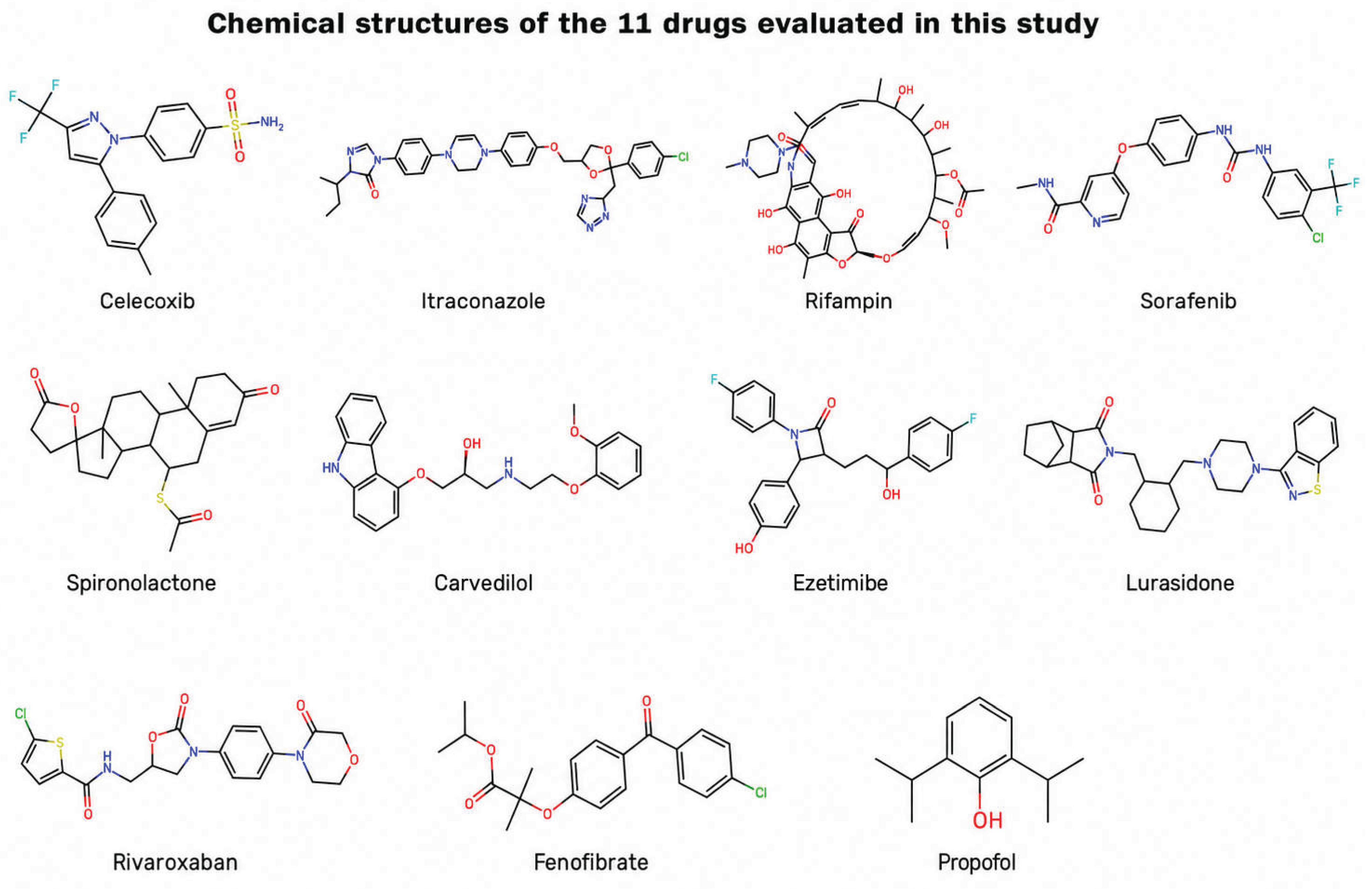
The eleven drugs selected for this study represent a chemically diverse group of poorly water-compatible small molecules. Their therapeutic activities differ widely, but their structures share a common formulation-relevant feature: each molecule contains substantial hydrophobic regions together with one or more polar, hydrogen-bonding, or heteroatom-containing groups. This mixed distribution of hydrophobic and polar elements provides a structural rationale for evaluating FAST as a rapid carrier-free self-assembly platform. In this context, “hydrophobic domains” refer to aromatic rings, fused ring systems, steroid-like scaffolds, alkyl substituents, halogenated rings, and other nonpolar regions, whereas “hydrophilic groups” refer to sulfonamide, amide, urea, hydroxyl, carbonyl, ether, ester, lactone, amine, piperazine, triazole, oxazolidinone, morpholinone, and related polar groups.
Celecoxib is a diaryl-substituted pyrazole with a molecular weight of approximately 381 g/mol and calculated XLogP around 3.9. Its hydrophobic character is contributed by two aromatic rings, a methyl-substituted phenyl group, and a trifluoromethyl group. Its polar region is concentrated in the pyrazole nitrogens and the sulfonamide group, which contains strong hydrogen-bonding and polar sulfonyl oxygen atoms. This domain-separated structure helps explain why celecoxib is a useful BCS Class II benchmark molecule for evaluating nanoscale formulation strategies [27].
Itraconazole is a much larger and more lipophilic azole antifungal drug, with a molecular weight of approximately 705 g/mol and calculated XLogP above 6. Its hydrophobic regions include multiple aromatic rings, a dichlorophenyl group, and an alkyl substituent. At the same time, itraconazole contains multiple nitrogen- and oxygen-containing polar groups, including triazole, triazolone, piperazine, ether, and dioxolane moieties. Although these heteroatoms provide hydrogen-bond acceptor sites, the molecule has no hydrogen-bond donor group and is dominated by bulky hydrophobic structure, contributing to its very poor aqueous solubility [28].
Rifampin is a large rifamycin antibiotic with a molecular weight of approximately 823 g/mol. It is structurally distinct from the other drugs because it contains a large macrocyclic ansa structure with multiple hydroxyl, carbonyl, amide, ether, acetate, and piperazinyl-imine groups. The molecule has both extensive polar functionality and a large hydrophobic framework composed of a conjugated macrocycle, aromatic/naphthohydroquinone-like core, methyl groups, and nonpolar carbon chain regions. This amphipathic but high-molecular-weight structure makes rifampin a particularly informative molecule for FAST because it contains many polar groups yet still presents major aqueous formulation and dissolution challenges [29].
Sorafenib is a multikinase inhibitor with a molecular weight of approximately 465 g/mol and calculated XLogP around 3.8. Its hydrophobic regions include aromatic rings, a chlorinated ring, and a trifluoromethyl-substituted phenyl group. Its polar groups include a pyridine nitrogen, a carboxamide, and a urea linker that provides both hydrogen-bond donor and acceptor capacity. This combination of hydrophobic aryl/halogenated domains and polar amide/urea groups is typical of many kinase inhibitors and contributes to both target binding and formulation difficulty [30].
Spironolactone is a steroid-like molecule with a molecular weight of approximately 417 g/mol and calculated XLogP around 3.4. Its hydrophobic character is dominated by the fused steroid nucleus and methyl substituents. In contrast, its polar groups are limited to ketone, lactone, and thioacetate functionalities, with no hydrogen-bond donor group. The large nonpolar steroid core and limited polar surface make spironolactone a useful example of a poorly water-soluble drug in which polar groups are present but insufficient to overcome the hydrophobic scaffold [31].
Carvedilol is an amphipathic β-blocker with a molecular weight of approximately 406 g/mol and calculated XLogP around 3.8. The molecule contains a hydrophobic carbazole ring system and a methoxyphenoxy aromatic region, connected through a flexible side chain. Its polar and ionizable groups include a secondary amine, secondary alcohol, ether linkages, and carbazole NH. This structure creates a molecule with both membrane-compatible hydrophobic regions and aqueous-interacting polar groups, but its overall water solubility remains limited [32].
Ezetimibe has a molecular weight of approximately 409 g/mol and calculated XLogP around 4.5. Its hydrophobic regions include two fluorinated phenyl rings and an additional phenyl ring. Its polar groups include a β-lactam carbonyl, a phenolic hydroxyl group, and a secondary alcohol. Although these groups provide hydrogen-bonding capability, the molecule remains strongly influenced by its aromatic and fluorinated hydrophobic regions, making ezetimibe a useful BCS Class II lipid-lowering drug for nanoformulation evaluation [33].
Lurasidone is a CNS-active drug with a molecular weight of approximately 493 g/mol and calculated XLogP around 5.2. Its hydrophobic regions include a cyclohexyl group, a fused imide-containing ring system, and a benzisothiazole moiety. Its polar groups include piperazine nitrogens, imide carbonyls, and heteroatoms in the benzisothiazole ring. Lurasidone has no hydrogen-bond donor group and contains multiple ring systems, giving it strong lipophilic character despite the presence of several heteroatoms. This makes lurasidone a useful molecule for evaluating FAST in the context of CNS drug formulation and possible future route-specific delivery studies [34].
Rivaroxaban is structurally different from the more lipophilic drugs in this panel. It has a molecular weight of approximately 435 g/mol, a relatively high polar surface area, and multiple hydrogen-bond acceptor groups. Its polar regions include an amide, an oxazolidinone carbonyl, a morpholinone carbonyl, ether oxygen atoms, and heteroatoms within the oxazolidinone and morpholinone rings. Its hydrophobic regions include a chlorothiophene group and an aromatic phenyl linker. Therefore, rivaroxaban illustrates that poor aqueous solubility is not always explained only by very high LogP; crystal packing, molecular rigidity, and solid-state properties can also strongly influence dissolution behavior [35].
Fenofibrate is a classic BCS Class II drug with a molecular weight of approximately 361 g/mol and calculated XLogP above 5. Its hydrophobic character is derived from a chlorobenzoyl phenoxy structure, aromatic rings, and alkyl ester substituents. Its polar groups are mainly hydrogen-bond acceptors, including ester, ketone, and ether oxygens, with no hydrogen-bond donor group. This strong imbalance between hydrophobic surface and limited donor capacity makes fenofibrate an excellent benchmark molecule for evaluating whether FAST can rapidly generate aqueous nanoscale dispersions of highly lipophilic drugs [36].
Propofol was included as an eleventh clinically important hydrophobic drug to introduce the “me-better” formulation concept. Propofol is much smaller than the other drugs, with a molecular weight of approximately 178 g/mol, but it has calculated XLogP around 4 and very limited polar functionality. Its hydrophobic region consists of a phenyl ring substituted with two isopropyl groups, while its only strongly polar group is a phenolic hydroxyl. This structure explains why propofol is highly lipophilic and why the approved intravenous product depends on an oil-in-water lipid emulsion. In the present study, propofol serves as a stress-test molecule for assessing whether FAST can generate carrier-free aqueous nanoscale dispersions of a clinically important drug whose current formulation is constrained by extreme hydrophobicity and lipid-emulsion dependence [37].
Together, these structural features support the selection of the eleven-drug panel. The drugs vary in size, aromaticity, heteroatom content, hydrogen-bonding capacity, lipophilicity, ionizable groups, and therapeutic class. This diversity allows FAST to be evaluated not as a single-compound formulation method, but as a platform approach for rapidly generating aqueous nanoscale dispersions from chemically distinct poorly water-soluble drugs [27,28,29,30,31,32,33,34,35,36,37].
2. Materials and Methods
2.1. Compounds and Reagents
Epigallocatechin-3-gallate-palmitate (EC16) was obtained from Camellix, LLC (Evans, GA, USA). Celecoxib (A10193-100), sorafenib (A17857-500), carvedilol (A11843-100), ezetimibe (A10379-50), lurasidone (A11214-50), rivaroxaban (A10800-50), fenofibrate (SKU A10385-5000), and propofol (A18293-100) were purchased from AdooQ BioSciences (Irvine, CA, USA). Itraconazole (J66390.03), rifampin (455620010), and spironolactone (207460010) were obtained from Fisher Scientific (Pittsburgh, PA, USA). Omyadent HA100 hydroxyapatite (Lot# OMD624231B) was provided by Nutrivene/International Nutrition Inc. (Middle River, MD, USA). Sterile double-distilled water, purified 0.1 µm-filtered water, glycerol, paraformaldehyde, glutaraldehyde, and uranyl acetate were used as described below. Vendor catalog numbers, lot numbers, and purity information will be added in the final manuscript.
2.2. Preparation of Drug Nanoparticles
Drug nanoparticles were prepared using the proprietary Facilitated Self-Assembling Technology (FAST) method, as previously described with modifications [5,6,7]. The FAST method is patent pending under U.S. Patent Application No. 63/896,918. Briefly, individual poorly water-soluble drugs were processed under compound-specific FAST conditions to generate nanoparticle stock suspensions. The resulting nanoparticle stocks were prepared at 0.1–1% w/v and stabilized in pure glycerol for storage. Before particle characterization and visual suspension evaluation, the glycerol stock suspensions were diluted 50-fold with sterile double-distilled water to final drug concentrations, unless otherwise specified. For TEM imaging the drug stocks were diluted 1:4 in purified double-distilled water.
2.3. Nanoparticle Tracking Analysis and Zeta Potential Measurement
Particle size distribution and particle concentration were measured using a ZetaView X20 Nanoparticle Tracking Analyzer (Particle Metrix, Meerbusch, Germany), following a method previously described [5,6,7]. The instrument measuring range for particle diameter was 10–2000 nm. Samples were diluted in purified 0.1 µm-filtered water before analysis. Measurements were performed at 25 °C with sensitivity 70, frame rate 30 frames per second, and shutter speed 100. Particle information was collected from 11 positions across the cell with two reading cycles. Post-acquisition parameters were set as follows: minimum brightness 20, minimum area 10, maximum area 1000, and trace length 15.
Zeta potential was measured using the same ZetaView X20 system. Nanoparticle suspensions were diluted in purified 0.1 µm-filtered water with low conductivity (<1000 µS/cm) before analysis. Zeta potential profiles were collected from 11-position measurements at 25 °C. Particle size, particle concentration, and zeta potential values were recorded for each drug nanoparticle suspension.
2.4. Transmission Electron Microscopy
Transmission electron microscopy was used to examine the morphology of selected drug nanoparticles. Drug nanoparticle stocks were diluted with double-distilled water and fixed in 4% paraformaldehyde and 2% glutaraldehyde. After mixing, 5 µL of each sample was transferred to a Formvar/carbon-coated copper 200-mesh grid and allowed to dry for 15 minutes. Excess liquid was removed using filter paper. Samples were then negatively stained by adding 5 µL of 2% aqueous uranyl acetate. Multiple images were captured from each sample using a JEM 1400 Flash transmission electron microscope (JEOL, Peabody, MA, USA) operated at 120 kV with a Gatan OneView digital camera (Gatan Inc., Pleasanton, CA, USA).
2.5. Visual Assessment of Drug Nanoparticle Water Suspensions
Drug nanoparticle stocks were diluted in double-distilled water to final concentrations of 0.01–0.02% w/v to generate aqueous nanoparticle suspensions. Most diluted nanoparticle suspensions appeared clear to slightly opalescent. Rifampin suspensions exhibited a light brown color after dilution from an amber-colored stock.
To evaluate short-term visual dispersion stability, diluted nanoparticle suspensions were stored at room temperature for at least 30 days. After storage, the 30-day diluted suspensions were photographed in glass tubes alongside freshly diluted nanoparticle suspensions of the same drugs. Visual assessment of color change, visible precipitation, sedimentation, and aggregation was performed using coded samples by three independent observers before documentation was finalized. For itraconazole, an additional centrifugation check was performed by spinning the suspension at approximately 13,300 rpm for 20 minutes and visually inspecting for sedimentation or pellet formation. Because the rotor radius was not recorded, the corresponding relative centrifugal force (x g) could not be calculated.
3. Results
3.1. DLS/ZetaView Characterization of FAST-Generated Aqueous Nanoscale Suspensions
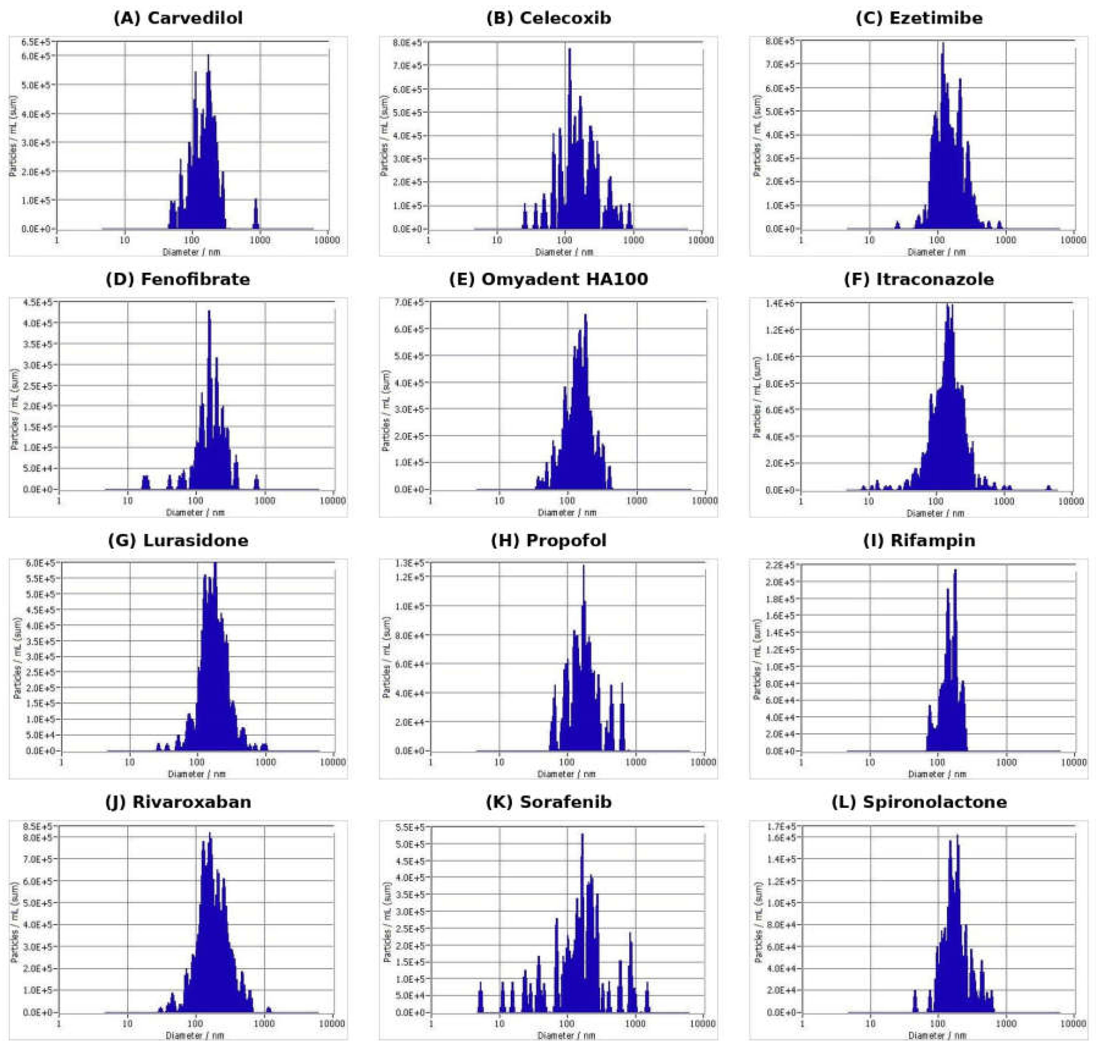
FAST processing generated measurable aqueous nanoscale suspensions across the selected poorly water-soluble drug panel and Omyadent HA100. Particle-size distribution profiles are organized alphabetically in Figure 2. The following subsections combine the formulation-relevant introduction for each compound with the corresponding DLS/ZetaView result so that the rationale and nanoparticle characterization are presented together for each material.
3.1.1. Carvedilol
Carvedilol is an amphipathic beta-blocker with a hydrophobic carbazole ring system and a methoxyphenoxy aromatic region connected through a flexible side chain. Its polar and ionizable groups include a secondary amine, secondary alcohol, ether linkages, and carbazole NH, but its overall water solubility remains limited.
FAST-generated carvedilol nanoparticles showed a median particle size of 145.5 nm, with a measured size range of 91.3-172.5 nm. At 0.02% carvedilol, the particle concentration was 6.6 × 10^8 particles/mL and the zeta potential was -46.93 mV.
3.1.2. Celecoxib
Celecoxib is a diaryl-substituted pyrazole with hydrophobic aromatic and trifluoromethyl-containing regions and a polar sulfonamide-containing domain. This domain-separated structure makes celecoxib a useful BCS Class II benchmark molecule for evaluating nanoscale formulation strategies [27].
FAST-generated celecoxib nanoparticles showed a median particle size of 149.5 nm, with a measured size range of 32.0-242.8 nm. At 0.02% celecoxib, the particle concentration was 4.51 × 10^8 particles/mL and the zeta potential was -35.72 mV.
3.1.3. Ezetimibe
Ezetimibe contains multiple aromatic and fluorinated hydrophobic regions together with polar beta-lactam carbonyl, phenolic hydroxyl, and secondary alcohol groups. Its low aqueous solubility makes it a clinically relevant BCS Class II lipid-lowering drug for nanoformulation evaluation.
FAST-generated ezetimibe nanoparticles showed a median particle size of 137.3 nm, with a measured size range of 92.3-276.0 nm. At 0.02% ezetimibe, the particle concentration was 1.9 × 10^9 particles/mL and the zeta potential was -44.53 mV.
3.1.4. Fenofibrate
Fenofibrate is a classic BCS Class II drug with strong hydrophobic character from a chlorobenzoyl phenoxy structure, aromatic rings, and alkyl ester substituents. Its limited hydrogen-bond donor capacity and poor aqueous solubility make it a benchmark molecule for evaluating aqueous nanoscale dispersion approaches.
FAST-generated fenofibrate nanoparticles showed a median particle size of 157.0 nm, with a measured size range of 119.3-282.3 nm. At 0.02% fenofibrate, the particle concentration was 3.5 × 10^8 particles/mL and the zeta potential was -28.21 mV.
3.1.5. Itraconazole
Itraconazole is a large and lipophilic azole antifungal drug with multiple aromatic rings, a dichlorophenyl group, and multiple nitrogen- and oxygen-containing polar groups. Despite these heteroatoms, its bulky hydrophobic structure contributes to very poor aqueous solubility.
FAST-generated itraconazole nanoparticles showed a median particle size of 150.8 nm, with a measured size range of 67.0-236.0 nm. At 0.02% itraconazole, the particle concentration was 1.8 × 10^9 particles/mL and the zeta potential was -41.1 mV.
3.1.6. Lurasidone
Lurasidone is a CNS-active drug with multiple ring systems, including a cyclohexyl group and benzisothiazole moiety, together with piperazine nitrogens and imide carbonyl groups. Its poor aqueous solubility and clinically important food effect make it a useful molecule for evaluating FAST-enabled formulation flexibility.
FAST-generated lurasidone nanoparticles showed a median particle size of 168.0 nm, with a measured size range of 74.7-220.1 nm. At 0.02% lurasidone, the particle concentration was 8.1 × 10^8 particles/mL and the zeta potential was -54.94 mV.
3.1.7. Omyadent HA100
Omyadent HA100 was included as an inorganic hydroxyapatite proof-of-concept material rather than a BCS Class II drug. Hydroxyapatite was selected because of its clinical and commercial relevance in dentistry and oral-care products, where biomimetic HA and nano-HA have been studied for remineralization, caries prevention, and dentin hypersensitivity applications [23,24,25,26]. FAST processing of HA100 was therefore evaluated to test whether the platform could generate nanoscale mineral clusters from a dental-grade HA material under mild aqueous conditions.
FAST-processed Omyadent HA100 showed a median particle size of 141.2 nm, with a measured size range of 60.9-270.3 nm. At 0.02% HA100, the particle concentration was 7.8 × 10^8 particles/mL and the zeta potential was -22.07 mV.
3.1.8. Propofol
Propofol is a clinically important hydrophobic intravenous anesthetic whose approved product depends on an oil-in-water lipid emulsion because of its very limited aqueous solubility. In this study, propofol served as a stress-test molecule for evaluating whether FAST can generate carrier-free aqueous nanoscale dispersions of a highly lipophilic drug.
FAST-generated propofol nanoparticles showed a median particle size of 150.47 nm, with a measured size range of 95.3-202.5 nm. At 0.02% propofol, the particle concentration was 5.3 × 10^8 particles/mL and the zeta potential was -38.12 mV.
3.1.9. Rifampin
Rifampin is a large rifamycin antibiotic with extensive polar functionality and a large hydrophobic macrocyclic framework. Its aqueous formulation and stability challenges make rifampin an informative molecule for testing FAST-generated nanoscale dispersion and visual stability [29].
FAST-generated rifampin nanoparticles showed a median particle size of 145.5 nm, with a measured size range of 75.4-225.4 nm. At 0.02% rifampin, the particle concentration was 3.1 × 10^8 particles/mL and the zeta potential was -37.45 mV.
3.1.10. Rivaroxaban
Rivaroxaban contains multiple polar groups, including amide, oxazolidinone carbonyl, morpholinone carbonyl, and ether oxygens, together with hydrophobic chlorothiophene and aromatic regions. It illustrates that poor aqueous solubility can arise from solid-state and molecular packing effects in addition to lipophilicity.
FAST-generated rivaroxaban nanoparticles showed a median particle size of 165.5 nm, with a measured size range of 128.9-254.7 nm. At 0.02% rivaroxaban, the particle concentration was 2.4 × 10^9 particles/mL and the zeta potential was -42.36 mV.
3.1.11. Sorafenib
Sorafenib is a multikinase inhibitor with hydrophobic aryl and halogenated domains and polar amide/urea groups. Its poor aqueous solubility, limited bioavailability, and formulation challenges make it a relevant anticancer drug for FAST-based nanoformulation evaluation.
FAST-generated sorafenib nanoparticles showed a median particle size of 159.2 nm, with a measured size range of 100.9-269.3 nm. At 0.02% sorafenib, the particle concentration was 1.4 × 10^9 particles/mL and the zeta potential was -29.9 mV.
3.1.12. Spironolactone
Spironolactone is a steroid-like molecule with a large hydrophobic fused-ring core and limited polar functionality, including ketone, lactone, and thioacetate groups. This hydrophobic scaffold makes spironolactone a useful example of a poorly water-soluble drug for nanoformulation evaluation.
FAST-generated spironolactone nanoparticles showed a median particle size of 145.5 nm, with a measured size range of 67.3-172.5 nm. At 0.02% spironolactone, the particle concentration was 6.6 × 10^8 particles/mL and the zeta potential was -51.15 mV.
3.2. Visual Appearance and Short-Term Suspension Stability
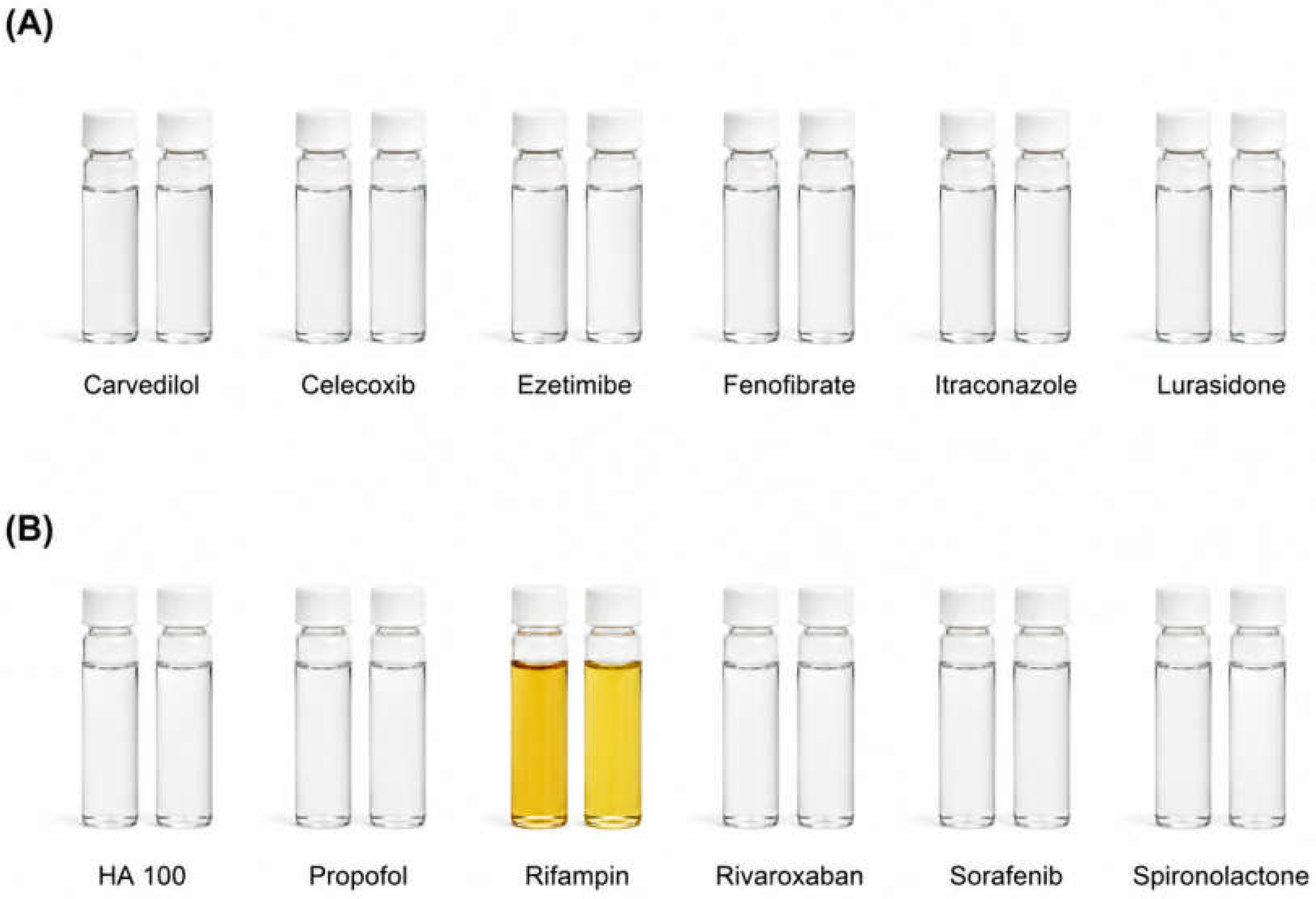
Diluted FAST-generated aqueous suspensions were visually compared as aged (>10 days in room temperature) versus freshly prepared (same day preparation) pairs. The paired visual assessment was organized into two panels to allow direct comparison of each suspension. Group 1 included carvedilol, celecoxib, ezetimibe, fenofibrate, itraconazole, and lurasidone. Group 2 included HA100, propofol, rifampin, rivaroxaban, sorafenib, and spironolactone.
No macroscopic precipitation, sedimentation, aggregation, phase separation, or foam formation was observed by visual inspection for the evaluated suspensions. Most aged and freshly prepared pairs remained visually comparable in color and overall appearance. Rifampin was the only sample that showed a clear visual color shift, with the aged suspension appearing darker yellow/amber compared with the freshly prepared rifampin suspension. This observation is consistent with the known aqueous color/stability sensitivity of rifampin and should be confirmed by analytical stability methods such as HPLC or LC-MS.
Figure 3.
Visual appearance and short-term stability assessment of FAST-generated aqueous suspensions. Each pair shows aged (left) and freshly prepared (right) suspensions for the indicated material. (A) Group 1: Carvedilol, Celecoxib, Ezetimibe, Fenofibrate, Itraconazole, and Lurasidone. (B) Group 2: HA100, Propofol, Rifampin, Rivaroxaban, Sorafenib, and Spironolactone. No visible precipitation, sedimentation, aggregation, phase separation, or foam formation was observed by visual inspection. Rifampin showed the most apparent color change, with the aged suspension appearing darker yellow/amber relative to the freshly prepared suspension.
Figure 3.
Visual appearance and short-term stability assessment of FAST-generated aqueous suspensions. Each pair shows aged (left) and freshly prepared (right) suspensions for the indicated material. (A) Group 1: Carvedilol, Celecoxib, Ezetimibe, Fenofibrate, Itraconazole, and Lurasidone. (B) Group 2: HA100, Propofol, Rifampin, Rivaroxaban, Sorafenib, and Spironolactone. No visible precipitation, sedimentation, aggregation, phase separation, or foam formation was observed by visual inspection. Rifampin showed the most apparent color change, with the aged suspension appearing darker yellow/amber relative to the freshly prepared suspension.
3.3. TEM Evaluation of Selected FAST-Generated Nanoparticles
TEM imaging was used to examine selected representative FAST-generated nanoparticle suspensions. Omyadent HA100 was selected as an inorganic mineral proof-of-concept material, propofol was selected as a clinically important hydrophobic small molecule whose current marketed formulation depends on an oil-in-water lipid emulsion, itraconazole was selected as a highly lipophilic azole antifungal drug with potential route-specific topical, ocular, and local antifungal formulation relevance, and rifampin was selected because of its amphipathic macrocyclic structure, known aqueous formulation sensitivity, and relevance to nanoparticle-based tuberculosis delivery concepts.
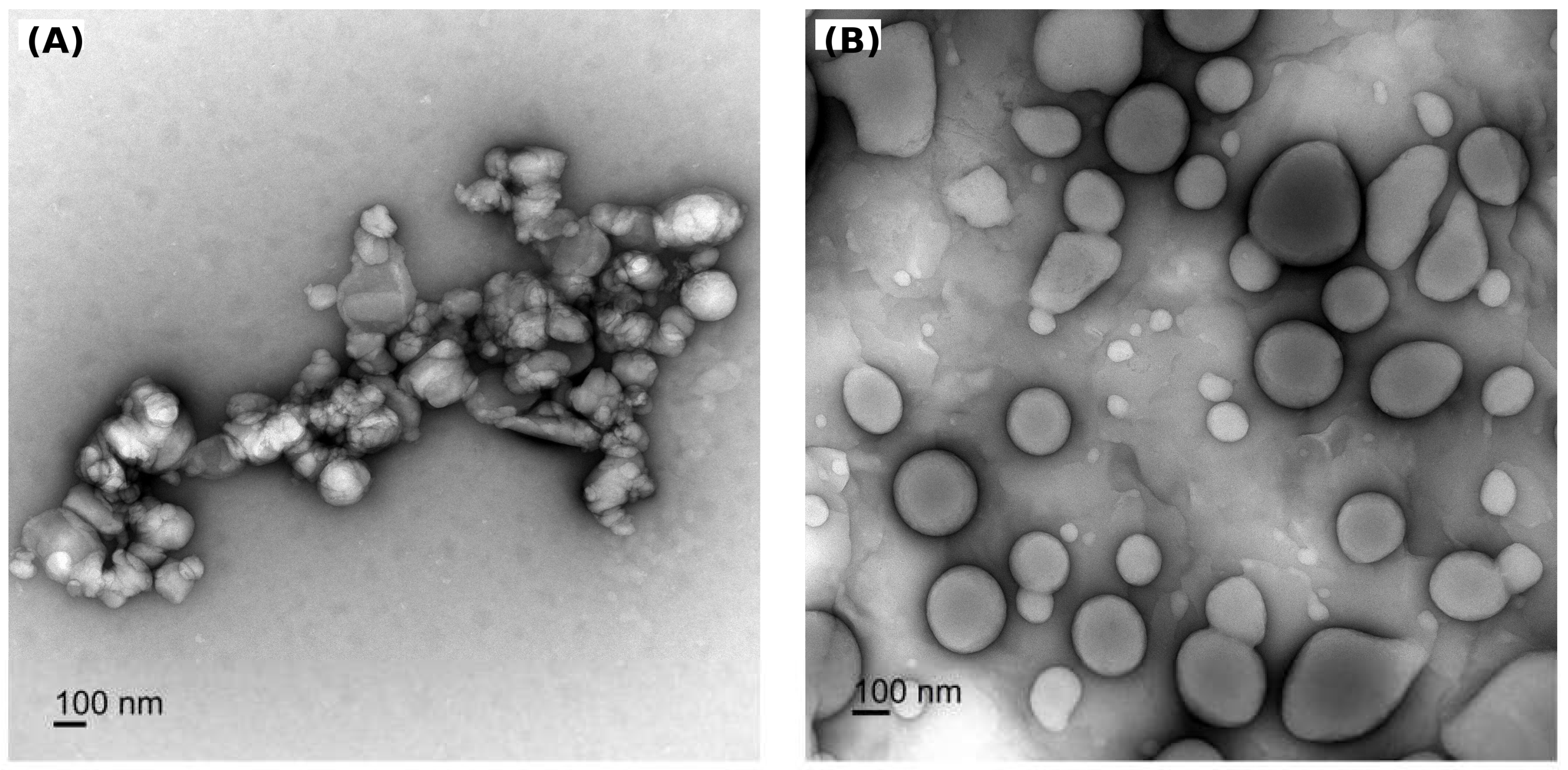
FAST-processed Omyadent HA100 showed abundant electron-dense, irregular, granular nanoscale clusters distributed across the TEM field. The morphology was not uniform or spherical; instead, the HA appeared as irregular nanoscale clusters composed of finer electron-dense substructures. Some larger aggregates were observed, indicating that the preparation represents a nanoparticle-enriched HA suspension rather than complete conversion into monodisperse isolated nanoparticles.
FAST-generated propofol nanoparticles appeared as abundant discrete spherical to near-spherical particles distributed across the TEM field. The observed particles were in the nanoscale range and were broadly consistent with the DLS/ZetaView particle-size results. The field showed some size heterogeneity, suggesting a polydisperse nanoparticle population, but no obvious needle-like or crystalline morphology was observed.
FAST-generated itraconazole showed two distinct nanoscale morphologies by TEM. One field showed rod-like electron-dense crystalline/nanocrystalline structures, while a separate field showed rounded nanoscale particles with darker internal substructure. The rod-like structures were small relative to the 100 nm scale bar and appeared as nanoscale or submicron crystalline particles rather than macroscopic precipitated crystals.
Consistent with this interpretation, the itraconazole suspension did not form a visible precipitate or pellet after centrifugation at approximately 13,300 rpm for 20 minutes. Although rpm should ideally be reported as relative centrifugal force (x g) when rotor radius is available, the absence of a visible pellet under this condition suggests that the crystal-like structures remained in the colloidally suspended fraction rather than behaving as large sedimenting crystals.
FAST-generated rifampin showed heterogeneous nanoscale morphology by TEM. Representative fields showed irregular clustered aggregates composed of smaller nanoscale substructures and separate rounded to oval nanoscale particles within a diffuse matrix. No long needle-like crystalline morphology was observed. Because the corresponding aqueous rifampin suspension appeared clear brown/amber without visible macroscopic precipitation or sedimentation, the aggregated TEM structures are interpreted cautiously as nanoscale rifampin particle clusters and/or drying/staining-associated aggregates rather than direct evidence of bulk precipitation.
Figure 4.
Transmission electron microscopy of selected FAST-generated nanoparticles. (A) Omyadent HA100 hydroxyapatite showed electron-dense, irregular granular nanoscale clusters distributed across the examined field. Some larger aggregates were also observed, and no obvious long needle-shaped HA particles were observed. (B) Propofol showed discrete spherical to near-spherical nanoparticles with nanoscale morphology and some size heterogeneity. Scale bars = 100 nm.
Figure 4.
Transmission electron microscopy of selected FAST-generated nanoparticles. (A) Omyadent HA100 hydroxyapatite showed electron-dense, irregular granular nanoscale clusters distributed across the examined field. Some larger aggregates were also observed, and no obvious long needle-shaped HA particles were observed. (B) Propofol showed discrete spherical to near-spherical nanoparticles with nanoscale morphology and some size heterogeneity. Scale bars = 100 nm.
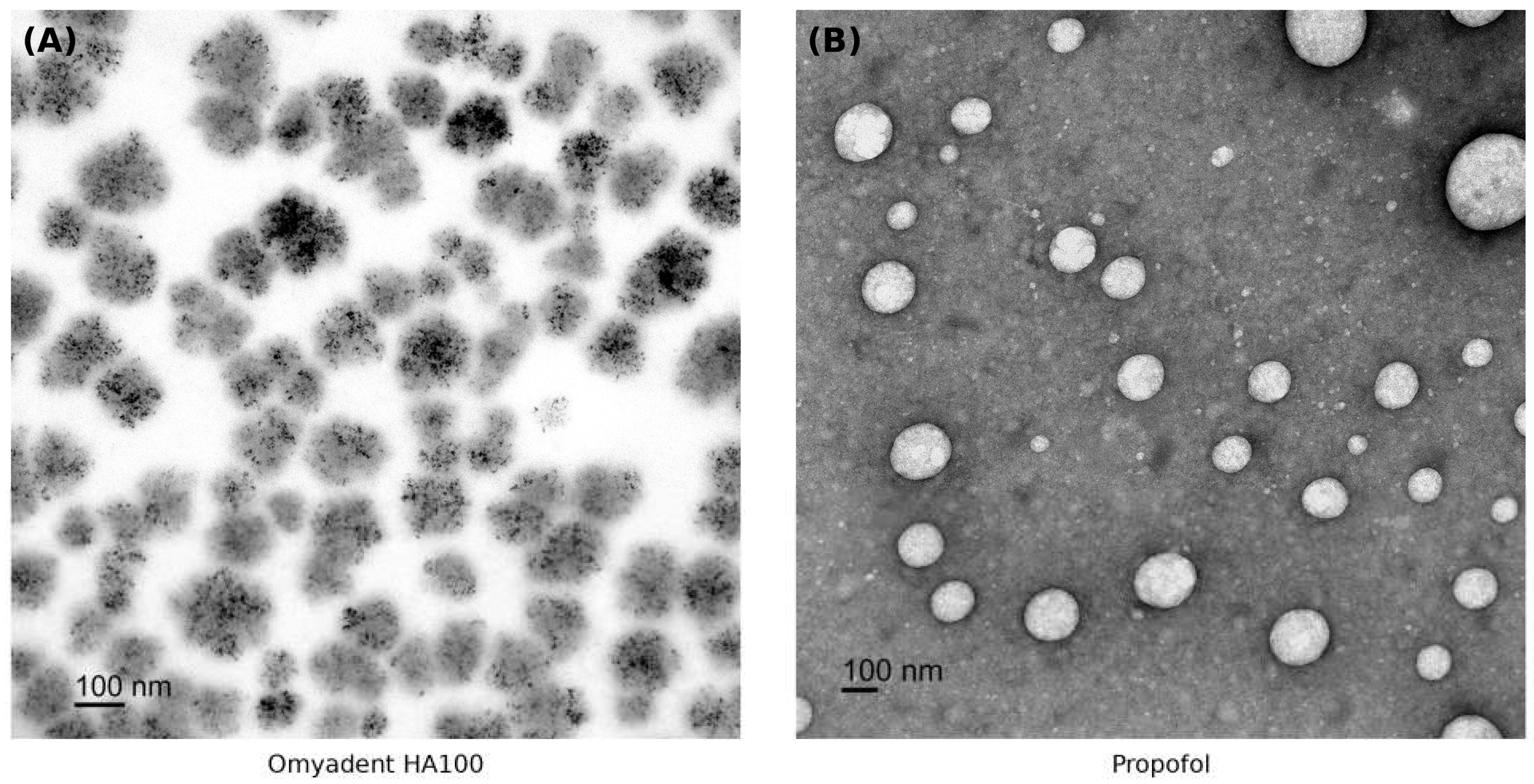
Figure 5.
Transmission electron microscopy of FAST-generated itraconazole. (A) Rod- or needle-like crystalline/nanocrystalline itraconazole structures were observed as small electron-dense particles relative to the 100 nm scale bar. (B) Rounded itraconazole nanoscale particles were also observed, indicating mixed morphology within the FAST preparation. Scale bars = 100 nm.
Figure 5.
Transmission electron microscopy of FAST-generated itraconazole. (A) Rod- or needle-like crystalline/nanocrystalline itraconazole structures were observed as small electron-dense particles relative to the 100 nm scale bar. (B) Rounded itraconazole nanoscale particles were also observed, indicating mixed morphology within the FAST preparation. Scale bars = 100 nm.
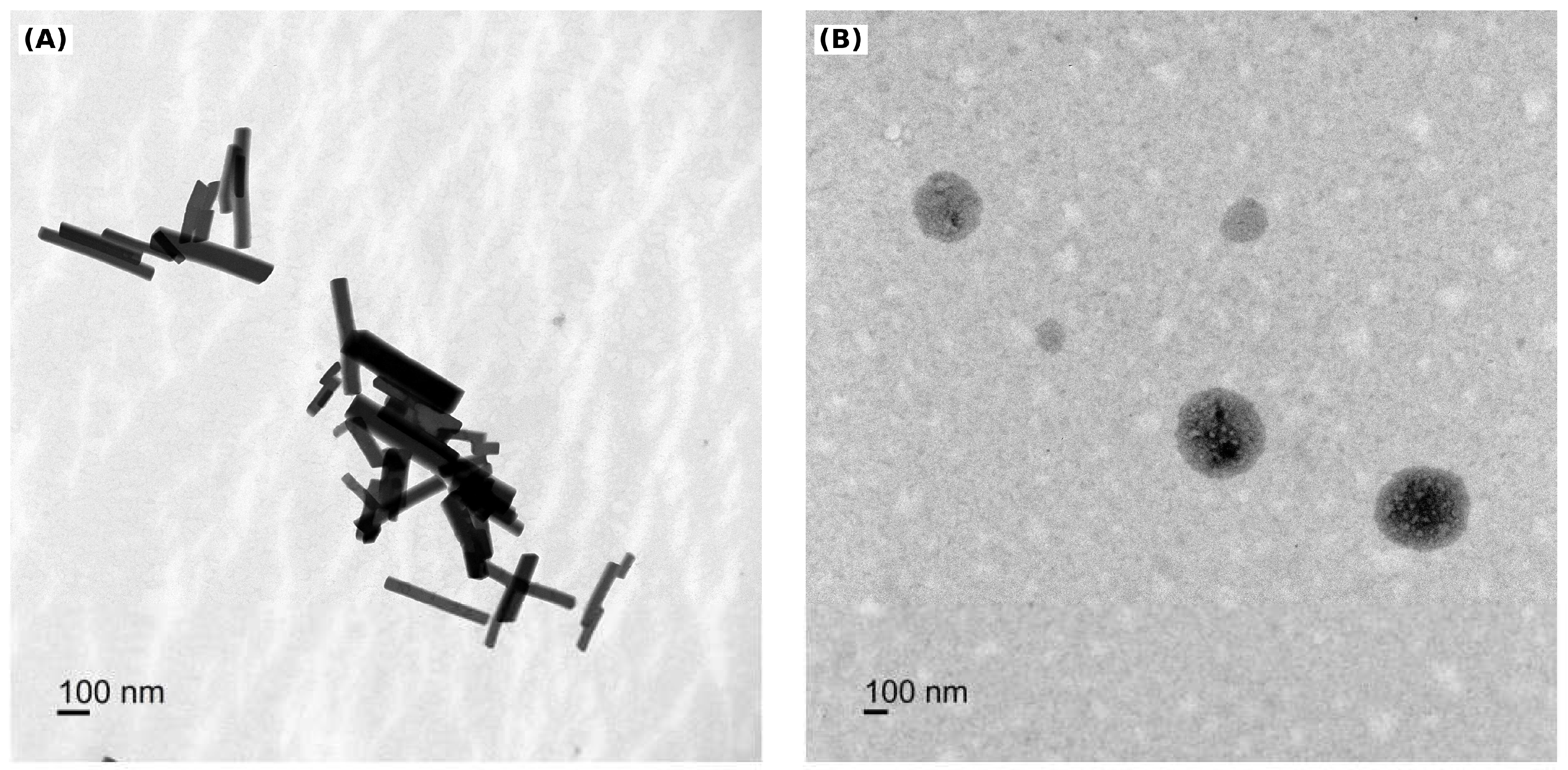
Figure 6.
Transmission electron microscopy of FAST-generated rifampin. (A) Representative rifampin field showing irregular aggregated clusters composed of smaller nanoscale substructures. (B) Representative field showing rounded to oval nanoscale particles within a diffuse matrix. Scale bars = 100 nm.
Figure 6.
Transmission electron microscopy of FAST-generated rifampin. (A) Representative rifampin field showing irregular aggregated clusters composed of smaller nanoscale substructures. (B) Representative field showing rounded to oval nanoscale particles within a diffuse matrix. Scale bars = 100 nm.
4. Discussion
4.1. Overall Interpretation of FAST-Generated Aqueous Nanoscale Suspensions
The present study supports FAST as a rapid formulation-feasibility platform for converting chemically diverse poorly water-soluble molecules into aqueous nanoscale suspensions. Across the drug panel, DLS/ZetaView analysis confirmed measurable nanoscale particle populations, and zeta potential measurements showed negative surface charge for all tested suspensions. The visual stability assessment further showed no macroscopic precipitation, sedimentation, aggregation, phase separation, or foam formation under the evaluated conditions, although rifampin displayed a visible yellow-to-darker-yellow/amber color shift after aging. These findings support the central conclusion that FAST can rapidly generate aqueous nanoparticle-enriched suspensions suitable for further formulation development, without yet claiming improved dissolution, pharmacokinetics, bioavailability, or clinical efficacy.
The broader formulation literature supports the rationale for nanoscale approaches in poorly soluble drug development. Nanocrystal and nanosuspension reviews describe increased surface area, improved dissolution, improved apparent solubility, and opportunities for integration into tablets, hydrogels, microneedles, microparticles, and functionalized systems as key advantages for poorly soluble drugs [4,38,39]. FAST differs from many conventional approaches because the current method is intended to be rapid, aqueous, carrier-free, and low-energy, although direct head-to-head comparisons with established nanocrystal, solid dispersion, lipid, and surfactant-based systems remain necessary.
4.2. Significance of FAST-Processed Omyadent HA100 Hydroxyapatite Nanoparticles
The preliminary Omyadent HA100 findings suggest that FAST may have broader applicability beyond hydrophobic organic drug molecules. Hydroxyapatite is an inorganic calcium phosphate material widely used in oral-care and dental applications because of its chemical similarity to mineral components of enamel and dentin [23,24,25,26]. Conventional commercial HA powders may be micron-sized or broadly distributed in particle size, which may limit access to nanoscale dental structures such as dentinal tubules, enamel surface defects, and microscopic mineral-loss regions. This provided the rationale for evaluating whether FAST could convert a commercially relevant HA source into a nanoparticle-enriched suspension under mild aqueous conditions.
In this study, Omyadent HA100 formed nanoscale HA particle clusters within approximately one minute under FAST conditions. This observation is notable because the process did not involve conventional nanoparticle engineering approaches, including mechanical size reduction, high-pressure homogenization, surfactant stabilization, polymer encapsulation, or surface modification. Instead, the data support FAST-enabled spontaneous nanoscale formation of HA clusters in an aqueous environment.
The NTA result of 141.2 +/- 71 nm, together with the TEM evidence of abundant irregular nanoscale HA clusters, suggests that FAST produced a nanoparticle-enriched HA suspension. The observed broad size distribution and partial aggregation are not unexpected for mineral nanoparticles and may reflect both the intrinsic surface properties of HA and artifacts associated with drying during TEM preparation. Therefore, the current data should be interpreted as evidence of nanoscale HA cluster formation rather than complete conversion into a uniform population of isolated nanoparticles.
From a dental-materials perspective, the formation of non-engineered nanoscale HA clusters may be important. Smaller HA particles may have improved access to microstructured tooth surfaces compared with larger micron-scale HA powders. Published oral-care literature supports the concept that nano-HA can deposit on tooth surfaces, contribute to remineralization processes, and occlude dentinal tubules in dentin hypersensitivity models [23,24,25,26]. In principle, FAST-generated nanoscale HA may better interact with enamel defects, dentin surfaces, and exposed tubules, although this functional advantage remains to be experimentally confirmed. Future studies should evaluate dentin tubule penetration, enamel surface deposition, remineralization potential, suspension stability, and performance in relevant oral-care formulations.
Safety also requires direct testing. Although the TEM images did not show obvious needle-shaped HA morphology, cytocompatibility cannot be assumed solely from size or morphology. The biological response to nanoscale HA can depend on particle concentration, exposure time, aggregation state, surface charge, cell type, and route of exposure. Therefore, future studies should include oral keratinocytes, gingival fibroblasts, dental pulp or odontoblast-like cells, and inflammatory-response assays to assess cytotoxicity, membrane damage, oxidative stress, and cytokine induction.
Overall, these findings support Omyadent HA100 (or other type of HA material) as a useful proof-of-concept material demonstrating that FAST can rapidly generate non-engineered nanoscale mineral clusters. This expands the potential scope of FAST from hydrophobic small molecules to inorganic oral-care materials and provides a foundation for future development of FAST-enabled HA formulations for dental and consumer oral-health applications.
4.3. Propofol: Potential Value of a Clean Aqueous Nanoformulation
Propofol is already a highly successful and widely used anesthetic, and the current oil-in-water lipid emulsion has been used clinically for decades without controversy regarding its core anesthetic utility. The goal of a FAST-generated propofol nanoformulation should therefore not be framed as replacing a failed product. Instead, it should be framed as a potential me-better formulation opportunity that may address specific formulation-related constraints while preserving the known pharmacology of propofol. Recent reviews emphasize that propofol remains limited by poor water solubility, lipid-emulsion dependence, strict aseptic handling requirements, injection-pain issues, microbial contamination concerns, and challenges in alternative route development [21,22].
The TEM image of FAST-generated propofol provides morphological support for a nanoparticle-enriched propofol suspension, with spherical to near-spherical particles in the nanoscale range. If a clean aqueous propofol nanoformulation can be optimized, potential advantages may include reduced or eliminated lipid excipient burden, improved formulation clarity for quality control, reduced emulsion-associated instability concerns, and improved flexibility for route-specific product design. However, these potential benefits remain hypotheses until free propofol concentration, release kinetics, hemocompatibility, sterility, endotoxin, preservative compatibility, vascular irritation, cardiopulmonary safety, anesthetic potency, and pharmacokinetics/pharmacodynamics are established.
From a regulatory perspective, an intravenous propofol nanoformulation containing the same active ingredient but a substantially different formulation might be evaluated through a 505(b)(2) pathway if an adequate scientific bridge can be established to an approved listed propofol product. FDA describes the 505(b)(2) NDA as a pathway in which at least some required safety or effectiveness information may come from studies not conducted by or for the applicant, including reliance on FDA findings for a listed drug, provided that differences between the proposed and listed product are supported and bridged scientifically [40]. For a clean propofol nanoparticle product, the development program would likely need comparative CMC, physicochemical characterization, free-drug and total-drug assays, bioavailability or pharmacokinetic bridging, nonclinical safety bridging, and possibly clinical pharmacodynamic bridging. If the formulation, route, indication, or exposure profile differs substantially from the approved product, FDA may require a broader nonclinical and clinical package, and the pathway could approach a full 505(b)(1) new drug development program.
Intranasal propofol is scientifically intriguing but should be discussed cautiously. Nose-to-brain delivery reviews describe the nasal route as a noninvasive strategy that may bypass the blood-brain barrier through olfactory and trigeminal pathways [6,41,42]. In principle, a nanoscale propofol formulation could be evaluated for nasal delivery because propofol acts rapidly in the central nervous system and because the nasal route could, in theory, reduce dependence on bloodborne delivery. However, this concept is highly speculative for propofol. A nasal propofol program would need to address dose capacity, nasal mucosal irritation, ciliotoxicity, local tissue retention, taste and odor, systemic versus direct CNS absorption, onset and offset predictability, sedation depth, respiratory depression risk, abuse potential, and emergency reversibility. Therefore, nasal propofol should be positioned as a future exploratory route-specific concept rather than a conclusion supported by the present data.
4.4. Potential Me-Better Opportunities for the Ten Oral BCS Class II Drugs
For the ten oral BCS Class II drugs, the key FAST opportunity is to determine whether rapid carrier-free nanoparticle formation can improve dissolution, dispersion uniformity, dose flexibility, and route-specific testing without adding complex excipient systems. The literature supports the concept that formulation improvement may increase performance for this class, but it also highlights drug-specific challenges that must be addressed individually [4,8,9,10,11,12,13,14,15,16,17,18,19,20,38,39]. FAST should therefore be framed as a screening and enabling platform rather than a universal final product format.
Celecoxib and fenofibrate represent benchmark BCS Class II drugs where nanosizing and related formulation strategies have already been shown to improve dissolution and oral performance. A 2023 celecoxib nanoformulation study reported improved solubility, dissolution rate, and oral bioavailability, supporting the idea that nanoscale celecoxib formulations can create clinically relevant performance gains [8]. Additional celecoxib nanosuspension work supports the broader concept that particle-size reduction can enhance bioavailability [9]. Fenofibrate has a long history of formulation optimization, including micronized, nanoparticle, and other improved formulations; recent nanocrystal reviews also identify fenofibrate as a model compound for nanosizing and bioavailability enhancement [10,38,39]. FAST-generated versions of these benchmark drugs can therefore serve as internal positive comparators for determining whether FAST provides a simpler or faster route to similar formulation-enabling outcomes.
Itraconazole, rifampin, and sorafenib illustrate drugs where formulation improvement may support route-specific me-better opportunities. Itraconazole is a highly lipophilic azole with strong formulation barriers; recent antifungal nanoformulation literature and itraconazole nanoparticle studies support advanced drug delivery formats for improving local or systemic delivery [14,15]. In the current study, itraconazole formed a high-concentration nanoscale suspension by DLS/ZetaView and showed a mixed TEM morphology consisting of small rod- or needle-like crystalline/nanocrystalline structures together with rounded nanoscale particles. Importantly, no visible precipitate or pellet formed after centrifugation at approximately 13,300 rpm for 20 minutes, suggesting that the crystal-like structures should be interpreted as nanoscale or colloidally suspended nanocrystalline material rather than large precipitated drug crystals. This mixed morphology may be relevant for future topical, ocular, and local antifungal reformulation, where a nanosuspension or nanocrystalline fraction could be useful if dissolution, tissue residence, drug release, and safety are confirmed. Rifampin nanoparticle and tuberculosis nanocarrier reviews emphasize the potential of nanocarriers for targeted tuberculosis therapy, enhanced delivery to infected tissues or macrophages, and controlled release, but rifampin stability remains a critical issue [16,17]. In the current study, rifampin formed nanoscale particles by DLS/ZetaView and showed a clear brown/amber suspension without visible sedimentation, but TEM revealed more heterogeneous morphology than propofol and itraconazole rounded-particle fields. The rifampin TEM images showed irregular aggregated clusters and rounded/oval nanoscale particles, which may reflect rifampin self-association, its complex amphipathic macrocyclic structure, concentration during grid drying, negative-stain/sample-preparation effects, or a combination of these factors. Therefore, the TEM aggregates should not be interpreted alone as bulk precipitation or formulation failure. However, because rifampin also showed a visible aging-related color shift, future studies should combine DLS/NTA before and after aging, centrifugation or filtration challenge, TEM under replicate preparation conditions, and HPLC or LC-MS analysis to distinguish physical clustering from chemical degradation. Sorafenib drug delivery reviews describe the importance of nanocarriers and formulation strategies for overcoming poor solubility, low bioavailability, and dose-limiting toxicities [18]. For these drugs, FAST may enable rapid aqueous screening and early biological testing, but efficacy and safety advantages must be validated by dissolution, stability, pharmacokinetic, and disease-model studies.
Carvedilol, ezetimibe, lurasidone, rivaroxaban, and spironolactone provide additional examples where FAST may support either oral performance improvement or alternative-route development. Carvedilol literature describes low solubility and poor bioavailability as major formulation barriers and summarizes nanosuspension, nanocrystal, nanoemulsion, dendrimer, and polymeric nanoparticle approaches for solubility enhancement [12]. Ezetimibe formulation reviews describe multiple strategies, including nanosizing, solid dispersions, and lipid-based systems, to improve dissolution and therapeutic potential [11]. Lurasidone has poor aqueous solubility and a clinically important food effect; recent intranasal nanostructured lipid carrier work and CNS nanocarrier reviews support the broader concept that nanoscale formulations may improve bioavailability or brain-targeted delivery for neuropsychiatric drugs [20,42]. Rivaroxaban studies and reviews summarize approaches including improved dissolution systems, SNEDDS, solid lipid nanoparticles, cocrystals, sustained release, and solid dispersion to address dose-dependent solubility and bioavailability issues [13]. Spironolactone nanoformulation reviews and topical delivery studies support the potential of nanoparticulate systems for targeted topical therapy in hyperandrogenic skin conditions and chronic wounds [19]. For each of these drugs, FAST-generated nanoparticles may serve as a rapid formulation prototype that can be compared against existing approaches in drug loading, dissolution, stability, permeability, tissue targeting, and safety.
4.5. Limitations
This study has several important limitations. First, the results establish nanoparticle formation and visual appearance stability, but they do not demonstrate improved dissolution, drug loading, permeability, pharmacokinetics, bioavailability, safety, or therapeutic efficacy. Second, DLS/ZetaView and TEM provide complementary but different measurements; hydrodynamic size, drying artifacts, aggregation state, and sample preparation can influence the apparent particle-size distribution and morphology. Third, visual stability is a useful first screen but cannot establish chemical stability. Rifampin color darkening especially requires HPLC or LC-MS analysis to quantify parent-drug loss and identify possible degradation products. Fourth, TEM confirmation is currently available for HA100, propofol, itraconazole, and rifampin, but most drugs in the panel still lack TEM confirmation. For rifampin, drying and negative staining may have exaggerated particle clustering on the TEM grid, so bulk suspension clarity, DLS/NTA, and chemical stability data should be considered together. Fifth, the itraconazole centrifugation observation was recorded in rpm because rotor radius was not available, so relative centrifugal force could not be calculated; future work should repeat this assessment using reported x g conditions, replicate samples, and quantitative particle recovery. Sixth, the FAST method is proprietary and patent pending, so the present manuscript must balance reproducibility with protection of undisclosed processing conditions. Finally, the regulatory and clinical pathways discussed for propofol and other me-better products are conceptual and would require formal FDA interaction, product-specific CMC development, nonclinical testing, and clinical bridging studies.
5. Conclusion
FAST generated aqueous nanoscale suspensions from a chemically diverse panel of ten BCS Class II drugs, propofol, and Omyadent HA100 hydroxyapatite. DLS/ZetaView analysis confirmed nanoscale particle populations with negative zeta potentials, visual assessment showed no macroscopic precipitation, sedimentation, aggregation, phase separation, or foam formation for the evaluated suspensions, and TEM imaging supported nanoscale morphology for HA100, propofol, itraconazole, and rifampin. Itraconazole showed both rounded nanoscale particles and small crystalline/nanocrystalline structures that remained visually suspended without forming a pellet after centrifugation at approximately 13,300 rpm for 20 minutes. Rifampin showed heterogeneous rounded/oval particles and irregular clustered structures by TEM and a clear aging-related color shift, highlighting the need for drug-specific physical and chemical stability testing. The findings support FAST as a rapid carrier-free formulation-feasibility platform, but additional studies are required to establish dissolution enhancement, chemical stability, drug loading, safety, pharmacokinetics, route-specific delivery, and efficacy. If confirmed in future studies, FAST may provide a practical platform for early formulation screening, AI-assisted drug discovery, and me-better development of approved drugs with formulation-limited performance.
References
- Bhalani, D.V.; Nutan, B.; Kumar, A.; Chandel, A.K.S. Bioavailability Enhancement Techniques for Poorly Aqueous Soluble Drugs and Therapeutics. Biomedicines 2022, 10, 2055. [Google Scholar] [CrossRef]
- Amidon, G.L.; Lennernas, H.; Shah, V.P.; Crison, J.R. A Theoretical Basis for a Biopharmaceutic Drug Classification: The Correlation of In Vitro Drug Product Dissolution and In Vivo Bioavailability. Pharm. Res. 1995, 12, 413–420. [Google Scholar] [CrossRef] [PubMed]
- Samineni, R.; Chimakurthy, J.; Konidala, S. Emerging Role of Biopharmaceutical Classification and Biopharmaceutical Drug Disposition System in Dosage Form Development: A Systematic Review. Turk. J. Pharm. Sci. 2022, 19, 706–713. [Google Scholar] [CrossRef] [PubMed]
- Kumari, L.; Choudhari, Y.; Patel, P.; Gupta, G.D.; Singh, D.; Rosenholm, J.M.; Bansal, K.K.; Kurmi, B.D. Advancement in Solubilization Approaches: A Step towards Bioavailability Enhancement of Poorly Soluble Drugs. Life 2023, 13, 1099. [Google Scholar] [CrossRef] [PubMed]
- Frank, N.; Dickinson, D.; Liu, Y.; Yu, H.; Cai, J.; Hsu, S. Facilitated Self-Assembling Technology (FAST) for the Preparation of Nanoparticles to Increase the Solubility and Bioavailability of Hydrophobic Molecules. Fortune J. Health Sci. 2025, 8, 283–295. [Google Scholar] [CrossRef]
- Frank, N.; Dickinson, D.; Lovett, G.; Liu, Y.; Yu, H.; Cai, J.; Yao, B.; Jiang, X.; Hsu, S. Evaluation of Novel Nasal Mucoadhesive Nanoformulations Containing Lipid-Soluble EGCG for Long COVID Treatment. Pharmaceutics 2024, 16, 791. [Google Scholar] [CrossRef] [PubMed]
- Cai, J.; Dudish, C.; Mouna, A.; Jacob, A.; James, W.; Dickinson, D.; Yu, H.; Liu, Y.; Sarker, A.K.; Culha, M.; Garrepally, D.; Kittaka, M.; Hsu, S. Food-Grade Preparation of Nutraceutical Nanoparticles Using Facilitated Self-Assembling Technology (FAST) for A New Generation of Nutritional Supplements. J. Nanotechnol. Res. 2026, 8, 07–17. [Google Scholar] [CrossRef]
- Arslan, A.; Yet, B.; Nemutlu, E.; Akdag Cayli, Y.; Eroglu, H.; Oner, L. Celecoxib Nanoformulations with Enhanced Solubility, Dissolution Rate, and Oral Bioavailability: Experimental Approaches over In Vitro/In Vivo Evaluation. Pharmaceutics 2023, 15, 363. [Google Scholar] [CrossRef] [PubMed]
- He, J.; Han, Y.; Xu, G.; Yin, L.; Neubi, M.N.; Zhou, J.; Ding, Y.; Zhou, J. Preparation and Evaluation of Celecoxib Nanosuspensions for Bioavailability Enhancement. RSC Adv. 2017, 7, 13053–13064. [Google Scholar] [CrossRef]
- Ling, H.; Luoma, J.T.; Hilleman, D. A Review of Currently Available Fenofibrate and Fenofibric Acid Formulations. Cardiol. Ther. 2013, 2, 141–153. [Google Scholar]
- Priani, S.E.; Chaerunisaa, A.Y.; Wilar, G.; Sopyan, I. Formulation Strategies for Ezetimibe and Its Combinations: Advancing Biopharmaceutical and Therapeutic Potential. Drug Des. Devel Ther. 2025, 19, 8555–8580. [Google Scholar] [CrossRef] [PubMed]
- Fernandes, G.J.; Kumar, L.; Sharma, K.; Tunge, R.; Rathnanand, M. A Review on Solubility Enhancement of Carvedilol-a BCS Class II Drug. J. Pharm. Innov. 2018, 13, 197–212. [Google Scholar] [CrossRef]
- Ozon, E.A.; Mati, E.; Karampelas, O.; Anuta, V.; Sarbu, I.; Musuc, A.M.; Gird, C.E.; Anghel, A.I.; Dinu-Pirvu, C.E. The Development of an Innovative Method to Improve the Dissolution Performance of Rivaroxaban. Heliyon 2024, 10, e33162. [Google Scholar] [CrossRef] [PubMed]
- Sil, D.; Roy, N.; Mukherjee, A.; Ghosh, M.; Das, D.; Das, S. Comprehensive Insights into the Role of Nanocarriers in Managing Ocular Fungal Infections. RSC Adv. 2025, 15, 26826–26850. [Google Scholar] [CrossRef] [PubMed]
- Hussain, S.; Malik, N.S.; Tulain, U.R.; Erum, A.; Mahmood, A.; Akram, S.; Javaid, A.; Jabeen, A.; Shao, C.; Wang, S.; Younas, A. Itraconazole-Loaded Polycaprolactone Nanoparticle Gel for Enhanced Transdermal Delivery: Development, Characterization, and ex vivo Evaluation. Int. J. Nanomed. 2025, 20, 15655–15681. [Google Scholar] [CrossRef] [PubMed]
- Kumar, M.; Jaiswal, A.; Kushwaha, A.K.; Jain, S.; Kumar, R.; Sharma, P.K. Nanocarriers in Tuberculosis Treatment: Challenges and Delivery Strategies. J. Drug Deliv. Sci. Technol. 2024, 95, 105622. [Google Scholar] [CrossRef]
- Alshammari, A.S.J.; Altewsan, A.M.K.; Alanazi, M.S.F.; Alali, D.; Alanazi, M.M.; Alanazi, A.R.A.A.; Alsudays, A.A.; Alduwayghiri, A.A.; Imran, M. Rifampicin-Loaded Nanoparticles for Targeted Tuberculosis Therapy: Enhancing Delivery and Efficacy. Asian J. Pharm. 2025, 19, 1521–1534. [Google Scholar] [CrossRef]
- Wang, L.; Chen, M.; Ran, X.; Tang, H.; Cao, D. Sorafenib-Based Drug Delivery Systems: Applications and Perspectives. Polymers 2023, 15, 2638. [Google Scholar] [CrossRef] [PubMed]
- Dereiah, S.; Ghori, M.U.; Conway, B.R. A Systematic Review of Spironolactone Nano-Formulations for Topical Treatment of Skin Hyperandrogenic Disorders and Chronic Wounds. Pharmaceutics 2025, 17, 27. [Google Scholar] [CrossRef] [PubMed]
- Kadam, T.; Amrutkar, C.; Patil, S.; Kshirsagar, S.; Raval, N.; et al. Novel Nanostructured Lipid Carriers with Lurasidone Hydrochloride for Intranasal Administration for Improved Bioavailability. Ther. Deliv. 2025, 16, 419–429. [Google Scholar] [CrossRef] [PubMed]
- U.S. Food and Drug Administration. DIPRIVAN (propofol) Injectable Emulsion, USP, Prescribing Information. FDA, 2017. [Google Scholar]
- Kazi, M.; Gaskari, A.; Shahba, A.A.; Ahmad, S.; Aldughaim, M.S.; Hussain, M.D. Propofol: Current Updates, Challenges, and Strategies for Improved Self-Nanoemulsifying Formulation. ACS Pharmacol. Transl. Sci. 2025, 8, 1013–1027. [Google Scholar] [CrossRef] [PubMed]
- Pepla, E.; Besharat, L.K.; Palaia, G.; Tenore, G.; Migliau, G. Nano-hydroxyapatite and its applications in preventive, restorative and regenerative dentistry: A review of literature. Ann. Stomatol (Roma) 2014, 5, 108–114. [Google Scholar] [PubMed]
- Chen, L.; Al-Bayatee, S.; Khurshid, Z.; Shavandi, A.; Brunton, P.; Ratnayake, J. Hydroxyapatite in Oral Care Products-A Review. Materials 2021, 14, 4865. [Google Scholar] [CrossRef] [PubMed]
- O’Hagan-Wong, K.; Enax, J.; Meyer, F.; Ganss, B. The Use of Hydroxyapatite Toothpaste to Prevent Dental Caries. Odontology 2022, 110, 223–230. [Google Scholar] [CrossRef] [PubMed]
- Limeback, H.; Enax, J.; Meyer, F. Clinical Evidence of Biomimetic Hydroxyapatite in Oral Care Products for Reducing Dentin Hypersensitivity: An Updated Systematic Review and Meta-Analysis. Biomimetics 2023, 8, 23. [Google Scholar] [CrossRef] [PubMed]
- National Center for Biotechnology Information. PubChem Compound Summary for CID 2662, Celecoxib. In PubChem; accessed; (accessed on 2 July 2026).
- National Center for Biotechnology Information. PubChem Compound Summary for CID 55283, Itraconazole. In PubChem; accessed; (accessed on 2 July 2026).
- National Center for Biotechnology Information. PubChem Compound Summary for CID 135398735, Rifampicin. In PubChem; accessed; (accessed on 2 July 2026).
- National Center for Biotechnology Information. PubChem Compound Summary for CID 216239, Sorafenib. In PubChem; accessed; (accessed on 2 July 2026).
- National Center for Biotechnology Information. PubChem Compound Summary for CID 5833, Spironolactone. In PubChem; accessed; (accessed on 2 July 2026).
- National Center for Biotechnology Information. PubChem Compound Summary for CID 2585, Carvedilol. In PubChem; accessed; (accessed on 2 July 2026).
- National Center for Biotechnology Information. PubChem Compound Summary for CID 150311, Ezetimibe. In PubChem; accessed; (accessed on 2 July 2026).
- National Center for Biotechnology Information. PubChem Compound Summary for CID 213046, Lurasidone. In PubChem; accessed; (accessed on 2 July 2026).
- National Center for Biotechnology Information. PubChem Compound Summary for CID 9875401, Rivaroxaban. In PubChem; accessed; (accessed on 2 July 2026).
- National Center for Biotechnology Information. PubChem Compound Summary for CID 3339, Fenofibrate. In PubChem; accessed; (accessed on 2 July 2026).
- National Center for Biotechnology Information. PubChem Compound Summary for CID 4943, Propofol. In PubChem; accessed; (accessed on 2 July 2026).
- Chary, P.S.; Shaikh, S.; Bhavana, V.; Rajana, N.; Vasave, R.; Mehra, N.K. Emerging Role of Nanocrystals in Pharmaceutical Applications: A Review of Regulatory Aspects and Drug Development Process. Appl. Mater. Today 2024, 40, 102334. [Google Scholar] [CrossRef]
- Rossier, B.; Jordan, O.; Allemann, E.; Rodriguez-Nogales, C. Nanocrystals and Nanosuspensions: An Exploration from Classic Formulations to Advanced Drug Delivery Systems. Drug Deliv. Transl. Res. 2024, 14, 3438–3451. [Google Scholar] [CrossRef] [PubMed]
- U.S. Food and Drug Administration. Overview of the 505(b)(2) Regulatory Pathway for New Drug Applications. FDA, 2022. [Google Scholar]
- Qiu, Y.; Zhang, Y.; Wang, J.; et al. The Nasal-Brain Drug Delivery Route: Mechanisms and Applications to Central Nervous System Diseases. MedComm 2025, 6, e70213. [Google Scholar] [CrossRef] [PubMed]
- Ferreira, M.D.; Costa, L.; Almeida, A.J.; et al. Nanosystems for Brain Targeting of Antipsychotic Drugs. Pharmaceutics 2023, 15, 581. [Google Scholar] [CrossRef] [PubMed]
Figure 2.
Particle-size distribution profiles of FAST-generated aqueous nanoscale suspensions arranged alphabetically. (A) Carvedilol, (B) Celecoxib, (C) Ezetimibe, (D) Fenofibrate, (E) Omyadent HA100, (F) Itraconazole, (G) Lurasidone, (H) Propofol, (I) Rifampin, (J) Rivaroxaban, (K) Sorafenib, and (L) Spironolactone. Profiles were obtained using ZetaView/NTA particle-size distribution analysis after dilution in water. Omyadent HA100 was included as an inorganic hydroxyapatite proof-of-concept material and is not a BCS Class II small-molecule drug. Note that the y-axis scale differs by panel.
Figure 2.
Particle-size distribution profiles of FAST-generated aqueous nanoscale suspensions arranged alphabetically. (A) Carvedilol, (B) Celecoxib, (C) Ezetimibe, (D) Fenofibrate, (E) Omyadent HA100, (F) Itraconazole, (G) Lurasidone, (H) Propofol, (I) Rifampin, (J) Rivaroxaban, (K) Sorafenib, and (L) Spironolactone. Profiles were obtained using ZetaView/NTA particle-size distribution analysis after dilution in water. Omyadent HA100 was included as an inorganic hydroxyapatite proof-of-concept material and is not a BCS Class II small-molecule drug. Note that the y-axis scale differs by panel.
Disclaimer/Publisher’s Note: The statements, opinions and data contained in all publications are solely those of the individual author(s) and contributor(s) and not of MDPI and/or the editor(s). MDPI and/or the editor(s) disclaim responsibility for any injury to people or property resulting from any ideas, methods, instructions or products referred to in the content. |
© 2026 by the authors. Licensee MDPI, Basel, Switzerland. This article is an open access article distributed under the terms and conditions of the Creative Commons Attribution (CC BY) license (http://creativecommons.org/licenses/by/4.0/).
Copyright: This open access article is published under a Creative Commons CC BY 4.0 license, which permit the free download, distribution, and reuse, provided that the author and preprint are cited in any reuse.


