

Submitted:
07 July 2026
Posted:
09 July 2026
You are already at the latest version
Abstract
Obstructive sleep apnoea (OSA) is associated with substantially elevated cardiovascular, neurodegenerative, and metabolic morbidity, yet the mechanistic chain linking nocturnal intermittent hypoxia to these clinical consequences remains incompletely understood. We highlight three apparent contradictions in the existing OSA erythrocyte literature: (i) accelerated erythropoiesis with normal haemoglobin concentration; (ii) unchanged daytime P50 and 2,3-diphosphoglycerate concentrations compared to non-OSA controls; yet (iii) consistently elevated systemic oxidative stress, vascular inflammation, oxidized low-density lipoprotein, and accelerated brain ageing. These observations are not easily reconciled within frameworks that treat OSA erythrocytes as a stable hypoxia-adapted phenotype. We propose the perpetual erythrocyte cohort churning hypothesis: OSA erythrocytes constitute a dynamically mixed population in which newly produced cells attempting to adopt a hypoxia-adapted phenotype are continually subjected to neocytolysis upon return to daytime normoxia. The molecular substrate is the miR-21-mediated catalase suppression demonstrated by Song and Prchal: the same molecular programme that fits cells for nocturnal hypoxia also embeds the antioxidant vulnerability that triggers their daytime clearance. The resulting cycle of production and lysis—occurring each twenty-four hours over years to decades—releases cell-free haemoglobin, free iron, membrane phospholipid fragments, and extracellular vesicles into the systemic circulation, generating a chronic background of oxidative stress that we propose drives the downstream cardiovascular and neurodegenerative consequences. The hypothesis is positioned within the Red Blood Cell Triphenotype Classification (RTC v2.0) as a paradigmatic case of dynamic mixed phenotype—a state in which a single individual harbours multiple coexisting erythrocyte functional phenotypes whose proportions vary diurnally. Four falsifiable predictions are advanced: erythrocyte churning intensity will distinguish OSA from controls and scale with severity; churning will correlate more strongly with downstream oxidative and inflammatory markers than the apnoea-hypopnoea index itself; CPAP therapy will attenuate churning, with residual churning identifying patients at continued cardiovascular risk; and individuals with disproportionately high churning despite moderate AHI will be at elevated long-term morbidity. A multicentre prospective observational study (CSMUH-OSA-RTC-2026-01) has been designed to test these predictions.
Keywords:
obstructive sleep apnoea
; erythrocyte phenotype
; neocytolysis
; intermittent hypoxia
; oxidative stress
; atherosclerosis
; cognitive decline
; cell-free haemoglobin
1. Introduction: Three Apparent Contradictions
Obstructive sleep apnoea (OSA) is among the most prevalent chronic disorders of adulthood, affecting an estimated 17-34% of middle-aged adults worldwide [1]. It is associated with substantially elevated risk of hypertension, myocardial infarction, stroke, atrial fibrillation, type 2 diabetes, non-alcoholic fatty liver disease, mild cognitive impairment, Alzheimer's disease, and accelerated all-cause mortality [2,3,4]. The clinical importance of OSA is therefore not in dispute. What remains incompletely understood is the mechanistic chain linking the disorder's signature physiological perturbation—nocturnal intermittent hypoxia—to its diverse and consequential clinical sequelae.
Across more than three decades of research, three observations stand out for their resistance to integration into a single mechanistic model:
First contradiction: the quantitative paradox of erythropoiesis. Hypoxia is the canonical stimulus for erythropoietin production and erythrocytosis. Yet, in a definitive 2023 analysis of 527 OSA patients, Song and colleagues found that more than 98% had normal haemoglobin concentrations [5]. The expected secondary erythrocytosis was absent. Through careful radionuclide-labelling experiments, the authors established that red cell mass was indeed normal—the absence of erythrocytosis was not artifactual. Yet measurements of erythropoietin and reticulocyte count revealed accelerated erythropoiesis. The reconciliation was provided by the same study: hypoxia-induced erythropoiesis in OSA is offset by neocytolysis, the lytic clearance of newly produced erythrocytes upon return to normoxic conditions during daytime wakefulness. The molecular substrate involves microRNA-21-mediated suppression of catalase and inflammation-induced restriction of iron availability by hepcidin.
Second contradiction: the qualitative paradox of unchanged oxygen kinetics. If OSA erythrocytes are conditioned by nocturnal hypoxia, one would predict elevation of 2,3-diphosphoglycerate (2,3-DPG) and rightward shift of the oxyhaemoglobin dissociation curve, by analogy with high-altitude adaptation. Clause and colleagues directly tested this prediction in 88 patients with polysomnographically confirmed OSA who were normoxaemic during daytime [6]. They found no significant difference in either P50 or 2,3-DPG concentration between OSA patients and non-apnoeic controls. There was also no change in either parameter following short-term continuous positive airway pressure (CPAP) treatment. By the criterion of bulk erythrocyte oxygen kinetics, daytime OSA erythrocytes appear indistinguishable from those of healthy individuals.
Third contradiction: the downstream consequences nonetheless emerge. Despite the apparent absence of bulk erythrocyte changes, OSA patients consistently exhibit elevated systemic oxidative stress [7,8], vascular endothelial dysfunction [9], elevated oxidized low-density lipoprotein [10], premature atherosclerotic plaque [2,11], accelerated arterial stiffening [12], increased blood-borne amyloid-β [13], hippocampal atrophy [14], and advanced brain age [15]. Treatment with CPAP partially reverses many of these abnormalities, providing causal support for the disorder's role [9,12]. The question is mechanistic: if the daytime erythrocyte oxygen-carrying machinery looks normal, where do these consequences come from?
Existing frameworks address each contradiction separately. The quantitative paradox is attributed to neocytolysis. The qualitative paradox is attributed to inadequate hypoxic dose or to compensatory mechanisms. The downstream consequences are attributed to sympathetic activation, intermittent hypoxia-reoxygenation cycling, hypoxia-inducible factor dysregulation, and metabolic comorbidity. These explanations are not wrong, but they are incomplete. They do not account for the temporal and biochemical features of OSA pathology, particularly its diurnal pattern and its specific oxidative and inflammatory signature.
In this perspective, we propose a single unifying hypothesis that integrates these three contradictions and accounts for the downstream clinical picture: the perpetual erythrocyte cohort churning hypothesis. The framework derives from the Red Blood Cell Triphenotype Classification (RTC), a hypothesis-driven functional taxonomy of circulating erythrocytes that we have proposed elsewhere [16,17]. The present manuscript is a companion to the RTC framework paper [16] and represents the framework's first detailed clinical application.
2. The Perpetual Erythrocyte Cohort Churning Hypothesis
2.1. Statement of the Hypothesis
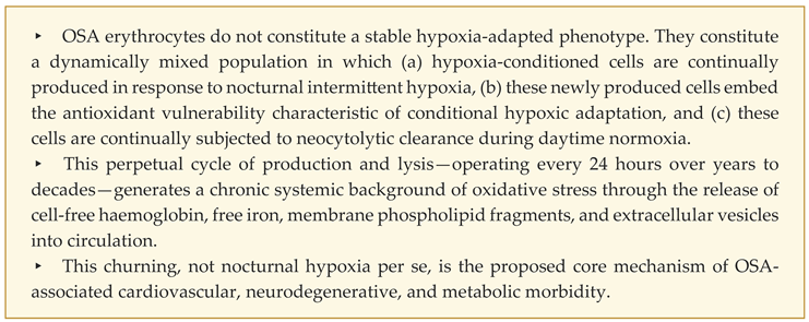
We propose that:
2.2. Molecular Basis of the Churning
The hypothesis is grounded in three established molecular mechanisms:
Mechanism 1: Nocturnal hypoxia drives band 3-GAPDH disinhibition. As Issaian and colleagues established in 2021 [18], deoxyhaemoglobin competitively displaces glyceraldehyde-3-phosphate dehydrogenase from the N-terminus of band 3 during deoxygenation, accelerating glycolytic flux. The Martí-Mateos 2026 demonstration that erythrocytes function as a primary systemic glucose sink in chronic hypoxia [19] confirms the consequence: hypoxic erythrocytes upregulate GLUT1, accelerate glucose uptake approximately three-fold, and redirect flux through the Luebering-Rapoport shunt to generate 2,3-DPG. In OSA, this programme is initiated nightly by repeated apnoeic desaturations.
Mechanism 2: The hypoxic programme embeds conditional antioxidant vulnerability. Concurrent with the metabolic reprogramming, hypoxia induces microRNA-21, which suppresses catalase expression in erythrocyte progenitors [20,21]. The biological logic is parsimonious: cells armed for hypoxia carry a built-in self-destruct mechanism that will activate when the hypoxic stimulus terminates and oxidative stress consequently rises. Prchal and colleagues have described this as "conditional phenotypic armament with built-in retirement" [20]. The molecular vulnerability includes not only catalase suppression but also retained reticulocyte mitochondrial mass (BNIP3L-dependent mitophagy defect), which becomes a reactive oxygen species source upon reoxygenation [5].
Mechanism 3: Daytime reoxygenation triggers selective clearance. Upon return to normoxic conditions during waking hours, the catalase-deficient, mitochondria-laden newly produced erythrocytes encounter an oxidative environment they cannot withstand. They are tagged for clearance—neocytolysis—through mechanisms including membrane phosphatidylserine externalization, complement deposition, and macrophage recognition. Risso and colleagues observed this clearance directly in altitude-descended humans [22]; Song and colleagues quantified its contribution in OSA [5]. The clearance is selective: it targets the recently produced cohort while sparing older, normoxic-conditioned cells in the circulation.
In OSA, the unique pathophysiological feature is that the cycle of hypoxia and normoxia is not a one-time event (as in altitude descent) but a daily reiteration. Each night, a new cohort is produced. Each day, that cohort is partially cleared. Over years, the cumulative biochemical exposure—not the snapshot composition at any single time point—is the relevant clinical variable.
2.3. Why the Three Contradictions Resolve
Resolution of contradiction 1 (the quantitative paradox). Haemoglobin remains normal because production and clearance are quantitatively balanced. The accelerated erythropoiesis observed by Song and colleagues [5] is real; so is the absence of erythrocytosis. Neither alone defines the OSA erythrocyte phenotype—their dynamic balance does.
Resolution of contradiction 2 (the qualitative paradox). Daytime bulk P50 and 2,3-DPG are unchanged because the recently produced hypoxia-conditioned cohort constitutes only a minority of circulating erythrocytes. The majority of circulating cells are older, normoxic-conditioned cells whose oxygen kinetics dominate the bulk measurement. The hypoxia-conditioned subpopulation is real but masked—as we will demonstrate, it can be detected by single-cell methods even when bulk measurements are silent.
Resolution of contradiction 3 (the downstream paradox). The systemic oxidative stress, vascular inflammation, oxidized LDL, and neurodegenerative consequences arise not from a different erythrocyte phenotype but from the lytic clearance products of the dynamically cleared subpopulation. Each day, a measurable fraction of circulating cells releases their contents into plasma: cell-free haemoglobin (a potent ROS generator via heme iron-catalysed Fenton chemistry), free iron, phospholipid membrane fragments (complement activators), and extracellular vesicles (proinflammatory cargo carriers).
3. Positioning in the RTC Framework
The hypothesis is most precisely articulated within the Red Blood Cell Triphenotype Classification (RTC) framework [16], which organizes erythrocyte function along three axes: Glucose Sink Capacity (GSC), Oxygen Kinetics State (OKS), and Antioxidant Reserve State (ARS), with ten defined phenotype categories (Types α through κ).
In RTC v1.0 [16], a single individual was assumed to occupy a single phenotype at any given time. The OSA case demonstrates that this assumption is insufficient. In RTC v2.0 [17], we introduce the concept of dynamic mixed phenotypes, in which an individual's circulating erythrocyte population comprises multiple coexisting phenotypes whose proportions vary diurnally.
OSA is the paradigmatic clinical example of dynamic mixed phenotype. An untreated severe OSA patient may be coded as:
RTC-α.MMH [80%] ⇌ β.HHM [8%] ⇌ δ.LLL [12%]
This notation captures the three coexisting subpopulations: the dominant older normoxic-conditioned cells (Type α), the recently produced hypoxia-adapted cohort (Type β), and the neocytolysis-bound cohort in transition (Type δ). The proportions are illustrative; empirical determination is the central task of the planned validation study [23]. After three months of effective CPAP, the same individual would be predicted to converge toward a stable Type α majority with progressive disappearance of the β and δ subpopulations.
This dynamic mixed phenotype notation distinguishes OSA from other clinical contexts. The pure altitude-trained athlete is RTC-β.HHM (single phenotype, stable). The OSA patient is fundamentally different: even at the same averaged P50, the heterogeneity, the rate of cohort turnover, and the consequent biochemical leakage differ.
4. From Churning to Disease: The Biochemical Chain
4.1. Released Mediators
The clearance of the hypoxia-conditioned cohort releases four classes of biochemical mediators into the systemic circulation:
Cell-free haemoglobin. Haemoglobin liberated from lysed cells consumes nitric oxide (impairing endothelial function), participates in Fenton chemistry (generating hydroxyl radicals), and serves as a damage-associated molecular pattern recognized by toll-like receptors [24]. The plasma haemoglobin elevation is typically below the threshold detected by routine clinical assays but is sufficient to drive sustained low-grade endothelial activation [25].
Free iron. Released as heme iron is processed, free iron in plasma drives Fenton-Haber-Weiss reactions producing hydroxyl radicals from peroxide. The hepcidin elevation observed in OSA patients [5] is partly a homeostatic response to this iron release. The system is incomplete: a portion of the released iron remains catalytically active.
Phospholipid membrane fragments. Erythrocyte membranes liberated by lysis carry phosphatidylserine and oxidatively modified phospholipids. These fragments activate complement, are recognized by scavenger receptors on macrophages, and contribute to the inflammatory milieu that characterizes OSA [26].
Extracellular vesicles (EVs). Erythrocyte-derived extracellular vesicles—including microparticles released during eryptotic processing—carry proinflammatory cargo including microRNAs, lipid mediators, and damage-associated proteins. EV-mediated signalling represents an under-appreciated mode of erythrocyte-to-tissue communication [27,28]. In OSA, where erythrocyte clearance is amplified, EV release is correspondingly increased.
4.2. The Cardiovascular Consequences
The mediators released by perpetual churning converge on the vascular endothelium. Cell-free haemoglobin consumes endothelial nitric oxide, impairing endothelium-dependent vasodilation. Free iron and EVs activate NADPH oxidase, generating additional superoxide. Membrane fragments and EVs activate complement and recruit monocytes. The result is the classical vascular phenotype of OSA: endothelial dysfunction, increased leukocyte-endothelial adhesion, accelerated low-density lipoprotein oxidation, and progressive atherosclerotic remodelling [9,10,11,12].
Critically, this mechanistic chain operates whether or not the patient has co-existing dyslipidaemia or hypertension. It explains why OSA confers cardiovascular risk independently of these classical risk factors, and why CPAP treatment partially reverses this risk: by interrupting the nocturnal hypoxia signal, CPAP allows the recently produced cohort to age normally rather than being cleared, ending the cycle.
4.3. The Neurodegenerative Consequences
The same released mediators have specific consequences for the brain. The blood-brain barrier is exposed to circulating cell-free haemoglobin and free iron through fenestrated regions including the choroid plexus and circumventricular organs. Endothelium-derived ROS reduces cerebral blood flow autoregulation. EVs traverse the blood-brain barrier and may carry inflammatory mediators directly to brain parenchyma. The hippocampus, exquisitely sensitive to oxidative damage and blood flow disturbance, exhibits the structural changes consistently observed in OSA neuroimaging [14,29,30,31].
This framework also explains why OSA increases amyloid-β accumulation [13]: chronic oxidative stress impairs cerebral amyloid clearance via the glymphatic system, while inflammation upregulates β- and γ-secretase activity. The recent finding that OSA hypoxic burden, rather than AHI per se, predicts hippocampal volume loss [29] is consistent with the churning hypothesis: hypoxic burden is the upstream driver of churning intensity, which is the proximate cause of neurodegenerative biochemistry.
4.4. Why CPAP Sometimes Fails
Several large randomised trials of CPAP for cardiovascular prevention in OSA have produced disappointing results [32,33], despite robust observational evidence that CPAP-treated patients fare better than untreated patients. The churning hypothesis offers a reconciling interpretation: in patients who respond well to CPAP, the nocturnal hypoxia stimulus is sufficiently interrupted that the production of hypoxia-conditioned cells ceases, the existing dynamic mixed phenotype resolves, and downstream pathology recedes. In patients who respond poorly—whether because of poor adherence, residual events, or other comorbidities maintaining the inflammatory milieu—churning persists, and cardiovascular risk remains elevated.
This interpretation generates a clinically tractable prediction: among CPAP-treated patients, those with persistent erythrocyte churning are the subset at continued cardiovascular risk. If validated, this would identify patients who might benefit from adjunctive antioxidant therapy, more aggressive lipid management, or alternative interventions targeting the residual churning pathway.
5. Testable Predictions
The hypothesis generates four falsifiable predictions, each amenable to investigation with the RTC Level 1 measurement protocol (flow cytometric GLUT1 with reticulocyte gating, plasma cell-free haemoglobin, reticulocyte microRNA-21, and reticulocyte count):
Prediction 1: Churning intensity scales with OSA severity. In an unselected cohort of severe OSA, moderate OSA, and healthy controls matched for age, sex, and body mass index, a composite churning intensity score (z-score sum of reticulocyte count, plasma cell-free haemoglobin, and reticulocyte microRNA-21) will be elevated in OSA versus controls and will correlate positively with the apnoea-hypopnoea index and oxygen desaturation index.
Prediction 2: Churning correlates more strongly with downstream pathology than does AHI. Churning intensity will correlate with systemic oxidative stress markers (urinary 8-hydroxydeoxyguanosine, plasma F2-isoprostanes) and vascular inflammation markers (high-sensitivity C-reactive protein, lipoprotein-associated phospholipase A2, oxidized LDL) more strongly than the apnoea-hypopnoea index correlates with these same markers. Statistical mediation analysis will identify churning intensity as a significant mediator of the AHI-to-inflammation pathway.
Prediction 3: CPAP reverses churning, with residual churning identifying continued-risk patients. Three months of effective CPAP therapy (adherence ≥4 hours per night) will reduce churning intensity by ≥50%, with parallel reduction in downstream inflammatory markers. A subset of patients—even with apparently effective CPAP by polysomnographic criteria—will exhibit persistent churning; this subset will correspond to patients with continued cardiovascular event risk.
Prediction 4: Bimodal GLUT1 distribution reflects dynamic mixed phenotype. Flow cytometric measurement of GLUT1 mean fluorescence intensity at the single-cell level, with reticulocyte (CD71+) gating, will reveal a bimodal distribution in severe OSA patients—reflecting coexistence of high-GLUT1 hypoxia-conditioned reticulocytes and normal-GLUT1 older cells—whereas healthy controls will show unimodal distribution. The bimodality coefficient will scale with OSA severity.
These predictions are addressed in the prospective observational study CSMUH-OSA-RTC-2026-01, which we have designed with 180 participants (60 severe OSA, 60 moderate OSA, 60 healthy controls) over 12 months [23]. Statistical power calculations indicate adequate power (≥85%) to detect the predicted effect sizes.
6. Alternative Interpretations and Distinguishing Evidence
6.1. Could the Churning Be Epiphenomenal?
A reviewer might argue that churning is real but epiphenomenal—merely a marker of OSA severity rather than a causal driver of cardiovascular consequence. The hypothesis distinguishes itself empirically through Prediction 2: if churning is epiphenomenal, AHI should correlate as strongly with inflammation as churning does. The hypothesis predicts the opposite, and mediation analysis on the planned cohort will adjudicate.
6.2. Could the Sympathetic Hypothesis Be Sufficient?
OSA-induced sympathetic activation is well-established and contributes substantially to hypertension and to several cardiovascular consequences [3]. We do not argue that sympathetic activation is unimportant. We argue that it is insufficient to explain the full spectrum of OSA pathology. In particular, sympathetic activation does not naturally explain the specific oxidative chemistry observed in OSA (haemoglobin-driven NO consumption, iron-driven Fenton chemistry, complement activation), nor does it explain why CPAP partial responders maintain residual risk despite normalisation of sympathetic activity. The churning hypothesis fills these explanatory gaps without contradicting the sympathetic mechanism.
6.3. Could Endothelial Dysfunction Be Primary?
OSA endothelial dysfunction is robustly established [9]. Whether it is primary or secondary to circulating mediators is unsettled. The churning hypothesis proposes a specific upstream source for the endothelial insult that has not previously been articulated. Experimental support comes from observations that erythrocyte-derived extracellular vesicles directly damage endothelium [25,28], and that cell-free haemoglobin directly impairs vasodilation [24].
7. Clinical and Research Implications
7.1. Reframing OSA Severity
Current OSA severity assessment relies on the apnoea-hypopnoea index, a per-hour rate of breathing events that captures dose of nocturnal hypoxia but is silent on physiological response. The churning framework suggests that a composite indicator—"AHI + churning intensity"—would be more clinically informative. Two patients with identical AHI but divergent churning intensities would receive different prognostic stratification and possibly different therapeutic intensity. The Level 1 protocol uses only blood-based markers feasible within standard clinical laboratory infrastructure.
7.2. Identifying Treatment Non-Responders Early
Among CPAP-treated patients, churning intensity at three months may identify those at residual cardiovascular risk who require additional intervention. This represents a precision-medicine approach to OSA management that is currently impossible: routine clinical follow-up at three months captures AHI normalization but not the residual subclinical pathology that determines long-term outcome.
7.3. Generalizing to Other Intermittent Hypoxia States
The framework potentially generalizes to other intermittent hypoxia states: chronic obstructive pulmonary disease with exacerbations, heart failure with nocturnal desaturation, preterm infants with oxygen-saturation fluctuations, and—possibly—high-altitude shift workers. Each represents a clinical state in which the dynamic mixed phenotype framework may have application.
7.4. Therapeutic Opportunity
If the hypothesis is correct, several therapeutic opportunities follow. First, antioxidant agents targeting catalase substitution or NADPH oxidase inhibition might attenuate the consequences of churning in patients in whom CPAP is inadequate. Second, iron-chelating strategies might reduce the Fenton-chemistry component of the oxidative burden. Third, agents that stabilize newly produced erythrocytes against neocytolysis—if developed—could fundamentally alter the disease trajectory by interrupting the cycle. These are speculative, but they follow naturally from the mechanism.
8. Limitations and Open Questions
Several limitations require explicit acknowledgement.
The hypothesis is presently hypothesis. No direct quantitative measurement of OSA erythrocyte churning—integrating production, clearance, and biochemical leakage in a single individual—has been performed. The proposed measurements are feasible and within the scope of the planned validation study, but the empirical data do not yet exist.
Translation from molecular mechanism to clinical mediator is partial. While each link in the proposed chain—nocturnal hypoxia → cohort production → daytime clearance → biochemical leakage → downstream pathology—has individual experimental support, the full chain has not been quantitatively demonstrated in OSA. The hypothesis is most rigorous at its molecular foundations and progressively more speculative at its clinical termini.
Individual variation may dominate. Across the spectrum of OSA patients, individual variability in haemopoietic response, antioxidant reserve, and inflammatory propensity will likely modulate churning intensity. This is a feature, not a bug: the hypothesis predicts that this variation will correspond to variation in clinical outcome. But the variation also means that group-level statistics may underestimate individual-level effects.
The role of obesity is not fully addressed. OSA is strongly associated with obesity, which independently drives inflammation and oxidative stress. Disentangling the contribution of OSA-specific churning from obesity-driven inflammation requires careful statistical adjustment and, ideally, comparison of OSA versus non-OSA obese individuals. The planned study includes this design feature.
Pre-clinical experimental support is needed. Direct demonstration of perpetual churning would benefit from animal models of intermittent hypoxia in which erythrocyte production and clearance can be tracked longitudinally. Such studies have not been performed with the proposed framework in mind.
9. Conclusion
Three decades of OSA research have established the disorder's substantial clinical impact and have identified many of its mediating pathways. What has remained elusive is a single mechanistic framework that integrates the apparent contradictions in the erythrocyte literature with the consistent downstream observations of oxidative stress, vascular inflammation, and neurodegeneration. We propose that the perpetual erythrocyte cohort churning hypothesis provides such a framework.
The hypothesis is parsimonious: it requires no new molecular discovery, only the integration of established findings—Issaian's band 3 switch, Martí-Mateos' glucose sink, Prchal's neocytolysis—into a unified clinical model. It is falsifiable: it makes specific predictions amenable to direct measurement. It is consequential: if correct, it reframes OSA severity assessment, identifies CPAP non-responders early, and opens novel therapeutic avenues.
Most fundamentally, the hypothesis reframes our conception of OSA pathophysiology. The clinical consequences arise not from what happens during the apnoeic episode itself, but from the daytime aftermath—from the continual lytic processing of erythrocytes that nightly hypoxia has conditioned and that morning normoxia has condemned. The disease lives, in this view, not at night but at day; not in the airway but in the blood; not in the moment of obstruction but in the perpetual cycle that obstruction sets in motion.
"In obstructive sleep apnoea, the apnoea is the trigger, but the disease is the daily processing of erythrocytes that the apnoea makes inevitable. To treat the disease, we must understand—and measure—the churning."
Author Contributions
M-YH conceived the hypothesis, performed the literature synthesis, drafted the manuscript, and approved the final version. No other individuals contributed to the conceptual development or writing. The author bears sole responsibility for all content.
Competing Interests
The author declares no competing financial or non-financial interests.
Funding
This work received no specific funding. The planned validation study (CSMUH-OSA-RTC-2026-01) is being prepared for submission to Taiwan's National Science and Technology Council (NSTC).
Data Availability
This is a perspective article; no original data were generated. All cited literature is publicly accessible.
AI Use Disclosure
Large language model assistance (Anthropic Claude) was used for literature integration, drafting, and language refinement. All conceptual claims, framework architecture, mechanistic interpretations, and final phrasings were authored, reviewed, and approved by the human author. This disclosure aligns with current ICMJE recommendations.
Preprint Status
This manuscript is intended for preprint submission to Preprints.org concurrent with the related RTC framework manuscript [16] and subsequent submission for peer review to Sleep Medicine Reviews, Chest, or Circulation Research.
References
- Benjafield, A.V.; Ayas, N.T.; Eastwood, P.R.; et al. Estimation of the global prevalence and burden of obstructive sleep apnoea: a literature-based analysis. Lancet Respir. Med. 2019, 7, 687–698. [Google Scholar] [CrossRef] [PubMed]
- Drager, L.F.; Polotsky, V.Y.; Lorenzi-Filho, G. Obstructive sleep apnea: an emerging risk factor for atherosclerosis. Chest 2011, 140, 534–542. [Google Scholar] [CrossRef] [PubMed]
- Arnaud, C.; Bochaton, T.; Pépin, J.L.; Belaidi, E. Chronic intermittent hypoxia-induced cardiovascular and renal dysfunction: from adaptation to maladaptation. J. Physiol. 2023. [Google Scholar] [CrossRef] [PubMed]
- Bubu, O.M.; Andrade, A.G.; Umasabor-Bubu, O.Q.; et al. Obstructive sleep apnea, cognition and Alzheimer's disease: A systematic review integrating three decades of multidisciplinary research. Sleep Med. Rev. 2019, 50, 101250. [Google Scholar] [CrossRef] [PubMed]
- Song, J.; Sundar, K.; Gangaraju, R.; Prchal, J.T. Increased blood reactive oxygen species and hepcidin in obstructive sleep apnea precludes expected erythrocytosis. Am. J. Hematol. 2023. [Google Scholar] [CrossRef] [PubMed]
- Clause, D.; Detry, B.; Rodenstein, D.; Liistro, G. Stability of oxyhemoglobin affinity in patients with obstructive sleep apnea-hypopnea syndrome without daytime hypoxemia. J. Appl. Physiol. 2008. [Google Scholar] [CrossRef] [PubMed]
- Lavie, L. Oxidative stress in obstructive sleep apnoea and intermittent hypoxia – revisited – the bad ugly and good: implications to the heart and brain. Sleep Med. Rev. 2015, 20, 27–45. [Google Scholar] [CrossRef] [PubMed]
- Maniaci, A.; Iannella, G.; Cocuzza, S.; et al. Oxidative Stress and Inflammation Biomarker Expression in Obstructive Sleep Apnea Patients. J. Clin. Med. 2021, 10, 277. [Google Scholar] [CrossRef] [PubMed]
- Jelic, S.; Padeletti, M.; Kawut, S.M.; et al. Inflammation, oxidative stress, and repair capacity of the vascular endothelium in obstructive sleep apnea. Circulation 2008, 117, 2270–2278. [Google Scholar] [CrossRef] [PubMed]
- Mészáros, M.; Bikov, A.; Kunos, L.; et al. The Role of Soluble Low-Density Lipoprotein Receptor-Related Protein-1 in Obstructive Sleep Apnoea. J. Clin. Med. 2021, 10, 1494. [Google Scholar] [PubMed]
- Douglas, R.M.; Bowden, K.; Pattison, J.; et al. Intermittent hypoxia and hypercapnia induce pulmonary artery atherosclerosis and ventricular dysfunction in low density lipoprotein receptor deficient mice. J. Appl. Physiol. 2013. [Google Scholar] [CrossRef] [PubMed]
- Wang, J.; Yu, W.; Gao, M.; et al. Impact of Obstructive Sleep Apnea Syndrome on Endothelial Function, Arterial Stiffening, and Serum Inflammatory Markers: An Updated Meta-analysis and Metaregression of 18 Studies. J. Am. Heart Assoc. 2015, 4, e002454. [Google Scholar] [CrossRef] [PubMed]
- Cavuoto, M.G.; Robinson, S.R.; O'Donoghue, F.J.; et al. Associations Between Amyloid Burden, Hypoxemia, Sleep Architecture, and Cognition in Obstructive Sleep Apnea. J. Alzheimers Dis. 2023, 96, 491–501. [Google Scholar] [CrossRef]
- Torelli, F.; Moscufo, N.; Garreffa, G.; et al. Cognitive profile and brain morphological changes in obstructive sleep apnea. NeuroImage 2010, 54, 787–793. [Google Scholar] [PubMed]
- Weihs, A.; Frenzel, S.; Wittfeld, K.; et al. Associations Between Sleep Apnoea and Advanced Brain Ageing in a Large-Scale Population Study. Sleep 2020, 44, zsaa176. [Google Scholar] [CrossRef]
- Hsieh, M.-Y. Red blood cell triphenotype classification (RTC): a functional framework integrating glucose sink capacity, oxygen kinetics, and antioxidant reserve [preprint]. Preprints.org. 2026. [Google Scholar] [CrossRef]
- Hsieh, M.-Y. RTC v2.0: dynamic mixed phenotypes [working paper]. CSMUH Evidence-Based Medicine Center. 2026. [Google Scholar]
- Issaian, A.; Hay, A.; Dzieciatkowska, M.; et al. The interactome of the N-terminus of band 3 erythrocytes regulates red blood cell metabolism through complex formation and recruitment to the membrane upon hypoxia. Haematologica 2021, 106, 2971–2985. [Google Scholar] [CrossRef] [PubMed]
- Martí-Mateos, Y.; Safari, Z.; Bevers, S.; et al. Red blood cells serve as a primary glucose sink to improve glucose tolerance at altitude. Cell Metab. 2026. [Google Scholar] [CrossRef] [PubMed]
- Song, J.; Yoon, D.; Christensen, R.D.; Horvathova, M.; Thiagarajan, P.; Prchal, J.T. HIF-mediated increased ROS from reduced mitophagy and decreased catalase causes neocytolysis. J. Mol. Med. 2015, 93, 857–866. [Google Scholar] [CrossRef] [PubMed]
- Risso, A.; Turello, M.; Biffoni, F.; Antonutto, G. Red blood cell senescence and neocytolysis in humans after high altitude acclimatization. Blood Cells Mol. Dis. 2007, 38, 83–92. [Google Scholar] [CrossRef] [PubMed]
- Mairbäurl, H. Neocytolysis: how to get rid of the extra erythrocytes formed by stress erythropoiesis upon descent from high altitude. Front Physiol. 2018, 9, 345. [Google Scholar] [CrossRef] [PubMed]
- Hsieh, M.-Y. CSMUH-OSA-RTC-2026-01: Perpetual erythrocyte cohort churning in OSA - a prospective mechanistic observational study [IRB protocol]. 2026. [Google Scholar]
- Schaer, D.J.; Buehler, P.W.; Alayash, A.I.; et al. Hemolysis and free hemoglobin revisited: exploring hemoglobin and hemin scavengers as a novel class of therapeutic proteins. Blood 2013, 121, 1276–1284. [Google Scholar] [CrossRef] [PubMed]
- Buehler, P.W.; D'Agnillo, F. Toxicological consequences of extracellular hemoglobin: biochemical and physiological perspectives. Antioxid. Redox Signal. 2010, 12, 275–291. [Google Scholar] [CrossRef] [PubMed]
- Lang, K.S.; Lang, P.A.; Bauer, C.; et al. Mechanisms of suicidal erythrocyte death. Cell Physiol. Biochem. 2005, 15, 195–202. [Google Scholar] [CrossRef] [PubMed]
- Said, A.S.; Doctor, A. Influence of red blood cell-derived microparticles upon vasoregulation. Blood Transfus. 2017, 15, 522–534. [Google Scholar] [CrossRef] [PubMed]
- Sadallah, S.; Eken, C.; Schifferli, J.A. Erythrocyte-derived ectosomes have immunosuppressive properties. J. Leukoc. Biol. 2008, 84, 1316–1325. [Google Scholar] [CrossRef] [PubMed]
- Settimi, M.; Schiavolin, S.; Sgaramella, M.; et al. Examining the association between sleep apnea and total hippocampal volumes in cognitive impairment. Alzheimer's Dement 2025. [Google Scholar] [CrossRef]
- Gosselin, N.; Baril, A.A.; Osorio, R.S.; et al. Obstructive Sleep Apnea and the Risk of Cognitive Decline in Older Adults. Am. J. Respir. Crit. Care Med. 2019, 199, 142–148. [Google Scholar] [CrossRef] [PubMed]
- Andrade, A.G.; Bubu, O.M.; Varga, A.W.; Osorio, R.S. The Relationship between Obstructive Sleep Apnea and Alzheimer's Disease. J. Alzheimers Dis. 2018, 64(s1), S255–S270. [Google Scholar] [CrossRef] [PubMed]
- McEvoy, R.D.; Antic, N.A.; Heeley, E.; et al. CPAP for Prevention of Cardiovascular Events in Obstructive Sleep Apnea. N Engl. J. Med. 2016, 375, 919–931. [Google Scholar] [CrossRef] [PubMed]
- Peker, Y.; Glantz, H.; Eulenburg, C.; et al. Effect of Positive Airway Pressure on Cardiovascular Outcomes in Coronary Artery Disease Patients with Nonsleepy Obstructive Sleep Apnea. The RICCADSA Randomized Controlled Trial. Am. J. Respir. Crit. Care Med. 2016, 194, 613–620. [Google Scholar] [CrossRef] [PubMed]
- Lavie, L. Obstructive sleep apnoea syndrome - an oxidative stress disorder. Sleep Med. Rev. 2003, 7, 35–51. [Google Scholar] [CrossRef] [PubMed]
- Prabhakar, N.R.; Peng, Y.J.; Nanduri, J. Hypoxia inducible factors (HIFs) and obstructive sleep apnea. J. Clin. Invest. 2020, 130, 5042–5051. [Google Scholar] [CrossRef] [PubMed]
- Mochol, J.; Gawrys, J.; Gajecki, D.; et al. Cardiovascular Disorders Triggered by Obstructive Sleep Apnea-A Focus on Endothelium and Blood Components. Int. J. Mol. Sci. 2021, 22, 5139. [Google Scholar] [CrossRef] [PubMed]
- Waltz, X.; Beaudin, A.E.; Belaidi, E.; et al. Impact of obstructive sleep apnoea and intermittent hypoxia on blood rheology: a translational study. Eur Respir J 2021. [Google Scholar] [CrossRef] [PubMed]
- Maliszewska-Cyna, E.; Bawa, K.K.; Kapustin, D.; et al. A Comparative Study Evaluating the Impact of Physical Exercise on Disease Progression in a Mouse Model of Alzheimer's Disease. J. Alzheimers Dis. 2017. [Google Scholar] [CrossRef]
- Liu, X.; Ma, Y.; Ouyang, R.; et al. The relationship between inflammation and neurocognitive dysfunction in obstructive sleep apnea syndrome. J. Neuroinflammation 2020, 17, 229. [Google Scholar] [CrossRef] [PubMed]
- Reisz, J.A.; Wither, M.J.; Dzieciatkowska, M.; et al. Oxidative modifications of glyceraldehyde 3-phosphate dehydrogenase regulate metabolic reprogramming of stored red blood cells. Blood 2016, 128, e32–e42. [Google Scholar] [CrossRef] [PubMed]
- Hsieh, M.-Y. The hyperoxia paradox: a missing axis in ICU stress hyperglycaemia [perspective draft]. 2026. [Google Scholar]
- Hsieh, M.-Y. HbA1c reconsidered: glycated haemoglobin in the era of dynamic erythrocyte glucose metabolism [perspective draft]. 2026. [Google Scholar]
Disclaimer/Publisher’s Note: The statements, opinions and data contained in all publications are solely those of the individual author(s) and contributor(s) and not of MDPI and/or the editor(s). MDPI and/or the editor(s) disclaim responsibility for any injury to people or property resulting from any ideas, methods, instructions or products referred to in the content. |
© 2026 by the authors. Licensee MDPI, Basel, Switzerland. This article is an open access article distributed under the terms and conditions of the Creative Commons Attribution (CC BY) license (http://creativecommons.org/licenses/by/4.0/).
Copyright: This open access article is published under a Creative Commons CC BY 4.0 license, which permit the free download, distribution, and reuse, provided that the author and preprint are cited in any reuse.


