
Submitted:
07 July 2026
Posted:
08 July 2026
You are already at the latest version
Abstract
Doxorubicin (DOX) is a potent chemotherapeutic agent widely used in the clinical treatment of various malignancies. However, its therapeutic application is significantly constrained by dose-dependent cardiotoxicity, with accelerated cardiac aging emerging as a particularly challenging issue. This presents a critical limitation to its clinical use, emphasizing the urgent need for targeted cardioprotective strategies to mitigate DOX-induced cardiotoxicity. Cellular senescence, marked by permanent cell cycle arrest, plays a key role in the development of DOX-induced cardiac aging. DOX has been shown to induce cellular stress and senescence in multiple cardiac cell types, leading to impaired cardiac function. Understanding the molecular pathways and mechanisms through which DOX triggers cardiac aging is essential for the development of effective interventions to address its cardiotoxic side effects. In this review, we discuss the molecular mechanisms underlying DOX-induced senescence in different cardiac cell types and summarize the current progress in anti-aging therapeutic strategies aimed at alleviating DOX-related cardiac complications.
Keywords:
cardiac aging
; doxorubicin
; treatment strategy
; Traditional Chinese Medicine
1. Introduction
Oncological diseases are among the most prevalent public health concerns and rank as the second leading cause of death, following cardiovascular diseases. Doxorubicin (DOX), commonly known as Adriamycin, is a widely used anthracycline antibiotic and a highly potent chemotherapy agent. It has shown remarkable therapeutic efficacy in treating a range of cancers, including breast, lung, leukemia, and colon cancers(Gergely et al., 2015). Doxorubicin (DOX) contains a planar tetracyclic ring and an amino sugar. Upon targeting tumor cells, the planar ring intercalates into DNA, forming a “DNA-TOP2α-DOX” ternary complex. This inhibits DNA replication and topoisomerase II (Top2) activity, resulting in persistent DNA double-strand breaks (DSBs) and inducing an oxidative stress-mediated tumor cell death(Wang et al., 2018). Despite its anticancer benefits, DOX causes multi-organ toxicity, including nephrotoxicity, hepatotoxicity, alopecia, bone marrow suppression, and chemotherapy-related cognitive impairment (“chemobrain”), particularly dose-limiting cardiotoxicity. This arises from ROS-mediated mitochondrial dysfunction, DNA damage, and calcium overload, culminating in cardiomyocyte apoptosis and myocardial injury(Narayan et al., 2017). This type of injury and functional decline is closely associated with pathological cardiac aging. It highlight the urgent need for interventions targeting DOX-induced heart aging, which could broaden its clinical applicability by mitigating dose-limiting cardiotoxicity.
Given DOX’s exceptional antitumor efficacy, it has become a cornerstone of modern chemotherapy regimens, and its therapeutic applications have been extensively studied. While recent reviews have focused on pharmacological toxicity of DOX in oncology, comprehensive analyses specifically exploring its role in pathological cardiac aging remain scarce(Liang et al., 2020). We systematically review the current advances and future prospects of key mechanisms underlying DOX-induced abnormal cardiac aging during cancer treatment. First, we outline the pathological features of DOX-induced cardiac aging. Next, we explore the mechanisms driving this process in detail. Finally, we discuss the current challenges and future research directions in this area (Figure 1).
2. Cell Types Involved in Doxorubicin-Induced Cardiac Aging
Cellular senescence is a heterogeneous phenotype that varies depending on cell type and context, characterized by an irreversible loss of proliferative capacity(Mao et al., 2012). It can be further categorized into subtypes, including replicative senescence, oncogene-induced senescence, and stress-induced premature senescence(Marescal and Cheeseman, 2020). Doxorubicin can lead to accelerated cardiac aging and associated diseases. Studies have shown that aging is associated with altered Sirtuin 6 (SIRT6) activity, contributing to the development of cardiac hypertrophy and fibrosis(Pillai et al., 2021). Insulin like growth factor binding protein 7 (IGFBP7) is a marker of senescence secretome, involved in multiple stages of immune system regulation(Bracun et al., 2022).
Subacute concentrations of doxorubicin induce cardiac aging through various forms of cellular senescence, involving cardiomyocytes, fibroblasts, endothelial cells, and other cell types. The mechanisms of cellular senescence differ between these cell types, resulting in distinct disease phenotype(Figure 2). In this review, we summarize the key mechanisms of DOX-induced cardiac aging in different cell types(Linders et al., 2024).
2.1. Cardiomyocytes
Cardiomyocytes account for 30–40% of the total cell population in the heart and 80% of its cellular volume(Pinto et al., 2016). Senescent cardiomyocytes exhibit increased cell size, elevated pacing frequency, contractile dysfunction, and metabolic impairment(Tang et al., 2020). Additionally, they can secrete SASP factors that induce senescence in neighboring cells(Anderson et al., 2019). Cardiomyocyte senescence induced by DOX is primarily associated with DNA damage, oxidative stress, telomere dysfunction, and alterations in mitochondrial dynamics and function (Figure 3).
The single-electron reduction of DOX to form semiquinone is often accompanied by the generation of reactive oxygen species (ROS), which is a primary source of oxidative stress induced by DOX(Tocchetti et al., 2019). Although DOX typically induces cardiomyocyte damage, low doses of DOX tend to promote oxidative stress and senescence phenotypes rather than apoptosis. Activation of p21cip1/waf1 by p53 acetylated by promyelocytic leukemia protein (PML) played an important role in this premature senescence(Maejima et al., 2008). DOX-induced cardiomyocyte senescence was significantly alleviated by reducing oxidative stress and modulating the expression of glutathione transferase, highlighting the link between oxidative stress and DOX-induced cardiomyocyte senescence(Bielak-Zmijewska et al., 2014). The accumulation of reactive oxygen species (ROS) significantly impacts mitochondrial dynamics and function. In DOX-induced senescent cardiomyocytes, elevated ROS lead to mitochondrial fragmentation and impaired energy metabolism, contributing to cardiac dysfunction(Wan et al., 2019). The cardiomyocytes exhibited increased expression of the mitochondrial fission protein dynamin-related protein1 (Drp1) and its phosphorylation at Ser616. This is accompanied by the downregulation of telomere-binding factors TRF1 and TRF2(Shi et al., 2021; Spallarossa et al., 2009). Testosterone could counteract DOX-induced downregulation of TRF2 through the PI3K/AKT/NOS-3 signaling pathway, thereby alleviating DOX-induced cardiomyocyte senescence(Altieri et al., 2016). In DOX-treated HL-1 murine cardiomyocytes, long intergenic non-coding RNA-p21 (lincRNA-p21) was significantly upregulated. This upregulation was associated with decreased cell proliferation and viability, increased expression of senescence markers p53 and p16, reduced telomere length, and diminished telomerase activity. Notably, silencing endogenous lincRNA-p21 alleviated these effects(Xie et al., 2018). Furthermore, C5aRA(complement component 5a receptor antagonist) also enhanced telomere length and telomerase activity in H9c2 and AC16 cardiomyocytes following DOX stimulation(Wen et al., 2021). Inflammatory markers promoted local inflammation in the cardiac microenvironment, triggering senescence in neighboring cells(Fallah et al., 2019). DOX-induced cardiomyocyte senescence was associated with increased TXNIP(thioredoxin-interactive protein) expression and activation of the NLRP3(NOD-like receptor family pyrin domain-containing 3) inflammasome, silencing TXNIP inhibited NLRP3 inflammasome activation(Huang et al., 2020). DOX also upregulated the expression of inflammatory factors, including NF- κB, TNF-α, IL-1β, and IL-6, resulting in an inflammatory microenvironment. Increased TNF-α release further promoted ROS generation, which was closely linked to oxidative stress(Fallah et al., 2019).
In the senescent state, cardiomyocytes exhibited increased cell volume, contributing to cardiac hypertrophy. Aging cardiomyocytes displayed altered contractility and abnormal conduction patterns, which can lead to cardiomyopathy or arrhythmias(Gude et al., 2018). Compared to normal mice, older mice exhibited a reduced ejection fraction, an increased heart-to-tibia length ratio, and signs of cardiac hypertrophy(Pillai et al., 2021). Doxorubicin induced a senescence-like phenotype in cardiomyocytes and disrupted troponin phosphorylation, which may impair efficient cardiac contraction(Maejima et al., 2008). Moreover, DOX-induced cardiomyocyte senescence also resulted in a dilated cardiomyopathy phenotype(Mitry et al., 2020).
2.2. Cardiac Fibroblasts
Cardiac fibroblasts (CFs) are the second most abundant cell type in the heart, comprising 24.3% of atrial tissue and 15.5% of ventricular tissue(Litviňuková et al., 2020). In both normal and aged hearts, cardiac fibroblasts have opposing roles. In the normal heart, cardiac fibroblasts promote healing following cardiac injury, whereas in the aged heart, they contribute to the development of cardiac fibrosis(Burstein et al., 2008; Saucerman et al., 2019). In the aging heart, fibroblasts differentiate into myofibroblasts, increasing the risk of cardiac disease. Senescence in cardiac fibroblasts was regulated by miR-17, which targeted the senescence-associated protein Par4. Ectopic expression of circ-Foxo3 induced cellular senescence in Mouse Embryonic Fibroblasts(MEFs) by interacting with the anti-aging proteins ID1 and E2F1, as well as the stress-response proteins FAK and HIF1α(Du et al., 2017). Furthermore, aspirin reduced the accumulation of p53 and p21, decreased cell viability and lowered Bcl-xL protein levels in DOX-treated human and mouse fibroblasts. Notably, COX-2 knockdown significantly attenuated aspirin’s inhibitory effect on DOX-induced p53 accumulation(Feng et al., 2019). Ginsenoside Rh2 (Rh2) could attenuated cardiac pathological remodelling induced by DOX through reducing fibroblast to myofibroblast transition (FMT)(Hou et al., 2022). Recent studies have shown that DOX-induced extracellular vesicle (EV) secretion may promote autophagy in rat cardiac fibroblasts by activating AMPKα, thereby mitigating the cellular senescence phenotype(Fujioka et al., 2023).
2.3. Endothelial Cell
Endothelial cells (ECs) constitute 12.2% of the atrial tissue and 7.8% of the ventricular tissue(Litviňuková et al., 2020). ECs senescence is closely linked to oxidative stress and mitochondrial metabolism. Recent studies have revealed that ECs can influence autophagy through extracellular vesicles, thereby affecting cellular senescence(Wallis et al., 2020). In the aging heart, endothelial cells play a crucial role in regulating vasoactivity and maintaining cardiac viability(Colliva et al., 2020). Doxorubicin could elevate P16INK4A protein expression and induce endothelial senescence by activating JNK and p38 MAPK signaling pathways, implying that DOX-induced endothelial senescence may be related to ROS accumulation(Spallarossa et al., 2010). Another study found that activation of oxidative stress induced premature senescence of endothelial cells, which was accompanied by a reduction in SIRT1 expression. However, resveratrol administration reversed this effect and protected against endothelial cell senescence(Kao et al., 2010). Moreover, DOX-induced senescent endothelial cells exhibited downregulated TRF2, suggesting that telomere dysfunction is one of the underlying mechanisms of senescence(Spallarossa et al., 2010). Study also revealed that DOX-induced senescence was significantly reduced by PI3K/AKT pathway inhibitors, with similar effects observed with BCL-2 inhibitors(Abdelgawad et al., 2023).
2.4. Vascular Smooth Muscle Cells (VSMCs)
Vascular smooth muscle cells (VSMCs) play a pivotal role in cardiac hemodynamics, working in coordination with endothelial cells to maintain vascular function and ensure hemodynamic stability in the aging heart. VSMC senescence can be triggered by telomere shortening, DNA damage, oxidative stress, and autophagy dysfunction. Senescent VSMCs exhibited increased SA-β-Gal activity and upregulated p53/p21、p16 expression, leading to aberrant processing of prelamin A into lamin A(Wang et al., 2015). This resulted in improper nuclear lamina formation, making cells more susceptible to DNA damage(Ragnauth et al., 2010). Compared to replicative senescence, DOX-triggered stress-induced senescence in VSMCs was more likely to arrest cells in the G2/M phase rather than G1 phase, and could be limited by upregulating IL-10 via the AMPKα/SIRT1/FOXO3a pathway(Chen et al., 2021).
DOX-induced premature senescence (SIPS) in VSMCs was also associated with elevated DNA double-strand breaks(DSB) and ROS levels. It was showed that lowering the level of ROS-producing enzyme-NOX4 oxidase below physiological level lead to cellular senescence of VSMCs which was correlated with secretion of pro-inflammatory cytokines(IL-6/IL-8)(Przybylska et al., 2016). Furthermore, DOX-induced ROS and DNA damage triggered a phenotypic shift in VSMCs, promoting the expression of senescent and inflammatory proteins(Herrmann et al., 2021).
2.5. Mesenchymal Stem Cells
Mesenchymal stem cells (MSCs) are multipotent adult stem cells derived from the mesoderm, possessing self-renewal capacity and the ability to differentiate into various cell types. MSCs can transform into endothelial cells and smooth muscle cells, contribute to the formation of new blood vessels. Additionally, they can stimulate resident cardiac cells in ischemic tissues, then improve the function of resident cardiomyocytes which are considered as important cellular components of the heart(Razeghian-Jahromi et al., 2021).
Doxorubicin damaged cultured MSCs, inducing premature senescence, but MSCs exhibited greater resistance to this damage compared to differentiated cells(Kozhukharova et al., 2018). MSCs may serve as a therapeutic strategy to counteract cellular senescence in the heart. When co-cultured with MSCs in the presence of doxorubicin, H9c2 cells exhibited enhanced proliferation and viability, reduced expression of p53 and p16, and increased telomere length and telomerase activity. This anti-aging effect was mediated through the VEGF/Jagged-1/Notch-1/TGF-β1 signaling pathway(Chen et al., 2018). Another study found that treating MSCs with macrophage migration inhibitory factor (MIF) suppressed miR-221-3p via LncRNA-NEAT1, leading to Sirt2 activation and mitigating DOX-induced cardiomyocyte senescence(Zhuang et al., 2020). Similarly, LncRNA-NEAT1 could upregulate miR-92a-3p expression, activating ATG4A and thereby suppressing DOX-induced cellular senescence(Xia et al., 2020). Moreover, MSCs could protect myocardial function against doxorubicin by mitigating lipid peroxidation, thereby preserving mitochondrial complex I activity, oxygen consumption, and ATP synthesis(Villa et al., 2021).
2.6. Other Cell Types
Beyond the previously mentioned cell types, other cells also play key roles in DOX-induced cardiac aging, including cardiac progenitor cells (CPCs), immune cells, and valve interstitial cells. In human hearts over 70 years old, more than half of the isolated CPCs exhibit senescence, characterized by DNA damage, altered telomere length and SASP. DOX-induced senescent CPCs displayed comparable phenotypes. Notably, removal of these stress-induced senescent cells activated the remaining progenitor cells in the mouse heart, restoring their differentiation capacity(Lewis-McDougall et al., 2019). Immune cells play a dual role in the aging heart. In certain cases, macrophages and other immune cells facilitated the clearance of senescent cells and improved cardiac function(Li-Zhen et al., 2021). Conversely, neutrophils could trigger inflammatory responses, negatively impacting surrounding cells and accelerating the aging process(Horckmans et al., 2017). Valve interstitial cells (VICs) are essential for maintaining valve function, and their aging significantly contributes to valve fibrosis and calcification. In aged valvular tissue, VICs exhibited downregulation of miR-17, miR-20a, miR-30d and let-7c, along with increased p21 expression and SA-β-gal activity. Additionally, aged VICs showed reduced viability and enhanced myofibroblast differentiation, further exacerbating valvular dysfunction(Yang et al., 2018).
3. Mechanisms of DOX-Induced Cardiac Aging (Figure 4)
3.1. Oxidative Stress
Oxidative stress refers to the reaction of highly reactive molecules, such as reactive oxygen species (ROS) and reactive nitrogen species (RNS), with the oxidation and antioxidant systems during the removal of aging cells or in response to various stimuli(Tocchetti et al., 2019). ROS are primarily produced in the mitochondria, with electrons supplied by enzymes like xanthine oxidase, NADPH oxidase (Noxs), uncoupled nitric oxide synthase (NOS), and peroxisomes. A single electron reduces doxorubicin into semiquinone, which then reacts with superoxide anions (O₂⁻) to generate ROS or RNS through a series of subsequent reactions(Cappetta et al., 2017; Štěrba et al., 2013).
There is a direct correlation between DOX-induced cardiotoxicity and the dose of DOX, mediated by oxidative stress. Low-dose doxorubicin did not induce apoptosis but instead promoted cellular oxidative stress and senescence-associated phenotypes, including elevated β-galactosidase activity(Maejima et al., 2008). One step in ROS generation is the neutralization of O₂⁻ by superoxide dismutase, converting it into hydrogen peroxide (H2O2)(Cappetta et al., 2017). Resveratrol could protect endothelial cells from H2O2-induced oxidative stress by activating SIRT1, which was associated with a decrease in the SA-β-Gal positive rate(Kao et al., 2010). DOX-induced upregulation of C5a(complement component 5a) and its receptor C5aR was accompanied by increased oxidative stress; treatment with the C5aR antagonist (C5aRA) suppressed TNF-αand IFN-γ expression, reduced ROS accumulation, and alleviated cellular senescence(Wen et al., 2021). Recent research has made notable progress in understanding how non-coding RNAs regulate DOX-induced oxidative stress. It was observed that the anti-senescent effect of modulating lincRNA-p21 relied on reducing oxidative stress. Additionally, lincRNA-p21 interacts with β-catenin, and silencing β-catenin eliminates the anti-senescence effect of lincRNA-p21 silencing(Xie et al., 2018). In conclusion, oxidative stress plays a crucial role in the mechanism of DOX-induced cardiac aging.
3.2. Telomere Dysfunction
Telomeres are short, repetitive, non-coding sequences bound to specific proteins, forming specialized structures that protect chromosome ends from fusion and degradation. They are essential for chromosome positioning, replication, stability, and the regulation of cell growth and lifespan(Li et al., 2017). With each cell cycle, telomeres progressively shorten. Once they reach a critical length, shelterin proteins can no longer protect the DNA loop, triggering the DDR(DNA Damage Response) system and initiating cell cycle arrest. This natural telomere shortening is a hallmark of aging(Fyhrquist et al., 2013). Stress-induced senescence is often associated with reduced telomerase activity, as indicated by decreased levels of telomerase reverse transcriptase (TERT) protein in DOX-treated neonatal cardiomyocytes(Maejima et al., 2008). Low doses of doxorubicin downregulated TRF1 and TRF2 while increasing senescence-associated β-galactosidase activity, leading to chromosomal abnormalities and cell cycle alterations. Doxorubicin regulates TRF1 and TRF2 differentially via p53 and MAPK in a dose-dependent manner, while TRF1 and TRF2 distinctly influence early apoptosis, senescence, and late-stage cell death caused by mitotic mutations(Spallarossa et al., 2009). The suppression of DOX-induced senescence and TRF2 downregulation represents another nongenomic effect of testosterone, mediated through PI3K, AKT, and NOS-3 signaling pathways(Altieri et al., 2016). Silencing endogenous lincRNA-p21 mitigates the associated reductions in cell proliferation, viability, telomere length, and telomerase activity in DOX-induced cardiac senescence(Xie et al., 2018). Additionally, C5aRA can enhance telomere length and telomerase activity in H9c2 and AC16 cardiomyocytes following DOX stimulation(Xie et al., 2018). Similar increases in mitochondrial DNA damage and telomere shortening were observed in the hearts of Sirt6-deficient mice compared to normally aging mice, suggesting that Sirt6 may be a potential target for addressing aging-related telomere dysfunction(Pillai et al., 2021).
3.3. Mitochondrial Dynamics and Dysfunction
As the energy powerhouse of cells, changes in mitochondrial morphology and function are often key drivers of cellular senescence. In senescent cells, mitochondrial fusion and fission become unbalanced, leading to mitochondrial dysfunction. A relative decrease in mitochondrial fission proteins, such as FIS1(Lee et al., 2007) or dynamin-related protein 1 (DRP1)(Park et al., 2010), or an increase in mitochondrial fusion proteins, including mitofusins 1 and 2 (MFN1/2)(Song et al., 2017) or OPA1, promotes a senescent phenotype characterized by hyperelongated mitochondria. Doxorubicin induces apoptosis in H9c2 cardiomyocytes by triggering abnormal mitochondrial fission, while miR-499-5p directly targets p21 and mitigates DOX-induced mitochondrial fission and apoptosis(Fallah et al., 2019). Luteolin effectively attenuated mitochondrial fission in cardiomyocytes through reducing DOX-mediated upregulation of the fission protein Drp1 and Ser616 phosphorylation(Shi et al., 2021). lncRNA-MALAT1 functions as a ceRNA by binding to miR-92a-3p, thereby activating ATG4A and enhancing mitochondrial metabolism and its function(Xia et al., 2020). Doxorubicin induce metabolic changes through p90RSK-ERK5 regulation and poly (ADP-ribose) polymerase (PARP) activation, leading to nicotinamide adenine dinucleotide (NAD⁺) depletion and subsequent mitochondrial shock(Kotla et al., 2021). The regulation of mitochondrial morphology and function contributes to the improvement of the DOX-induced cardiac aging phenotype, emphasizing the pivotal role of mitochondrial dynamics and dysfunction in the development of DOX-induced cardiac aging.
3.4. Senescence-Associated Secretory Phenotype (SASP)
Senescent cells secrete a variety of factors, including pro-inflammatory cytokines, chemokines, growth regulators, angiogenic factors, and matrix metalloproteinases, collectively referred to as the senescence-associated secretory phenotype (SASP)(Birch and Gil, 2020). SASP is a characteristic hallmark of senescent cells, mediating their pathophysiological effects by promoting communication with the surrounding environment and influencing their fate. Low doses of doxorubicin can mildly increase inflammatory markers, thereby inducing cellular senescence(Fallah et al., 2019). In contrast, selective removal of senescent cells can reduce SASP secretion(Lewis-McDougall et al., 2019). Therefore, SASP has been targeted to alleviate DOX-induced senescence in cardiac cells. VSMC calcification is a characteristic phenotype of DOX action, and its induction relies on NLRP3 activity. IL-1β, as a downstream target of NLRP3, promotes a pro-calcifying VSMC phenotype(Herrmann et al., 2021). The inhibition of DOX-induced HAEC senescence by VitD3, through the AMPKα/SIRT1/FOXO3a pathway to regulate IL-10, further supports the feasibility of targeting SASP in DOX-induced cardiac senescence(Chen et al., 2021).
3.5. Non-coding RNA
Non-protein-coding RNAs (ncRNAs) regulate gene expression, including microRNAs (miRNAs), long non-coding RNAs (lncRNAs), and circular RNAs (circRNAs)(Vasilas, 2024). Non-coding RNAs play a key role in DOX-induced cardiac aging. Circ-Foxo3 is highly expressed in the tissues of aged mice and patients, and its levels correlate with senescence markers in DOX-induced aging mice. Ectopic expression of circ-Foxo3 exacerbated DOX-induced cardiomyopathy, while silencing circ-Foxo3 reversed this effect(Du et al., 2017). LincRNA-p21 interacts with β-catenin, while silencing β-catenin abolished the anti-senescence effect of lincRNA-p21 silencing(Xie et al., 2018). Exosomal EMIF, through LncRNA-NEAT1 transfer, inhibited miR-221-3p and activates Sirt2, making it a promising anti-aging effector(Zhuang et al., 2020). Furthermore, miR-34a mediated DOX-induced senescence in H9c2 cells by targeting the phosphatase1 nuclear-targeting subunit (PNUTS)(Liu et al., 2019). Cardiac-specific overexpression of miR-199a-3p promoted cell cycle reentry and proliferation, thereby alleviating cardiac senescence. Upon DOX exposure, miR-199a-3p was downregulated in cardiomyocytes(Xia et al., 2021).
3.6. Autophagy
Autophagy is a hallmark of aging, playing a key role in longevity, the aging process, and the development of age-associated pathologies(Revuelta and Matheu, 2017). Studies showed that autophagy can either promote or prevent senescence, highlighting its complex and dual role as a “double-edged sword”(Rajendran et al., 2019). Low doses of DOX activate protective autophagy to retard cardiac senescence, whereas high doses induce detrimental autophagy through blocking autophagic flux, which accelerates cardiac senescence(Wang et al., 2025). While inhibiting TOR signaling is sufficient to promote longevity in an autophagy-dependent manner(Xia et al., 2023), Both autophagy and autophagic flux are generally reduced in aging heart, and murine models with autophagy loss-of-function exhibit severe cardiac dysfunction, characterized by the accumulation of misfolded proteins and dysfunctional organelles(Shirakabe et al., 2016). In a DOX-induced senescence state, the accumulation of autophagy markers, including LC3B II and p62/SQSTM1, has been observed(Bojko et al., 2020). Moreover, Macrophage migration inhibitory factor(MIF) replenishment alleviated DOX-induced premature cell senescence, while MIF knockdown exacerbated this process, as indicated by increased SA-β-gal accumulation(Xu et al., 2016). Based on this, it is found that Sirt3 promoted autophagy to counteract DOX-induced senescence by enhancing autophagic flux and inhibiting the PI3K/AKT/mTOR signaling pathway(Fan et al., 2023). Additionally, Parkin combated DOX-induced damage and alleviated cardiac aging by facilitating mitophagy through K63-linked polyubiquitination of TBK1(Gao et al., 2021).
3.7. Apoptosis
Doxorubicin activated different regulatory mechanisms depending on the dose: high doses induce apoptosis, while lower doses lead to senescence followed by late cell death via mitotic catastrophe(Eom et al., 2005). Doxorubicin induced senescence or apoptosis in rat neonatal cardiomyocytes by modulating the expression of telomere-binding factors1 and 2(Spallarossa et al., 2009). Studies indicated that in isolated human cardiac progenitor cells (hCPCs), doxorubicin induced DNA damage response, causing early apoptosis followed by telomere shortening and senescence at later stages(Piegari et al., 2013). Cathepsin K deficiency attenuates both caspase-dependent and caspase-independent apoptosis in cardiac aging(Hua et al., 2015). However, Klotho may protect against cardiac aging by activating autophagy and inhibiting apoptosis(Li-Zhen et al., 2021). Furthermore, berberine upregulated Klotho expression and protected the heart from DOX-induced senescence through its antioxidant and anti-apoptotic effects, as well as by mitigating mitochondrial dysfunction(Li et al., 2022). Moreover, adipose-derived mesenchymal stem cells (ADMSCs) mitigated age-related cardiac damage by reducing apoptosis and downregulating senescence markers p21 and β-gal(Chang et al., 2021). Based on these findings, transfection of H9c2 cells with the proapoptotic factor CHOP siRNA effectively blocked the DOX-induced activation of cellular senescence signaling(p53, p21 and p16)(Ye et al., 2021). Additionally, resveratrol mitigates DOX-induced cardiotoxicity in aging hearts by restoring SIRT1 activity and reducing USP7-mediated catabolic and pro-apoptotic signaling pathways(Sin et al., 2015).
4. Interventions for DOX-Induced Cardiac Aging
Senescence-related markers were notably upregulated in the left ventricles of patients with DOX-induced cardiotoxicity, and signs of senescence were particularly evident in the hearts of those with severe cardiotoxicity(Linders et al., 2023). Doxorubicin treatment of H9c2 cells elicited distinct senescence markers, such as β-galactosidase positivity, elevated mRNA levels of p16 and p21, cell cycle arrest, and DNA damage(de Jesus and Blasco, 2012). Both EA.hy926 cells and HUVECs exhibited similar senescence phenotype induced by doxorubicin(Abdelgawad et al., 2023).
4.1. Holistic Intervention
4.1.1. Nutritional Intervention
Folic acid mitigates age-related cardiac remodeling and dysfunction. In aged mice, folic acid supplementation decreased left ventricular(LV) hypertrophy via the ER stress pathway and preserved LV function. Additionally, it reduced remodeling, fibrosis, apoptosis, and oxidative stress. Senescence-associated β-galactosidase staining showed that folic acid alleviated cardiac senescence by downregulating p53, p21, and p16 expression(Ye et al., 2021). Erucic acid (EA) decreased doxorubicin’s ability of inhibiting S-phase but enhanced its efficacy in inducing a senescent morphology(Altinoz et al., 2018). Citrus flavonoids are known to reduce cardiovascular disease (CVD) risks, largely due to their antioxidant effects. Therefore, Citrus maxima fruit juice may be a valuable functional food to protect cells from oxidative cell damage, enhance phase II GSTP enzyme activity, and reduce the DOX-induced senescent phenotype(Bielak-Zmijewska et al., 2014; Chularojmontri et al., 2013). The cardioprotective effects of Naringenin were also observed in senescent H9c2 cardiomyoblasts(Testai et al., 2017).
4.1.2. Mechanistic Intervention
Sirt6 over-expression protected cardiomyocytes against DOX-induced senescence(Pillai et al., 2021). As a downstream effector of p38 MAPK, Redd1 promoted cardiomyocyte senescence through p65 phosphorylation and nuclear translocation. Redd1 knockdown prevented DOX-induced cardiac senescence by inhibiting p38 MAPK-mediated NF-κB signaling(Huang et al., 2021). Studies revealed that miR-199a-3p was downregulated in cardiomyocytes exposed to doxorubicin. Cardiac-specific overexpression of miR-199a-3p promoted cell cycle re-entry and proliferation, alleviating cardiac senescence(Xia et al., 2021). B7-H3 prevented cellular senescence and growth arrest via the AKT/TM4SF1/SIRT1 pathway, inhibition of this pathway reversed B7-H3-mediated resistance to senescence(Wang et al., 2021). Exosome lncRNA-NEAT1, derived from MIF-treated mesenchymal stem cells, protected against DOX-induced cardiac senescence by inhibiting miR-221-3p and leading to Sirt2 activation(Zhuang et al., 2020), indicating the potential of exosome to serve as a cardioprotective therapeutic agent.
4.1.3. pharmacological Intervention
Recent studies in mice have demonstrated that the combination of dasatinib and quercetin, which effectively reduces senescent cell abundance, is associated with improved left ventricular function, suggesting a potential therapeutic approach for age-related cardiac decline(Dookun et al., 2022). Aspirin treatment following juvenile exposure to DOX improved body weight gain, alleviated long-term adverse effects, and reduced senescence marker levels(Feng et al., 2019). Through regulation of the Klotho/SIRT1 signaling pathway, berberine upregulated Klotho(KL) expression, thereby preventing DOX-induced cardiac senescence by exerting anti-oxidative and anti-apoptotic effects, as well as alleviating mitochondrial dysfunction(Li et al., 2022). Honokiol(Hnk) effectively protected H9c2 cardiomyocytes from DOX-induced senescence, shown by reduced senescence-associated β-galactosidase (SA-β-gal) staining and decreased expression of p16INK4A and p21. Moreover, Honokiol mitigated TXNIP expression and NLRP3 inflammasome activation in DOX-treated H9c2 cardiomyocytes(Huang et al., 2020). Cardiac-specific over-expression of FNDC5 or infusion of irisin significantly suppressed NLRP3 inflammasome activation and cardiac inflammation, thereby mitigating aging-related cardiac remodeling and dysfunction, serving as a cardioprotectant against DOX-induced cardiomyopathy. FNDC5 protected against aging-related cardiac dysfunction by activating AMPKα(Hu et al., 2022). Testosterone protected cardiomyocytes at least partially, by modulating telomere binding factor2 (TRF2) through an androgen receptor-mediated pathway involving PI3K/AKT and nitric oxide synthase 3 (NOS3)(Altieri et al., 2016).
4.1.4. Exercise Intervention
Mounting evidence underscores the cardioprotective benefits of exercise training, positioning it as a key non-pharmacological intervention to combat cardiotoxicity during and after chemotherapy. Exercise triggers diverse physiological adaptations within the heart, including enhanced antioxidant capacity, improved mitochondrial function, and altered calcium handling, ultimately reducing susceptibility to DOX-induced cardiac aging(Gaytan et al., 2023). Physical exercise downregulated DOX-induced p53 overexpression and prevented cardiomyocyte apoptosis in wild-type mice by upregulating telomere-stabilizing proteins and ultimately attenuating senescence(Gurtner et al., 2008). ADAR2, an enzyme responsible for converting adenosine into inosine in double-stranded RNA, played a critical role in RNA editing. Recent studies have shown that ADAR2 levels are elevated in the hearts of exercised animals, where it exerts a protective effect against DOX-induced cardiotoxicity(Wu et al., 2022).
4.2. Targeted Anti-Senescence
4.2.1. Inhibition of Senescence Phenotype
Ginsenoside Rh2 (Rh2) exerted cardioprotective effects against doxorubicin by targeting multiple aspects of cardiac damage. Specifically, Rh2 reduced cardiotoxicity through the inhibition of cardiac histopathological changes, apoptosis, necrosis, and resulting inflammation. These mechanisms contribute to Rh2’s ability to weaken pathological cardiac remodeling, protect against DOX-induced injury during cancer treatment, and potentially enhance doxorubicin’s antitumor effects while lessening cardiotoxicity(Hou et al., 2022). Metformin mitigated DOX-induced senescence and ameliorated the hyper-inflammatory response to LPS, effects associated with the decreased secretion of SASP factors and adhesion molecules, and significant inhibition of the JNK and NF-κB pathways. These findings demonstrate that metformin may protect against DOX-induced vascular aging and subsequent cardiac dysfunction(Abdelgawad et al., 2023).
4.2.2. Targeting Senescent Cells
Cellular senescence plays a causal role in age-related phenotypes and that eliminating senescent cells can prevent or delay tissue dysfunction while extending healthspan(Baker et al., 2011). Therefore, targeting senescent cells offers a promising therapeutic strategy for mitigating DOX-induced cardiac aging. Therapeutic approaches that eliminate senescent cells may alleviate cardiac deterioration with aging and restore the regenerative capacity of the heart(Lewis-McDougall et al., 2019). A commonly used therapeutic strategy involves senolytics agents that selectively eliminate senescent cells and reduce their harmful effects on cardiac tissue. Co-administration of the senolytic Navitoclax in various formulations significantly reduced markers of senescence and cardiotoxicity, while restoring cardiac function and delaying the progression of cardiac aging in mice(Lérida-Viso et al., 2022). The senolytic cocktail, dasatinib plus quercetin, selectively targeted senescent cells, decreased their abundance and the release of pro-inflammatory cytokines in human(Xu et al., 2018).
5. Conclusions and Future Perspectives
The clinical efficacy of doxorubicin is severely compromised by dose-dependent cardiotoxicity, a pathology characterized as accelerated cardiac aging. Accumulating evidences implicate mitochondrial dysfunction as the primary driver of this senescent phenotype. DOX accumulation disrupts the mitochondrial respiratory chain and membrane integrity, precipitating calcium dysregulation and a surge in reactive oxygen species(Pal, 2025). This oxidative stress catalyzes lipid peroxidation and DNA damage, triggering regulated cell death pathways, including apoptosis, pyroptosis, and ferroptosis(Tuersuntuoheti et al., 2024; Vitale et al., 2024). Concomitantly, DOX disrupts mitochondrial dynamics by promoting fission, leading to organelle fragmentation and bioenergetic failure. Consequently, restoring mitochondrial homeostasis-specifically by inhibiting fission regulators like Drp1-represents a critical therapeutic strategy to retard cardiac senescence(Ge et al., 2025).
In contrast to single-target pharmacotherapies, Traditional Chinese Medicine (TCM) offers a holistic intervention strategy. Studies demonstrate that TCM formulations restore mitochondrial quality control through distinct signaling axes: Shenmai Injection and Fermented Cordyceps sinensis preserve mitochondrial homeostasis via PI3K/Akt and AMPK pathways(Li et al., 2020) (Wu et al., 2018), while Linggui Zhugan Decoction (LGZGD) rebalances fusion dynamics through the AMPK-FOXO3a axis(Kong et al., 2025). Furthermore, agents such as Qishen Granule and Ginsenoside Rb1 have been shown to mitigate ROS and ferroptosis by regulating SIRT3 and autophagy(Zhai et al., 2024; Zhang et al., 2024). Notably, modern pharmacology validates the traditional concept of “Yang Qi” as a functional analogue of mitochondrial bioenergetics(Luo et al., 2022). This paradigm is exemplified by Danggui Buxue Decoction (DBD), a classic Qi-tonifying formulation that counteracts DOX-induced senescence by enhancing ATP synthesis and suppressing mitochondrial ROS, thereby preserving myocardial function(Chen et al., 2016; Fang-He et al., 2020; Liu et al., 2018). Collectively, accumulating preclinical evidence has verified the promising efficacy and molecular mechanisms of TCM in ameliorating DOX-induced cardiac aging via targeting mitochondrial dysfunction. Nevertheless, current studies remain insufficient to support large-scale clinical translation, with unresolved challenges including unclear precise molecular targets of multi-component TCM formulas, lack of validated early diagnostic biomarkers, and insufficient standardized clinical evidence for optimal medication regimens.
To address the above research gaps and accelerate the clinical transformation of TCM for the prevention and treatment of DOX-induced cardiac aging, future studies should prioritize three critical research directions: (1) the application of multi-omics technologies to elucidate the pharmacodynamics and precise molecular targets of complex formulations like DBD; (2) the validation of sensitive, non-invasive biomarkers-including circulating mitochondrial DNA and senescence-associated secretory phenotype (SASP) factors-for early risk evaluation; and (3) To conduct rigorous clinical trials to delineate optimal therapeutic windows, particularly by comparing prophylactic Qi-tonification interventions with restorative treatment regimens. Ultimately, DOX-induced cardiac aging is primarily driven by mitochondrial dysfunction; targeting this core mechanism through TCM offers a scientifically grounded, integrative strategy to preserve cardiovascular health in cancer survivors.
Author Contributions
Conceptualization, Liu D.L.; methodology, Bai Y.L.; software, Yang W. and Zhang Zh.P.; formal analysis, Wang Z.R.; data curation, Yang Q.Q.; writing—original draft preparation, Yang W.; writing—review and editing, Bai Y.L. and Liu D.L.; visualization, Liu J.L. and Qi Y.F.; supervision, Liu D.L.; project administration, Liu D.L.; funding acquisition, Liu D.L. and Bai Y.L. All authors have read and agreed to the published version of the manuscript.
Funding
This project was supported by the National Natural Science Foundation of China (grant number 82360790), Gansu Provincial Science and Technology Plan Project (grant numbers 23JRRA1203, 23JR6KA028 and 21JR1RA256).
Institutional Review Board Statement
Not applicable.
Informed Consent Statement
Not applicable.
Written
Not applicable.
Data Availability Statement
No new data were created or analyzed in this study. Data sharing is not applicable to this article. All data cited in this review are derived from publicly published literatures.
Acknowledgments
During the preparation of this manuscript, the authors used ChatGPT 4.0 for language polishing and sentence reorganization. The authors have reviewed and edited the output and take full responsibility for the content of this publication.
Conflicts of Interest
The authors declare no conflicts of interest.
References
- Abdelgawad, I.Y.; Agostinucci, K.; Sadaf, B.; Grant, M.K.; Zordoky, B.N.J.F.i.A. Metformin mitigates SASP secretion and LPS-triggered hyper-inflammation in Doxorubicin-induced senescent endothelial cells. 2023, 4, 1170434. [Google Scholar] [PubMed]
- Altieri, P.; Barisione, C.; Lazzarini, E.; Garuti, A.; Bezante, G.P.; Canepa, M.; Spallarossa, P.; Tocchetti, C.G.; Bollini, S.; Brunelli, C.J.J.o.t.A.H.A. Testosterone antagonizes doxorubicin-induced senescence of cardiomyocytes. 2016, 5, e002383. [Google Scholar] [CrossRef] [PubMed]
- Altinoz, M.A.; Bilir, A.; Elmaci, İ.J.C.-b.i. Erucic acid, a component of Lorenzo’s oil and PPAR-δ ligand modifies C6 glioma growth and toxicity of doxorubicin. Experimental data and a comprehensive literature analysis. 2018, 294, 107–117. [Google Scholar] [PubMed]
- Anderson, R.; Lagnado, A.; Maggiorani, D.; Walaszczyk, A.; Dookun, E.; Chapman, J.; Birch, J.; Salmonowicz, H.; Ogrodnik, M.; Jurk, D.J.T.E.j. Length-independent telomere damage drives post-mitotic cardiomyocyte senescence. 2019, 38, e100492. [Google Scholar] [CrossRef] [PubMed]
- Baker, D.J.; Wijshake, T.; Tchkonia, T.; LeBrasseur, N.K.; Childs, B.G.; Van De Sluis, B.; Kirkland, J.L.; Van Deursen, J.M.J.N. Clearance of p16Ink4a-positive senescent cells delays ageing-associated disorders. 2011, 479, 232–236. [Google Scholar] [PubMed]
- Bielak-Zmijewska, A.; Wnuk, M.; Przybylska, D.; Grabowska, W.; Lewinska, A.; Alster, O.; Korwek, Z.; Cmoch, A.; Myszka, A.; Pikula, S.J.B. A comparison of replicative senescence and doxorubicin-induced premature senescence of vascular smooth muscle cells isolated from human aorta. 2014, 15, 47–64. [Google Scholar] [PubMed]
- Birch, J.; Gil, J. Senescence and the SASP: many therapeutic avenues. Genes Dev. 2020, 34, 1565–1576. [Google Scholar] [CrossRef] [PubMed]
- Bojko, A.; Staniak, K.; Czarnecka-Herok, J.; Sunderland, P.; Dudkowska, M.; Śliwińska, M.A.; Salmina, K.; Sikora, E.J.I.j.o.m.s. Improved autophagic flux in escapers from doxorubicin-induced senescence/polyploidy of breast cancer cells. 2020, 21, 6084. [Google Scholar] [PubMed]
- Bracun, V.; van Essen, B.; Voors, A.A.; van Veldhuisen, D.J.; Dickstein, K.; Zannad, F.; Metra, M.; Anker, S.; Samani, N.J.; Ponikowski, P.J.E.H.F. Insulin-like growth factor binding protein 7 (IGFBP7), a link between heart failure and senescence. 2022, 9, 4167–4176. [Google Scholar] [CrossRef] [PubMed]
- Burstein, B.; Libby, E.; Calderone, A.; Nattel, S.J.C. Differential behaviors of atrial versus ventricular fibroblasts: a potential role for platelet-derived growth factor in atrial-ventricular remodeling differences. 2008, 117, 1630–1641. [Google Scholar] [CrossRef] [PubMed]
- Cappetta, D.; De Angelis, A.; Sapio, L.; Prezioso, L.; Illiano, M.; Quaini, F.; Rossi, F.; Berrino, L.; Naviglio, S.; Urbanek, K.J.O.m.; longevity, c. Oxidative stress and cellular response to doxorubicin: a common factor in the complex milieu of anthracycline cardiotoxicity. 2017, 1521020. [Google Scholar] [CrossRef] [PubMed]
- Chang, Y.-M.; Shibu, M.A.; Chen, C.-S.; Tamilselvi, S.; Tsai, C.-T.; Tsai, C.-C.; Kumar, K.A.; Lin, H.-J.; Mahalakshmi, B.; Kuo, W.-W.J.J.o.E. Adipose derived mesenchymal stem cells along with Alpinia oxyphylla extract alleviate mitochondria-mediated cardiac apoptosis in aging models and cardiac function in aging rats. 2021, 264, 113297. [Google Scholar] [CrossRef] [PubMed]
- Chen, L.; Holder, R.; Porter, C.; Shah, Z.J.P.O. Vitamin D3 attenuates doxorubicin-induced senescence of human aortic endothelial cells by upregulation of IL-10 via the pAMPKα/Sirt1/Foxo3a signaling pathway. 2021, 16, e0252816. [Google Scholar] [PubMed]
- Chen, L.; Xia, W.; Hou, M.J.I.J.o.M.M. Mesenchymal stem cells attenuate doxorubicin-induced cellular senescence through the VEGF/Notch/TGF-β signaling pathway in H9c2 cardiomyocytes. 2018, 42, 674–684. [Google Scholar] [PubMed]
- Chen, S.-T.; Lee, T.-Y.; Tsai, T.-H.; Lin, Y.-C.; Lin, C.-P.; Shieh, H.-R.; Hsu, M.-L.; Chi, C.-W.; Lee, M.-C.; Chang, H.-H.J.M. The traditional Chinese medicine DangguiBuxue Tang sensitizes colorectal cancer cells to chemoradiotherapy. 2016, 21, 1677. [Google Scholar] [CrossRef] [PubMed]
- Chularojmontri, L.; Gerdprasert, O.; Wattanapitayakul, S.K. Pummelo protects Doxorubicin-induced cardiac cell death by reducing oxidative stress, modifying glutathione transferase expression, and preventing cellular senescence. Evid.-Based Complement. Altern. Med. eCAM 2013, 254835. [Google Scholar]
- Colliva, A.; Braga, L.; Giacca, M.; Zacchigna, S.J.T.J.o.p. Endothelial cell–cardiomyocyte crosstalk in heart development and disease. 2020, 598, 2923–2939. [Google Scholar] [PubMed]
- de Jesus, B.B.; Blasco, M.A.J.C.r. Assessing cell and organ senescence biomarkers. 2012, 111, 97–109. [Google Scholar] [CrossRef]
- Dookun, E.; Passos, J.F.; Arthur, H.M.; Richardson, G.D.J.C.D.; Therapy. Therapeutic potential of senolytics in cardiovascular disease. 2022, 36, 187–196. [Google Scholar] [PubMed]
- Du, W.W.; Yang, W.; Chen, Y.; Wu, Z.-K.; Foster, F.S.; Yang, Z.; Li, X.; Yang, B.B.J.E.h.j. Foxo3 circular RNA promotes cardiac senescence by modulating multiple factors associated with stress and senescence responses. 2017, 38, 1402–1412. [Google Scholar] [PubMed]
- Eom, Y.-W.; Kim, M.; Park, S.S.; Goo, M.J.; Kwon, H.J.; Sohn, S.; Kim, W.-H.; Yoon, G.; Choi, K.S.J.O. Two distinct modes of cell death induced by doxorubicin: apoptosis and cell death through mitotic catastrophe accompanied by senescence-like phenotype. 2005, 24, 4765–4777. [Google Scholar] [CrossRef] [PubMed]
- Fallah, M.; Mohammadi, H.; Shaki, F.; Hosseini-Khah, Z.; Moloudizargari, M.; Dashti, A.; Ziar, A.; Mohammadpour, A.; Mirshafa, A.; Modanloo, M.J.L.s. Doxorubicin and liposomal doxorubicin induce senescence by enhancing nuclear factor kappa B and mitochondrial membrane potential. 2019b, 232, 116677. [Google Scholar]
- Fan, X.; He, Y.; Wu, G.; Chen, H.; Cheng, X.; Zhan, Y.; An, C.; Chen, T.; Wang, X.J.B.e.B.A.-M.C.R. Sirt3 activates autophagy to prevent DOX-induced senescence by inactivating PI3K/AKT/mTOR pathway in A549 cells. 2023, 1870, 119411. [Google Scholar] [CrossRef] [PubMed]
- Fang-He, L.; Huang, X.-L.; Hui, W.; Shu-Wen, G.; Ping, L.J.C.J.o.N.M. Protective effect of Yi-Qi-Huo-Xue Decoction against ischemic heart disease by regulating cardiac lipid metabolism. 2020, 18, 779–792. [Google Scholar]
- Feng, M.; Kim, J.; Field, K.; Reid, C.; Chatzistamou, I.; Shim, M.J.F.B. Aspirin ameliorates the long-term adverse effects of doxorubicin through suppression of cellular senescence. 2019, 1, 579–590. [Google Scholar] [CrossRef] [PubMed]
- Fujioka, Y.; Otani, K.; Kodama, T.; Okada, M.; Yamawaki, H.J.J.o.V.M.S. Autocrine role of senescent cardiac fibroblasts-derived extracellular vesicles. 2023, 85, 1157–1164. [Google Scholar] [CrossRef] [PubMed]
- Fyhrquist, F.; Saijonmaa, O.; Strandberg, T.J.N.R.C. The roles of senescence and telomere shortening in cardiovascular disease. 2013, 10, 274–283. [Google Scholar] [CrossRef] [PubMed]
- Gao, B.; Yu, W.; Lv, P.; Liang, X.; Sun, S.; Zhang, Y.J.B.e.B.A.-M.B.o.D. Parkin overexpression alleviates cardiac aging through facilitating K63-polyubiquitination of TBK1 to facilitate mitophagy. 2021, 1867, 165997. [Google Scholar] [CrossRef] [PubMed]
- Gaytan, S.L.; Lawan, A.; Chang, J.; Nurunnabi, M.; Bajpeyi, S.; Boyle, J.B.; Han, S.M.; Min, K.J.F.i.p. The beneficial role of exercise in preventing doxorubicin-induced cardiotoxicity. 2023, 14, 1133423. [Google Scholar] [CrossRef] [PubMed]
- Ge, W.; Zhang, X.; Lin, J.; Wang, Y.; Zhang, X.; Duan, Y.; Dai, X.; Zhang, J.; Zhang, Y.; Jiang, M.J.C.D. Rnd3 protects against doxorubicin-induced cardiotoxicity through inhibition of PANoptosis in a Rock1/Drp1/mitochondrial fission-dependent manner. Disease 2025, 16, 2. [Google Scholar]
- Gergely, S.; Hegedűs, C.; Lakatos, P.; Kovács, K.; Gáspár, R.; Csont, T.; Virág, L.J.O.m.; longevity. High throughput screening identifies a novel compound protecting cardiomyocytes from doxorubicin-induced damage. 2015, 178513. [Google Scholar] [PubMed]
- Gude, N.A.; Broughton, K.M.; Firouzi, F.; Sussman, M.A.J.N.R.C. Cardiac ageing: extrinsic and intrinsic factors in cellular renewal and senescence. 2018, 15, 523–542. [Google Scholar] [CrossRef] [PubMed]
- Gurtner, G.C.; Werner, S.; Barrandon, Y.; Longaker, M.T.J.N. Wound repair and regeneration. 2008, 453, 314–321. [Google Scholar] [CrossRef] [PubMed]
- Herrmann, J.; Xia, M.; Gummi, M.R.; Greco, A.; Schacke, A.; van der Giet, M.; Tölle, M.; Schuchardt, M.J.F.i.c.m. Stressor-induced “inflammaging” of vascular smooth muscle cells via Nlrp3-mediated pro-inflammatory auto-loop. 2021, 8, 752305. [Google Scholar] [CrossRef] [PubMed]
- Horckmans, M.; Ring, L.; Duchene, J.; Santovito, D.; Schloss, M.J.; Drechsler, M.; Weber, C.; Soehnlein, O.; Steffens, S.J.E.h.j. Neutrophils orchestrate post-myocardial infarction healing by polarizing macrophages towards a reparative phenotype. 2017, 38, 187–197. [Google Scholar] [PubMed]
- Hou, J.; Yun, Y.; Cui, C.; Kim, S.J.C.P. Ginsenoside Rh2 mitigates doxorubicin-induced cardiotoxicity by inhibiting apoptotic and inflammatory damage and weakening pathological remodelling in breast cancer-bearing mice. 2022, 55, e13246. [Google Scholar] [PubMed]
- Hu, C.; Zhang, X.; Hu, M.; Teng, T.; Yuan, Y.P.; Song, P.; Kong, C.Y.; Xu, S.C.; Ma, Z.G.; Tang, Q.Z.J.A.C. Fibronectin type III domain-containing 5 improves aging-related cardiac dysfunction in mice. 2022, 21, e13556. [Google Scholar] [PubMed]
- Hua, Y.; Robinson, T.J.; Cao, Y.; Shi, G.P.; Ren, J.; Nair, S.J.A.c. Cathepsin K knockout alleviates aging-induced cardiac dysfunction. 2015, 14, 345–351. [Google Scholar] [PubMed]
- Huang, P.-P.; Fu, J.; Liu, L.-H.; Wu, K.-F.; Liu, H.-X.; Qi, B.-M.; Liu, Y.; Qi, B.-L.J.I.J.o.M.M. Honokiol antagonizes doxorubicin-induced cardiomyocyte senescence by inhibiting TXNIP-mediated NLRP3 inflammasome activation. 2020, 45, 186–194. [Google Scholar] [PubMed]
- Huang, P.; Bai, L.; Liu, L.; Fu, J.; Wu, K.; Liu, H.; Liu, Y.; Qi, B.; Qi, B.J.A. Redd1 knockdown prevents doxorubicin-induced cardiac senescence. 2021, 13, 13788. [Google Scholar] [CrossRef] [PubMed]
- Kao, C.-L.; Chen, L.-K.; Chang, Y.-L.; Yung, M.-C.; Hsu, C.-C.; Chen, Y.-C.; Lo, W.-L.; Chen, S.-J.; Ku, H.-H.; Hwang, S.-J.J.J.o.a.; thrombosis. Resveratrol protects human endothelium from H2O2-induced oxidative stress and senescence via SirT1 activation. 2010, 17, 970–979. [Google Scholar] [PubMed]
- Kong, L.; Liu, Y.; Wang, J.-h.; Lv, M.-j.; Wang, Y.-z.; Sun, W.-p.; Cao, H.-m.; Guo, R.-b.; Zhang, L.; Yu, Y.J.P. Linggui Zhugan decoction ameliorating mitochondrial damage of doxorubicin-induced cardiotoxicity by modulating the AMPK-FOXO3a pathway targeting BTG2. 2025, 139, 156529. [Google Scholar] [PubMed]
- Kotla, S.; Zhang, A.; Imanishi, M.; Ko, K.A.; Lin, S.H.; Gi, Y.J.; Moczygemba, M.; Isgandarova, S.; Schadler, K.L.; Chung, C.J.R.B. Nucleus-mitochondria positive feedback loop formed by ERK5 S496 phosphorylation-mediated poly (ADP-ribose) polymerase activation provokes persistent pro-inflammatory senescent phenotype and accelerates coronary atherosclerosis after chemo-radiation. 2021, 47, 102132. [Google Scholar] [PubMed]
- Kozhukharova, I.; Zemelko, V.; Kovaleva, Z.; Alekseenko, L.; Lyublinskaya, O.; Nikolsky, N.J.I.j.o.h. Therapeutic doses of doxorubicin induce premature senescence of human mesenchymal stem cells derived from menstrual blood, bone marrow and adipose tissue. 2018, 107, 286–296. [Google Scholar] [PubMed]
- Lee, S.; Jeong, S.-Y.; Lim, W.-C.; Kim, S.; Park, Y.-Y.; Sun, X.; Youle, R.J.; Cho, H.J.J.o.B.C. Mitochondrial fission and fusion mediators, hFis1 and OPA1, modulate cellular senescence. 2007, 282, 22977–22983. [Google Scholar] [CrossRef] [PubMed]
- Lérida-Viso, A.; Estepa-Fernández, A.; Morellá-Aucejo, Á.; Lozano-Torres, B.; Alfonso, M.; Blandez, J.F.; Bisbal, V.; Sepúlveda, P.; García-Fernández, A.; Orzáez, M.J.P.R. Pharmacological senolysis reduces doxorubicin-induced cardiotoxicity and improves cardiac function in mice. 2022, 183, 106356. [Google Scholar] [CrossRef] [PubMed]
- Lewis-McDougall, F.C.; Ruchaya, P.J.; Domenjo-Vila, E.; Shin Teoh, T.; Prata, L.; Cottle, B.J.; Clark, J.E.; Punjabi, P.P.; Awad, W.; Torella, D.J.A.c. Aged-senescent cells contribute to impaired heart regeneration. 2019, 18, e12931. [Google Scholar] [PubMed]
- Li-Zhen, L.; Chen, Z.-c.; Wang, S.-S.; Liu, W.-b.; Zhuang, X.-d. J.E.J.o.P. Klotho deficiency causes cardiac ageing by impairing autophagic and activating apoptotic activity. 2021, 911, 174559. [Google Scholar] [PubMed]
- Li, C.; Jiang, S.; Wang, H.; Wang, Y.; Han, Y.; Jiang, J.J.B. Pharmacotherapy Berberine exerts protective effects on cardiac senescence by regulating the Klotho/SIRT1 signaling pathway. 2022, 151, 113097. [Google Scholar] [CrossRef] [PubMed]
- Li, J.S.Z.; Miralles Fusté, J.; Simavorian, T.; Bartocci, C.; Tsai, J.; Karlseder, J.; Lazzerini Denchi, E.J.S. TZAP: A telomere-associated protein involved in telomere length control. 2017, 355, 638–641. [Google Scholar] [CrossRef] [PubMed]
- Li, L.; Li, J.; Wang, Q.; Zhao, X.; Yang, D.; Niu, L.; Yang, Y.; Zheng, X.; Hu, L.; Li, Y.J.F.i.P. Shenmai injection protects against doxorubicin-induced cardiotoxicity via maintaining mitochondrial homeostasis. 2020, 11, 815. [Google Scholar] [CrossRef] [PubMed]
- Liang, X.; Wang, S.; Wang, L.; Ceylan, A.F.; Ren, J.; Zhang, Y.J.P.R. Mitophagy inhibitor liensinine suppresses doxorubicin-induced cardiotoxicity through inhibition of Drp1-mediated maladaptive mitochondrial fission. 2020, 157, 104846. [Google Scholar] [PubMed]
- Linders, A.N.; Dias, I.B.; López Fernández, T.; Tocchetti, C.G.; Bomer, N.; Van der Meer, P.J.n.A. A review of the pathophysiological mechanisms of doxorubicin-induced cardiotoxicity and aging. 2024, 10, 9. [Google Scholar] [CrossRef] [PubMed]
- Linders, A.N.; Dias, I.B.; Ovchinnikova, E.S.; Vermeer, M.C.; Hoes, M.F.; Markousis Mavrogenis, G.; Deiman, F.E.; Arevalo Gomez, K.F.; Bliley, J.M.; Nehme, J.J.C.O. Evaluation of senescence and its prevention in doxorubicin-induced cardiotoxicity using dynamic engineered heart tissues. 2023, 5, 298–315. [Google Scholar] [CrossRef] [PubMed]
- Litviňuková, M.; Talavera-López, C.; Maatz, H.; Reichart, D.; Worth, C.L.; Lindberg, E.L.; Kanda, M.; Polanski, K.; Heinig, M.; Lee, M.J.N. Cells of the adult human heart. 2020, 588, 466–472. [Google Scholar] [CrossRef] [PubMed]
- Liu, K.; Ren, X.-m.; You, Q.-s.; Gu, M.-M.; Wang, F.; Wang, S.; Ma, C.-H.; Li, W.-N.; Ye, Q.J.B.R.I. Ameliorative Effect of Dangguibuxue Decoction against Cyclophosphamide-Induced Heart Injury in Mice. 2018, 8503109. [Google Scholar] [PubMed]
- Liu, Y.; Liu, Z.; Xie, Y.; Zhao, C.; Xu, J.J.J.o.C.T.R. Serum extracellular vesicles retard H9C2 cell senescence by suppressing miR-34a expression. 2019, 12, 45–50. [Google Scholar] [PubMed]
- Luo, J.; Shen, S.; Xia, J.; Wang, J.; Gu, Z.J.P. Mitochondria as the essence of Yang Qi in the human body. 2022, 2, 336–348. [Google Scholar] [CrossRef] [PubMed]
- Maejima, Y.; Adachi, S.; Ito, H.; Hirao, K.; Isobe, M.J.A.c. Induction of premature senescence in cardiomyocytes by doxorubicin as a novel mechanism of myocardial damage. 2008, 7, 125–136. [Google Scholar] [PubMed]
- Mao, Z.; Ke, Z.; Gorbunova, V.; Seluanov, A.J.A. Replicatively senescent cells are arrested in G1 and G2 phases. 2012, 4, 431. [Google Scholar] [CrossRef] [PubMed]
- Marescal, O.; Cheeseman, I.M.J.D.c. Cellular mechanisms and regulation of quiescence. 2020, 55, 259–271. [Google Scholar] [CrossRef] [PubMed]
- Mitry, M.A.; Laurent, D.; Keith, B.L.; Sira, E.; Eisenberg, C.A.; Eisenberg, L.M.; Joshi, S.; Gupte, S.; Edwards, J.G.J.A.J.o.P.-C.P. Accelerated cardiomyocyte senescence contributes to late-onset doxorubicin-induced cardiotoxicity. 2020, 318, C380–C391. [Google Scholar] [CrossRef] [PubMed]
- Narayan, H.K.; Finkelman, B.; French, B.; Plappert, T.; Hyman, D.; Smith, A.M.; Margulies, K.B.; Ky, B.J.C. Detailed echocardiographic phenotyping in breast cancer patients: associations with ejection fraction decline, recovery, and heart failure symptoms over 3 years of follow-up. 2017, 135, 1397–1412. [Google Scholar] [PubMed]
- Pal, C.J.C.T. Small molecules targeting mitochondria: a mechanistic approach to combating doxorubicin-induced cardiotoxicity. 2025, 25, 216–247. [Google Scholar] [PubMed]
- Park, Y.-Y.; Lee, S.; Karbowski, M.; Neutzner, A.; Youle, R.J.; Cho, H.J.J.o.c.s. Loss of MARCH5 mitochondrial E3 ubiquitin ligase induces cellular senescence through dynamin-related protein 1 and mitofusin 1. 2010, 123, 619–626. [Google Scholar] [CrossRef] [PubMed]
- Piegari, E.; De Angelis, A.; Cappetta, D.; Russo, R.; Esposito, G.; Costantino, S.; Graiani, G.; Frati, C.; Prezioso, L.; Berrino, L.J.B.r.i.c. Doxorubicin induces senescence and impairs function of human cardiac progenitor cells. 2013, 108, 1–18. [Google Scholar] [CrossRef]
- Pillai, V.B.; Samant, S.; Hund, S.; Gupta, M.; Gupta, M.P.J.A. The nuclear sirtuin SIRT6 protects the heart from developing aging-associated myocyte senescence and cardiac hypertrophy. 2021, 13, 12334. [Google Scholar] [CrossRef] [PubMed]
- Pinto, A.R.; Ilinykh, A.; Ivey, M.J.; Kuwabara, J.T.; D’antoni, M.L.; Debuque, R.; Chandran, A.; Wang, L.; Arora, K.; Rosenthal, N.A.J.C.r. Revisiting cardiac cellular composition. 2016, 118, 400–409. [Google Scholar] [CrossRef] [PubMed]
- Przybylska, D.; Janiszewska, D.; Goździk, A.; Bielak-Zmijewska, A.; Sunderland, P.; Sikora, E.; Mosieniak, G.J.O. NOX4 downregulation leads to senescence of human vascular smooth muscle cells. 2016, 7, 66429. [Google Scholar] [CrossRef] [PubMed]
- Ragnauth, C.D.; Warren, D.T.; Liu, Y.; McNair, R.; Tajsic, T.; Figg, N.; Shroff, R.; Skepper, J.; Shanahan, C.M. Prelamin A acts to accelerate smooth muscle cell senescence and is a novel biomarker of human vascular aging. Circulation 2010, 121, 2200–2210. [Google Scholar] [CrossRef] [PubMed]
- Rajendran, P.; Alzahrani, A.M.; Hanieh, H.N.; Kumar, S.A.; Ben Ammar, R.; Rengarajan, T.; Alhoot, M.A.J.J.o.c.p. Autophagy and senescence: A new insight in selected human diseases. 2019, 234, 21485–21492. [Google Scholar] [CrossRef] [PubMed]
- Razeghian-Jahromi, I.; Matta, A.G.; Canitrot, R.; Zibaeenezhad, M.J.; Razmkhah, M.; Safari, A.; Nader, V.; Roncalli, J.J.S.C.R.; Therapy. Surfing the clinical trials of mesenchymal stem cell therapy in ischemic cardiomyopathy. 2021, 12, 361. [Google Scholar] [CrossRef] [PubMed]
- Revuelta, M.; Matheu, A.J.A.c. Autophagy in stem cell aging. 2017, 16, 912–915. [Google Scholar] [CrossRef] [PubMed]
- Saucerman, J.J.; Tan, P.M.; Buchholz, K.S.; McCulloch, A.D.; Omens, J.H.J.N.R.C. Mechanical regulation of gene expression in cardiac myocytes and fibroblasts. 2019, 16, 361–378. [Google Scholar] [CrossRef] [PubMed]
- Shi, Y.; Li, F.; Shen, M.; Sun, C.; Hao, W.; Wu, C.; Xie, Y.; Zhang, S.; Gao, H.; Yang, J.J.F.i.C.M. Luteolin prevents cardiac dysfunction and improves the chemotherapeutic efficacy of doxorubicin in breast cancer. 2021, 8, 750186. [Google Scholar] [CrossRef] [PubMed]
- Shirakabe, A.; Ikeda, Y.; Sciarretta, S.; Zablocki, D.K.; Sadoshima, J.J.C.r. Aging and autophagy in the heart. 2016, 118, 1563–1576. [Google Scholar] [CrossRef] [PubMed]
- Sin, T.K.; Tam, B.T.; Yung, B.Y.; Yip, S.P.; Chan, L.W.; Wong, C.S.; Ying, M.; Rudd, J.A.; Siu, P.M.J.T.J.o.p. Resveratrol protects against doxorubicin-induced cardiotoxicity in aged hearts through the SIRT1-USP7 axis. 2015, 593, 1887–1899. [Google Scholar] [CrossRef] [PubMed]
- Song, M.; Franco, A.; Fleischer, J.A.; Zhang, L.; Dorn, G.W.J.C.m. Abrogating mitochondrial dynamics in mouse hearts accelerates mitochondrial senescence. 2017, 26, 872–883. e875. [Google Scholar] [CrossRef] [PubMed]
- Spallarossa, P.; Altieri, P.; Aloi, C.; Garibaldi, S.; Barisione, C.; Ghigliotti, G.; Fugazza, G.; Barsotti, A.; Brunelli, C.J.A.J.o.P.-H.; Physiology, C. Doxorubicin induces senescence or apoptosis in rat neonatal cardiomyocytes by regulating the expression levels of the telomere binding factors 1 and 2. 297, H2169-H2181. 2009. [Google Scholar] [PubMed]
- Spallarossa, P.; Altieri, P.; Barisione, C.; Passalacqua, M.; Aloi, C.; Fugazza, G.; Frassoni, F.; Podestà, M.; Canepa, M.; Ghigliotti, G.J.P.O. p38 MAPK and JNK antagonistically control senescence and cytoplasmic p16INK4A expression in doxorubicin-treated endothelial progenitor cells. 2010, 5, e15583. [Google Scholar] [CrossRef] [PubMed]
- Štěrba, M.; Popelová, O.; Vávrová, A.; Jirkovský, E.; Kovaříková, P.; Geršl, V.; Šimůnek, T.J.A.; signaling, r. Oxidative stress, redox signaling, and metal chelation in anthracycline cardiotoxicity and pharmacological cardioprotection. 2013, 18, 899–929. [Google Scholar] [CrossRef] [PubMed]
- Tang, X.; Li, P.-H.; Chen, H.-Z.J.F.i.e. Cardiomyocyte senescence and cellular communications within myocardial microenvironments. 2020, 11, 280. [Google Scholar] [CrossRef] [PubMed]
- Testai, L.; Da Pozzo, E.; Piano, I.; Pistelli, L.; Gargini, C.; Breschi, M.C.; Braca, A.; Martini, C.; Martelli, A.; Calderone, V.J.F.i.p. The citrus flavanone naringenin produces cardioprotective effects in hearts from 1 year old rat, through activation of mitoBK channels. 2017, 8, 71. [Google Scholar] [CrossRef] [PubMed]
- Tocchetti, C.G.; Cadeddu, C.; Di Lisi, D.; Femmino, S.; Madonna, R.; Mele, D.; Monte, I.; Novo, G.; Penna, C.; Pepe, A.J.A.; signaling, r. From molecular mechanisms to clinical management of antineoplastic drug-induced cardiovascular toxicity: a translational overview. 2019, 30, 2110–2153. [Google Scholar] [CrossRef] [PubMed]
- Tuersuntuoheti, M.; Peng, F.; Li, J.; Zhou, L.; Gao, H.; Gong, H.J.B.P. PLCE1 enhances mitochondrial dysfunction to promote GSDME-mediated pyroptosis in doxorubicin-induced cardiotoxicity. 2024, 223, 116142. [Google Scholar] [CrossRef] [PubMed]
- Vasilas, K. Non-coding RNA classifier; Πανεπιστήμιο Πειραιώς, 2024. [Google Scholar]
- Villa, F.; Bruno, S.; Costa, A.; Li, M.; Russo, M.; Cimino, J.; Altieri, P.; Ruggeri, C.; Gorgun, C.; De Biasio, P.J.C. The human fetal and adult stem cell secretome can exert cardioprotective paracrine effects against cardiotoxicity and oxidative stress from cancer treatment. 2021, 13, 3729. [Google Scholar] [CrossRef] [PubMed]
- Vitale, R.; Marzocco, S.; Popolo, A.J.I.J.o.M.S. Role of oxidative stress and inflammation in doxorubicin-induced cardiotoxicity: a brief account. 2024, 25, 7477. [Google Scholar] [CrossRef] [PubMed]
- Wallis, R.; Mizen, H.; Bishop, C.L.J.M.o.a.; development. The bright and dark side of extracellular vesicles in the senescence-associated secretory phenotype. 2020, 189, 111263. [Google Scholar] [CrossRef] [PubMed]
- Wan, Q.; Xu, T.; Ding, W.; Zhang, X.; Ji, X.; Yu, T.; Yu, W.; Lin, Z.; Wang, J.J.F.i.g. miR-499-5p attenuates mitochondrial fission and cell apoptosis via p21 in doxorubicin cardiotoxicity. 2019, 9, 734. [Google Scholar] [CrossRef] [PubMed]
- Wang, J.; Uryga, A.K.; Reinhold, J.; Figg, N.; Baker, L.; Finigan, A.; Gray, K.; Kumar, S.; Clarke, M.; Bennett, M. Vascular Smooth Muscle Cell Senescence Promotes Atherosclerosis and Features of Plaque Vulnerability. Circulation 2015, 132, 1909–1919. [Google Scholar] [CrossRef] [PubMed]
- Wang, R.; Sun, L.; Xia, S.; Wu, H.; Ma, Y.; Zhan, S.; Zhang, G.; Zhang, X.; Shi, T.; Chen, W.J.C.d. B7-H3 suppresses doxorubicin-induced senescence-like growth arrest in colorectal cancer through the AKT/TM4SF1/SIRT1 pathway. disease 2021, 12, 453. [Google Scholar] [CrossRef]
- Wang, S.; Wang, L.; Cheng, H.; Li, H.; Zhang, Q.; He, C.; Fu, C.; Wei, Q. Targeting autophagy in doxorubicin-induced cardiotoxicity: A comprehensive review of scientific landscapes and therapeutic innovations. Ageing Res. Rev. 2025, 110, 102818. [Google Scholar] [CrossRef] [PubMed]
- Wang, Z.; Wang, M.; Liu, J.; Ye, J.; Jiang, H.; Xu, Y.; Ye, D.; Wan, J.J.O.M.; Longevity, C. Inhibition of TRPA1 attenuates doxorubicin-induced acute cardiotoxicity by suppressing oxidative stress, the inflammatory response, and endoplasmic reticulum stress. 2018, 5179468. [Google Scholar] [PubMed]
- Wen, Y.; Shen, F.; Wu, H.J.E.; Medicine, T. Role of C5a and C5aR in doxorubicin-induced cardiomyocyte senescence. 2021, 22, 1114. [Google Scholar] [CrossRef] [PubMed]
- Wu, R.; Yao, P.A.; Wang, H.L.; Gao, Y.; Yu, H.L.; Wang, L.; Cui, X.H.; Xu, X.; Gao, J.P.J.M.m.r. Effect of fermented Cordyceps sinensis on doxorubicin-induced cardiotoxicity in rats. 2018, 18, 3229–3241. [Google Scholar] [PubMed]
- Wu, X.; Wang, L.; Wang, K.; Li, J.; Chen, R.; Wu, X.; Ni, G.; Liu, C.; Das, S.; Sluijter, J.P.J.M.T. ADAR2 increases in exercised heart and protects against myocardial infarction and doxorubicin-induced cardiotoxicity. 2022, 30, 400–414. [Google Scholar] [CrossRef] [PubMed]
- Xia, J.; Liu, X.G.; Dong, R.; Xia, C.L.; Chen, J. Cepharanthine Inhibits Doxorubicin-Induced Cellular Senescence by Activating Autophagy via the mTOR Signaling Pathway. Discov. Med. 2023, 35. [Google Scholar] [PubMed]
- Xia, W.; Chang, B.; Li, L.; Hu, T.; Ye, J.; Chen, H.; Li, W.; Zan, T.; Hou, M.J.A. MicroRNA therapy confers anti-senescent effects on doxorubicin-related cardiotoxicity by intracellular and paracrine signaling. 2021, 13, 25256. [Google Scholar] [PubMed]
- Xia, W.; Chen, H.; Xie, C.; Hou, M.J.A. Long-noncoding RNA MALAT1 sponges microRNA-92a-3p to inhibit doxorubicin-induced cardiac senescence by targeting ATG4a. 2020, 12, 8241. [Google Scholar] [PubMed]
- Xie, Z.; Xia, W.; Hou, M. Long intergenic non-coding RNA-p21 mediates cardiac senescence via the Wnt/β-catenin signaling pathway in doxorubicin-induced cardiotoxicity. Mol. Med. Rep. 2018a, 17, 2695–2704. [Google Scholar]
- Xu, M.; Pirtskhalava, T.; Farr, J.N.; Weigand, B.M.; Palmer, A.K.; Weivoda, M.M.; Inman, C.L.; Ogrodnik, M.B.; Hachfeld, C.M.; Fraser, D.G.J.N.m. Senolytics improve physical function and increase lifespan in old age. 2018, 24, 1246–1256. [Google Scholar] [CrossRef] [PubMed]
- Xu, X.; Pang, J.; Chen, Y.; Bucala, R.; Zhang, Y.; Ren, J.J.S.r. Macrophage migration inhibitory factor (MIF) deficiency exacerbates aging-induced cardiac remodeling and dysfunction despite improved inflammation: role of autophagy regulation. 2016, 6, 22488. [Google Scholar] [CrossRef] [PubMed]
- Yang, V.K.; Tai, A.K.; Huh, T.P.; Meola, D.M.; Juhr, C.M.; Robinson, N.A.; Hoffman, A.M.J.P.O. Dysregulation of valvular interstitial cell let-7c, miR-17, miR-20a, and miR-30d in naturally occurring canine myxomatous mitral valve disease. 2018, 13, e0188617. [Google Scholar] [CrossRef] [PubMed]
- Ye, S.; Zhou, X.; Chen, P.; Lin, J.-F.J.L.S. Folic acid attenuates remodeling and dysfunction in the aging heart through the ER stress pathway. 2021, 264, 118718. [Google Scholar] [CrossRef] [PubMed]
- Zhai, Y.; Bai, J.; Peng, Y.; Cao, J.; Fang, G.; Dong, Y.; Wang, Z.; Lu, Y.; Wang, M.; Liu, M.J.B.; Communications, B.R. Ginsenoside Rb1 attenuates doxorubicin induced cardiotoxicity by suppressing autophagy and ferroptosis. 2024, 710, 149910. [Google Scholar] [CrossRef] [PubMed]
- Zhang, J.; Li, W.; Xue, S.; Gao, P.; Wang, H.; Chen, H.; Hong, Y.; Sun, Q.; Lu, L.; Wang, Y.J.J.o.e. Qishen granule attenuates doxorubicin-induced cardiotoxicity by protecting mitochondrial function and reducing oxidative stress through regulation of Sirtuin3. 2024, 319, 117134. [Google Scholar] [PubMed]
- Zhuang, L.; Xia, W.; Chen, D.; Ye, Y.; Hu, T.; Li, S.; Hou, M.J.J.o.n. Exosomal LncRNA–NEAT1 derived from MIF-treated mesenchymal stem cells protected against doxorubicin-induced cardiac senescence through sponging miR-221-3p. 2020, 18, 1–16. [Google Scholar]
Figure 1.
DOX-induced cardiac aging: target cells, mechanisms, and countermeasures. Doxorubicin targets multiple cardiac cell populations, including cardiomyocytes, cardiac fibroblasts, endothelial cells, vascular smooth muscle cells, and valve interstitial cells, triggering core aging-related mechanisms. These mechanisms encompass apoptosis, oxidative stress, mitochondrial dysfunction, senescence-associated secretory phenotype (SASP) secretion, telomere dysfunction, and non-protein-coding RNA dysregulation, which collectively promote cellular senescence and accelerate cardiac aging. Potential countermeasures to retard this aging progression include mechanism-based intervention, nutritional intervention, exercise training, senolytic application, and senescent phenotypic inhibition. The illustration was created by BioRender.com.
Figure 1.
DOX-induced cardiac aging: target cells, mechanisms, and countermeasures. Doxorubicin targets multiple cardiac cell populations, including cardiomyocytes, cardiac fibroblasts, endothelial cells, vascular smooth muscle cells, and valve interstitial cells, triggering core aging-related mechanisms. These mechanisms encompass apoptosis, oxidative stress, mitochondrial dysfunction, senescence-associated secretory phenotype (SASP) secretion, telomere dysfunction, and non-protein-coding RNA dysregulation, which collectively promote cellular senescence and accelerate cardiac aging. Potential countermeasures to retard this aging progression include mechanism-based intervention, nutritional intervention, exercise training, senolytic application, and senescent phenotypic inhibition. The illustration was created by BioRender.com.
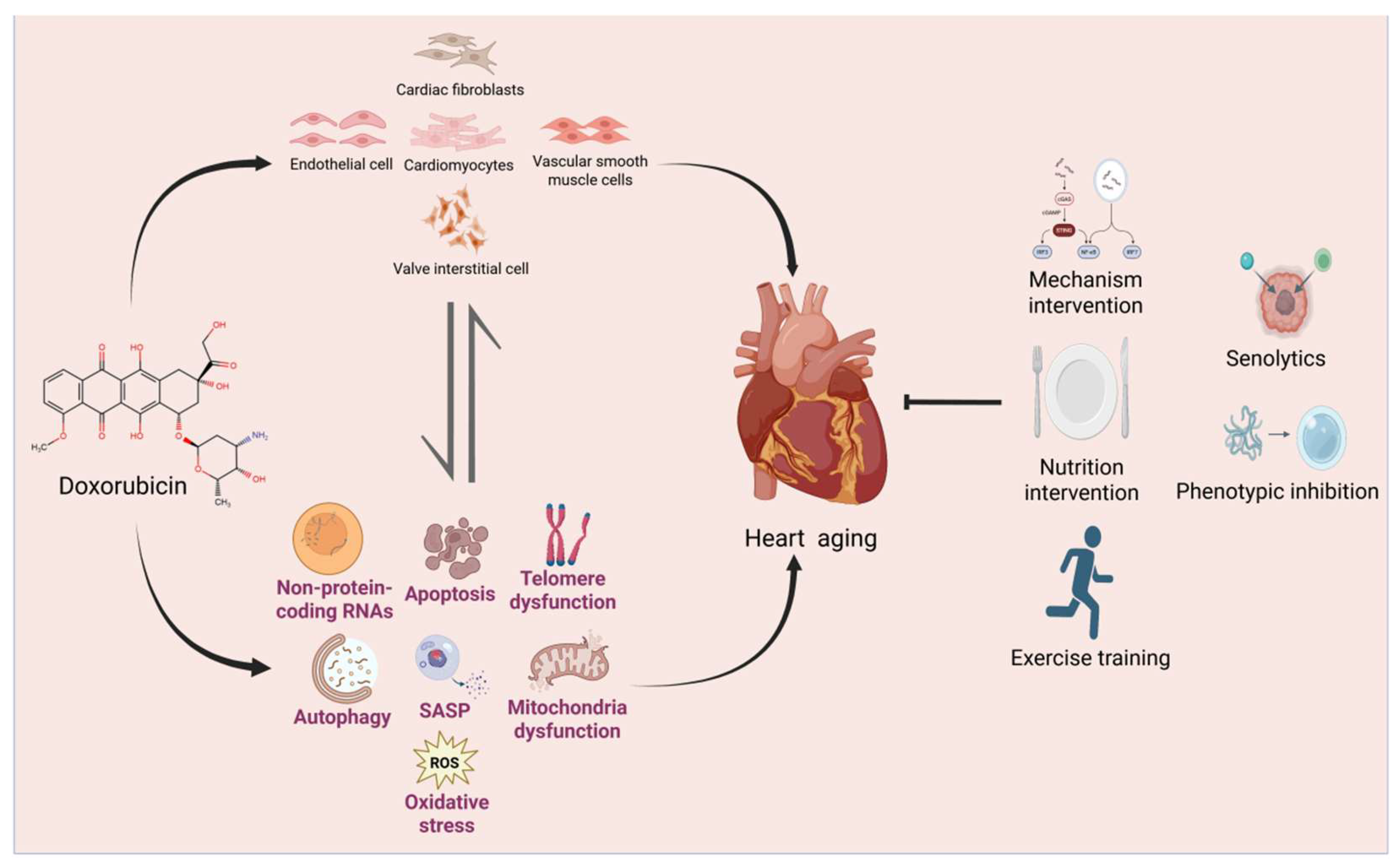
Figure 2.
Doxorubicin acts on different types of cardiac cells and induces cellular dysfunction and senescence through various mechanisms. (1)Cardiomyocytes: senescence of cardiomyocytes can lead to arrhythmia, dilated cardiomyopathy, and inefficient cardiac contraction. The process of doxorubicin-induced cardiomyocyte senescence includes the following aspects: acetylation of p53 promotes the transcription of p21, resulting in cell cycle arrest; imbalance of mitochondrial dynamics mediated by the PI3K/AKT/NOS-3 signaling pathway, with up-regulated expression of Drp1 protein and down-regulated expression of TRF1 and TRF2; activation of C5aR, which shortens telomere length and reduces telomerase activity; release of inflammatory factors such as NF-κB, TNF-α, IL-1β, and IL-6; activation of the NLRP3 inflammasome by the TXNIP . (2) Cardiac fibroblasts: upon transformation into myofibroblasts, they directly activates a profibrotic phenotype, resulting in excessive extracellular matrix deposition and cardiac fibrosis. The main involved processes include regulation of miR-17 and its target Par-4 protein; the forkhead factor Foxo-3 interacting with ID-3, E2F1, FAK, and HIF1α; as well as increased protein levels of cyclooxygenase-2 (COX2) and Bcl-xL. (3) Endothelial cells: DOX-induced senescence in endothelial cells is mediated through JNK, p38 MAPK, and PI3K/AKT signaling pathways, resulting in upregulation of P16INK4A and SIRT1 protein expression. Endothelial cells can also differentiate into myofibroblasts through EndMT, thereby participating in the process of myocardial fibrosis. (4)Vascular smooth muscle cell: DOX treatment activates the p53/(p21, p16) pathway to induce cellular senescence, downregulates the expression of NOX4, promoting the secretion of Il-6 and Il-8. Cellular calcification is dependent on Nlrp-3 and its downstream target Il-1β, ultimately leading to atherosclerosis. (5)Valvular interstitial cells (VICs): cellular senescence is accompanied by downregulated microRNAs (miRs) including miR-17, miR-20a, miR-30d, and let-7c, along with elevated p21 levels and increased senescence-associated β-galactosidase (SA-β-gal) activity-ultimately driving valvular fibrosis and calcification. (6)Other cells: immune cells function as a double-edged sword in senescence. Macrophages exert partial senescent cell-clearing effects, whereas neutrophil-mediated inflammation exacerbates senescence. In contrast, mesenchymal stem cells (MSCs) differentiate into endothelial cells and vascular smooth muscle cells, alleviating cardiac senescence. The illustration was created by BioRender.com.
Figure 2.
Doxorubicin acts on different types of cardiac cells and induces cellular dysfunction and senescence through various mechanisms. (1)Cardiomyocytes: senescence of cardiomyocytes can lead to arrhythmia, dilated cardiomyopathy, and inefficient cardiac contraction. The process of doxorubicin-induced cardiomyocyte senescence includes the following aspects: acetylation of p53 promotes the transcription of p21, resulting in cell cycle arrest; imbalance of mitochondrial dynamics mediated by the PI3K/AKT/NOS-3 signaling pathway, with up-regulated expression of Drp1 protein and down-regulated expression of TRF1 and TRF2; activation of C5aR, which shortens telomere length and reduces telomerase activity; release of inflammatory factors such as NF-κB, TNF-α, IL-1β, and IL-6; activation of the NLRP3 inflammasome by the TXNIP . (2) Cardiac fibroblasts: upon transformation into myofibroblasts, they directly activates a profibrotic phenotype, resulting in excessive extracellular matrix deposition and cardiac fibrosis. The main involved processes include regulation of miR-17 and its target Par-4 protein; the forkhead factor Foxo-3 interacting with ID-3, E2F1, FAK, and HIF1α; as well as increased protein levels of cyclooxygenase-2 (COX2) and Bcl-xL. (3) Endothelial cells: DOX-induced senescence in endothelial cells is mediated through JNK, p38 MAPK, and PI3K/AKT signaling pathways, resulting in upregulation of P16INK4A and SIRT1 protein expression. Endothelial cells can also differentiate into myofibroblasts through EndMT, thereby participating in the process of myocardial fibrosis. (4)Vascular smooth muscle cell: DOX treatment activates the p53/(p21, p16) pathway to induce cellular senescence, downregulates the expression of NOX4, promoting the secretion of Il-6 and Il-8. Cellular calcification is dependent on Nlrp-3 and its downstream target Il-1β, ultimately leading to atherosclerosis. (5)Valvular interstitial cells (VICs): cellular senescence is accompanied by downregulated microRNAs (miRs) including miR-17, miR-20a, miR-30d, and let-7c, along with elevated p21 levels and increased senescence-associated β-galactosidase (SA-β-gal) activity-ultimately driving valvular fibrosis and calcification. (6)Other cells: immune cells function as a double-edged sword in senescence. Macrophages exert partial senescent cell-clearing effects, whereas neutrophil-mediated inflammation exacerbates senescence. In contrast, mesenchymal stem cells (MSCs) differentiate into endothelial cells and vascular smooth muscle cells, alleviating cardiac senescence. The illustration was created by BioRender.com.
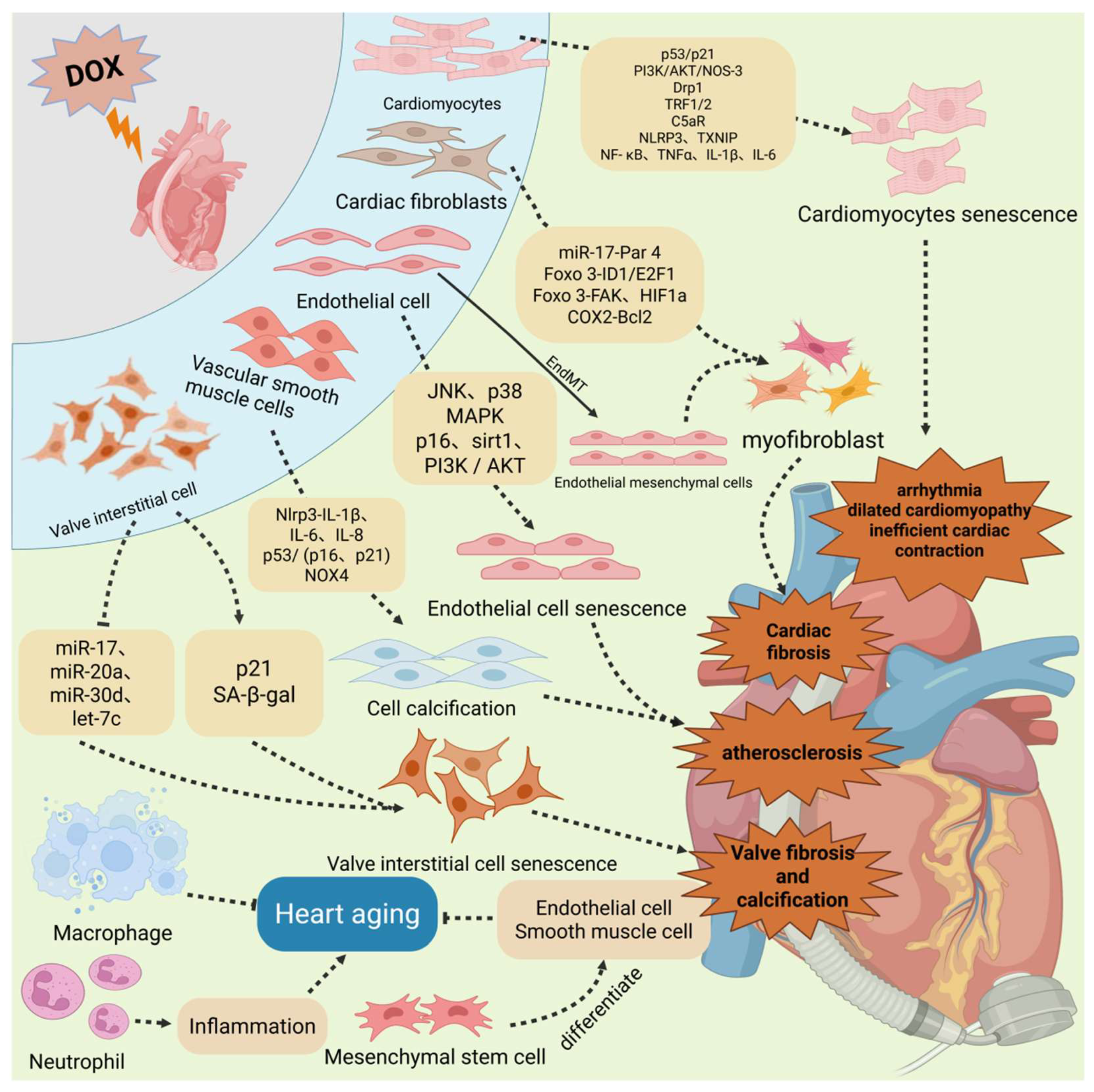
Figure 3.
Doxorubicin impairs cardiomyocyte function through three primary pathways: inflammation, telomere dysfunction, and disrupted mitochondrial dynamics. (1) Inflammation: DOX induces oxidative stress by promoting ROS accumulation. It upregulates TXNIP expression, activates the NLRP3 inflammasome, and stimulates the release of pro-inflammatory factors (NF-κB, TNF-α, IL-1β, IL-6). DOX also activates complement component C5a, further amplifying ROS production and cardiomyocyte injury. This cascade can be significantly mitigated by the C5a receptor antagonist (C5aRA). (2) Telomere Dysfunction: DOX triggers telomere impairment by promoting p53 phosphorylation and upregulating p21 and p16, leading to cell cycle arrest. Concurrent downregulation of Sirt6 and telomere-binding factors TRF1/2 further exacerbates telomere dysfunction. (3) Mitochondrial Dynamics: DOX enhances mitochondrial fission by promoting phosphorylation of the fission protein Drp1 at Ser616. Additionally, DOX-induced downregulation of Sirt6 suppresses expression of fusion proteins MFN1/2 and OPA1, collectively leading to mitochondrial fragmentation. The illustration was created by BioRender.com.
Figure 3.
Doxorubicin impairs cardiomyocyte function through three primary pathways: inflammation, telomere dysfunction, and disrupted mitochondrial dynamics. (1) Inflammation: DOX induces oxidative stress by promoting ROS accumulation. It upregulates TXNIP expression, activates the NLRP3 inflammasome, and stimulates the release of pro-inflammatory factors (NF-κB, TNF-α, IL-1β, IL-6). DOX also activates complement component C5a, further amplifying ROS production and cardiomyocyte injury. This cascade can be significantly mitigated by the C5a receptor antagonist (C5aRA). (2) Telomere Dysfunction: DOX triggers telomere impairment by promoting p53 phosphorylation and upregulating p21 and p16, leading to cell cycle arrest. Concurrent downregulation of Sirt6 and telomere-binding factors TRF1/2 further exacerbates telomere dysfunction. (3) Mitochondrial Dynamics: DOX enhances mitochondrial fission by promoting phosphorylation of the fission protein Drp1 at Ser616. Additionally, DOX-induced downregulation of Sirt6 suppresses expression of fusion proteins MFN1/2 and OPA1, collectively leading to mitochondrial fragmentation. The illustration was created by BioRender.com.
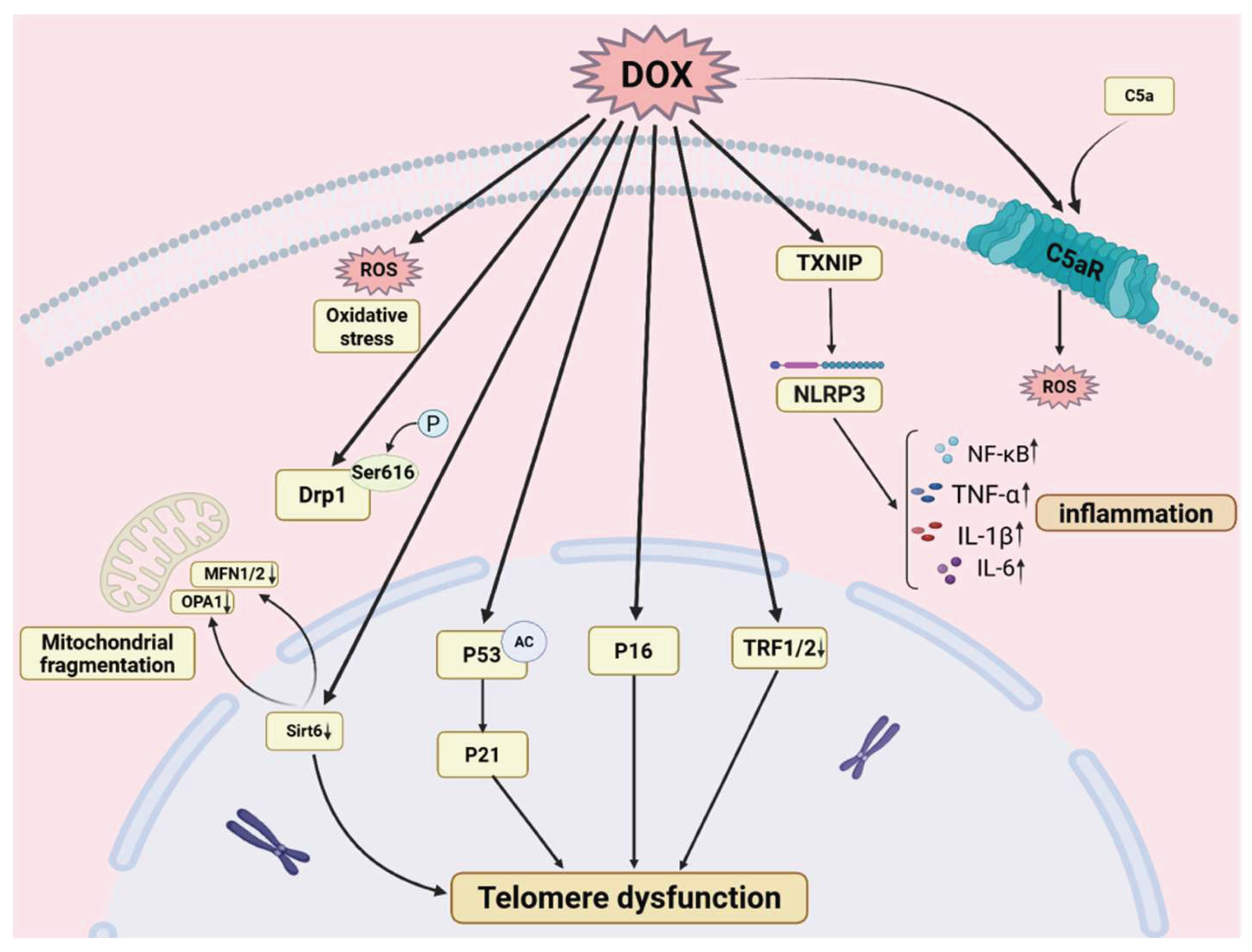
Figure 4.
4. DOX-induced cardiac aging involves multiple mechanisms, mainly including: (1) Oxidative stress: ROS/RNS-mediated oxidation-antioxidant imbalance, which is a key mechanism. (2) Telomere dysfunction: DOX disrupts telomere homeostasis (telomere shortening, reduced telomerase activity, TRF1/TRF2 downregulation) to activate DNA damage response(DDR), inducing cardiomyocyte cycle arrest and senescence. (3) Mitochondrial dysfunction: Doxorubicin perturbs mitochondrial fission-fusion dynamics and metabolic function, underlying cardiac aging. (4) SASP: low-dose DOX induces senescence-associated secretory phenotype(SASP) and vascular smooth muscle cell (VSMC) calcification, while targeting SASP alleviates DOX-induced cardiac-related cell senescence. (5) Non-coding RNAs: circ-Foxo3, lincRNA-p21, miR-199a-3p, etc., modulate cardiac aging via downstream target gene/pathway regulation. (6) Autophagy: Sirt3/Parkin-mediated autophagy/mitophagy activation mitigates DOX-induced cardiac aging by enhancing autophagic flux and inhibiting PI3K/AKT/mTOR. (7)Apoptosis: DOX induces dose-dependent apoptosis. High doses trigger direct apoptosis, while lower doses lead to senescence followed by late cell death via mitotic catastrophe. Inhibiting apoptosis alleviates DOX-induced cardiac aging. The illustration was created by BioRender.com.
Figure 4.
4. DOX-induced cardiac aging involves multiple mechanisms, mainly including: (1) Oxidative stress: ROS/RNS-mediated oxidation-antioxidant imbalance, which is a key mechanism. (2) Telomere dysfunction: DOX disrupts telomere homeostasis (telomere shortening, reduced telomerase activity, TRF1/TRF2 downregulation) to activate DNA damage response(DDR), inducing cardiomyocyte cycle arrest and senescence. (3) Mitochondrial dysfunction: Doxorubicin perturbs mitochondrial fission-fusion dynamics and metabolic function, underlying cardiac aging. (4) SASP: low-dose DOX induces senescence-associated secretory phenotype(SASP) and vascular smooth muscle cell (VSMC) calcification, while targeting SASP alleviates DOX-induced cardiac-related cell senescence. (5) Non-coding RNAs: circ-Foxo3, lincRNA-p21, miR-199a-3p, etc., modulate cardiac aging via downstream target gene/pathway regulation. (6) Autophagy: Sirt3/Parkin-mediated autophagy/mitophagy activation mitigates DOX-induced cardiac aging by enhancing autophagic flux and inhibiting PI3K/AKT/mTOR. (7)Apoptosis: DOX induces dose-dependent apoptosis. High doses trigger direct apoptosis, while lower doses lead to senescence followed by late cell death via mitotic catastrophe. Inhibiting apoptosis alleviates DOX-induced cardiac aging. The illustration was created by BioRender.com.
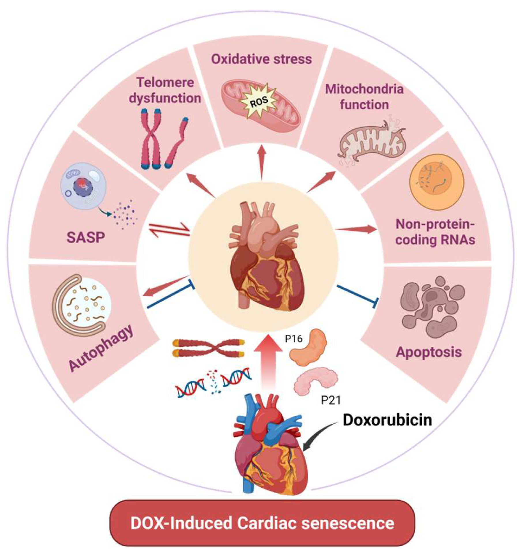
Disclaimer/Publisher’s Note: The statements, opinions and data contained in all publications are solely those of the individual author(s) and contributor(s) and not of MDPI and/or the editor(s). MDPI and/or the editor(s) disclaim responsibility for any injury to people or property resulting from any ideas, methods, instructions or products referred to in the content. |
© 2026 by the authors. Licensee MDPI, Basel, Switzerland. This article is an open access article distributed under the terms and conditions of the Creative Commons Attribution (CC BY) license (http://creativecommons.org/licenses/by/4.0/).
Copyright: This open access article is published under a Creative Commons CC BY 4.0 license, which permit the free download, distribution, and reuse, provided that the author and preprint are cited in any reuse.


