
Submitted:
06 July 2026
Posted:
07 July 2026
You are already at the latest version
Abstract
Background: Minimally invasive cardiac valve surgery (MIVS) is established as a safe alternative to conventional sternotomy, yet the technical complexity of annular suturing remains a significant barrier to procedural efficiency. Automated annular suturing devices (RAM®) have been introduced to mechanize and standardize suture placement specifically during valve replacements. This study evaluates the clinical feasibility, safety, and device usability of automated suturing during its institutional introductory phase. Methods: We retrospectively reviewed 43 consecutive patients undergoing MIVS supported by the RAM® system between 2021 and 2025. The cohort was stratified into RAM-supported Mitral Valve Replacements (RAM-MVR, n=26) and Aortic Valve Replacements (RAM-AVR, n=17). Technical parameters, device configurations, and perioperative outcomes were descriptively analyzed and contextualized within our mature institutional percutaneous MIVS benchmark platform (n=182). Results: Automated annular suturing achieved 100% technical feasibility with zero mechanical malfunctions or suture-line failures. In the RAM-MVR group, the median aortic cross-clamp and cardiopulmonary bypass (CPB) times were 73.0 minutes (IQR 64.0–91.0) and 114.5 minutes (IQR 94.8–129.5), respectively. RAM-AVR cases demonstrated a median cross-clamp time of 58.0 minutes (IQR 50.0–63.0). Conclusion: Automated annular suturing represents a robust and predictable technology that integrates seamlessly into a mature percutaneous MIVS environment. The system provides reliable standardization of valve anchoring across varying positions, yielding stable procedural times and excellent hemodynamic performance without introducing device-specific morbidity.
Keywords:
minimally invasive surgery
; percutaneous cannulation
; mitral valve surgery
1. Introduction
Minimally invasive cardiac valve surgery (MIVS) has evolved over the past two decades into a well-established and robust alternative to conventional median sternotomy for selected patients requiring mitral or aortic valve interventions [1,2,3,4,5]. Since the pioneering work of Cohn et al., which demonstrated that limited-access valve surgery significantly improved patient satisfaction and reduced costs, the field has transitioned toward increasingly sophisticated techniques. Numerous systematic reviews, meta-analyses, and large-scale registry studies have confirmed that minimally invasive approaches achieve clinical outcomes and safety profiles comparable to conventional surgery while offering distinct physiological advantages [6,7]. These benefits include reduced surgical trauma, shorter hospital stays, accelerated postoperative recovery, and superior cosmetic results. Consequently, MIVS has been increasingly adopted as the standard of care for both isolated and selected combined valve procedures in high-volume centers, or even in patients with thoracic malformations [8].
Despite these clear clinical advantages, MIVS remains technically demanding and the reliance on restricted access, deep operative fields, and long-shafted instrumentation—often coupled with endoscopic visualization—substantially increases procedural complexity [3]. These factors place high ergonomic and cognitive demands on the surgical team, which can impact workflow efficiency. Challenges are most pronounced during the annular suturing phase, a step that requires absolute precision in needle placement and controlled tissue handling within confined anatomical spaces. Unlike conventional surgery, where the surgeon has direct 3D visualization and manual dexterity, MIVS often necessitates specialized suturing techniques to ensure the long-term stability of the prosthesis and to prevent paravalvular leaks [4]. Because of these constraints, prolonged cardiopulmonary bypass (CPB) and aortic cross-clamp times are frequently reported, particularly during the developmental phase of a minimally invasive program [9,10,11,12].
The clinical implications of operative duration cannot be overstated. Prolonged ischemic and bypass times are major concerns in cardiac surgery, as aortic cross-clamp duration has been identified as an independent predictor of morbidity and mortality across diverse patient populations [9,10,11,13]. Strategies aimed at stabilizing or reducing these operative times without compromising procedural safety or repair quality are therefore of paramount interest. In this context, technological innovation has played a central role in mitigating the technical hurdles of limited-access surgery. For instance, automated knot-fastening devices have been widely adopted in both minimally invasive and robotic valve surgery, with several meta-analyses associating their use with significant reductions in cross-clamp and CPB times [7,14,15,16]. Building upon the success of mechanized knot-tying, automated annular suturing devices—such as the RAM® system—have been developed to further mechanize the procedure by automating the placement of the annular sutures themselves [15,17].
The RAM® device is designed to improve reproducibility, reduce ergonomic strain, and enhance sharps safety by automating a traditionally manual and high-risk step. Early feasibility studies and technical descriptions have demonstrated that these devices can be safely applied in both minimally invasive aortic and mitral valve surgery. Recent reports have even described the successful implementation of automated suturing within fully endoscopic workflows, emphasizing its role in standardizing operative integration [18]. However, a significant gap remains in the literature. Most published studies on automated suturing focus on selected patient cohorts, single valve positions, or purely technical descriptions. Furthermore, while the term “usability” is frequently cited, it is rarely operationally defined or assessed in the context of real-world clinical practice. In a routine surgical setting, usability encompasses not only technical success but also seamless workflow integration, reproducibility across varying anatomies, and the ability to apply technology consistently without introducing new sources of error.
At the Passau Heart Center, the development of MIVS has been characterized by a decade of structured maturation [19]. Our program has documented a systematic transition from traditional open femoral cut-down to ultrasound-guided percutaneous cannulation supported by advanced technologies such as the MANTA closure device [20,21,22,23]. This “institutional evolution” has resulted in measurable improvements in procedural efficiency, including significantly shorter CPB and total operative times, as well as reduced ICU and hospital stays. Perhaps most importantly, the adoption of percutaneous techniques has virtually eliminated lymphatic morbidity, which was a common complication prior to 2021, when the groin cannulation was performed via cut-down technique (20). Our center’s experience indicates that MIVS programs mature not through the simplification of surgery, but through the progressive acquisition of confidence in advanced techniques and the standardization of team-based workflows.
As part of this ongoing maturation, we have integrated automated annular suturing into our surgical armamentarium. While our broader MIVS program has seen an increase in the complexity of reconstructive mitral repairs—including high rates of combined annuloplasty and neochordae implantation—the RAM® device has been utilized specifically for mitral and aortic valve replacements. This specialized focus allows for a unique analysis of how mechanization influences the most standardized form of valve intervention. Given that our center has already established a high-performing program for MIVS, it is essential to evaluate new technologies not in isolation, but against this modern institutional benchmark.
The objective of the present study is to provide a comprehensive descriptive analysis of a single-center consecutive experience using an automated annular suturing device in minimally invasive mitral and aortic valve replacement. By defining usability pragmatically through technical feasibility, documentation completeness, and operative efficiency metrics, we aim to demonstrate how automated suturing integrates into an already matured percutaneous MIVS program. This analysis captures the institutional introduction of mechanized suturing, evaluating whether procedural stability and the quality standards established during our decade of program development are maintained. By profiling the RAM® cohort within the context of our broader institutional benchmark, this study provides real-world data to optimize workflows in mechanized valve surgery.
2. Materials and Methods
2.1. Study Design, Population and Ethical Considerations
This retrospective, single-center descriptive observational study was conducted at the Passau Heart Center. The analysis focused on 43 consecutive patients undergoing minimally invasive cardiac valve surgery (MIVS) supported by the RAM® automated annular suturing device. To provide institutional context, this cohort was benchmarked against the broader SoC cohort of the last 4 years (2021–2025) of the center, which included 182 patients treated with standardized ultrasound-guided groin access [20]. The study protocol for the longitudinal experience was approved by the Ethics Committee of the University of Regensburg (protocol code 25-4426-104, approval date is 3 December 2025), while formal approval for the specific anonymized RAM usability analysis was waived according to local regulations.
All patients undergoing MIVS during the study period were identified via the institutional surgical database. Inclusion required undergoing isolated mitral or aortic valve replacement performed through a right-sided minimally invasive access. Routine preoperative evaluation included CT angiography to assess the suitability of femoral vessels for peripheral cannulation. Specific exclusion criteria for peripheral access included a femoral artery diameter of less than 6 mm, significant anterior calcification at the intended puncture site, or the presence of aortic thrombosis.
2.2. Surgical Technique
All procedures were performed by a single experienced surgeon under general anesthesia with continuous intraoperative transesophageal echocardiographic guidance. A 4–5 cm right mini-thoracotomy in the 3th or 4th intercostal space was used for both mitral and aortic valve procedures. Cardiopulmonary bypass was established via percutaneous femoral cannulation using a 19- or 21-Fr Bio-Medicus™ arterial cannula (Medtronic, Minneapolis, MN, USA) and a SMART Cannula® for venous drainage (Smartcanula LLC, Lausanne, Switzerland) (Figure 1).
After initiation of cardiopulmonary bypass, the ascending aorta was cross-clamped using a Chitwood transthoracic clamp. Myocardial protection was achieved with a single antegrade dose of 1800 mL histidine–tryptophan–ketoglutarate (HTK; Bretschneider) cardioplegia administered over approximately 6 min. Carbon dioxide insufflation of the operative field and transesophageal echocardiographic guidance were routinely used throughout the procedure. Exclusively biological prosthetic valves were implanted in all patients.
Minimally Invasive Mitral Valve Replacement
Mitral valve replacement was performed through a right anterolateral mini-thoracotomy in the fourth intercostal space. After cardioplegic arrest, the left atrium was opened through Sondergaard’s groove, and the mitral valve was exposed using a dedicated atrial retractor. The native mitral valve was excised while preserving the subvalvular apparatus whenever technically feasible.
Interrupted pledgeted 2-0 braided polyester annular sutures were placed circumferentially around the mitral annulus using the RAM® 5.0 automated suture fastening device (LSI Solutions, Victor, NY, USA) (Figure 2).
After placing the sutures, all sutures where passed through the prosthetic valve using the SEW-EASY Device 5.0 (LSI Solutions, Victor, NY, USA) (Figure 3).
Then the prosthesis was deployed in the mitral annulus and the sutures ehere tightened with CorKnot (LSI Solutions, Victor, NY, USA). The device deploys a titanium fastener while simultaneously trimming the excess suture material, eliminating the need for manual knot tying. Following confirmation of appropriate prosthesis seating, the left atrium was closed in the standard fashion. After de-airing and removal of the aortic cross-clamp, valve function was assessed by intraoperative transesophageal echocardiography.
Minimally Invasive Aortic Valve Replacement
Minimally invasive aortic valve replacement was performed through the same right mini-thoracotomy approach. Following cardioplegic arrest, an oblique aortotomy was performed, and the diseased native aortic valve was excised. Annular decalcification was carried out as required to allow optimal prosthesis implantation.
Interrupted pledgeted 2-0 braided polyester sutures were placed circumferentially around the aortic annulus in the similar matter as for the MVRs and secured with the CorKnot (LSI Solutions, Victor, NY, USA) automated suture fastening device. The aortotomy was subsequently closed in the standard fashion. Following meticulous de-airing, the aortic cross-clamp was removed, and prosthetic valve function was confirmed by intraoperative transesophageal echocardiography before weaning from cardiopulmonary bypass.
2.3. Data Collection and Variables
Data were extracted from the institutional surgical database and reviewed manually. Variables included: Baseline characteristics: Age, sex, obesity (BMI > 30 kg/m2), EuroSCORE II, pre-operative NYHA class, COPD, chronic dialysis, and prior cardiac surgery (any previous commissurotomy, Harpoon, mitral valve replacement, bypass, aortic valve replacement, or prior sternotomy). Operative variables: Cardiopulmonary bypass time, aortic cross-clamp time, total operation time, ICU stay, hospital stay, and need for re-exploration for bleeding. Access-site and device outcomes: Groin complications, lymph fistula, wound healing disorder, use of MANTA closure, MANTA-related complications (ischemia or bleeding). Mortality: <30-day and >30-day mortality. Groin complications were defined as a composite endpoint including access-site hematoma requiring intervention, pseudoaneurysm, arterial or venous thrombosis, limb ischemia, access-site infection, seroma, or need for surgical or endovascular vascular intervention. Lymph fistula and wound-healing disorders were analyzed separately, as lymphatic injury represents a distinct mechanism related to surgical dissection.
2.4. Statistical Analysis
All data were extracted from the institutional database and reviewed manually. Continuous variables demonstrated a non-normal distribution and were therefore expressed as median and interquartile range (IQR). Categorical variables were presented as absolute counts and percentages. All analyses were performed using Python 3.13 with pandas and SciPy libraries.
3. Results
3.1. Patient Population and Baseline Characteristics
The analyzed study population comprises the introductory consecutive cohort of automated annular suturing at our institution. A total of 43 patients underwent minimally invasive valve surgery (MIVS) supported by the RAM® system. This mechanized cohort was stratified by anatomical position into a mitral valve replacement group (RAM-MVR, n = 26) and an aortic valve replacement group (RAM-AVR, n = 17). To contextualize these groups within our modern standardized workflow, they are presented alongside the overarching institutional standard of care (SoC) benchmark cohort (n = 182).
The baseline demographics and clinical profiles of the automated groups demonstrated characteristics typical of contemporary limited-access valve replacement candidates. In the RAM-MVR subgroup, the median age was 67.0 years (IQR 56.0–74.0), which was identical to the median age of 67.0 years (IQR 59.5–74.0) observed in the RAM-AVR group, and closely aligned with the institutional benchmark population (median 65.0 years, IQR 57.0–74.0). Female patients accounted for 53.8% (14/26) of the mitral cohort and 17.6% (3/17) of the aortic cohort, compared to 32.4% (59/182) in the overarching institutional benchmark.
Preoperative risk scores and symptomatic severity reflected distinct clinical patterns between the two anatomical categories. The median EuroSCORE II was higher in the mitral cohort at 2.61% (IQR 1.03–4.90) than in the aortic cohort, which presented with a median score of 0.92% (IQR 0.76–1.30). Preoperative functional impairment was highly pronounced among the mitral patients, with a median NYHA functional class of 3 (IQR 2–3), and 58.1% of the RAM-MVR cohort presenting in NYHA class III or IV. The RAM-AVR cohort exhibited a descriptive lower symptomatic burden, with a median NYHA class of 2 (IQR 1–3). Preoperative left ventricular ejection fraction (LVEF) was stable and well-preserved across both active automated groups, showing a median of 56.5% (IQR 55.0–60.0%) in the mitral replacement patients and 60.0% (IQR 55.0–60.0%) in the aortic replacement patients. Baseline demographics are summarized in Table 1.
3.2. Operative Metrics and Procedural Efficiency
Procedural characteristics for the automated cohort demonstrated highly consistent performance profiles across both positions. For patients undergoing mitral valve replacement (RAM-MVR), the median aortic cross-clamp duration was 73.0 min (IQR 64.0–91.0), with an associated total cardiopulmonary bypass time of 114.5 min (IQR 94.8–129.5). Total surgical skin-to-skin time for the mitral cohort was 201.0 min (IQR 184.8–226.3). In comparison, the aortic subgroup (RAM-AVR) exhibited median cross-clamp and CPB times of 58.0 min (IQR 50.0–63.0) and 92.0 min (IQR 86.0–108.0), respectively, translating to a total operative duration of 171.0 min (IQR 160.0–191.0). These timeframes aligned well within the overall baseline of the institution’s mature percutaneous MIVS experience., as seen in Table 2.
3.3. Learning Curve and Procedural Maturation Analysis
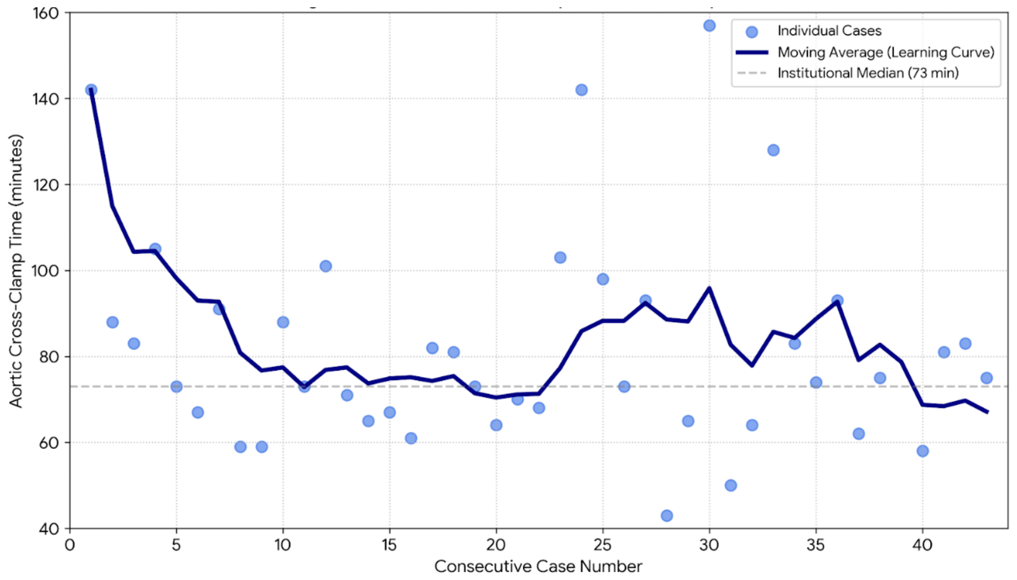
There was a reduction in aortic cross-clamp times. While early cases frequently required over 90 min of ischemic time, the cross-clamp times reached a stable plateau of approximately 70–75 min following the completion of the first 20 cases. These findings confirm a rapid institutional maturation, with the “safety learning curve” for the automated device stabilizing after approximately 18 cases for mitral and 14 cases for aortic replacements.
Figure 2.
Learning Curve of Aortic Cross-Clamp Times during RAM Implementation.
3.4. Device Configuration and Usability
The pragmatic usability of the mechanization system was reflected in a 100% technical success rate, with no conversions to manual suturing or structural device failures noted. Complete data documentation regarding active jaw components was achieved in 88.4% of cases. The deployment of specific jaw sizes followed clear anatomical requirements. In the mitral position, the larger 5.0 mm jaw configuration was used almost exclusively (96.0% of cases) to facilitate deeper tissue incorporation within the voluminous mitral annulus. Conversely, the aortic position demanded a predominant utilization of the smaller 3.5 mm jaw profile (71.0%), allowing optimized maneuverability within the confined sub-aortic space. No suture line disruption, paravalvular leakage, or prosthesis misalignments were recorded intraoperatively or predischarge, as seen in Table 3.
3.6. Postoperative Outcomes
The 30-day mortality for the RAM series was 4.7%. No paravalvular leaks or suture dehiscence were recorded. Median ICU and hospital stays were 2.0 and 8.0 days, respectively.
Hemodynamic performance was a key secondary endpoint to ensure that the mechanization of the annular suturing did not compromise the effective orifice area or the valve seating. In the RAM-MVR group, the median postoperative mean pressure gradient was 3.0 mmHg (IQR 2.0–4.0), demonstrating excellent, preserved hemodynamic performance compared to the institutional benchmark (3.5 mmHg, IQR 3.0–4.8). For the RAM-AVR group, the median mean gradient was 8.0 mmHg (IQR 6.0–10.0), confirming excellent procedural quality and valve alignment across all automated cases, as seen in Table 4.
Postoperative cardiac function remained stable (median LVEF 55% across all cohorts), and recovery metrics showed no descriptive difference in ICU or hospital stay compared to the institutional benchmark.
4. Discussion
The integration of automated annular suturing technology into minimally invasive mitral and aortic valve surgery represents a strategic pivot toward procedural standardization at our institution. By profiling the introductory cohort of the RAM® device within our mature clinical environment, the primary findings demonstrate that automated suturing is not only highly feasible but achieves excellent operative efficiency that aligns seamlessly with established institutional standards from the early adoption phase.
4.1. Procedural Efficiency and Workflow Integration
The clinical introduction of the RAM® automated annular suturing device at our institution demonstrated highly stable perioperative metrics that integrated effectively into our standardized percutaneous MIVS program. In limited-access cardiac surgery, the aortic cross-clamp duration is widely considered the primary indicator of intracardiac technical proficiency and surgical workflow efficiency [9,10,13]. In our initial series of mechanized cases, the RAM-MVR cohort achieved a median cross-clamp time of 73.0 min (IQR 64.0–91.0). This profile indicates that the automated approach provides predictable and reproducible surgical timeframes from the early stages of its adoption curve.
Mechanizing the placement of annular sutures appears to streamline the procedural steps within the deep, restricted operative field of the mitral valve. By transitioning from manual needle handling to automated deployment, the surgical team bypasses the ergonomic challenges associated with working through limited port access [15,16]. This consistency is also reflected in the median CPB times for the RAM-MVR group (114.5 min, IQR 94.8–129.5), which aligned with the established institutional benchmark of 115.0 min (IQR 98.0–141.0) representing our mature percutaneous platform. The automated system acts as a reliable technical standardizer, stabilizing operative duration during the implementation of a new device workflow [13].
4.2. Optimization of Operative Efficiency and Technical Maturation
In our series, the cumulative average cross-clamp time for RAM-supported cases followed a distinct downward trajectory, achieving institutional stabilization after the initial 15–20 cases. This maturation period is consistent with the learning curves observed in other centers adopting automated suturing technologies, where the initial “overhead” of device preparation is gradually offset by the speed of mechanized suture deployment.
The observed median cross-clamp time of 73.0 min in the RAM-MVR group demonstrates remarkable procedural stability from the outset. In the context of MIVS, where myocardial ischemia is linearly associated with a systemic inflammatory response, achieving reliable clamp times well within safe clinical boundaries provides a critical safety margin. The efficiency and consistency of the workflow are further reflected in the total surgical duration, with the RAM-MVR group requiring a median of 201.0 min. This confirms that the initial setup of the automated device does not introduce disruptive operational delays.
This stabilization of operative times is not merely a reflection of the surgeon’s manual dexterity but rather an institutional team maturation. We identified one critical factor that define this efficiency gain. The RAM® system requires a specific coordination between the surgeon, the assistant and the scrub nurse. Unlike manual suturing, where each stitch is managed individually, the automated device handles dual-needle deployment. As the assistant becomes familiar with the tensioning and organization of the pre-placed sutures, the interval between individual deployments shrinks significantly.
By standardizing the most technically variable portion of the operation—the annular suturing—the RAM® system acts as a mechanized bridge to proficiency. It allows the surgical team to bypass the prolonged “trial and error” phase typically associated with mastering manual suturing in MIVS, ultimately aligning the procedural speed with our established Institutional Benchmark.
4.3. Anatomical and Risk Considerations: Mitral vs. Aortic
The baseline analysis reveals distinct clinical and risk profiles between the two active automated cohorts. The RAM-AVR subgroup represented a low-risk surgical population with a median EuroSCORE II of 0.92% (IQR 0.76–1.30). In contrast, the mitral replacement cohort exhibited a higher risk profile, with a median EuroSCORE II of 2.61% (IQR 1.03–4.90). This descriptive variance reflects clinical reality, as patients requiring mitral valve replacement frequently present with advanced structural disease, illustrated by the 58.1% of RAM-MVR patients presenting in severe NYHA functional class III or IV.
Despite these divergent preoperative risk profiles and the higher technical demands inherent to the mitral position, the mechanized operative metrics remained consistent across both subgroups. The device demonstrated operational versatility in adapting to these differing anatomical requirements. The mitral annulus, typically being more voluminous, required a larger tissue bite to ensure stable anchoring of the prosthetic sewing cuff, which explains the significant institutional preference for the 5.0 mm jaw configuration (utilized in 96.0% of mitral cases) [29]. Conversely, the restricted space of the aortic root was effectively managed using the smaller 3.5 mm jaw configuration in 71.0% of the cases, which allowed for optimized maneuverability around the coronary ostia without compromising suture lines. This split-profile utilization highlights the necessity of anatomical-specific device selection to ensure optimal clinical integration.
4.5. Synergy with the Institutional Benchmark
The successful implementation of automated suturing was inextricably linked to our center’s broader transition to the SoC cohort [20,23]. As documented in our 10-year experience, the adoption of ultrasound-guided femoral access and the MANTA® closure device revolutionized our MIVS program by eliminating lymphatic morbidity and standardizing vascular access [20,23].
By the time the RAM device was introduced, the “access trauma” and “access anxiety” had been largely resolved. This allowed the surgical team to focus exclusively on the intracardiac portion of the procedure. The RAM device fits perfectly into this philosophy of procedural standardization. Just as ultrasound guidance removed the variability of femoral access, automated suturing removes the variability of manual annular needle placement. The cumulative effect of these technologies is a more “industrialized” and reproducible surgical workflow, which is essential for the continued expansion of minimally invasive programs.
A critical facilitator for the successful rollout of the RAM® device was the pre-existing institutional proficiency in a standardized percutaneous workflow. Having already transitioned through a decade-long maturation—moving from open femoral cut-down to ultrasound-guided access and routine use of the MANTA® closure device—the surgical team operated within a stable procedural platform [19,21,22,24]. This maturation effectively removed the technical ‘noise’ and morbidity associated with vascular access, such as lymphatic complications or groin infections. Consequently, the team could dedicate its full cognitive and technical resources to the intracardiac mechanized suturing process, which likely explains why immediate parity in cross-clamp times was achieved despite the introduction of a new technology.
4.6. Technical Considerations and Usability
Pragmatic usability was confirmed by the high documentation rate (88.4%) and the fact that the device was used successfully in every intended case. The surgeon’s ability to choose between 3.5 mm and 5.0 mm jaws allows for flexibility across different annuli. The preference for the larger jaw in mitral cases is consistent with the need to capture more robust tissue in the mitral ring compared to the often calcified or more delicate aortic annulus [29,32]. The lack of any paravalvular leaks or suture dehiscence in the RAM cohort further supports the technical reliability of mechanized annular fixation.
The implementation of automated annular suturing shifted the procedural demands from a purely surgeon-centric task to a highly coordinated ‘team-sport’ workflow. Unlike manual suturing, where the assistant primarily provides suction and suture tensioning, the RAM® device requires the assistant to actively manage the device ‘magazine’ and coordinate the sequential delivery of sutures within the restricted port space. We observed that the stabilization of the learning curve was as much dependent on the assistant’s proficiency in maintaining optimal suture trajectory as it was on the surgeon’s precision in jaw placement. This synchronization is critical to prevent suture entanglement, a common pitfall in deep-seated minimally invasive mitral replacements.
Our data revealed a statistically significant preference for the 5.0 mm jaw configuration in mitral replacements (96.0%) compared to the aortic position (p = 0.004). This selection is driven by the fundamental anatomical differences between the two valves. The mitral annulus, particularly in patients with chronic regurgitation, often presents as more voluminous and less calcified than the aortic annulus, necessitating a deeper ‘tissue bite’ to ensure stable anchoring of the prosthetic sewing cuff. In contrast, the 3.5 mm jaw was found to be sufficient for the often more restricted and calcified aortic annulus, where a smaller footprint allows for easier maneuverability around the coronary ostia without compromising suture security [32].
4.7. Visual Field, Device Ergonomics, and Port Usability
A critical component of implementing advanced mechanization inside restricted fields is the geometric footprint of the instrument itself. Because the automated suturing device possesses a larger shaft and tip profile than a conventional long-shafted manual needle holder, concerns regarding visual clutter or camera obstruction are valid.
In our experience, however, this spatial footprint did not hinder procedural visualization. The dual-needle mechanical design allows for a single, well-defined entry into the annulus, eliminating the repetitive, multi-angle shifting required by traditional manual suturing. While the device tips necessitate a deliberate and focused camera path, the workflow transitions from an isolated, surgeon-centric movement to a highly structured team routine. The visual presence of the device is offset by the rapidity and precision of its tissue engagement, eliminating “trial-and-error” parallax errors frequently encountered under standard endoscopic visibility.
4.8. Mortality
Regarding early safety outcomes, a descriptive difference in 30-day mortality between the RAM cohort (4.7%) and the broader institutional average (1.6%) has been found.
Crucially, a granular review of the causes of death in the RAM cohort revealed that both fatalities were unrelated to technical device performance or the automated suturing mechanism. One patient succumbed to septic shock, and the other to sepsis-induced multi-organ failure (MOV), both of which were rooted in the patients’ high-risk preoperative status (58% NYHA III/IV) rather than procedural complications.
4.9. Limitations
This study is limited by its retrospective design and the relatively small sample size of the individual subgroups. While the results show comparable operative metrics with a favorable numerical trend for automated suturing, the study was not powered to show statistical superiority in operative times. Finally, long-term follow-up is needed to ensure that mechanized suturing maintains the same durability as traditional hand-placed sutures over 5–10 years.
5. Conclusions
The integration of automated annular suturing at the Passau Heart Center represents a significant stride toward the complete standardization of minimally invasive valve surgery. By evaluating our consecutive series of 43 RAM®-supported procedures within the context of our mature percutaneous standard of care benchmark, this study provides definitive evidence that mechanization offers immediate efficiency parity and consistent procedural stability in limited-access environments. Our findings confirm the technical robustness of the technology, with a 100% feasibility rate and zero device malfunctions. Most importantly, the descriptive analysis demonstrated that automated suturing integrates smoothly into existing settings without an artificial learning-curve penalty [28].
This parity suggests that the ergonomic relief provided by mechanized suturing—particularly in the deep and restricted operative field of the mitral annulus—allows the surgical team to focus on procedural flow rather than the technical minutiae of needle handling.
The anatomical insights gained from this study further refine the application of the technology. When combined with the advancements of our SoC cohort specifically ultrasound-guided access and the use of the MANTA® closure device automated annular suturing completes a modern, “access-trauma-minimal” surgical workflow.
In conclusion, automated annular suturing is a safe, efficient, and highly reproducible method for minimally invasive mitral and aortic valve replacement. By providing a mechanized bridge that stabilizes operative times and procedural complications, the technology allows centers to reach institutional maturity more rapidly. While larger prospective trials are warranted to evaluate long-term paravalvular leakage and durability, these real-world data support the RAM® system as a potential cornerstone of the next generation of standardized, minimally invasive cardiac surgery [33,34].
Acknowledgments
The authors thank the surgical, anesthesia, perfusion, and intensive care teams of Klinikum Passau for their dedicated and continued support in developing and refining the minimally invasive mitral valve repair program. We also acknowledge the contributions of the nursing staff and physician assistants whose commitment to perioperative care made this work possible.
Author Contributions
Conceptualization, P.M. and R.B.; methodology, P.M, R.B. and J.S.; validation, P.M and K.B; formal analysis, M.M.H and R.B.; investigation, K.B. and R.B.; resources, K.B. and R.B.; data curation, J.S. and R.B writing—original draft preparation, J.S.; writing—review and editing, P.M. and R.B.; supervision, R.B: project administration, P.M. All authors have read and agreed to the published version of the manuscript.
Funding
No external funding was received for this study.
Informed Consent Statement
Informed consent was obtained from all subjects involved in the study. Written informed consent has been obtained from the patient(s) to publish this paper.
Conflicts of Interest
The authors declare no conflict of interest.
References
- Cohn, L.H.; et al. Minimally invasive mitral valve surgery: A multi-center experience with 707 patients. Ann. Surg. 1997. [Google Scholar] [PubMed]
- Schmitto, J.; Mokashi, S.; Cohn, L. Minimally-invasive valve surgery. J. Am. Coll. Cardiol. 2010, 56, 455–462. [Google Scholar] [CrossRef] [PubMed]
- Holubec, T.; et al. Minimally invasive cardiac surgery: State of the art and current challenges. Front. Cardiovasc Med. 2023. [Google Scholar] [CrossRef] [PubMed]
- Chitwood, W.R., Jr. Historical evolution of robot-assisted cardiac surgery: A 25-year journey. Ann. Cardiothorac. Surg. 2022. [Google Scholar] [CrossRef] [PubMed]
- Suri, R.M.; et al. Robotic mitral valve repair for all comers: Is it safe, effective and reproducible? J. Thorac. Cardiovasc Surg. 2013. [Google Scholar]
- Decker, S.; et al. Outcomes of minimally invasive vs. conventional mitral valve replacement. Sci. Rep. 2021. [Google Scholar]
- Gillinov, A.M.; et al. Early results of robotically assisted mitral valve surgery. J. Thorac. Cardiovasc Surg. 2018. [Google Scholar] [CrossRef] [PubMed]
- Harpa, M.M.; et al. Endoscopic Mitral Surgery in Noonan Syndrome—Case Report and Considerations. J. Clin. Med. 2025, 14, 583. [Google Scholar] [CrossRef] [PubMed]
- Al-Sarraf, N.; et al. Cross-clamp time is an independent predictor of mortality and morbidity in low- and high-risk cardiac patients. Int. J. Surg. 2011. [Google Scholar] [PubMed]
- Salis, S.; et al. Cardiopulmonary bypass duration is an independent predictor of morbidity and mortality after cardiac surgery. J. Cardiothorac. Vasc. Anesth. 2008. [Google Scholar] [CrossRef] [PubMed]
- Gaudino, M.; et al. Identification of a threshold for the association of aortic cross-clamp time with mortality and morbidity. JAMA Cardiol. 2021. [Google Scholar] [CrossRef] [PubMed]
- Harpa, M.M.; et al. Successful Treatment of Unilateral Pulmonary Edema as Minimally Invasive Mitral Valve Surgery Complication—Case Presentation. J. Clin. Med. 2024, 13, 7654. [Google Scholar] [CrossRef] [PubMed]
- Doenst, T.; et al. Cardiopulmonary bypass predicts late survival after mitral valve repair. J. Thorac. Cardiovasc Surg. 2002. [Google Scholar] [CrossRef]
- Salmasi, M.Y.; et al. Automated titanium suture fasteners in valvular surgery: A systematic review and meta-analysis. Interact. Cardiovasc Thorac. Surg. 2019. [Google Scholar] [CrossRef] [PubMed]
- Cody, A.J.; et al. Automated suture fasteners reduce operative times in minimally invasive mitral valve surgery. Innovations 2021. [Google Scholar] [CrossRef]
- Lee, R.; et al. Comparison of manual vs. automated suture fastening in robotic MIVS. J. Thorac. Cardiovasc Surg. 2018. [Google Scholar] [CrossRef] [PubMed]
- Gammie, J.S.; et al. Automated Annular Suturing: A technical description and early experience. Ann. Thorac. Surg. 2021. [Google Scholar] [CrossRef] [PubMed]
- Chen, Y.; et al. Totally endoscopic mitral valve surgery: Experience in 188 patients. J. Cardiothorac. Surg. 2021. [Google Scholar] [CrossRef] [PubMed]
- Yankah, C.A.; et al. Minimally invasive mitral valve surgery: Passau experience. Thorac. Cardiovasc Surg. 2013. [Google Scholar] [CrossRef]
- Schwarz, J.; Massoudy, P.; Harpa, M.M.; et al. Evolution of Femoral Cannulation Techniques in Minimally Invasive Mitral Valve Surgery: A 10-Year Experience. Med. Sci. 2026, 14, 182. [Google Scholar] [CrossRef] [PubMed]
- Schaefer, A.; Sarwari, H.; Reichenspurner, H.; Conradi, L. Plug-based vascular closure device for percutaneous femoral artery closure in minimally invasive valve surgery. Front. Cardiovasc Med. 2021, 8, 682321. [Google Scholar] [PubMed]
- Van Mieghem, N.; Latib, A.; van der Heyden, J. Plug-based arteriotomy closure device for large-bore access: Multicenter prospective study. JACC Cardiovasc Interv. 2017, 10, 613–619. [Google Scholar] [CrossRef] [PubMed]
- Moccetti, F.; et al. Safety and efficacy of the MANTA vascular closure device after percutaneous MIVS access. Catheter Cardiovasc Interv. 2021. [Google Scholar] [PubMed]
- Vora, A.N.; et al. US-guided femoral access in patients with vascular closure devices: The UNIVERSAL trial. JAMA 2023. [Google Scholar]
- Novick, R.J.; et al. CUSUM analysis for monitoring surgical performance. Ann. Thorac. Surg. 1999. [Google Scholar] [CrossRef]
- Holzhey, D.M.; Seeburger, J.; Misfeld, M.; Borger, M.A.; Mohr, F.W. Learning minimally invasive mitral valve surgery: A 10-year experience. Eur. J. Cardiothorac. Surg. 2013, 44, 1072–1077. [Google Scholar]
- Rogers, C.A.; et al. Control charts in public reporting: An aid or a distraction? BMJ 2004. [Google Scholar] [CrossRef] [PubMed]
- Elsebaie, A.; et al. Learning Curves in MIVS: A Systematic Review. Curr. Cardiol. Rep. 2022. [Google Scholar]
- Ho, S.Y. Anatomy of the mitral valve. Heart 2002. [Google Scholar] [CrossRef] [PubMed]
- Seeburger, J.; et al. Re-exploration for bleeding after MIVS. Eur. J. Cardiothorac. Surg. 2010. [Google Scholar]
- Mazine, A.; El-Hamamsy, I.; Bouhout, I. Minimally invasive mitral valve surgery: Influence of aortic occlusion technique on outcomes. Ann. Thorac. Surg. 2013, 96, 778–784. [Google Scholar]
- Piazza, N.; et al. Anatomy of the aortic root and sigmoid valves. EuroIntervention 2008. [Google Scholar] [CrossRef] [PubMed]
- Willems, L.; et al. Safety and efficiency of automated suture fasteners. J. Card. Surg. 2022. [Google Scholar] [CrossRef]
- Vasilakis, V.; et al. Comparison of suture techniques for prosthetic valve anchoring. J. Card. Surg. 2022. [Google Scholar]
Figure 1.
Percutaneus cannulation with the 21-Fr Bio-Medicus™ arterial cannula (Medtronic, Minneapolis, MN, USA) and a SMART Cannula® for venous drainage (Smartcanula LLC, Lausanne, Switzerland).
Figure 1.
Percutaneus cannulation with the 21-Fr Bio-Medicus™ arterial cannula (Medtronic, Minneapolis, MN, USA) and a SMART Cannula® for venous drainage (Smartcanula LLC, Lausanne, Switzerland).
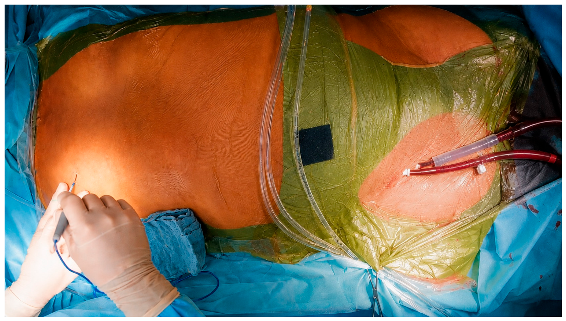
Figure 2.
Placement of the annular sutures with the RAM 5.0 automated sutureing device (LSI Solutions, Victor, NY, USA).
Figure 2.
Placement of the annular sutures with the RAM 5.0 automated sutureing device (LSI Solutions, Victor, NY, USA).
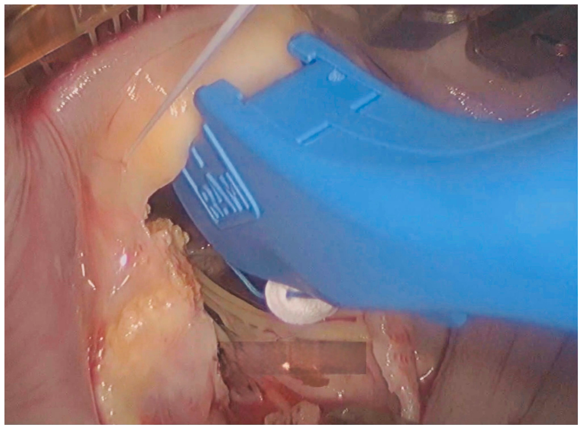
Figure 3.
The annular sutures are passed through the prosthesis with the SEW Easy Device (LSI Solutions, Victor, NY, USA).
Figure 3.
The annular sutures are passed through the prosthesis with the SEW Easy Device (LSI Solutions, Victor, NY, USA).
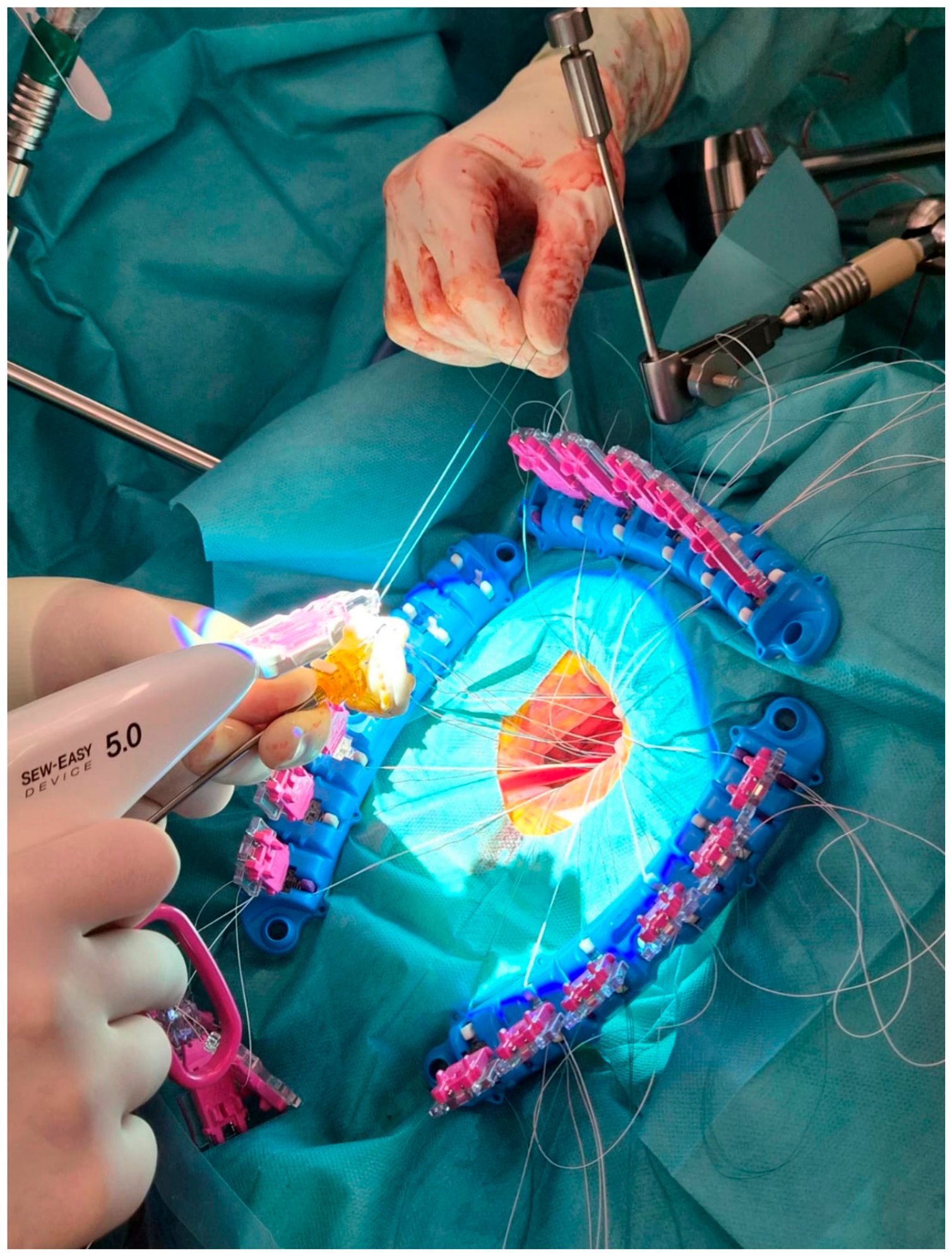

Table 1.
Baseline characteristics of the patient population.
| Parameter | RAM-MVR (n = 26) | RAM-AVR (n = 17) | Institutional benchmark (n = 182) |
| Demographics | |||
| Age (years) [Median (IQR)] | 67.0 (56.0–74.0) | 67.0 (59.5–74.0) | 65.0 (57.0–74.0) |
| Female sex [% (n)] | 53.8% (14/26) | 17.6% (3/17) | 32.4% (59/182) |
| Clinical Status | |||
| EuroSCORE II (%) [Median (IQR)] | 2.61 (1.03–4.90) | 0.92 (0.76–1.30) | 1.21 (0.73–2.51) |
| NYHA Class [Median (IQR)] | 3 (2–3) | 2 (1–3) | 3 (2–3) |
| Echocardiography | |||
| LVEF (%) [Median (IQR)] | 56.5 (55.0–60.0) | 60.0 (55.0–60.0) | N/A |
Table 2.
Surgical characteristics.
| Outcome Parameter | RAM-MVR (n = 26) | RAM-AVR (n = 17) | Institutional benchmark (n = 182) |
| Aortic X-clamp [Median (IQR)] | 73.0 (64.0–91.0) | 58.0 (50.0–63.0) | N/A |
| CPB time [Median (IQR)] | 114.5 (94.8–129.5) | 92.0 (86.0–108.0) | 115.0 (98.0–141.0) |
| Total surgery time [Median (IQR)] | 201.0 (184.8–226.3) | 171.0 (160.0–191.0) | 210.0 (185.0–245.0) |
| Clinical Outcomes | |||
| 30-day mortality [% (n)] | 3.8% (1) | 5.9% (1) | 1.6% (3) |
| Recovery & Function | |||
| ICU stay (days) [Median (IQR)] | 2.0 (1.0–3.0) | 2.0 (1.0–3.0) | 2.0 (1.0–4.0) |
| Hospital stay (days) [Median (IQR)] | 10.0 (9.0–13.3) | 10.0 (8.0–12.0) | 8.0 (7.0–12.0) |
| Postoperative EF (%) [Median] | 55.0% | 60.0% | 55.0% |
Table 3.
RAM Usage.
| Parameter | RAM-MVR (n = 26) | RAM-AVR (n = 17) | Institutional Benchmark (n = 182) |
|---|---|---|---|
| Vascular Access & Closure | |||
| Percutaneous Access [% (n)] | 100% (26) | 100% (17) | 100% (182) |
| MANTA® Closure Device [% (n)] | 100% (26) | 100% (17) | 100% (182) |
| RAM® Configuration | |||
| Technical Feasibility [%] | 100% | 100% | N/A |
| Jaw Size: 5.0 mm [% (n)] | 96.0% (25) | ~29% (5) * | N/A |
| Jaw Size: 3.5 mm [% (n)] | ~4% (1) * | ~71% (12) | N/A |
| Surgical Outcomes | |||
| Suture Line Failure [% (n)] | 0% (0) | 0% (0) | 0% (0) |
| Paravalvular Leak [%] | 0% | 0% | <1% |
| Device Malfunction [%] | 0% | 0% | N/A |
Table 4.
Postoperative mean pressure gradients.
| Parameter | RAM |
| Mean Pressure Gradient mitral valve | 3.0 (2.0–4.0) mmHg |
| Mean Pressure Gradient aortic valve | 8.0 (6.0–10.0) mmHg |
Disclaimer/Publisher’s Note: The statements, opinions and data contained in all publications are solely those of the individual author(s) and contributor(s) and not of MDPI and/or the editor(s). MDPI and/or the editor(s) disclaim responsibility for any injury to people or property resulting from any ideas, methods, instructions or products referred to in the content. |
© 2026 by the authors. Licensee MDPI, Basel, Switzerland. This article is an open access article distributed under the terms and conditions of the Creative Commons Attribution (CC BY) license (http://creativecommons.org/licenses/by/4.0/).
Copyright: This open access article is published under a Creative Commons CC BY 4.0 license, which permit the free download, distribution, and reuse, provided that the author and preprint are cited in any reuse.


