

Submitted:
03 July 2026
Posted:
07 July 2026
You are already at the latest version
Abstract
This retrospective study evaluated clinical and radiographic outcomes in dogs wei-ghing < 10 kg undergoing surgical stabilization of radius–ulna fractures using either the Vitcone® internal fixator system (Group A) or Veterinary Instrumentation locking plates (Group B) between January 2021 and December 2025. Dogs were included if they presented with complete diaphyseal or metaphyseal fractures without concurrent orthopedic or neurological disease. Preoperative radiographs were assessed by two independent observers, and fractures were managed with open reduction and internal fixation using craniomedial or craniolateral approach. Postoperative care included re-stricted activity for six weeks and standardized lameness scoring at the eight week re-check. Radiographic healing was evaluated using a Visual Assessment Scale (VAS), and interobserver agreement was quantified using the intraclass correlation coefficient (ICC). Ninety three dogs met inclusion criteria (59 in Group A, 34 in Group B). Most fractures resulted from low energy trauma and involved the distal metaphysis or diaphysis. In Group A 52/59 dogs showed a lameness score of 0/4 at eight weeks. Group B showed similar recovery in 26/34 dogs, although 5/34 exhibited ROM re-duction. Radiographic healing was comparable between groups, with cortical brid-ging typically observed within eight weeks and excellent interobserver agreement (ICC > 0.75). Major complications occurred in 5/59 (8.4%) dogs in Group A, 4/5 re-fractures after early implant removal in young, small dogs and 1/5 implant breakage. Group B showed a significantly similar major complication rates including refractures in 3/34 (8%). No differences were statistically observed (p > 0.05). Owner follow up confirmed complete weight bearing in all contacted cases. These findings suggest that small breed dogs with radial fractures treated with Vitcone® fixation, have similar cli-nical outcome of those achieved with Veterinary Instrumentation system.
Keywords:
radio-ulna
; fracture
; Vitcone® internal fixator
1. Introduction
Fractures of the radius and ulna are common orthopedic injuries in canine patients [1,2]. In dogs weighitng less then 10 kg, the distal antebrachium is characterized by low vascular blood supply, making it highly susceptible to vascular compromise following trauma [3]. Small-breed dogs, particularly Chihuahuas, Pomeranians, Toy Poodles, and Yorkshire Terriers, exhibit a well-documented predisposition to distal radial fractures, often resulting from low-energy trauma such as jumping from furniture. These fractures tend to be transverse or short-oblique and are notorious for progressing to non-union if not managed with surgical fixation [4]. These anatomical constraints have important implications for fracture healing. In small and toy breeds, the vascular supply to the distal radius is especially tenuous, predisposing these patients to delayed union, non-union, and implant failure when stabilization is inadequate or biological support is insufficient [5]. This phenomenon has been well recognized for decades and remains a major factor influencing treatment decisions in these breeds.
The mechanical environment of the antebrachium further complicates fracture management. The radius supports the majority of the axial load transmitted through the forelimb, while the ulna contributes to joint stability and provides attachment sites for key musculotendinous structures [6]. Inadequate fixation can lead to non-union, malalignment, altered load distribution, and secondary complications such as synostosis, elbow incongruity, or degenerative joint disease. Achieving and maintaining anatomical alignment is therefore essential, particularly in the distal third of the radius, where even minor deviations can significantly affect limb function [7].
Historically, a variety of treatment modalities have been employed for radial and ulnar fractures, ranging from conservative management with external coaptation to surgical stabilization using external skeletal fixation, dynamic compression plates, and, more recently, steel and titanium locking plate technology [4,8,9,10,11,12,13,14].
The introduction of locking compression plates (LCPs) has represented a major advancement in the management of antebrachial fractures. Locking constructs provide angular stability, reduce the need for precise plate-to-bone contouring, and preserve periosteal blood supply by minimizing plate-bone contact [15].
These features are particularly advantageous in small-breed dogs, where biological fragility and limited bone stock increase the risk of fixation failure [16]. Several studies have demonstrated improved outcomes with locking plates compared with traditional non-locking constructs, including higher union rates, reduced complication rates, and better maintenance of alignment [8,9,11,17,18,19]. Both open reduction and minimally invasive approaches habe been described [20,21,22]. The major postoperative complications requiring revision surgery for fracture treatment using conventional and locking plate systems were 3% and 9% respectively [21,23]. Nevertheless, the optimal fixation strategy remains a topic of debate. A paper published reported proper reduction of the caudal cortex of the readius is a critical point to promote bone healing preventing implant failure [24].
Given the complexity of these injuries and the diversity of clinical presentations, there is a continued need for high-quality research that clarifies optimal treatment strategies and identifies predictors of successful outcomes.
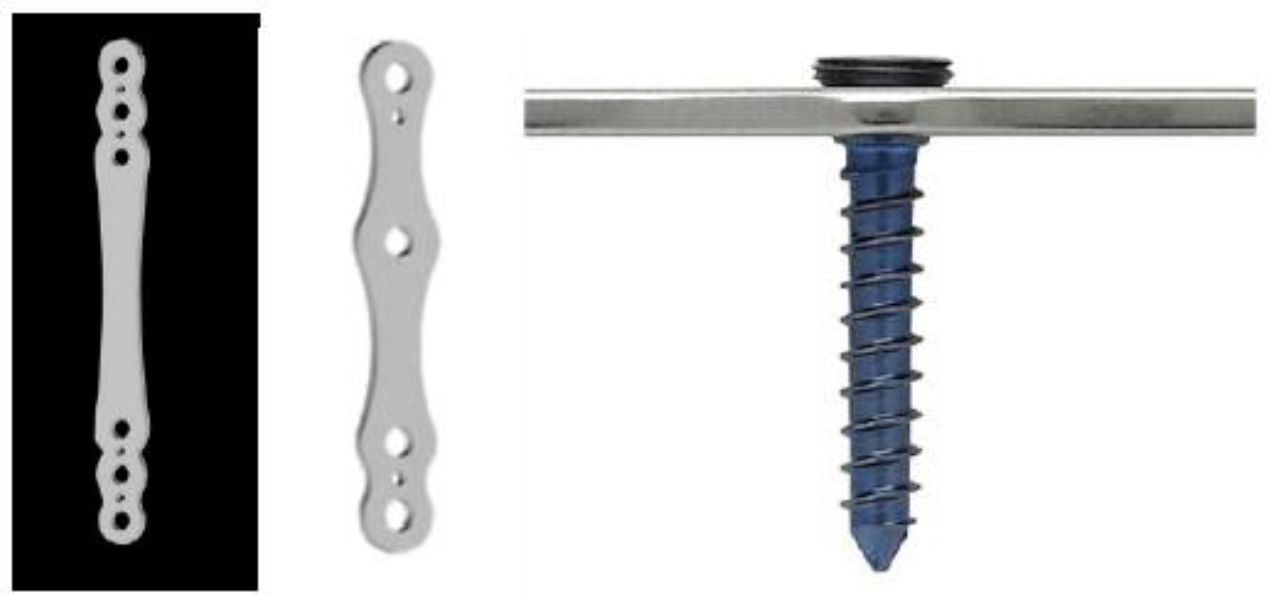
The Vitcone® plate are made in AISI 316LVM stainless steel or Ti-6Al-4V titanium alloy locking plate system that achieves angular stability through a direct conical coupling between the plate and screw heads. The self-tapping and self-locking screws are produced in Ti-6Al-4V titanium alloy, although they can be made of AISI 316LVM steel (Figure 1).
Up to date no papers described the application of the internal fixator system for management of radio-ulnar fractures in small breed dogs.
The present study aims to evaluate outcome and complications rates in dogs undergoing radial and ulnar fractures stabilized with Vitcone® internal fixator system comparing the results with those achieved using the Veterinary Instrumentation locking fixation system.
2. Materials and Methods
This retrospective study evaluated dogs dogs weighting < 10 kg undergoing surgical stabilization of radius–ulna fractures using the Vitcone® internal fixator fixation system and Veterinary Instrumentation internal lockin fixation between January 2021 and December 2025. Informed owner consent was obtained for all cases. Dogs were eligible for inclusion if they presented with a complete diaphyseal or metaphyseal fracture of the radius and ulna, had no concurrent orthopedic or neurological conditions affecting the same limb. Exclusion criteria included open fractures, pathological fractures secondary to neoplasia or metabolic disease, incomplete medical records, or lack of postoperative radiographic follow-up.
All patients were divided in two groups as follow: group A (Viticone), group B dogs (Veterinary Instrumentation).
For each patient, signalment, body weight, fracture etiology, and time from injury to surgery were recorded. Fractures were classified by two independent observers (AU, UM). Mediolateral and craniocaudal preoperative radiographs views were evaluated for fracture configuration, comminution, displacement, and bone quality. All surgeries were performed by experted orthopedic surgeons.
Dogs were premedicated with methadone (0.2 mg/kg) and dexmedetomidine (5 mcg/kg IM), then induced for endotracheal intubation with propofol (4 mg/kg) and maintained on isoflurane in oxygen.
A medial or lateral approach to the radius was used. Soft tissue dissection was minimized to preserve periosteal blood supply. Fracture reduction was achieved by an open reduction internal fixation approach followed by direct manipulation and pointed reduction forceps.
In simple fractures, anatomical reconstruction was attempted; in comminuted fractures, a bridging technique was used without reduction of intermediate fragments. Care was taken to avoid penetration of the radiocarpal joint and to maintain appropriate alignment in all planes. Intraoperative and postoperative radiographs were used to confirm implant placement.
Postoperative management included a modified Robert Jones bandage for 2-3 days, restricted activity for six weeks with leash walks (5-10 minutes, three times daily), and progressive return to normal exercise thereafter. All dogs received antibioc and non-teroidal antiinflammatory drugs (NSAIDs) for ten day after surgery. The limb function was subjectively assessed by the in-charged orthopedic surgeon scoring the lameness during the eight-week recheck as follow: 0/4 no lameness; 1/4 slight; 2/4 mild; 3/4 moderate; 4/4 no weight-bearing.
Follow-up radiographs were obtained at least at 4, 8 or 12 weeks, or until complete healing was documented. Radiographs were independently evaluated by two blinded observers with experience in orthopedic imaging.
Radiographic bone healing was assessed using the Visual assessment scale (VAS) [25]. On each medio-lateral and caudo-cranial views the VAS was given as follow by each observer: 0-10% no healing, 10-79% (partial healing) 80-100% (complete healing). The VAS that defined union as fusion of the fracture line, or the presence of bridging callus, or disappearance of the fracture lines, as seen on at least one aspect of each radiographic view: absence of osteotomy line on both radiological projections 100% healing, 5% less for each line of osteotomy displayed in the different projections.
Data about VAS score were tested for normality with Shapiro-Wilk, were analyzed with the intraclass correlation coefficient (ICC) for interobserver agreement. Mean and standard deviations were calculated for each observer evaluation. The ICC ranged from 0 (no agreement) to 1 (excellent agreement). We considered the values below 0.4 to be poor agreement, the values between 0.4 and 0.59 to be fair agreement, values between 0.6 and 0.74 to be good agreement, and values higher than 0.75 to be excellent agreement.
Complications were classified as minor or major according to the criteria described by Cook et al. [26]. Minor complications were managed conservatively, whereas major complications required surgical intervention or adversely affected the expected clinical outcome.
Statistical analysis of major complication rates between two groups were performed using GraphPad Prism 10. using Fisher’s exact test with significance set at p < 0.05.
Telephonic interview was performed up to one year after surgery asking to the owners if the patient was lame or not on the the treated fore limb.
3. Results
Over the five-year observation period, a total of 93 dogs affected by unilateral radius–ulna fractures were treated, showing a wide distribution in breed, age and body weight, ranging from 6 month to 10-year-old. Breeds included were mixed in 36/93, Pinscher 18/93, Yorkshire 9/93, Chiuhahua 4/93, Pomeranian dog 4/93, Poodle 18/93, Whippet 4/93.
Fractures involved predominantly diaphyseal or distal metaphyseal bone and were primarily injured (77%) by low energy trauma. Right and left fore limb factures were observed in 56 out of 93 and 36 out of 93 respectively.
In group A the medio-lateral approach was used in 25/59 (43%) and was then progressive adoption of the cranio-lateral approach for distal fractures, used in 34/59 (57%).
In group B cranio-medial radial approach was used in all cases.
In group A implant size was selected according to patient size, using 2.5 mm plates in small dogs (<10 < 5g) and micro-plates (1.8 mm screws) in toy breeds.
In group B implant size was selected according to patient size, using 2.4 mm plates in small dogs (<10 < 5g) and 2.0 plates in toy breeds.
In group A the eight-week recheck showed lameness score 0/4 in 52/59. The functional recovery within 8 weeks. No radio-carpal ROM deficiency was recorded in the cranio-lateral radial approach.
In group B the eight- week showed lameness score 0/4 in 26/34. Radio-carpal ROM deficiency was recorded in 5/34.
In group A implant removal was performed in 40/59 cases (68%) undergoing planned removal, typically performed minimally invasively.
In 12/40 en block removal was performed. In 12/40 double staged removal was planned. In 16/40 plate was left in situ after removal (Figure 3).
In group B implant removal was performed in 12/34 cases (35%) undergoing double stage planned removal.
Radiographic progression typically showed cortical bridging within 8 weeks in both groups. The mean VAS score obtained in the group A was 97% ± 0.04 (Figure 2). The mean VAS score of each observer were: 96% ± 0.03 in group B. The ICC was > 0.75 in both groups and considered as excellent agreement.
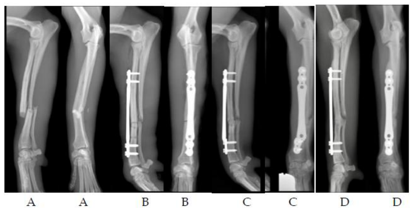
In group A major complications were observed in 5/59 cases (8,4%). Four refractures were recorded exclusively after early implant removal (Figure 4). In one case implant breakage was observed 29 days after surgery and was successfully revised using the same size of implant (Figure 5). No minor complications were recorded.
In group B complications were recorded in 6/34 cases (17,4%). Major complications included refracture after implant removal in 3/34 (8%). All dogs underwent a successfull revision surgery. Minor complications were observed in three cases as self-limiting swelling.
Statistiscal comparison about major complications rates was significant p > 0.05. Telephonic interview was performed in 47/59 in the group A, and 23/34 in group B of cases within one year after surgery: owners reported complete weight bearing in all of them.
Figure 2.
Dog Chihuahua, m, 1 year-old, 1.9 kg; right distal radius and ulna fracture; medio-lateral (ML) and cranio-caudal (Cr-Cd) preoperative (A), postoperative (B) and 3 month follow up (C) radiographic views: evidence of complete healing. Implant length 35,6 mm, thickness 1.3mm, holes n. 4. VAS 100%.
Figure 2.
Dog Chihuahua, m, 1 year-old, 1.9 kg; right distal radius and ulna fracture; medio-lateral (ML) and cranio-caudal (Cr-Cd) preoperative (A), postoperative (B) and 3 month follow up (C) radiographic views: evidence of complete healing. Implant length 35,6 mm, thickness 1.3mm, holes n. 4. VAS 100%.
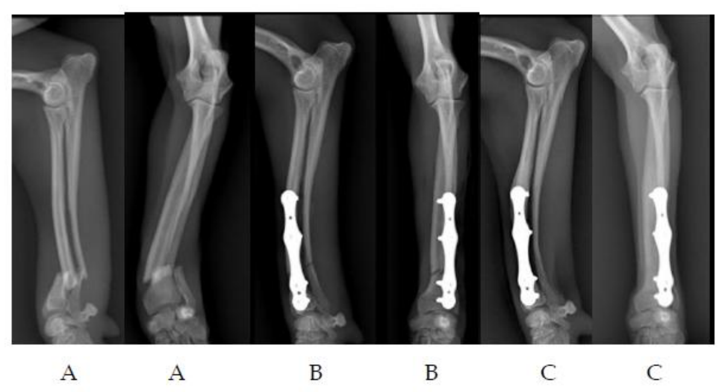
Figure 3.
Poodle, m, 7 month-old, 3.9 kg. left diaphyseal radius and ulna fracture ; ML and Cr-.Cd preoperative (A), postoperative (B) views. Two month (C), four month (D) and six month (E) postoperative radiographic views: all screws were progressively removed leaving the plate in situ.
Figure 3.
Poodle, m, 7 month-old, 3.9 kg. left diaphyseal radius and ulna fracture ; ML and Cr-.Cd preoperative (A), postoperative (B) views. Two month (C), four month (D) and six month (E) postoperative radiographic views: all screws were progressively removed leaving the plate in situ.
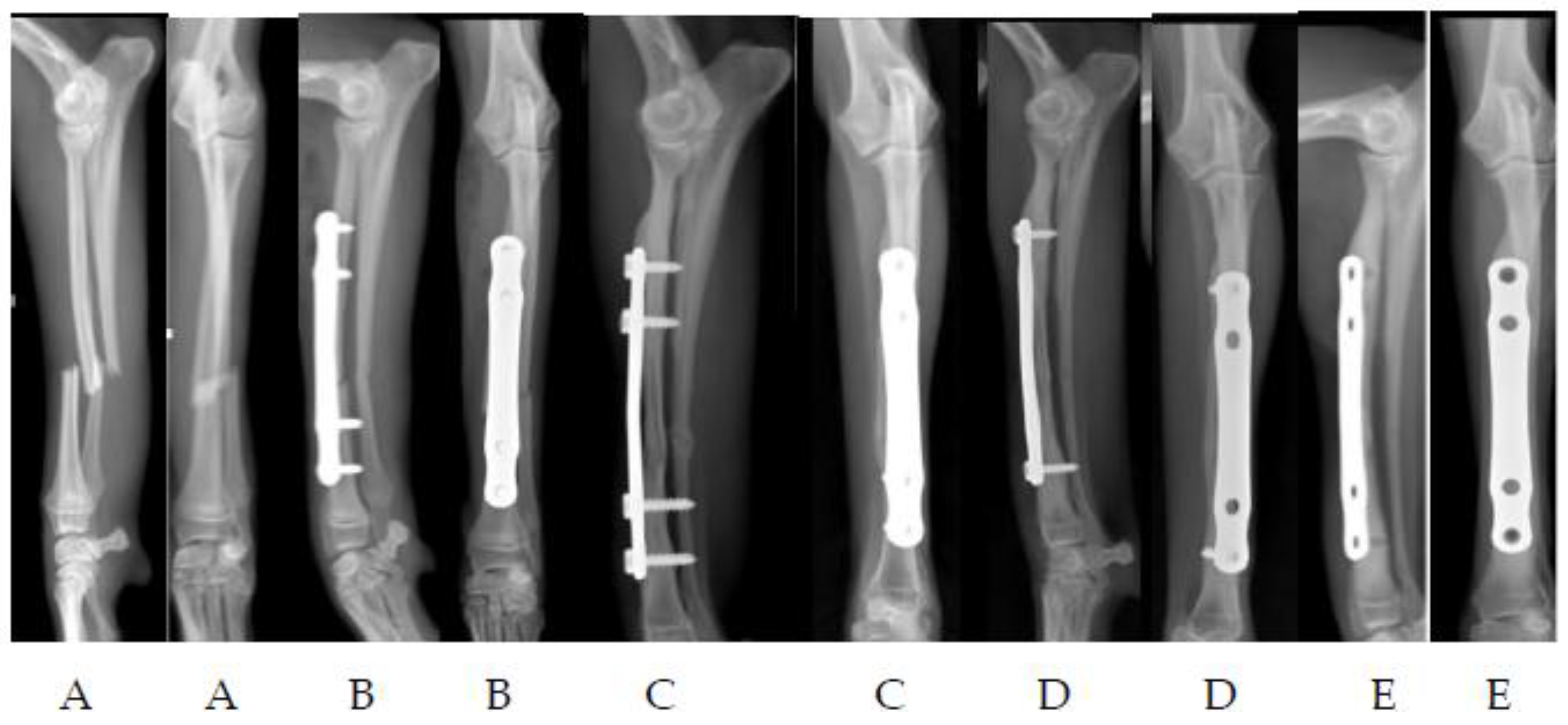
Figure 4.
Poodle, m, 1 year-old, 5kg; left distal radius and ulna fracture; ML and Cr-Cd radiographic views: evidence of refracture 17 days after implant removal (D); revision surgery was performed(E).
Figure 4.
Poodle, m, 1 year-old, 5kg; left distal radius and ulna fracture; ML and Cr-Cd radiographic views: evidence of refracture 17 days after implant removal (D); revision surgery was performed(E).
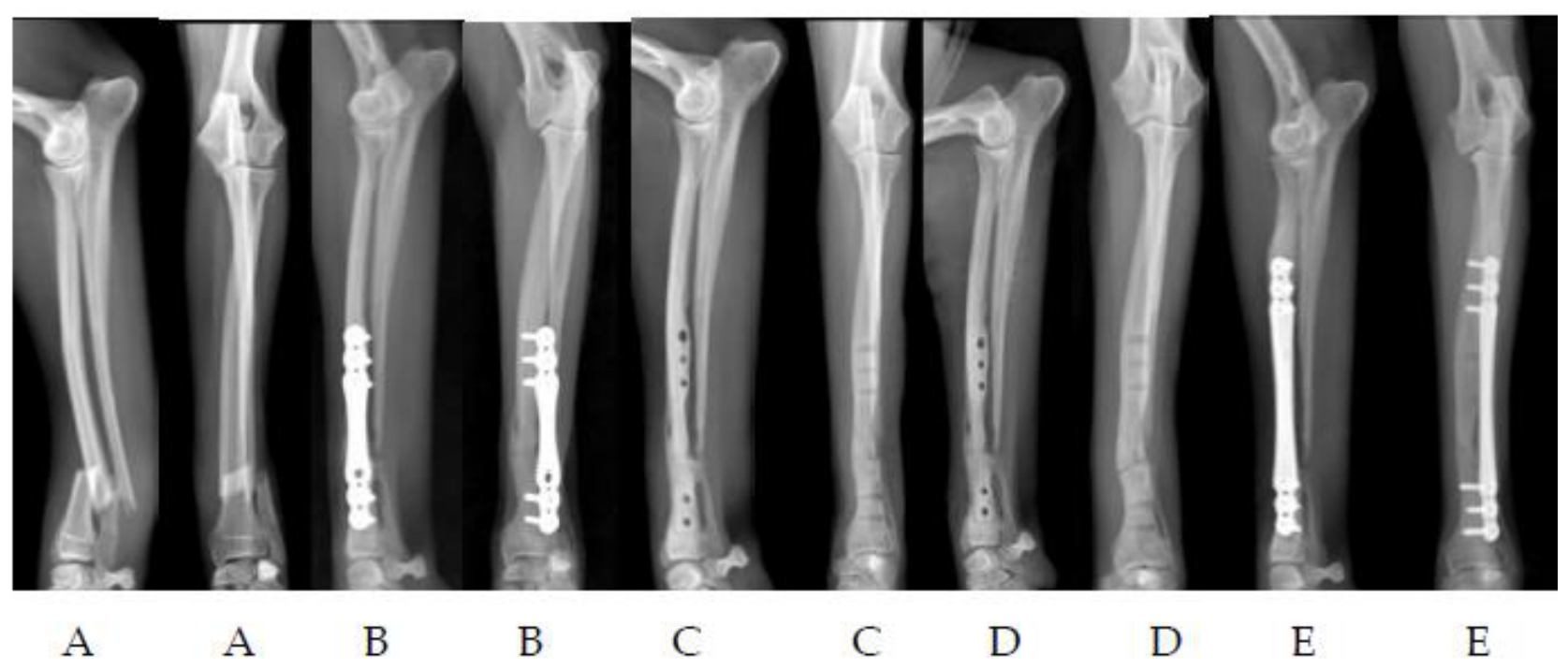
Figure 5.
Pincher, f, 7year-old, 3kg; right diaphyseal radius and ulna fracture; ML and Cr-Cd preoperative (A), postoperative (B) views. Twenty-nine day after surgery evidence of plate breakage was observed (C); two months after revision surgery, bone healing was achieved (D).
Figure 5.
Pincher, f, 7year-old, 3kg; right diaphyseal radius and ulna fracture; ML and Cr-Cd preoperative (A), postoperative (B) views. Twenty-nine day after surgery evidence of plate breakage was observed (C); two months after revision surgery, bone healing was achieved (D).
4. Discussion
The present study provides a comparison of clinical outome of unilateral radius–ulna fractures in dogs weighting < 10 kg stabilized with Vitcone System and Veterinary Instrumentation locking system, with a particular focus on surgical approach, clincal/radiographic outcome and complication rates. The demographic distribution observed in this cohort—small and toy breeds affected by low-energy trauma—is consistent with the epidemiology widely reported in the literature [1,21]. Small-breed dogs, especially toy breeds, are predisposed to distal radial fractures due to their unique bone morphology, reduced vascularity, and limited soft-tissue envelope [3]. The age distribution in our population, ranging from six months to ten years, further reflects the susceptibility of both immature and adult small dogs to these injuries.
In the present study Group A (Vitcone) employed a combination of medio-lateral and craniolateral approaches, with a progressive shift toward the latter for distal fractures. Group B, in contrast, relied exclusively on the craniomedial approach. The craniomedial approach has traditionally been recommended for radial fractures because it provides direct access to the tension surface of the radius [6]. However, several authors have noted that this approach may increase the risk of soft-tissue morbidity, particularly due to its proximity to the extensor carpi radialis tendon and the limited ability to retract surrounding structures safely [27,28]. The cranio-lateral approach provided similar exposure to the cranio medial one, but minimizing minimized morbidity by avoiding interference with the extensor carpi radialis tendon, while allowing safe lateral retraction of the extensor digitorum communis [27,28]. Our findings support these concerns: dogs in Group B exhibited a higher incidence of radiocarpal range-of-motion deficits, whereas Group A—especially when the craniolateral approach was used—did not show such limitations. This suggests that the craniolateral approach may offer a biomechanical and anatomical advantage in distal fractures by reducing interference with critical extensor tendons and providing a more favorable corridor for implant placement.
Implant selection also differed between groups. Group A used 2.5 mm plates in small dogs and micro-plates in toy breeds, while Group B used 2.4 mm and 2.0 mm plates. Despite these differences, radiographic healing progressed similarly in both groups, with cortical bridging typically observed within eight weeks. This timeframe is consistent with previously reported healing intervals for stable internal fixation of radial fractures in small breeds [6]. The high VAS scores and excellent inter-observer agreement (ICC > 0.75) further support the reliability of radiographic assessment across the study.
Major complications rates were similar in both groups that experienced refractures following implant removal. This finding is consistent with previous reports indicating that removal of implants in immature dogs increases the risk of refracture due to incomplete remodeling and persistent cortical weakness [23]. A paper published in 2025 reported the primary risk factor associated with the refractures after plate removal on distal radius in toy breeds was the reduced radial tickhness at the fracture site [29]. Because stress protection weakens the radial bone, the timing of plate removal is important to reduce risk of refracture. In the present study the decision to remove implants in both groups was not standardized and related to the radiographic bone healing progression associated with the young age.
Long-term functional outcomes were excellent in both groups, as confirmed by owner interviews reporting complete weight bearing in all contacted cases. This aligns with the literature, which consistently demonstrates that, when appropriately managed, radial fractures in small dogs have a high likelihood of returning to full function [14]. The absence of catastrophic complications in both groups further supports the overall safety of the surgical techniques employed.
Another important aspect of this study is the difference in implant removal strategies. Group A adopted a proactive removal policy in 68% of cases, often using minimally invasive techniques, motivated by concerns about stress shielding in young dogs. Group B performed removal in only 35% of cases, typically in a staged manner. The implant removal strategy in small-breed radial fractures remains uncertain. Our findings suggest that early removal may be beneficial in selected cases to avoid the stress protection phenomenon but must be approached cautiously in young, small dogs due to their increased susceptibility to refracture.
Overall, the results of this study suggest that the craniolateral approach, combined with careful implant selection may reduce complication rates and optimize recovery in small-breed dogs with radial fractures.
Main limitations of the present study is related to its retrospective nature and to the different sample size between the two groups.
Future prospective studies with standardized protocols would help further clarify the biomechanical and clinical implications of these findings and may contribute to the development of evidence-based guidelines for the management of radial fractures in small dogs.
5. Conclusions
This study provides evidence supporting the use of the Vitcone internal fixator system for radius–ulna fractures in small animals. The combination of elastic fixation, strategic screw placement, anatomically informed surgical approaches, and individualized postoperative care results in predictable healing and excellent functional outcomes. Future comparative studies with other plating systems would further clarify the relative advantages of the internal fixator, but the present data already position it as a highly effective tool in the management of these challenging fractures.
Author Contributions
For research articles with several authors, a short paragraph specifying their individual contributions must be provided. The following statements should be used “Conceptualization, A.U. and R.T.; methodology, A.U., A.P.; formal analysis, U.M., F.C.; investigation, A.U.; data curation, U.M.; writing—original draft preparation, A.U.; writing—review and editing, R.T.; supervision, R.T. All authors have read and agreed to the published version of the manuscript.” Please turn to the CRediT taxonomy for the term explanation. Authorship must be limited to those who have contributed substantially to the work reported.
Funding
This research received no external funding.
Informed Consent Statement
Informed consent was obtained from all subjects involved in the study.
Data Availability Statement
The original contributions presented in the study are included in the article, further inquiries can be directed to the corresponding author.
Conflicts of Interest
Andrea Urizzi is the patent holder of the internal fixator system. The other authors declare no conflicts of interest
References
- Anggoro, D.; Purba, M.S.; Jiang, F.; Nishida, N.; Itoh, H.; Itamoto, K.; Nemoto, Y.; Nakaichi, M.; Sunahara, H.; Tani, K. Elucidation of the Radius and Ulna Fracture Mechanisms in Toy Poodle Dogs Using Finite Element Analysis. J Vet Med Sci 2024, 86, 575–583. [CrossRef]
- Milovancev, M.; Ralphs, S.C. Radius/Ulna Fracture Repair. Clin Tech Small Anim Pract 2004, 19, 128–133. [CrossRef]
- Welch, J.A.; Boudrieau, R.J.; DeJardin, L.M.; Spodnick, G.J. The Intraosseous Blood Supply of the Canine Radius: Implications for Healing of Distal Fractures in Small Dogs. Vet Surg 1997, 26, 57–61. [CrossRef]
- Gibert, S.; Ragetly, G.R.; Boudrieau, R.J. Locking Compression Plate Stabilization of 20 Distal Radial and Ulnar Fractures in Toy and Miniature Breed Dogs. Vet Comp Orthop Traumatol 2015, 28, 441–447. [CrossRef]
- Marshall, W.G.; Filliquist, B.; Tzimtzimis, E.; Fracka, A.; Miquel, J.; Garcia, J.; Fontana, M.D. Delayed Union, Non-Union and Mal-Union in 442 Dogs. Vet Surg 2022, 51, 1087–1095. [CrossRef]
- DeCamp, C.; Johnston, S.; Déjardin, L.; Schaefer, S. Brinker, Piermattei and Flo’s Handbook of Small Animal Orthopedics and Fracture RepairHandbook of Small Animal Orthopedics and Fracture Repair; 5th ed.; Elsevier: St.Louis, 2016;
- Houlton, J. Advances in Canine Elbow Disease. Vet Surg 2009, 38, 133–134. [CrossRef]
- Swepson, R.; Crowley, J.; Glyde, M.; de Bruyn, B.; Wills, D.; Beierer, L.; Newman, M.; Tan, C. Clinical and Radiographic Outcomes of 1.5-Mm Locking Plate Fixation for 30 Radial and Ulnar Fractures in Dogs. Vet Comp Orthop Traumatol 2025, 38, 308–315. [CrossRef]
- Aikawa, T.; Miyazaki, Y.; Shimatsu, T.; Iizuka, K.; Nishimura, M. Clinical Outcomes and Complications after Open Reduction and Internal Fixation Utilizing Conventional Plates in 65 Distal Radial and Ulnar Fractures of Miniature- and Toy-Breed Dogs. Vet Comp Orthop Traumatol 2018, 31, 214–217. [CrossRef]
- Lakshmi, N.D.; Ganesh, T.N.; Ayyappan, S.; Shafiuzama, M.D.; Sureshkumar, R. Management of a Distal Metaphyseal Radial Fracture in a Dog with a Mini T-Plate. Vet Rec 2007, 161, 791–792.
- McCartney, W.; Kiss, K.; Robertson, I. Treatment of Distal Radial/Ulnar Fractures in 17 Toy Breed Dogs. Vet Rec 2010, 166, 430–432. [CrossRef]
- Bierens, D.; Unis, M.D.; Cabrera, S.Y.; Kass, P.H.; Owen, T.J.; Mueller, M.G. Radius and Ulna Fracture Repair with the IMEX Miniature Circular External Skeletal Fixation System in 37 Small and Toy Breed Dogs: A Retrospective Study. Vet Surg 2017, 46, 587–595. [CrossRef]
- Piras, L.; Cappellari, F.; Peirone, B.; Ferretti, A. Treatment of Fractures of the Distal Radius and Ulna in Toy Breed Dogs with Circular External Skeletal Fixation: A Retrospective Study. Vet Comp Orthop Traumatol 2011, 24, 228–235. [CrossRef]
- Crovace, A.M.; Guadalupi, M.; Belvito, R.; Monopoli, C.; Artiles, A.; Sanna Passino, E.; Crovace, A. A Retrospective Study of Clinical and Radiographic Outcomes and Exploratory Analysis of Implant Failure in Dogs ≤5 Kg Treated with Titanium Locking Mini-Plates for Radial and Ulnar Fractures. Vet Sci 2026, 13, 286. [CrossRef]
- Stoffel, K.; Dieter, U.; Stachowiak, G.; Gächter, A.; Kuster, M.S. Biomechanical Testing of the LCP--How Can Stability in Locked Internal Fixators Be Controlled? Injury 2003, 34 Suppl 2, B11-19. [CrossRef]
- Watrous, G.K.; Moens, N.M.M.; Runciman, J.; Gibson, T.W.G. Biomechanical Properties of the 1.5 Mm Locking Compression Plate: Comparison with the 1.5 and 2.0 Mm Straight Plates in Compression and Torsion. Vet Comp Orthop Traumatol 2018, 31, 438–444. [CrossRef]
- Nelson, L.L. Surgical Site Infections in Small Animal Surgery. Vet Clin North Am Small Anim Pract 2011, 41, 1041–1056, viii. [CrossRef]
- Higuchi, M.; Katayama, M. Clinical Outcomes of Orthogonal Plating to Treat Radial and Ulnar Fractures in Toy-Breed Dogs. J Small Anim Pract 2021, 62, 1001–1006. [CrossRef]
- Kang, B.-J.; Ryu, H.-H.; Park, S.; Kim, Y.; Kweon, O.-K.; Hayashi, K. Clinical Evaluation of a Mini Locking Plate System for Fracture Repair of the Radius and Ulna in Miniature Breed Dogs. Vet Comp Orthop Traumatol 2016, 29, 522–527. [CrossRef]
- Rehnblom, E.R.; Murphy, S.M.; Won, W.W.; Suber, J.T.; Arnold, J.J. Open Reduction and Internal Fixation of 75 Small Animal Fractures Treated with the OrthoLineTM Fracture System. Vet Surg 2025, 54, 1571–1581. [CrossRef]
- De Arburn Parent, R.; Benamou, J.; Gatineau, M.; Clerfond, P.; Planté, J. Open Reduction and Cranial Bone Plate Fixation of Fractures Involving the Distal Aspect of the Radius and Ulna in Miniature- and Toy-Breed Dogs: 102 Cases (2008-2015). J Am Vet Med Assoc 2017, 250, 1419–1426. [CrossRef]
- Lederer, X.J.; Lewis, D.D.; Evans, R.B.; Johnson, M.D.; Kim, S.E. Retrospective Comparison of Minimally Invasive Plate Osteosynthesis and Open Reduction and Plate Stabilization of Antebrachial Fractures in 105 Dogs (2017-2022). Vet Surg 2025, 54, 714–723. [CrossRef]
- Vallefuoco, R.; Le Pommellet, H.; Savin, A.; Decambron, A.; Manassero, M.; Viateau, V.; Gauthier, O.; Fayolle, P. Complications of Appendicular Fracture Repair in Cats and Small Dogs Using Locking Compression Plates. Vet Comp Orthop Traumatol 2016, 29, 46–52. [CrossRef]
- Korchek, K.A.; Kim, S.Y.; Chou, P.-Y.; Kapatkin, A.S.; Hayashi, K. Association of Fracture Gap with Implant Failure in Radius and Ulna Fractures in Toy Breed Dogs-A Multicenter Retrospective Cohort Study. Vet Surg 2025, 54, 287–296. [CrossRef]
- Leal, R.A.; Lambrechts, N.E.; Crowley, J.D.; Griffin, J.F.; Karnia, J.J.; Torres, B.T.; Maritato, K.C.; Kieves, N.R.; Duerr, F.M. Comparison of Radiographic Scoring Systems for Assessment of Bone Healing after Tibial Plateau Leveling Osteotomy in Dogs. Front Vet Sci 2023, 10, 1147386. [CrossRef]
- Cook, J.L.; Evans, R.; Conzemius, M.G.; Lascelles, B.D.X.; McIlwraith, C.W.; Pozzi, A.; Clegg, P.; Innes, J.; Schulz, K.; Houlton, J.; et al. Proposed Definitions and Criteria for Reporting Time Frame, Outcome, and Complications for Clinical Orthopedic Studies in Veterinary Medicine. Vet Surg 2010, 39, 905–908. [CrossRef]
- Lin, K.-T.; Degner, D.A.; DeCamp, C.E. Does the Craniolateral Approach Provide Better Exposure to the Radius than the Craniomedial Approach for Internal Fracture Fixation in Dogs? Vet Comp Orthop Traumatol 2025, 38, 97–102. [CrossRef]
- Crowdis, S.K.; Lewis, D.D. Comparison of Exposure of the Radius Afforded by the Craniomedial and Craniolateral Open Surgical Approaches in Dog Cadavers. J S Afr Vet Assoc 2024. [CrossRef]
- Muroi, N.; Kanno, N.; Harada, Y.; Hara, Y. A Retrospective Study of Risk Factors Associated with Refracture after Repair of Radial-Ulnar Fractures in Small-Breed Dogs. Vet Comp Orthop Traumatol 2025, 38, 77–86. [CrossRef]
Figure 1.
Vitcone® screw and plate. The conical locking occurs between the non-protruding portion of the screw head and the conical hole in the plate; part of the screw head protrudes above the plate.
Figure 1.
Vitcone® screw and plate. The conical locking occurs between the non-protruding portion of the screw head and the conical hole in the plate; part of the screw head protrudes above the plate.
Disclaimer/Publisher’s Note: The statements, opinions and data contained in all publications are solely those of the individual author(s) and contributor(s) and not of MDPI and/or the editor(s). MDPI and/or the editor(s) disclaim responsibility for any injury to people or property resulting from any ideas, methods, instructions or products referred to in the content. |
© 2026 by the authors. Licensee MDPI, Basel, Switzerland. This article is an open access article distributed under the terms and conditions of the Creative Commons Attribution (CC BY) license (http://creativecommons.org/licenses/by/4.0/).
Copyright: This open access article is published under a Creative Commons CC BY 4.0 license, which permit the free download, distribution, and reuse, provided that the author and preprint are cited in any reuse.


