
Submitted:
05 July 2026
Posted:
07 July 2026
You are already at the latest version
Abstract
Introduction: The superficial palmar branch of the radial artery (SPBRA) typically originates 10 to 20 mm proximal to the distal wrist crease, before the radial artery winds around the carpal bones toward the dorsum of the hand. From its origin, the SPBRA courses along the palmar aspect of the hand, traverses the fibers of the thenar muscles, and anastomoses with the ulnar artery to form the superficial palmar arch. Anatomical variations of this branch are described in the literature; given its potential as a vascular pedicle for thenar flaps in the reconstruction of distal finger and hand injuries, knowledge of its morphology is indispensable for surgical safety. Objective: To report a proximal origin of the SPBRA and discuss its possible clinical and surgical implications. Case Report: During routine dissection, an anatomical variation in the origin of the SPBRA was identified in a right forearm disarticulated at the elbow level. The radial artery followed its usual course in the anterior forearm until it prematurely emitted the SPBRA in the distal third, 61.05 mm proximal to the wrist joint, characterizing a high origin compared to the pattern described in the literature. The branch directed itself obliquely in a distal and medial direction, penetrated the fibers of the abductor pollicis brevis muscle, and, after this intramuscular segment, anastomosed with the ulnar artery to form the superficial palmar arch. The total length of the SPBRA was 92.68 mm. Conclusion: The high origin of the SPBRA is a rare anatomical variation of great clinical relevance, the recognition of which is fundamental in planning surgical and reconstructive procedures of the hand, especially in the design of thenar flaps. In addition to corroborating previous literature findings, the present study contributes an unprecedented measurement of the distance between the high origin of the branch and the wrist joint, providing relevant data for the anatomical characterization of this variation.
Keywords:
radial artery
; superficial palmar branch
; anatomical variation
; high origin
; thenar flap
1. Introduction
The superficial palmar branch of the radial artery (SPBRA) typically originates from the bifurcation of the main trunk of the radial artery, 10 to 20 mm proximal to the distal wrist crease—rarely exceeding 30 mm from this landmark—just before the radial artery (RA) winds around the carpal bones toward the dorsum of the hand [1,2,3]. The SPBRA courses toward the palmar aspect of the hand, traversing the fibers of the thenar muscles, and terminates by anastomosing with the ulnar artery to form the superficial palmar arch [4,5,6].
Anatomical variations of the SPBRA are frequently described in the literature. Tagil et al. [3] identified the SPBRA coursing transversely over the flexor retinaculum without traversing the thenar eminence. Singer et al. [7] located the SPBRA describing its course in the subcutaneous tissue over the abductor pollicis brevis muscle, so superficially that it caused pain. Furthermore, Nasr [8] consolidated these findings by demonstrating that the anatomical variability of the RA and its branches is significant, encompassing differences in origin, caliber, course, and relationships with adjacent structures. The explanation for such extensive variability can be found in the different developmental stages of these vessels, which exhibit high embryonic vascular complexity with numerous patterns of regression and persistence of angiogenesis [9].
The SPBRA has been widely used as a vascular pedicle for thenar flaps in soft tissue reconstruction of distal finger and hand injuries, yielding satisfactory functional and aesthetic results [10]. Therefore, understanding variations in the origin, course, and branching of the SPBRA is crucial for reducing the risk of iatrogenic injury during wrist and hand surgeries [5]. The present study aims to report a proximal or high origin of the SPBRA and its possible clinical and surgical implications.
2. Case Report
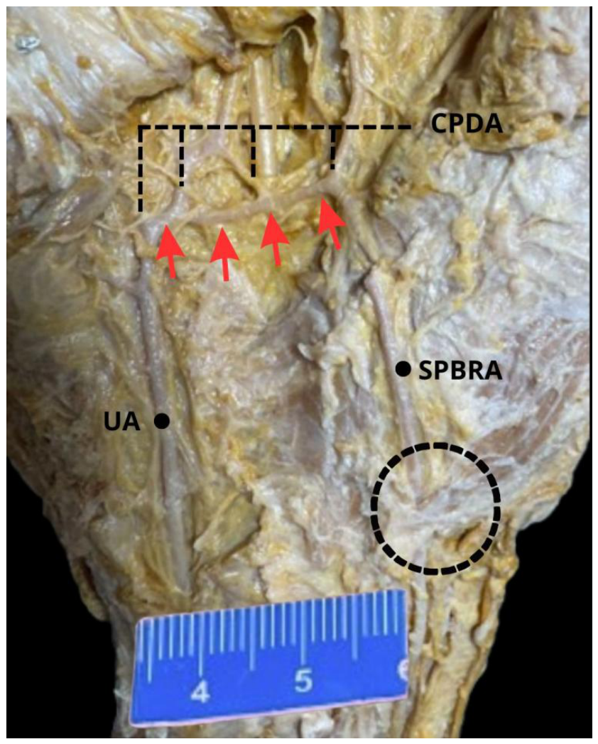
During routine dissection at the Anatomy Laboratory of the Federal University of Sergipe, an anatomical variation in the origin of the SPBRA was identified in a right forearm, disarticulated at the elbow level, from an adult cadaver fixed in a 10% formalin solution. The specimen exhibited satisfactory tissue preservation, with vascular and muscular structures intact for morphological and morphometric identification. The RA followed its usual course along the anterior aspect of the forearm, between the tendons of the flexor carpi radialis and brachioradialis muscles, until the premature emission of the SPBRA in the distal third of the forearm, 61.05 mm proximal to the wrist joint—characterizing a high origin relative to the pattern described in the literature (Figure 1). From this point, the SPBRA directed itself obliquely in a distal and medial direction, reached the palmar aspect of the hand, and entered the muscular fibers of the abductor pollicis brevis. After this intramuscular segment, the branch reappeared on the palmar surface, anastomosing with the ulnar artery to form the superficial palmar arch (Figure 2). Using a digital caliper with 0.01 mm precision, the following measurements were taken: a total SPBRA length of 92.68 mm; an external diameter of 1.60 mm at the radial end, 1.25 mm at the middle third, and 1.20 mm at the ulnar end, evidencing a progressive reduction in caliber along its course. After emitting the SPBRA, the RA proceeded distally along the anterior aspect of the forearm to the wrist, wound around the carpal bones, passed deep to the radial styloid process, and reached the anatomical snuffbox, continuing its usual dorsal course.
3. Discussion
The radial artery is of paramount importance for the vascularization of the hand, which directly underscores its significance in clinical practice. Primarily, it contributes effectively to the formation of the deep palmar arch and, through its SPBRA, joins with the ulnar artery to form the superficial palmar arch. This vascular configuration exhibits various anatomical variations or instances of dominance of this branch [6]. Clinically, the radial artery is the preferred access route for procedures such as cardiac catheterization and percutaneous coronary interventions due to the lower risk of hemorrhagic complications compared to the femoral approach, as well as greater patient comfort and lower morbidity [11]. In myocardial revascularization surgery, the RA is considered a superior graft compared to the great saphenous vein due to its higher long-term patency, which has been associated in the literature with better clinical outcomes [12]. However, the significant presence of anatomical variations, including a superficial course of the SPBRA or a high bifurcation of the radial artery, can substantially impact the safety and effectiveness of these procedures, increasing the risk of iatrogenic injuries [3].
The anatomical literature provides isolated data regarding the high origin variation of the SPBRA. In an analysis of 100 upper limbs from cadavers, Nasr [8] observed the SPBRA in 95 cases, of which 89 originated routinely at the level of the wrist joint, while six exhibited a high emergence. This type of variation was also documented by Weinand et al. [13], who identified a case of high bifurcation between the SPBRA and the RA through arteriographic examinations in a patient with an aneurysm. Although the present study corroborates the existence of this rare variation, it advances uniquely by quantifying the exact distance between the high origin of the branch and the wrist joint—an essential metric parameter still absent from the literature for characterizing this anomalous behavior. The external diameter of the SPBRA is a frequently reported measurement in the scientific literature, as it may be related to both its length and its irrigation area [14]. In this context, Ilić et al. [14] found the SPBRA diameter to vary from 0.8 mm to 2.7 mm, measured at its origin from the radial artery. In contrast, Radunovic et al. [15] observed an even wider range, with values oscillating between 0.55 mm and 4.3 mm; they concluded from their findings that there is no dependent relationship between the diameter and the length of the SPBRA. However, the authors themselves acknowledge a significant limitation in their study: the small sample size, which favors the occurrence of extreme values and compromises the robustness of the statistical analysis. This suggests that a larger sample would yield more reliable and representative results.
The SPBRA holds significant clinical and surgical importance, primarily due to its potential as a source for vascular flaps widely employed in reconstructive microsurgery for digital injuries. Although traditionally described as a small-caliber branch that contributes inconsistently to the superficial palmar arch [16], anatomical studies have demonstrated that the SPBRA can be found at varying frequencies. When present and meeting an adequate caliber pattern, it can be utilized in the construction of microsurgical flaps [15]. Flaps based on the SPBRA have been successfully used in distal phalanx reconstruction, offering patency and adequate distal vascularization without the need to sacrifice the main radial artery [10]. Furthermore, anatomical variations of the SPBRA—such as a high origin, anomalous superficial course, or direct anastomosis with the superficial palmar arch—can cause compressive symptoms (pain, paresthesia) or lead to erroneous interpretations of the Allen test, which is a direct assessment of the ulnar artery's ability to adequately supply the hand in the event of radial artery harvesting [7].
Therefore, knowledge regarding the diverse morphological expressions of the SPBRA is indispensable for hand surgeons, interventional radiologists, and anesthesiologists [8]. Thenar flaps are excellent surgical options for digital reconstruction, especially in distal pulp and nail region injuries, primarily utilizing fasciocutaneous transposition [17]. It is worth noting that these procedures essentially aim to resolve local skin defects while maintaining the morphological, morphometric, and sensory characteristics of the recipient site. Based on the vascularization provided by the SPBRA or direct branches of the radial artery, these flaps allow for the coverage of tissue loss with preserved innervation, resulting in excellent functional and aesthetic recovery [2]. The classic thenar flap, primarily described as pedicled, is widely used for trauma to the distal phalanx of the middle, ring, and little fingers, being considered a relatively simple technique with a high success rate [18].
The superficial palmar arch is classically formed by the anastomosis between the SPBRA and the ulnar artery, although the literature acknowledges wide variability in the behavior of these vessels [6,16]. Aiming to characterize this anatomical diversity, Al-Turk and Metcalf [19] conducted an ultrasonographic study of 50 hands, in which they identified different constitutions of the superficial palmar arch and organized them into two major groups: the complete arch, observed in 84% of cases, and the incomplete arch, present in the remaining 16%. The complete arch group was further subdivided into three types—A, B, and C—according to the particularities of its formation. Type A, the most prevalent, was found in 78% of the evaluated hands and corresponds exactly to the classic configuration of anastomosis between the SPBRA and the ulnar artery. Types B and C, in turn, showed considerably lower frequencies, corresponding to 4% and 2% of cases, respectively. In the present report, a Type A complete arch configuration was identified, which is in line with the predominant statistical trend in the studied population.
4. Conclusions
It is concluded that the high origin of the SPBRA is a rare anatomical variation of significant clinical importance. Its recognition is fundamental in surgical and reconstructive procedures of the hand, especially in the design of thenar flaps. In addition to confirming literature findings, this study adds the measurement of the distance between the high origin of the SPBRA and the wrist joint, providing relevant data for a better anatomical characterization of this variation.
Author Contributions
Conceptualization: Wallance Geovane Alexandre Lima, Gilvan Paixão Santos Junior, Lucas Morais Paixão, Giovanna de Oliveira Sá Costa Methodology: Adler Oliveira Silva Jacó Carvalho, Henrique Montalvão Routman da Cunha Writing – Original Draft Preparation: José Aderval Aragão, Deise Maria Furtado de Mendonça Writing – Review & Editing: Francisco Prado Reis, Iapunira Catarina Sant’Anna Aragão, Felipe Matheus Sant’Anna AragãoSupervision: José Aderval Aragão.
Funding
“This research received no external funding”.
Institutional Review Board Statement
Brazilian Law No. 8501, enacted on November 30, 1992, allows the donation of unclaimed bodies by forensic medical institutes to universities for studies or scientific research, and includes other provisions.
Informed Consent Statement
“Not applicable.”.
Acknowledgments
We thank Marcelo Diaz Nascimento, Gladson Gomes de Souza, and Luís Henrique Santos Fortes, anatomy laboratory technicians in the Federal University of Sergipe, for their support in preparing the cadavers, since without their collaboration, this work could not have been accomplished. The authors wish to sincerely thank those who donated their bodies to science so that anatomical research could be carried out. The results of this research can potentially im-prove patient care and increase the overall knowledge of humanity. Therefore, these donors and their families deserve our utmost gratitude. The authors have reviewed and edited the output and take full responsibility for the content of this publication.”.
Conflicts of Interest
“The authors declare no conflicts of interest.”.
Abbreviations
The following abbreviations are used in this manuscript:
| APB | Abductor Pollicis Brevis Muscle |
| Blue Arrow | Superficial Palmar Branch of the Radial Artery |
| BR | Brachioradialis Muscle |
| Circumference | Intramuscular Pathway of the Superficial Palmar Branch of the Radial Artery |
| CPDA | Common Palmar Digital Arteries |
| FCR | Flexor Carpi Radialis Muscle |
| FCU | Flexor Carpi Ulnaris Muscle |
| PL | Palmaris Longus Muscle |
| RA | Radial Artery |
| Red Arrow | Superficial Palmar Arch |
| SPBRA | Superficial Palmar Branch of the Radial Artery |
| UA | Ulnar Artery |
References
- Omokawa, S.; Ryu, J.; Tang, J.-B.; Han, J. Vascular and neural anatomy of the thenar area of the hand: its surgical applications. Plast. Reconstr. Surg. 1997, 99, 116–21. [Google Scholar] [CrossRef]
- Lee, T.; Liao, C.; Wu, I.; Yu, C.; Chen, S. Free Flap From the Superficial Palmar Branch of the Radial Artery ( SPBRA Flap ) for Finger Reconstruction. J. Trauma 2009, 66, 1173–9. [Google Scholar] [CrossRef] [PubMed]
- Tagil, S.; Bozkurt, M.; Kuvat, S.; Cicekcibasi, A. A variant course of the superficial palmar branch of the radial artery. Surg. Radiol. Anat. 2012, 34, 871–3. [Google Scholar] [CrossRef] [PubMed]
- Kumar, V.D.; Rajasekhar, S.S.S.N.; Sankaranarayanan, G. Superficial palmar branch of the radial artery in a fibromuscular tunnel : a case report. Surg. Radiol. Anat. 2020, 42, 277–80. [Google Scholar] [CrossRef] [PubMed]
- Rakaba, M.; Ross, O.; Juttu, A.; Bhattacharya, A. Anatomical Variation of the Superficial Palmar Branch of the Radial Artery : A Cadaveric Case Report. Cureus 2026, 18, e104407. [Google Scholar] [CrossRef] [PubMed]
- Standring, S. (Ed.) Gray’s anatomy: the anatomical basis of clinical practice, 40th ed.; Churchill Livingstone Elsevier: London, 2008. [Google Scholar]
- Singer, G.; Marterer, R.; Till, H.; Schmidt, B. A rare anatomic variation of the superficial palmar branch of the radial artery causing pain. Surg. Radiol. Anat. 2018, 40, 349–52. [Google Scholar] [CrossRef] [PubMed]
- Nasr, A.Y. The radial artery and its variations: Anatomical study and clinical implications. Folia Morphol. 2012, 71, 252–62. [Google Scholar]
- Singer, E. Embryological pattern persisting in the arteries of the arm. Anat. Rec. 1933, 55, 403–9. [Google Scholar] [CrossRef]
- Zhao, W.; Song, K.; Ma, B.; Liu, Y. Clinical application of the free superficial palmar branch of radial artery flap for soft-tissue reconstruction of distal digital injury. J. Orthop. Surg. Res. 2024, 19, 553. [Google Scholar] [CrossRef] [PubMed]
- Jolly, S.S.; Yusuf, S.; Cairns, J.; Niemelä, K.; Xavier, D.; Widimsky, P.; et al. Radial versus femoral access for coronary angiography and intervention in patients with acute coronary syndromes ( RIVAL ): a randomised, parallel group, multicentre trial. Lancet 2011, 377, 1409–20. [Google Scholar] [CrossRef] [PubMed]
- Gaudino, M.; Benedetto, U.; Fremes, S.; Biondi-Zoccai, G.; Puskas, J.; Buxton, B. Radial-artery or saphenous-vein grafts in coronary-artery bypass surgery. N Engl. J. Med. 2018, 378, 2069–77. [Google Scholar] [CrossRef] [PubMed]
- Weinand, C.; Akbari, C.; O’Donnell, S. A High Bifurcation of the Dorsal Branch with Dominant Superficial Palmar Branch of the Radial Artery: A Case Report of an Aberrant Radial Artery with Traumatic Aneurysm. J. Hand Microsurg 2011, 3, 78–81. [Google Scholar] [CrossRef] [PubMed]
- Ilić, M.; Milisavljević, M.; Maliković, A.; Laketić, D.; Erić, D.; Boljanović, J.; et al. The superficial palmar branch of the radial artery : a corrosion cast study. Folia Morphol. 2018, 77, 649–55. [Google Scholar] [CrossRef] [PubMed]
- Radunovic, M.; Vukcevic, B.; Radojevic, N.; Vukcevic, N.; Vuksanovic-Bozaric, A. Superficial palmar branch of the radial artery: A study of occurrence and diameter. Ann. Anat. 2018, 220, 55–9. [Google Scholar] [CrossRef] [PubMed]
- Coleman, S.S.; Anson, B.J. Arterial patterns in the hand based upon a study of 650 specimens. Surg. Gynecol. Obstet. 1961, 113, 409–24. [Google Scholar] [PubMed]
- Hugon, S.; Castus, P.; Schoofs, M. Index reconstruction by means of a fasciocutaneous thenar flap. Plast. Reconstr. Surg. 2010, 126, 43e–44e. [Google Scholar] [CrossRef] [PubMed]
- Kasikcioglu, N.; Govsa, F.; Bicer, A.; Demir, O.F.; Gokmen, G. Superficial palmar branch of radial artery flap for digital skin reconstruction: anatomical study and clinical applications. Surg. Radiol. Anat. 2024, 47, 45. [Google Scholar] [CrossRef] [PubMed]
- Al-Turk, M.; Metcalf, W.K. A study of the superficial palmar arteries using the Doppler Ultrasonic Flowmeter. J. Anat. 1984, 138, 27–32. [Google Scholar] [PubMed]
Figure 1.
Anterior view of the forearm showing the high origin of the superficial palmar branch of the radial artery. Legend: RA: Radial Artery; UA: Ulnar Artery; APB: Abductor Pollicis Brevis Muscle; FCU: Flexor Carpi Ulnaris Muscle; PL: Palmaris Longus Muscle; FCR: Flexor Carpi Radialis Muscle; BR: Brachioradialis Muscle; Red Arrow: Superficial Palmar Arch; Blue Arrow: Superficial Palmar Branch of the Radial Artery; Circumference: Intramuscular Pathway of the Superficial Palmar Branch of the Radial Artery.
Figure 1.
Anterior view of the forearm showing the high origin of the superficial palmar branch of the radial artery. Legend: RA: Radial Artery; UA: Ulnar Artery; APB: Abductor Pollicis Brevis Muscle; FCU: Flexor Carpi Ulnaris Muscle; PL: Palmaris Longus Muscle; FCR: Flexor Carpi Radialis Muscle; BR: Brachioradialis Muscle; Red Arrow: Superficial Palmar Arch; Blue Arrow: Superficial Palmar Branch of the Radial Artery; Circumference: Intramuscular Pathway of the Superficial Palmar Branch of the Radial Artery.
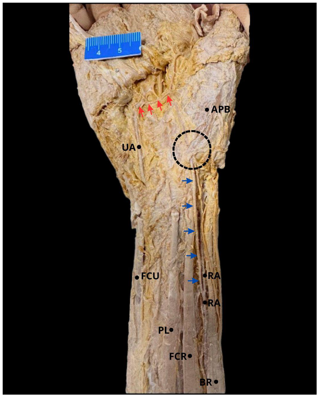
Figure 2.
Course of the superficial palmar branch of the radial artery through the palmar aspect of the hand and the formation of the superficial palmar arch. Legend: UA: Ulnar Artery; SPBRA: Superficial Palmar Branch of the Radial Artery; CPDA: Common Palmar Digital Arteries; Red Arrow: Superficial Palmar Arch; Circumference: Intramuscular Pathway of the Superficial Palmar Branch of the Radial Artery.
Figure 2.
Course of the superficial palmar branch of the radial artery through the palmar aspect of the hand and the formation of the superficial palmar arch. Legend: UA: Ulnar Artery; SPBRA: Superficial Palmar Branch of the Radial Artery; CPDA: Common Palmar Digital Arteries; Red Arrow: Superficial Palmar Arch; Circumference: Intramuscular Pathway of the Superficial Palmar Branch of the Radial Artery.
Disclaimer/Publisher’s Note: The statements, opinions and data contained in all publications are solely those of the individual author(s) and contributor(s) and not of MDPI and/or the editor(s). MDPI and/or the editor(s) disclaim responsibility for any injury to people or property resulting from any ideas, methods, instructions or products referred to in the content. |
© 2026 by the authors. Licensee MDPI, Basel, Switzerland. This article is an open access article distributed under the terms and conditions of the Creative Commons Attribution (CC BY) license.
Copyright: This open access article is published under a Creative Commons CC BY 4.0 license, which permit the free download, distribution, and reuse, provided that the author and preprint are cited in any reuse.


