

Submitted:
03 July 2026
Posted:
06 July 2026
You are already at the latest version
Abstract
Background: Contrast media are essential tools in diagnostic and interventional imaging, but their relationship with acute kidney injury remains clinically relevant and conceptu-ally debated. This narrative review aimed to synthesize current evidence on contrast me-dia-associated nephrotoxicity, including terminology, epidemiology, pathophysiology, risk stratification, prevention, pharmacotherapeutic management, and clinical deci-sion-making. Methods: A structured literature search was conducted in major biomedical databases and complemented by international guidelines and consensus statements ad-dressing contrast-associated and contrast-induced acute kidney injury in adults exposed to intravascular contrast media. Discussion: Contemporary evidence emphasizes the dis-tinction between contrast-associated acute kidney injury, which reflects a temporal asso-ciation after exposure, and contrast-induced acute kidney injury, which implies causality. The renal risk directly attributable to modern intravenous iodinated contrast media ap-pears to have been historically overestimated, although clinically relevant risk persists in vulnerable patients. Proposed mechanisms include renal vasoconstriction, medullary hy-poxia, oxidative stress, mitochondrial dysfunction, tubular epithelial injury, endothelial dysfunction, and inflammatory or apoptotic pathways. Preventive strategies should be in-dividualized, with isotonic saline remaining the main intervention when indicated, whereas routine pharmacologic prophylaxis is not supported by consistent clinically meaningful benefit. Conclusions: Renal safety in contrast-enhanced imaging requires a balanced approach that minimizes avoidable kidney injury in high-risk patients without unnecessarily delaying clinically indicated diagnostic or therapeutic procedures.
Keywords:
contrast-associated acute kidney injury
; contrast-induced acute kidney injury
; iodinated contrast media
; gadolinium-based contrast agents
; nephrotoxicity
; acute kidney injury
; chronic kidney disease
; risk stratification
; isotonic saline
; pharmacologic prophylaxis
1. Introduction
Contrast media are agents administered to the body to enhance the differentiation and visualization of organs, blood vessels, tissues, and internal structures across different imaging studies. They are used in X-ray–based techniques, such as radiography, computed tomography (CT), and angiography, as well as in non-ionizing imaging modalities such as magnetic resonance imaging (MRI), where gadolinium-based contrast agents are primarily employed [1]. These agents are essential tools for the diagnosis, stratification, and follow-up of multiple clinical conditions, including cardiovascular, oncologic, infectious, neurologic, and traumatic diseases. Their use has improved diagnostic accuracy and supported timely therapeutic decision-making; however, they have historically been associated with a relevant risk of acute deterioration in kidney function [1,2].
In recent years, emerging evidence has prompted a critical reassessment of this concept. Several international guidelines and consensus statements have emphasized the need to distinguish between CA-AKI and CI-AKI. The consensus statement from the American College of Radiology and the National Kidney Foundation (ACR–NKF 2020) indicates that the risk of AKI following intravenous iodinated contrast administration in patients with reduced kidney function has probably been overestimated, partly because of the historical lack of adequate control groups capable of separating temporally associated kidney injury from kidney injury truly induced by contrast media [3].
From a diagnostic perspective, AKI after contrast exposure is usually identified by applying the general KDIGO criteria for AKI, defined as an increase in serum creatinine of at least 0.3 mg/dL within 48 hours, an increase to 1.5 times or more from baseline within the previous 7 days, or a reduction in urine output to less than 0.5 mL/kg/h for at least 6 hours [4]. However, when these criteria are met after contrast administration, the finding should initially be interpreted as CA-AKI, since the definition itself does not establish direct causality with the administered agent.
Nevertheless, causal attribution of kidney function deterioration to contrast media remains a matter of growing controversy [5]. A substantial proportion of AKI episodes observed after contrast administration occurs in patients with multiple concomitant factors, such as chronic kidney disease, sepsis, hypovolemia, heart failure, hemodynamic instability, or simultaneous exposure to other nephrotoxic agents. Therefore, these events do not necessarily reflect kidney injury directly induced by contrast exposure [6].
In this context, the American College of Radiology has noted that one of the main diagnostic limitations lies in the low sensitivity and specificity of serum creatinine for differentiating between distinct forms of AKI [3]. Consequently, criteria based exclusively on changes in serum creatinine cannot reliably distinguish between CI-AKI, in which a direct causal relationship is assumed, and CA-AKI, which describes only a temporal association after exposure. This distinction has relevant clinical implications. An overly conservative interpretation of risk may delay or prevent necessary imaging studies, potentially causing harm through delayed or inaccurate diagnosis [7].
Given this background, a critical synthesis of the current scientific evidence on AKI associated with contrast media is warranted, addressing its conceptual evolution, risk factors, proposed pathophysiologic mechanisms, preventive strategies, and clinical-pharmacotherapeutic implications in the hospital setting. This review aims to provide a critical synthesis useful for clinical pharmacists, nephrologists, radiologists, internists, emergency physicians, and hospital teams responsible for patient safety.
2. Materials and Methods
2.1. Study Design
This manuscript was developed as a narrative review supported by a structured literature search. The objective was to synthesize clinical, pathophysiological, pharmacotherapeutic, and guideline-based evidence on AKI associated with the use of intravascular contrast media. Although this work was not designed as a systematic review or meta-analysis, the search strategy, information sources, eligibility criteria, selection process, and synthesis method were predefined to improve the transparency and reproducibility of the approach.
2.2. Guiding Question
The review was guided by the following question: What is the current scientific evidence regarding the risk, mechanisms, predisposing factors, preventive strategies, and clinical management of AKI associated with the use of intravascular contrast media in adult patients?
2.3. Information Sources and Search Strategy
A structured search was conducted in PubMed/MEDLINE, Embase, Scopus or Web of Science, and the Cochrane Library. In addition, official documents, clinical practice guidelines, and consensus statements from relevant scientific societies were reviewed, including the American College of Radiology, the European Society of Urogenital Radiology, Kidney Disease: Improving Global Outcomes, the National Institute for Health and Care Excellence, the Canadian Association of Radiologists, and the joint consensus statements of the American College of Radiology and the National Kidney Foundation.
The search strategy combined controlled vocabulary and free-text terms related to contrast-associated kidney injury, iodinated contrast media, risk factors, prevention, and special clinical populations. The core search structure was as follows and was adapted according to the requirements of each database: (“contrast-associated acute kidney injury” OR “contrast-induced acute kidney injury” OR “contrast-induced nephropathy” OR “post-contrast acute kidney injury”) AND (“iodinated contrast media” OR “contrast media”) AND (“risk factors” OR “prevention” OR “hydration” OR “N-acetylcysteine” OR “sodium bicarbonate” OR “metformin” OR “chronic kidney disease” OR “eGFR”). Complementary targeted searches were performed to identify specific evidence on terminology and epidemiology, the risk associated with intravenous versus intra-arterial contrast administration, preventive strategies, medication management, and guideline-based recommendations.
2.4. Inclusion and Exclusion Criteria
Clinical practice guidelines, international consensus statements, narrative reviews, systematic reviews, meta-analyses, randomized clinical trials, and observational studies were included when they provided clinically relevant information on adult patients exposed to intravascular iodinated contrast media. Studies were considered eligible if they addressed terminology, epidemiology, pathophysiology, risk stratification, preventive interventions, pharmacotherapeutic management, or clinical outcomes related to CA-AKI.
The search covered literature published between January 2000 and May 2026, with interpretative emphasis on studies, guidelines, and consensus documents published from 2013 onward. This emphasis was justified by the greater availability, since that decade, of observational studies with control groups and more robust adjustment for confounding factors. Only publications in English were included.
Studies conducted exclusively in animals were excluded, except when they provided relevant mechanistic context. Isolated case reports without broad clinical relevance, studies focused exclusively on older high-osmolality contrast media without historical or pathophysiological value, studies lacking a clear definition of AKI, investigations that did not adequately differentiate between intravenous and intra-arterial exposure, opinion articles without bibliographic support, and non-peer-reviewed literature were also excluded, except for official documents from scientific societies or regulatory bodies.
2.5. Study Selection and Data Extraction
Search results were initially screened by title and abstract to identify potentially relevant documents. Full texts were then assessed according to their clinical relevance, methodological quality, recency, and applicability to decision-making in the hospital setting. Reference lists of relevant guidelines, consensus statements, and reviews were also examined to identify additional studies.
2.6. Evidence Synthesis
Findings were synthesized qualitatively and organized into the following thematic domains: terminology and definitions; epidemiology and current controversies; proposed pathophysiological mechanisms; patient- and procedure-related risk factors; risk stratification before contrast exposure; preventive strategies; pharmacotherapeutic management; post-contrast monitoring; special clinical scenarios; and hospital-based decision-making.
Given the narrative nature of this review, no formal risk-of-bias assessment or certainty-of-evidence grading using GRADE was performed. However, the evidence was critically interpreted by considering study design, the presence of control groups, adjustment for confounding factors, the AKI definition used, sample size, clinical applicability, and consistency with contemporary guideline recommendations.
3. Discussion
3.1. Epidemiology and Current Controversy
The reported incidence of CA-AKI varies according to the definition used, the population studied, baseline kidney function, route of administration, and type of procedure. In general studies, approximately 6–7% of patients develop some degree of AKI after contrast exposure; however, severe outcomes, such as substantial kidney function deterioration or the need for kidney replacement therapy, are much less frequent, generally occurring in less than 1–2% of cases [4]. In a meta-analysis of contrast-enhanced computed tomography, the need for post-procedural dialysis was approximately 0.3% [8]. These figures represent events occurring after contrast administration and are not necessarily caused by it.
For decades, the renal risk attributed to contrast media was probably overestimated because many studies lacked adequate control groups. In such designs, any increase in serum creatinine after exposure tended to be considered contrast-related, even though many patients had concomitant conditions capable of causing AKI, such as sepsis, hypovolemia, hypotension, heart failure, hemodynamic instability, or exposure to nephrotoxic drugs. Without an appropriate comparator group, it is difficult to distinguish between AKI temporally associated with contrast exposure and AKI truly induced by contrast media [8].
Contemporary evidence, based on observational studies with improved control groups and propensity score–matching techniques, has modified this interpretation. A meta-analysis of 25,950 patients found a similar frequency of AKI among patients who received intravenous contrast and those who did not: 6.4% versus 6.5%, respectively [8]. Similarly, another meta-analysis of matched studies found no significant overall increase in risk associated with intravenous contrast, although it identified greater vulnerability among patients with eGFR ≤30 mL/min/1.73 m2 and those with arterial hypertension [9].
These findings suggest that the risk of AKI directly induced by modern intravenous contrast media is lower than historically estimated, particularly in clinically stable patients with eGFR ≥30 mL/min/1.73 m2. Nevertheless, this reduction in risk does not imply its absence, especially in patients with active AKI, advanced chronic kidney disease, hemodynamic instability, or intra-arterial exposure in selected procedures.
3.2. Risk Factors, Pathophysiological Mechanisms, and Clinical Stratification
It is necessary to distinguish between factors associated with the development of CA-AKI and those that specifically increase the likelihood of CI-AKI. The former include all conditions capable of impairing kidney function during the post-procedural period, even in the absence of a causal relationship with contrast exposure. These include chronic kidney disease, active AKI, diabetes mellitus, advanced age, heart failure, sepsis, anemia, hypotension, hypovolemia, hemodynamic instability, and concomitant exposure to nephrotoxic medications [10].
Baseline kidney function is the main element for risk stratification. Although an eGFR <60 mL/min/1.73 m2 identifies the presence of chronic kidney disease, this threshold alone does not imply a high risk of injury induced by intravenous contrast [10]. Clinical concern increases mainly in patients with active AKI or eGFR <30 mL/min/1.73 m2. In patients with eGFR between 30 and 44 mL/min/1.73 m2, risk should be assessed individually according to clinical stability and the coexistence of other predisposing factors [11].
Procedure-related factors include contrast volume, osmolality and viscosity of the agent, repeated administration within short intervals, and route of exposure. The use of modern low-osmolality or iso-osmolar agents has reduced the risk compared with older high-osmolality contrast media. Likewise, the lowest dose that provides diagnostically adequate imaging should be used, without reducing the dose to the point of compromising study quality [10].
3.3. Toxicity of Iodinated Contrast Media
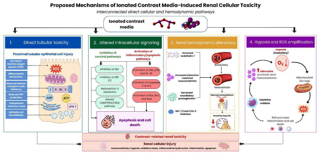
The renal toxicity of iodinated contrast media is mainly related to two interconnected mechanisms: direct cellular toxicity and alterations in renal hemodynamics. These processes involve oxidative stress, mitochondrial dysfunction, changes in intracellular survival and apoptotic pathways, and an imbalance between vasoconstrictor and vasodilatory mechanisms [12,13].
Iodinated contrast media may exert direct toxicity on proximal tubular epithelial cells. Reported alterations include increased production of reactive oxygen species (ROS), reduced efficiency of the mitochondrial electron transport chain and ATP production, endoplasmic reticulum stress, and epithelial cell vacuolization [12].
At the intracellular level, contrast media may inhibit cell survival pathways, including Akt and ERK1/2, and reduce Bcl-2 expression. They may also alter the cAMP/PKA/CREB pathway and activate proteins involved in inflammatory responses and cell death, such as JNK, p38, and NF-κB. Activation of caspases 3 and 9 and proapoptotic proteins such as Bax, Bim, and Bad promotes apoptosis and cell death [12,14].
Hypoxia and mitochondrial injury also promote ROS formation, both directly and through adenosine production and xanthine oxidase activation. Increased ROS contributes to cellular damage and inflammation. In addition, adenosine production promotes renal vasoconstriction, which may further intensify hypoxia and cellular injury [12,15].
Hemodynamic alterations also include increased endothelin-1 and decreased vasodilatory prostaglandins related to inhibition of ERK1/2 and COX-2. Together, increased vasoconstrictor mechanisms and reduced vasodilation contribute to decreased renal perfusion and amplification of cellular injury [16,17].
Therefore, the renal toxicity associated with iodinated contrast media appears to result from the interaction between vasoconstriction, hypoxia, oxidative stress, mitochondrial dysfunction, inhibition of cell survival pathways, and activation of inflammatory and apoptotic mechanisms.
Figure 1.
Proposed mechanisms of iodinated contrast media–induced renal cellular toxicity, including direct tubular injury, altered intracellular signaling, renal hemodynamic changes, medullary hypoxia, oxidative stress, and apoptosis.
Figure 1.
Proposed mechanisms of iodinated contrast media–induced renal cellular toxicity, including direct tubular injury, altered intracellular signaling, renal hemodynamic changes, medullary hypoxia, oxidative stress, and apoptosis.
3.4. Toxicity of Gadolinium-Based Contrast Agents
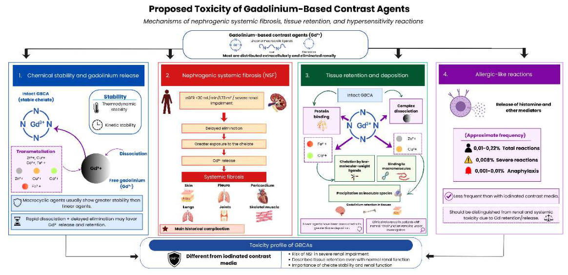
Gadolinium-based contrast agents, used in magnetic resonance imaging, have a toxicity profile that differs from that of iodinated contrast media. These agents consist of trivalent gadolinium (Gd3+) bound to linear or macrocyclic organic ligands, with the purpose of facilitating elimination and preventing the toxicity of free gadolinium in tissues. Their main historically recognized complication is nephrogenic systemic fibrosis, described particularly in patients with severe kidney impairment, especially those with an estimated glomerular filtration rate below 30 mL/min/1.73 m2. In this setting, delayed elimination of gadolinium chelates may promote transmetallation and the release of free gadolinium, a highly toxic form associated with scleroderma-like cutaneous fibrosis and systemic involvement of tissues such as the pleura, pericardium, lungs, joints, and skeletal muscle [18,19,20].
The chemical stability of gadolinium-based agents depends on two complementary properties: thermodynamic stability, which describes the tendency of the complex to release gadolinium under defined conditions, and kinetic stability, which refers to the rate at which gadolinium dissociates from the chelate. Rapid dissociation, combined with delayed elimination, may favor the release and retention of free gadolinium. One proposed mechanism for this release is transmetallation, a process in which chelated gadolinium is replaced by endogenous metal ions. Although macrocyclic agents generally have greater stability than linear agents, from a clinical perspective, gadolinium-based agents approved for human use bind gadolinium strongly and are considered stable and safe when used appropriately [18].
In addition to nephrogenic systemic fibrosis, trace gadolinium retention has been described in tissues such as the brain, bone, and skin, even in patients with preserved kidney function. The exact mechanisms of biological distribution and the chemical form of retained gadolinium have not been fully elucidated; however, several possible pathways have been proposed. These include persistence of the gadolinium-based agent in its intact form, protein binding, dissociation of the complex with release of free gadolinium, exchange with endogenous cations such as iron, calcium, zinc, or copper, precipitation as insoluble species, chelation by low-molecular-weight ligands, and binding to macromolecules. Together, these processes may contribute to tissue gadolinium retention, although its clinical relevance, particularly in patients with normal kidney function, remains an area of ongoing investigation [18,21,22].
Allergic-like reactions to gadolinium-based agents are uncommon and occur less frequently than those observed with iodinated contrast media. Overall reported rates are approximately 0.01–0.22%, with severe reactions occurring in around 0.008% of cases and anaphylaxis being extremely rare, estimated at 0.001–0.01% [6]. Although these reactions are relevant to patient safety, they should be distinguished from renal and systemic mechanisms related to gadolinium retention or release [23,24].
Figure 2.
Proposed toxicity mechanisms of gadolinium-based contrast agents, including chemical stability, gadolinium release, nephrogenic systemic fibrosis, tissue retention, and allergic-like reactions.
Figure 2.
Proposed toxicity mechanisms of gadolinium-based contrast agents, including chemical stability, gadolinium release, nephrogenic systemic fibrosis, tissue retention, and allergic-like reactions.
3.5. Prevention, Pharmacotherapeutic Management, and Clinical Decision-Making
3.5.1. Risk Assessment and Indication for Prophylaxis
Prophylaxis to reduce the risk of CA-AKI should not be applied universally to all patients exposed to iodinated contrast media, but should be reserved for those with greater renal vulnerability. According to contemporary recommendations, intravenous volume expansion is mainly considered in patients with active AKI or advanced chronic kidney disease, particularly when the estimated glomerular filtration rate is below 30 mL/min/1.73 m2 and the patient is not receiving chronic dialysis [24,25].
In patients with stable estimated glomerular filtration rate values equal to or greater than 30 mL/min/1.73 m2, prophylaxis is not routinely recommended, since the risk of clinically significant kidney injury attributable to modern intravenous contrast appears to be low. However, in patients with values between 30 and 44 mL/min/1.73 m2, the decision may be individualized when high-risk circumstances are present, such as recent AKI, multiple predisposing factors, clinical instability, borderline kidney function, or intra-arterial exposure during complex procedures [24,26].
The main prophylactic strategy is intravenous volume expansion with isotonic solutions, particularly 0.9% normal saline. Protocols are usually started approximately 1 hour before contrast administration and continued for 3 to 12 hours after the procedure. Longer regimens, closer to 12 hours, appear to be associated with a lower risk of CA-AKI compared with shorter protocols. Doses may be administered as fixed volumes, such as 500 mL before and after the procedure, or adjusted according to body weight, commonly in the range of 1 to 3 mL/kg/h [24,25].
Before indicating prophylaxis, the balance between the potential renal benefit and the risks associated with volume expansion should be assessed. This consideration is particularly relevant in patients with heart failure, reduced ejection fraction, volume overload, or hypervolemic conditions, in whom non-individualized hydration may precipitate congestion or clinical decompensation [27,28].
In patients receiving chronic dialysis, prophylaxis aimed at preventing CA-AKI is not routinely indicated. However, in non-anuric patients or those with residual kidney function, the decision should be individualized according to the clinical objective, the need to preserve residual function, contrast volume, and the patient’s volume status [27,28].
3.5.2. Adjustment of Prophylaxis in Patients at Risk of Volume Overload
In patients with heart failure, reduced ejection fraction, advanced chronic kidney disease, or signs of hypervolemia, prophylaxis with intravenous volume expansion should be individualized. Although 0.9% normal saline is the main preventive strategy, non-adjusted hydration may precipitate congestion, dyspnea, hypoxemia, or pulmonary edema. Therefore, before initiating hydration, volume status, cardiac function, diuretic use, and the presence of edema, orthopnea, or pulmonary crackles should be assessed [29,30].
In these patients, it may be necessary to reduce the infusion rate, shorten the duration of the protocol, or perform close clinical monitoring. Some protocols recommend reducing hydration to approximately 0.5 mL/kg/h in patients with left ventricular ejection fraction ≤35% or New York Heart Association functional class >II. When standard hydration is not safe, prevention should focus on optimizing hemodynamic status, avoiding nephrotoxic agents, minimizing contrast volume, and using low-osmolality or iso-osmolar contrast media [29,30].
3.5.3. Preventive Pharmacological Interventions
Several pharmacological interventions have been evaluated for the prevention of CA-AKI; however, none has demonstrated a sufficiently consistent clinical benefit to support routine use. Although some meta-analyses have reported reductions in creatinine-based outcomes, these findings have not been uniformly translated into clinically relevant benefits, such as a lower need for kidney replacement therapy, lower mortality, or reduced persistent kidney dysfunction. In addition, much of the evidence comes from studies in coronary angiography or percutaneous coronary intervention, limiting its applicability to contrast-enhanced computed tomography with intravenous contrast. This information is summarized in Table 1.
Taken together, pharmacological prevention should be considered an area of limited and heterogeneous evidence. The main preventive strategy remains the identification of high-risk patients, optimization of volume status, individualized isotonic hydration when indicated, reduction of unnecessary contrast exposure, and review of potentially nephrotoxic medications. In cardiovascular procedures, statin use should be guided by standard cardiometabolic indications and not solely by the intention to prevent contrast-associated kidney injury.
3.5.4. Pharmacotherapeutic Review: Metformin and Nephrotoxic Drugs
Table 2 summarizes practical recommendations for the prevention and clinical management of patients at risk of CA-AKI, integrating contrast selection, medication review, metformin management, and strategies that should not be routinely used. These recommendations include identifying high-risk patients, optimizing volume status, avoiding dehydration, appropriately selecting the type and volume of contrast, reviewing potentially nephrotoxic medications, and adjusting metformin management according to kidney function and the presence of AKI.
4. Limitations
This review has some limitations. First, because it is a narrative review, no formal risk-of-bias assessment or systematic grading of the certainty of evidence using tools such as GRADE was performed. Although a structured search strategy was used and guidelines, consensus statements, systematic reviews, meta-analyses, and clinically relevant studies were prioritized, the selection and interpretation of the evidence followed a qualitative approach.
Second, the available evidence on CA-AKI is heterogeneous. Studies use different definitions of AKI, different serum creatinine thresholds, diverse clinical populations, distinct routes of administration, and variable contrast exposures. In addition, many reported outcomes are based on transient changes in serum creatinine, which do not always translate into clinically relevant outcomes such as the need for kidney replacement therapy, mortality, or persistent kidney dysfunction.
Another important limitation is that part of the evidence on preventive pharmacological interventions comes from coronary procedures involving intra-arterial contrast, such as coronary angiography or percutaneous coronary intervention. Therefore, these findings are not always directly extrapolable to patients undergoing contrast-enhanced computed tomography with intravenous contrast, where both the risk and clinical context may differ.
Finally, this review incorporated recommendations from the KDIGO 2026 public review draft on acute kidney injury and acute kidney disease. This document does not yet correspond to the final official version of the guideline; therefore, its recommendations should be interpreted with caution until final publication. However, for the topics addressed in this review, such as the preferential use of isotonic saline, the lack of consistent benefit of prophylactic agents such as N-acetylcysteine, ascorbic acid, furosemide, dopamine, or fenoldopam, and the recommendation against prophylactic pericontrast hemodialysis, the draft recommendations are consistent with contemporary guidelines and consensus statements. Therefore, substantial changes are not anticipated in these specific areas, although this review should be updated in the future if the final KDIGO 2026 guideline or new clinical guidelines modify current recommendations.
5. Conclusions
CA-AKI remains a clinically relevant issue, but its interpretation requires precise conceptual differentiation. Contemporary evidence suggests that the risk of kidney injury directly attributable to modern intravenous contrast media was probably overestimated in historical studies, mainly because of the lack of adequate control groups and the presence of multiple confounding factors in hospitalized patients. In this context, it is essential to distinguish between CA-AKI, which describes a temporal relationship after exposure, and CI-AKI, which implies a direct causal relationship. This distinction is not merely terminological; it directly influences risk estimation, the indication for prophylaxis, and clinical decision-making.
Renal risk is not uniform across all patients. The greatest vulnerability is concentrated among individuals with active AKI, advanced chronic kidney disease, particularly those with an estimated glomerular filtration rate below 30 mL/min/1.73 m2, hemodynamic instability, heart failure, hypovolemia, complex intra-arterial exposure, or concomitant use of nephrotoxic medications. In contrast, clinically stable patients with preserved kidney function or mild to moderate chronic kidney disease have a low risk of clinically significant kidney injury attributable to modern intravenous contrast. Therefore, individualized risk stratification should be the central axis of any preventive strategy.
Prevention should be proportional to risk and focused on interventions with stronger clinical support. These include assessment of baseline kidney function, optimization of volume status, prevention of dehydration, use of low-osmolality or iso-osmolar contrast media, administration of the lowest diagnostically adequate dose, and avoidance of unnecessary repeated exposures. In high-risk patients without volume overload, volume expansion with 0.9% isotonic saline remains the main preventive intervention when indicated. However, this strategy should be individualized in patients with heart failure, reduced ejection fraction, or risk of congestion, in whom non-adjusted hydration may cause adverse effects.
Preventive pharmacological interventions have not demonstrated sufficiently consistent clinical benefit to support routine use. Although N-acetylcysteine, sodium bicarbonate, statins, theophylline, and ascorbic acid have shown favorable signals in some studies or meta-analyses, the results are heterogeneous and often limited to outcomes defined by changes in serum creatinine. To date, there is no solid evidence of a consistent reduction in clinically relevant outcomes such as the need for kidney replacement therapy, mortality, or persistent kidney dysfunction. Therefore, so-called “nephroprotective” drugs should not replace the fundamental measures based on risk stratification, individualized hydration, appropriate contrast selection, and pharmacotherapeutic review.
Clinical management should integrate the review of potentially nephrotoxic medications, metformin management according to kidney function and the presence of AKI, and individualized decision-making in complex patients. Temporary discontinuation of non-essential medications may be considered in high-risk patients when clinically feasible, whereas prophylactic pericontrast hemodialysis is not recommended. At the same time, clinically indicated contrast-enhanced studies should not be delayed or withheld without justification, especially in urgent scenarios in which the diagnostic or therapeutic benefit outweighs the potential renal risk.
In conclusion, renal safety associated with contrast media requires a balance between avoiding underestimation of risk in vulnerable patients and preventing unnecessary restriction of essential contrast-enhanced studies. Decision-making should be interdisciplinary and involve radiology, nephrology, internal medicine, emergency medicine, cardiology, and clinical pharmacy.
Author Contributions
Conceptualization, E.Z.-M., J.M.-J. and S.A.-C.; methodology, E.Z.-M., J.M.-J., S.A.-C. and K.C.-M.; investigation, E.Z.-M., J.M.-J., K.C.-M. and S.A.-C.; resources, E.Z.-M., L.G.H.-J. and J.M.C.-F.; data curation, E.Z.-M., J.M.-J. and K.C.-M.; formal analysis, E.Z.-M., J.M.-J. and S.A.-C.; writing—original draft preparation, E.Z.-M. and J.M.-J.; writing—review and editing, E.Z.-M., J.M.-J., K.C.-M., S.A.-C., L.G.H.-J. and J.M.C.-F.; visualization, E.Z.-M., J.M.-J. and K.C.-M.; supervision, J.M.C.-F. and E.Z.-M.; project administration, E.Z.-M. All authors contributed substantially to the development, critical revision, and final approval of the manuscript. All authors have read and agreed to the published version of the manuscript.
Funding
This research received no external funding.
Institutional Review Board Statement
Not applicable.
Informed Consent Statement
Not applicable.
Data Availability Statement
No new data were created or analyzed in this study. Data sharing is not applicable to this article.
Acknowledgments
The authors would like to thank Dr. Carrillo for her valuable support and for contributing additional perspectives from her experience as a pharmacist, which helped enrich the clinical and pharmacotherapeutic approach of this narrative review. During the preparation of this manuscript, the authors used ChatGPT (OpenAI) for language editing, translation assistance, and drafting support. The authors have reviewed and edited the output and take full responsibility for the content of this publication.
Conflicts of Interest
E.Z.-M., J.M.-J., K.C.-M. and S.A.-C. are affiliated with Clínica Bíblica, and L.G.H.-J. and J.M.C.-F. are affiliated with the University of Costa Rica. The authors declare that the research was conducted in the absence of any commercial or financial relationships that could be construed as a potential conflict of interest.
Abbreviations
The following abbreviations are used in this manuscript:
| ACEi | Angiotensin-converting enzyme inhibitor |
| ACR | American College of Radiology |
| ACR–NKF | American College of Radiology–National Kidney Foundation |
| AKD | Acute kidney disease |
| AKI | Acute kidney injury |
| ARB | Angiotensin II receptor blocker |
| ATP | Adenosine triphosphate |
| CA-AKI | Contrast-associated acute kidney injury |
| CI | Confidence interval |
| CI-AKI | Contrast-induced acute kidney injury |
| CKD | Chronic kidney disease |
| COX-2 | Cyclooxygenase-2 |
| CT | Computed tomography |
| eGFR | Estimated glomerular filtration rate |
| ERK1/2 | Extracellular signal-regulated kinases 1/2 |
| ESUR | European Society of Urogenital Radiology |
| GBCA | Gadolinium-based contrast agent |
| Gd3+ | Trivalent gadolinium |
| GRADE | Grading of Recommendations Assessment, Development and Evaluation |
| KDIGO | Kidney Disease: Improving Global Outcomes |
| KRT | Kidney replacement therapy |
| MRI | Magnetic resonance imaging |
| NAC | N-acetylcysteine |
| NF-κB | Nuclear factor kappa B |
| NICE | National Institute for Health and Care Excellence |
| NSAIDs | Nonsteroidal anti-inflammatory drugs |
| NYHA | New York Heart Association |
| OR | Odds ratio |
| PCI | Percutaneous coronary intervention |
| RASi | Renin-angiotensin system inhibitors |
| ROS | Reactive oxygen species |
| RR | Risk ratio |
References
- Clinical Safety Evaluation of Contrast Agents Based on Real-world Evidence - Jiang - 2021 - Journal of Clinical Pharmacy and Therapeutics - Wiley Online Library. Available online: https://onlinelibrary.wiley.com/doi/10.1111/jcpt.13485 (accessed on 12 June 2026).
- Schöckel, L.; Jost, G.; Seidensticker, P.; Lengsfeld, P.; Palkowitsch, P.; Pietsch, H. Developments in X-Ray Contrast Media and the Potential Impact on Computed Tomography. Invest Radiol. 2020, 55, 592–597. [Google Scholar] [CrossRef] [PubMed]
- Andersen, P.E. Patient Selection and Preparation Strategies for the Use of Contrast Material in Patients with Chronic Kidney Disease. World J. Radiol. 2012, 4, 253–257. [Google Scholar] [CrossRef] [PubMed]
- Mehran, R.; Dangas, G.D.; Weisbord, S.D. Contrast-Associated Acute Kidney Injury. N. Engl. J. Med. 2019, 380, 2146–2155. [Google Scholar] [CrossRef] [PubMed]
- Mehran, R.; Owen, R.; Chiarito, M.; Baber, U.; Sartori, S.; Cao, D.; Nicolas, J.; Pivato, C.A.; Nardin, M.; Krishnan, P.; et al. A Contemporary Simple Risk Score for Prediction of Contrast-Associated Acute Kidney Injury after Percutaneous Coronary Intervention: Derivation and Validation from an Observational Registry. Lancet 2021, 398, 1974–1983. [Google Scholar] [CrossRef] [PubMed]
- Yi, T.; O’Hara, D.V.; Levin, A. Global Kidney Health: Are We Failing the Silent Pandemic? J. Intern. Med. 2026, 300, 26–38. [Google Scholar] [CrossRef] [PubMed]
- Weisbord, S.D.; Gallagher, M. Iodinated Contrast and Acute Kidney Injury. In Evidence-Based Nephrology; John Wiley & Sons, Ltd., 2022; pp. 145–162. ISBN 978-1-119-10595-4. [Google Scholar]
- McDonald, J.S.; McDonald, R.J.; Comin, J.; Williamson, E.E.; Katzberg, R.W.; Murad, M.H.; Kallmes, D.F. Frequency of Acute Kidney Injury Following Intravenous Contrast Medium Administration: A Systematic Review and Meta-Analysis. Radiology 2013, 267, 119–128. [Google Scholar] [CrossRef] [PubMed]
- Obed, M.; Gabriel, M.M.; Dumann, E.; Vollmer Barbosa, C.; Weißenborn, K.; Schmidt, B.M.W. Risk of Acute Kidney Injury after Contrast-Enhanced Computerized Tomography: A Systematic Review and Meta-Analysis of 21 Propensity Score-Matched Cohort Studies. Eur. Radiol. 2022, 32, 8432–8442. [Google Scholar] [CrossRef] [PubMed]
- Ludwig, U.; Keller, F. Prophylaxis of Contrast-Induced Nephrotoxicity. BioMed Res. Int. 2014, 2014, 308316. [Google Scholar] [CrossRef] [PubMed]
- Mehran, R.; Nikolsky, E. Contrast-Induced Nephropathy: Definition, Epidemiology, and Patients at Risk. Kidney Int. Suppl. 2006, S11–15. [Google Scholar] [CrossRef] [PubMed]
- Molecular Mechanisms of Renal Cellular Nephrotoxicity Due to Radiocontrast Media - Michael - 2014 - BioMed Research International - Wiley Online Library. Available online: https://onlinelibrary.wiley.com/doi/10.1155/2014/249810 (accessed on 24 June 2026).
- Ward, D.B.; Valentovic, M.A. Contrast Induced Acute Kidney Injury and Direct Cytotoxicity of Iodinated Radiocontrast Media on Renal Proximal Tubule Cells. J. Pharmacol. Exp. Ther. 2025, 370, 160–171. [Google Scholar] [CrossRef] [PubMed]
- Yano, T.; Itoh, Y.; Kubota, T.; Sendo, T.; Oishi, R. A Prostacyclin Analog Beraprost Sodium Attenuates Radiocontrast Media-Induced LLC-PK1 Cells Injury. Kidney Int. 2004, 65, 1654–1663. [Google Scholar] [CrossRef] [PubMed]
- Vasoconstrictor and Vasodilator Effects of Adenosine in the Kidney | American Journal of Physiology-Renal Physiology | American Physiological Society. Available online: https://journals.physiology.org/doi/full/10.1152/ajprenal.00051.2003?rfr_dat=cr_pub++0pub (accessed on 24 June 2026).
- Jabbour, H.N.; Sales, K.J.; Boddy, S.C.; Anderson, R.A.; Williams, A.R.W. A Positive Feedback Loop That Regulates Cyclooxygenase-2 Expression and Prostaglandin F2α Synthesis via the F-Series-Prostanoid Receptor and Extracellular Signal-Regulated Kinase 1/2 Signaling Pathway. Endocrinology 2005, 146, 4657–4664. [Google Scholar] [CrossRef] [PubMed]
- Endothelin-1 Gene Regulation - Stow - 2011 - The FASEB Journal - Wiley Online Library. Available online: https://faseb.onlinelibrary.wiley.com/doi/10.1096/fj.10-161612 (accessed on 24 June 2026).
- Starekova, J.; Pirasteh, A.; Reeder, S.B. Update on Gadolinium-Based Contrast Agent Safety, From the AJR Special Series on Contrast Media. AJR Am. J. Roentgenol. 2024, 223, e2330036. [Google Scholar] [CrossRef] [PubMed]
- Blomqvist, L.; Nordberg, G.F.; Nurchi, V.M.; Aaseth, J.O. Gadolinium in Medical Imaging-Usefulness, Toxic Reactions and Possible Countermeasures-A Review. Biomolecules 2022, 12, 742. [Google Scholar] [CrossRef] [PubMed]
- American College of Cardiology Foundation Task Force on Expert Consensus Documents; Hundley, W.G.; Bluemke, D.A.; Finn, J.P.; Flamm, S.D.; Fogel, M.A.; Friedrich, M.G.; Ho, V.B.; Jerosch-Herold, M.; Kramer, C.M.; et al. ACCF/ACR/AHA/NASCI/SCMR 2010 Expert Consensus Document on Cardiovascular Magnetic Resonance: A Report of the American College of Cardiology Foundation Task Force on Expert Consensus Documents. J. Am. Coll. Cardiol. 2010, 55, 2614–2662. [Google Scholar] [CrossRef] [PubMed]
- Frenzel, T.; Apte, C.; Jost, G.; Schöckel, L.; Lohrke, J.; Pietsch, H. Quantification and Assessment of the Chemical Form of Residual Gadolinium in the Brain After Repeated Administration of Gadolinium-Based Contrast Agents: Comparative Study in Rats. Invest Radiol. 2017, 52, 396–404. [Google Scholar] [CrossRef] [PubMed]
- Radbruch, A. Gadolinium Deposition in the Brain: We Need to Differentiate between Chelated and Dechelated Gadolinium. Radiology 2018, 288, 434–435. [Google Scholar] [CrossRef] [PubMed]
- Wang, P.I.; Chong, S.T.; Kielar, A.Z.; Kelly, A.M.; Knoepp, U.D.; Mazza, M.B.; Goodsitt, M.M. Imaging of Pregnant and Lactating Patients: Part 1, Evidence-Based Review and Recommendations. AJR Am. J. Roentgenol. 2012, 198, 778–784. [Google Scholar] [CrossRef] [PubMed]
- ACR Manual on Contrast Media. Available online: https://www.acr.org/Clinical-Resources/Clinical-Tools-and-Reference/Contrast-Manual (accessed on 25 June 2026).
- VA.Gov | Veterans Affairs. Available online: https://www.healthquality.va.gov/guidelines/cd/ckd/index.asp (accessed on 29 June 2026).
- Davenport, M.S.; Perazella, M.A.; Yee, J.; Dillman, J.R.; Fine, D.; McDonald, R.J.; Rodby, R.A.; Wang, C.L.; Weinreb, J.C. Use of Intravenous Iodinated Contrast Media in Patients with Kidney Disease: Consensus Statements from the American College of Radiology and the National Kidney Foundation. Radiology 2020, 294, 660–668. [Google Scholar] [CrossRef] [PubMed]
- KDIGO 2024 Clinical Practice Guideline for the Evaluation and Management of Chronic Kidney Disease - Kidney International. Available online: https://www.kidney-international.org/article/S0085-2538(23)00766-4/fulltext (accessed on 29 June 2026).
- McCullough, P.A.; Choi, J.P.; Feghali, G.A.; Schussler, J.M.; Stoler, R.M.; Vallabahn, R.C.; Mehta, A. Contrast-Induced Acute Kidney Injury. J. Am. Coll. Cardiol. 2016, 68, 1465–1473. [Google Scholar] [CrossRef] [PubMed]
- Mehran, R.; Dangas, G.D.; Weisbord, S.D. Contrast-Associated Acute Kidney Injury. N. Engl. J. Med. 2019, 380, 2146–2155. [Google Scholar] [CrossRef] [PubMed]
- Briguori, C.; D’Amore, C.; De Micco, F.; Signore, N.; Esposito, G.; Visconti, G.; Airoldi, F.; Signoriello, G.; Focaccio, A. Left Ventricular End-Diastolic Pressure Versus Urine Flow Rate-Guided Hydration in Preventing Contrast-Associated Acute Kidney Injury. JACC Cardiovasc Interv. 2020, 13, 2065–2074. [Google Scholar] [CrossRef] [PubMed]
- Subramaniam, R.M.; Suarez-Cuervo, C.; Wilson, R.F.; Turban, S.; Zhang, A.; Sherrod, C.; Aboagye, J.; Eng, J.; Choi, M.J.; Hutfless, S.; et al. Effectiveness of Prevention Strategies for Contrast-Induced Nephropathy: A Systematic Review and Meta-Analysis. Ann. Intern Med. 2016, 164, 406–416. [Google Scholar] [CrossRef] [PubMed]
- Walker, H.; Guthrie, G.D.; Lambourg, E.; Traill, P.; Zealley, I.; Plumb, A.; Bell, S. Systematic Review and Meta-Analysis of Prophylaxis Use with Intravenous Contrast Exposure to Prevent Contrast-Induced Nephropathy. Eur. J. Radiol. 2022, 153, 110368. [Google Scholar] [CrossRef] [PubMed]
- Su, X.; Xie, X.; Liu, L.; Lv, J.; Song, F.; Perkovic, V.; Zhang, H. Comparative Effectiveness of 12 Treatment Strategies for Preventing Contrast-Induced Acute Kidney Injury: A Systematic Review and Bayesian Network Meta-Analysis. Am. J. Kidney Dis. 2017, 69, 69–77. [Google Scholar] [CrossRef] [PubMed]
- Acute Kidney Injury (AKI) and Acute Kidney Disease (AKD) – KDIGO. Available online: https://kdigo.org/guidelines/acute-kidney-injury/ (accessed on 29 June 2026).
- Macdonald, D.B.; Hurrell, C.; Costa, A.F.; McInnes, M.D.F.; O’Malley, M.E.; Barrett, B.; Brown, P.A.; Clark, E.G.; Hadjivassiliou, A.; Kirkpatrick, I.D.C.; et al. Canadian Association of Radiologists Guidance on Contrast Associated Acute Kidney Injury. Can. Assoc. Radiol. J. 2022, 73, 499–514. [Google Scholar] [CrossRef] [PubMed]
- Notice. Kidney Int. Suppl. 2012, 2, 1. [CrossRef]
- Cho, Aj.; Lee, Y.-K.; Sohn, S.Y. Beneficial Effect of Statin on Preventing Contrast-Induced Acute Kidney Injury in Patients with Renal Insufficiency: A Meta-Analysis. Medicine 2020, 99, e19473. [Google Scholar] [CrossRef] [PubMed]
- Lee, J.M.; Park, J.; Jeon, K.-H.; Jung, J.-H.; Lee, S.E.; Han, J.-K.; Kim, H.-L.; Yang, H.-M.; Park, K.W.; Kang, H.-J.; et al. Efficacy of Short-Term High-Dose Statin Pretreatment in Prevention of Contrast-Induced Acute Kidney Injury: Updated Study-Level Meta-Analysis of 13 Randomized Controlled Trials. PLoS ONE 2014, 9, e111397. [Google Scholar] [CrossRef] [PubMed]
- Effects of Saline, Mannitol, and Furosemide on Acute Decreases in Renal Function Induced by Radiocontrast Agents | New England Journal of Medicine. Available online: https://www.nejm.org/doi/full/10.1056/NEJM199411243312104?utm_source=openevidence (accessed on 29 June 2026).
- Diuretics for Preventing and Treating Acute Kidney Injury - Hashimoto, H - 2025 | Cochrane Library. Available online: https://www.cochranelibrary.com/cdsr/doi/10.1002/14651858.CD014937.pub2/full (accessed on 29 June 2026).
- Meta-Analysis of the Effects of Furosemide Combined with Hydration Therapy on Contrast-Induced Acute Kidney Injury after Coronary Intervention - PubMed. Available online: https://pubmed.ncbi.nlm.nih.gov/34337720/ (accessed on 29 June 2026).
- Stone, G.W.; McCullough, P.A.; Tumlin, J.A.; Lepor, N.E.; Madyoon, H.; Murray, P.; Wang, A.; Chu, A.A.; Schaer, G.L.; Stevens, M.; et al. Fenoldopam Mesylate for the Prevention of Contrast-Induced Nephropathy: A Randomized Controlled Trial. JAMA 2003, 290, 2284–2291. [Google Scholar] [CrossRef] [PubMed]
- Fenoldopam for Preventing and Treating Acute Kidney Injury - Esezobor, CI - 2024 | Cochrane Library. Available online: https://www.cochranelibrary.com/cdsr/doi/10.1002/14651858.CD012905.pub2/full (accessed on 29 June 2026).
- Lauschke, A.; Teichgräber, U.K.M.; Frei, U.; Eckardt, K.-U. ‘Low-Dose’ Dopamine Worsens Renal Perfusion in Patients with Acute Renal Failure. Kidney Int. 2006, 69, 1669–1674. [Google Scholar] [CrossRef] [PubMed]
- Friedrich, J.O.; Adhikari, N.; Herridge, M.S.; Beyene, J. Meta-Analysis: Low-Dose Dopamine Increases Urine Output but Does Not Prevent Renal Dysfunction or Death. Ann. Intern Med. 2005, 142, 510–524. [Google Scholar] [CrossRef] [PubMed]
- Navarese, E.P.; Gurbel, P.A.; Andreotti, F.; Kołodziejczak, M.M.; Palmer, S.C.; Dias, S.; Buffon, A.; Kubica, J.; Kowalewski, M.; Jadczyk, T.; et al. Prevention of Contrast-Induced Acute Kidney Injury in Patients Undergoing Cardiovascular Procedures-a Systematic Review and Network Meta-Analysis. PLoS ONE 2017, 12, e0168726. [Google Scholar] [CrossRef] [PubMed]

Table 1.
Summary of information by drug as a prophylactic benefit prior to the use of contrast media.
Table 1.
Summary of information by drug as a prophylactic benefit prior to the use of contrast media.
| Intervention | Summarized evidence | Clinical interpretation | Guideline-based practical recommendation |
|---|---|---|---|
| N-acetylcysteine (NAC) | The evidence is inconsistent. A 2016 systematic review found that low-dose NAC plus intravenous saline reduced contrast-induced nephropathy compared with saline alone (RR 0.75; 95% CI: 0.63–0.89), albeit with low certainty of evidence. In patients receiving low-osmolality contrast media, NAC plus saline also showed a risk reduction (RR 0.69; 95% CI: 0.58–0.84) with moderate certainty [31]. A 2022 systematic review focused on intravenous contrast reported a reduction in the incidence of contrast-induced nephropathy (risk difference −0.07; 95% CI: −0.13 to −0.01), but no reduction in renal replacement therapy, mortality, or persistent renal failure [32]. A 2017 network meta-analysis found a lower risk with NAC alone versus hydration (OR 0.67; 95% CI: 0.54–0.81) [33]. | Although some meta-analyses show a reduction in creatinine-defined outcomes, the clinical benefit is uncertain. NAC may increase tubular secretion of creatinine, which makes changes in serum creatinine difficult to interpret and may create a false impression of nephroprotection. | Not recommended routinely as pharmacologic prophylaxis. KDIGO 2026 notes that NAC has not demonstrated a consistent benefit as a preventive measure for CA-AKI [34]. Similarly, the Canadian Association of Radiologists guideline does not recommend the use of NAC for CA-AKI prophylaxis [35]. |
| Sodium bicarbonate | KDIGO 2012 considered sodium bicarbonate an alternative to isotonic saline for volume expansion in at-risk patients [36]. However, subsequent evidence has not demonstrated superiority of bicarbonate over saline. A 2022 systematic review found no significant reduction in contrast-induced nephropathy compared with saline (risk difference −0.02; 95% CI: −0.04 to 0.01) [32], and a network meta-analysis showed a non-significant trend favoring bicarbonate (OR 0.78; 95% CI: 0.59–1.01) [33]. Consistently, KDIGO 2026 recommends periprocedural volume expansion with 0.9% saline over sodium bicarbonate, hypotonic solutions, or no hydration in high-risk adults without volume overload [34]. | Bicarbonate had pathophysiologic plausibility and initial interest, but it has not demonstrated clinical superiority over 0.9% saline. Therefore, the use of 0.9% saline is recommended instead of hypotonic solutions for periprocedural volume expansion. | Do not prefer over 0.9% normal saline. In high-risk adults without volume overload, KDIGO 2026 favors periprocedural expansion with 0.9% saline over bicarbonate, hypotonic solutions, or no hydration [34]. Bicarbonate could be considered only if there is an institutional protocol or a specific clinical condition, such as metabolic acidosis, but not as a superior or routine strategy to prevent CA-AKI [35]. |
| Statins | The evidence suggests a potential benefit mainly in patients undergoing coronary angiography or percutaneous coronary intervention. A meta-analysis in patients with mild-to-moderate renal impairment found that statin pretreatment significantly reduced the risk of CA-AKI (RR 0.59; 95% CI: 0.44–0.79; p=0.0003) [37]. Another meta-analysis in coronary catheterization reported a similar risk reduction (RR 0.54; 95% CI: 0.38–0.78; p=0.001) [38]. In addition, high-dose statin use reduced the incidence of CA-AKI (RR 0.45; 95% CI: 0.35–0.57; NNT=16) [38]. A network meta-analysis showed a benefit with high-dose statins plus NAC (OR 0.31; 95% CI: 0.14–0.60) and with high-dose statins alone (OR 0.37; 95% CI: 0.19–0.64), whereas low-dose statins showed no significant benefit (OR 0.98; 95% CI: 0.41–2.07) [33]. | Statins, especially at high doses, could reduce creatinine-defined CA-AKI events in patients undergoing coronary procedures, particularly in acute coronary syndrome, chronic kidney disease, or exposure to high contrast volumes. However, the evidence comes almost exclusively from intra-arterial contrast in interventional cardiology, with little or no evidence in contrast-enhanced computed tomography with intravenous contrast. Moreover, a reduction in clinically relevant outcomes, such as hemodialysis or mortality, has not been consistently demonstrated. | Do not start routinely solely to prevent CA-AKI. Statins should be used when there is a cardiovascular indication. In patients undergoing coronary angiography or percutaneous coronary intervention who already have an indication for high-intensity statin therapy, their use could provide an additional renal benefit. There is insufficient evidence to recommend them as general pharmacologic prophylaxis in patients receiving intravenous contrast for computed tomography. |
| Furosemide and mannitol | The evidence does not support the use of furosemide or mannitol as CA-AKI prophylaxis. In the trial by Solomon et al., conducted in patients with chronic kidney disease undergoing cardiac angiography, mannitol added to saline showed no benefit in patients with or without diabetes, whereas furosemide added to saline was associated with greater acute deterioration of renal function [39]. A 2025 Cochrane review on diuretics for AKI prevention found very-low-certainty evidence for furosemide (RR 0.82; 95% CI: 0.58–1.16) and mannitol (RR 0.76; 95% CI: 0.42–1.38), without demonstrating a clear benefit [40]. In patients undergoing coronary intervention, a meta-analysis of furosemide plus hydration also showed no significant reduction in CI-AKI (OR 0.85; 95% CI: 0.46–1.60; p=0.62) [41]. | The use of diuretics as prophylaxis may be counterproductive if it promotes hypovolemia, renal hemodynamic changes, or reduced renal perfusion. Although forced-diuresis strategies with controlled fluid replacement, such as RenalGuard, have been evaluated in coronary angiography, the evidence is heterogeneous and does not allow recommending this approach as standard. There is insufficient specific evidence in contrast-enhanced computed tomography with intravenous contrast. |
Not recommended routinely to prevent CA-AKI. [35]. Contemporary guidelines advise against the prophylactic use of furosemide or mannitol. Isotonic saline alone is preferable to the combination of saline with mannitol or furosemide, unless there is an independent clinical indication to treat volume overload [35]. |
| Fenoldopam | The evidence does not demonstrate a benefit for the prevention of CA-AKI. In the CONTRAST trial, conducted in patients with chronic kidney disease undergoing coronary or peripheral angiography, fenoldopam did not reduce the incidence of contrast-induced renal injury versus placebo (33.6% vs 30.1%; p=0.61) [42]. A Cochrane review in procedures with radiologic contrast likewise showed no reduction in AKI (RR 1.00; 95% CI: 0.70–1.42) or in renal replacement therapy (RR 1.36; 95% CI: 0.31–5.97) [43]. | Although fenoldopam has physiologic plausibility owing to its renal vasodilator effect, clinical studies have not demonstrated a preventive benefit. In addition, it may cause systemic hypotension and tachycardia, potentially harmful effects in high-risk patients. The available evidence comes mainly from intra-arterial procedures, without sufficient data in contrast-enhanced computed tomography with intravenous contrast. | Not recommended for CA-AKI prophylaxis [35]. Current guidelines, including KDIGO and ACR, do not recommend fenoldopam to prevent contrast-associated renal injury because of the lack of consistent benefit and the risk of adverse effects [34]. |
| Dopamine | Low-dose dopamine has not demonstrated a consistent benefit as a preventive measure for CA-AKI [35,44]. KDIGO 2026 notes that dopamine has not shown a consistent preventive benefit and, furthermore, recommends against using low-dose dopamine as a renal protection strategy in patients at risk of AKI or with AKI/AKD (1A) [34,45]. | It does not prevent AKI, does not improve renal recovery, and does not reduce the need for renal replacement therapy; in addition, it may be associated with adverse effects. | Not recommended as a renoprotective strategy or for CA-AKI prophylaxis [34]. It should be used only if there is an independent hemodynamic indication. |
| Theophylline | KDIGO 2012 suggests not using theophylline to prevent contrast-induced renal injury [36]. Although a network meta-analysis showed a reduction in CA-AKI with methylxanthines versus saline (OR 0.48; 95% CI: 0.26–0.82) and an association with lower mortality (OR 0.12; 95% CI: 0.01–0.94), these findings have not been incorporated as a routine recommendation by the major guidelines [46]. | Theophylline has physiologic plausibility owing to its effect on adenosine-mediated vasoconstriction; however, the evidence is heterogeneous, limited, and not robust enough to change clinical practice. In addition, its safety profile may be problematic because of the risk of cardiovascular and neurologic adverse effects. | Not recommended routinely for CA-AKI prophylaxis [35]. Despite some favorable signals in meta-analyses, the ACR and KDIGO guidelines do not recommend its systematic preventive use because of the heterogeneity of the evidence, the absence of consistent clinical benefit, and its safety profile [34]. |
| Ascorbic acid / vitamin C | A 2016 systematic review found that ascorbic acid versus intravenous saline showed a clinically favorable but not statistically significant difference (RR 0.72; 95% CI: 0.48–1.01) [31]. Although a network meta-analysis showed a lower risk with vitamins and analogues compared with standard hydration alone (OR 0.64; 95% CI: 0.41–0.95), the overall evidence remains inconsistent and does not demonstrate a consistent clinical benefit [33]. | Although it has been proposed for its potential antioxidant effect, the available evidence does not demonstrate a consistent clinical benefit or a clear reduction in relevant outcomes such as dialysis, mortality, or persistent renal deterioration. | Not recommended routinely for CA-AKI prophylaxis [34]. It may be mentioned as a studied intervention, but it should not be proposed as a standard preventive strategy [35]. |
Table 2.
Practical recommendations for the management of patients at risk of CA-AKI.
| Intervention | Summarized evidence |
|---|---|
| Differentiate intravenous from intra-arterial contrast | Risk and preventive measures should be individualized according to the route of administration, the clinical context, and the type of procedure. Intra-arterial procedures, especially complex cardiovascular ones, usually require a stricter risk assessment [34]. |
| Use low-osmolality or iso-osmolar contrast media | In patients at risk of CA-AKI, it is recommended to avoid high-osmolality media and to use modern low-osmolality or iso-osmolar agents, according to availability, cost, and procedure characteristics [34]. |
| Use the minimum necessary contrast dose | The smallest contrast volume that allows an adequate diagnostic image should be used. Excessive dose reduction must not compromise diagnostic quality or delay important clinical decisions [34]. |
| Discontinue non-essential nephrotoxic drugs in high-risk patients | In patients with AKI or eGFR <30 mL/min/1.73 m2, temporarily withdrawing potentially nephrotoxic, non-essential medications may be considered, such as NSAIDs, diuretics, aminoglycosides, amphotericin B, platinum agents, zoledronate, or methotrexate, approximately 24–48 hours before and 48 hours after contrast exposure, when clinically feasible [34]. |
| Metformin management | In patients with eGFR >30 mL/min/1.73 m2 and no evidence of AKI, metformin does not need to be discontinued before contrast, and routine subsequent monitoring of renal function is not required [34]. In patients with AKI or eGFR ≤30 mL/min/1.73 m2, metformin should be discontinued at the time of or before contrast administration and restarted after at least 48 hours, only if renal function remains stable and the clinical team reassesses its continuation [33]. |
| Avoid dehydration in vulnerable patients | In non-dialysis patients with eGFR <30 mL/min/1.73 m2 or AKI receiving intravenous contrast, dehydration should be avoided and volume status optimized, considering the risk of fluid overload [35]. |
| Do not use renoprotective drugs routinely | N-acetylcysteine, ascorbic acid, furosemide, dopamine, fenoldopam, and calcium channel blockers have not demonstrated a consistent preventive benefit for CA-AKI, so they should not be used routinely as pharmacologic prophylaxis [35]. |
| Do not perform prophylactic peri-contrast hemodialysis | Prophylactic hemodialysis after contrast is not recommended, as it has not demonstrated benefit and may be potentially harmful [34]. |
| Do not routinely discontinue ACEi/ARB | KDIGO suggests not routinely discontinuing renin-angiotensin system inhibitors before iodinated contrast procedures in adults. The decision should be individualized according to blood pressure, volume status, renal function, and clinical context [34]. |
Disclaimer/Publisher’s Note: The statements, opinions and data contained in all publications are solely those of the individual author(s) and contributor(s) and not of MDPI and/or the editor(s). MDPI and/or the editor(s) disclaim responsibility for any injury to people or property resulting from any ideas, methods, instructions or products referred to in the content. |
© 2026 by the authors. Licensee MDPI, Basel, Switzerland. This article is an open access article distributed under the terms and conditions of the Creative Commons Attribution (CC BY) license (http://creativecommons.org/licenses/by/4.0/).
Copyright: This open access article is published under a Creative Commons CC BY 4.0 license, which permit the free download, distribution, and reuse, provided that the author and preprint are cited in any reuse.


