
Submitted:
01 July 2026
Posted:
03 July 2026
You are already at the latest version
Abstract
Accurate segmentation of non-small cell lung cancer (NSCLC) on positron emission tomography/computed tomography (PET/CT) is a critical foundation for automated metabolic tumor volume (MTV) quantification and staging. Although deep learning models achieve high performance on large-scale datasets, their generalization across different clinical domains remains challenged by variations in imaging protocols and patient demographics. This study aims to evaluate several deep learning architectures and investigate a transfer learning strategy to mitigate domain shift on a dataset of 400 PET/CT scans from Vietnamese NSCLC patient cohort. Three architectures, including 3D U-Net, nnU-Net v2, and Swin UNETR, were benchmarked from scratch and compared with a fine-tuned nnU-Net initialized with AutoPET II weights. Results on the internal dataset showed that the fine-tuned nnU-Net achieved a Dice similarity coefficient (DSC) of 83.4 ± 6.5%, a 95% Hausdorff distance (HD95) of 5.1 ± 3.6 mm, and precision of 89.6 ± 8.2%. Compared to the nnU-Net v2, the fine-tuned nnU-Net improved the absolute DSC by 6.8% while reducing the training time by 37.5%. The fine-tuned nnU-Net model also demonstrated a good correlation between the MTV and the ground truth (Pearson r = 0.96, p < 0.001), indicating its potential as a reliable automated approach for quantitative MTV extraction and NSCLC prognostic related analysis.
Keywords:
NSCLC
; PET/CT
; deep learning
; nnU-Net
; domain shift
; transfer learning
; tumor segmentation
1. Introduction
Non-small cell lung cancer (NSCLC) is a leading cause of cancer-related mortality globally, with a significantly increasing incidence in Asian countries, including Vietnam. In modern clinical diagnostics, 18F-FDG positron emission tomography/computed tomography (PET/CT) plays a crucial role in tumor staging and treatment response assessment by synergizing anatomical details from CT with metabolic characteristics from PET. This multimodal integration has been proven to improve diagnostic accuracy by 20% to 30% compared to standalone CT [1]. However, manual 3D tumor segmentation remains a substantial challenge: it is time-consuming, highly subjective, and suffers from significant inter-observer variability [2]. Such inconsistencies directly compromise the reliability of quantitative biomarkers like metabolic tumor volume (MTV) and total lesion glycolysis (TLG), which are critical for prognosticating patient outcomes [3].
The growth of deep learning, particularly self-configuring architectures like nnU-Net, has established new state-of-the-art benchmarks in medical image segmentation [4]. Recent international challenges, such as AutoPET, have demonstrated that automated models can achieve a Dice similarity coefficient (DSC) of approximately 0.80 on large-scale datasets [5,6]. Despite these advancements, a generalization gap appears when deploying these models across different clinical environments. The performance of PET-based segmentation algorithms is influenced by domain shift, which originates not only from variations in patient demographics and pathological characteristics but also from differences in imaging hardware, acquisition protocols, and reconstruction methods across institutions. [7].
Furthermore, the incompliance of histologically confirmed datasets in contemporary AI research fundamentally limits the assessment of true clinical accuracy [8]. While several large-scale public PET/CT datasets exist (e.g., AutoPET, LUNG-PET-CT-Dx), they often lack comprehensive histological validation or remain unoptimized for evaluating domain shift across diverse ethnicities [8]. To address these limitations, our study introduces and benchmarks a large-scale dataset comprising 400 PET/CT scans from a Vietnamese patient cohort [9]. The novelty of this research lies in three primary contributions: (1) all of the NSCLC cases are confirmed via histopathological biopsy; (2) A rigorous multi-rater annotation protocol involving three experienced physicians, coupled with a probabilistic consensus algorithm STAPLE to mitigate subjective bias [10,11]; and (3) The implementation of specialized preprocessing pipelines to maximize tumor-to-background signal ratios.
Conducting research on this dataset not only enhances the reliability of the model but also contributes to efforts to eliminate inequalities regarding ethnic diversity in global medical AI development by conducting benchmarks for current state of the art algorithms on this Vietnamese PET/CT dataset [13,14], evaluating domain shift and cross-generalization capabilities through testing on international public datasets [6,15], and analyzing the correlation between AI-predicted volumetric metrics and actual clinical outcomes, strictly adhering to the latest biomedical evaluation guidelines [16].
2. Materials and Methods
The framework for automated NSCLC segmentation is shown in Figure 1. The evaluated framework operates in three sequential steps. First, in the data preparation stage, raw 3D PET/CT images are pre-processed and a consensus ground truth is generated from three expert annotations using the STAPLE algorithm, followed by conversion into NiFTI format. Then, in the training stage, the dataset is augmented and evaluated using stratified 5-fold cross-validation. The fine-tuned nnU-Net v2 is trained and compared with baseline models (nnU-Net v2, Swin UNETR, and 3D U-Net) under a unified configuration (hybrid loss, patch size of 128×128×128, AdamW optimizer). Finally, in the inference and evaluation stage, predictions are generated using a sliding-window strategy with test-time augmentation, followed by post-processing to obtain final 3D predicted masks. Model performance is then assessed using spatial metrics (DSC, HD95, ASSD) and clinical relevance via MTV correlation.
2.1. Dataset Preparation
This study utilized an extended version of the Vietnamese NSCLC PET/CT dataset [9]. Data were collected between June 2019 and June 2023 from three leading oncology centers: Vietnam National Cancer Hospital, Hanoi Oncology Hospital, and Ho Chi Minh City Oncology Hospital. Clinicopathological characteristics of the patient cohort and PET/CT dataset are shown in Table 1. A total of 416 whole-body PET/CT scans were initially retrieved. After excluding 16 cases with respiratory motion artifacts, the final database comprising 400 cases was included for analysis. This cohort consisted of 284 NSCLC patients (with a total of 342 primary and metastatic lesions, averaging 1.2 lesions per patient) and 116 negative controls comprising healthy individuals or those with benign pathologies. Retaining the 116 negative control cases was important as it was designed to evaluate the model’s capacity to control the false-positive rate in regions with physiological FDG uptake, analogous to the design of the AutoPET dataset. All 284 NSCLC cases were validated using the gold standard of histopathology via biopsy or surgery.
To address the imaging domain characteristics, the acquisition parameters across the participating centers were standardized. Following standard clinical preparation, patients were instructed to fast for at least 6 hours prior to the scan, and blood glucose levels were verified to be within normal limits. 18F-FDG was administered intravenously with a weight-based activity ranging from 3.7 to 5.5 MBq/kg, followed by a standardized uptake time of approximately 60 minutes.
Whole-body PET/CT scans were primarily performed using Siemens Biograph mCT systems across the Vietnam National Cancer Hospital, Hanoi Oncology Hospital, and Ho Chi Minh City Oncology Hospital. Unenhanced low-dose CT scans were acquired for attenuation correction and anatomical localization. The native CT images were reconstructed with a matrix size of 512 × 512 pixels, an in-plane pixel spacing of 1.037 × 1.037 mm2, and a slice thickness of 3.26 mm. Consequently, the PET data were acquired in 3D mode and reconstructed with a matrix size of 192 × 192 pixels, an in-plane resolution of 3.646 × 3.646 mm2, and a matching slice thickness of 3.26 mm.
Figure 2 illustrates the multimodal imaging inputs and the rigorous ground-truth generation workflow utilized in this study. By fusing anatomical details from unenhanced CT scans with metabolic activity from 18F-FDG PET, NSCLC lesions were precisely localized and subsequently manually delineated by nuclear medicine experts using 3D Slicer software. To address the inherent inter-observer variability among the independent annotations, the STAPLE algorithm [10] was applied to synthesize them into a unified probability map. This process yields a robust, probabilistic final consensus that effectively minimizes subjective human bias [11], providing a highly reliable and objective reference standard for training the deep learning architectures. Finally, the dataset was partitioned using a 5-fold cross-validation strategy, stratified by tumor volume and SUVmax, and standardized into 3D binary matrices, where a value of 1 represents the tumor region and 0 denotes healthy tissues.
2.2. Image Preprocessing
The entire preprocessing pipeline was implemented synchronously via the nnU-Net v2 framework [4]. To harmonize the native heterogeneous spatial resolutions described above (CT: 1.037 × 1.037 × 3.26 mm3 vs. PET: 3.646 × 3.646 × 3.26 mm3), both PET and CT images were uniformly resampled to an isotropic voxel spacing of 2.0 × 2.0 × 2.0 mm3. This was executed using cubic spline interpolation for intensity data and nearest-neighbor interpolation for mask labels to preserve the exact integrity of the multi-rater STAPLE tumor boundaries.
- CT Channel: Window clipping was applied at [-1000, 400] HU to focus on the lung parenchyma while removing noise from bone or external objects, followed by Z-score normalization based on the mean and standard deviation of the entire dataset.
- PET Channel: Radioactivity intensity was converted to Standardized Uptake Value (SUV) based on body weight and clipped at [0, 20] to mitigate the impact of extreme outliers. It was then Z-score normalized so the neural network could focus on metabolic contrast rather than absolute intensity variation.
Regarding data augmentation, it is important to note that transformations were applied dynamically on-the-fly during the training process using the batchgenerators framework, rather than expanding the dataset offline. Consequently, an exact finite number of augmented images generated per original image cannot be defined. Instead, every time a 3D patch is sampled to formulate a training batch, it is subjected to a stochastic pipeline of transformations (e.g., random rotation, scaling, Gaussian noise simulation) with predefined application probabilities (typically 20% per transformation). This dynamic approach exposes the network to a virtually infinite variation of the original data, significantly enhancing its robustness against overfitting.
2.3. Segmentation Models
The selection of deep learning architectures in this study aims not only to achieve the highest performance but also to establish a comprehensive benchmarking system to evaluate the adaptability of advanced algorithms to the specific anthropometric characteristics and image distributions of Vietnamese patients. The nnU-Net v2 was utilized as the baseline model due to its superior self-configuring capabilities based on data characteristics, allowing for the automatic determination of training parameters without manual intervention. With an input patch size of 128 × 128 × 128 voxels, this model simultaneously ingests multi-channel PET and CT data, generating high stability in delineating anatomical boundaries [13].
To address the limitations of traditional CNN networks, which are constrained by local receptive fields, the Swin UNETR architecture was integrated to leverage the self-attention mechanism of the Transformer network. By tokenizing 3D data into patches and processing them through hierarchical Swin Transformer blocks with a 7 × 7 × 7 window size, this model is capable of modeling the long-range dependencies of biomedical signals. This characteristic is particularly suitable for identifying large NSCLC tumors or those with complex invasive morphologies in the dataset, contributing to the highest overall sensitivity [14].
Finally, the deployment of a 3D U-Net [17] based on the MONAI framework (v1.3+) [18] combined with a ResNet backbone served as an architectural control for the efficacy of feature fusion at the encoder level. The performance contrast between this model and the fine-tuned nnU-Net version, which achieved a highest Dice score of 83.44%, affirms that standard architectures, if not subjected to transfer learning and fine-tuning on the target data domain, will struggle to overcome domain shift.
2.4. Training and Implementation Details
To handle the substantial computational workload from multimodal 3D data and complex transformer models, the entire training and evaluation process was conducted on a server system equipped with an NVIDIA RTX Pro 6000 Blackwell Max-Q Edition GPU (96GB GDDR7). The substantial advantages in bandwidth and substantial VRAM capacity enabled the maintenance of an input patch size of 128 × 128 × 128 voxels without requiring a reduction in native spatial resolution.
Patch extraction was performed during the training process. To mitigate the class imbalance between the relatively small NSCLC tumors and the vast background space, a forced oversampling strategy was employed by the nnU-Net framework. Specifically, one-third of the extracted patches within each training batch were rigidly guaranteed to be centered on a foreground class (tumor voxel), while the remaining (two-thirds) were sampled uniformly at random across the entire 3D image volume. Furthermore, the total number of patches extracted per original image is not a fixed integer; instead, patches are continuously and stochastically sampled across the entire training dataset to formulate 250 training iterations per epoch.
Training regimen:
For the nnU-Net architecture, the training process adhered to the default schedule with maximum of 1000 epochs. Notably, the nnU-Net model (pretrained + fine-tuned) was initialized with the optimal weights from the winning solution of the AutoPET II 2023 challenge (publicly shared on the Zenodo platform by Isensee et al. [19]) and fine-tuned for 500 additional epochs. In this framework, algorithmic convergence was defined by the exhaustion of the polynomial learning rate (polyLR) schedule, wherein the learning rate incrementally decayed to near zero. The optimal network weights were selected based on the highest exponential moving average of the pseudo-Dice score evaluated on the validation set to prevent overfitting. For architectures deployed via the MONAI framework (3D U-Net and Swin UNETR), the models were trained for 300 epochs using the AdamW optimizer. The initial learning rate was set at 1 × 10⁻⁴ with a cosine annealing decay schedule and a weight decay coefficient of 1 × 10⁻⁵. The overall batch size was established at 8 through a gradient accumulation mechanism.
Loss function:
A hybrid loss function was utilized to optimize the network, combining the stability of the cross-entropy loss with the capability of handling class imbalance inherent to the soft-Dice loss. The formula is defined as follows:
In which, the components are calculated according to the formula:
Where is the actual ground truth label at the i-th voxel; is the predicted probability of the model at that voxel; N is the total number of voxels in the 3D image space; and is a very small smoothing constant to prevent division by zero errors.
Inference and Post-processing:
The inference process on the test set applied a sliding-window approach with a 50% boundary overlap, combined with test-time augmentation (TTA) techniques by averaging predictions across image mirroring operations. The post-processing step utilized a connected component analysis (CCA) algorithm to eliminate small noise regions, retaining significant lesion clusters. Establishing a probability threshold of 0.5 in conjunction with TTA and CCA demonstrated its efficacy through an internal ablation study, revealing that this post-processing pipeline reduced the false-positive area in regions of physiological FDG uptake (e.g., brown fat, myocardium) by 8% compared to the initial raw predicted masks
2.5. Evaluation Metrics
The evaluation of spatial accuracy and detection performance strictly adhered to the rigorous guidelines from Metrics Reloaded [16], with computations executed via the monai.metrics library:
- DSC: Measures the degree of spatial overlap between the predicted label (P) and the ground truth (G).
- HD95: Computes the maximum distance between two sets of surface points while excluding the top 5% of outliers, thereby mitigating noise caused by isolated, highly divergent predictions.
- ASSD: Measures the average distance from the surface of the automated segmentation to the surface of the manual annotation, and vice versa. A lower ASSD demonstrates that the predicted contour closely adheres to the original label.
3. Results
3.1. Performance on Internal Dataset
The results of the 5-fold cross-validation on the Vietnamese patient PET/CT dataset are reported in detail to identify the optimal model architecture. Table 2 presents the quantitative segmentation performance of three advanced deep learning models (trained from scratch), compared to the model employing a transfer learning strategy (fine-tuned from pretrained weights).
It can be seen from Table 2, the nnU-Net model (pretrained + fine-tuned) demonstrated the best overall performance. Leveraging global features combined with the fine-tuning process on the endemic data enabled this model to achieve the highest DSC at 83.44 ± 6.5%. Concurrently, this model recorded the lowest spatial distance errors, with an HD95 of only 5.1 ± 3.6 mm and an ASSD of 2.0 ± 2.2 mm, demonstrating precise adherence to the tumor boundaries. The precision of the fine-tuned architecture also achieved the highest performance (89.60 ± 8.2%), indicating improved control over false positives in regions of physiological FDG uptake.
Among the architectures trained from scratch, the nnU-Net v2 (baseline) exhibited the highest stability with a DSC of 76.6 ± 7.5% and HD95 of 7.3 ± 8.6 mm. The Swin UNETR ranked next, achieving a DSC of 71.2 ± 8.6%. Although its overall DSC was lower than that of nnU-Net, Swin UNETR recorded the highest sensitivity among all models, reaching 80.4 ± 8.1%. Swin UNETR’s capability to minimize missed lesions (false negatives) can be attributed to the self-attention mechanism characteristic of the transformer networks, which facilitates more effective modeling of global contexts. Conversely, the 3D U-Net (MONAI) demonstrated the poorest performance on this dataset, achieving a DSC of only 65.0 ± 10.2% and HD95 of 15.8 ± 8.5 mm.
Figure 3 presents representative qualitative results from two patient cases, visually comparing the segmentation outputs of the different training architectures. Visually, the baseline nnU-Net and Swin UNETR models struggle to delineate complex tumor boundaries accurately. In contrast, the fine-tuned nnU-Net exhibits the best overlap with the STAPLE consensus, particularly in regions of heterogeneous FDG uptake. Overall, given its superior spatial accuracy and precision, the fine-tuned nnU-Net was utilized for all downstream analyses in this study.
3.2. Inter-Observer Agreement
Evaluating an AI model against human inter-observer variability plays an important role in determining its feasibility for clinical application. In this study, the intrinsic human baseline, measured as the average pairwise Dice coefficient among the three independent nuclear medicine experts, was 80.2 ± 6.6%.
As expected, the fine-tuned nnU-Net achieved its highest correlation (DSC = 81.8%) when evaluated against the STAPLE consensus label, since this probabilistic map served as its training target. However, the actual measure of the model’s robustness lies in its independent agreement with individual human raters. As detailed in Table 3, when evaluated individually against Rater 1, Rater 2, and Rater 3, the fine-tuned model attained consistent DSC values of 81.5, 82.2, and 81.8, respectively, resulting in an average DSC of 81.8%.
The automated model demonstrated higher agreement with individual physicians than the agreement observed among physicians themselves. Furthermore, the AI segmentation performance exceeded the human inter-rater error margin in 68% of the evaluated cases. These findings suggest that the fine-tuned framework can effectively capture the consensus characteristics while maintaining robust segmentation consistency beyond the variability associated with manual delineation. This level of reliability supports its potential application for automated metabolic tumor volume (MTV) quantification in clinical workflows.
3.3. Performance on International Datasets
To evaluate domain generalization capabilities and the model sensitivity to domain shift phenomena, the segmentation architectures were applied directly (zero-shot, without retraining) on two completely independent external datasets: the AutoPET II set (n = 200 test cases) and the LUNG-PET-CT-Dx (TCIA) set (n = 50 test cases).
As illustrated in Table 4, the models trained from scratch on the internal dataset suffered performance degradation when comparing with external data. The baseline nnU-Net v2 and Swin UNETR models recorded substantial average DSC drops of up to -21.0% and -20.1%, respectively. This significant decline in performance primarily stems from systematic differences in scanner protocols, SUV normalization techniques, as well as the demographic variations between Asian and Western patient populations.
Conversely, the fine-tuned nnU-Net model demonstrated a improved robustness, maintaining high DSCs of 81.1% on AutoPET II and 79.8% on LUNG-PET-CT-Dx. With an exceedingly modest average performance drop of merely -3.5%, the fine-tuning framework suggested improved generalization capacity that vastly outperforms architectures trained locally from scratch.
The superiority of the fine-tuned model in generalization capability is more clearly illustrated through the statistical distribution in Figure 4. The chart indicates that when facing domain shift phenomena on external datasets, the baseline architectures (nnU-Net baseline and Swin UNETR) not only suffered sharp declines in median values but also revealed substantial data dispersion. The interquartile ranges (IQR) of these models were significantly expanded, accompanied by numerous outliers where performance plummeted to critically low thresholds (< 0.4). In contrast, the fine-tuned nnU-Net model maintained stability. The dispersion margin of the proposed model on both AutoPET II and LUNG-PET-CT-Dx datasets remained very narrow and comparable to the distribution on the internal dataset. This suggests that the fine-tuned AI framework not only preserves overall accuracy but also minimizes failures caused by differences in imaging protocols and different types of equipment.
3.4. Ablation Study: Effect of Pretrained Fine-Tuning
To systematically isolate the empirical benefits of domain-specific fine-tuning from the inherent capabilities of the pretrained model, an ablation study was conducted. We compared three distinct configurations: a baseline model trained from scratch with random initialization, a zero-shot pretrained model (initialized with AutoPET II weights but without any local retraining), and the fine-tuned model (Table 5).
As presented in Table 5, the zero-shot pretrained model yielded a Dice similarity coefficient (DSC) of 76.7%, which is similar to the baseline model trained entirely from scratch (76.6%). However, a large discrepancy is shown in the spatial boundary error. The zero-shot Pretrained model exhibited a high 95% Hausdorff Distance (HD95) of 198.1 mm, compared to 7.30 mm in the baseline model. This spatial error suggests the presence of substantial domain shift between the training and target datasets. When directly applying the AutoPET II pretrained weights to the Vietnamese dataset, the model produced distant false-positive predictions (likely in regions of physiological FDG uptake such as the brain, myocardium, or urinary tract), driven by systematic differences in scanner protocols, SUV normalization, and the specific focus on NSCLC versus a multi-cancer cohort.
The fine-tuned nnU-Net not only surged the overall spatial overlap to a DSC of 83.4% but also slashed the HD95 down to 5.10 mm. These results further revealed that deploying global pretrained weights without local adaptation is highly susceptible to substantial domain shift artifacts. The transfer learning strategy indicates that transfer learning benefits from an appropriate fine-tuning strategy on the target domain, enabling the model to adapt to institution-specific imaging protocols, data characteristics, and disease-related variations.
3.5. Clinical Assessment of Metabolic Tumor Volume
Beyond metrics evaluating spatial overlap (such as DSC or HD95), the core value of an AI model in radiation oncology lies in its accuracy when extracting quantitative biological parameters, especially the MTV factor. To assess this capability, we compared the tumor volumes automatically predicted by the model against the volumes derived from the STAPLE consensus labels of the experts.
Figure 5A presents the statistical analysis results demonstrating a strong linear correlation between AI predictions and manual assessments, achieving a Pearson correlation coefficient of r = 0.96 (p < 0.001). Moreover, the Bland-Altman analysis (Figure 5B) illustrates a high level of clinical agreement, recording a mean bias of only +1.8 mL. The 95% limits of agreement fell within a narrow range from -11.5 mL to +15.0 mL, a margin of error that is acceptable for automated metabolic tumor volume (MTV) quantification, providing a reliable quantitative basis for tumor staging and prognostic evaluation.
In particular, the fine-tuning process on the internal data substantially resolved one of the most significant drawbacks of deep learning models on PET images: false-positive phenomena in regions of physiological FDG uptake. Compared to the baseline nnU-Net model, the misidentification rate in areas such as brown fat, myocardium, or the brain was significantly reduced from 14% to 6% following fine-tuning. This improvement indicates that the model not only learned tumor morphology but also effectively differentiated pathological signals from the patient specific physiological metabolic activities
Figure 6 illustrates the discrepancies between the STAPLE consensus label (solid green line) and the fine-tuned nnU-Net (dashed red line), in which the white arrows indicate notable regions of discordance. Despite its overall robustness, the fine-tuned nnU-Net model occasionally exhibited segmentation discordance in challenging boundary regions. In instances of close anatomical proximity, such as tumor-pleura interfaces, the model resulted in over-segmentation due to partial volume effects blurring the boundaries (Figure 6A). Conversely, Figure 6B demonstrates a typical case of under-segmentation in a tumor containing a non-FDG-avid necrotic core. In such scenarios, the model’s prediction was exclusively confined to the hypermetabolic rim, failing to encompass the entire internal necrotic volume delineated by the expert consensus. These specific failure cases highlight the ongoing challenges in segmenting highly heterogeneous tumors.
4. Discussion
This study implemented and validated an automated NSCLC tumor segmentation framework on multimodal 18F-FDG PET/CT images based on nnU-Net v2, specifically optimized for a Vietnamese patient population using a dataset of 400 cases (100% histologically confirmed, STAPLE consensus labels from three nuclear medicine experts). The quantitative results in Section 3 provide clear evidence of the fine-tuned nnU-Net architecture’s superior performance over architectures trained from scratch, as well as its robust generalization capability on external data.
In the internal evaluation using 5-fold cross-validation, the fine-tuned nnU-Net model achieved the highest DSC (83.4 ± 6.5%), the lowest HD95 (5.1 ± 3.6 mm), and the leading precision (89.6 ± 8.2%). Compared to the baseline nnU-Net v2 (DSC 76.6 ± 7.5%; HD95 7.3 ± 8.6 mm), fine-tuned nnU-Net architecture significantly improved both spatial accuracy and precision, while reducing surface error (ASSD from 2.5 ± 2.3 mm down to 2.0 ± 2.2 mm). This result is consistent with with the recommendations of Isensee et al. [13] regarding the benefits of fine-tuning nnU-Net on target data, as well as recent studies focusing on domain adaptation in PET/CT segmentation [12,14].
The fine-tuned nnU-Net model also demonstrated clinical consistency that surpassed human inter-observer variability. While the pairwise Dice among the three physicians only reached 80.2%, the fine-tuned model achieved an average DSC of 81.8% when compared independently against each rater, and peaked at 82.2% against the STAPLE consensus label. The AI performance exceeded the experts error margin in 68% of the cases, supporting the clinical feasibility of the model.
Generalization capability was proven through cross-evaluation on two independent external datasets (AutoPET II and LUNG-PET-CT-Dx). Models trained from scratch (base line nnU-Net v2 and Swin UNETR) suffered average DSC drops of -21.0% and -20.1%, respectively, while the fine-tuned nnU-Net model only dropped by -3.5% (maintaining a DSC of 81.1% on AutoPET II and 79.8% on LUNG-PET-CT-Dx). The boxplot illustrates that the fine-tuned model possesses a narrow dispersion range and fewer outliers, proving its robustness against domain shift caused by differences in demographics, SUV distributions, and scanner protocols.
The ablation analysis clearly confirms the value of transfer learning: initialization with pretrained weights combined with fine-tuning not only raised the DSC from 0.766 to 0.834 but also reduced the HD95 from 7.30 mm to 5.10 mm, and saved 37.5% in training time (from 16 hours down to 10 hours per fold). This result reinforces the vital role of transfer learning in reducing computational costs and accelerating convergence rates while preserving high performance.
Regarding clinical value, the fine-tuned model achieved a goog linear correlation with the MTV manually measured by experts (Pearson r = 0.96, p < 0.001), with a Bland-Altman mean bias of only +1.8 mL (95% LoA: -12.4 to +16.0 mL). Simultaneously, the false-positive rate in regions of physiological uptake (e.g., brown fat, myocardium, brain) decreased from 14% to 6%, proving that the model not only learned the morphological features of the tumor but also effectively differentiated pathological signals from normal physiological metabolic activities.
Despite achieving a good overall accuracy, the in-depth qualitative analysis (Figure 6) revealed certain limitations of the fine-tuned model at complex anatomical boundaries. Typically, as observed in Figure 6A, the model occasionally encountered difficulties in delineating the exact boundary between the tumor and adjacent structures such as the pleura, leading to over-segmentation. This is likely exacerbated by the partial volume effect inherent to PET imaging, which blurs signals at the edges. Conversely, for large tumors with central necrosis (Figure 6B), the neural network exhibited a tendency toward under-segmentation. Because the necrotic region lacks FDG avidity, the model primarily relied on the high PET signal spectrum for delineation, resulting in the omission of the tumor core despite the CT channel providing tissue density information. These localized boundary discordances explain the residual error margin in the Dice score (~16.5%) and the HD95 distance (5.1 mm). Future algorithmic refinements could focus on integrating boundary-aware loss functions or enhancing attention mechanisms for the CT image feature stream, compelling the AI to recognize morphology comprehensively rather than relying overly on FDG metabolism.
5. Conclusions
This study investigated an automated NSCLC tumor segmentation framework for 18F-FDG PET/CT based on a fine-tuned nnU-Net v2 architecture using a histologically confirmed dataset of 400 cases from a Vietnamese patient cohort. The fine-tuned nnU-Net model demonstrated improved segmentation performance compared with models trained from scratch, including baseline nnU-Net and Swin UNETR, achieving strong agreement with expert-derived MTV measurements (r = 0.93) and reducing physiological false-positive detections from 14% to 6%.
The proposed framework maintained robust performance on external datasets, achieving DSC values of 81.1% on AutoPET II and 79.8% on LUNG-PET-CT-Dx. Compared with the baseline model, the fine-tuned approach exhibited reduced performance degradation under domain shift conditions, with an average DSC reduction of 3.5% compared with 21.0% for the baseline model. In addition, the fine-tuning strategy reduced training time by 37.5%, providing advantages in computational efficiency.
The findings highlight that domain shift in PET/CT segmentation is a bidirectional challenge, where both locally trained models applied to external datasets and globally pretrained models applied to local datasets may experience performance degradation without appropriate adaptation. Transfer learning with target-domain fine-tuning enables the model to better accommodate variations in imaging protocols, scanner characteristics, and disease distributions, supporting more reliable automated MTV quantification. Further multi-center evaluation will be valuable to assess the broader clinical applicability of the proposed framework.
Author Contributions
Conceptualization, Methodology, Writing - original draft: Quang Tuan Ho, Ngoc Ha Bui. Data curation, Formal analysis, Validation: Duong Tran Thuy, Quang Huy Khuat, Van Thai Nguyen, Tat Thang Nguyen, Dinh Thuy Mai, Quang Duy To, Dinh Chau Nguyen, Nguyen Huong Giang Trinh, Khac Nam Vo. Software, Review: Xuan Chung Le, Huu Quyet Nguyen. Visualization, Editing: Van Chinh Cao, Tien Hung Bui. Translating & editing: Thu Trang Vu. Supervision, Review & Editing: Ngoc Toan Tran, Hai Quan Ho.
Funding
This research was funded by the National Foundation for Science and Technology Development (NAFOSTED) under grant number No. NCUD.01-2025.32.
Institutional Review Board Statement
The study was conducted in accordance with the Declaration of Helsinki, and approved by the Institutional Review Board (or Ethics Committee) of Ho Chi Minh Oncology Hospital, Vietnam.
Informed Consent Statement
Informed consent was obtained from all subjects involved in the study.
Data Availability Statement
The raw whole-body 3D PET/CT imaging datasets analyzed in this study are not publicly available due to institutional regulations and ethical restrictions related to patient privacy. De-identified datasets may be available from the corresponding author upon reasonable request and subject to approval by the Institutional Review Board of Ho Chi Minh Oncology Hospital, Vietnam.
Acknowledgments
The authors would like to express their sincere gratitude to the medical staff, radiologists, and nuclear medicine experts at Vietnam National Cancer Hospital, Ho Chi Minh City Oncology Hospital, and 108 Military Central Hospital for their invaluable support in data collection and STAPLE consensus annotation. We also extend our appreciation to the creators of the AutoPET II and LUNG-PET-CT-Dx datasets for providing the public cohorts used for our cross-domain generalization testing.
Conflicts of Interest
The authors declare no conflict of interest.
References
- Xiang, D., Zhang, B., Lu, Y., & Deng, S. Modality-specific segmentation network for lung tumor segmentation in PET-CT images. IEEE Journal of Biomedical and Health Informatics. 2022 27(3); pp. 1237-1248.
- Hershman, M., Yousefi, B., Serletti, L., Galperin-Aizenberg, M., Roshkovan, L., Luna, J. M., ... and Katz, S. I. Impact of Interobserver Variability in Manual Segmentation of Non-Small Cell Lung Cancer (NSCLC) Applying Low-Rank Radiomic Representation on Computed Tomography. Cancers (Basel). 2021;13(23):5985.
- Liao, S., Penney, B. C., Wroblewski, K., Zhang, H., Simon, C. A., Kampalath, R., ... and Pu, Y. Prognostic value of metabolic tumor burden on 18F-FDG PET in nonsurgical patients with non-small cell lung cancer. European journal of nuclear medicine and molecular imaging. 2012 39(1), 27-38.
- Isensee, F., Jaeger, P. F., Kohl, S. A., Petersen, J., & Maier-Hein, K. H . nnU-Net: a self-configuring method for deep learning-based biomedical image segmentation. Nature Methods. 2021 (18); pp. 203-211.
- Gatidis, S., Früh, M., Fabritius, M. P., Gu, S., Nikolaou, K., Fougère, C. L., ... & Küstner, T. Results From the AutoPET Challenge on Fully Automated Lesion Segmentation in Oncologic PET/CT Imaging. Nature Machine Intelligence. 2024 (6); pp. 1396-1405.
- Dexl, J., Gatidis, S., Früh, M., Jeblick, K., Mittermeier, A., Stüber, A. T., ... & Küstner, T. AutoPET Challenge on Fully Automated Lesion Segmentation in Oncologic PET/CT Imaging, Part 2: Domain Generalization. Journal of Nuclear Medicine. 2026 (67).
- Guan, H., and Liu, M. Domain adaptation for medical image analysis: a survey. IEEE Transactions on Biomedical Engineering. 2021:69(3); pp. 1173-1185.
- Ciarmiello, A., Yosifov, N., Masciale, D., Ferrando, O., Foppiano, F., Milano, A., ... and Alfano, B. Global and regional accuracy of deep learning-based tumor segmentation from whole-body [18F] fluorodeoxyglucose PET/CT images. EJNMMI Res 16, 3 (2026) .
- Tuan H. Q., Duong T. T., Ha B. N., et al. Development of a Vietnamese PET/CT Dataset for Machine Learning-Based Analysis of Non-Small Cell Lung Cancer Images. Atom Indonesia. 2025 51(2); pp. 137-143.
- Warfield S. K., Zou K. H., Wells W. M. Simultaneous truth and performance level estimation (STAPLE): an algorithm for the validation of image segmentation. IEEE Transactions on Medical Imaging. 2004 23(7); pp. 903-921.
- Anne-Sophie Dewalle-Vignion, Nacim Betrouni, Clio Baillet and Maximilien Vermandel. Is STAPLE algorithm confident to assess segmentation methods in PET imaging?. Physics in Medicine & Biology. 2015 60(24); pp. 9473-9492.
- Wang, H., Chen, W., Luo, X., Xing, Z., Liu, L., Qin, J., ... and Zhu, L. Toward fair and accurate cross-domain medical image segmentation: A vlm-driven active domain adaptation paradigm. In: Proceedings of the IEEE/CVF International Conference on Computer Vision. 2025; pp. 24102-24112.
- Isensee, F., Wald, T., Ulrich, C., Baumgartner, M., Roy, S., Maier-Hein, K., & Jaeger, P. F. nnU-net revisited: A call for rigorous validation in 3d medical image segmentation. In: International Conference on Medical Image Computing and Computer-Assisted Intervention; 2024. pp. 488-498. Cham: Springer Nature Switzerland.
- Hatamizadeh, A., Nath, V., Tang, Y., Yang, D., Roth, H. R., & Xu, D. (2021, September). Swin unetr: Swin transformers for semantic segmentation of brain tumors in mri images. In International MICCAI brainlesion workshop. 2021; pp. 272-284. Cham: Springer International Publishing.
- Li, P., Wang, S., Li, T., Lu, J., HuangFu, Y., & Wang, D. A Large-Scale CT and PET/CT Dataset for Lung Cancer Diagnosis (Lung-PET-CT-Dx) [Data set]. The Cancer Imaging Archive. 2020; [CrossRef]
- Maier-Hein, L., Reinke, A., Godau, P., Tizabi, M. D., Buettner, F., Christodoulou, E., ... and Jäger, P. F. Metrics reloaded: recommendations for image analysis validation. Nature methods. 2024 21(2); pp. 195-212.
- Çiçek, Ö., Abdulkadir, A., Lienkamp, S. S., Brox, T., & Ronneberger, O. 3D U-Net: learning dense volumetric segmentation from sparse annotation. In: International conference on medical image computing and computer-assisted intervention. 2016; pp. 424-432. Cham: Springer International Publishing.
- Cardoso, M. J., Li, W., Brown, R., Ma, N., Kerfoot, E., Wang, Y., ... & Feng, A. MONAI: An open-source framework for deep learning in healthcare. arXiv preprint arXiv:2211.02701. 2022.
- Isensee, F., & Maier-Hein, K. H. Look Ma, no code: fine tuning nnU-Net for the AutoPET II challenge by only adjusting its JSON plans. arXiv preprint arXiv:2309.13747. 2023.
- Bland, J. M., and Altman, D. (1986). Statistical methods for assessing agreement between two methods of clinical measurement. The Lancet. 1986 327(8476); pp. 307-310.
Figure 1.
Overview of the implemented validation end-to-end framework for automated NSCLC segmentation.
Figure 1.
Overview of the implemented validation end-to-end framework for automated NSCLC segmentation.
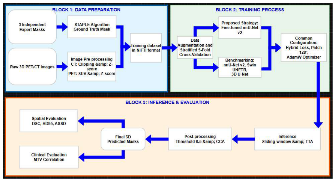
Figure 2.
Visualization of multimodal PET/CT data and the ground-truth generation process. (A) Unenhanced CT; (B) 18F-FDG PET (SUV map); (C) Fused PET/CT; (D) Inter-observer variability among three experts; (E) STAPLE probability map; and (F) Final probabilistic consensus used as the reference standard.
Figure 2.
Visualization of multimodal PET/CT data and the ground-truth generation process. (A) Unenhanced CT; (B) 18F-FDG PET (SUV map); (C) Fused PET/CT; (D) Inter-observer variability among three experts; (E) STAPLE probability map; and (F) Final probabilistic consensus used as the reference standard.
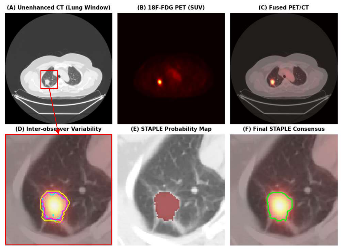
Figure 3.
Representative qualitative results with different training architectures.
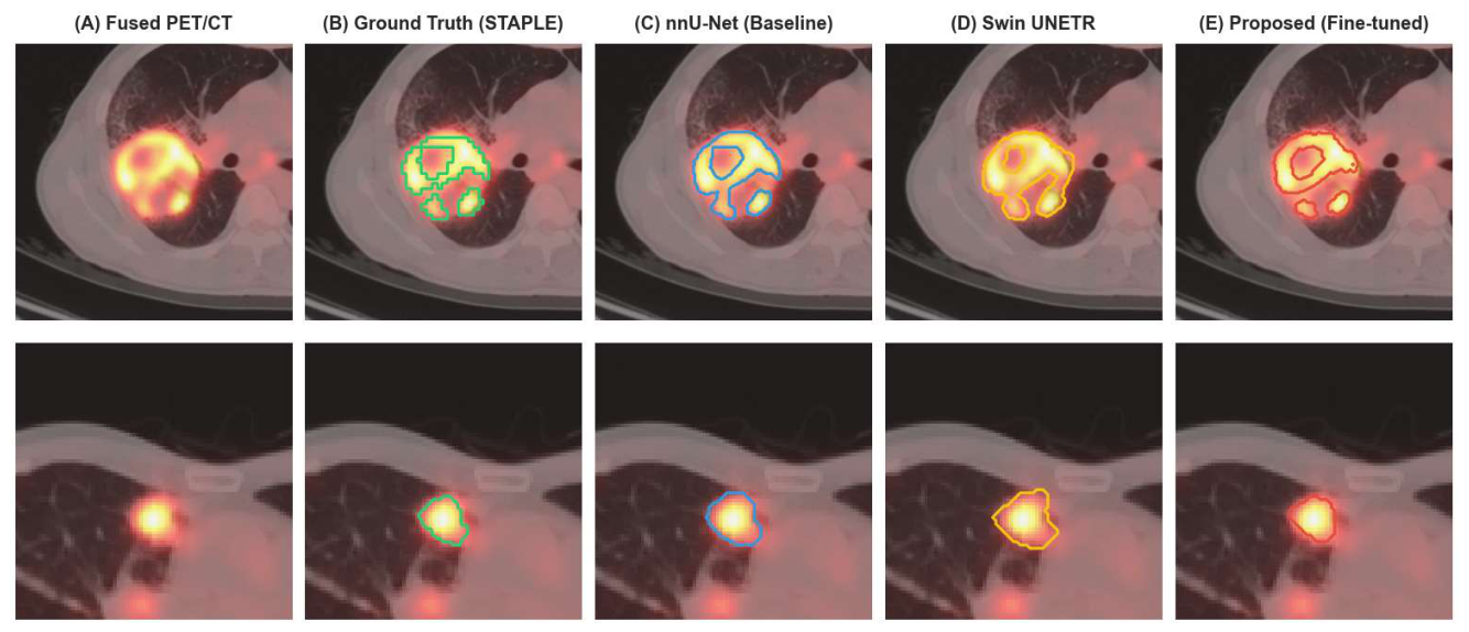
Figure 4.
Distribution of DSC across internal and external datasets. The boxplots illustrate the median, interquartile range (IQR), and outliers circles for the benchmarked models, highlighting the superior stability of the fine-tuned framework under domain shift.
Figure 4.
Distribution of DSC across internal and external datasets. The boxplots illustrate the median, interquartile range (IQR), and outliers circles for the benchmarked models, highlighting the superior stability of the fine-tuned framework under domain shift.
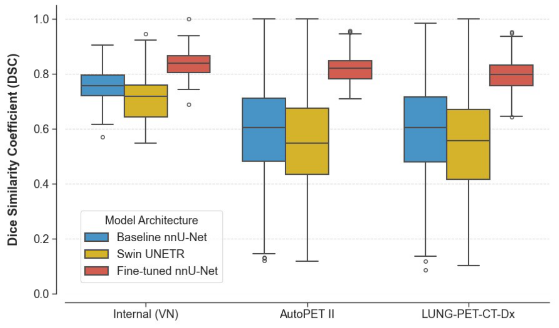
Figure 5.
Clinical agreement analysis of MTV extracted by the fine-tuned nnU-Net model versus the expert STAPLE consensus.
Figure 5.
Clinical agreement analysis of MTV extracted by the fine-tuned nnU-Net model versus the expert STAPLE consensus.
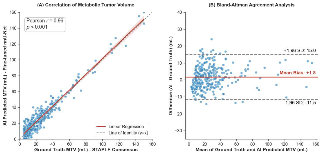
Figure 6.
Qualitative analysis of segmentation discordance in challenging anatomical boundary regions: (A) pleural over-segmentation; (B) under-segmentation of a non-FDG-avid necrotic tumor core.
Figure 6.
Qualitative analysis of segmentation discordance in challenging anatomical boundary regions: (A) pleural over-segmentation; (B) under-segmentation of a non-FDG-avid necrotic tumor core.
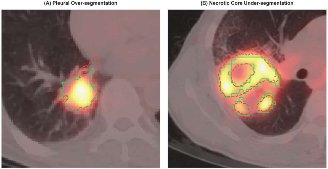

Table 1.
Clinicopathological characteristics of the patient cohort and PET/CT dataset.
| Parameter | Values |
|---|---|
| Total number of cases analyzed | 400 cases |
| + NSCLC cases (histologically confirmed) | 284 cases |
| + Negative controls | 116 cases |
| Dice score (DSC) of 3 Raters | 80.2 ± 6.6% |
| Mean age (years) | 61.2±9.9 |
| Gender (Male/Female) | 231 / 185 |
| Lesion Characteristics | |
| +Total NSCLC lesions | 342 |
| +Mean tumor size (cm) | 2.8±1.0 |
| Histological Subtype | |
| + Adenocarcinoma | 198 cases (69.7%) |
| + Squamous cell carcinoma | 86 cases (30.3%) |
| Mean SUVmax by Stage | |
| + Stage I - II | 4.2±1.8 |
| + Stage III - IV | 7.4±3.1 |
Table 2.
Quantitative comparison of segmentation performance across different deep learning architectures on the internal Vietnamese.
Table 2.
Quantitative comparison of segmentation performance across different deep learning architectures on the internal Vietnamese.
| Model | DSC (%) ↑ | HD95 (mm) ↓ | ASSD (mm) ↓ | Sensitivity (%) ↑ | Precision (%) ↑ | p-value (vs. Fine-tuned) |
|---|---|---|---|---|---|---|
| 3D U-Net (MONAI) | 65.0 ± 10.2 | 15.8 ± 8.5 | 6.2 ± 2.1 | 66.2 ± 10.5 | 68.1 ± 12.0 | < 0.001 |
| Swin UNETR | 71.2 ± 8.6 | 9.5 ± 6.2 | 3.1 ± 1.5 | 80.4 ± 8.1 | 77.5 ± 10.2 | < 0.001 |
| nnU-Net v2 | 76.6 ± 7.5 | 7.3 ± 8.6 | 2.5 ± 2.3 | 77.2 ± 7.4 | 81.3 ± 8.5 | < 0.01 |
| nnU-NET (pretrained + fine-tuned) | 83.4 ± 6.5 | 5.1 ± 3.6 | 2.0 ± 2.2 | 79.2 ± 8.2 | 89.6 ± 8.2 | - |
| Note: The symbol ↑ indicates higher is better; ↓ indicates lower is better | ||||||
Table 3.
Agreement DSC analysis between the fine-tuned model and individual expert radiologists.
| Model | Rater 1 | Rater 2 | Rater 3 | STAPLE Consensus |
|---|---|---|---|---|
| nnU-Net (pretrained + fine-tuned) | 81.5% | 82.2% | 81.8% | 81.8% |
Table 4.
Zero-shot generalization performance of the segmentation models on independent international datasets (AutoPET II and LUNG-PET-CT-Dx).
Table 4.
Zero-shot generalization performance of the segmentation models on independent international datasets (AutoPET II and LUNG-PET-CT-Dx).
| Model | Internal (VN) | AutoPET II | LUNG-PET-CT-Dx | Average Drop |
|---|---|---|---|---|
| nnU-Net v2 (baseline) | 76.6% | 61.2% | 59.8% | -21.0% |
| Swin UNETR | 71.2% | 57.7% | 56.0% | -20.1% |
| nnU-Net (pretrained + fine-tuned) | 83.4% | 81.1% | 79.8% | -3.5% |
Table 5.
Ablation study results evaluating the impact of transfer learning on segmentation accuracy and training efficiency.
Table 5.
Ablation study results evaluating the impact of transfer learning on segmentation accuracy and training efficiency.
| Configuration | Initialization | DSC | HD95 (mm) |
|---|---|---|---|
| nnU-Net v2 (baseline) | Random Weights | 76.6% | 7.30 |
| Zero-shot Pretrained | AutoPET II Weights | 76.7% | 198.1 |
| Fine-tuned nnU-Net | AutoPET II Weights | 83.4% | 5.10 |
Disclaimer/Publisher’s Note: The statements, opinions and data contained in all publications are solely those of the individual author(s) and contributor(s) and not of MDPI and/or the editor(s). MDPI and/or the editor(s) disclaim responsibility for any injury to people or property resulting from any ideas, methods, instructions or products referred to in the content. |
© 2026 by the authors. Licensee MDPI, Basel, Switzerland. This article is an open access article distributed under the terms and conditions of the Creative Commons Attribution (CC BY) license (http://creativecommons.org/licenses/by/4.0/).
Copyright: This open access article is published under a Creative Commons CC BY 4.0 license, which permit the free download, distribution, and reuse, provided that the author and preprint are cited in any reuse.


