

Submitted:
02 July 2026
Posted:
03 July 2026
You are already at the latest version
Abstract
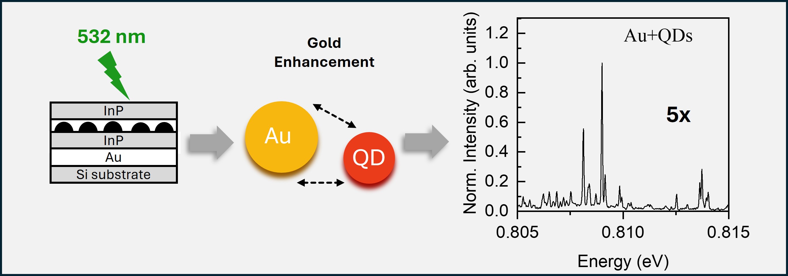
We demonstrate the integration of molecular beam epitaxy (MBE)-grown InP-based quantum dots (QDs) on gold thin films, achieving a fivefold enhancement of emission at telecom wavelengths compared with QDs grown on distributed Bragg reflectors (DBRs). Micro-photoluminescence (µ-PL) spectroscopy reveals a pronounced enhancement of the PL intensity, which is attributed to plasmon-assisted light–matter interaction at the metal–semiconductor interface. Reflectivity measurements reveal a characteristic dip near the QD emission wavelength, consistent with the excitation of surface plasmon resonances (SPRs), indicating favorable spectral overlap between the plasmonic modes and the QD emission. Power-dependent measurements reveal background-free exciton and biexciton emission from single QDs with resolution-limited linewidths. Polarization-dependent measurements demonstrate an ultra-small excitonic fine-structure splitting as low as ~2 μeV. Finally, a statistical analysis of multiple QDs confirms the reproducibility of the observed optical properties.
Keywords:
quantum dots
; photoluminescence
; extraction efficiency
; telecom wavelengths
; gold mirrors
1. Introduction
Quantum information technologies such as optical quantum computing, quantum teleportation, and advanced quantum communication protocols rely on efficient sources of highly indistinguishable single photons [1,2,3,4]. Semiconductor quantum dots (QDs) have emerged as promising sources due to their discrete energy levels and compatibility with standard semiconductor fabrication techniques [5]. Their capability to generate single-photons on demand further enhances their appeal for quantum applications, including on-chip integration and scalable photonic circuits. Although extensive studies have developed QDs emitting at wavelengths below 1 µm, their integration with fiber-optic systems remains limited due to the high attenuation of silica fibers in this spectral range [6,7,8]. In addition to their emission spectra, QDs offer tunable optical properties through modification of their band structure and strain engineering.
To address the challenges in fiber-based quantum photonic applications, research has increasingly shifted toward QDs operating in the telecom bands, where optical transmission losses in silica fibers are minimal [9,10,11]. Telecom wavelengths not only offer strong compatibility with integrated quantum photonic platforms but are also well suited for quantum photonic circuit technologies based on silicon or SiN. Within III-V based semiconductor heterostructures. The InAs/InP material system exhibits a lattice mismatch of approximately 3.2%, substantially smaller than the 7.2% mismatch in the InAs/GaAs system. This reduced mismatch enables higher crystal quality and lower defect densities, making InP-based QDs attractive candidates for single-photon sources operating at telecom wavelengths and for long-distance quantum communication. At the same time, GaAs-based materials have demonstrated successful single-photon emission in the C-band, highlighting significant progress in overcoming the inherent material limitations of these systems [12,13,14].
Realizing bright, efficient, and stable single-photon sources, however, requires careful optimization of QD properties through advanced semiconductor layer engineering and integration with photonic structures such as photonic crystals [15] and distributed Bragg reflectors (DBRs) [16,17]. In recent years, we have demonstrated single-photon emission from InAs QDs embedded in an InAlGaAs matrix with a fine-structure splitting (FSS) of 20 µeV [16]. Building on this work, we achieved further FSS reduction by embedding QDs in an InP matrix and coupling them to InP-based photonic crystal microcavities, reaching FSS values as low as 5 µeV [15] and down to 2 µeV on DBR-integrated structures [17].
Among the different approaches developed to enhance the light extraction from quantum emitters, metal-based materials have gained attention due to their ability to manipulate the electromagnetic field at nanoscale levels. In particular, thin gold (Au) films are highly promising because of their broadband reflectivity spanning the near-infrared to far-infrared region. Their high reflectivity, chemical stability, resistance to oxidation, and relatively small optical skin depth make Au films attractive for optical and plasmonic devices[18,19,20,21,22]. In addition, Au-based nanostructures exhibit strong plasmonic effects and offer significant design versatility, enabling precise control over optical parameters such as spectral response, phase shift, and group velocity dispersion [23,24]. When patterned into nanostructures, Au films support localized surface plasmon resonances that can enhance light absorption and spontaneous emission through strong coupling to nearby quantum emitters [25,26].
Building on these advantages, we present an approach using thin Au films to improve the collected photoluminescence intensity of epitaxially grown InAs QDs by MBE. Rather than relying on complex nanocavity architectures, this approach emphasizes high material quality combined with carefully engineered metal integration, a broadband reflective layer that modifies the optical environment and enhances photon extraction. In this work, we systematically investigate Au-integrated single InAs/InP QDs emitting at 1550 nm. Morphological characterization was performed using atomic force microscopy (AFM), while optical investigations, including macro- and micro-photoluminescence (PL) measurements, demonstrate a fivefold enhancement in PL intensity compared with QDs grown on distributed Bragg reflector (DBR) structures.
2. Materials and Methods
The samples were grown by molecular beam epitaxy (MBE) on an InP substrate. The InAs quantum dots (QDs) were embedded between InP layers, with the QD active region grown on top of a thick InGaAs layer. The InGaAs layer served as a sacrificial layer for subsequent device fabrication. Low-density QDs were formed via the Stranski–Krastanov (SK) growth mode using a procedure similar to that described in [17], resulting in emission in the telecom C-band. For morphological characterization of the QDs, an additional uncapped QD layer was grown under identical conditions.
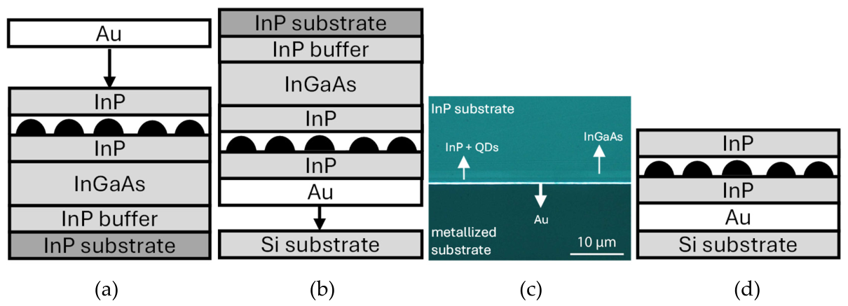
Figure 1.
Schematic illustration of the fabrication procedure: (a) As-grown structure and deposition of a gold layer on the sample surface; (b) bonding of the gold-coated sample to a substrate; (c) scanning electron microscope (SEM) image of the resulting bonded structure; and (d) final structure, consisting of the gold-coated semiconductor layer bonded to the substrate, used for optical characterization.
Figure 1.
Schematic illustration of the fabrication procedure: (a) As-grown structure and deposition of a gold layer on the sample surface; (b) bonding of the gold-coated sample to a substrate; (c) scanning electron microscope (SEM) image of the resulting bonded structure; and (d) final structure, consisting of the gold-coated semiconductor layer bonded to the substrate, used for optical characterization.
The MBE-grown samples were coated with approximately 1.0 nm of titanium (Ti), serving as an adhesion layer, followed by 200 nm of gold (Au). The Ti layer improves the adhesion between the Au film and the underlying semiconductor surface, preventing delamination [27]. After metal deposition, the sample was flip-chip bonded upside down onto a silicon substrate using conductive silver epoxy, with precise alignment achieved using a micromanipulator.
The heterostructure contains an InGaAs sacrificial layer that exhibits significant optical absorption near the 1.55 µm emission wavelength of the quantum dots (QDs). To enable optical access to the QDs from the substrate side, both the InP substrate and the InGaAs layer were selectively removed by wet chemical etching.
For macro-photoluminescence (PL) measurements, excitation and collection were performed using a lens with a focal length of approximately 15 cm. To investigate the optical properties of individual QDs, micro-photoluminescence (µ-PL) measurements were carried out at 4 K using a helium-flow cryostat (Figure 2b–d). The sample was mounted on the cryostat cold finger and excited with a 532 nm laser focused to a spot size of approximately 1 µm through a microscope objective with a numerical aperture (NA) of 0.7. The emitted PL was collected through the same objective, dispersed by a 0.75 m spectrometer, and detected using an InGaAs detector. Polarization-resolved measurements were performed using a rotating linear polarizer.
3. Results and Discussion
The reflectivity spectrum of the fabricated sample was measured at room temperature, as shown in Figure 2a (black curve), revealing a reflectivity of approximately 99% in the telecom C-band, coinciding with the designed cavity resonance and the spectral region of maximum optical absorption. Macro-PL (Figure 2a, dashed black line) and µ-PL (Figure 2a, blue curve) measurements were performed at 10 K under 532 nm laser excitation. The µ-PL spectrum exhibits sharp, discrete emission lines characteristic of individual quantum dots, whereas the macro-PL spectrum is centered at the cavity resonance and closely overlaps the absorption maximum.
Background-free µ-PL spectra of individual QDs were recorded at telecom wavelengths under low excitation powers of 406 and 650 nW (Figure 2b). The full width at half maximum (FWHM) of the emission line (Figure 2c) is instrument-limited (38 µeV). These resolution-limited linewidths are consistent with the excellent optical quality of the individual QDs and suggest that their intrinsic linewidths are narrower than the instrumental resolution [28,29].
A comparative optical study was performed between QDs grown on distributed Bragg reflectors (DBRs) and QDs integrated with a thin gold layer. µ-PL spectra of both samples were recorded at 4 K under identical excitation conditions (λₑₓc = 532 nm). Under the same excitation power of 6.4 µW, the QDs on the gold layer exhibited a fivefold increase in the measured PL intensity compared with those grown on DBRs (Figure 2d,e). This enhancement may arise from plasmonic interactions at the Au–semiconductor interface, which can modify the local density of optical states and alter the spontaneous emission dynamics, while also improving excitation and photon extraction efficiencies. Further studies, including time-resolved measurements, are required to distinguish the relative contributions of these mechanisms. A detailed investigation of the enhancement mechanism is currently underway and will be reported elsewhere. Notably, the emission linewidths remain resolution-limited in both structures, indicating that the gold layer does not introduce measurable spectral broadening within the resolution of our measurement system.
The observed PL enhancement may result from a combination of (i) enhanced excitation due to increased local electromagnetic fields [26], (ii) modification of the local density of optical states that alters the spontaneous emission rate, and (iii) improved photon extraction resulting from reflection by the gold layer. The relative contributions of these mechanisms remain under investigation.
To investigate the excitonic dynamics, power-dependent μ-PL measurements were performed. Figure 3a shows the exciton (X) and biexciton (XX) emission intensities from a single QD as a function of excitation power. At low excitation powers, only weak exciton emission is observed owing to the low probability of carrier capture into the QD. As the excitation power increases, both the exciton and biexciton emission intensities increase significantly. The double-logarithmic plot of the μ-PL intensity versus excitation power (Figure 3b) shows that the exciton emission scales approximately linearly with excitation power, as expected for single-exciton recombination under non-resonant excitation. In contrast, the biexciton emission exhibits an approximately quadratic dependence, reflecting the sequential occupation of two excitons in the QD and becoming pronounced at higher excitation powers [15,16].
Polarization-dependent μ-PL measurements were performed to investigate the fine-structure splitting (FSS) and the structural symmetry of the QDs (Figure 3b,c). The polarization-resolved energy maps (Figure 3c), plotted as a function of the linear polarization angle, exhibit the characteristic sinusoidal energy oscillations of the X and XX transitions. Fitting the polarization dependence yields a fine-structure splitting (FSS) of 2.3 μeV, indicating a high degree of in-plane structural symmetry. The small FSS, comparable to values reported for QDs grown on DBRs [15], is attributed primarily to the nearly circular in-plane geometry of the QDs, which suppresses anisotropic electron–hole exchange interactions.
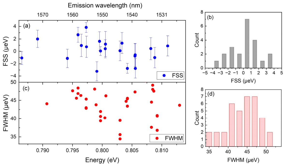
A statistical analysis of FSS and FWHM was conducted for individual QDs emitting at telecom wavelengths. As shown in Figure 4a, the FSS values remain consistently low across the emission range, consistent with a high degree of structural symmetry, an essential condition for entangled photon pair generation via the biexciton–exciton cascade [30,31].
High uniformity of the investigated QD ensemble is further supported by the distributions shown in Figure 4b. Linewidth analysis across multiple QDs (Figure 4c,d) reveals narrow emission lines of 30–35 µeV, approaching the resolution limit of our setup and thus representing an upper bound on the intrinsic linewidth. Such narrow linewidths are consistent with reduced spectral diffusion and low charge noise under the measurement conditions, reflecting excellent crystal quality.
Collectively, the combination of low FSS and narrow linewidths indicates that QDs on thin gold films are promising candidates for entangled photon generation via the biexciton–exciton cascade, which is essential for advanced quantum photonic applications.
4. Conclusions
We have demonstrated an efficient approach to enhance the photon extraction efficiency of high-quality InAs quantum dots (QDs) emitting at telecom wavelengths by integrating them on gold thin films. The gold layers provide broadband reflectivity and chemical stability and improve light outcoupling, potentially supported by plasmonic effects depending on emitter–metal separation. Polarization- and power-dependent μ-PL measurements confirm the presence of exciton and biexciton emission lines, low fine-structure splitting, and narrow linewidths, indicating high optical quality of the emitters. These results highlight the potential of gold-integrated QDs as reliable and spectrally pure single-photon sources for quantum communication applications.
Author Contributions
Conceptualization, M.B.; validation, M.B.; formal analysis, R.K.; investigation, R.K. and M.A.; resources, M.B.; supervision, M.B.; project administration, M.B.; review and editing, M.B.; funding acquisition, M.B.
Funding
This work was financially supported by the BMFTR_QR.N-Project (contract no. 16KIS2204), DFG-DeLiCom project number 418390659, DFG project number 430159793, and DFG-Heisenberg grant project number 505496601).
Data Availability Statement
The data that supports the findings of this study are available from the corresponding author upon reasonable request.
Acknowledgments
We gratefully acknowledge technical support provided by Vitalii Sichkovskyi and Uwe Gernhardt.
Conflicts of Interest
The authors declare no conflicts of interest.
Abbreviations
The following abbreviations are used in this manuscript:
| QDs | Quantum Dots |
| MBE | Molecular Beam epitaxy |
| DBR | Distributed Bragg reflectors |
| µ-PL | Micro-photoluminescence |
| AFM | Atomic force microscopy |
| SEM | Scanning electron microscopy |
| DBR | Distributed Bragg reflectors |
| FSS | Fine structure splitting |
References
- The physics of quantum information: quantum cryptography, quantum teleportation, quantum computation; Springer, 2000.
- Benyoucef, M. Photonic Quantum Technologies: Science and Applications; John Wiley & Sons, 2023. [Google Scholar]
- Langenfeld, S.; Welte, S.; Hartung, L.; Daiss, S.; Thomas, P.; Morin, O.; Distante, E.; Rempe, G. Quantum teleportation between remote qubit memories with only a single photon as a resource. Phys. Rev. Lett. 2021, 126(13), 130502. [Google Scholar] [CrossRef] [PubMed]
- Nielsen, M.A.; Chuang, I.L. Quantum computation and quantum information; Cambridge university press, 2010. [Google Scholar]
- Shields, A.J. Semiconductor quantum light sources. Nat. Photonics 2007, 1(4), 215–223. [Google Scholar] [CrossRef]
- Beveratos, A.; Abram, I.; Gérard, J.-M.; Robert-Philip, I. Quantum optics with quantum dots: Towards semiconductor sources of quantum light for quantum information processing. Eur. Phys. J. D. 2014, 68(12), 377. [Google Scholar]
- Bimberg, D.; Grundmann, M.; Ledentsov, N.N. Quantum dot heterostructures; John Wiley & Sons, 1999. [Google Scholar]
- Carosini, L.; Giorgino, F.; Sund, P.I.; Hansen, L.M.; Hamel, R.R.; Mousavi, S.M.A.; Rozema, L.A.; Poletti, F.; Slavík, R.; Walther, P. Quantum communication with quantum dots beyond telecom wavelengths via hollow-core fibers. Opt. Quantum 2026, 4(2), 82–88. [Google Scholar] [CrossRef]
- Yu, Y.; Liu, S.; Lee, C.-M.; Michler, P.; Reitzenstein, S.; Srinivasan, K.; Waks, E.; Liu, J. Telecom-band quantum dot technologies for long-distance quantum networks. Nat. Nanotechnol. 2023, 18(12), 1389–1400. [Google Scholar] [PubMed]
- Benyoucef, M.; Musiał, A. Telecom Wavelengths InP-Based Quantum Dots for Quantum Communication. Photonic Quantum Technol. Sci. Appl. 2023, 2, 463–507. [Google Scholar]
- Ge, Z.; Chung, T.; He, Y.-M.; Benyoucef, M.; Huo, Y. Polarized and bright telecom C-band single-photon source from InP-based quantum dots coupled to elliptical Bragg gratings. Nano Lett. 2024, 24(5), 1746–1752. [Google Scholar] [PubMed]
- Holewa, P.; Mikulicz, M.G.; Musiał, A.; Srocka, N.; Quandt, D.; Strittmatter, A.; Rodt, S.; Reitzenstein, S.; Sęk, G. Thermal stability of emission from single InGaAs/GaAs quantum dots at the telecom O-band. Sci. Rep. 2020, 10(1), 21816. [Google Scholar] [PubMed]
- Stevens, M.A.; McKenzie, W.; Baumgartner, G.; Grim, J.Q.; Carter, S.G.; Bracker, A.S. InAs quantum emitters at telecommunication wavelengths grown by droplet epitaxy. J. Vac. Sci. Technol. A 2023, 41(3). [Google Scholar] [CrossRef]
- Zeuner, K.D.; Jons, K.D.; Schweickert, L.; Reuterskiöld Hedlund, C.; Nuñez Lobato, C.; Lettner, T.; Wang, K.; Gyger, S.; Scholl, E.; Steinhauer, S. On-demand generation of entangled photon pairs in the telecom C-band with InAs quantum dots. ACS Photonics 2021, 8(8), 2337–2344. [Google Scholar] [PubMed]
- Kors, A.; Fuchs, K.; Yacob, M.; Reithmaier, J.P.; Benyoucef, M. Telecom wavelength emitting single quantum dots coupled to InP-based photonic crystal microcavities. Appl. Phys. Lett. 2017, 110(3). [Google Scholar]
- Benyoucef, M.; Yacob, M.; Reithmaier, J.P.; Kettler, J.; Michler, P. Telecom-wavelength (1.5 μm) single-photon emission from InP-based quantum dots. Appl. Phys. Lett. 2013, 103(16). [Google Scholar]
- Kors, A.; Reithmaier, J.P.; Benyoucef, M. Telecom wavelength single quantum dots with very small excitonic fine-structure splitting. Appl. Phys. Lett. 2018, 112(17). [Google Scholar] [CrossRef]
- Johnson, P.B.; Christy, R.-W. Optical constants of the noble metals. Phys. Rev. B 1972, 6(12), 4370. [Google Scholar] [CrossRef]
- Jun, H.K.; Careem, M.A.; Arof, A.K. Plasmonic effects of quantum size gold nanoparticles on dye-sensitized solar cell. Mater. Today Proc. 2016, 3, S73–S79. [Google Scholar] [CrossRef]
- Karakurt, O.; Alemdar, E.; Erer, M.C.; Cevher, D.; Gulmez, S.; Taylan, U.; Cevher, S.C.; Ozsoy, G.H.; Ortac, B.; Cirpan, A. Boosting the efficiency of organic solar cells via plasmonic gold nanoparticles and thiol functionalized conjugated polymer. Dye. Pigment. 2023, 208, 110818. [Google Scholar]
- Pfeiffer, M.; Lindfors, K.; Wolpert, C.; Atkinson, P.; Benyoucef, M.; Rastelli, A.; Schmidt, O.G.; Giessen, H.; Lippitz, M. Enhancing the optical excitation efficiency of a single self-assembled quantum dot with a plasmonic nanoantenna. Nano Lett. 2010, 10(11), 4555–4558. [Google Scholar] [CrossRef] [PubMed]
- Zheng, D.; Schwob, C.; Prado, Y.; Ouzit, Z.; Coolen, L.; Pauporté, T. How do gold nanoparticles boost the performance of perovskite solar cells? Nano Energy 2022, 94, 106934. [Google Scholar] [CrossRef]
- Amendola, V.; Pilot, R.; Frasconi, M.; Maragò, O.M.; Iatì, M.A. Surface plasmon resonance in gold nanoparticles: a review. J. Phys. Condens. Matter 2017, 29(20), 203002. [Google Scholar] [CrossRef] [PubMed]
- Perveen, A.; Zhang, X.; Tang, J.-L.; Han, D.-B.; Chang, S.; Deng, L.-G.; Ji, W.-Y.; Zhong, H.-Z. Sputtered gold nanoparticles enhanced quantum dot light-emitting diodes. Chin. Phys. B 2018, 27(8), 86101. [Google Scholar] [CrossRef]
- Fischbach, S.; Kaganskiy, A.; Tauscher, E.B.Y.; Gericke, F.; Thoma, A.; Schmidt, R.; Strittmatter, A.; Heindel, T.; Rodt, S.; Reitzenstein, S. Efficient single-photon source based on a deterministically fabricated single quantum dot-microstructure with backside gold mirror. Appl. Phys. Lett. 2017, 111(1). [Google Scholar]
- Etrich, C.; Fahr, S.; Hedayati, M.K.; Faupel, F.; Elbahri, M.; Rockstuhl, C. Effective optical properties of plasmonic nanocomposites. Materials 2014, 7(2), 727–741. [Google Scholar] [CrossRef] [PubMed]
- Olson, D.H.; Freedy, K.M.; McDonnell, S.J.; Hopkins, P.E. The influence of titanium adhesion layer oxygen stoichiometry on thermal boundary conductance at gold contacts. Appl. Phys. Lett. 2018, 112(17). [Google Scholar] [CrossRef]
- Mereni, L.O.; Dimastrodonato, V.; Young, R.J.; Pelucchi, E. A site-controlled quantum dot system offering both high uniformity and spectral purity. Appl. Phys. Lett. 2009, 94(22). [Google Scholar] [CrossRef]
- Savchenko, S.S.; Weinstein, I.A. Inhomogeneous broadening of the exciton band in optical absorption spectra of InP/ZnS nanocrystals. Nanomaterials 2019, 9(5), 716. [Google Scholar] [PubMed]
- Bayer, M.; Ortner, G.; Stern, O.; Kuther, A.; Gorbunov, A.A.; Forchel, A.; Hawrylak, P.; Fafard, S.; Hinzer, K.; Reinecke, T.L. Fine structure of neutral and charged excitons in self-assembled In (Ga) As/(Al) GaAs quantum dots. Phys. Rev. B 2002, 65(19), 195315. [Google Scholar] [CrossRef]
- Gaisler, A.V.; Yaroshevich, A.S.; Derebezov, I.A.; Kalagin, A.K.; Bakarov, A.K.; Toropov, A.I.; Shcheglov, D.V.; Gaisler, V.A.; Latyshev, A.V.; Aseev, A.L. Fine structure of the exciton states in InAs quantum dots. JETP Lett. 2013, 97(5), 274–278. [Google Scholar] [CrossRef]
Figure 2.
(a) Macro-PL (black dashed curve) and µ-PL (blue curve) spectra of the sample with a gold layer, together with the reflectivity spectrum (black solid curve). Inset: Atomic force microscopy (AFM) image of a single QD. (b) µ-PL spectra of two individual QDs from the sample with the gold layer. (c) Resolution-limited linewidth of a single QD (QD1 + Au) emitting in the telecom C-band. (d,e) Comparison of the µ-PL spectra of QDs grown on a distributed Bragg reflector (DBR) and on gold structures, measured under identical excitation conditions.
Figure 2.
(a) Macro-PL (black dashed curve) and µ-PL (blue curve) spectra of the sample with a gold layer, together with the reflectivity spectrum (black solid curve). Inset: Atomic force microscopy (AFM) image of a single QD. (b) µ-PL spectra of two individual QDs from the sample with the gold layer. (c) Resolution-limited linewidth of a single QD (QD1 + Au) emitting in the telecom C-band. (d,e) Comparison of the µ-PL spectra of QDs grown on a distributed Bragg reflector (DBR) and on gold structures, measured under identical excitation conditions.
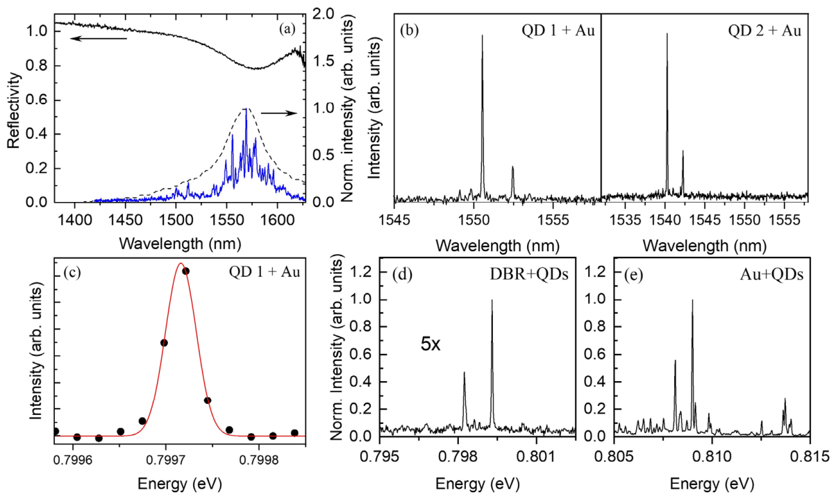
Figure 3.
(a) Power-dependent µ-PL spectra of a single QD measured at increasing excitation powers, showing the exciton (X) and biexciton (XX) emission lines. (b) Integrated PL intensities of the X and XX emissions as a function of excitation power, exhibiting linear and quadratic power dependences, respectively. (c) Polarization-dependent PL intensity map of a single QD. (d) Emission energies of the X and XX transitions as a function of the linear polarization angle, indicating a very small fine-structure splitting (FSS).
Figure 3.
(a) Power-dependent µ-PL spectra of a single QD measured at increasing excitation powers, showing the exciton (X) and biexciton (XX) emission lines. (b) Integrated PL intensities of the X and XX emissions as a function of excitation power, exhibiting linear and quadratic power dependences, respectively. (c) Polarization-dependent PL intensity map of a single QD. (d) Emission energies of the X and XX transitions as a function of the linear polarization angle, indicating a very small fine-structure splitting (FSS).
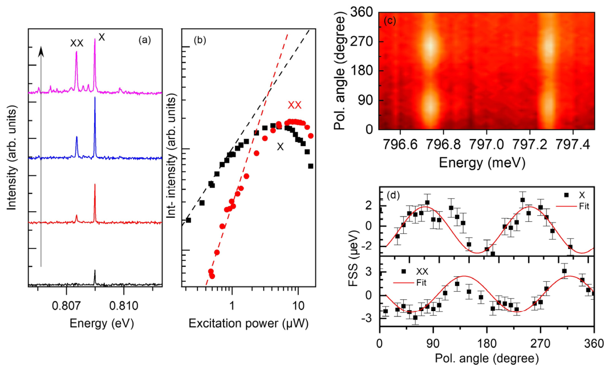
Figure 4.
(a) Neutral exciton (X) fine-structure splitting (FSS) as a function of exciton emission energy for approximately 20 quantum dots (QDs) measured from the processed sample, demonstrating consistently low FSS values. (b) Histogram of the exciton FSS values, showing a narrow statistical distribution. (c) Full width at half maximum (FWHM) of the exciton emission from the same QDs, indicating narrow emission linewidths. (d) Histogram showing the statistical distribution of the corresponding FWHM values.
Figure 4.
(a) Neutral exciton (X) fine-structure splitting (FSS) as a function of exciton emission energy for approximately 20 quantum dots (QDs) measured from the processed sample, demonstrating consistently low FSS values. (b) Histogram of the exciton FSS values, showing a narrow statistical distribution. (c) Full width at half maximum (FWHM) of the exciton emission from the same QDs, indicating narrow emission linewidths. (d) Histogram showing the statistical distribution of the corresponding FWHM values.
Disclaimer/Publisher’s Note: The statements, opinions and data contained in all publications are solely those of the individual author(s) and contributor(s) and not of MDPI and/or the editor(s). MDPI and/or the editor(s) disclaim responsibility for any injury to people or property resulting from any ideas, methods, instructions or products referred to in the content. |
© 2026 by the authors. Licensee MDPI, Basel, Switzerland. This article is an open access article distributed under the terms and conditions of the Creative Commons Attribution (CC BY) license (http://creativecommons.org/licenses/by/4.0/).
Copyright: This open access article is published under a Creative Commons CC BY 4.0 license, which permit the free download, distribution, and reuse, provided that the author and preprint are cited in any reuse.


