

Submitted:
01 July 2026
Posted:
03 July 2026
You are already at the latest version
Abstract
Background and Objectives: Rhizarthrosis is a degenerative disease of the trapeziometacarpal joint characterized by pain, functional limitation, and a reduction in quality of life. Although orthotics and therapeutic exercise represent the first-line conservative treatment, the role of high-intensity laser therapy remains poorly defined. This study evaluated the efficacy of an innovative multimodal high-energy laser therapy protocol, combined with stimulation of lymphatic pathways and periarticular trigger points, in the treatment of rhizarthrosis. Materials and Methods: This prospective, randomized study enrolled 42 patients with Eaton-Littler stage I-II rhizarthrosis and assigned them to an experimental group (THEAL group) (n=21 subjects) or a control group (exercise group) (n=21 subjects). The experimental protocol included 10 sessions on alternate days, including treatment of the lymph nodes, the trapezium-metacarpal joint, the forearm muscles, and related trigger points. The control group completed a 4-week exercise protocol. All recruited subjects used a first finger splint during the study. Outcomes assessed at baseline, 1, 3, and 6 months included pain (VAS), hand function (FIHOA), upper limb disability (DASH), and patient satisfaction (Roles and Maudsley score). Results: Both groups showed significant improvement over time for all outcomes assessed (p<0.001). In the comparison between recruitment and 6 months, the THEAL group (5.33 ± 1.02) showed a significantly greater reduction in VAS, FIHOA, and DASH scores than the exercise group (p<0.001). Multivariate regression analysis confirmed an independent association between THEAL treatment and greater pain reduction at 6 months (p<0.001). Favorable differences were also observed for the DASH score and the Roles and Maudsley score in the THEAL group. Conclusions: A multimodal high-energy laser therapy protocol combined with a brace resulted in greater clinical improvements compared to conventional conservative treatment in patients with rhizarthrosis, with benefits for pain and functional recovery. The results also suggest a possible contribution from lymphatic stimulation and trigger point treatment, aspects that merit further investigation.
Keywords:
thumb carpometacarpal osteoarthritis
; high-intensity laser therapy
; photobiomodulation
; conservative treatment
1. Introduction
Trapeziometacarpal osteoarthritis, also called rhizarthrosis, is a degenerative pathology with inflammatory aspects affecting the joint of the first finger of the hand, which causes pain, functional limitation and disability in daily activities [1].
It occurs more frequently in postmenopausal women, reaching prevalence values between 7.3% and 39%, depending on age [2]. Recent studies are investigating new pathophysiological aspects responsible for the disease, in addition to the already known ones of cartilage degeneration, osteophyte formation, joint subluxation, subchondral bone edema, and the presence of synovitis [3].
Indeed, the presence of inflammatory mediators in peri-articular tissues could alter local microcirculation, compromising lymphatic drainage and causing persistent chronic inflammation [4]. Furthermore, chronic pain and altered thumb biomechanics may contribute to trigger point activation, protective muscle hyperactivity, and functional overload of the upper limb kinetic chain [5,6,7].
For the conservative management of thumb arthritis suggest a multimodal approach focused on orthoses to stabilize the trapeziometacarpal joint and exercise programs aimed at strengthening muscles and improving thumb motor control [3,8]. Available evidence also suggests that integrating these strategies is more effective than individual interventions alone.
Physical therapies are currently not among the recommended first-line interventions for rhizarthrosis; however, there are biological premises that suggest a potential adjuvant role in the modulation of pain and inflammatory processes, particularly for photobiomodulation techniques such as low- and high-intensity laser [9,10].
High-intensity laser therapy has emerged as a promising noninvasive therapeutic modality for various musculoskeletal conditions due to its analgesic, anti-inflammatory, and biostimulatory properties [11]. In patients with rhizarthrosis, preliminary evidence indicates that high-energy laser can reduce pain and improve upper limb function [9,12].
Currently available protocols focus primarily on local joint treatment. Recent studies have shown that high-energy laser can improve microvascular perfusion, modulate inflammatory cytokine activity, and promote tissue repair processes [11,13]. Furthermore, photobiomodulation therapies have been associated with effects on fluid dynamics and tissue drainage, suggesting a potential influence on lymphatic and interstitial homeostasis [14,15].
Therefore, the aim of this study is to evaluate the efficacy of high-energy laser therapy in the treatment of rhizarthrosis, providing an integrated approach that, in addition to treatment of the trapezium-metacarpal joint, includes stimulation of the lymphatic pathways and periarticular trigger points of the upper limb, in order to improve clinical outcomes compared to conventional local treatment.
2. Materials and Methods
We designed a prospective, randomized study to evaluate the effects of high-energy laser in the treatment of rhizarthrosis. The Territorial Ethics Committee of the “Consorziale Policlinico” University Hospital authorized the study (Interregional Ethics Committee, approval no. 7905, meeting of 8 October 2025). Participants gave their written informed consent. The trial was registered at https://clinicaltrials.gov/ with the trial registration number NCT07566507.
Patients were examined in the outpatient clinic of the Orthopedics and Traumatology Unit of the Policlinico di Bari, Italy. All patients with rhizarthrosis, after evaluation by a hand surgeon, were included in the study if eligible. Inclusion criteria were: trapeziometacarpal osteoarthritis with stage 1 or 2 of the Eaton-Littler radiographic classification and pain (recent x-ray within 6 months) [16]; clinical condition with onset at least 6 months prior; pain rated at least 4/10 on a VAS scale.
Exclusion criteria were: rheumatoid arthritis or trauma to the affected area, contraindications to THEAL treatment (neoplasia, pregnancy, thrombocytopenia, epilepsy, uncompensated heart disease or arrhythmia, pacemaker, local infections), corticosteroid injections, or physical therapy in the previous 4 weeks. Forty-two patients were enrolled, of whom 21 were indicated for laser therapy and bracing (THEAL group) and an additional 21 were indicated for exercises and bracing (Exercise group).
THEAL group
The multimode high energy laser therapy treatment with thermal control (THEAL: Temperature controlled High Energy Adjustable multimode emission Laser) was delivered with an Ixyon XP Ultra device (Mecmedix – Mectronic Medicale srl, Grassobbio (BG), Italy) which allows the delivery of 6 wavelengths (650 nm, 780 nm, 810 nm, 905 nm, 980 nm and 1064 nm), with continuous and pulsed mode, average power up to 50 W, administering 10 sessions on alternate days. Each single session was divided into four phases, the energy parameters of each phase are described in Table 1 and Figure 1. The protocol was defined in accordance with the literature [17].
The first phase consists of activating the subclavian, axillary, and cubital lymphatic stations. The treatment of each lymphatic station takes 1 minute and 40 seconds and is accompanied by light manual mechanical pressure. The protocol involves the use of the IR-Compact applicator (diameter 23 mm), the delivery of 600 J for each lymphatic station, and a temperature control between 39 and 43 °C.
The second phase consists of anti-inflammatory and biostimulating action targeted at the area affected by the pathology. The protocol involves treatment of both the external area and the internal area of the palm of the hand. The treatment area is approximately circular and measures about 5 cm in diameter. By entering the precise dimensions into the software, the exact duration of the treatment and the exact amount of energy to be administered are calculated (the protocol specifies 35 J/cm2). The applicator to be used is IR-Compact with a temperature control set between 38° and 41 °C.
The third phase of treatment is performed in the therapeutic window relating to the forearm muscles (flexors and extensors) with the aim of performing a decontracting and anti-inflammatory action. By entering the dimensions of the area to be treated into the software, the time and joules relating to the phase will be calculated (expected dosage of 33 J/cm2). The treatment must be performed with a Large Spherical applicator or an IR-Large applicator with the temperature control set between 38 and 41 °C.
The final phase focuses on treating latent trigger points related to the condition. The protocol involves working on two of the following trigger points:
- Long abductor of the thumb,
- Short extensor of the thumb,
- Long extensor of the thumb,
- Long flexor of the thumb.
60 joules are administered to each trigger point using a collimated applicator.
Exercise group
Patients performed exercise for 4 weeks after recruitment [18]. Patients in this group were instructed on home exercises to improve dynamic stability of the carpal-metacarpal (CMC) joint of the thumb (Figure 2).
- First interspace stretch: Passive thumb abduction to increase the opening between the first and second digits; hold for 30 seconds, 2–4 repetitions [19].
- Active range of motion of the first dorsal interosseous: active radial deviation of the index finger with the hand resting on the table. In the absence of pain, the exercise was progressively performed against resistance (manual or elastic); in the case of pain, only active execution was maintained. The exercise consisted of 10 repetitions [19,20,21].
FIRST FINGER SPLINTING
Patients in both groups were instructed to wear a splint during the day for 4 weeks following enrollment.
CLINICAL EVALUATIONS
Assessments were performed at baseline (T0), 1 month (T1), 3 months (T2), and 6 months (T3). At each time point, the VAS scale, the Functional Index for Osteoarthritis of the Hand (FIHOA), and the Disabilities of the Arm, Shoulder, and Hand (DASH) questionnaire were administered. The Roles and Maudsley score was assessed at T1, T2, and T3. Treatment-related adverse events, including skin irritation, increased pain, post-exercise soreness, splint-related discomfort, and any other unexpected symptoms, were monitored throughout the study.
Pain was measured using a visual analogue scale (VAS), which consists of a 10-cm horizontal line (with 0 cm corresponding to no pain and 10 cm corresponding to the most severe pain ever experienced) [25].
Hand function was assessed using the Functional Index for Osteoarthritis of the Hand (FIHOA), a 10-item questionnaire with four possible answers for each question (0 = possible without difficulty, 1 = possible with mild difficulty, 2 = possible with significant difficulty, 3 = impossible); the score ranges from 0 (no limitation) to 30 (maximum limitation)[26].
Disability is assessed using the DASH (Disabilities of the Arm, Shoulder, and Hand) questionnaire: 11 questions with five possible answers each (1 = No difficulty, 2 = Mild difficulty, 3 = Moderate difficulty, 4 = Severe difficulty, 5 = Inability) and a total score ranging from 11 (no difficulty) to 55 (inability) [27].
The Roles and Maudsley score assess the patient's perception of improvement, ranging from 1 (excellent outcome with no symptoms after treatment) to 4 (poor, symptoms identical to or worse than before treatment) [28].
Statistical Analysis
Qualitative variables were described as proportions and percentages. Quantitative variables were described as means and standard deviations (SD). Normality of distributions was assessed based on skewness and kurtosis.
In order to verify whether the VAS, FIHOA and DASH scores changed over time, Student’s t test for paired samples was employed, separating results by study group (THEAL versus control). Changes at each checkpoint were then confronted via Mann-Whitney’s rank sum test due to non-normality of these variables’ distribution. Further, since normally distributed, Student’s t test for independent samples was employed to confront the RM score at each checkpoint.
The main study outcome was represented by the change of the VAS score from t0 to t3 (calculated as the first reported score minus the last reported one). Secondary outcomes were represented by the same changes for the FIHOA and DASH scores, respectively. For each outcome, the impact of therapy was assessed via linear regression with robust standard errors, in order to account for non-normality and avoid violation of homoscedasticity assumptions. Three models were built for each outcome:
- M1: raw regression;
- M2: adjustment for sex and age;
- M3: M2 with further adjustment for body mass index (BMI), smoking habit, concordance of the hand (i.e., the treated hand was the dominant one), months since the beginning of symptoms, hypertensive cardiopathy, endocrine dysfunction/condition, previous or ongoing therapy with nonsteroidal anti-inflammatory drugs, previous physiokinesis therapy, echography classification of disease.
Sensitivity analysis was then performed restricting regression models to subjects aged ≤65 years. A p-value <0.05 was considered statistically significant. The study database was built via Microsoft Excel®. All calculations were performed via STATA MP17®.
3. Results
The study enrolled 42 patients, randomly assigned to either laser therapy or exercise (Figure 3).
The study population’s characteristics are synthetized in Table 2. All enrolled patients completed the study through the final follow-up visit. No dropouts, treatment discontinuations, or adverse events were recorded during the study period.
The reduction of the VAS score over time was significant in both laser-treated (t: 24.04; p-value <0.001) and control subjects (t: 11.36; p-value <0.001). Likewise, a significant reduction was observed for the other scores in both groups (Table 3).
The non-parametric Mann-Whitney test for independent samples identified significant differences in the average change of the VAS score between the two groups both overall (z: -4.90; p-value <0.001), at the t1 (z: -3.60; p-value <0.001) and t3 checkpoints (z: -4.16; p-value <0.001), while the difference was non-significant between t1 and t2 (z: -0.61; p-value: 0.567). The overall change of the DASH score was also significantly different between the two groups (z: -3.00; p-value: 0.002). On the other hand, the FIHOA score changes did not differ significantly (z: -1.83; p-value: 0.068). Student’s t test (one-sided p-value, hypothesizing superiority of the study treatment) identified a significant difference in terms of reported RM score for the two groups. In particular, a significantly lower score was observed in the study group at both t1 (t: 3.16; p-value: 0.001), t2 (t: 3.48; p-value <0.001) and t3 (t: 4.56; p-value <0.001). Further specifics are provided in Table 4.
Regression analysis
Regression analysis’s results are synthetized in Table 5.
As far as fully adjusted linear regression models are concerned, only the VAS score changes showed significant association with the treatment, laser therapy showing significantly greater reduction of perceived pain (ß: 3.49; 95%CI: 2.65 – 4.32; p-value <0.001). The DASH score, albeit showing significantly positive impact of laser therapy in partially adjusted regression (ß: 9.16; 95%CI: 4.00 – 14.32; p-value: 0.001), lost significance of the association in M3 (ß: 9.20; 95%CI: -0.84 – 19.23; p-value: 0.071). Sensitivity analysis fully confirmed these results.
4. Discussion
This study evaluated the efficacy of high-energy laser therapy (THEAL) combined with bracing, compared to a conservative program of bracing and therapeutic exercise, in the treatment of rhizarthrosis. Results showed significant improvement in clinical outcomes in both groups during follow-up, with overall greater benefits in the experimental treatment group. These results suggest that the addition of laser therapy to standard conservative bracing may result in additional clinical effects compared to the bracing and therapeutic exercise approach.
The improvement observed in the control group is consistent with current scientific evidence and international recommendations for the conservative management of rhizarthrosis. Guidelines identify orthotics and therapeutic exercise as the cornerstones of non-surgical treatment [3,8,29]. Stabilization of the trapeziometacarpal joint achieved through the brace reduces mechanical stress during daily activities, while therapeutic exercise promotes recovery of function through improved muscle strength, coordination, and motor control of the thumb [8].
The most significant aspect emerging from this study concerns the potential of high-energy laser therapy in the conservative treatment of rhizarthrosis. Although both groups showed favorable outcomes over time, those treated with THEAL achieved superior results during follow-up, suggesting a potential additive effect of laser therapy compared to the benefits obtained with conventional treatment alone.
These results appear consistent with preliminary evidence available in the literature. The protocol by Cantero-Téllez and colleagues [9] involved the application of high-intensity laser therapy (HILT, class IV KLaser K1200) exclusively locally to the trapeziometacarpal joint, with standardized parameters (peak power 3 W, average power 1.5 W, wavelengths 800 + 970 nm, frequency 2 Hz, dose 75 J per session) and administration three times a week for four weeks. In this study, laser treatment was the only therapeutic intervention, without association with exercise, orthoses, or other rehabilitation strategies, and therefore the isolated effect of high-energy laser therapy on the symptoms of rhizarthrosis was evaluated. The results reported a reduction in pain sensitivity and an improvement in motor performance in patients with trapeziometacarpal osteoarthritis subjected to high-energy laser.
Similarly, Guo et al. [12] administered high-energy laser therapy in combination with functional hand exercises over a 4-week course. Parameters included a high-intensity laser system with near-infrared emission, with typical wavelengths in the 808–980 nm range (dual-wavelength laser), pulsed mode, and scanning application over the joint area of the base of the thumb. Energy was delivered according to standard clinical protocols, with progressive dosages adjusted to patient tolerance. The researchers observed significant clinical improvements in patients treated with high-intensity laser therapy combined with functional hand training. Although the methodologies and protocols used differ from those of the present study, the results converge in suggesting that high-energy laser therapy may represent an effective therapeutic strategy in the conservative treatment of rhizarthrosis.
The mechanisms responsible for the observed effects are not yet fully elucidated. High-energy laser therapy has been associated with analgesic, anti-inflammatory, and biostimulatory effects, likely mediated by modulating cellular activity, the inflammatory response, and tissue repair processes [11]. Furthermore, experimental studies have demonstrated that high-energy laser therapy is able to increase local microcirculation and tissue perfusion, promoting a biological environment potentially more favorable for symptom reduction and functional recovery [13].
A distinctive feature of this study is the therapeutic approach adopted. Unlike most previous studies, which focused primarily on local treatment of the trapeziometacarpal joint [9,12], the experimental protocol also included stimulation of specific areas related to the main drainage pathways of the upper limb and periarticular trigger points.
The high-energy multi-mode emission laser (THEAL) allows for a dose- and wavelength-dependent therapeutic approach, in which power density qualitatively modulates biological targets compared to low-energy photobiomodulation (PBM) alone [30]. At low energy doses, the effect is predominantly photobiomodulatory and mitochondrial, while the use of high energy also determines a controlled photothermal component with effects on microcirculation, tissue viscosity and blood drainage, extending the action from the cellular to deep tissue level.
The 650 and 780 nm wavelengths act mainly on superficial and cellular targets, with absorption prevalent on cytochrome c oxidase and modulation of fibroblasts, keratinocytes, and macrophages, through redox-sensitive pathways (ROS, NF-κB), with anti-inflammatory and pro-reparative effects [31,32]. The 810 and 905 nm wavelengths reach deeper districts (deep dermis, muscle-tendon tissue, and peripheral nervous system), modulating stromal fibroblasts, endothelia, and nociceptive Aδ/C fibers, reducing nociceptive transmission and promoting anti-edema effects through microvascular regulation [33,34].
The 980 and 1064 nm wavelengths present maximum penetration and prevalent interaction with water and hemoglobin, determining controlled photothermal effects on the extracellular matrix and deep microvascular compartment, with increased blood flow, angiogenic stimulation (VEGF), and MMP-mediated collagen remodeling [17,33]. The combination of continuous-wave, PBM, and E2C modalities therefore allows for an integrated multi-target action, from redox-dependent cellular modulation to the deep neurovascular response. This rationale is supported by clinical evidence demonstrating significant improvements in tendinopathies, Dupuytren's disease and plantar fasciitis, attributed to the reduction of inflammation and edema and the improvement of tissue repair processes [35,36,37].
In recent years, interest in the possible role of microcirculation and tissue fluid homeostasis in chronic inflammatory processes has grown. Some evidence from photobiomodulation studies suggests that laser stimulation may influence drainage mechanisms and interstitial fluid dynamics [14]. Similarly, studies conducted in patients with lymphedema have reported favorable effects of photobiomodulation on lymphatic system function [15]. Although these mechanisms have not yet been specifically demonstrated in rhizarthrosis, they could help explain, at least in part, the results obtained with the protocol used in the present study.
Similarly, trigger point treatment may have played a complementary role. Chronic pain associated with rhizarthrosis frequently leads to changes in motor patterns and compensatory strategies that involve the entire upper limb [5]. The presence of myofascial trigger points and alterations in muscle function have been described as a possible contributing factor to the persistence of musculoskeletal pain (Simons et al. 1998; Zhai et al. 2024). In this context, a therapeutic approach also targeting myofascial components may have contributed to the observed improvements.
From a clinical perspective, the study results are particularly significant as they confirm the importance of a multimodal approach in the management of rhizarthrosis. Current guidelines emphasize that no single intervention can comprehensively address the complexity of the disease; therefore, integrating complementary therapeutic strategies is a priority for clinical research. In this context, high-energy laser therapy could be a useful adjuvant treatment, especially in patients with persistent pain, high levels of clinical irritability, or functional limitations not fully addressed by conventional interventions.
Current international guidelines identify treatment based on orthoses and therapeutic exercise as first-line intervention, with proven effects on pain reduction and improved function thanks to the biomechanical stabilization of the trapeziometacarpal joint and the recovery of neuromuscular control of the thumb [3,8].
Among adjuvant physical therapies in the treatment of osteoarthritis, traditional modalities show an overall limited level of evidence. TENS primarily exerts a short-term neuromodulatory analgesic effect, with no consistent evidence on functional or structural outcomes [38]. Therapeutic ultrasound has not demonstrated clinically relevant benefits compared to other conservative interventions, with an overall low level of evidence [39]. An intermediate level of evidence could be attributed to pulsed electromagnetic field (PEMF) magnetic therapy, for which some systematic reviews report short-term improvements in pain and function, albeit with high methodological heterogeneity and low to moderate quality of evidence [40].
More recently, physical therapies based on mechanical-energetic biostimulation appear to show more promising results, even in the model of hand osteoarthritis. In the SWEX-TO study, extracorporeal shock wave therapy (ESWT) demonstrated significant improvements in pain and function in the medium term compared to therapeutic exercise, suggesting a possible persistent biological effect on periarticular tissues [35].
Taken together, these data suggest a progressive shift in the therapeutic paradigm toward physical strategies capable of inducing more complex biological responses than symptomatic modulation alone. However, significant limitations remain related to the heterogeneity of protocols, the variability of treatment parameters, and the need for direct comparative studies to more clearly define the clinical role of each modality within rhizarthrosis rehabilitation programs.
The results, however, must be interpreted in light of some limitations. First, the study design does not allow distinguishing the specific effect of joint treatment from that resulting from stimulation of drainage pathways and trigger points. Furthermore, the follow-up duration may not be sufficient to assess the long-term persistence of the effects. Further studies with larger sample sizes, longer follow-ups, and comparative protocols will be needed to confirm the results obtained and clarify the underlying biological mechanisms.
5. Conclusions
In conclusion, both conventional conservative treatment and the protocol including high-energy laser therapy resulted in significant improvements in patients with rhizarthrosis. However, the high-energy laser therapy group showed greater benefits during follow-up, suggesting that this intervention may enhance the effects of standard conservative treatment. These results support the use of THEAL therapy as a complementary strategy within a multimodal rehabilitation approach and provide the basis for further studies aimed at defining its clinical role and mechanisms of action.
Author Contributions
Conceptualization, N.A. and C. I.; methodology, N.A., D.L.A.; validation, N.A., C.I.. and S.G.; investigation, R.F., L.I, and D.G.S.; data curation, R.F., C.I., N.A. and D.L.A.; writing—original draft preparation, N.A. and S.G.; writing—review and editing, N.A., C.I. and D.G.S.; visualization, D.G.S. and D.L.A.; supervision, N.A.; project administration, N.A. and S.G. All authors have read and agreed to the published version of the manuscript.
Funding
This research received no external funding. The APC was funded by Mectronic Medical S.r.l. (BG, Italy).
Institutional Review Board Statement
The study was conducted by the Declaration of Helsinki, and approved by the Ethics Committee of AOU Consorziale Policlinico di Bari (Interregional Ethics Committee) (protocol code protocol no. 7905, October 8h, 2025).
Informed Consent Statement
Informed consent was obtained from all subjects involved in the study. Written informed consent has been obtained from the patients to publish this paper.
Data Availability Statement
The data presented in this study are available upon request from the corresponding author. The data are not publicly available due to privacy concerns.
Conflicts of Interest
The authors declare no conflict of interest.
Abbreviations
The following abbreviations are used in this manuscript:
| THEAL | Temperature controlled High Energy Adjustable multimode emission Laser |
| VAS | Visual Analogue Scale |
| FIHOA | Functional Index for Osteoarthritis of the Hand |
| DASH | Disabilities of the Arm, Shoulder, and Hand) |
| RM | Roles and Maudley Score |
| CW | Continuous Wave |
| PBM | Photo-Biomodulation Mechanism |
| E2C | Superpulsed stochastic emission |
| IR | Infra-red |
| CMC | Carpal-Metacarpal |
| SD | Standard Deviation |
| BMI | Body Mass Index |
| CI | Confidence Interval |
| HILT | High Intensity Laser Therapy |
| ROS | Reactive Oxygen Species |
| NF-kB | Nuclear factor NF-kappa-B |
| PEMF | Pulsed Electromagnetic Field, |
| ESWT | Extracorporeal Shock Waves Therapy |
References
- Teunissen, J.S.; Wouters, R.M.; Bierma-Zeintra, S.M.A.; van Meurs, J.B.J.; Schreuders, T.A.R.; Zuidam, J.M.; Selles, R.W. The prevalence, incidence, and progression of radiographic thumb base osteoarthritis in a population-based cohort: the Rotterdam Study. Osteoarthr. Cartil. 2022, 30(4), 578–585. [Google Scholar] [CrossRef]
- van der Oest, M.J.W.; Duraku, L.S.; Andrinopoulou, E.R.; Wouters, R.M.; Bierma-Zeinstra, S.M.A.; Selles, R.W.; Zuidam, J.M. The prevalence of radiographic thumb base osteoarthritis: a meta-analysis. Osteoarthr. Cartil. 2021, 29(6), 785–792. [Google Scholar] [CrossRef]
- Kloppenburg, M.; Kroon, F.P.; Blanco, F.J.; Doherty, M.; Dziedzic, K.S.; Greibrokk, E.; Haugen, I.K.; Herrero-Beaumont, G.; Jonsson, H.; Kjeken, I.; Maheu, E.; Ramonda, R.; Ritt, M.J.; Smeets, W.; Smolen, J.S.; Stamm, T.A.; Szekanecz, Z.; Wittoek, R.; Carmona, L. 2018 update of the EULAR recommendations for the management of hand osteoarthritis. Ann. Rheum. Dis. 2019, 78(1), 16–24. [Google Scholar] [CrossRef] [PubMed]
- Oliver, G.; Kipnis, J.; Randolph, G.J.; Harvey, N.L. The Lymphatic Vasculature in the 21st Century: Novel Functional Roles in Homeostasis and Disease. Cell 2020, 182(2), 270–296. [Google Scholar] [CrossRef] [PubMed]
- Hodges, P.W.; Tucker, K. Moving differently in pain: a new theory to explain the adaptation to pain. Pain 2011, 152((3) Suppl, S90–S98. [Google Scholar] [CrossRef] [PubMed]
- Simons, D.G.; Travell, J.G.; Simons, L.S. Myofascial Pain and Dysfunction: The Trigger Point Manual; Lippincott Williams & Wilkins: Baltimore, 1998. [Google Scholar]
- Zhai, T.; Jiang, F.; Chen, Y.; Wang, J.; Feng, W. Advancing musculoskeletal diagnosis and therapy: a comprehensive review of trigger point theory and muscle pain patterns. Front Med. . 2024, 11, 1433070. [Google Scholar] [CrossRef]
- Algar, L.; Naughton, N.; Ivy, C.; Loomis, K.; McGee, C.; Strouse, S.; Fedorczyk, J. Assessment and treatment of nonsurgical thumb carpometacarpal joint osteoarthritis: A modified Delphi-based consensus paper of the American Society of Hand Therapists. J. Hand Ther. 2023, 36(4), 982–999. [Google Scholar] [CrossRef] [PubMed]
- Cantero-Téllez, R.; Villafañe, J.H.; Valdes, K.; García-Orza, S.; Bishop, M.D.; Medina-Porqueres, I. Effects of High-Intensity Laser Therapy on Pain Sensitivity and Motor Performance in Patients with Thumb Carpometacarpal Joint Osteoarthritis: A Randomized Controlled Trial. Pain Med. 2020, 21(10), 2357–2365. [Google Scholar] [PubMed]
- Rahman, M.M.; Shakoor, M.A.; Ferdous, N.; Alam, M.O.; Farhad, S.; Mehedi, A.B.M.; Sarker, S.; Moyeenuzzaman, M. Low-Level Laser Therapy for Thumb Carpometacarpal Joint Osteoarthritis: A Randomized Controlled Trial. Cureus 2024, 16(4), e57883. [Google Scholar] [CrossRef] [PubMed]
- Arroyo-Fernández, R.; Aceituno-Gómez, J.; Serrano-Muñoz, D.; Avendaño-Coy, J. High-Intensity Laser Therapy for Musculoskeletal Disorders: A Systematic Review and Meta-Analysis of Randomized Clinical Trials. J. Clin. Med. 2023, 12(4), 1479. [Google Scholar] [PubMed]
- Guo, Q.; Zhang, Z.M.; Fan, J.H.; Zhu, J.Y.; Wei, X.L. High-intensity laser therapy combined with targeted hand function training for the treatment of grade 1-2 thumb carpometacarpal osteoarthritis. Zhongguo Gu Shang 2023, 36(8), 743–7. [Google Scholar] [PubMed]
- Brandl, A.; Egner, C.; Reisser, U.; Lingenfelder, C.; Schleip, R. Influence of high-energy laser therapy to the patellar tendon on its ligamentous microcirculation: An experimental intervention study. PLoS ONE 2023, 18(3), e0275883. [Google Scholar] [CrossRef] [PubMed]
- Semyachkina-Glushkovskaya, O.; Abdurashitov, A.; Dubrovsky, A.; Klimova, M.; Agranovich, I.; Terskov, A.; Shirokov, A.; Vinnik, V.; Kuzmina, A.; Lezhnev, N.; Blokhina, I.; Shnitenkova, A.; Tuchin, V.; Rafailov, E.; Kurths, J. Photobiomodulation of lymphatic drainage and clearance: perspective strategy for augmentation of meningeal lymphatic functions. BioMed Opt Express 2020, 11(2), 725–734. [Google Scholar] [CrossRef] [PubMed]
- Baxter, G.D.; Liu, L.; Petrich, S.; Gisselman, A.S.; Chapple, C.; Anders, J.J.; Tumilty, S. Low level laser therapy (Photobiomodulation therapy) for breast cancer-related lymphedema: a systematic review. BMC Cancer 2017, 17(1), 833. [Google Scholar] [CrossRef] [PubMed]
- Eaton, E.G.; Littler, J.W. Ligament reconstruction for the painful thumb carpometacarpal joint. J. Bone Jt. Surg. Am. 1973, 55(8), 1655–1666. [Google Scholar] [CrossRef]
- Notarnicola, A.; Covelli, I.; De Giorgi, S.; Moretti, B. High intensity laser therapy in the treatment of tendinopathy: A brief narrative review and update of current literature. Minerva Orthop. 2024, 75, 32–42. [Google Scholar] [CrossRef]
- Pisano, K.; Wolfe, T.; Lubahn, J.; Cooney, T. Effect of a stabilization exercise program versus standard treatment for thumb carpometacarpal osteoarthritis: A randomized trial. J. Hand Ther. 2023, 36, 546–559. [Google Scholar] [PubMed]
- O’Brien, V.H.; Giveans, M.R. Effects of a dynamic stability approach in conservative intervention of the carpometacarpal joint of the thumb: A retrospective study. J. Hand Ther. 2013, 26, 44–51, quiz 52. [Google Scholar] [CrossRef] [PubMed]
- Albrect, J. Caring for the Painful Thumb: More Than a Splint; Corporate Graphics: North Mankato, MN, USA, 2008. [Google Scholar]
- Mobargha, N.; Esplugas, M.; Garcia-Elias, M.; Lluch, A.; Megerle, K.; Hagert, E. The effect of individual isometric muscle loading on the alignment of the base of the thumb metacarpal: A cadaveric study. J. Hand Surg. Eur. 2016, 41, 374–379. [Google Scholar]
- Valdes, K.; von der Heyde, R. An exercise program for carpometacarpal osteoarthritis based on biomechanical principles. J. Hand Ther. 2012, 25, 251–262, quiz 263. [Google Scholar] [CrossRef] [PubMed]
- Pellegrini, V.D. Osteoarthritis at the base of the thumb. Orthop. Clin. N. Am. 1992, 23, 83–102. [Google Scholar] [CrossRef]
- Moulton, M.J.; Parentis, M.A.; Kelly, M.J.; Jacobs, C.; Naidu, S.H.; Pellegrini, V.D. Influence of metacarpophalangeal joint position on basal joint-loading in the thumb. J. Bone Jt. Surg. Am. 2001, 83, 709–716. [Google Scholar] [CrossRef]
- Shafshak, T.S.; Elnemr, R. The Visual Analogue Scale Versus Numerical Rating Scale in Measuring Pain Severity and Predicting Disability in Low Back Pain. J. Clin. Rheumatol. 2021, 27, 282–285. [Google Scholar] [PubMed]
- Dreiser, R.L.; Maheu, E.; Guillou, G.B.; Caspard, H.; Grouin, J.M. Validation of an algofunctional index for osteoarthritis of the hand. Rev. Rhum. Engl. Ed. 1995, 62((6) Suppl 1, 43S–53S. [Google Scholar] [PubMed]
- Moraes, V.Y.; Faria, J.C.V.; Fernandes, M.; Raduan-Neto, J.; Okamura, A.; Belloti, J.C. Disability of Arm, Shoulder and Hand and Michigan Hand Outcomes Questionnaires: Exploring Responsiveness and Diagnostic Performance in a Sample of Outpatients with and without Hand and Wrist Complaints. Rev. Bras. Ortop. 2022, 57, 449–454. [Google Scholar]
- Roles, N.C.; Maudsley, R.H. Radial tunnel syndrome: Resistant tennis elbow as a nerve entrapment. J. Bone Jt. Surg. Br. 1972, 54, 499–508. [Google Scholar]
- Fuggle, N.; Bere, N.; Bruyère, O.; Rosa, M.M.; Prieto Yerro, M.C.; Dennison, E.; Dincer, F.; Gabay, C.; Haugen, I.K.; Herrero-Beaumont, G.; Hiligsmann, M.; Hochberg, M.C.; Laslop, A.; Matijevic, R.; Maheu, E.; Migliore, A.; Pelletier, J.P.; Radermecker, R.P.; Rannou, F.; Uebelhart, B.; Uebelhart, D.; Veronese, N.; Vlaskovska, M.; Rizzoli, R.; Mobasheri, A.; Cooper, C.; Reginster, J.Y. Management of hand osteoarthritis: from an US evidence-based medicine guideline to a European patient-centric approach. Aging Clin. Exp. Res. 2022, 34(9), 1985–1995. [Google Scholar] [PubMed]
- Hamblin, M.R. Mechanisms and applications of the anti-inflammatory effects of photobiomodulation. AIMS Biophys. 2017, 4(3), 337–361. [Google Scholar] [CrossRef] [PubMed]
- Hassan, M.; Al Balah, O.; Osama, M. Physiological action of Photobiomodulation using 650 nm diode laser for treating frozen shoulder: a comprehensive review. Lasers Med. Sci. 2025, 40(1), 520. [Google Scholar] [CrossRef] [PubMed]
- Rochkind, S.; Drory, V.; Alon, M.; Nissan, M.; Ouaknine, G.E. Laser phototherapy (780 nm), a new modality in treatment of long-term incomplete peripheral nerve injury: a randomized double-blind placebo-controlled study. Photomed. Laser Surg. 2007, 25(5), 436–42. [Google Scholar] [CrossRef] [PubMed]
- Golpasandhagh, L.; Yazdanpanah, A.; Ariankia, A.; Dorestan, N. The Effect of Low-Level 980-810 nm Diode Laser on Postoperative Pain of Crown Lengthening Surgery. Maedica (Bucur) 2024, 19(4), 736–741. [Google Scholar] [CrossRef] [PubMed]
- Svobodova, B.; Kloudova, A.; Ruzicka, J.; Kajtmanova, L.; Navratil, L.; Sedlacek, R.; Suchy, T.; Jhanwar-Uniyal, M.; Jendelova, P.; Machova Urdzikova, L. The effect of 808 nm and 905 nm wavelength light on recovery after spinal cord injury. Sci. Rep. 2019, 9(1), 7660. [Google Scholar] [CrossRef] [PubMed]
- Covelli, I.; De Giorgi, S.; Di Lorenzo, A.; Moretti, B.; Solarino, G.; Notarnicola, A. The Role of Bone Edema in Plantar Fasciitis Treated with Temperature-Controlled High-Energy Adjustable Multi-Mode Emission Laser (THEAL) and Exercise: A Prospective Randomized Clinical Trial. Biomedicines 2024, 12(8), 1729. [Google Scholar] [PubMed]
- Notarnicola, A.; Maccagnano, G.; Rifino, F.; Pesce, V.; Gallone, M.; Covelli, I.; Moretti, B. Short-term effect of shockwave therapy, temperature controlled high energy adjustable multi-mode emission laser or stretching in Dupuytren's disease: a prospective randomized clinical trial. J. Biol. Regul. Homeost. Agents 2017, 31(3), 775–784. [Google Scholar] [PubMed]
- Notarnicola, A.; Covelli, I.; Macchiarola, D.; Bianchi, F.P.; Cassano, G.D.; Moretti, B. The Efficacy of Temperature-Controlled High-Energy Polymodal Laser Therapy in Tendinopathy of the Shoulder. J. Clin. Med. 2023, 12(7), 2583. [Google Scholar] [PubMed]
- Reichenbach, S.; Jüni, P.; Hincapié, C.A.; Schneider, C.; Meli, D.N.; Schürch, R.; Streit, S.; Lucas, C.; Mebes, C.; Rutjes, A.W.S.; da Costa, B.R. Effect of transcutaneous electrical nerve stimulation (TENS) on knee pain and physical function in patients with symptomatic knee osteoarthritis: the ETRELKA randomized clinical trial. Osteoarthr. Cartil. 2022, 30(3), 426–435. [Google Scholar] [CrossRef]
- Jamtvedt, G.; Dahm, K.T.; Christie, A.; Moe, R.H.; Haavardsholm, E.; Holm, I.; Hagen, K.B. Physical therapy interventions for patients with osteoarthritis of the knee: an overview of systematic reviews. Phys. Ther. 2008, 88(1), 123–36. [Google Scholar] [CrossRef] [PubMed]
- Markovic, L.; Wagner, B.; Crevenna, R. Effects of pulsed electromagnetic field therapy on outcomes associated with osteoarthritis: A systematic review of systematic reviews. Wien. Klin. Wochenschr. 2022, 134(11-12), 425–433. [Google Scholar] [PubMed]
Figure 1.
Four-phase therapeutic protocol for the treatment of the upper limb: stimulation of the lymphatic stations [subclavian (a), axillary (b), and cubital(c)], anti-inflammatory and biostimulating action on the hand (d, e), treatment of the forearm muscles with a decontracting and anti-inflammatory effect (f), and finally treatment of thumb muscle trigger points (g) using a collimated applicator.
Figure 1.
Four-phase therapeutic protocol for the treatment of the upper limb: stimulation of the lymphatic stations [subclavian (a), axillary (b), and cubital(c)], anti-inflammatory and biostimulating action on the hand (d, e), treatment of the forearm muscles with a decontracting and anti-inflammatory effect (f), and finally treatment of thumb muscle trigger points (g) using a collimated applicator.

Figure 2.
Therapeutic hand exercise protocol focused on thumb and first interspace rehabilitation: thenar massage (a), passive thumb abduction stretch (b), “C” contraction exercise (c), active range of motion of the first dorsal interosseous muscle (d), active thumb abduction €, and active flexion of the trapeziometacarpal joint (f), with progressive introduction of resistance when pain-free.
Figure 2.
Therapeutic hand exercise protocol focused on thumb and first interspace rehabilitation: thenar massage (a), passive thumb abduction stretch (b), “C” contraction exercise (c), active range of motion of the first dorsal interosseous muscle (d), active thumb abduction €, and active flexion of the trapeziometacarpal joint (f), with progressive introduction of resistance when pain-free.

Figure 3.
CONSORT flow-diagram of participants throughout the study period.
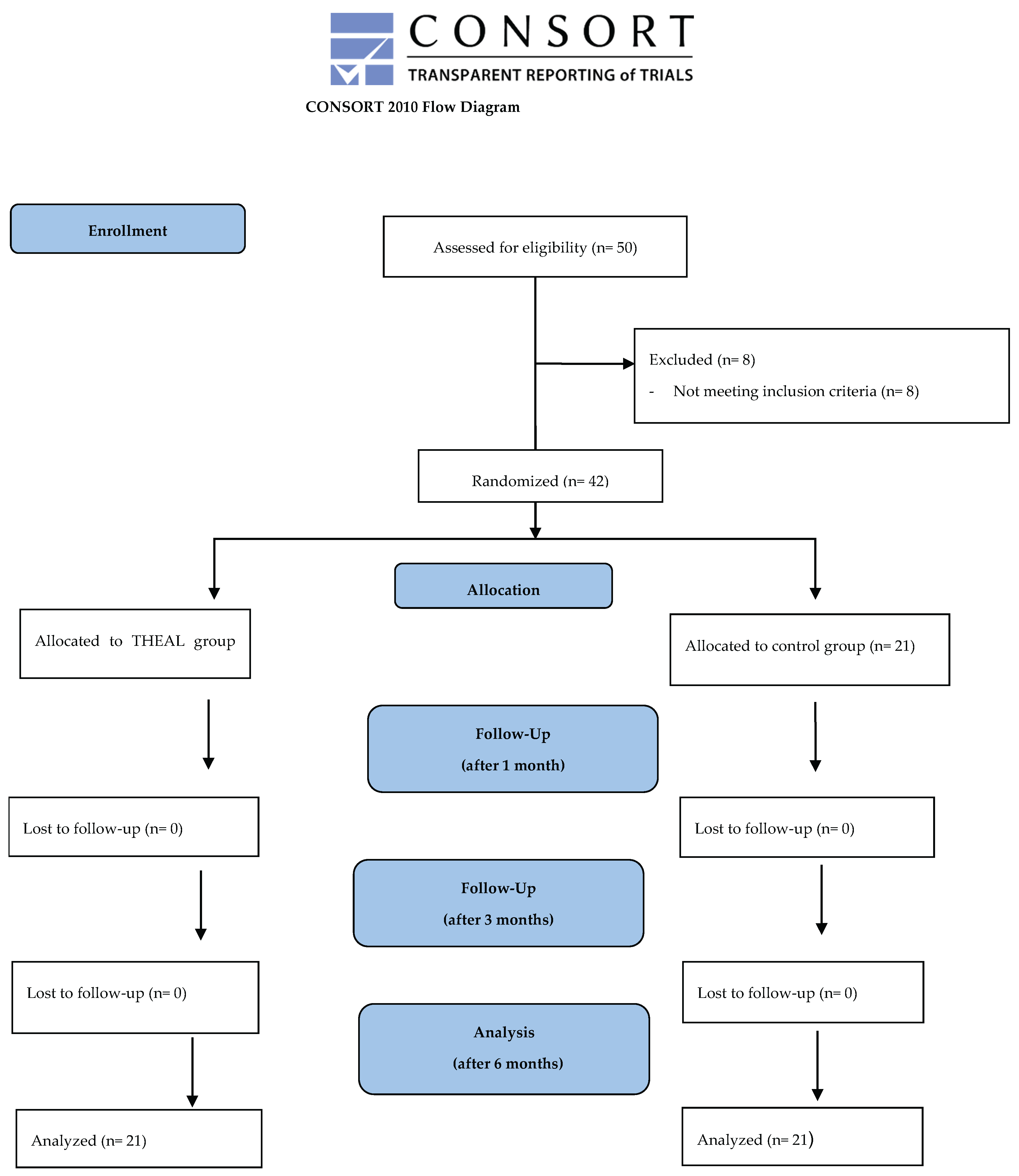

Table 1.
Energetic parameters of the four phases of each session of the THEAL treatment. The first phase aim to activate the lymph node stations activation, the second phase consists of anti-inflammatory and biostimulating action targeted at the area affected by the pathology, the third phase is performed in the therapeutic window relating to the forearm muscles (flexors and extensors) with the aim of performing a decontracting and anti-inflammatory action. The final phase focuses on treating latent trigger points related to the condition. CW: continuous-wave; PBM: photobiomodulation mechanism; E2C: superpulsed stochastic emission.
Table 1.
Energetic parameters of the four phases of each session of the THEAL treatment. The first phase aim to activate the lymph node stations activation, the second phase consists of anti-inflammatory and biostimulating action targeted at the area affected by the pathology, the third phase is performed in the therapeutic window relating to the forearm muscles (flexors and extensors) with the aim of performing a decontracting and anti-inflammatory action. The final phase focuses on treating latent trigger points related to the condition. CW: continuous-wave; PBM: photobiomodulation mechanism; E2C: superpulsed stochastic emission.
| Phase | Wavelength | Power | Emission modality | Source | Total energy | Termic control |
| 1 | 650 nm 780 nm 810 nm 905 nm 980 nm 1064 nm |
6W | CW | Laser diode | 600 J | 39 – 43°C |
| 2 | 650 nm 780 nm 810 nm 905 nm 980 nm 1064 nm |
9.56W | CW + PBM | Laser diode | 35 J/cm2 | 38 – 41°C |
| 3 | 650 nm 780 nm 810 nm 905 nm 980 nm 1064 nm |
10.64W | CW + PBM | Laser diode | 33 J/cm2 | 38 – 41°C |
| 4 | 650 nm 780 nm 810 nm 905 nm 980 nm 1064 nm |
6W | CW | Laser diode | 60 J | Without thermal control |
Table 2.
Study population characteristics.
| Control group | THEAL group | Overall | |||
| Categorical variables (cases and percentages) | |||||
| Sex | Female | 10 (47.62%) | 11 (52.38%) | 21 (50.00%) | |
| Male | 11 (52.38%) | 10 (47.62%) | 21 (50.00%) | ||
| Smoking habit | 2 (9.52%) | 4 (19.05%) | 6 (14.29%) | ||
| Treated hand is dominant | 13 (61.90%) | 10 (47.62%) | 23 (54.76%) | ||
| Hypertensive cardiopathy | 7 (33.33%) | 13 (61.90%) | 20 (47.62%) | ||
| Endocrine dysfunction | 5 (23.81%) | 12 (57.14%) | 17 (40.48%) | ||
| Previous therapy with nonsteroidal anti-inflammatory drugs | 4 (19.05%) | 2 (9.52%) | 6 (14.29%) | ||
| Previous physiokinesis therapy | 3 (14.29%) | / | 3 (7.14%) | ||
| Echography class | 1 | 4 (19.05%) | 11 (52.38%) | 15 (35.71%) | |
| 2 | 17 (80.95%) | 10 (47.62%) | 27 (64.29%) | ||
| Continuous variables (means ±SD) | |||||
| Age (years) | 65.43 ±6.83 | 64.38 ±7.68 | 64.90 ±7.20 | ||
| Body mass index (kg/m2) | 25.33 ±2.22 | 27.00 ±2.28 | 26.16 ±2.38 | ||
| Months since the symptoms began | 10.90 ±7.71 | 21.81 ±8.65 | 16.36 ±9.79 | ||
| VAS | t0 | 7.00 ±0.89 | 7.29 ±1.10 | 7.14 ±1.00 | |
| t1 | 4.71 ±1.10 | 4.14 ±1.15 | 4.43 ±1.15 | ||
| t2 | 3.38 ±1.40 | 2.71 ±0.78 | 3.05 ±1.17 | ||
| t3 | 4.14 ±1.28 | 1.95 ±0.92 | 3.05 ±1.56 | ||
| FIHOA | t0 | 19.67 ±5.34 | 21.24 ±7.24 | 20.45 ±6.34 | |
| t1 | 9.76 ±3.16 | 8.43 ±3.23 | 9.09 ±3.23 | ||
| t2 | 7.71 ±6.06 | 7.62 ±2.35 | 7.67 ±4.54 | ||
| t3 | 8.57 ±6.30 | 6.67 ±1.39 | 7.62 ±4.61 | ||
| DASH | t0 | 28.62 ±7.37 | 32.24 ±12.18 | 30.43 ±10.11 | |
| t1 | 21.57 ±9.15 | 17.90 ±10.46 | 19.74 ±9.88 | ||
| t2 | 16.90 ±7.35 | 14.05 ±5.02 | 15.48 ±6.38 | ||
| t3 | 18.48 ±6.46 | 12.38 ±3.31 | 15.43 ±10.87 | ||
| RM | t1 | 2.57 ±0.51 | 2.14 ±0.36 | 2.36 ±0.48 | |
| t2 | 2.14 ±0.48 | 1.62 ±0.50 | 1.88 ±0.55 | ||
| t3 | 2.55 ±0.89 | 1.52 ±0.51 | 2.02 ±0.88 | ||
Table 3.
Score changes over time (t0-t3) and significance thereof. Comparison was performed via Student’s t test for paired samples, using a one-sided test (null hypothesis: stability or increase of score).
Table 3.
Score changes over time (t0-t3) and significance thereof. Comparison was performed via Student’s t test for paired samples, using a one-sided test (null hypothesis: stability or increase of score).
| Score | Group | t0-t3 change | SD | t | p-value |
| VAS | Control group | 2.86 | 1.15 | 11.36 | <0.001 |
| THEAL group | 5.33 | 1.02 | 24.04 | <0.001 | |
| FIHOA | Control group | 11.09 | 6.78 | 7.50 | <0.001 |
| THEAL group | 14.57 | 6.79 | 9.83 | <0.001 | |
| DASH | Control group | 10.14 | 6.44 | 7.21 | <0.001 |
| THEAL group | 19.86 | 11.68 | 7.79 | <0.001 |
Table 3.
Mean comparison tests between the control and study group for the study scores’ changes over each checkpoint, as well as for the RM score upon checkpoint 1, 2, and 3. The Mann-Whitney non-parametric test was used for changes of the VAS, FIHOA and DASH score due to non-normal distribution, while Student’s t test was used for the RM score (hypothesizing lower values for the experimental group).
Table 3.
Mean comparison tests between the control and study group for the study scores’ changes over each checkpoint, as well as for the RM score upon checkpoint 1, 2, and 3. The Mann-Whitney non-parametric test was used for changes of the VAS, FIHOA and DASH score due to non-normal distribution, while Student’s t test was used for the RM score (hypothesizing lower values for the experimental group).
| Variable | Test statistics | p-value | |
| ΔVAS | t0-1 | z: -3.60 | <0.001* |
| t1-2 | z: -0.61 | 0.567 | |
| t2-3 | z: -4.16 | <0.001* | |
| t0-3 | z: -4.90 | <0.001* | |
| ΔFIHOA | t0-1 | z: -1.48 | 0.142 |
| t1-2 | z: 1.55 | 0.122 | |
| t2-3 | z: -2.74 | 0.005* | |
| t0-3 | z: -1.83 | 0.068 | |
| ΔDASH | t0-1 | z: -3.29 | <0.001* |
| t1-2 | z: 1.42 | 0.160 | |
| t2-3 | z: -2.64 | 0.008* | |
| t0-3 | z: -3.00 | 0.002* | |
| RM score | t1 | t: 3.16 | 0.001* |
| t2 | t: 3.48 | 0.006* | |
| t3 | t: 4.56 | <0.001* | |
Table 5.
Regression analysis. The determinant is represented by the treatment option, considered as a binomial variable with value =1 for the experimental group. Sensitivity analysis with exclusion of subjects over 65 years of age is reported at the bottom of the table. Due to non-normal distribution of all outcomes, correction with robust standard errors was employed for all linear regression models.
Table 5.
Regression analysis. The determinant is represented by the treatment option, considered as a binomial variable with value =1 for the experimental group. Sensitivity analysis with exclusion of subjects over 65 years of age is reported at the bottom of the table. Due to non-normal distribution of all outcomes, correction with robust standard errors was employed for all linear regression models.
| Outcome | Model | ß | 95%CI | p-value |
| ΔVAS (t0-3) | M1 | 2.48 | 1.80 – 3.15 | <0.001* |
| M2 | 2.42 | 1.78 – 3.06 | <0.001* | |
| M3 | 3.49 | 2.65 – 4.32 | <0.001* | |
| ΔFIHOA (t0-3) | M1 | 3.48 | -0.76 – 7.71 | 0.105 |
| M2 | 3.26 | -0.92 – 7.44 | 0.122 | |
| M3 | 6.51 | -1.06 – 14.08 | 0.089 | |
| ΔDASH (t0-3) | M1 | 9.71 | 3.83 – 15.60 | 0.002* |
| M2 | 9.16 | 4.00 – 14.32 | 0.001* | |
| M3 | 9.20 | -0.84 – 19.23 | 0.071 | |
| Sensitivity analysis | ||||
| ΔVAS (t0-3) | M1 | 2.35 | 1.15 – 3.55 | 0.001* |
| M2 | 2.32 | 1.36 – 3.29 | <0.001* | |
| M3 | 2.36 | 0.47 – 4.25 | 0.021* | |
| ΔFIHOA (t0-3) | M1 | 1.64 | -5.51 – 8.79 | 0.637 |
| M2 | 0.63 | -6.29 – 7.55 | 0.849 | |
| M3 | -6.39 | -14.71 – 1.93 | 0.114 | |
| ΔDASH (t0-3) | M1 | 10.84 | 1.02 – 20.67 | 0.032* |
| M2 | 10.01 | 1.15 – 18.86 | 0.029* | |
| M3 | 18.40 | -0.31 – 37.11 | 0.053 | |
95%CI: 95% confidence interval. M1: raw regression. M2: M1 with adjustment for sex and age. M3: M2 with further adjustment for body mass index, smoking habit, concordance of the hand, months since the beginning of symptoms, hypertensive cardiopathy, endocrine dysfunction/condition, therapy with nonsteroidal anti-inflammatory drugs, previous physiokinesis therapy, echography classification of disease.
Disclaimer/Publisher’s Note: The statements, opinions and data contained in all publications are solely those of the individual author(s) and contributor(s) and not of MDPI and/or the editor(s). MDPI and/or the editor(s) disclaim responsibility for any injury to people or property resulting from any ideas, methods, instructions or products referred to in the content. |
© 2026 by the authors. Licensee MDPI, Basel, Switzerland. This article is an open access article distributed under the terms and conditions of the Creative Commons Attribution (CC BY) license (http://creativecommons.org/licenses/by/4.0/).
Copyright: This open access article is published under a Creative Commons CC BY 4.0 license, which permit the free download, distribution, and reuse, provided that the author and preprint are cited in any reuse.