

Submitted:
02 July 2026
Posted:
03 July 2026
You are already at the latest version
Abstract
Metabolic diseases, such as obesity, type 2 diabetes (T2DM), and metabolic dysfunction-associated steatotic liver disease (MASLD, formerly NAFLD), emerge from chronic nutrient overload that progressively disrupts cellular homeostasis and precipitates persistent inflammatory remodeling. Two stress-adaptive programs sit at the center of such pathological transition: autophagy, the homeostatic engine responsible for lysosomal recycling and organelle quality control, and cellular senescence, a tissue repair system that becomes deleterious when persistent, driving dysfunction through stable growth arrest and senescence-associated secretory phenotype (SASP)-associated inflammatory remodeling. Emerging evidence identifies lysine acetylation as a nutrient-sensitive molecular rheostat that orchestrates the reciprocal regulation of these two processes across metabolic tissues. Because acetylation dynamics are governed by the availability of acetyl-CoA and the NAD+-dependent deacetylation capacity of sirtuins, metabolic stress reshapes the enzymatic balance between acetyltransferases (KATs, e.g., p300/CBP) and deacetylases. This shift triggers a “double blow” to cellular health; the simultaneous suppression of autophagic flux and the epigenetic stabilization of senescent state. This review synthesizes mechanistic evidence showing how acetylation inhibits autophagy at multiple checkpoints, ranging from the activity of core autophagy proteins to lysosomal biogenesis, while concurrently reinforcing senescence through chromatin hyperacetylation and the activation of non-histone effectors such as NF-κB, p53 and FOXO proteins. We place particular emphasis on the mTORC1-p300-SIRT1 axis as the primary integration hub coupling nutrient status to this dual regulation. Finally, we discuss tissue-specific manifestations in the liver, adipose tissue, and other metabolic organs and highlight emerging therapeutic strategies, such as KAT inhibition/sirtuin activation, aimed at restoring the acetylation rheostat to improve autophagic competence while restraining senescence-driven metabolic decline.
Keywords:
protein acetylation
; acetyl CoA and NAD+
; EP300
; sirtuins
; autophagy
; senescence
; SASP
; metabolic diseases
1. Introduction
Metabolic diseases, including obesity, type 2 diabetes mellitus (T2DM) and metabolic dysfunction-associated steatotic liver disease (MASLD/NAFLD), are characterized by chronic nutrient surplus and the progressive deterioration of tissue resilience (Hotamisligil, 2017). At the cellular level, these pathologies reflect a failure of two coupled stress-adaptive programs, such as autophagy and cellular senescence. These programs function as homeostatic engines, integrating nutrient signaling (via hubs such as mTORC1 and SIRT1) to maintain cellular quality control through lysosomal recycling and the removal of irreparable cells (Kitada & Koya, 2021; Lee et al., 2021). However, chronic overnutrition triggers a reciprocal ‘vicious cycle’ where impaired autophagic flux directly fuels the transition toward a persistent, pro-inflammatory senescence program. The resulting accumulation of damaged components and the SASP drive systemic insulin resistance and broader tissue dysfunction, including β-cell tissue failure (Chaib et al., 2026; Kitada & Koya, 2021). Consequently, the balance between these two processes has emerged as a central determinant of whether metabolic stress is resolved or progresses into long term or irreversible pathology (Kitada & Koya, 2021; Liu et al., 2020).
Emerging evidence identifies protein acetylation as a nutrient-sensitive integrator that coordinates the autophagy-senescence balance across metabolic tissues (Yinfeng Xu & Wei Wan, 2023; Yu et al., 2025). This regulatory layer functions as a molecular rheostat, where the availability of acetyl-CoA fuels lysine acetyltransferases (KATs, e.g., EP300/CBP) and NAD+ levels dictate sirtuin- mediated deacetylation. Through this metabolic coupling, acetylation controls autophagy at multiple checkpoints, ranging from the direct modification of core ATG proteins and ULK/Beclin-1 complexes to the transcriptional orchestration of lysosomal biogenesis (Yinfeng Xu & Wei Wan, 2023). Simultaneously, acetylation stabilizes the senescent state by driving enhancer remodeling and modulating the activity of non-histone effectors, including p53, NF-kB, and FOXO proteins (Sen et al., 2019; Xu et al., 2025). Under chronic metabolic stress, the depletion of NAD+ and subsequent loss of sirtuin activity trigger a shift toward pathological hyperacetylation. This imbalance suppresses autophagic flux while reinforcing pro-inflammatory chromatin landscapes, ultimately locking cells into a state of persistent damage accumulation and metabolic decline (Hwang et al., 2013; Palmer et al., 2022).
This review synthesizes the evidence for protein acetylation as a pivotal metabolic integrator coordinating autophagic flux and cellular senescence. We propose the “Acetylation Rheostat” hypothesis, which posits that cellular homeostasis is dictated by the relative availability of acetyl-CoA and NAD+. This metabolic state modulates the activity of the EP300-sirtuin axis, thereby determining the balance between homeostatic quality control and inflammatory remodeling. When this regulatory hub is skewed by chronic overnutrition, it triggers the ‘double blow’ of impaired clearance and persistent senescence that drive the progression of obesity, T2DM, and MASLD. This review establishes a comprehensive roadmap of the biochemical sensors of nutrient status before detailing the molecular toggle switches (e.g., p53, FOXO, mTORC1) that translate acetylation signals into cell-fate decisions. Finally, we examine these dynamics from a tissue-specific perspective and highlight therapeutic ‘rebalancing’ strategies aimed at restoring cellular resilience at its regulatory root, rather than merely treating its downstream pathological consequences.
2. The Metabolic Crisis: Autophagy Failure and Senescence Accumulation
2.1. Autophagy Failure in Metabolic Tissues
In metabolic tissues, autophagy acts as a core homeostatic engine that couples nutrient signaling, primarily via the AMPK-mTORC1 axis, to lysosomal recycling in order to sustain proteostasis, mitochondrial integrity, and lipid handling during feeding-fasting cycles. By sequestering damaged mitochondria (mitophagy) and other dysfunctional macromolecules into autophagosomes and clearing them via lysosomes, autophagic flux provides continuous cellular ‘housekeeping’, thereby protecting the insulin signaling network from chronic stress (Cao et al., 2021; Liu et al., 2023). Although autophagy is crucial for maintaining cellular homeostasis, autophagic efficiency declines with overnutrition, aging and metabolic derangements including obesity, insulin resistance, and T2DM, thereby amplifying metabolic stress and tissue dysfunction (Jakubek et al., 2024; Kitada & Koya, 2021; Raza, 2024). Further, the impairment of autophagy through the genetic loss of Atg7 or pharmacological inhibition disrupts adipose tissue mass and differentiation while enhancing pro-inflammatory cytokine expression. Consequent to the decline in autophagic flux, the accumulation of damaged and superfluous cargo raises reactive oxygen species (ROS) and amplifies pro-inflammatory signaling along with increasing NLRP3 inflammasome-linked inflammatory tone, leading to insulin resistance and tissue dysfunction (Biasizzo & Kopitar-Jerala, 2020).
However, compelling evidence indicates that enhancing autophagic flux can effectively ameliorate various metabolic diseases. In adipose tissue, autophagy serves as a protective mechanism that restrains obesity-associated fibrosis and maintains the functional integrity of visceral white adipose tissue by regulating tissue purine nucleoside metabolism (Piletic et al., 2025). Similarly, hepatic autophagy has emerged as a metabolic control node. For example, activation of melanocortin 3 receptor signaling has been shown to reduce adipose tissue lipid accumulation and restore body weight in obese mouse models via the regulation of hepatic autophagy (Patel et al., 2025). Mechanistic studies further demonstrate that GLP-1 receptor agonists can reduce hepatic steatosis by inducing autophagy through the AMPK/SIRT1 signaling pathway (Yu et al., 2025). Furthermore, preclinical research has expanded this concept to include TFEB-centered lysosomal- autophagy programs and metabolic intermediates. Examples include the Qigui Jiangzhi Formula, which improves MASLD via modulation of the AMPK/SIRT1-TFEB pathway (Zhang et al., 2025), and the use of aminobutyric acid to halt MASLD progression in mice by inducing autophagic flux (Felicianna et al., 2025). Consistent with these mechanistic pathways, research on autophagy emphasizes that it is not simply ‘bulk degradation’ mechanism, but a highly selective and flux-dependent pathway critical for adaptation to metabolic stress and the prevention of chronic disease development (Glick et al., 2010; Kitada & Koya, 2021; Lee et al., 2025; Tao et al., 2025).
2.2. Senescence Burden in Metabolic Disease
By contrast, cellular senescence contributes to metabolic homeostasis only when it is transient, such as during wound repair or tumor suppression. However, in metabolic disease, it frequently becomes persistent, causing durable growth arrest along with a SASP that drives inflammation, extracellular matrix remodeling, immune-cell recruitment, and insulin resistance (Palmer et al., 2022; Saliev & Singh, 2025; Spinelli et al., 2023). Recent evidence underscores the critical role of senescent cells in curtailing both health-span as well as lifespan. Their chronic accumulation is now recognized as a key driver of various pathologies, including metabolic dysfunction and accelerated aging syndromes (Chaib et al., 2026; Papatheodoridi et al., 2020; Suda et al., 2025). Research utilizing genetic and pharmacological senescent-cell clearance models has shown that reducing the senescent cell burden in obese mice effectively alleviates metabolic dysfunction, improves glucose homeostasis, and enhances overall longevity. For example, the selective clearance of p16Ink4a+ senescent cells can mitigate systemic inflammation and improve glucose metabolism in obese mice (Suda et al., 2025). Similarly, in another study, senescent hepatocytes have been identified as central drivers of MASLD. therefore, eliminating these cells presents a promising therapeutic strategy for treating liver disease and its associated multi-organ complications (Du et al., 2025). Further, human atlas studies have revealed that while weight loss has the potential to reverse adipose senescence and improved metabolic remodeling, an “obesogenic epigenetic memory” can persist in these tissues, potentially leading to a rebound in metabolic dysfunction. Thus, elimination of such senescent cells represents a key therapeutic target to improve metabolic health (Hinte et al., 2024; Miranda et al., 2025). These insights further extend to skeletal muscle, where multi-omics profiling has identified Maraviroc as a potential senotherapeutic strategy to combat sarcopenia-associated loss of muscle mass and strength (Y. Li et al., 2025). Finally, a pivotal study showed that inhibition of TGFβ can hinder senescence transmission from liver to other organs, thereby preventing systemic multi-organ dysfunction; thus, providing powerful evidence that senescence is an active driver rather than a mere bystander in metabolic homeostasis (Kiourtis et al., 2024; Saliev & Singh, 2025).
2.3. Mechanistic Crosstalk Between Autophagy and Senescence
Mechanistically, metabolic stress initiates a ‘vicious cycle’ where autophagy failure acts as persistent pro-senescence signal which accelerates senescence, and the established senescent state further represses autophagic capacity. Impaired autophagy exacerbates mitochondrial dysfunction, amplifying ROS and DNA damage signaling that enforces senescence commitment (Figure 2). However, autophagy restoration at this stage can break this cycle and restore regenerative function in aged cells (Bahar et al., 2026; García-Prat et al., 2016; Rajendran et al., 2019). Once senescence is established, SASP-associated inflammatory signaling (often via NF-κB) can up-regulate autophagy inhibitors such as A20, Bcl-2/Bcl-xL while suppressing pro-autophagy mediators such as BNIP3 and JNK1, further reducing cellular clearance capacity and augmenting damage accumulation (Salminen et al., 2012). In addition, autophagy-dependent RNA splicing and translational rewiring during senescence actively direct inflammatory output, thereby perpetuating defective clearance and SASP maintenance (Kim et al., 2025). Moreover, the negative regulator of autophagy (Rubicon) rises with age to down-regulate autophagic flux, predisposing tissues to damage accumulation and senescence pressure (Nakamura et al., 2019). In parallel, senescence has been evidenced to robustly suppress mitophagy, locking cells into an elevated ROS and pro-inflammatory state that further perpetuates the SASP program (Chaib et al., 2026; Kelly et al., 2024; Suda et al., 2025).
These coupled failures of autophagy and senescence control suggest the existence of an upstream metabolic regulator, a “master switch”, that can orchestrate both mechanisms simultaneously. Under metabolic stress, changes in nutrient availability reshape intracellular acetyl-CoA and NAD+ pools, thereby shifting the balance between acetyltransferases and deacetylases. This places “protein acetylation” in a prime position to function as a nutrient-sensitive regulatory layer that links the metabolic state to the control of both autophagy and senescence (Keller & Nakamura, 2024; Xu et al., 2025). For instance, on one hand, chronic mTORC1 signaling activates p300, impairing autophagic quality control via the acetylation of core autophagic machinery, while; on the other hand acetylation-dependent chromatin remodeling, including p300-driven super-enhancer formation and BRD4 recruitment, stabilizes senescence and SASP transcription. Ultimately, it supports the view that a single regulatory layer, i.e., “protein acetylation”, is capable of pushing both arms of this metabolic ‘double blow’ in the same pathological direction (García-Prat et al., 2016; Ghosh, 2021; Tasdemir et al., 2016; Wan et al., 2017; Y. Xu & W. Wan, 2023).
3. Protein Acetylation: The Nutrient-Sensitive Sensor of Cellular Metabolic Status
3.1. Biochemical Foundation of Protein Acetylation
Protein acetylation predominantly occurs as N-terminal acetylation and lysine ε-N-acetylation, with lysine acetylation being the major reversible form regulating chromatin, metabolism, and signaling (Ali et al., 2018; Zhou & Cole, 2025). Lysine acetylation is a reversible post-translational modification (PTM), involving dynamically regulated addition of an acetyl group (CH3CO) to the ε-amino group of lysine residue. This modification neutralizes the positive charge of the lysine residue, which can significantly alter its protein conformation, chromatin accessibility, enzymatic activity, and molecular interactions. This process is enzymatically catalyzed by lysine acetyltransferase (KATs), such as p300/CBP; while the removal of acetyl groups is performed by deacetylases, including histone deacetylases (HDACs) and nicotinamide adenine dinucleotide NAD+-dependent sirtuins (Ali et al., 2018; Y. Xu & W. Wan, 2023; Yazıcı & McIntyre, 2025). For example, in mitochondria, the high concentration of acetyl-CoA, alkaline matrix pH and the higher density of reactive metabolites favor acetyl transfer reactions. Consequently, non-enzymatic protein acetylation represents the most prevalent post-translational modification in this organelle. In this environment, sirtuins (such as SIRT3) function as a key ‘deacetylation repair’ system, counterbalancing the non-enzymatic accumulation of acetyl groups to preserve mitochondrial oxidative metabolism and quality control. This biochemical foundation positions acetylation as a vital upstream regulator that integrates nutrient availability with cellular stress-adaptive responses (Hosp et al., 2017; M. Q. Li et al., 2025; Shimazu et al., 2025).
3.2. Metabolic Dependence of Acetylation and Compartmentalized Acetyl-CoA/NAD+ Pools
Having outlined the biochemical basis of lysine acetylation, it is also important to its intrinsic link to cellular metabolism. This dependency arises because the activities of the modifying enzymes are governed by the availability of metabolic substrates and cofactors. Specifically, KAT activity depends on local acetyl-CoA availability, while sirtuin-regulated deacetylation requires NAD+. Therefore, alternations in nutrient supply, mitochondrial function, as well as redox status can directly shift the local and global acetylation tone, allowing this modification to serve as a versatile metabolic sensor (Shi & Tu, 2015; Sivanand et al., 2018; Wellen et al., 2009).
A defining reason for acetylation being metabolically informative and versatile is that acetyl-CoA is compartmentalized into separate, functionally distinct pools. The mitochondrial acetyl-CoA pool (derived primarily from β-oxidation and the pyruvate dehydrogenase complex) is regulated independently and serves explicit local metabolic functions and is not freely interchangeable with nuclear/cytosolic pools. Despite the permeability of nuclear pores to acetyl-CoA, recent metabolic evidence suggests that nuclear and cytosolic acetyl-CoA pools are largely distinct and gate specific local acetylation programs (Guertin & Wellen, 2023; Sivanand et al., 2018). The nuclear-cytosolic acetyl-CoA is generated from two canonical precursors; citrate, via ATP-citrate lyase (ACLY), and acetate, via acyl-CoA synthetase short-chain 2 (ACSS2). Moreover, alternative shuttling routes, such as acetyl carnitine-associated transfer, help sustain nuclear and cytosolic acetylation when primary sources are constrained (Izzo et al., 2023).
Such principles are especially relevant in metabolic diseases, where these compartmentalized dynamics allow acetylation to function as a metabolic master switch that simultaneously tunes autophagy and senescence to maintain metabolic homeostasis (Morales-Tarré et al., 2021; Wang & Lei, 2018). The same metabolites that are sensitive to nutrient status, such as acetyl-CoA and NAD+, control the opposing activities of EP300/CBP and sirtuins, which in turn regulate both autophagic machinery and senescence programs. For instance, under nutrient-rich conditions with high acetyl-CoA levels, activated EP300 acetylates autophagy core ATG proteins, impairing autophagic flux. Simultaneously, up-regulated EP300 activity promotes a hyperacetylated enhancer landscape that boosts the expression of senescence-associated genes. Conversely, during starvation or NAD+-favored conditions, sirtuin-mediated deacetylation restores autophagic competence while restraining pro-senescent chromatin remodeling (Lee et al., 2008; Lee & Finkel, 2009; Mariño et al., 2014; Sen et al., 2019). In brief, acetylation is uniquely positioned to convert the cellular metabolic state into the coordinated regulation of intracellular quality control and cell-fate remodeling, and thus changes in the acetylation state can shift both processes simultaneously (Eisenberg et al., 2009; Kang & Elledge, 2016; Lee et al., 2021; Sun et al., 2021; Xu et al., 2025).
4. The Molecular Toggle Switch: Reciprocal Regulation of Pathological Programs
Having established the biochemical basis of acetylation, we now turn to its role in regulating autophagy and senescence simultaneously. Acetylation is not merely a downstream marker of metabolic stress; it functions as a bidirectional molecular toggle that converts changes in acetyl-CoA and NAD+ availability into coordinated shifts in autophagic flux and senescence programs. In metabolic diseases, prolonged nutrient surplus can bias cells towards KAT-driven acetylation and away from sirtuin-mediated deacetylation, leading to simultaneous autophagic suppression and senescence stabilization, consequently driving metabolic disease progression (Lee et al., 2008; Lee & Finkel, 2009; Mariño et al., 2014; Rajendran et al., 2019; Sen et al., 2019) (Figure 1).
Figure 1.
Acetylation rheostat model linking nutrient status to reciprocal regulation of autophagy and senescence in metabolic diseases. Nutrient-sensitive acetylation acts as a rheostat that reciprocally regulates autophagy and senescence. In nutrient-rich states, increased acetyl-CoA and mTORC1 activity favors p300-dependent acetylation, suppressing autophagic flux and promoting senescence-associated chromatin remodeling, p16/p21 induction, and SASP expression. While, in nutrient scarce states, increased NAD+ and reduced mTORC1 activity favors sirtuin-mediated deacetylation, restoring autophagy and restraining senescence. Chronic nutrient excess shifts this balance towards impaired cellular quality control and persistent inflammatory remodeling in metabolic disease.
Figure 1.
Acetylation rheostat model linking nutrient status to reciprocal regulation of autophagy and senescence in metabolic diseases. Nutrient-sensitive acetylation acts as a rheostat that reciprocally regulates autophagy and senescence. In nutrient-rich states, increased acetyl-CoA and mTORC1 activity favors p300-dependent acetylation, suppressing autophagic flux and promoting senescence-associated chromatin remodeling, p16/p21 induction, and SASP expression. While, in nutrient scarce states, increased NAD+ and reduced mTORC1 activity favors sirtuin-mediated deacetylation, restoring autophagy and restraining senescence. Chronic nutrient excess shifts this balance towards impaired cellular quality control and persistent inflammatory remodeling in metabolic disease.
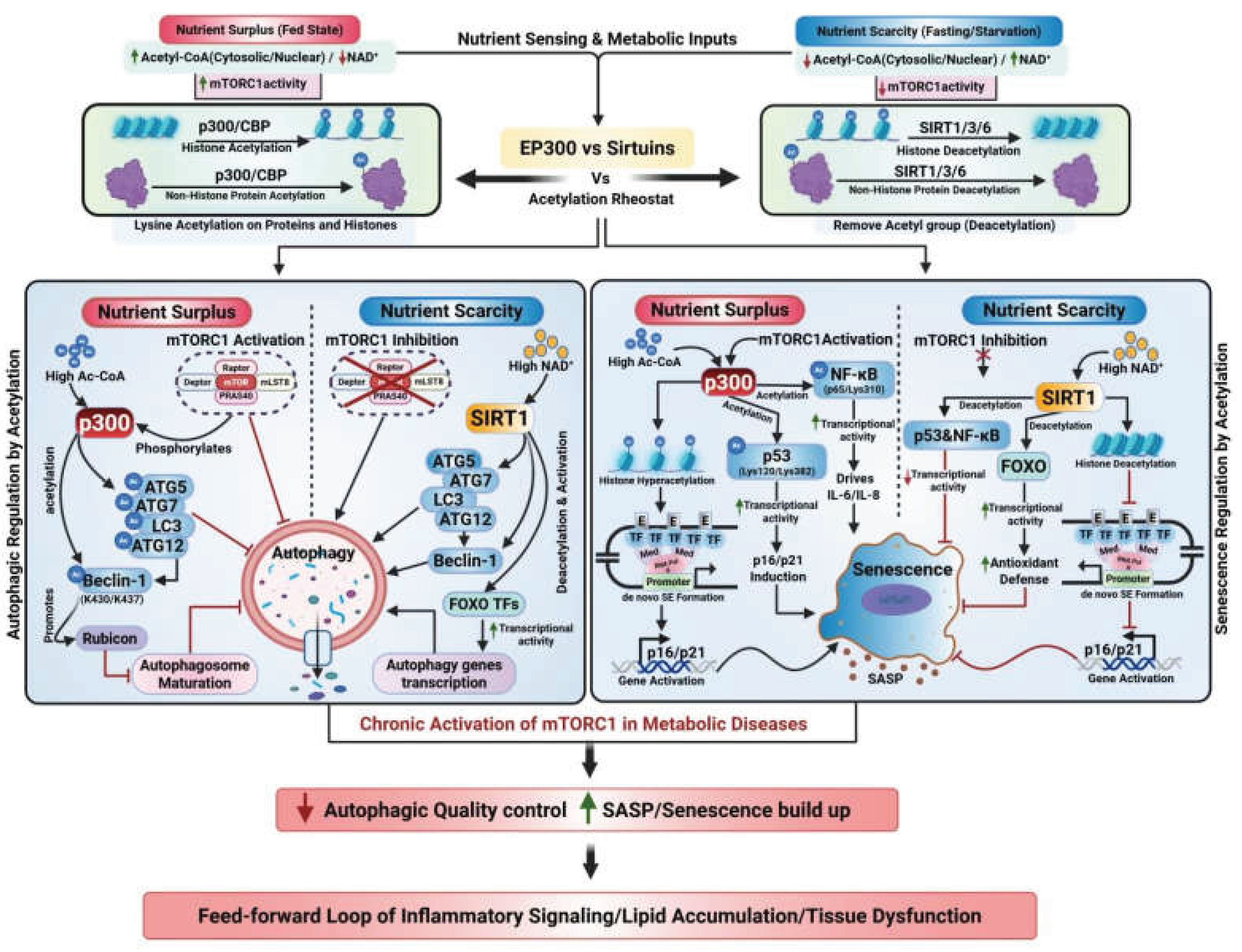
Figure 2.
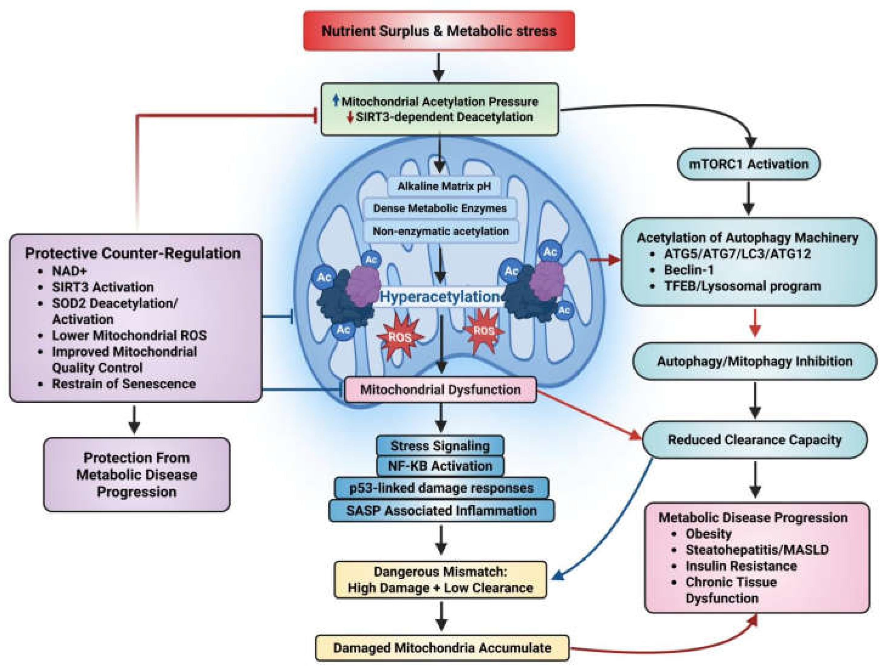
Mitochondrial acetylation: Mitophagy mismatch links nutrient surplus to senescence and metabolic disease progression. Nutrient surplus /metabolic stress elevates mitochondrial acetylation pressure, while weakening SIRT3-dependent deacetylation, promoting mitochondrial hyperacetylation, and ROS generation. It leads to mitochondrial dysfunction and activation of senescence-associated signaling. Simultaneously, mTORC1 activation suppresses mitophagy via acetylation of core autophagic machinery, reducing clearance capacity. The resulting mismatch between mitochondrial damage and mitochondrial clearance drives damaged mitochondria accumulation, inflammatory and senescence associated signaling, and metabolic disease progression. Protective NAD+/SIRT3-dependent counter-regulation limits these effects by restoring mitochondrial quality control.
Figure 2.
Mitochondrial acetylation: Mitophagy mismatch links nutrient surplus to senescence and metabolic disease progression. Nutrient surplus /metabolic stress elevates mitochondrial acetylation pressure, while weakening SIRT3-dependent deacetylation, promoting mitochondrial hyperacetylation, and ROS generation. It leads to mitochondrial dysfunction and activation of senescence-associated signaling. Simultaneously, mTORC1 activation suppresses mitophagy via acetylation of core autophagic machinery, reducing clearance capacity. The resulting mismatch between mitochondrial damage and mitochondrial clearance drives damaged mitochondria accumulation, inflammatory and senescence associated signaling, and metabolic disease progression. Protective NAD+/SIRT3-dependent counter-regulation limits these effects by restoring mitochondrial quality control.
4.1. The mTORC1-p300 Integration Hub: Feeding-Induced Autophagy Suppression and Senescence Stabilization
Upstream nutrient sensing is most directly regulated by acetylation-linked control via mTORC1-p300 axis, which couples the fed states to the suppression of autophagy and enhanced lipogenic programs. Under nutrient-rich conditions, such as high amino acid or insulin levels, mTORC1 is activated and directly phosphorylates p300 at several C-terminal serine residues, thereby stimulating its acetyltransferase activity. Activated p300/CBP suppresses autophagy at both early and late checkpoints. Specifically, p300 acetylates core autophagy proteins, such as ATG5, ATG7, ATG8/LC3, and ATG12, sequestering them in the nucleus and preventing their participation in cytoplasmic autophagosome formation (Lee & Finkel, 2009; Wu et al., 2021). Furthermore, p300-mediated acetylation of Beclin-1 (at K430/K437) promotes the recruitment of Rubicon, which constrains autophagosome maturation and endocytic trafficking. This mechanism ultimately lowers net autophagic flux even in the presence of initiation signals (Sun et al., 2015; Y. Xu & W. Wan, 2023; Yazıcı & McIntyre, 2025). Thus, consistent with this model, pharmacological inhibition of p300/CBP has been shown to rapidly reduce lipogenesis in liver and adipose tissue, suppress hepatic gluconeogenesis, and alleviate insulin resistance, positioning p300 as a promising therapeutic target for treating autophagy-related metabolic disorders (Ansari et al., 2023; Y. Xu & W. Wan, 2023; Zhou et al., 2020) (Figure 3).
In parallel, p300 activity is crucial during senescence entry, acting as a primary driver by inducing de novo super enhancers (SEs) formation and promoting the hyper-acetylated chromatin state that establishes the senescence-specific gene expression program. For example, during replicative senescence, p300 reshapes large regions of the non-coding genome into these SEs to sustain the expression of senescence associated genes, including cell cycle inhibitors like p16 and p21 (Sen et al., 2019; Yu et al., 2025). While p300 initiates this state, the maintenance stage relies on the downstream recruitment of BRD4, an acetyl-lysine reader that binds to p300-induced acetylation marks to sustain transcriptional activity. This creates a positive feedback loop that strengthens the senescent phenotype. Mechanistically, BRD4 recruitment to newly activated SEs near SASP genes facilitates robust pro-inflammatory transcription and paracrine signaling, a mechanism that drives persistent inflammation in MASLD, obesity and T2DM (Di Giorgio et al., 2021; Tasdemir et al., 2016). Moreover, recent literature underscores the pivotal role of BRD4 as a key epigenetic reader that governs the maintenance of SASP and broader aging-associated transcriptional programs (Sun et al., 2024; Wang et al., 2024). Recent work further supports p300 as an epigenetic driver of premature senescence and highlights that the stable growth arrest and inflammatory remodeling associated with stress-induced senescence can be effectively delayed or mitigated through the pharmacological inhibition of p300 (Di Fede et al., 2025).
In conclusion, in metabolic disease, the chronic activation of mTORC1 pushes the system via p300 to simultaneously repress autophagic quality control and lock in senescence and SASP programs, creating a pathological feed-forward loop of inflammatory signaling, lipid accumulation, and tissue dysfunction (Sen et al., 2019; Son et al., 2024; Sun et al., 2015; Wan et al., 2017) (Figure 1).
4.2. Acetylation Modification of Core Regulatory Hubs: p53, FOXO, and NF-κB
Beyond the regulation of the upstream integration hub, acetylation also governs cell fate through the modification of various other core regulatory hubs, specifically key transcription factors, including p53, FOXO, and NF-κB. Once stress and nutrient signals are detected, the acetylation-dependent regulation of these “gatekeeping” factors jointly determines whether a cell undergoes stress adaptation via autophagy or transitions into durable senescence characterized by inflammatory remodeling (Son et al., 2025).
Increased acetylation of p53 significantly boosts its transcriptional activity. For example, the acetylation of p53 at Lys120 by MOZ leads to p21/p16 induction and pushing the cells towards stable growth arrest (Hu et al., 2023). However, when stress is intermittent, deacetylases such as SIRT1 act as a negative regulators by deacetylating p53 at Lys382, thereby restraining its transcriptional output and reducing the probability of permanent arrest (Yu et al., 2025). This balance is critical in metabolic disease, where nutrient surplus often increases acetylation pressure while suppressing deacetylation capacity. This environment makes p53-regulated arrest more robust and harder to resolve, facilitating chronic tissue dysfunction (Vaziri et al., 2001; Wang et al., 2023; Zhang et al., 2017). Interestingly, dietary interventions such as glucose restriction (via carbohydrate-free dietary regimens) have been shown to induce a specific form of p53 acetylation that routes the protein for degradation via autophagy, highlighting a potential therapeutic mechanism to clear pro-senescent factors (Rodriguez et al., 2012).
NF-κB functions as the primary transcriptional engine for SASP chemokines/cytokines, with its output becoming specifically “hard-wired” through enhancer remodeling during the progression of senescence. Acetylation of the p65 subunit of NF-κB at Lys310 significantly enhances its transcriptional activity, which is essential for the maintenance of the SASP state. Conversely, the deacetylation of p65 by SIRT1 inhibits this process, effectively reducing the production of potent pro-inflammatory cytokines, such as IL-6 and IL-8 (Yeung et al., 2004; Yu et al., 2025). Mechanistically, this acetylation event enables the recruitment of acetyl-lysine reader BRD4 to newly activated super-enhancers adjacent to SASP genes. This BRD4 recruitment is significantly involved in SASP regulation and downstream paracrine signaling. While the activation of NF-κB initiates early in the stress response, BRD4-mediated super-enhancer control is predominantly critical for the long-term maintenance of senescence and amplification of SASP. This epigenetic mechanism represents a major disease-driving component in conditions such as MASLD, obesity and T2DM (Brown et al., 2014; Tasdemir et al., 2016). Furthermore comparative studies show that such enhanced NF-κB signaling during aging and cellular senescence actively suppresses cellular quality control by up-regulating the expression of autophagy inhibitors, (e.g., A20, and Bcl-2/Bcl-xL) and repressing the essential autophagy activators, such as BNIP3, JNK1, and ROS (Salminen et al., 2012).
Beyond p53 and NF-κB, the FOXO transcription factor family (notably FOXO1 and FOXO3a) serves as another major regulatory hub whose function is tightly governed by acetylation. For example, the acetylation of FOXO1 or FOXO3a transcription factors inhibits their transcriptional activity and diminishes the cellular antioxidant defenses, thereby driving cell fate towards senescence. Conversely, oxidative stress-induced deacetylation enhances FOXO activity, promoting mitochondrial resilience and the expression of autophagy-related genes while simultaneously suppressing the cellular senescence program (Gupta et al., 2025; Kuo et al., 2025; Zhang et al., 2013). By integrating with sirtuin signaling and mitochondrial status, the FOXO family functions as a critical ‘bridge-hub’ that coordinates the reciprocal relationship between autophagy and senescence.
4.3. Organelle-Specific Acetylation Regulation of Mitophagy, ROS, and Senescence
Beyond the nucleus and cytosol, mitochondrial acetylation establishes a critical “damage loa” that escalates the demand for autophagy/mitophagy, the selective autophagic degradation of mitochondria, while simultaneously fueling senescence pressure via ROS and inflammatory signaling (Figure2). Mitochondria are uniquely susceptible to lysine acetylation due to their alkaline matrix pH, high acetyl-CoA availability, and high c density of metabolic enzymes. In this environment, much of the protein acetylation occurs non-enzymatically, making the mitochondrial acetyl-proteome a powerful metabolic sensor of the organelle’s internal state. This spontaneous accumulation of acetyl groups is counterbalanced by NAD+-dependent deacetylase SIRT3, the dominant sirtuin in mitochondrial matrix (Baeza et al., 2016; Eftekhari et al., 2025). SIRT3 acts as a vital “deacetylation repair” system, removing inhibitory acetyl groups to preserve oxidative metabolism and mitochondrial quality control. For instance, caloric restriction boosts SIRT3 activity, which promotes the deacetylation and activation of SOD2 (superoxide dismutase 2), a primary antioxidant enzyme that neutralizes mitochondrial ROS. Elevated mitochondrial ROS serves as a leading upstream trigger of DNA damage signaling, NF-κB up-regulation, as well as pro-senescent SASP programs activation (Giralt & Villarroya, 2012; Qiu et al., 2010; Shimazu et al., 2025; Xu et al., 2024). Critically for metabolic disease, a high-fat diet leads to SIRT3 down-regulation and a subsequent surge in mitochondrial protein hyperacetylation. This pathological cascade accelerates obesity-associated phenotypes, such as steatohepatitis and insulin resistance, indicating that mitochondrial acetylation imbalance is a primary driver of disease progression rather than a mere correlation (Hirschey et al., 2011; Xian et al., 2025). This interpretation is also supported by a recent work linking age-related SIRT3 loss to the spread of pro-senescent signaling across metabolically active tissues (Šešelja et al., 2025).
Mechanistically, mitochondrial hyperacetylation leads to the organelle dysfunction, creating an increased “cargo burden” that upsurges the need for clearance via mitophagy. However, in the context of metabolic syndrome, a dangerous mismatch occurs; while mitochondrial damage increases the need for clearance, cytosolic autophagy is simultaneously constrained by the nutrient-activated mTORC1-p300 axis. This creates a state of ‘low clearance plus high damage’, which traps the cell into a cycle of chronic inflammation, defective quality control, and progressive senescent remodeling (Baeza et al., 2016; Hirschey et al., 2011). In this state sirtuins, especially SIRT3, serve as the central gatekeepers of mitochondrial quality control system (Lagunas-Rangel, 2025; Šešelja et al., 2025). Taken together, the mitochondrion exemplifies how organelle-specific acetylation dynamics establish a homeostatic “set-point”. Restoration of mitochondrial deacetylation capacity, specifically through SIRT3 activation or NAD+ supplementation, represents a mechanistically validated therapeutic strategy to restore cellular quality control and limit senescence-driven metabolic decline ( Figure 2) (Šešelja et al., 2025).
5. Tissue-Specific Manifestations of Metabolic Dysfunction
Having established the biochemical and molecular framework of the metabolic acetylation rheostat, we next examine how this regulatory system translates into tissue-specific pathologies. Although the acetylation-mediated regulation of autophagy and senescence represents a common cellular response to metabolic stress, its functional consequences differ across different organs according to their unique cellular composition, distinct metabolic functions, as well as their adaptive demands. In this section, we explore how this shared regulatory logic manifests in liver, adipose tissue, and other metabolically active organs; thereby linking a common upstream regulatory mechanism to diverse yet interconnected forms of metabolic dysfunction.
5.1. The Hepatic Acetylation Rheostat: Lipophagy vs. Fibrogenic Senescence
In the liver, acetylation-mediated regulation of autophagy and senescence is tightly coupled to the nutrient and stress signals through the EP300-SIRT1 axis, which serves as a molecular rheostat (Lee & Finkel, 2009; Mariño et al., 2014). Under nutrient-replete states, high levels of cytosolic acetyl-CoA stimulate EP300 activity, leading to the acetylation of various core ATG proteins (such as ATG5, ATG7, and ATG8/LC3), which suppresses autophagic flux. Conversely, during fasting states, the NAD+-dependent deacetylase SIRT1 removes these acetyl groups from the autophagic machinery and activates FOXO-regulated autophagy programs, effectively restoring cellular clearance (Lee et al., 2008; Lee & Finkel, 2009; Mariño et al., 2014). This acetylation rheostat is functionally critical for hepatic lipophagy, the selective autophagic degradation of lipid droplets. Specifically, ATGL-driven triglyceride mobilization requires the induction of SIRT1-dependent lipophagy for efficient lipid droplet catabolism and subsequent fatty-acid oxidation (Sathyanarayan et al., 2017). Consequently, a deacetylation deficit caused by impaired SIRT1 signaling weakens hepatic quality control by hindering both ATG protein function and FOXO-dependent autophagic programs (Cho et al., 2017).
In the context of chronic metabolic injury and steatohepatitis (MASH, formerly NASH), this same acetylation circuitry shifts to favor senescence. Senescent hepatocytes accumulate in steatotic livers, through the secretion of SASP-linked inflammatory and fibrogenic mediators, actively stimulate the transition of hepatic stellate cells into collagen-producing myofibroblasts. Furthermore, p300-regulated enhancer hyperacetylation reinforces these senescence-associated transcriptional programs, locking the liver into a state of chronic inflammation (Bonnet et al., 2022; Sen et al., 2019; Wijayasiri et al., 2022). Counter-regulatory SIRT1 signaling remains highly protective in this setting; for example, SIRT1 overactivation can reduce p53 acetylation within hepatic sinusoidal endothelial cells and hepatocytes, thereby blunting the p53/p21-dependent senescence and inflammatory signaling that drives liver fibrosis (Liu et al., 2024; Luo et al., 2021). Collectively, these findings support a model in which the hepatic acetylation state simultaneously dictates the balance between quality control and cell-fate remodeling. In this model, EP300-dominant states impair autophagic homeostasis and promote pro-senescent chromatin remodeling, while SIRT1-dominant states promote lipophagy and restrain the senescence-associated inflammatory milieu (Lee et al., 2008; Lee & Finkel, 2009; Mariño et al., 2014; Sen et al., 2019).
5.2. The Adipose Acetylation Rheostat: Autophagic Plasticity and Inflammatory Senescence
Expanding the tissue-specific landscape, the adipose tissue presents a highly compartmentalized acetylation profile that varies significantly across mature adipocytes, adipocyte progenitors, and stromal-vascular immune cells. Consequently, the same acetylation enzyme can produce cell-state-dependent outcomes that differ fundamentally from those observed in liver. In mature adipocytes, SIRT1 overactivation suppresses both autophagic flux and adipogenesis through a distinct tissue-specific mechanism. SIRT1 deacetylates and activates upstream signaling proteins, such as AKT and STAT3, which in turn engage the mTOR-ULK1 and p55 cascades to inhibit the autophagy pathway, ultimately restraining lipid storage (Tao et al., 2021).
However, the basal SIRT1 activity remains essential for adipose homeostasis. The loss of adipocyte-specific SIRT1 triggers obesity and insulin resistance characterized by defective autophagy and increased exosome release, mechanistically linking autophagic failure to endocrine and paracrine drivers of systemic insulin resistance (Li et al., 2019; Tao et al., 2021). During overnutrition, a reduction in SIRT1 levels leads to histone hyperacetylation, which facilitates ectopic pro-inflammatory transcription and macrophage recruitment. This establishes an inflammatory milieu that favors senescence-associated dysfunction. Collectively, these findings support a model in which the SIRT1 rheostat links autophagy and senescence in adipose tissue. While excessive SIRT1 activation may suppress necessary adipogenesis, and insufficient SIRT1 promotes inflammatory transcription, autophagic failure, and pro-senescent signaling (Gillum et al., 2011; Li et al., 2019; Tao et al., 2021).
In this context, autophagy serves as an intersecting remodeling pathway within the acetylation-senescence network: general autophagy is required for white adipocyte differentiation, while mitophagy is essential for the transition of beige adipocytes back to white adipocytes following thermogenic withdrawal (Altshuler-Keylin et al., 2016). A key metabolic marker in this process is PPARγ, whose acetylation increases as cells become senescent. This modification is oppositely regulated by p300 and SIRT1, as persistent PPARγ acetylation is considered pathogenic in aging and obesity. Conversely, SIRT1-regulated deacetylation of PPARγ at Lys268/Lys293 recruits PRDM16, pushing the transcriptional program towards a healthier, metabolically more protective brown or beige adipocyte (Han et al., 2010; He et al., 2023; Qiang et al., 2012). Concurrently, autophagy-driven mitochondrial turnover helps determine whether this thermogenic state is reversed or maintained. Finally, the acetylation of p53 directly tunes adipocyte senescence. For example, recent evidence indicates that the necdin-regulated blockade of the p53-p300 interaction reduces adipocyte p53 acetylation, senescence markers, as well as SASP in aged mouse models. This positions p300-depenent acetylation as a critical switch for adipose senescence during metabolic decline (Yang et al., 2025). Because p300 is also a primary regulator of autophagy suppression, it represents a definitive mechanistic point at which autophagy restraint and senescence signaling converge to drive adipose tissue dysfunction (Lee & Finkel, 2009; Yazıcı & McIntyre, 2025).
5.3. The Acetylation Rheostat in Other Metabolic Organs: Autophagy and Senescence in Inter-Organ Metabolic Regulation
Beyond the liver and adipose tissue, growing evidence indicates that the autophagy-senescence framework serves as a systemic regulator across diverse metabolically active organs, where acetylation-sensitive pathways dictate tissue adaptation or dysfunction.
Pancreas: In the pancreas, β-cell failure is driven by a combination of defective autophagy and persistent senescence triggered by glucolipotoxic stress, culminating in impaired insulin secretion and diabetes progression. However, SIRT1-dependent deacetylation is increasingly recognized as a critical upstream regulator that enhances β-cell fitness, stress resistance and anti-senescent adaptation, thereby linking acetylation status directly to insulin secretory capacity and survival (Hoseini et al., 2025; Jiao et al., 2025; Nguyen et al., 2024).
Intestine: In the intestine, autophagy conserves epithelial barrier integrity along with mucosal and microbial homeostasis, whereas intestinal aging often leads to epithelial dysfunction, inflammation, and senescence-associated barrier failure. Here again, acetylation is relevant; as recent work shows that SIRT1 helps preserve intestinal epithelial homeostasis by modulating stress and inflammatory signaling through acetylation-sensitive pathways. This suggests that the loss of intestinal deacetylation capacity may connect gut aging to systemic metabolic inflammation (Law et al., 2024; Raza, 2024).
Stomach: In the stomach, metabolic regulation is associated with gastric endocrine signaling rather than bulk tissue remodeling alone. Ghrelin-producing gastric cells influence nutrient sensing, appetite, systemic energy balance, and autophagy, and this axis is altered in obesity. Although direct evidence of senescence in stomach is less developed, but ghrelin is notably an acetylated hormone and its biology intersects with both autophagic pathways and aging-related metabolic decline. Further, its dependence on nutrient-responsive post-translational processing highlights a wider role for metabolic signaling modification in the gastric regulation of metabolic disease (Skoracka et al., 2025; Wu et al., 2024).
Hypothalamus: In the brain, specifically the hypothalamus, obesity-associated inflammation disrupts central energy circuits, whereas aging-associated neuroinflammation increasingly mimics senescence-like remodeling that worsens systemic metabolic control. In these settings, SIRT1 works as a central metabolic integrator, coordinating nutrient sensing, autophagy, and mitochondrial function while restraining inflammation. Consequently, reduced SIRT1 signaling in the brain is a major contributor to obesity-linked neural dysfunction and aging-associated metabolic decline (Le Thuc & García-Cáceres, 2024; Thapa et al., 2024; Yi et al., 2024).
Ultimately, these findings (as presented in Table 1) reveal that the acetylation-regulated autophagy-senescence axis is not limited to primary metabolic organs, but functions as a pervasive inter-organ system involving endocrine, neuroendocrine, and even central regulatory tissues.
6. Therapeutic Implications: Restoring Cellular Resilience
Having established that the acetylation-dependent dysregulation of autophagy and senescence occurs across multiple metabolic tissues, the next critical question is whether this pathological disruption can be therapeutically reversed. Since chronic metabolic stress drives the rheostat toward p300/CBP-mediated hyperacetylation and reduced sirtuin activity, restoring cellular resilience requires therapeutic strategies that relives both acetylation-regulated autophagic brakes and restrain senescence-linked inflammatory remodeling (Figure 3 and Table 1).
6.1. Targeting Acetyltransferase to Release Autophagic Brakes
Therapeutically, acetyltransferase, especially p300/CBP, represent highly appealing targets in metabolic disease because they sit at the intersection points of nutrient-associated transcription, and pro-senescent gene programs (Ghosh, 2021; Tabibzadeh, 2023). Targeting these acetyltransferases offers a direct mechanism to shift the cellular state towards enhanced autophagy and reduced senescence. Because p300/CBP functions as an endogenous repressor of autophagy, its pharmacological inhibition can effectively remove these brakes to restore cellular proteostasis and metabolic lipid handling (Y. Xu & W. Wan, 2023). For example, functional inhibitors such as spermidine, which inhibits EP300 activity, have been shown to promote autophagy across various animal models and cell types. This intervention is frequently categorized as a caloric-restriction-mimetic (CRM) route, providing the benefits of fasting, including dampened senescence-linked signalling, without the need for actual nutrient deprivation (Pietrocola et al., 2015).
In parallel, because p300 acts as a senescence amplifier by promoting the formation of de novo super-enhancers that sustain SASP expression, its inhibition serves as a powerful anti-senescence strategy. For example, A-485, a catalytic inhibitor of p300/CBP has shown significant metabolic benefits in preclinical models, including reduced hepatic gluconeogenesis and lipogenesis (Sen et al., 2019; Zhou et al., 2020). This illustrates how the targeted inhibition of acetyltransferases can reshape metabolic transcription to simultaneously favor autophagic promotion and the suppression of pro-senescent chromatin states (Table 1).
6.2. Enhancing Deacetylase Activity to Counteract Senescence
While inhibiting acetyltransferases removes the “brakes” on cellular quality control, targeting deacetylases, specifically NAD+-dependent sirtuins, provides a complementary route to actively restore autophagic flux and restrain senescence. SIRT1 emerges as a primary candidate in this strategy. It deacetylates core autophagy proteins (such as ATG5, ATG7, and ATG8/LC3) and related components to induce autophagy during nutrient or metabolic stress. This makes SIRT1 activation a logical therapeutic pathway for restoring the cellular autophagic clearance capacity that is typically lost in metabolic diseases (Lee et al., 2008).
In the context of senescence, SIRT1 functions as a critical negative regulator of p53-mediated growth arrest. By deacetylating p53 at Lys382, SIRT1 restrains its transcriptional activity, thereby helping to prevent or limit the transition into a stable, irreversible senescent state (Vaziri et al., 2001). Beyond SIRT1, SIRT6 exerts a powerful chromatin-based anti-senescence effect. SIRT6 specifically deacetylates H3K9 at NF-κB target promoters, effectively suppressing the NF-κB-driven inflammatory transcription that characterizes the SASP in metabolic tissues (Kawahara et al., 2009).
Evidence from disease models demonstrates that sirtuins-based strategies can simultaneously address both arms of the ‘double blow’. For example, resveratrol has been shown to alleviate hepatic steatosis by up-regulating SIRT1-deriven autophagic flux. This deacetylation-linked approach improves cellular clearance and reduces the chronic stress signals that would otherwise feed into the progression of cellular senescence (Table 1) (Ding et al., 2017).
6.3. Combined Rebalancing Strategies for Systemic Homeostasis
Given that aging and metabolic disease often create a dual imbalance, characterized by both excessive acetylation pressure and reduced deacetylation capacity, a combined strategy may be the more effective than aggressively targeting a single node hard. In this scenario, coupling KAT inhibition (such as functional EP300 inhibition with spermidine or catalytic p300/CBP inhibition with A-485) with sirtuin activation (via SIRT1/SIRT6-linked activators or NAD+ precursors) provides a synergistic mechanism to relieve the acetylation-imposed blockade on autophagy while repressing the epigenetic and transcriptional programs that lock cells into a senescent, pro-inflammatory SASP state (Kawahara et al., 2009; Lee & Finkel, 2009; Pietrocola et al., 2015; Sen et al., 2019; Vaziri et al., 2001). Ultimately, this rebalancing strategy targets the regulatory roots of metabolic decline rather than its downstream consequences, providing a robust framework for treating chronic disorders such as MASLD, obesity, and T2DM (Table 1).
6.4. Current Clinical Status and Drug-Development Landscape
From a translational perspective, the therapeutic strategies discussed above are advancing at different stages of development. Among them, KAT inhibitors, particularly those targeting CBP/p300, remain the least advanced for metabolic diseases. Although their mechanistic rationale is strong and several agents have entered early-phase oncology trials, their application in obesity, T2DM, and MASLD remains largely preclinical (Wu et al., 2025). In contrast, NAD+ precursors such as nicotinamide riboside (NR) and nicotinamide mononucleotide (NMN) have already progressed into multiple human studies, including trials examining obesity-related phenotypes, mitochondrial function, insulin sensitivity, and broader metabolic outcomes. However, their clinical efficacy remains variable across tissues, patient populations, and dosing regimens (Chen et al., 2024; Vinten et al., 2025).
Sirtuin-based interventions occupy an intermediate position. For example, natural compounds, such as resveratrol have been evaluated in several human studies and remain attractive because of their favorable metabolic and anti-inflammatory profiles (Mansouri et al., 2025). Furthermore, selective synthetic activators, such as SRT2104 have reached phase I and early clinical studies, including investigations in healthy older adults and individuals with T2DM, but they have not yet matured into established metabolic therapies (Chang et al., 2024). Finally, spermidine is emerging as one of the most clinically tractable autophagy-enhancing natural compounds. Its progression into human interventional studies and an ongoing phase II trial highlights its promise as a CRM candidate (Thorup et al., 2025).
Taken together, the field is moving from mechanistic plausibility towards clinical translation, but various challenges remain, including the need to establish tissue-specific target engagement, develop reliable biomarkers of acetylation-state correction, ensure long term safety for chronic administration, and identify the patient populations most likely to benefit from rebalancing the acetylation rheostat.
7. Concluding Remarks and Future Directions
Overall, the evidence reviewed supports the protein acetylation as a fundamental, nutrient-responsive control layer that mechanistically couples acetyl-CoA/NAD+ homeostasis to the coordinated regulation of autophagic flux and cellular senescence in T2DM, obesity, and MASLD. In the context of aging and chronic overnutrition, this balance shifts toward persistent p300/CBP/EP300-driven acetylation with a concomitant decline in sirtuin-mediated deacetylation, creating a metabolic “double blow” to tissue homeostasis. On one hand, autophagic competence is progressively impaired at multiple levels, including core ATG protein function, ULK1/Beclin-1complex regulation, autophagosome maturation, and TFEB-driven lysosomal programs. On the other hand, senescence is simultaneously stabilized through chromatin and enhancer hyperacetylation together with the activation of non-histone effectors like p53, FOXO, and NF-κB, thereby reinforcing permanent growth arrest, SASP persistence, and chronic inflammatory remodeling.
Despite this strong mechanistic framework, clinical translation remains inadequate because of several unsettled bottlenecks. A major technical barrier is that acetyl-CoA and NAD+ are compartmentalized and highly dynamic metabolites, thus their real-time subcellular quantification within the living systems is intrinsically difficult (Brown et al., 2024; Eftekhari et al., 2025; Smith et al., 2025). Techniques such as mass spectrometry, although powerful, requires destructive sampling and thus cannot capture live metabolic flux, while current fluorescent biosensors still have limited resolution and provide incomplete spatial and temporal coverage (Smith et al., 2025). A related challenge is mechanistic attribution; since it remains technically difficult to distinguish enzymatic from non-enzymatic acetylation in vivo, particularly in mitochondria, it is often hard to determine which acetylation events are true drivers rather than secondary consequences of metabolic stress (Ali et al., 2018; Eftekhari et al., 2025).
Mechanistic translation is further complicated by target-related limitations. Because CBP/p300 are global transcriptional co-activators, and their direct inhibition may relieve autophagic suppression and dampen senescence programs, but it also carries the risk of broad off-target effects on essential gene networks, including developmental and tumor-suppression pathways (Gou & Zhang, 2024; Yazıcı & McIntyre, 2025). Similarly, SIRT1-regulated interventions remain limited due to the poor bioavailability, pleiotropic target engagement, and erratic clinical performance observed with compounds such as resveratrol (Brown et al., 2024; Salla et al., 2024).
Tissue specificity poses another translational obstacle. Although the acetylation-dependent regulation of autophagy and senescence clearly differs across liver, adipose tissue, intestine, pancreas, and brain; the approaches for organ-selective modulation remain underdeveloped. This raises the need for tissue-directed delivery systems, including prodrugs, nanoparticle or liposomal formulations, antibody conjugates, and other related targeted approaches capable of restoring the acetylation rheostat in diseased organs without upsetting parallel programs in other organs (Couvreur et al., 2023; Unagolla et al., 2024; Yang & Wang, 2024).
Furthermore, the causality problem is equally substantial. The majority of current evidence is derived from cell lines and animal models, whereas the direct causal link between altered acetylation, defective autophagic flux, and senescence stabilization in human tissues is still weakly established. This reflects not only the lack of some adequately specified perturbation tools, but also the fact that autophagic flux is a dynamic process rather than a static marker, and senescence, being a heterogenous state, cannot be captured by a single biomarker (Bahar et al., 2026; S. Li et al., 2025).
Thus, to overcome such limitations, future studies must prioritize integrated human-centered approaches, including biopsies, spatial multi-omics, inducible perturbation systems, and patient-derived organoid or iPSC platforms capable of resolving cell-type-specific metabolic and acetylation states. Equally important will be to define the functional outcomes of site-specific protein acetylation on shared autophagy-senescence nodes, such as ATG proteins, p53, PPARγ, NF-κB, FOXOs, and TFEB, while also developing standardized assays capable of robustly quantifying autophagic flux and senescence in human tissues. These advances will be instrumental not only for identifying the disease-driving acetylation events, but also for designing safer, tissue-informed rebalancing therapies. Whether through selective KAT inhibition (e.g., via A-485 or spermidine), enhancement of sirtuin-driven deacetylation (e.g., via SIRT1/SIRT6 activation) or rational combination approaches, the long-term objective of these strategies will be to restore the acetylation rheostat to its homeostatic set-point with sufficient mechanistic precision and clinical tolerability. Thus, if successfully translated, these strategies can lead to improved cellular quality control, limit senescence- and SASP-linked tissue damage, and ultimately slow metabolic decline and extend the health-span of patients with chronic metabolic disorders.
Compliance with Ethics Requirements: Ethics Statement
This review does not involve new experiments involving human participants or animals, nor did it involve new field sampling or collection of biological material. Therefore, institutional ethics approval and informed consent were not required.
Authors Contribution Statement
FN, FW and PL conceptualized the study; FN wrote the first draft; RAG, XFW, and WBR helped write and reviewed the manuscript; FW and CBF critically revised and supervised the manuscript.
Authors Competing Interest Statement
All authors declare that they do not have any competing interest with current study.
Funding
This work was support by Key Project of Jiangxi Provincial Key Research and Development Program (grant numbers 20243BCC31009), National Natural Science Foundation of China (grant numbers 82505123), and the Starting Grants of the Lushan Botanical Garden, Chinese Academy of Sciences, Jiujiang (project no 2023ZWZX13).
Use of Artificial Intelligence
Generative AI assistance (ChatGPT, OpenAI) was used only to improve grammar, clarity, and readability. No scientific claims, data, analyses, citations, or conclusions were generated by AI. The authors reviewed and verified all content and take full responsibility for the accuracy, integrity, and originality of the manuscript.
Acknowledgments
We thank all the lab members for critical comments on the manuscript. We also acknowledge BioRender.com for providing the platform to create the scientific illustrations in this manuscript.
Abbreviations
ACLY, ATP-citrate lyase; ACSS2, acyl-CoA synthetase short-chain family member 2; AMPK, AMP-activated protein kinase; ATG, autophagy-related protein/gene; BRD4, bromodomain-containing protein 4; CBP, CREB-binding protein; CRM, caloric-restriction mimetic; EP300/p300, E1A-binding protein p300; FOXO, forkhead box O; GCN5, general control non-derepressible 5; GLP-1, glucagon-like peptide-1; HDAC, histone deacetylase; KAT, lysine acetyltransferase; LC3, microtubule-associated protein 1 light chain 3; MASLD, metabolic dysfunction-associated steatotic liver disease; MASH, metabolic dysfunction-associated steatohepatitis; mTORC1, mechanistic target of rapamycin complex 1, NAD+, nicotinamide adenine dinucleotide; NAFLD, non-alcoholic fatty liver disease; NASH, non-alcoholic steatohepatitis; NF-kB, nuclear factor kappa B; NLRP3, NOD-like receptor family pyrin domain-containing 3; NMN, nicotinamide mononucleotide; NR, nicotinamide riboside; PPARγ, peroxisome proliferator-activated receptor gamma; PRDM16, PR domain-containing protein 16; PTM, post-translational modification; ROS, reactive oxygen species, SASP, senescence-associated secretory phenotype; SE, super-enhancer; SIRT, sirtuin; SOD2, superoxide dismutase 2; T2DM, type 2 diabetes mellitus; TFEB, transcription factor EB; TGFβ, transforming growth factor beta; ULK1, Unc-51-like autophagy activating kinase 1.
References
- Ali, I.; Conrad, R. J.; Verdin, E.; Ott, M. Lysine Acetylation Goes Global: From Epigenetics to Metabolism and Therapeutics. Chem. Rev. 2018, 118(3), 1216–1252. [Google Scholar] [CrossRef] [PubMed]
- Altshuler-Keylin, S.; Shinoda, K.; Hasegawa, Y.; Ikeda, K.; Hong, H.; Kang, Q.; Yang, Y.; Perera, R. M.; Debnath, J.; Kajimura, S. Beige Adipocyte Maintenance Is Regulated by Autophagy-Induced Mitochondrial Clearance. Cell Metab. 2016, 24(3), 402–419. [Google Scholar] [CrossRef] [PubMed]
- Ansari, M. S. Z.; Stagni, V.; Iuzzolino, A.; Rotili, D.; Mai, A.; Del Bufalo, D.; Lavia, P.; Degrassi, F.; Trisciuoglio, D. Pharmacological targeting of CBP/p300 drives a redox/autophagy axis leading to senescence-induced growth arrest in non-small cell lung cancer cells. Cancer Gene Ther. 2023, 30(1), 124–136. [Google Scholar] [CrossRef] [PubMed]
- Baeza, J.; Smallegan, M. J.; Denu, J. M. Mechanisms and Dynamics of Protein Acetylation in Mitochondria. Trends Biochem Sci. 2016, 41(3), 231–244. [Google Scholar] [CrossRef] [PubMed]
- Bahar, M. E.; Hwang, J. S.; Lai, T. H.; Akter, K. M.; Maulidi, R. F.; Kim, D. R. The autophagy-senescence axis as a threshold model of aging and therapeutic targeting. Redox Biol. 2026, 91, 104079. [Google Scholar] [CrossRef] [PubMed]
- Biasizzo, M.; Kopitar-Jerala, N. Interplay Between NLRP3 Inflammasome and Autophagy. Front Immunol. 2020, 11, 591803. [Google Scholar] [CrossRef] [PubMed]
- Bonnet, L.; Alexandersson, I.; Baboota, R. K.; Kroon, T.; Oscarsson, J.; Smith, U.; Boucher, J. Cellular senescence in hepatocytes contributes to metabolic disturbances in NASH. Front Endocrinol. 2022, 13, 957616. [Google Scholar] [CrossRef] [PubMed]
- Brown, J. D.; Lin, C. Y.; Duan, Q.; Griffin, G.; Federation, A.; Paranal, R. M.; Bair, S.; Newton, G.; Lichtman, A.; Kung, A.; Yang, T.; Wang, H.; Luscinskas, F. W.; Croce, K.; Bradner, J. E.; Plutzky, J. NF-κB directs dynamic super enhancer formation in inflammation and atherogenesis. Mol. Cell 2014, 56(2), 219–231. [Google Scholar] [CrossRef] [PubMed]
- Brown, K.; Theofanous, D.; Britton, R. G.; Aburido, G.; Pepper, C.; Sri Undru, S.; Howells, L. Resveratrol for the Management of Human Health: How Far Have We Come? A Systematic Review of Resveratrol Clinical Trials to Highlight Gaps and Opportunities. Int. J. Mol. Sci. 2024, 25(2). [Google Scholar] [CrossRef] [PubMed]
- Cao, W.; Li, J.; Yang, K.; Cao, D. An overview of autophagy: Mechanism, regulation and research progress. Bull. Du. Cancer 2021, 108(3), 304–322. [Google Scholar] [CrossRef]
- Chaib, S.; Palmer, A. K.; Wyles, S. P.; Musi, N.; Kirkland, J. L.; Tchkonia, T. Translating cellular senescence research into clinical practice for metabolic disease. Nat. Rev. Endocrinol. 2026, 22(2), 102–115. [Google Scholar] [CrossRef] [PubMed]
- Chang, N.; Li, J.; Lin, S.; Zhang, J.; Zeng, W.; Ma, G.; Wang, Y. Emerging roles of SIRT1 activator, SRT2104, in disease treatment. Sci. Rep. 2024, 14(1), 5521. [Google Scholar] [CrossRef] [PubMed]
- Chen, F.; Zhou, D.; Kong, A. P.-S.; Yim, N. T.; Dai, S.; Chen, Y. N.; Hui, L. L. Effects of Nicotinamide Mononucleotide on Glucose and Lipid Metabolism in Adults: A Systematic Review and Meta-analysis of Randomised Controlled Trials. Curr. Diabetes Rep. 2024, 25(1), 4. [Google Scholar] [CrossRef] [PubMed]
- Cho, J. H.; Kim, G. Y.; Pan, C. J.; Anduaga, J.; Choi, E. J.; Mansfield, B. C.; Chou, J. Y. Downregulation of SIRT1 signaling underlies hepatic autophagy impairment in glycogen storage disease type Ia. PLoS Genet 2017, 13(5), e1006819. [Google Scholar] [CrossRef] [PubMed]
- Couvreur, P.; Lepetre-Mouelhi, S.; Garbayo, E.; Blanco-Prieto, M. J. Self-assembled lipid–prodrug nanoparticles. Nat. Rev. Bioeng. 2023, 1(10), 749–768. [Google Scholar] [CrossRef]
- Di Fede, E.; Taci, E.; Castiglioni, S.; Rebellato, S.; Ancona, S.; Grazioli, P.; Parodi, C.; Colombo, E. A.; Bernardelli, C.; Lesma, E.; Krantz, I. D.; Corti, S.; Priori, A.; Fazio, G.; Gervasini, C.; Massa, V.; Lettieri, A. p300 inhibition delays premature cellular senescence. npj Aging 2025, 11(1), 62. [Google Scholar] [CrossRef] [PubMed]
- Di Giorgio, E.; Paluvai, H.; Dalla, E.; Ranzino, L.; Renzini, A.; Moresi, V.; Minisini, M.; Picco, R.; Brancolini, C. HDAC4 degradation during senescence unleashes an epigenetic program driven by AP-1/p300 at selected enhancers and super-enhancers. Genome Biol. 2021, 22(1), 129. [Google Scholar] [CrossRef] [PubMed]
- Ding, S.; Jiang, J.; Zhang, G.; Bu, Y.; Zhang, G.; Zhao, X. Resveratrol and caloric restriction prevent hepatic steatosis by regulating SIRT1-autophagy pathway and alleviating endoplasmic reticulum stress in high-fat diet-fed rats. PLoS ONE 2017, 12(8), e0183541. [Google Scholar] [CrossRef] [PubMed]
- Du, K.; Umbaugh, D. S.; Wang, L.; Jun, J. H.; Dutta, R. K.; Oh, S. H.; Ren, N.; Zhang, Q.; Ko, D. C.; Ferreira, A.; Hill, J.; Gao, G.; Pullen, S. S.; Jain, V.; Gregory, S.; Abdelmalek, M. F.; Diehl, A. M. Targeting senescent hepatocytes for treatment of metabolic dysfunction-associated steatotic liver disease and multi-organ dysfunction. Nat. Commun. 2025, 16(1), 3038. [Google Scholar] [CrossRef] [PubMed]
- Eftekhari, A.; Sabir, U.; Kasumov, T. The role of lysine acetylation in metabolic sensing and proteostasis. Pharmacol. Ther. 2025, 274, 108908. [Google Scholar] [CrossRef] [PubMed]
- Eisenberg, T.; Knauer, H.; Schauer, A.; Büttner, S.; Ruckenstuhl, C.; Carmona-Gutierrez, D.; Ring, J.; Schroeder, S.; Magnes, C.; Antonacci, L.; Fussi, H.; Deszcz, L.; Hartl, R.; Schraml, E.; Criollo, A.; Megalou, E.; Weiskopf, D.; Laun, P.; Heeren, G.; Madeo, F. Induction of autophagy by spermidine promotes longevity. Nat. Cell Biol. 2009, 11(11), 1305–1314. [Google Scholar] [CrossRef] [PubMed]
- Felicianna; Lo, E. K. K.; Chen, C.; Ismaiah, M. J.; Zhang, F.; Leung, H. K. M.; El-Nezami, H. Alpha-aminobutyric acid ameliorates diet-induced metabolic dysfunction-associated steatotic liver disease (MASLD) progression in mice via enhancing AMPK/SIRT1 pathway and modulating the gut-liver axis. J. Nutr. Biochem 2025, 140, 109885. [Google Scholar] [CrossRef] [PubMed]
- García-Prat, L.; Martínez-Vicente, M.; Perdiguero, E.; Ortet, L.; Rodríguez-Ubreva, J.; Rebollo, E.; Ruiz-Bonilla, V.; Gutarra, S.; Ballestar, E.; Serrano, A. L.; Sandri, M.; Muñoz-Cánoves, P. Autophagy maintains stemness by preventing senescence. Nature 2016, 534(7607), S3–S4. [Google Scholar] [CrossRef]
- Ghosh, A. K. Acetyltransferase p300 Is a Putative Epidrug Target for Amelioration of Cellular Aging-Related Cardiovascular Disease. Cells 2021, 10(11). [Google Scholar] [CrossRef] [PubMed]
- Gillum, M. P.; Kotas, M. E.; Erion, D. M.; Kursawe, R.; Chatterjee, P.; Nead, K. T.; Muise, E. S.; Hsiao, J. J.; Frederick, D. W.; Yonemitsu, S.; Banks, A. S.; Qiang, L.; Bhanot, S.; Olefsky, J. M.; Sears, D. D.; Caprio, S.; Shulman, G. I. SirT1 regulates adipose tissue inflammation. Diabetes 2011, 60(12), 3235–3245. [Google Scholar] [CrossRef] [PubMed]
- Giralt, A.; Villarroya, F. SIRT3, a pivotal actor in mitochondrial functions: metabolism, cell death and aging. Biochem J. 2012, 444(1), 1–10. [Google Scholar] [CrossRef] [PubMed]
- Glick, D.; Barth, S.; Macleod, K. F. Autophagy: cellular and molecular mechanisms. J. Pathol. 2010, 221(1), 3–12. [Google Scholar] [CrossRef] [PubMed]
- Gou, P.; Zhang, W. Protein lysine acetyltransferase CBP/p300: A promising target for small molecules in cancer treatment. BioMed Pharmacother. 2024, 171, 116130. [Google Scholar] [CrossRef] [PubMed]
- Guertin, D. A.; Wellen, K. E. Acetyl-CoA metabolism in cancer. Nat. Rev. Cancer 2023, 23(3), 156–172. [Google Scholar] [CrossRef] [PubMed]
- Gupta, S.; Afzal, M.; Agrawal, N.; Almalki, W. H.; Rana, M.; Gangola, S.; Chinni, S. V.; Kumar, K. B.; Ali, H.; Singh, S. K.; Jha, S. K.; Gupta, G. Harnessing the FOXO-SIRT1 axis: insights into cellular stress, metabolism, and aging. Biogerontology 2025, 26(2), 65. [Google Scholar] [CrossRef] [PubMed]
- Han, L.; Zhou, R.; Niu, J.; McNutt, M. A.; Wang, P.; Tong, T. SIRT1 is regulated by a PPAR{γ}-SIRT1 negative feedback loop associated with senescence. Nucleic Acids Res. 2010, 38(21), 7458–7471. [Google Scholar] [CrossRef] [PubMed]
- Hariharan, N.; Maejima, Y.; Nakae, J.; Paik, J.; Depinho, R. A.; Sadoshima, J. Deacetylation of FoxO by Sirt1 Plays an Essential Role in Mediating Starvation-Induced Autophagy in Cardiac Myocytes. Circ. Res. 2010, 107(12), 1470–1482. [Google Scholar] [CrossRef] [PubMed]
- He, Y.; Zhang, R.; Yu, L.; Zahr, T.; Li, X.; Kim, T. W.; Qiang, L. PPARγ Acetylation in Adipocytes Exacerbates BAT Whitening and Worsens Age-Associated Metabolic Dysfunction. Cells 2023, 12(10). [Google Scholar] [CrossRef] [PubMed]
- Hinte, L. C.; Castellano-Castillo, D.; Ghosh, A.; Melrose, K.; Gasser, E.; Noé, F.; Massier, L.; Dong, H.; Sun, W.; Hoffmann, A.; Wolfrum, C.; Rydén, M.; Mejhert, N.; Blüher, M.; von Meyenn, F. Adipose tissue retains an epigenetic memory of obesity after weight loss. Nature 2024, 636(8042), 457–465. [Google Scholar] [CrossRef] [PubMed]
- Hirschey, M. D.; Shimazu, T.; Jing, E.; Grueter, C. A.; Collins, A. M.; Aouizerat, B.; Stančáková, A.; Goetzman, E.; Lam, M. M.; Schwer, B.; Stevens, R. D.; Muehlbauer, M. J.; Kakar, S.; Bass, N. M.; Kuusisto, J.; Laakso, M.; Alt, F. W.; Newgard, C. B.; Farese, R. V., Jr.; Verdin, E. SIRT3 deficiency and mitochondrial protein hyperacetylation accelerate the development of the metabolic syndrome. Mol. Cell 2011, 44(2), 177–190. [Google Scholar] [CrossRef] [PubMed]
- Hoseini, R.; Hoseini, Z.; Kamangar, A. The role of the SIRT1 and mTOR pathways in exercise-induced β-cell senescence reduction in type 2 diabetes mellitus. Cell. Mol. Life Sci. 2025, 82(1), 374. [Google Scholar] [CrossRef] [PubMed]
- Hosp, F.; Lassowskat, I.; Santoro, V.; De Vleesschauwer, D.; Fliegner, D.; Redestig, H.; Mann, M.; Christian, S.; Hannah, M. A.; Finkemeier, I. Lysine acetylation in mitochondria: From inventory to function. Mitochondrion 2017, 33, 58–71. [Google Scholar] [CrossRef] [PubMed]
- Hotamisligil, G. S. Inflammation, metaflammation and immunometabolic disorders. Nature 2017, 542(7640), 177–185. [Google Scholar] [CrossRef] [PubMed]
- Hu, J.; Leisegang, M. S.; Looso, M.; Drekolia, M. K.; Wittig, J.; Mettner, J.; Karantanou, C.; Kyselova, A.; Dumbovic, G.; Li, X.; Li, Y.; Guenther, S.; John, D.; Siragusa, M.; Zukunft, S.; Oo, J. A.; Wittig, I.; Hille, S.; Weigert, A.; Bibli, S. I. Disrupted Binding of Cystathionine γ-Lyase to p53 Promotes Endothelial Senescence. Circ. Res. 2023, 133(10), 842–857. [Google Scholar] [CrossRef] [PubMed]
- Hwang, J. W.; Yao, H.; Caito, S.; Sundar, I. K.; Rahman, I. Redox regulation of SIRT1 in inflammation and cellular senescence. Free Radic. Biol. Med. 2013, 61, 95–110. [Google Scholar] [CrossRef] [PubMed]
- Izzo, L. T.; Trefely, S.; Demetriadou, C.; Drummond, J. M.; Mizukami, T.; Kuprasertkul, N.; Farria, A. T.; Nguyen, P. T. T.; Murali, N.; Reich, L.; Kantner, D. S.; Shaffer, J.; Affronti, H.; Carrer, A.; Andrews, A.; Capell, B. C.; Snyder, N. W.; Wellen, K. E. Acetylcarnitine shuttling links mitochondrial metabolism to histone acetylation and lipogenesis. Sci. Adv. 2023, 9(18), eadf0115. [Google Scholar] [CrossRef] [PubMed]
- Jakubek, P.; Pakula, B.; Rossmeisl, M.; Pinton, P.; Rimessi, A.; Wieckowski, M. R. Autophagy alterations in obesity, type 2 diabetes, and metabolic dysfunction-associated steatotic liver disease: the evidence from human studies. Intern. Emerg. Med. 2024, 19(5), 1473–1491. [Google Scholar] [CrossRef] [PubMed]
- Jiao, Y.; Lu, W.; Wang, X.; Sun, W.; Hu, B.; Zhao, J.; Fang, J.; Lu, Y.; Wan, C. Nucleolar stress facilitates islet β cell senescence via hijacking the DNA damage response pathways. Iscience 2025, 28(10), 113508. [Google Scholar] [CrossRef] [PubMed]
- Kang, C.; Elledge, S. J. How autophagy both activates and inhibits cellular senescence. Autophagy 2016, 12(5), 898–899. [Google Scholar] [CrossRef] [PubMed]
- Kawahara, T. L.; Michishita, E.; Adler, A. S.; Damian, M.; Berber, E.; Lin, M.; McCord, R. A.; Ongaigui, K. C.; Boxer, L. D.; Chang, H. Y.; Chua, K. F. SIRT6 links histone H3 lysine 9 deacetylation to NF-kappaB-dependent gene expression and organismal life span. Cell 2009, 136(1), 62–74. [Google Scholar] [CrossRef] [PubMed]
- Keller, M. A.; Nakamura, M. Acetyltransferase in cardiovascular disease and aging. J. Cardiovasc Aging 2024, 4(26). [Google Scholar] [CrossRef] [PubMed]
- Kelly, G.; Kataura, T.; Panek, J.; Ma, G.; Salmonowicz, H.; Davis, A.; Kendall, H.; Brookes, C.; Ayine-Tora, D. M.; Banks, P.; Nelson, G.; Dobby, L.; Pitrez, P. R.; Booth, L.; Costello, L.; Richardson, G. D.; Lovat, P.; Przyborski, S.; Ferreira, L.; Korolchuk, V. I. Suppressed basal mitophagy drives cellular aging phenotypes that can be reversed by a p62-targeting small molecule. Dev. Cell 2024, 59(15), 1924–1939.e1927. [Google Scholar] [CrossRef] [PubMed]
- Kim, J.; Lee, Y.; Jeon, T.; Ju, S.; Kim, J. S.; Kim, M. S.; Kang, C. Autophagy-dependent splicing control directs translation toward inflammation during senescence. Dev. Cell 2025, 60(3), 364–378.e367. [Google Scholar] [CrossRef] [PubMed]
- Kiourtis, C.; Terradas-Terradas, M.; Gee, L. M.; May, S.; Georgakopoulou, A.; Collins, A. L.; O’Sullivan, E. D.; Baird, D. P.; Hassan, M.; Shaw, R.; Tan, E. H.; Müller, M.; Engelmann, C.; Andreola, F.; Hsieh, Y.-C.; Reed, L. H.; Borthwick, L. A.; Nixon, C.; Clark, W.; Bird, T. G. Hepatocellular senescence induces multi-organ senescence and dysfunction via TGFβ. Nat. Cell Biol. 2024, 26(12), 2075–2083. [Google Scholar] [CrossRef] [PubMed]
- Kitada, M.; Koya, D. Autophagy in metabolic disease and ageing. Nat. Rev. Endocrinol. 2021, 17(11), 647–661. [Google Scholar] [CrossRef] [PubMed]
- Kuo, C. J.; Manastireanu, D. M.; Nieto-Torres, J. L.; Kumsta, C. Transcriptional Dysregulation of Autophagy in Aging and Potential Interventions: Insights Into TFEB and FOXOs. Front Biosci. (Landmark Ed) 2025, 30(9), 38730. [Google Scholar] [CrossRef] [PubMed]
- Lagunas-Rangel, F. A. Sirtuins in mitophagy: key gatekeepers of mitochondrial quality. Mol. Cell. Biochem. 2025, 480(12), 5877–5896. [Google Scholar] [CrossRef] [PubMed]
- Law, M.; Wang, P. C.; Zhou, Z. Y.; Wang, Y. From Microcirculation to Aging-Related Diseases: A Focus on Endothelial SIRT1. Pharmaceuticals 2024, 17(11). [Google Scholar] [CrossRef] [PubMed]
- Le Thuc, O.; García-Cáceres, C. Obesity-induced inflammation: connecting the periphery to the brain. Nat. Metab. 2024, 6(7), 1237–1252. [Google Scholar] [CrossRef] [PubMed]
- Lee, I. H.; Cao, L.; Mostoslavsky, R.; Lombard, D. B.; Liu, J.; Bruns, N. E.; Tsokos, M.; Alt, F. W.; Finkel, T. A role for the NAD-dependent deacetylase Sirt1 in the regulation of autophagy. Proc. Natl. Acad. Sci. U S A 2008, 105(9), 3374–3379. [Google Scholar] [CrossRef] [PubMed]
- Lee, I. H.; Finkel, T. Regulation of autophagy by the p300 acetyltransferase. J. Biol. Chem. 2009, 284(10), 6322–6328. [Google Scholar] [CrossRef] [PubMed]
- Lee, S. W.; Park, H.; Lee, M.; Lee, H.; Kang, E. S. Sodium-Glucose Cotransporter-2 Inhibitor Enhances Hepatic Gluconeogenesis and Reduces Lipid Accumulation via AMPK-SIRT1 Activation and Autophagy Induction. Endocrinol. Metab. 2025, 40(4), 583–597. [Google Scholar] [CrossRef] [PubMed]
- Lee, Y.; Kim, J.; Kim, M. S.; Kwon, Y.; Shin, S.; Yi, H.; Kim, H.; Chang, M. J.; Chang, C. B.; Kang, S. B.; Kim, V. N.; Kim, J. H.; Kim, J. S.; Elledge, S. J.; Kang, C. Coordinate regulation of the senescent state by selective autophagy. Dev. Cell 2021, 56(10), 1512–1525.e1517. [Google Scholar] [CrossRef] [PubMed]
- Li, F.; Li, H.; Jin, X.; Zhang, Y.; Kang, X.; Zhang, Z.; Xu, M.; Qian, Z.; Ma, Z.; Gao, X.; Zhao, L.; Wu, S.; Sun, H. Adipose-specific knockdown of Sirt1 results in obesity and insulin resistance by promoting exosomes release. Cell Cycle 2019, 18(17), 2067–2082. [Google Scholar] [CrossRef] [PubMed]
- Li, M. Q.; Gao, P. P.; Li, N.; Zhang, H. J.; Chen, Y. N.; Liu, W. X.; Xu, N. N.; Xu, Y. Q.; Huang, Y.; Sun, W. Y. Sirtuin 3, a pivotal actor in liver diseases: research progress and implications. Int. J. Biol. Macromol. 2025, 321 Pt 3, 146364. [Google Scholar] [CrossRef] [PubMed]
- Li, S.; Agudelo Garcia, P. A.; Aliferis, C.; Becich, M. J.; Calyeca, J.; Cosgrove, B. D.; Elisseeff, J.; Farzad, N.; Fertig, E. J.; Glass, C.; Gu, L.; Hu, Q.; Ji, Z.; Königshoff, M.; LeBrasseur, N. K.; Li, D.; Ma, A.; Ma, Q.; Menon, V.; SenNet, C. Advancing biological understanding of cellular senescence with computational multiomics. Nat. Genet. 2025, 57(10), 2381–2394. [Google Scholar] [CrossRef] [PubMed]
- Li, Y.; Li, C.; Zhou, Q.; Liu, X.; Qiao, Y.; Xie, T.; Sun, H.; Ong, M. T.-Y.; Wang, H. Multiomics and cellular senescence profiling of aging human skeletal muscle uncovers Maraviroc as a senotherapeutic approach for sarcopenia. Nat. Commun. 2025, 16(1), 6207. [Google Scholar] [CrossRef] [PubMed]
- Liu, J.; Ma, X.; Guo, W.; Lu, B.; Yue, Y.; Yang, X.; Wang, R.; Wu, C.; Zhang, B.; Li, X.; Luo, X. Deacetylation of HnRNP U mediated by sirtuin1 ameliorates aged rat with liver fibrosis via inhibiting p53-related senescence and NLRP3-related inflammation. Int. Immunopharmacol. 2024, 141, 113026. [Google Scholar] [CrossRef] [PubMed]
- Liu, S.; Yao, S.; Yang, H.; Liu, S.; Wang, Y. Autophagy: Regulator of cell death. Cell Death Dis. 2023, 14(10), 648. [Google Scholar] [CrossRef] [PubMed]
- Liu, Z.; Wu, K. K.; Jiang, X.; Xu, A.; Cheng, K. K. The role of adipose tissue senescence in obesity-and ageing-related metabolic disorders. Clin. Sci. 2020, 134(2), 315–330. [Google Scholar]
- Luo, X.; Bai, Y.; He, S.; Sun, S.; Jiang, X.; Yang, Z.; Lu, D.; Wei, P.; Liang, Y.; Peng, C.; Wang, Y.; Sheng, R.; Han, S.; Li, X.; Zhang, B. Sirtuin 1 ameliorates defenestration in hepatic sinusoidal endothelial cells during liver fibrosis via inhibiting stress-induced premature senescence. Cell Prolif. 2021, 54(3), e12991. [Google Scholar] [CrossRef] [PubMed]
- Mansouri, F.; Feliziani, G.; Bordoni, L.; Gabbianelli, R. Impact of Resveratrol Supplementation on Human Sirtuin 1: A Grading of Recommendations Assessment, Development and Evaluation-Assessed Systematic Review and Dose-Response Meta-Analysis of Randomized Controlled Trials. J. Acad. Nutr. Diet. 2025, 125(9), 1299–1314.e1291. [Google Scholar] [CrossRef] [PubMed]
- Mariño, G.; Pietrocola, F.; Eisenberg, T.; Kong, Y.; Malik, S. A.; Andryushkova, A.; Schroeder, S.; Pendl, T.; Harger, A.; Niso-Santano, M.; Zamzami, N.; Scoazec, M.; Durand, S.; Enot, D. P.; Fernández Á, F.; Martins, I.; Kepp, O.; Senovilla, L.; Bauvy, C.; Kroemer, G. Regulation of autophagy by cytosolic acetyl-coenzyme A. Mol. Cell 2014, 53(5), 710–725. [Google Scholar] [CrossRef] [PubMed]
- Miranda, A. M. A.; McAllan, L.; Mazzei, G.; Andrew, I.; Davies, I.; Ertugrul, M.; Kenkre, J.; Kudo, H.; Carrelha, J.; Patel, B.; Newton, S.; Zhang, W.; Pollard, A.; Cross, A.; McCallion, O.; Jang, M.; Choi, K. L.; Brown, S.; Rasool, Y.; Scott, W. R. Selective remodelling of the adipose niche in obesity and weight loss. Nature 2025, 644(8077), 769–779. [Google Scholar] [CrossRef] [PubMed]
- Morales-Tarré, O.; Alonso-Bastida, R.; Arcos-Encarnación, B.; Pérez-Martínez, L.; Encarnación-Guevara, S. Protein lysine acetylation and its role in different human pathologies: a proteomic approach. Expert Rev. Proteom. 2021, 18(11), 949–975. [Google Scholar] [CrossRef]
- Nakamura, S.; Oba, M.; Suzuki, M.; Takahashi, A.; Yamamuro, T.; Fujiwara, M.; Ikenaka, K.; Minami, S.; Tabata, N.; Yamamoto, K.; Kubo, S.; Tokumura, A.; Akamatsu, K.; Miyazaki, Y.; Kawabata, T.; Hamasaki, M.; Fukui, K.; Sango, K.; Watanabe, Y.; Yoshimori, T. Suppression of autophagic activity by Rubicon is a signature of aging. Nat. Commun. 2019, 10(1), 847. [Google Scholar] [CrossRef] [PubMed]
- Nguyen, H. T.; Wiederkehr, A.; Wollheim, C. B.; Park, K.-S. Regulation of autophagy by perilysosomal calcium: a new player in β-cell lipotoxicity. Exp. Mol. Med. 2024, 56(2), 273–288. [Google Scholar] [CrossRef] [PubMed]
- Palmer, A. K.; Tchkonia, T.; Kirkland, J. L. Targeting cellular senescence in metabolic disease. Mol. Metab. 2022, 66, 101601. [Google Scholar] [CrossRef] [PubMed]
- Papatheodoridi, A. M.; Chrysavgis, L.; Koutsilieris, M.; Chatzigeorgiou, A. The Role of Senescence in the Development of Nonalcoholic Fatty Liver Disease and Progression to Nonalcoholic Steatohepatitis. Hepatology 2020, 71(1), 363–374. [Google Scholar] [CrossRef] [PubMed]
- Patel, T. P.; Jun, J. Y.; Seo, A. Y.; Levi, N. J.; Elizondo, D. M.; Chen, J.; Wong, A. M.; Tugarinov, N.; Altman, E. K.; Gehle, D. B.; Jung, S. M.; Patel, P.; Ericson, M.; Haskell-Luevano, C.; Demby, T. C.; Cougnoux, A.; Wolska, A.; Yanovski, J. A. Melanocortin 3 receptor regulates hepatic autophagy and systemic adiposity. Nat. Commun. 2025, 16(1), 1690. [Google Scholar] [CrossRef] [PubMed]
- Pietrocola, F.; Lachkar, S.; Enot, D. P.; Niso-Santano, M.; Bravo-San Pedro, J. M.; Sica, V.; Izzo, V.; Maiuri, M. C.; Madeo, F.; Mariño, G.; Kroemer, G. Spermidine induces autophagy by inhibiting the acetyltransferase EP300. Cell Death Differ. 2015, 22(3), 509–516. [Google Scholar] [CrossRef] [PubMed]
- Piletic, K.; Kayvanjoo, A. H.; Richter, F. C.; Borsa, M.; Lechuga-Vieco, A. V.; Popp, O.; Grenet, S.; Ko, J. K. L.; Luo, L.; Zec, K.; Kyriazi, M.; Haysom, H. K.; Koneva, L.; Sansom, S.; Mertins, P.; Powrie, F.; Alsaleh, G.; Simon, A. K. Autophagy acts as a brake on obesity-related fibrosis by controlling purine nucleoside signalling. Nat. Commun. 2025, 16(1), 9220. [Google Scholar] [CrossRef] [PubMed]
- Qiang, L.; Wang, L.; Kon, N.; Zhao, W.; Lee, S.; Zhang, Y.; Rosenbaum, M.; Zhao, Y.; Gu, W.; Farmer, S. R.; Accili, D. Brown remodeling of white adipose tissue by SirT1-dependent deacetylation of Pparγ. Cell 2012, 150(3), 620–632. [Google Scholar] [CrossRef] [PubMed]
- Qiu, X.; Brown, K.; Hirschey, M. D.; Verdin, E.; Chen, D. Calorie restriction reduces oxidative stress by SIRT3-mediated SOD2 activation. Cell Metab. 2010, 12(6), 662–667. [Google Scholar] [CrossRef] [PubMed]
- Rajendran, P.; Alzahrani, A. M.; Hanieh, H. N.; Kumar, S. A.; Ben Ammar, R.; Rengarajan, T.; Alhoot, M. A. Autophagy and senescence: A new insight in selected human diseases. J. Cell. Physiol. 2019, 234(12), 21485–21492. [Google Scholar] [CrossRef] [PubMed]
- Raza, S. Autophagy and metabolic aging: Current understanding and future applications. Biochim Biophys. Acta Mol. Cell Res. 2024, 1871(6), 119753. [Google Scholar] [CrossRef] [PubMed]
- Rodriguez, O. C.; Choudhury, S.; Kolukula, V.; Vietsch, E. E.; Catania, J.; Preet, A.; Reynoso, K.; Bargonetti, J.; Wellstein, A.; Albanese, C.; Avantaggiati, M. L. Dietary downregulation of mutant p53 levels via glucose restriction: mechanisms and implications for tumor therapy. Cell Cycle 2012, 11(23), 4436–4446. [Google Scholar] [CrossRef] [PubMed]
- Saliev, T.; Singh, P. B. Targeting Senescence: A Review of Senolytics and Senomorphics in Anti-Aging Interventions. Biomolecules 2025, 15(6). [Google Scholar] [CrossRef] [PubMed]
- Salla, M.; Karaki, N.; El Kaderi, B.; Ayoub, A. J.; Younes, S.; Abou Chahla, M. N.; Baksh, S.; El Khatib, S. Enhancing the Bioavailability of Resveratrol: Combine It, Derivatize It, or Encapsulate It? Pharmaceutics 2024, 16(4). [Google Scholar] [CrossRef] [PubMed]
- Salminen, A.; Hyttinen, J. M.; Kauppinen, A.; Kaarniranta, K. Context-Dependent Regulation of Autophagy by IKK-NF-κB Signaling: Impact on the Aging Process. Int. J. Cell Biol. 2012, 2012, 849541. [Google Scholar] [CrossRef] [PubMed]
- Sathyanarayan, A.; Mashek, M. T.; Mashek, D. G. ATGL Promotes Autophagy/Lipophagy via SIRT1 to Control Hepatic Lipid Droplet Catabolism. Cell Rep. 2017, 19(1), 1–9. [Google Scholar] [CrossRef] [PubMed]
- Sen, P.; Lan, Y.; Li, C. Y.; Sidoli, S.; Donahue, G.; Dou, Z.; Frederick, B.; Chen, Q.; Luense, L. J.; Garcia, B. A. Histone acetyltransferase p300 induces de novo super-enhancers to drive cellular senescence. Mol. Cell 2019, 73(4), 684–698. e688. [Google Scholar] [CrossRef] [PubMed]
- Šešelja, K.; Šimunić, E.; Sobočanec, S.; Podgorski, I.I.; Pinterić, M.; Hadžija, M. P.; Balog, T.; Belužić, R. SIRT3-Mediated Mitochondrial Regulation and Driver Tissues in Systemic Aging. Genes 2025, 16(12). [Google Scholar] [CrossRef] [PubMed]
- Shi, L.; Tu, B. P. Acetyl-CoA and the regulation of metabolism: mechanisms and consequences. Curr. Opin. Cell Biol. 2015, 33, 125–131. [Google Scholar] [CrossRef] [PubMed]
- Shimazu, T.; Kataoka, A.; Suzuki, T.; Dohmae, N.; Shinkai, Y. Mitochondrial hyper-acetylation induced by an engineered acetyltransferase promotes cellular senescence. Iscience 2025, 28(9). [Google Scholar] [CrossRef] [PubMed]
- Sivanand, S.; Viney, I.; Wellen, K. E. Spatiotemporal Control of Acetyl-CoA Metabolism in Chromatin Regulation. Trends Biochem Sci. 2018, 43(1), 61–74. [Google Scholar] [CrossRef] [PubMed]
- Skoracka, K.; Hryhorowicz, S.; Schulz, P.; Zawada, A.; Ratajczak-Pawłowska, A. E.; Rychter, A. M.; Słomski, R.; Dobrowolska, A.; Krela-Kaźmierczak, I. The role of leptin and ghrelin in the regulation of appetite in obesity. Peptides 2025, 186, 171367. [Google Scholar] [CrossRef] [PubMed]
- Smith, J. J.; Valentino, T. R.; Ablicki, A. H.; Banerjee, R.; Colligan, A. R.; Eckert, D. M.; Desjardins, G. A.; Diehl, K. L. A genetically encoded fluorescent biosensor for visualization of acetyl-CoA in live cells. Cell Chem. Biol. 2025, 32(2), 325–337.e310. [Google Scholar] [CrossRef] [PubMed]
- Son, S. M.; Park, S. J.; Breusegem, S. Y.; Larrieu, D.; Rubinsztein, D. C. p300 nucleocytoplasmic shuttling underlies mTORC1 hyperactivation in Hutchinson–Gilford progeria syndrome. Nat. Cell Biol. 2024, 26(2), 235–249. [Google Scholar] [CrossRef] [PubMed]
- Son, Y.; Han, M.; Wu, X.; Roh, Y. S. SIRT1-Mediated Redox and Senescence Regulation in Cancer: Mechanisms and Therapeutic Implications. Antioxidants 2025, 14(9). [Google Scholar] [CrossRef] [PubMed]
- Spinelli, R.; Baboota, R. K.; Gogg, S.; Beguinot, F.; Blüher, M.; Nerstedt, A.; Smith, U. Increased cell senescence in human metabolic disorders. J. Clin. Investig. 2023, 133(12). [Google Scholar] [CrossRef] [PubMed]
- Suda, M.; Chaib, S.; Langhi Prata, L. G. P.; Zhu, Y.; Tripathi, U.; Paul, K. H.; Palmer, A. K.; Pirtskhalava, T.; Kulshreshtha, V.; Inman, C. L.; Johnson, K. O.; Giorgadze, N.; Huang, R.; Roos, C. M.; Leon-Sanchez, L. F.; Miller, J. D.; White, T.; Laux, L.; Niedernhofer, L. J.; Kirkland, J. L. Endothelial senescent-cell-specific clearance alleviates metabolic dysfunction in obese mice. Cell Metab. 2025, 37(12), 2455–2465.e2456. [Google Scholar] [CrossRef] [PubMed]
- Sun, J.; Gui, Y.; Zhou, S.; Zheng, X. L. Unlocking the secrets of aging: Epigenetic reader BRD4 as the target to combatting aging-related diseases. J. Adv. Res. 2024, 63, 207–218. [Google Scholar] [CrossRef] [PubMed]
- Sun, J.; Tai, S.; Tang, L.; Yang, H.; Chen, M.; Xiao, Y.; Li, X.; Zhu, Z.; Zhou, S. Acetylation Modification During Autophagy and Vascular Aging. Front Physiol. 2021, 12, 598267. [Google Scholar] [CrossRef] [PubMed]
- Sun, T.; Li, X.; Zhang, P.; Chen, W. D.; Zhang, H. L.; Li, D. D.; Deng, R.; Qian, X. J.; Jiao, L.; Ji, J.; Li, Y. T.; Wu, R. Y.; Yu, Y.; Feng, G. K.; Zhu, X. F. Acetylation of Beclin 1 inhibits autophagosome maturation and promotes tumour growth. Nat. Commun. 2015, 6, 7215. [Google Scholar] [CrossRef] [PubMed]
- Tabibzadeh, S. Role of autophagy in aging: The good, the bad, and the ugly. Aging Cell 2023, 22(1), e13753. [Google Scholar] [CrossRef] [PubMed]
- Tao, M.; Zhang, L. L.; Zhou, G. H.; Wang, C.; Luo, X. Inhibition of metabotropic glutamate receptor-5 alleviates hepatic steatosis by enhancing autophagy via activation of the AMPK signaling pathway. World J. Gastroenterol. 2025, 31(7), 98852. [Google Scholar] [CrossRef] [PubMed]
- Tao, Z.; Shi, L.; Parke, J.; Zheng, L.; Gu, W.; Dong, X. C.; Liu, D.; Wang, Z.; Olumi, A. F.; Cheng, Z. Sirt1 coordinates with ERα to regulate autophagy and adiposity. Cell Death Discov. 2021, 7(1), 53. [Google Scholar] [CrossRef] [PubMed]
- Tasdemir, N.; Banito, A.; Roe, J. S.; Alonso-Curbelo, D.; Camiolo, M.; Tschaharganeh, D. F.; Huang, C. H.; Aksoy, O.; Bolden, J. E.; Chen, C. C.; Fennell, M.; Thapar, V.; Chicas, A.; Vakoc, C. R.; Lowe, S. W. BRD4 Connects Enhancer Remodeling to Senescence Immune Surveillance. Cancer Discov. 2016, 6(6), 612–629. [Google Scholar] [CrossRef] [PubMed]
- Thapa, R.; Moglad, E.; Afzal, M.; Gupta, G.; Bhat, A. A.; Hassan Almalki, W.; Kazmi, I.; Alzarea, S. I.; Pant, K.; Singh, T. G.; Singh, S. K.; Ali, H. The role of sirtuin 1 in ageing and neurodegenerative disease: A molecular perspective. Ageing Res. Rev. 2024, 102, 102545. [Google Scholar] [CrossRef] [PubMed]
- Thorup, C. V.; Jeppesen, C. N. A.; Jensen, T. H.; Tinggaard, A. B.; Hvas, C. L.; Rud, C. L.; Skou, M. K.; Mortensen, J. K.; Reggiori, F.; Dengjel, J.; Wang, J.; Farup, J.; Jessen, N.; Kim, W. Y.; Wiggers, H. POLYamine treatment in elderly patients with Coronary Artery Disease (POLYCAD): study protocol for a Danish randomised, double-blind, placebo-controlled trial of spermidine treatment versus placebo. Trials 2025, 26(1), 452. [Google Scholar] [CrossRef] [PubMed]
- Unagolla, J. M.; Das, S.; Flanagan, R.; Oehler, M.; Menon, J. U. Targeting chronic liver diseases: Molecular markers, drug delivery strategies and future perspectives. Int. J. Pharm. 2024, 660, 124381. [Google Scholar] [CrossRef] [PubMed]
- Vaziri, H.; Dessain, S. K.; Ng Eaton, E.; Imai, S. I.; Frye, R. A.; Pandita, T. K.; Guarente, L.; Weinberg, R. A. hSIR2(SIRT1) functions as an NAD-dependent p53 deacetylase. Cell 2001, 107(2), 149–159. [Google Scholar] [CrossRef] [PubMed]
- Vinten, K. T.; Trętowicz, M. M.; Coskun, E.; van Weeghel, M.; Cantó, C.; Zapata-Pérez, R.; Janssens, G. E.; Houtkooper, R. H. NAD+ precursor supplementation in human ageing: clinical evidence and challenges. Nat. Metab. 2025, 7(10), 1974–1990. [Google Scholar] [CrossRef] [PubMed]
- Wan, W.; You, Z.; Xu, Y.; Zhou, L.; Guan, Z.; Peng, C.; Wong, C. C.; Su, H.; Zhou, T.; Xia, H. mTORC1 phosphorylates acetyltransferase p300 to regulate autophagy and lipogenesis. Mol. Cell 2017, 68(2), 323–335. e326. [Google Scholar] [CrossRef] [PubMed]
- Wang, B.; Han, J.; Elisseeff, J. H.; Demaria, M. The senescence-associated secretory phenotype and its physiological and pathological implications. Nat. Rev. Mol. Cell Biol. 2024, 25(12), 958–978. [Google Scholar] [CrossRef] [PubMed]
- Wang, P.; Yang, C.; Lu, J.; Ren, Y.; Goltzman, D.; Miao, D. Sirt1 protects against intervertebral disc degeneration induced by 1,25-dihydroxyvitamin D insufficiency in mice by inhibiting the NF-κB inflammatory pathway. J. Orthop. Transl. 2023, 40, 13–26. [Google Scholar] [CrossRef] [PubMed]
- Wang, Y.-P.; Lei, Q.-Y. Metabolite sensing and signaling in cell metabolism. Signal Transduct. Target. Ther. 2018, 3(1), 30. [Google Scholar] [CrossRef] [PubMed]
- Wang, Y.; Huang, Y.; Liu, J.; Zhang, J.; Xu, M.; You, Z.; Peng, C.; Gong, Z.; Liu, W. Acetyltransferase GCN5 regulates autophagy and lysosome biogenesis by targeting TFEB. EMBO Rep. 2019, 21(1), EMBR201948335. [Google Scholar] [CrossRef] [PubMed]
- Wellen, K. E.; Hatzivassiliou, G.; Sachdeva, U. M.; Bui, T. V.; Cross, J. R.; Thompson, C. B. ATP-citrate lyase links cellular metabolism to histone acetylation. Science 2009, 324(5930), 1076–1080. [Google Scholar] [CrossRef] [PubMed]
- Wijayasiri, P.; Astbury, S.; Kaye, P.; Oakley, F.; Alexander, G. J.; Kendall, T. J.; Aravinthan, A. D. Role of Hepatocyte Senescence in the Activation of Hepatic Stellate Cells and Liver Fibrosis Progression. Cells 2022, 11(14). [Google Scholar] [CrossRef] [PubMed]
- Wu, W.; Li, K.; Guo, S.; Xu, J.; Ma, Q.; Li, S.; Xu, X.; Huang, Z.; Zhong, Y.; Tettamanti, G.; Cao, Y.; Li, S.; Tian, L. P300/HDAC1 regulates the acetylation/deacetylation and autophagic activities of LC3/Atg8-PE ubiquitin-like system. Cell Death Discov. 2021, 7(1), 128. [Google Scholar] [CrossRef] [PubMed]
- Wu, W.; Zhu, L.; Dou, Z.; Hou, Q.; Wang, S.; Yuan, Z.; Li, B. Ghrelin in Focus: Dissecting Its Critical Roles in Gastrointestinal Pathologies and Therapies. Curr. Issues Mol. Biol. 2024, 46(1), 948–964. [Google Scholar] [CrossRef] [PubMed]
- Wu, X.; Zhang, X.; Tang, S.; Wang, Y. The important role of the histone acetyltransferases p300/CBP in cancer and the promising anticancer effects of p300/CBP inhibitors. Cell Biol. Toxicol. 2025, 41(1), 32. [Google Scholar] [CrossRef] [PubMed]
- Xian, Y.; Liu, B.; Shen, T.; Yang, L.; Peng, R.; Shen, H.; An, X.; Wang, Y.; Ben, Y.; Jiang, Q.; Guo, B. Enhanced SIRT3 expression restores mitochondrial quality control mechanism to reverse osteogenic impairment in type 2 diabetes mellitus. Bone Res. 2025, 13(1), 30. [Google Scholar] [CrossRef] [PubMed]
- Xu, G.; Quan, S.; Schell, J.; Gao, Y.; Varmazyad, M.; Sreenivas, P.; Cruz, D.; Jiang, H.; Pan, M.; Han, X.; Palavicini, J. P.; Zhao, P.; Sun, X.; Marchant, E. D.; Rasmussen, B. B.; Li, G.; Katsumura, S.; Morita, M.; Munkácsy, E.; Gius, D. Mitochondrial ACSS1-K635 acetylation knock-in mice exhibit altered metabolism, cell senescence, and nonalcoholic fatty liver disease. Sci. Adv. 2024, 10(20), eadj5942. [Google Scholar] [CrossRef] [PubMed]
- Xu, M.; Wang, W.; Lu, S.; Xiong, M.; Zhao, T.; Yu, Y.; Song, C.; Yang, J.; Zhang, N.; Cao, L.; Sun, G.; Chen, S.; Wang, P. The advances in acetylation modification in senescence and aging-related diseases. Front Physiol. 2025, 16, 1553646. [Google Scholar] [CrossRef] [PubMed]
- Xu, Y.; Wan, W. Acetylation in the regulation of autophagy. Autophagy 2023, 19(2), 379–387. [Google Scholar] [PubMed]
- Xu, Y.; Wan, W. Emerging roles of p300/CBP in autophagy and autophagy-related human disorders. J. Cell Sci. 2023, 136(12). [Google Scholar] [CrossRef] [PubMed]
- Yang, G.; Hong, E.; Kang, H.; Oh, S.; Kim, E. Lactobacillus amylovorus KU4 inhibits adipocyte senescence in aged mice through necdin regulation of p53 activity. Aging 2025, 17(9), 2351–2368. [Google Scholar] [CrossRef] [PubMed]
- Yang, Y.; Wang, X. Nano-drug delivery systems (NDDS) in metabolic dysfunction-associated steatotic liver disease (MASLD): current status, prospects and challenges. Front Pharmacol. 2024, 15, 1419384. [Google Scholar] [CrossRef] [PubMed]
- Yazıcı, E.; McIntyre, J. The complex network of p300/CBP regulation: Interactions, posttranslational modifications, and therapeutic implications. J. Biol. Chem. 2025, 301(11), 110715. [Google Scholar] [CrossRef] [PubMed]
- Yeung, F.; Hoberg, J. E.; Ramsey, C. S.; Keller, M. D.; Jones, D. R.; Frye, R. A.; Mayo, M. W. Modulation of NF-κB-dependent transcription and cell survival by the SIRT1 deacetylase. EMBO J. 2004, 23(12), 2369–2380. [Google Scholar] [CrossRef] [PubMed]
- Yi, W.; Chen, F.; Yuan, M.; Wang, C.; Wang, S.; Wen, J.; Zou, Q.; Pu, Y.; Cai, Z. High-fat diet induces cognitive impairment through repression of SIRT1/AMPK-mediated autophagy. Exp. Neurol. 2024, 371, 114591. [Google Scholar] [CrossRef] [PubMed]
- Yu, M.; Liu, J.; Shi, W.; Jiang, M.; Hou, H.; Xue, Y.; Xu, H.; Jiang, L.; Liu, H.; Cheng, D. Ubiquitination and Acetylation in Cellular Senescence: Mechanisms, Crosstalk, and Therapeutic Implications. FASEB J. 2025, 39(24), e71354. [Google Scholar] [CrossRef] [PubMed]
- Zhang, B.; Cui, S.; Bai, X.; Zhuo, L.; Sun, X.; Hong, Q.; Fu, B.; Wang, J.; Chen, X.; Cai, G. SIRT3 overexpression antagonizes high glucose accelerated cellular senescence in human diploid fibroblasts via the SIRT3-FOXO1 signaling pathway. Age (Dordr) 2013, 35(6), 2237–2253. [Google Scholar] [CrossRef] [PubMed]
- Zhang, F.; Zhang, M.; Wang, A.; Xu, M.; Wang, C.; Xu, G.; Zhang, B.; Zou, X.; Zhuge, Y. TWEAK increases SIRT1 expression and promotes p53 deacetylation affecting human hepatic stellate cell senescence. Cell Biol. Int. 2017, 41(2), 147–154. [Google Scholar] [CrossRef] [PubMed]
- Zhang, L.; Zheng, Y.; Shao, M.; Chen, A.; Liu, M.; Sun, W.; Li, T.; Fang, Y.; Dong, Y.; Zhao, S.; Luo, H.; Feng, J.; Wang, Q.; Li, L.; Zheng, Y. AlphaFold-based AI docking reveals AMPK/SIRT1-TFEB pathway modulation by traditional Chinese medicine in metabolic-associated fatty liver disease. Pharmacol. Res. 2025, 212, 107617. [Google Scholar] [CrossRef] [PubMed]
- Zhou, F.; Liu, Q.; Zhang, L.; Zhu, Q.; Wang, S.; Zhu, K.; Deng, R.; Liu, Y.; Yuan, G.; Wang, X. Selective inhibition of CBP/p300 HAT by A-485 results in suppression of lipogenesis and hepatic gluconeogenesis. Cell Death Dis. 2020, 11(9), 745. [Google Scholar] [CrossRef] [PubMed]
- Zhou, M.-M.; Cole, P. A. Targeting lysine acetylation readers and writers. Nat. Rev. Drug Discov. 2025, 24(2), 112–133. [Google Scholar] [CrossRef] [PubMed]
Figure 3.
Therapeutic rebalancing of the acetylation rheostat in metabolic diseases. Chronic nutrient surplus, aging and metabolic stress shift the acetylation rheostat toward increased acetyl-CoA, reduced NAD+, p300/CBP dominance, and weakened sirtuins activity; resulting in pathological hyperacetylation, defective autophagic quality control, SASP amplification and metabolic disease progression. Therapeutic rebalancing strategies correct this imbalance through either coordinated inhibition of KAT/p300 activity, using agents such as A-485 and spermidine; or activation of sirtuin pathways, particularly SIRT1, SIRT3, and SIRT6; or restoration of NAD+ availability via NMN, NR, and related NAD+ precursors; or enhancement of autophagy-lysosome function through AMPK-SIRT1-TFEB signaling; or restraint of senescence/SASP signaling through suppression of NF-KB, BRD4, and inflammatory transcriptional programs. Together, these interventions restore autophagic flux, improve lysosomal clearance of damaged cargo, reduce senescence and inflammatory burden, and promote metabolic resilience by lowering ROS, lipid accumulation, insulin resistance, and progression of obesity, MASLD/steatohepatitis, and chronic tissue dysfunction.
Figure 3.
Therapeutic rebalancing of the acetylation rheostat in metabolic diseases. Chronic nutrient surplus, aging and metabolic stress shift the acetylation rheostat toward increased acetyl-CoA, reduced NAD+, p300/CBP dominance, and weakened sirtuins activity; resulting in pathological hyperacetylation, defective autophagic quality control, SASP amplification and metabolic disease progression. Therapeutic rebalancing strategies correct this imbalance through either coordinated inhibition of KAT/p300 activity, using agents such as A-485 and spermidine; or activation of sirtuin pathways, particularly SIRT1, SIRT3, and SIRT6; or restoration of NAD+ availability via NMN, NR, and related NAD+ precursors; or enhancement of autophagy-lysosome function through AMPK-SIRT1-TFEB signaling; or restraint of senescence/SASP signaling through suppression of NF-KB, BRD4, and inflammatory transcriptional programs. Together, these interventions restore autophagic flux, improve lysosomal clearance of damaged cargo, reduce senescence and inflammatory burden, and promote metabolic resilience by lowering ROS, lipid accumulation, insulin resistance, and progression of obesity, MASLD/steatohepatitis, and chronic tissue dysfunction.
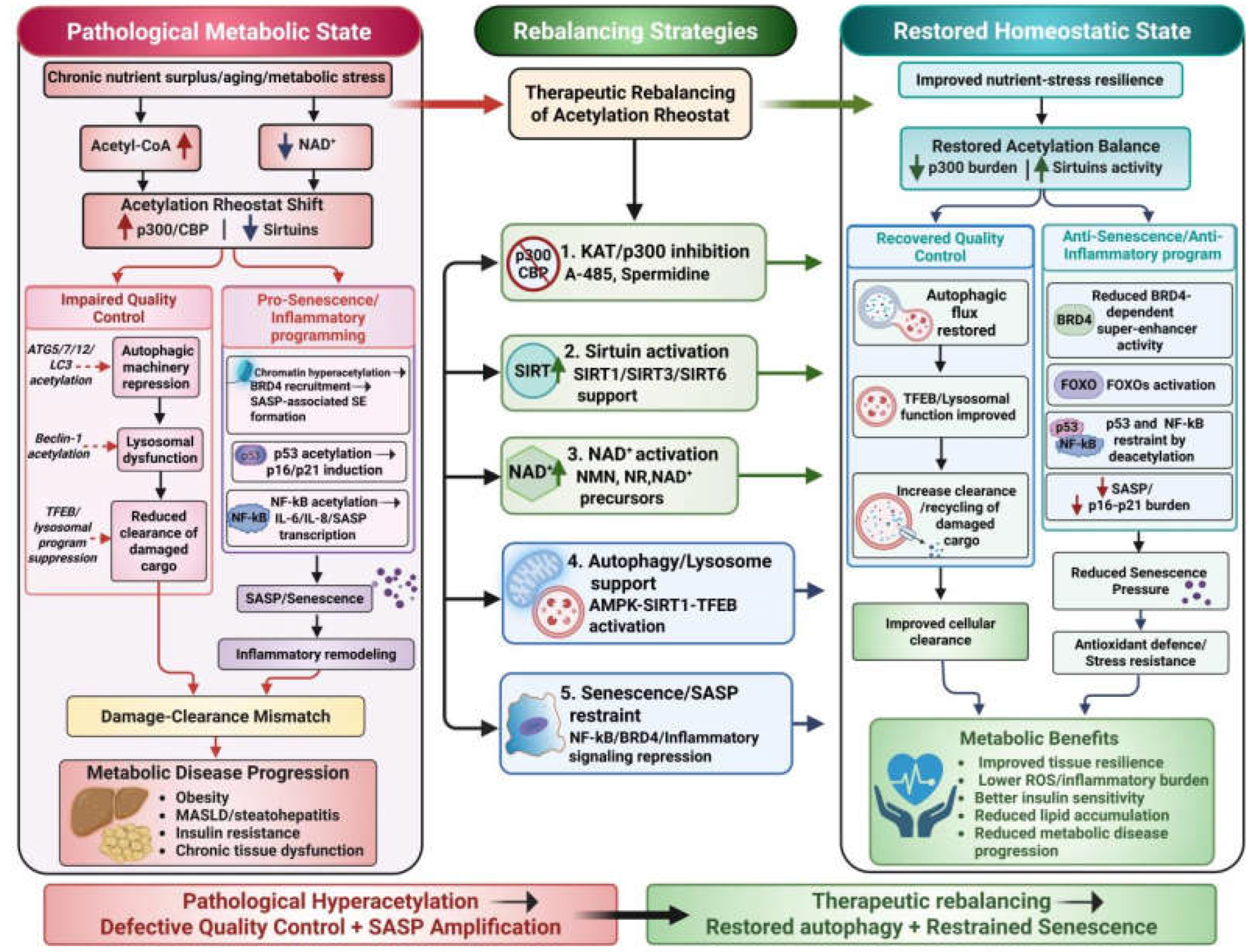

Table 1.
Key Acetylation regulators coordinating autophagy and senescence in metabolic diseases.
| Acetylation Regulator/ Node |
Acetylation Mechanism | Effect on Autophagy | Effect on Senescence | Tissue/Disease Relevance | Probable Therapeutic Implications | References |
|---|---|---|---|---|---|---|
|
EP300/p300-CBP |
Acetylates core ATG proteins (ATG5/7/8/12); promotes hyperacetylated chromatin /enhancer remodeling | Suppresses autophagosome formation and net autophagic flux | Augments p16/p21 expression, super-enhancer formation, BRD4 recruitment, and SASP programs | Adipose tissues, liver, MASLD/NAFLD, obesity, T2DM | KAT inhibition (e.g., A-485, Spermidine) may restores autophagic flux and declines SASP/senescence | (Lee & Finkel, 2009; Sen et al., 2019; Zhou et al., 2020) |
|
mTORC1-p300 axis |
Nutrient excess triggers mTORC1-mediated phosphorylation/ activation of p300 | Suppress autophagy under fed conditions, including ATG/Beclin-1 and lysosomal programs | Stabilizes senescence indirectly via p300-driven chromatin remodeling and inflammatory programming | Obesity, overnutrition-related metabolic syndromes, hepatic lipid accumulation |
Targeting this pathway may help uncouple nutrient overload from autophagy repression and senescence reinforcement | (Son et al., 2024; Wan et al., 2017) |
|
SIRT1 |
Promotes NAD+-driven deacetylation of core ATGs, FOXO, NF-κB, p53, and tissue specific targets | Promotes starvation-induced autophagy, FOXO-linked programs, and hepatic lipophagy | Restrains stress-induced senescence by limiting NF-κB and p53 transcriptional output | adipose tissues, liver, obesity, steatohepatitis, insulin resistance | SIRT1 activation or NAD+ support may augment autophagic homeostasis and restrain pro-senescent signaling | (Ding et al., 2017; Lee et al., 2008; Vaziri et al., 2001; Yeung et al., 2004) |
|
Beclin-1 |
P300-regulated acetylation at K430/K437 promotes Rubicon recruitment | Obstructs autophagosome maturation, endocytic trafficking, and autophagic flux | Indirectly favors senescence by increasing cellular stress and damaged cargo load | General metabolic stress states | Preventing Beclin-1 acetylation may improve autophagic flux and clearance capacity |
(Sun et al., 2015; Y. Xu & W. Wan, 2023) |
|
Acetylation Regulator/ Node |
Acetylation Mechanism | Effect on Autophagy | Effect on Senescence | Tissue/Disease Relevance | Probable Therapeutic Implications |
References |
|
TFEB/ lysosome |
GCN5-mediated TFEB acetylation | Inhibits lysosomal biogenesis and autophagosome-lysosome function | Drives senescence pressure via accumulation of damaged cargo | Lysosomal clearance hubs; MASLD/ NAFLD | Target TFEB acetylation or AMPK/SIRT1-TFEB signaling to restore flux |
(Wang et al., 2019; Zhang et al., 2025) |
|
p53 |
Acetylation of p53 at Lys120/Lys382 increases p53 transcription; SIRT1 removes Lys382 acetylation |
Indirectly suppresses adaptive autophagy via arrest/ stress signaling | Promotes p21-mediated growth arrest and stable senescence | Adipose tissues, liver, aging, metabolic decay | Reducing p53 acetylation may lower senescent burden and fibrosis/inflammation | (Hu et al., 2023; Liu et al., 2024; Vaziri et al., 2001) |
|
NF-κB (p65) |
Acetylation of RelA/p65 subunit of NF-κB at Lys310 enhances NF-κB activity; SIRT1/SIRT6 counteract it |
Induces autophagy-inhibitors (A20, Bcl-2/xL) and represses pro-autophagy mediators | Sustains persistent SASP and chronic inflammatory remodeling |
Obesity, T2DM, MASLD/NAFLD | SIRT1/SIRT6-linked suppression of NF-κB acetylation may limit senescence/SASP | (Kawahara et al., 2009; Salminen et al., 2012; Yeung et al., 2004) |
|
FOXO (1/3a) |
Reversible acetylation; deacetylation by SIRT1 enhances FOXO stress-response activity |
Promotes autophagy genes, mitochondrial resilience, and stress resistance | Reduces senescence by countering oxidative stress | Adipose progenitors, liver, metabolic stress hubs | FOXO-supportive deacetylation may improve cellular resilience | (Gupta et al., 2025; Hariharan et al., 2010; Kuo et al., 2025) |
|
Acetylation Regulator/ Node |
Acetylation Mechanism | Effect on Autophagy | Effect on Senescence | Tissue/Disease Relevance | Probable Therapeutic Implications | References |
|
PPARγ |
p300 acetylates PPARγ; SIRT1 deacetylates Lys268/Lys293 and recruits PRDM16 | Intersects with adipocytes remodeling and mitochondrial turnover |
Links inflammatory aging, adipocytes differentiation and whitening to metabolic fitness | Adipose tissues, obesity, and aging-associated dysfunction metabolic dysfunction |
Enhancing SIRT1-linked deacetylation may promote a healthier beige/brown adipocyte program | (Han et al., 2010; He et al., 2023; Qiang et al., 2012) |
|
Histone/ Super-enhancers |
P300-mediated hyperacetylation and BRD4 recruitment at senescence/SASP enhancers | Chronic low-clearance states may be transcriptionally worsened |
Establishes transcriptional memory and SASP persistence | Epigenetic remodeling in chronic metabolic tissues | Epigenetic acetylation-targeting drugs may limit senescence persistence | (Sen et al., 2019; Tasdemir et al., 2016; Wang et al., 2024) |
Disclaimer/Publisher’s Note: The statements, opinions and data contained in all publications are solely those of the individual author(s) and contributor(s) and not of MDPI and/or the editor(s). MDPI and/or the editor(s) disclaim responsibility for any injury to people or property resulting from any ideas, methods, instructions or products referred to in the content. |
© 2026 by the authors. Licensee MDPI, Basel, Switzerland. This article is an open access article distributed under the terms and conditions of the Creative Commons Attribution (CC BY) license (http://creativecommons.org/licenses/by/4.0/).
Copyright: This open access article is published under a Creative Commons CC BY 4.0 license, which permit the free download, distribution, and reuse, provided that the author and preprint are cited in any reuse.