

Submitted:
02 July 2026
Posted:
02 July 2026
You are already at the latest version
Abstract
Background: Bicyclomycin (BCM) is a unique antibiotic that selectively inhibits the bacterial transcription termination factor, Rho. Recent genomic analyses have revealed widespread distribution of BCM biosynthetic gene clusters across diverse bacterial species, suggesting additional biological functions beyond antimicrobial activity. We hypothesized that BCM may possess anti-inflammatory properties that contribute to its evolutionary conservation. Methods: We investigated the anti-inflammatory effects of BCM using LPS-stimulated RAW 264.7 macrophages. Cell viability was assessed using MTT assays, and inflammatory mediator production (nitric oxide, TNF-α, IL-6, and IL-1β) was measured using colorimetric and ELISA-based assays. Mechanistic insights were explored through pathway analysis. Results: BCM demonstrated dose-dependent anti-inflammatory activity in LPS-stimulated macrophages. Treatment with BCM (25-100 μM) significantly reduced production of nitric oxide, TNF-α, IL-6, and IL-1β compared to LPS-only controls. These effects occurred at concentrations that maintained cell viability above 100 %. Pathway analysis suggested involvement of NF-kB-mediated signaling cascades. Conclusions: This study provides the first evidence for anti-inflammatory properties of bicyclomycin, revealing a previously unrecognized therapeutic potential. The widespread distribution of BCM biosynthetic gene clusters may reflect evolutionary pressure for dual antimicrobial and anti-inflammatory functions. These findings suggest new therapeutic applications for BCM and highlight the importance of investigating pleiotropic effects of antibiotics with unique mechanisms of action.
Keywords:
anti‐inflammatory
; LPS
; macrophages
; NF‐kB pro‐inflammatory cytokines
; pro‐inflammatory mediators
1. Introduction
The current understanding of antibiotic function has undergone a profound transformation, evolving from a narrow focus on antimicrobial activity to recognition of complex pleiotropic effects that extend far beyond pathogen elimination [1]. This expanded perspective has been catalyzed by accumulating clinical evidence demonstrating therapeutic benefits of antibiotics in conditions where antimicrobial effects alone cannot account for observed improvements, leading to systematic investigations of immunomodulatory, anti-inflammatory, and cytoprotective properties across diverse antibiotic classes [2]. These discoveries have not only revealed new therapeutic applications for existing compounds but have also fundamentally altered our understanding of how these molecules function in complex biological systems.
Bicyclomycin (BCM), also known as bicozamycin occupies a unique position within the antibiotic contribution, distinguished by its unprecedented mechanism of action as the sole natural product known to selectively inhibit Rho, the essential bacterial transcription termination factor [3]. This remarkable selectivity, combined with BCM’s established clinical safety profile and potent activity against Gram-negative pathogens, has positioned the compound as both a valuable therapeutic agent and an important research tool for understanding bacterial transcription regulation [4]. However, despite decades of research focused on its antimicrobial properties and mechanism of action, the potential for bicyclomycin to exhibit biological activities beyond bacterial killing remains largely unexplored [5]. The recent identification and characterization of BCM biosynthetic gene clusters has provided unprecedented insights into the genetic basis for compound production while simultaneously revealing one of the most remarkable patterns of biosynthetic gene distribution ever documented [6,7,8]. Comprehensive genomic surveys have identified bicyclomycin-like clusters in hundreds of phylogenetically diverse bacterial species, spanning multiple phyla and encompassing organisms from vastly different ecological niches [9]. This extraordinary distribution pattern includes the presence of functional clusters in opportunistic pathogens such as Pseudomonas aeruginosa, Burkholderia species, and various members of the Enterobacteriaceae, suggesting potential roles for bicyclomycin production in pathogenesis and host-microbe interactions [10].
Anti-inflammatory models in the cellular studies widely are designed, assisting LPS-induced inflammation in RAW 264.7 murine macrophages due to the signaling cascade after the LPS challenge to healthy murine macrophages. The central role of TLR4 has been associated with chronic inflammatory conditions generally, and has been recorded as essential target in anti-inflammatory studies. The LPS-induced inflammation mainly initiated toll-like receptor (TLR4)/ myeloid differentiation factor 2 (MD2) signaling transduction [11]. Once, the TLR4 is stimulated by LPS, the downstream signaling is transmitted and finally transcribed the pro-inflammatory gene expression by the transcription factor, nuclear factor-kB (NF- kB) [12]. In this loop, phosphorylation of inhibitor of κBα (IκBα) and IκB kinase α/β (IKKα/β) also happen prior to the NF-kB nuclear translocation. Eventually, the pro-inflammatory gene expression includes interleukins, such as interleukin 6 and interleukin 1β (IL), (IL-6 and IL-1β), the cytokines, including tumor necrosis factor- alpha (TNF-α), and inflammatory mediators including nitric oxide (NO), and prostaglandin E2 (PGE2), showing as the whole organism level inflammatory markers [13,14,15]. At the same time, the inflammatory mediator genes are transcribed including inducible nitric oxide synthase (iNOS/NOS2) and cyclooxygenase 2 (COX2) [16]. We hypothesized that BCM may have anti-inflammatory potential due to its unique mechanism of inhibiting Rho-dependent transcription termination [17,18]. This action influences key bacterial processes, such as stress responses, metabolism, and virulence, which are closely linked to how bacteria interact with the host and trigger immune responses. [19]. Furthermore, the demonstrated ability of other antibiotic classes to exhibit potent anti-inflammatory effects through diverse molecular mechanisms provides precedent for investigating similar properties in bicyclomycin [20]. However, the mechanistic and proper biomarker evaluation had not performed to evaluate the anti-inflammatory activity of BCM. The clinical significance of identifying anti-inflammatory properties in bicyclomycin would be substantial, given the compound’s well-established safety profile and unique mechanism of action [21]. Current anti-inflammatory therapeutics, including nonsteroidal anti-inflammatory drugs (NSAIDs), corticosteroids, and targeted biologics, are associated with significant limitations including gastrointestinal toxicity, immunosuppression, cardiovascular risks, and high cost [22]. Bicyclomycin’s minimal systemic absorption, proven safety record, and distinct molecular mechanism position it as a potentially valuable addition to the anti-inflammatory therapeutic armamentarium, particularly for conditions where safer alternatives to conventional treatments are needed [5,23,24].
Therefore, we evaluated the anti-inflammatory characteristics of BCM using LPS-induced inflammation in RAW 264.7 macrophages. Further, we evaluated the effectiveness of BCM in modulating pro-inflammatory mediator levels cytokine production while maintaining the cell viability. Furthermore, molecular docking studies were performed to explore the potential binding of BCM to the TLR4 receptor, proving the beginning of mechanism of BCM’s anti-inflammatory action. By discovering the anti-inflammatory effects and the mechanistic approach of BCM, we aimed to establish its potential as a therapeutic option for the prophylaxis of chronic inflammation.
2. Materials and Methods
2.1. Chemical Reagents and Antibodies
Bicyclomycin was purchased from Santa Cruz Biotechnology, Inc. (SC-391755). Dulbecco’s Modified Eagle’s Medium (DMEM), penicillin–streptomycin (10,000 U/mL), fetal bovine serum (FBS) were purchased from Thermo Fisher Scientific (Waltham, MA, USA). BCA protein quantification assay kit (Do GEN Bio) used for protein quantification was purchased from Doozen Bio Co., Ltd., Seoul). Polyvinylidene fluoride (PVDF) membrane filter used for Western blotting, NE-PER (Nuclear and Cytoplasmic Extraction Reagents) protein extraction kit, and Ponceau S Staining Solution (A40000278) were purchased from Thermo Fisher Scientific (Waltham, MA, USA). Reagents, (4,5-dimethylthiazol-2-yl)-2,5-diphenyltetrazolium bromide (MTT), a protease inhibitor cocktail used for cell lysis were obtained from Sigma-Aldrich (St. Louis, MO, USA). The 2× Laemmli sample buffer and 10% Tween 20 used for protein lysate and TBST buffer preparation were purchased from Bio-Rad (Hercules, CA, USA). bovine serum albumin (BSA) was obtained from Bovostar (Bovogen, Melbourne, Australia). Dimethyl sulfoxide (DMSO), radioimmunoprecipitation assay (RIPA) buffer, 20× TBS buffer (pH 7.6) used to prepare 1× TBST, 10X Tris-glycine buffer (SDS) (TR2015-100-00), and phosphate-buffered saline (PBS) were purchased from Biosesang (Seongnam, Republic of Korea). The primary antibodies including COX-2 (SC-7951, C-62), NOS2 (SC-7271, C-11) were obtained from Santa Cruz Biotechnology, Inc. The primary antibodies including Phospho-NF-kappaB p65 (Ser536) (93H1) (S535), NF-kappaB1 p105/p50 (3035S), Phospho-IkappaB alpha (Ser32) (2859) (14D4), Lamin B1 (13435) and beta-Actin (45 kDa) (4967) were obtained from Cell Signaling TECHNOLOGY (Danvers, MA, USA). The secondary IgG antibodies including Anti-mouse IgG, HRP-linked, and (7076) were purchased from Cell Signaling TECHNOLOGY (Danvers, MA, USA). The ELISA kits for IL-6 ( BD OptEIA™ Mouse IL-6 ELISA Set, 555240 ), TNF (BD OptEIA™ Mouse TNF ELISA Kit, 560478) were obtained from BD Biosciences (San Jose, CA, USA). Human IL-1 beta/IL-1F2 Quantikine ELISA Kit (DLB50), and Prostaglandin E2 Parameter Assay Kit (KA0326) were purchased from R&D Systems, Minneapolis, MN, USA. Total NF-κB p65 ELISA Kit was purchased from Abcam, Cambridge, UK.
2.2. Cell Culture and Bicyclomycin Treatment
RAW 264.7 murine macrophage cells were obtained from the the Korean Cell Line Bank (KCLB, Seoul, Republic of Korea) and maintained in Dulbecco’s Modified Eagle Medium (DMEM) supplemented with 10% fetal bovine serum (FBS), 100 U/mL penicillin, and 100 μg/mL streptomycin. Cells were cultured in a humidified atmosphere containing 5% CO2 at 37 °C. For experiments, cells were seeded in appropriate plates and allowed to adhere overnight before treatment. BCM was dissolved in DMSO to prepare stock solutions. Cells were pretreated with various concentrations of BCM (0-100 μM) for 2 hours, followed by stimulation with lipopolysaccharide (LPS) from Escherichia coli O111:B4 (1 μg/mL) for 24 hours or 1 hour. Control groups included unstimulated cells and LPS-only stimulated cells.
2.3. Cell Viability Assay
Cell viability was assessed using the MTT (3-(4,5-dimethylthiazol-2-yl)-2,5-diphenyltetrazolium bromide) assay. The cells were seeded at a density of 1 × 10⁵ cells/mL and allowed to adhere the cells for 18 h. After treatment, cells were incubated with MTT solution (0.5 mg/mL) for 1 hour at 37 °C. The formed formazan crystals were dissolved in DMSO, and absorbance was measured at 570 nm using the Epoch™ microplate spectrophotometer (BioTek Instruments, Winooski, VT, USA).
Viability (%) = (Abs_sample/Abs_control) × 100
2.4. Griess Assay
RAW 264.7 cells were seeded into 24-well plates at a cell density of 1 × 10⁵ cells/mL and allowed to adhere the cells for 18 h. The cells were pretreated with different concentrations of BCM (0‒100 μM) for2 hours, followed by stimulation with 1 µg/mL LPS for 24 hours. After the incubation, the culture supernatants were collected and used in order to NO quantification using Griess reagent assay (using Griess A and B).
2.5. Measurement of PGE2 and Pro-Inflammatory Cytokines
The effects of BCM on the production of prostaglandin E2 (PGE2) and pro-inflammatory cytokines (IL-6, TNF-α, and IL-1β,) were measured using an enzyme-linked immunosorbent assay (ELISA) kits. RAW264.7 cells were seeded at 1.0 × 10⁵ cells/well into 24-well plates and incubated to adhere the cells for 18 hours. Cells were then treated with BCM in the presence of LPS and incubated for 24 h. After incubation, the culture medium was collected and centrifuged at 15,000 rpm for 20 min to remove debris. The supernatants were used for ELISA assays following the manufacturer’s protocols.
2.6. Total Protein Extraction and Western Blotting
RAW 264.7 cells were lysed using RIPA buffer supplemented with protease and phosphatase inhibitors (Sigma-Aldrich) to extract total protein. The lysates were centrifuged at 15 000 × g for 15 min at 4oC, and the supernatant containing the proteins was collected. Protein concentrations were determined using the BCA protein quantification assay kit (Do GEN Bio) (Doozen Bio Co., Ltd., Seoul). Equal amounts of protein (20 µg per well) were loaded onto 10% sodium dodecyl sulfate-polyacrylamide (SDS-PAGE) gel for electrophoretic separation. After separation, the proteins were transferred onto a polyvinylidene difluoride membrane using the Trans-Blot Turbo™ Transfer System (Bio-Rad Laboratories, Hercules, CA, USA; Cat. No. 1704150). The membranes were blocked with5% non-fat dry milk in Tris-buffered saline containing Tween-20for 1 hour to prevent non-specific binding. The membranes were incubated for 16 hours at 4◦C with primary antibodies specific to the target proteins (1:1000 dilution), followed by incubation with horseradish peroxidase (HRP)-conjugated secondary antibodies (1:1000 dilution) for 2 hours. Protein bands were detected using visualized using enhanced chemiluminescence (ECL) and Protein bands were visualized using the Fusion Solo S gel documentation system (Vilber Lourmat, Collégien, France). The intensity of the protein bands was quantified using ImageJ (Fiji) software (National Institutes of Health,Bethesda, MD, USA), and β-actin and Lamin B1 were used as loading controls to normalize the expression levels of the target proteins.
2.7. Statistical Analysis
Data are presented as mean ± standard error of the mean (SEM) from at least three independent experiments. Statistical significance was determined using one-way analysis of variance (ANOVA) followed by Bonferroni test. P-values less than 0.05 were considered statistically significant (*P < 0.05, **P < 0.01, ***P < 0.001, ###P < 0.001 compared to LPS control).
3. Results
3.1. Bicyclomycin (BCM) Exhibits Non-Toxic Ranges Cell Viability for Potential Further Evaluation
To establish the non-cytotoxic concentration, range of BCM, we performed MTT assays on RAW 264.7 cells treated with increasing concentrations of BCM (0-100 μM). As shown in Figure 1, BCM exhibited minimal cytotoxicity at concentrations up to 100 μM, with cell viability remaining above 100.000 ± 1.839% at all tested concentrations except at 200 μM of BCM. These results indicate that BCM can be safely used at concentrations up to 100 μM for subsequent anti-inflammatory studies.
3.2. BCM Suppresses LPS-Induced Anti-Inflammatory Mediators in RAW 264.7 Macrophages
Nitric oxide and cyclooxygenase 2 (COX2) is a key inflammatory mediator produced by activated macrophages through inducible nitric oxide synthase (iNOS/NOS2). LPS stimulation dramatically increased NO production in RAW 264.7 cells compared to untreated controls. Pretreatment with BCM dose-dependently inhibited LPS-induced NO production, with significant reductions observed at 25, 50, and 100 μM concentrations (P < 0.001). The inhibitory effect was most pronounced at 50 and 100 μM, where NO levels were reduced to 1.89 ± 6.1300e-3 (at 50 μM) and 1.816 ± 0.0123 (at 100 μM) respectively, compared to LPS-only controls (41.19 ± 0.23). LPS stimulation significantly increased NO production compared to the untreated control group (1.000 ± 0.0162, P < 0.001, ###). Treatment with bicyclomycin at concentrations of 25, 50, and 100 μM dose-dependently reduced LPS-induced NO production, with all three concentrations showing statistically significant suppression compared to the LPS-only group (p < 0.001, ***) (Figure 2A). Consistent with these findings, LPS-induced PGE2 levels in ELISA were markedly elevated relative to the control (p < 0.001, ###), and bicyclomycin treatment at 50 and 100 μM significantly attenuated PGE2 production (p < 0.001, ***), while the 25 μM concentration showed a partial but non-significant reduction (Figure 2B).
To investigate the underlying molecular mechanisms, protein expression levels of NOS2 and COX-2 were examined by western blot analysis. LPS stimulation markedly induced NOS2 protein expression to a relative density of 1.000 ± 0.265 compared to 0.316 ± 0.117 at the untreated control (p < 0.001, ###). Bicyclomycin treatment at 25 μM partially reduced NOS2 relative density to 0.614 ± 0.001 (p < 0.05, *), while treatment at 50 and 100 μM further suppressed expression to 0.292 ± 0.013 and 0.256 ± 0.017, representing a near-complete reduction (p < 0.001, ***), as confirmed by densitometry quantification normalized to β-actin (Figure 2C). In a similar manner, COX-2 protein expression was strongly upregulated upon LPS treatment to a relative density of 0.967 ± 0.073 (p < 0.001, ###) and was dose-dependently suppressed by bicyclomycin, with 25 μM reducing expression to 0.507 ± 0.023, and 50 and 100 μM further reducing it to 0.167 ± 0.012 and 0.097 ± 0.005 (p < 0.001, ***) (Figure 2D).
Validating the molecular markers, the morphological examination of macrophages revealed that LPS stimulation induced notable changes in cell shape, including increased spreading and irregular morphology consistent with macrophage activation. Bicyclomycin treatment progressively attenuated these LPS-induced morphological changes in a dose-dependent manner, with cells treated at higher concentrations (50 and 100 μM) displaying a more rounded morphology resembling the unstimulated control group. No overt cytotoxic effects or significant loss of cell viability were observed at any of the tested concentrations (Figure 2E). Altogether, these results demonstrate that bicyclomycin suppresses LPS-induced inflammatory mediator production by downregulating NOS2 and COX-2 expression and attenuates macrophage activation morphology.
3.3. BCM Attenuates the LPS-Induced Pro-Inflammatory Cytokine Production in RAW264.7 Cells
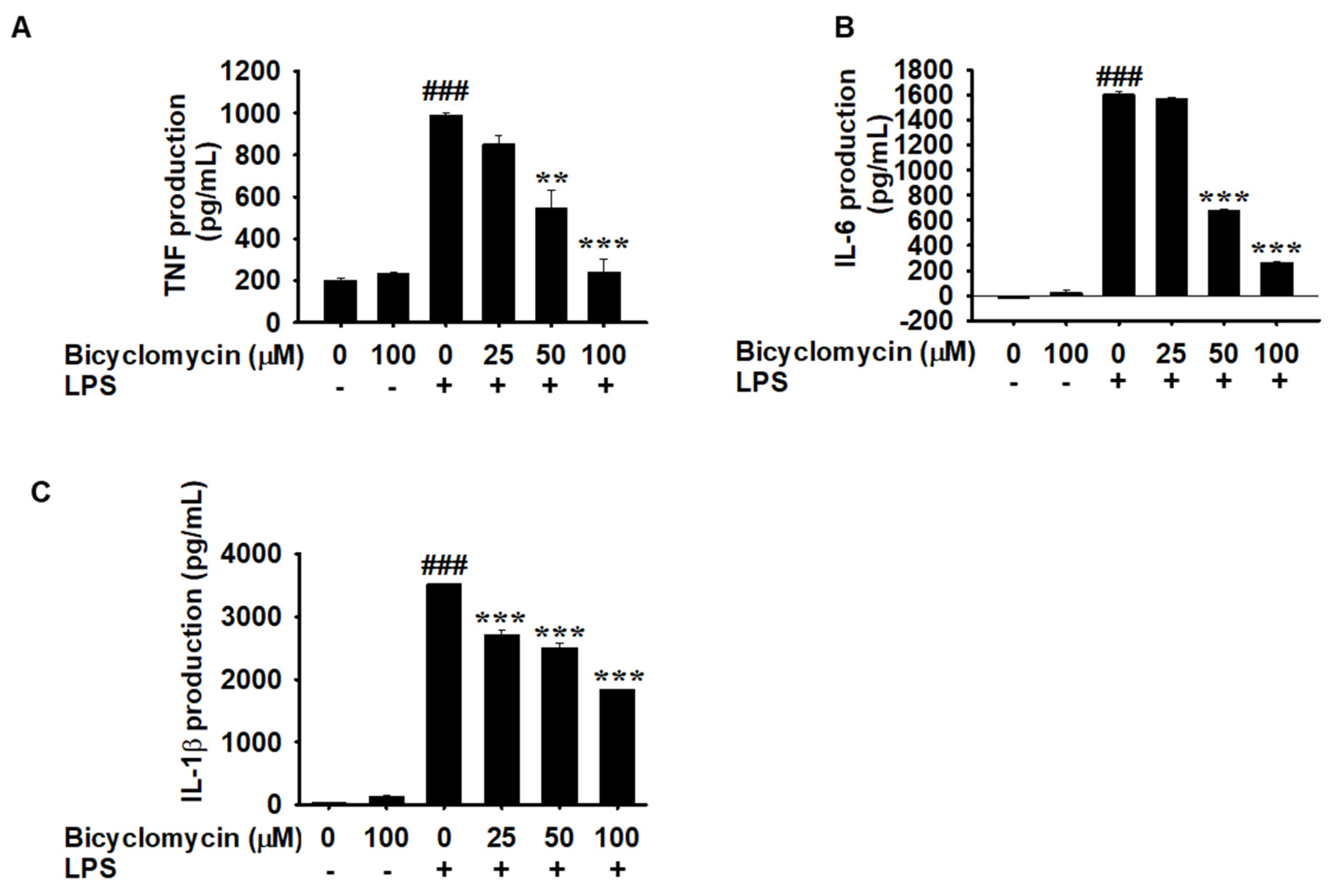
TNF-α is a central pro-inflammatory cytokine involved in the acute phase response [25]. LPS stimulation resulted in an increase markedly in TNF-α production 989.600 ± 14.000 pg/mL. BCM pretreatment significantly attenuated this response in a dose-dependent manner. At 25 μM, BCM reduced TNF-α levels to 853.100 ± 39.500 pg/mL (P < 0.05), while 50 and 100 μM concentrations reduced the production of TNF-α levels to 549.600 ± 83.000 pg/mL and 241.60 ± 61.000 pg/mL, respectively (P < 0.001).
Interleukin-6 (IL-6) is a pleiotropic cytokine with both pro- and anti-inflammatory properties, playing crucial roles in the acute phase response. LPS stimulation induced significant IL-6 production to 1604.800 ± 27.000 pg/mL in RAW 264.7 cells (P < 0.001) [26], while . BCM treatment significantly reduced IL-6 levels at all tested concentrations, with the most pronounced effects observed at 50 and 100 μM, resulting 679.800 ± 10.000 and 261.467 ± 13.667 pg/mL of IL-6 production (P < 0.001), while untreated control exhibited IL-6 production. The reduction in IL-6 production consisted with the effects observed with TNF-α, suggesting a broad anti-inflammatory mechanism.
IL-1β is a potent pro-inflammatory cytokine that plays critical roles in inflammatory cascades and fever responses [27]. LPS stimulation significantly increased IL-1β production, exhibiting 3521.500 ± 6.250 pg/mL in macrophages compared to the untreated control group, which showed 27.750 ± 10.000 pg/mL (P < 0.001). BCM treatment dose-dependently reduced IL-1β levels, with significant inhibition observed at 25 and 50 μM concentrations, exhibiting 2715.250 ± 65.000 pg/mL and 2511.500 ± 76.250 pg/mL while 100 μM of BCM reduced the IL-1β levels to 1836.500 ± 6.250 pg/mL (P < 0.001). The inhibitory effect on IL-1β production was consistent with the broader pattern of anti-inflammatory activity observed with other mediators.
Figure 3.
Bicyclomycin attenuates the LPS-induced pro-inflammatory cytokine production in RAW264.7 cells. The cells were seeded at 1 × 10⁵ cells/mL and kept for 18 hours, then pretreated with BCM (0-100 μM) for 2 hours followed treatment by LPS (1000 ng/mL) for 24 h. The levels of (A-C) TNF-α, IL-6, and IL-1β levels were measured in the culture supernatant at 24 hours-post LPS stimulation using an ELISA kit. Data are presented as mean ± SEM from three independent experiments. ###p < 0.001 vs. unstimulated control group; **p < 0.05 ***p < 0.001 vs. LPS-treated group.
Figure 3.
Bicyclomycin attenuates the LPS-induced pro-inflammatory cytokine production in RAW264.7 cells. The cells were seeded at 1 × 10⁵ cells/mL and kept for 18 hours, then pretreated with BCM (0-100 μM) for 2 hours followed treatment by LPS (1000 ng/mL) for 24 h. The levels of (A-C) TNF-α, IL-6, and IL-1β levels were measured in the culture supernatant at 24 hours-post LPS stimulation using an ELISA kit. Data are presented as mean ± SEM from three independent experiments. ###p < 0.001 vs. unstimulated control group; **p < 0.05 ***p < 0.001 vs. LPS-treated group.
3.4. BCM Attenuates LPS-Induced NF-kB Nuclear Translocation
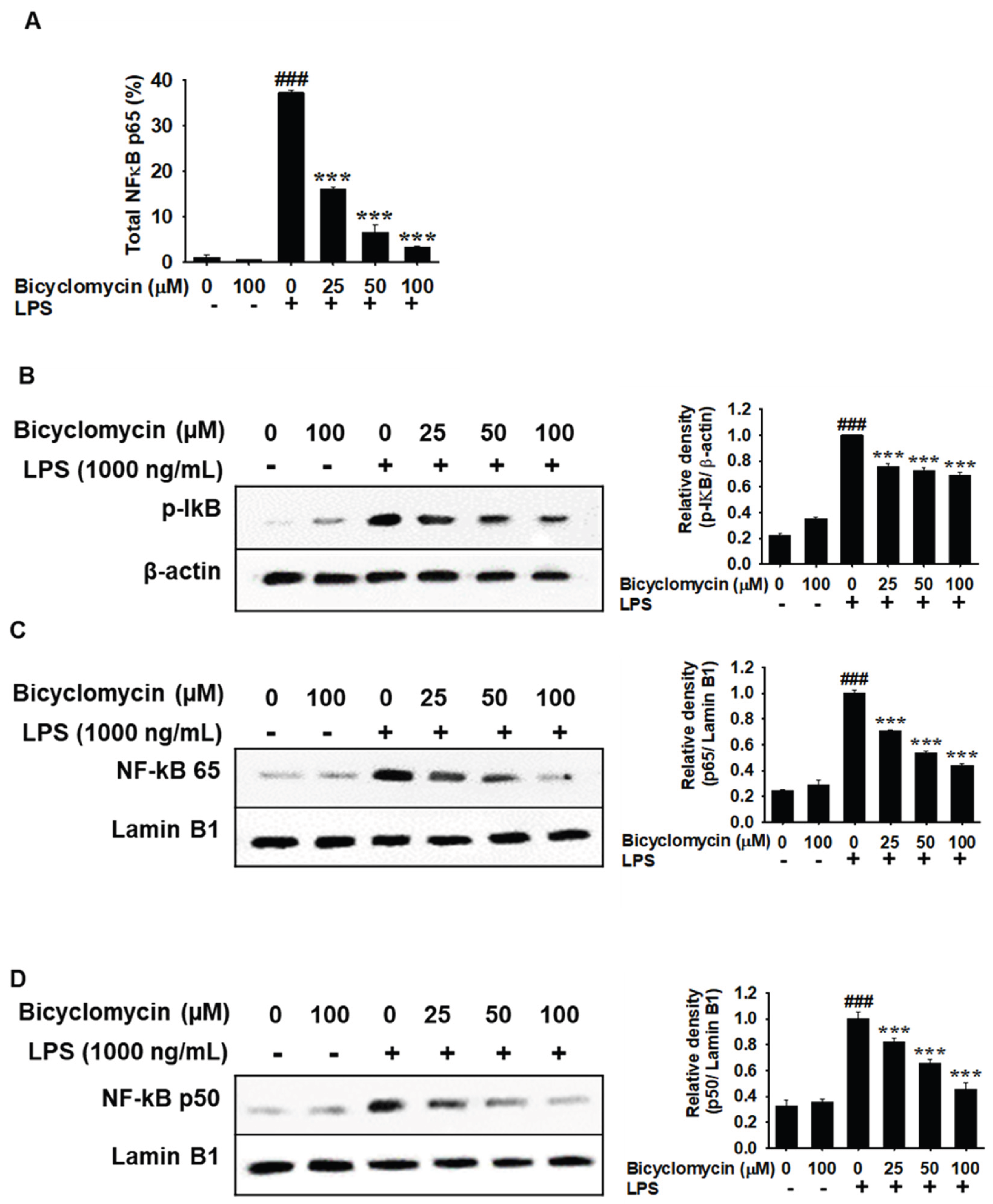
To define the molecular mechanisms underlying BCM’s anti-inflammatory effects, we performed pathway analysis focusing on NF-kB signaling cascades [11,28]. To functionally assess the effect of BCM on NF-κB activation, we quantified nuclear translocation of the p65 subunit by ELISA following LPS stimulation (1000 ng/mL). LPS treatment markedly increased nuclear NF-κB p65 levels to 37.184 ± 0.574% compared to the untreated control, which was 0.986 ± 0.645% after 1 hour of incubation time of LPS treatment (###p < 0.001), while the only-BCM treatment (50 μM) did not show any significant difference compared to the untreated control. Pre-treatment with BCM 2 hours prior to the LPS treatment at 25, 50, and 100 μM significantly attenuated LPS-induced nuclear p65 accumulation in a concentration-dependent manner exhibiting the expression levels at 15.965 ± 0.564%, 6.590 ± 1.578%, and 3.240 ± 0.338% (***p < 0.001 vs. LPS alone; Figure 4A), indicating that BCM inhibits canonical NF-κB nuclear translocation.
To further investigate and confirm the inhibition of the downregulation of LPS-induced molecular mechanism of NF-kB nuclear translocation by BCM, we performed western blot analysis and it revealed that LPS stimulation substantially elevated phosphorylated IκB (p-IκB) levels to 1.000± 0.000 relative to untreated control, which showed 0.221 ± 0.013 of relative density (###p < 0.001) in cytoplasm fraction, reflecting upstream NF-κB pathway activation. Treatment with BCM at 25, 50, and 100 μM dose-dependently reduced p-IκB expression (***p < 0.001 vs. LPS group), proportionated with β-actin serving as a loading control, by keeping the expression levels at 0.759 ± 0.020, 0.728 ± 0.019, and 0.691 ± 0.020 (Figure 4B). These results indicate that BCM suppresses NF-κB signaling upstream by inhibiting IκB phosphorylation. Consistent with the translocation data, western blot analysis of nuclear fractions demonstrated that LPS-only treatment significantly increased both NF-κB p65and NF-κB p50 protein levels to 1.000 ± 0.021 and 1.000 ± 0.055 (###p < 0.001), proportionated with Lamin B1 used as a nuclear loading control. BCM treatment at 25, 50, and 100 μM significantly reduced nuclear p65 to 0.708 ± 0.006, 0.534 ± 0.017, and 0.438 ± 0.017 respectively (Figure 4C). The data were consistent for p50 of pre-treatment of BCM in LPS-induced conditions and BCM treatment at 25, 50, and 100 μM significantly reduced nuclear p50 protein expression to 0.821 ± 0.028, 0.655 ± 0.030, and 0.453 ± 0.056 relative densities in a concentration-dependent manner compared to LPS-stimulated cells (***p < 0.001, Figure 4D). Collectively, these data demonstrate that BCM effectively suppresses the activation and nuclear translocation of the NF-κB complex, likely through its interaction with TLR4 and subsequent inhibition of the IκB phosphorylation cascade. The results suggest that BCM may interfere with key signaling nodes downstream of TLR4 activation, including NF-κB pathway. This mechanistic framework is consistent with the broad spectrum of inflammatory mediators affected by BCM treatment.
4. Discussion
The current study provides the first experimental evidence for anti-inflammatory properties of Bicyclomycin (BCM), revealing a previously unrecognized therapeutic potential for this unique antibiotic even though vast array of antibiotics has been proven to have anti-inflammatory properties [29]. Our findings demonstrate that BCM significantly reduces the production of key inflammatory mediators in LPS-stimulated macrophages, including nitric oxide and the pro-inflammatory cytokines TNF-α, IL-6, and IL-1β. Consistent with these data, BCM demonstrated that BCM potentially downregulated the LPS-induced NF-kB signaling cascade. These effects occur at concentrations that maintain acceptable cell viability, confirming a favorable therapeutic option for potential clinical applications in the anti-inflammation.
The significance of this finding is further amplified by BCM’s distinctive mechanism of action as a selective Rho inhibitor, setting it apart from other antibiotics with reported anti-inflammatory activity such as macrolides, tetracyclines, and fluoroquinolones, which primarily exert their immunomodulatory effects through suppression of cytokine transcription or direct inhibition of inflammatory kinases [30,31,32].
The BCM biosynthetic gene cluster, responsible for its extensive oxidative modifications, is remarkably widespread across phylogenetically diverse bacteria through horizontal gene transfer and is notably encoded by hundreds of clinically relevant Pseudomonas aeruginosa isolates worldwide. Rho-dependent transcription termination is essential for the regulation of bacterial gene expression, including the suppression of antisense transcription and the coordination of coupled transcription-translation, making it an attractive and largely unexploited antimicrobial target [7]. In the present era of escalating antimicrobial resistance, where conventional antibiotic classes are increasingly compromised by resistance mechanisms, BCM’s unique target offers a compelling rationale for renewed research interest and potential clinical development [33]. Notably, Rho is highly conserved across gram-negative bacteria, including clinically problematic pathogens such as Escherichia coli, Klebsiella pneumoniae, and Pseudomonas aeruginosa, yet shares no functional homolog in mammalian cells, providing an inherent selectivity that reduces the risk of host toxicity [34]. The favorable cytotoxicity profile demonstrated in this study, with RAW264.7 cell viability maintained above 100% at concentrations up to 100 μM, is consistent with this selectivity and further supports BCM’s potential as a safe therapeutic agent.
The observed suppression of NO and PGE2 production, alongside the downregulation of their respective biosynthetic enzymes NOS2 and COX-2, is consistent with the anti-inflammatory profiles reported for other naturally derived compounds and antibiotic classes. Macrolide antibiotics such as azithromycin and clarithromycin have been shown to suppress NOS2 and COX-2 expression in LPS-activated macrophages even though the mechanistic role has not been proven through NF-κB-dependent pathways [29,35].Our findings position BCM within this broader class of antibiotic-derived anti-inflammatory agents, while distinguishing it by its unique structural and mechanistic characteristics. Notably, the near-complete suppression of COX-2 expression achieved at 50 and 100 μM BCM is comparable in magnitude to that reported for established COX-2 inhibitors, suggesting a potent and potentially clinically relevant degree of inhibition. This is particularly significant given that COX-2-derived PGE2 is a central mediator not only of acute inflammation but also of chronic inflammatory conditions and cancer-associated inflammation, broadening the potential therapeutic scope of BCM beyond acute macrophage-driven responses [36].
Biologic therapies targeting individual cytokines, such as anti-TNF agents and IL-6 receptor antagonists, have transformed the management of chronic inflammatory diseases but are associated with considerable costs, immunosuppressive risks, and the requirement for parenteral administration [37]. Furthermore, in the context of infectious inflammatory diseases where bacterial infection and host inflammatory response coexist, BCM’s dual capacity to suppress bacterial growth through its unique Rho-targeting mechanism, while simultaneously attenuating the host inflammatory response via TLR4-NF-κB pathway inhibition, represents a mechanistically distinct therapeutic strategy that differentiates it from other antibiotics with reported immunomodulatory properties [38].
The mechanistic insight provided by our NF-κB pathway analysis offers a compelling explanation for BCM’s broad-spectrum anti-inflammatory activity. NF-κB activation is a central regulatory hub governing the transcription of NOS2, COX-2, TNF-α, IL-6, and IL-1β, among numerous other inflammatory mediators [11,28,39]. Furthermore, the functional confirmation of NF-κB suppression through inhibition of IκB phosphorylation and dose-dependent reduction of nuclear p65 and p50 translocation provides robust mechanistic evidence consistent with the observed downstream suppression of inflammatory mediators, and aligns BCM with other naturally derived NF-κB inhibitors such as curcumin, resveratrol, and certain antibiotic derivatives [40]. Importantly, the upstream positioning of BCM’s inhibitory effect at the level of IκB phosphorylation, rather than at individual cytokine or enzyme levels, provides a mechanistic rationale for the broad and simultaneous suppression of multiple inflammatory mediators observed across our experimental models [41,42].
BCM is conceivable that inhibition of Rho-dependent processes may modulate cytoskeletal dynamics involved in macrophage activation, consistent with the attenuation of LPS-induced morphological changes observed in this study, or may interfere with Rho GTPase-mediated intracellular signaling pathways that contribute to inflammatory gene expression [43].
The discovery of anti-inflammatory properties in BCM also carries broader biological significance when considered alongside recent genomic findings regarding the widespread distribution of BCM biosynthetic gene clusters across hundreds of phylogenetically diverse bacterial species, including opportunistic pathogens such as Pseudomonas aeruginosa [43]. The presence of these clusters in organisms that frequently colonize and infect mammalian hosts raises the intriguing possibility that BCM production may represent an evolutionarily conserved microbial strategy for modulating host immune responses to facilitate colonization or persistence. Our findings support the hypothesis that anti-inflammatory properties may contribute to the selective pressure for maintaining these complex biosynthetic pathways, and invite a broader re-evaluation of how microbial natural products function in host-microbe interactions beyond their conventional antimicrobial roles. From a clinical translational perspective, BCM’s combination of antimicrobial and anti-inflammatory properties presents a compelling and differentiated therapeutic profile. Current anti-inflammatory therapies, including NSAIDs and corticosteroids, are associated with well-documented adverse effects including gastrointestinal toxicity, cardiovascular risk, and immunosuppression that limit their long-term use [44].
However, several limitations of this study should be accepted and should be addressed in the future studies. All investigations were conducted using in vitro RAW264.7 macrophage models, and in vivo validation will be necessary to confirm therapeutic relevance clinically and pharmacokinetic feasibility. While our pathway analysis demonstrates involvement of NF-κB signaling, the crosstalk between BCM’s Rho inhibitory activity and its anti-inflammatory properties remains to be definitively established experimentally. Furthermore, the direct binding of BCM to TLR4-MD2 complex. Additionally, the differential sensitivity of individual inflammatory mediators to BCM, as suggested by the partial non-significant reduction in PGE2 at 25 μM alongside significant cytokine suppression at the same concentration, requires further mechanistic characterization.
Therefore, future research directions should include comprehensive in vivo studies in relevant inflammatory and infectious disease models, detailed mechanistic investigations to clarify the role of in BCM’s immunomodulatory effects, and structure-activity relationship studies to optimize both antimicrobial and anti-inflammatory potency while maintaining safety. The availability of genomic information regarding BCM biosynthesis also opens exciting possibilities for engineering derivatives with enhanced pharmacological profiles, and the identification of BCM biosynthetic gene clusters in clinically relevant pathogens warrants investigation into whether endogenously produced BCM contributes to immune evasion during infection.
5. Conclusions
This study establishes BCM as a compound with significant anti-inflammatory properties in the LPS-induced conditions in RAW264.7 macrophages. The anti-inflammatory activity of BCM exhibited via NF-kB pathway, leading to the inhibition of key pro-inflammatory mediators and pro-inflammatory cytokines. Furthermore, our findings provide a potential confirmation for the widespread distribution of BCM biosynthetic gene clusters across diverse bacterial species, highlighting the importance of investigating pleiotropic effects of microbial natural products. As we continue to explore the complex relationships between microbial metabolism and host immune function, compounds like BCM may serve as important tools for developing next-generation therapeutics that leverage natural solutions to biological challenges.
Author Contributions
Conceptualization; C.-G.H and J.A.A.C. J.; methodology C.-G.H and J.A.A.C. J; software, C.-G.H and J.A.A.C. J.; validation C.-G.H and J.A.A.C. J.; formal analysis, C.-G.H and J.A.A.C. J.; investigation, C.-G.H and J.A.A.C. J.; resources, C.-G.H and J.A.A.C. J.; data curation C.-G.H and J.A.A.C. J.; writing—original draft preparation, J.A.A.C. J.; writing—review and editing, C.-G.H and J.A.A.C. J.; visualization, J.A.A.C. J.; supervision, C.-G.H and J.A.A.C. J.; project administration, C.-G.H.; funding acquisition, C.-G.H. All authors have read and agreed to the published version of the manuscript.
Funding
This research was supported by the Regional Innovation System & Education (RISE) program through the Jeju RISE center, funded by the Ministry of Education (MOE) and the Jeju Special Self-Governing Province, Republic of Korea. (2026-RISE-17-001).
Informed Consent Statement
All participants involved in the study provided written informed consent.
Data Availability Statement
All data generated or analyzed during this study are fully available within this article.
Conflicts of Interest
The authors declare no conflicts of interest.
Abbreviations
The following abbreviations are used in this manuscript:
| BCM | Bicyclomycin |
| COX-2 | Cyclooxygenase 2 |
| IkB α | Inhibitor of nuclear factor kappa B alpha |
| MD-2 | Myeloid Differentiation Factor 2 |
| IL-β1 | Interleukin beta 1 |
| IL-6 | Interleukin 6 |
| NOS2 | Nitric Oxide Synthase 2 |
| NF-kB | Nuclear Factor Kappa B |
| PGE2 | Prostaglandin E2 |
| TLR4 | Toll Like Receptor 4 |
| TNF-α | Tumor Necrosis Factor-alpha |
References
- Calogero, A.E.; et al. Conservative nonhormonal options for the treatment of male infertility: antibiotics, anti-inflammatory drugs, and antioxidants. BioMed Res. Int. 2017, 2017, 4650182. [Google Scholar] [CrossRef] [PubMed]
- Ruh, C.; et al. Immunomodulatory effects of antimicrobial drugs. Immunol. Investig. 2017, 46, 847–863. [Google Scholar] [CrossRef]
- Skordalakes, E.; et al. Structural mechanism of inhibition of the Rho transcription termination factor by the antibiotic bicyclomycin. Structure 2005, 13, 99–109. [Google Scholar] [CrossRef] [PubMed]
- Wang, W.; et al. Bicyclomycin activity against multidrug-resistant gram-negative pathogens. Microbiol. Spectr. 2023, 11, e03790–22. [Google Scholar] [PubMed]
- Kohn, H.; Widger, W. The molecular basis for the mode of action of bicyclomycin. Curr. Drug Targets-Infect. Disord. 2005, 5, 273–295. [Google Scholar] [CrossRef] [PubMed]
- Williams, R.M. Natural products synthesis: enabling tools to penetrate nature’s secrets of biogenesis and biomechanism. J. Org. Chem. 2011, 76, 4221–4259. [Google Scholar] [CrossRef] [PubMed]
- Vior, N.M.; et al. Discovery and Biosynthesis of the Antibiotic Bicyclomycin in Distantly Related Bacterial Classes. Appl. Environ. Microbiol. 2018, 84, e02828–17. [Google Scholar] [CrossRef] [PubMed]
- Vior Natalia, M.; et al. Discovery and Biosynthesis of the Antibiotic Bicyclomycin in Distantly Related Bacterial Classes. Appl. Environ. Microbiol. 2018, 84, e02828–17. [Google Scholar] [CrossRef] [PubMed]
- Qi, D.; et al. Insights into antibiotic resistomes from gut meta-genome-assembled genomes of the free-range chickens. Available at SSRN 5547345.
- Ambreetha, S.; et al. Genomic and metabolic versatility of Pseudomonas aeruginosa contributes to its inter-kingdom transmission and survival. J. Med. Microbiol. 2024, 73, 001791. [Google Scholar] [CrossRef]
- Jayasingha, J.A.C.C.; et al. Sakuranin Is a Novel Anti-Inflammatory Agent Targeting TLR4–NF-κB Signaling Pathways. Chem. Biodivers. 2025, 22, e01167. [Google Scholar] [PubMed]
- Koh, W.; et al. Anti-inflammatory effect of Cortex Eucommiae via modulation of the toll-like receptor 4 pathway in lipopolysaccharide-stimulated RAW 264.7 macrophages. J. Ethnopharmacol. 2017, 209, 255–263. [Google Scholar] [PubMed]
- Chen, Y.; et al. Roburic acid suppresses NO and IL-6 production via targeting NF-κB and MAPK pathway in RAW264. 7 cells. Inflammation 2017, 40, 1959–1966. [Google Scholar] [PubMed]
- Chun, S.-C.; et al. Anti-inflammatory activity of the methanol extract of moutan cortex in LPS-activated RAW264. 7 cells. Evid.-Based Complement. Altern. Med. 2007, 4, 327–333. [Google Scholar]
- Chen, Y.C.; et al. Nitric oxide and prostaglandin E2 participate in lipopolysaccharide/interferon-γ-induced heme oxygenase 1 and prevent RAW264. 7 macrophages from UV-irradiation-induced cell death. J. Cell. Biochem. 2002, 86, 331–339. [Google Scholar] [CrossRef] [PubMed]
- Kim, E.-A.; et al. Anti-inflammatory effect of Apo-9′-fucoxanthinone via inhibition of MAPKs and NF-kB signaling pathway in LPS-stimulated RAW 264.7 macrophages and zebrafish model. Int. Immunopharmacol. 2018, 59, 339–346. [Google Scholar] [PubMed]
- Kang, W.; et al. Rho-kinase inhibitor Y-27632 downregulates LPS-induced IL-6 and IL-8 production via blocking p38 MAPK and NF-κB pathways in human gingival fibroblasts. J. Periodontol. 2018, 89, 883–893. [Google Scholar] [PubMed]
- Tsoupras, A.B.; et al. In vitro anti-inflammatory and anti-coagulant effects of antibiotics towards Platelet Activating Factor and thrombin. J. Inflamm. 2011, 8, 17. [Google Scholar]
- Do, T.D.; et al. Rho-dependent transcription termination: mechanisms and roles in bacterial fitness and adaptation to environmental changes. RNA 2025, 31, 1207–1234. [Google Scholar] [CrossRef] [PubMed]
- Buret, A.G. Immuno-modulation and anti-inflammatory benefits of antibiotics: the example of tilmicosin. Can. J. Vet. Res. 2010, 74, 1–10. [Google Scholar] [PubMed]
- Butler, M.S.; et al. A review of antibacterial candidates with new modes of action. ACS Infect. Dis. 2024, 10, 3440–3474. [Google Scholar] [CrossRef] [PubMed]
- Scarpignato, C.; et al. Safe prescribing of non-steroidal anti-inflammatory drugs in patients with osteoarthritis–an expert consensus addressing benefits as well as gastrointestinal and cardiovascular risks. BMC Med. 2015, 13, 55. [Google Scholar] [PubMed]
- Balachandra, C.; Padhi, D.; Govindaraju, T. Cyclic dipeptide: A privileged molecular scaffold to derive structural diversity and functional utility. ChemMedChem 2021, 16, 2558–2587. [Google Scholar] [CrossRef] [PubMed]
- Manchineella, S.; Govindaraju, T. Molecular self-assembly of cyclic dipeptide derivatives and their applications. ChemPlusChem 2017, 82, 88–106. [Google Scholar] [PubMed]
- Silva, L.B.; et al. The role of TNF-α as a proinflammatory cytokine in pathological processes. Open Dent. J. 2019, 13. [Google Scholar] [CrossRef]
- Xing, Z.; et al. IL-6 is an antiinflammatory cytokine required for controlling local or systemic acute inflammatory responses. J. Clin. Investig. 1998, 101, 311–320. [Google Scholar] [CrossRef] [PubMed]
- Pyrillou, K.; Burzynski, L.C.; Clarke, M.C. Alternative pathways of IL-1 activation, and its role in health and disease. Front. Immunol. 2020, 11, 613170. [Google Scholar] [CrossRef] [PubMed]
- Lai, J.-l.; et al. Indirubin inhibits LPS-induced inflammation via TLR4 abrogation mediated by the NF-kB and MAPK signaling pathways. Inflammation 2017, 40, 1–12. [Google Scholar] [PubMed]
- Ianaro, A.; et al. Anti-inflammatory activity of macrolide antibiotics. J. Pharmacol. Exp. Ther. 2000, 292, 156–163. [Google Scholar] [CrossRef] [PubMed]
- Zimmermann, P.; et al. The immunomodulatory effects of macrolides—a systematic review of the underlying mechanisms. Front. Immunol. 2018, 9, 302. [Google Scholar] [CrossRef] [PubMed]
- Dunston, C.R. The anti-inflammatory action of tetracyclines; University of Aston in Birmingham, 2008. [Google Scholar]
- Kashida, Y.; Kato, M. Characterization of fluoroquinolone-induced Achilles tendon toxicity in rats: comparison of toxicities of 10 fluoroquinolones and effects of anti-inflammatory compounds. Antimicrob. Agents Chemother. 1997, 41, 2389–2393. [Google Scholar] [CrossRef] [PubMed]
- Balter, R. Strategies to Address the Growing Impact of the Antibiotic Resistance Crisis. Sci. J. Lander Coll. Arts Sci. 2025, 18, 66–81. [Google Scholar]
- Lemonnier, M.; Landraud, L.; Lemichez, E. Rho GTPase-activating bacterial toxins: from bacterial virulence regulation to eukaryotic cell biology. FEMS Microbiol. Rev. 2007, 31, 515–534. [Google Scholar] [CrossRef] [PubMed]
- Ianaro, A.; et al. Anti-very late antigen-1 monoclonal antibody modulates the development of secondary lesion and T-cell response in experimental arthritis. Lab. Investig. 2000, 80, 73–80. [Google Scholar] [CrossRef] [PubMed]
- Berg, J.; et al. The analgesic NSAID lornoxicam inhibits cyclooxygenase (COX)-1/-2, inducible nitric oxide synthase (iNOS), and the formation of interleukin (IL)-6 in vitro. Inflamm. Res. 1999, 48, 369–379. [Google Scholar] [CrossRef] [PubMed]
- Bosani, M.; Ardizzone, S.; Porro, G.B. Biologic targeting in the treatment of inflammatory bowel diseases. Biologics 2009, 3, 77–97. [Google Scholar] [CrossRef] [PubMed]
- Peters, J.M.; et al. Rho directs widespread termination of intragenic and stable RNA transcription. Proc. Natl. Acad. Sci. 2009, 106, 15406–15411. [Google Scholar] [CrossRef] [PubMed]
- Lai, X.; et al. Metformin alleviates colitis-associated colorectal cancer via inhibition of the TLR4/MyD88/NFκB/MAPK pathway and macrophage M2 polarization. Int. Immunopharmacol. 2025, 144, 113683. [Google Scholar] [PubMed]
- Xie, Q.-F.; et al. Comparation of Anti-Inflammatory and Antioxidantactivities of Curcumin, Tetrahydrocurcuminand Octahydrocurcuminin LPS-Stimulated RAW264. 7 Macrophages. Evid.-Based Complement. Altern. Med. 2020, 2020, 8856135. [Google Scholar]
- Zhao, Y.; et al. Demethyleneberberine alleviated the inflammatory response by targeting MD-2 to inhibit the TLR4 signaling. Front. Immunol. 2023, 14, 1130404. [Google Scholar] [CrossRef] [PubMed]
- Wang, Y.; et al. Curcumin analog L48H37 prevents lipopolysaccharide-induced TLR4 signaling pathway activation and sepsis via targeting MD2. J. Pharmacol. Exp. Ther. 2015, 353, 539–550. [Google Scholar] [PubMed]
- Pixley, F.J.; et al. BCL6 suppresses RhoA activity to alter macrophage morphology and motility. J. Cell Sci. 2005, 118, 1873–1883. [Google Scholar] [CrossRef] [PubMed]
- Garmish, O.; et al. The effects of disease-modifying antirheumatic drugs on cardiovascular risk in inflammatory joint diseases: current evidence and uncertainties. Vasc. Health Risk Manag. 2025, 593–605. [Google Scholar] [CrossRef] [PubMed]
Figure 1.
Bicyclomycin maintains the RAW264.7 cell viability up to100 μM. RAW264.7. (A) The chemical structure of Bicyclomycin. (B) The cells were seeded at 1 × 10⁵ cells/mL and kept for 18 hours, then treated with BCM at increasing concentrations (0-200 μM) for 24 h. Cell viability was assessed by the MTT assay and expressed as a percentage relative to the untreated control. Data are presented as mean ± SD from three independent experiments. ###p < 0.001 vs. untreated control group.
Figure 1.
Bicyclomycin maintains the RAW264.7 cell viability up to100 μM. RAW264.7. (A) The chemical structure of Bicyclomycin. (B) The cells were seeded at 1 × 10⁵ cells/mL and kept for 18 hours, then treated with BCM at increasing concentrations (0-200 μM) for 24 h. Cell viability was assessed by the MTT assay and expressed as a percentage relative to the untreated control. Data are presented as mean ± SD from three independent experiments. ###p < 0.001 vs. untreated control group.
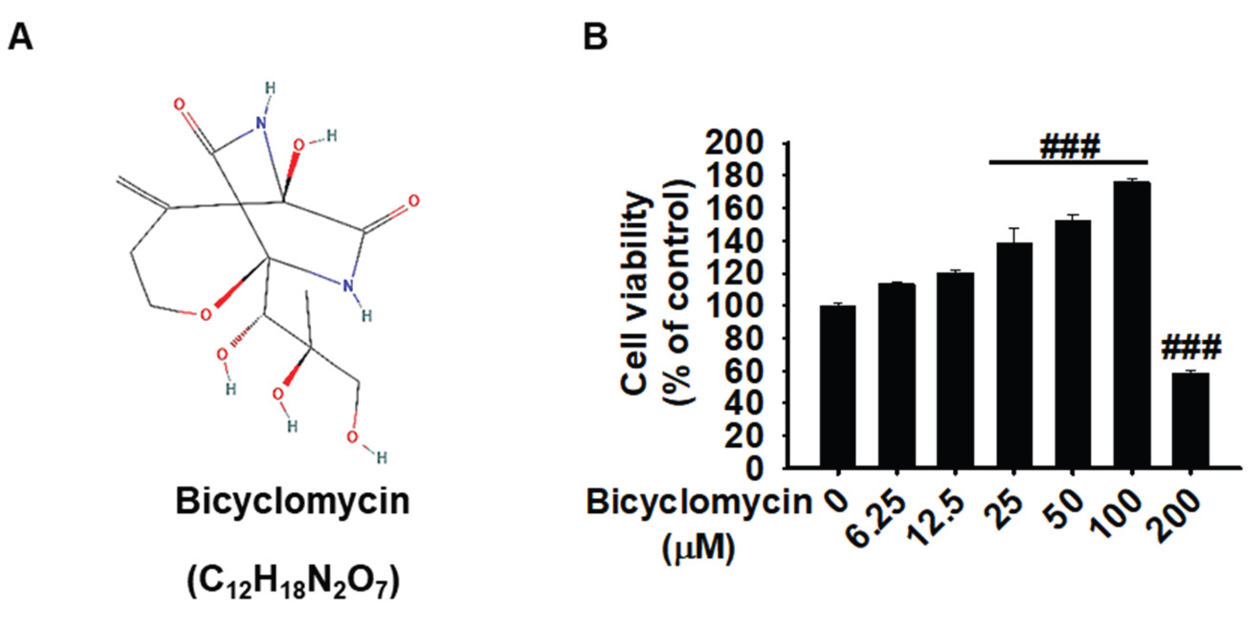
Figure 2.
Bicyclomycin suppresses LPS-induced NO and PGE2 production and downregulates NOS2 and COX-2 expression in macrophages. The cells were seeded at 1 × 10⁵ cells/mL and kept for 18 hours, then pretreated with BCM (0-100 μM) for 2 hours prior to stimulation with LPS (1000 ng/mL) for 24 h. (A) The NO production level was accessed using the Griess reagent assay and expressed as fold change relative to the untreated control. (B) PGE2 levels in the culture supernatants were determined by ELISA. (C- D) The total protein lysates were isolated 24 hours after the LPS induction, the western blot analysis was conducted to evaluate the protein expression level at translational level. Protein expression levels of NOS2 and COX-2 were analyzed by western blotting with β-actin as a loading control. Representative blot images and densitometry quantification normalized to β-actin are shown. (E) Phase-contrast microscopy images showing cell morphology under each treatment condition compared to the LPS-induced conditions. The red bar indicates the scale bar. Data are presented as mean ± SEM from three independent experiments. ###p < 0.001 vs. unstimulated control group; *p < 0.05, ***p < 0.001 vs. LPS-treated group.
Figure 2.
Bicyclomycin suppresses LPS-induced NO and PGE2 production and downregulates NOS2 and COX-2 expression in macrophages. The cells were seeded at 1 × 10⁵ cells/mL and kept for 18 hours, then pretreated with BCM (0-100 μM) for 2 hours prior to stimulation with LPS (1000 ng/mL) for 24 h. (A) The NO production level was accessed using the Griess reagent assay and expressed as fold change relative to the untreated control. (B) PGE2 levels in the culture supernatants were determined by ELISA. (C- D) The total protein lysates were isolated 24 hours after the LPS induction, the western blot analysis was conducted to evaluate the protein expression level at translational level. Protein expression levels of NOS2 and COX-2 were analyzed by western blotting with β-actin as a loading control. Representative blot images and densitometry quantification normalized to β-actin are shown. (E) Phase-contrast microscopy images showing cell morphology under each treatment condition compared to the LPS-induced conditions. The red bar indicates the scale bar. Data are presented as mean ± SEM from three independent experiments. ###p < 0.001 vs. unstimulated control group; *p < 0.05, ***p < 0.001 vs. LPS-treated group.
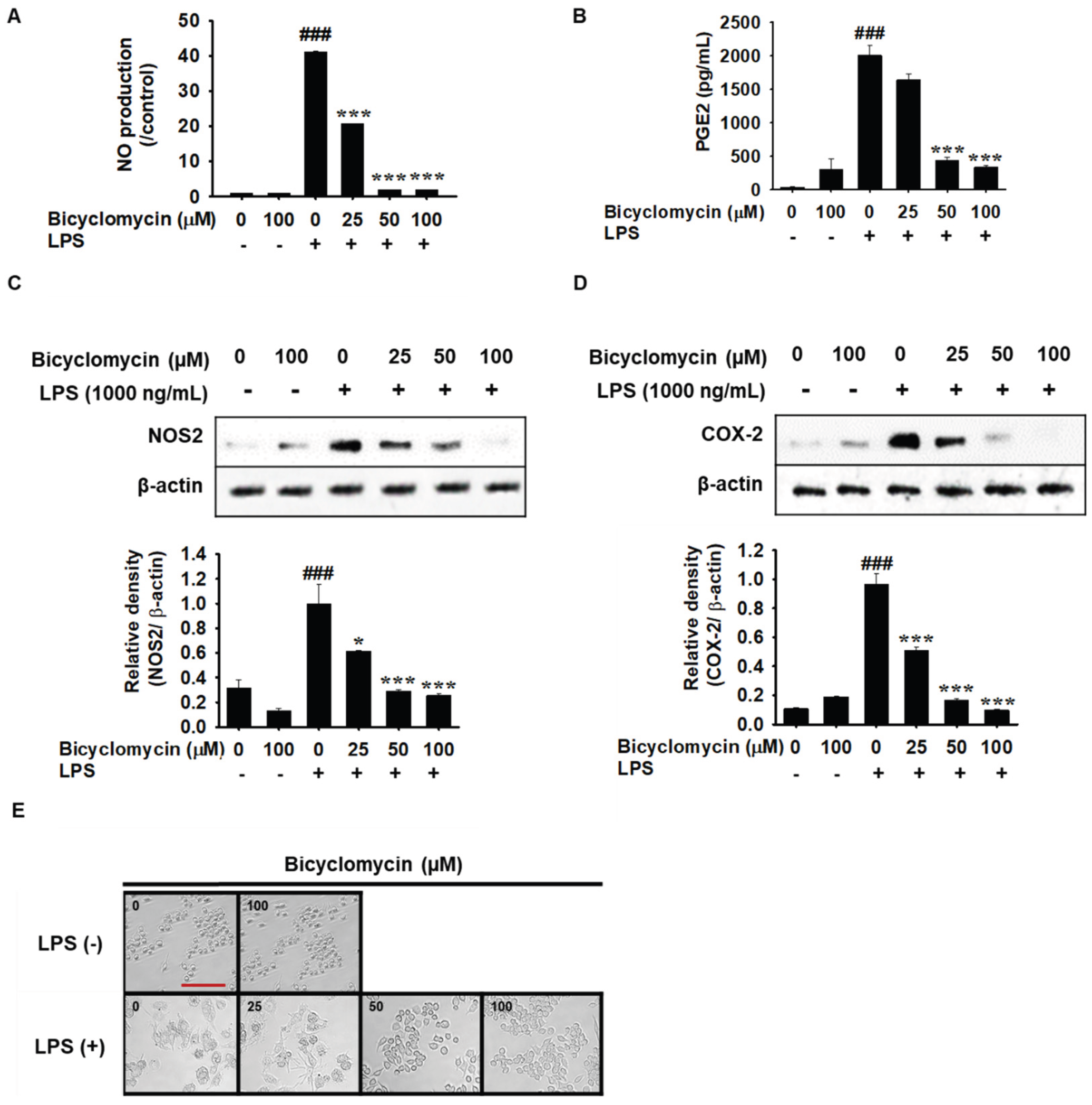
Figure 4.
Bicyclomycin attenuates LPS-induced NF-kB nuclear translocation. The in silico analysis was performed in order to evaluate the possibility of BCM block the binding of LPS to TLR4 –MD2 complex. The RAW264.7 cells were seeded at 1 × 10⁵ cells/mL and kept for 18 hours, then pre-treated with BCM (0-100 μM) for 2 hours, then exposed to LPS (1000 ng/mL), and incubated for 1 hour. (A) Nuclear NF-κB p65 translocation was quantified by total NF-κB p65 ELISA in cells pre-treated with the indicated concentrations of BCM (0-100 μM) for 1 h, followed by stimulation with LPS (1000 ng/mL) for 1 h. Results are expressed as a percentage of total NF-κB p65 relative to the LPS-only group and presented as mean ± SEM (n = 3, independent experiments). (B–D) Cells were pre-treated with BCM (0-100 μM) for 2 hours prior to LPS stimulation (1000 ng/mL) for the indicated time, LPS treated for 1 hour. Cytoplasmic and nuclear fractions were subjected to SDS-PAGE and western blot analysis. Representative western blot images were quantified by ImageJ (Fiji) software and corresponding densitometry quantification normalized to the respective loading control (β-actin and Lamin-B1 are shown for (D) phosphorylated IκB (p-IκB), with β-actin as a loading control; (E) nuclear NF-κB p65, with Lamin B1 as a nuclear fraction loading control; and (F) nuclear NF-κB p50, with Lamin B1 as a nuclear fraction loading control. Relative band intensities were normalized to the loading control and expressed relative to the LPS-only group. Data are represented as mean ± SEM from a minimum of three independent experiments. Statistical significance was determined by one-way ANOVA followed by bonferroni test. ###p < 0.001 compared with the untreated control group; ***p < 0.001 compared with the LPS-induced control group.
Figure 4.
Bicyclomycin attenuates LPS-induced NF-kB nuclear translocation. The in silico analysis was performed in order to evaluate the possibility of BCM block the binding of LPS to TLR4 –MD2 complex. The RAW264.7 cells were seeded at 1 × 10⁵ cells/mL and kept for 18 hours, then pre-treated with BCM (0-100 μM) for 2 hours, then exposed to LPS (1000 ng/mL), and incubated for 1 hour. (A) Nuclear NF-κB p65 translocation was quantified by total NF-κB p65 ELISA in cells pre-treated with the indicated concentrations of BCM (0-100 μM) for 1 h, followed by stimulation with LPS (1000 ng/mL) for 1 h. Results are expressed as a percentage of total NF-κB p65 relative to the LPS-only group and presented as mean ± SEM (n = 3, independent experiments). (B–D) Cells were pre-treated with BCM (0-100 μM) for 2 hours prior to LPS stimulation (1000 ng/mL) for the indicated time, LPS treated for 1 hour. Cytoplasmic and nuclear fractions were subjected to SDS-PAGE and western blot analysis. Representative western blot images were quantified by ImageJ (Fiji) software and corresponding densitometry quantification normalized to the respective loading control (β-actin and Lamin-B1 are shown for (D) phosphorylated IκB (p-IκB), with β-actin as a loading control; (E) nuclear NF-κB p65, with Lamin B1 as a nuclear fraction loading control; and (F) nuclear NF-κB p50, with Lamin B1 as a nuclear fraction loading control. Relative band intensities were normalized to the loading control and expressed relative to the LPS-only group. Data are represented as mean ± SEM from a minimum of three independent experiments. Statistical significance was determined by one-way ANOVA followed by bonferroni test. ###p < 0.001 compared with the untreated control group; ***p < 0.001 compared with the LPS-induced control group.
Disclaimer/Publisher’s Note: The statements, opinions and data contained in all publications are solely those of the individual author(s) and contributor(s) and not of MDPI and/or the editor(s). MDPI and/or the editor(s) disclaim responsibility for any injury to people or property resulting from any ideas, methods, instructions or products referred to in the content. |
© 2026 by the authors. Licensee MDPI, Basel, Switzerland. This article is an open access article distributed under the terms and conditions of the Creative Commons Attribution (CC BY) license (http://creativecommons.org/licenses/by/4.0/).
Copyright: This open access article is published under a Creative Commons CC BY 4.0 license, which permit the free download, distribution, and reuse, provided that the author and preprint are cited in any reuse.