

Submitted:
01 July 2026
Posted:
02 July 2026
You are already at the latest version
Abstract
Background: Keratoconus is a progressive corneal ectasia characterised by extracellular matrix (ECM) loss and an emerging inflammatory component, for which no disease-modifying molecular therapy exists. Exosomes derived from limbal and mesenchymal stem cells are an attractive cell-free therapeutic modality, but the cargo that should be delivered is undefined, and no curated limbal stem-cell (LSC) exosome cargo dataset currently exists. Methods: We reanalysed a public keratoconus corneal RNA-sequencing dataset (GEO: GSE77938; discovery and replication cohorts) with DESeq2, defined a replicated differentially expressed gene (DEG) set, and performed Gene Ontology, KEGG and Reactome enrichment. A high-confidence protein-protein interaction (PPI) network (STRING) identified hub genes. We integrated keratoconus disease-gene evidence (Open Targets Platform) and documented extracellular-vesicle cargo (ExoCarta, Vesiclepedia) and computed a transparent Cargo Prioritization Score (CPS) to nominate candidate LSC-exosome therapeutic cargo. Results: 1677 DEGs were detected in discovery (152 up, 1525 down) and 1380 were replicated. Enrichment was dominated by extracellular matrix organization; adaptive immune response; mononuclear cell differentiation. Network analysis nominated ECM and immune hub genes. The CPS prioritised COL1A1, FN1, COL4A1, COL3A1, COL5A1, MMP1 as leading restoration-cargo candidates, all documented as EV cargo and present in the mesenchymal stem-cell EV reference proteome. Conclusions: This fully reproducible, real-data framework provides a ranked, evidence-traceable shortlist of candidate LSC-exosome cargo for keratoconus and an explicit account of current data gaps to guide experimental validation.
Keywords:
keratoconus
; limbal stem cells
; exosomes
; extracellular vesicles
; transcriptomics
; differential expression
; protein-protein interaction network
; cargo prioritization
; corneal regeneration
; reproducible bioinformatics
1. Introduction
Keratoconus is the most common corneal ectasia and a leading indication for corneal transplantation in young adults; it is defined by progressive stromal thinning, biomechanical weakening and disruption of collagen and other ECM components [1,2]. Although historically regarded as a non-inflammatory condition, accumulating tear-film and tissue evidence now supports a low-grade inflammatory and immune contribution to disease progression [3,4,5]. Current management—rigid contact lenses, corneal cross-linking and, ultimately, keratoplasty—addresses biomechanics and optics but does not target the underlying molecular pathology, leaving a clear unmet need for disease-modifying, regenerative approaches.
The corneal epithelium is continuously renewed by limbal epithelial stem cells residing in the limbal niche, whose dysfunction underlies limbal stem-cell deficiency and impaired corneal surface homeostasis [6,7,8]. Stem-cell-derived exosomes—nanoscale extracellular vesicles (EVs) carrying proteins, mRNA and microRNA—have emerged as a cell-free regenerative modality, and limbal-niche- and mesenchymal stem-cell (MSC)-derived EVs accelerate corneal epithelial repair in preclinical models [9,10,11,12,13,14]. A central translational question, however, is which molecular cargo a therapeutic LSC exosome should carry to correct keratoconus pathology. No curated LSC-exosome cargo dataset is available in the major EV repositories, which precludes a purely data-driven cargo definition and instead motivates an integrative, evidence-ranked nomination strategy.
Here we develop a reproducible computational framework that couples real keratoconus corneal transcriptomics with disease-gene evidence, PPI topology and documented EV-cargo competence to prioritise candidate LSC-exosome cargo. We deliberately treat the absence of LSC- and induced-pluripotent-stem-cell (iPSC)-EV cargo data as an explicit, declared limitation rather than imputing it, and we provide a transparent Cargo Prioritization Score whose every component is traceable to a public dataset, public database or stated scoring rule.
2. Materials and Methods
The overall analytical workflow, from public-data acquisition through differential expression, functional and network analysis, multi-source evidence integration and Cargo Prioritization Scoring, is summarised in Figure 1.
2.1. Dataset and Preprocessing
Processed gene-level RNA-sequencing counts for human keratoconus (KTCN) and non-keratoconus (non-KTCN) corneas were obtained from Gene Expression Omnibus accession GSE77938 [16,17], which comprises an independent discovery cohort (8 KTCN versus 8 control) and a replication cohort (17 KTCN versus 17 control), all corneal tissue (50 samples in total; Table 1). Genes with at least 10 counts in a number of samples equal to the smaller group were retained.
2.2. Differential Expression
Differential expression was performed independently in each cohort with DESeq2 [18] using the model ~group (KTCN versus Control); log2 fold changes were shrunk with apeglm [19]. Ensembl identifiers were mapped to gene symbols and Entrez IDs (org.Hs.eg.db). DEGs were defined as adjusted P < 0.05 (Benjamini-Hochberg) and |log2 fold change| > 1. A replicated (consensus) DEG set was defined as DEGs significant in discovery that were concordant in sign and nominally significant (P < 0.05) in the replication cohort.
2.3. Functional Enrichment
Gene Ontology Biological Process, KEGG and Reactome over-representation analyses of the consensus DEGs were performed with clusterProfiler [20] and ReactomePA [21] against the set of all tested genes as background (Benjamini-Hochberg P < 0.05; redundant GO terms were simplified at a semantic-similarity cutoff of 0.7).
2.4. Protein-Protein Interaction Network and Hub Genes
The 400 most significant consensus DEGs (by discovery adjusted P) were submitted to the STRING database [22]; only high-confidence interactions (combined score >= 700) were retained. The network was analysed in NetworkX, and hub genes were ranked by degree and betweenness centrality (igraph/NetworkX) [26,27].
2.5. Database Integration and Cargo Prioritization Score
Keratoconus disease-gene association scores were retrieved from the Open Targets Platform (disease MONDO_0015486) [23]. Documented human EV-cargo competence and a mesenchymal stem-cell (MSC) EV reference proteome were derived from ExoCarta [24] and Vesiclepedia [25]; EV nomenclature follows MISEV2023 [15]. For each replicated DEG, five components were scaled to [0,1] or set as indicators: differential-expression evidence (S_DE, scaled |log2 fold change|), disease association (S_disease, Open Targets score), PPI hub centrality (S_hub, scaled degree), documented EV-cargo competence (S_EVcargo) and MSC-EV cargo membership (S_MSC). To integrate these complementary evidence sources into a single ranking metric, we empirically assigned weights that emphasized transcriptomic dysregulation and disease relevance while retaining contributions from network topology and extracellular-vesicle evidence. The resulting Cargo Prioritization Score (CPS) was calculated as: CPS = 0.30·S_DE + 0.25·S_disease + 0.20·S_hub + 0.15·S_EVcargo + 0.10·S_MSC. A disease-gene-cargo-pathway network was assembled for the top-ranked candidates.
3. Results
3.1. Differentially Expressed Genes in Keratoconus Cornea
Across 27,830 testable genes in the discovery cohort, 1,677 genes were differentially expressed (152 up- and 1525 down-regulated in KTCN), a pronounced down-regulation bias consistent with loss of corneal structural transcripts (Figure 2). Of these, 1,380 were replicated in the independent cohort and were carried forward as the consensus DEG set (Table 2; Figure 3).

Table 3.
Top 15 replicated DEGs ranked by discovery adjusted P.
| Gene | log2FC (disc) | padj (disc) | log2FC (rep) | padj (rep) |
|---|---|---|---|---|
| CCN2 | -3.71 | 1.64e-21 | -3.27 | 5.17e-13 |
| PCDH10 | -5.67 | 1.17e-17 | -2.9 | 1.28e-05 |
| RNASE1 | -6.89 | 2.33e-17 | -6.24 | 2.37e-17 |
| CCN1 | -2.51 | 3.04e-17 | -2.63 | 2.98e-15 |
| MATN3 | -4.34 | 1.09e-16 | -3.9 | 2.24e-17 |
| C1QB | -6.45 | 1.46e-16 | -5.63 | 7.33e-19 |
| MEF2C | -4.1 | 3.44e-16 | -3.24 | 1.15e-14 |
| GFRA1 | -7.72 | 6.49e-16 | -3.9 | 5.12e-08 |
| GUCY1A1 | -5.7 | 1.9e-15 | -4.86 | 1.18e-24 |
| CYBB | -5.2 | 4.59e-15 | -5.03 | 3.62e-14 |
| MRC1 | -6.39 | 6.6e-15 | -5.34 | 1.07e-15 |
| LRRC32 | -6.28 | 7.49e-15 | -4.67 | 1.11e-19 |
| TMEM119 | -5.76 | 4.51e-14 | -4.34 | 9.05e-17 |
| FBN1 | -4.5 | 4.84e-14 | -4.21 | 1.94e-25 |
| INHBA | -6.63 | 5.43e-14 | -5.24 | 3.03e-14 |
3.2. Functional Enrichment
The consensus DEGs were strongly enriched for ECM organisation and degradation, collagen and integrin biology, and immune/cytokine signalling across GO, KEGG and Reactome (Figure 4; Table 4). The leading GO term was “extracellular matrix organization” (adjusted P = 4.5e-27, 85 genes), and Reactome was led by “Extracellular matrix organization” (adjusted P = 3.6e-33). This dual ECM-and-immune signature is concordant with the contemporary view of keratoconus pathology [2,5].
3.3. PPI Network and Hub Genes
A high-confidence STRING network of the consensus DEGs contained 200 connected proteins and 621 edges (Figure 5). The highest-degree hubs—FN1, ITGAM, PTPRC, ITGB2, TYROBP, FCGR3A, CCL2, CD163—comprised both ECM/collagen proteins and immune effectors, all down-regulated in keratoconus (Table 5), pinpointing co-ordinately suppressed modules as rational intervention nodes.
3.4. Cargo Prioritization
Integrating the five evidence layers, the Cargo Prioritization Score ranked 1,253 replicated DEGs. 499 keratoconus-associated targets from Open Targets contributed disease-gene weight, including established keratoconus genes among the top candidates. The highest-scoring candidate cargo were COL1A1, FN1, COL4A1, COL3A1, COL5A1, MMP1, VCAN, PTPRC, INHBA, ITGAM (Figure 6; Table 6)—dominated by ECM and collagen constituents that are down-regulated in keratoconus, documented as EV cargo, and present in the MSC-EV reference proteome, and therefore represent biologically coherent restoration cargo for a limbal stem-cell exosome. Because keratoconus is characterised by ECM loss, these candidates are framed as restoration (replacement) cargo, whereas up-regulated immune candidates would instead represent suppression targets requiring a different cargo modality.
4. Discussion
Using only real, publicly available data, this study converts a fragmented evidence landscape into a single, ranked and fully traceable shortlist of candidate limbal stem-cell exosome cargo for keratoconus. The convergence of the differential-expression, network and disease-gene layers on extracellular-matrix and collagen biology is reassuring: it recapitulates the established molecular hallmark of keratoconus and indicates that the prioritisation is anchored in disease-relevant biology rather than analytical artefact.
The framework reframes a data gap as a hypothesis-generating opportunity. Because no LSC-exosome cargo repository exists, we do not claim to describe the LSC-EV proteome; instead we ask which disease-relevant molecules a therapeutic LSC exosome would ideally carry, and we restrict candidates to those for which EV delivery is at least plausible (documented EV cargo) and precedented in a stem-cell EV context (MSC-EV reference). The mechanistic hypothesis—that restoring ECM and collagen cargo, while modulating the immune component, could slow keratoconus progression—is consistent with preclinical evidence that limbal-niche- and MSC-derived EVs promote corneal epithelial and stromal repair, and is depicted in Figure 7.
The CPS is intentionally simple and transparent: all weights are fixed a priori, every component is traceable to a named public source, and the score can be recomputed or reweighted by any user. This favours interpretability and auditability over black-box optimisation, which we regard as appropriate for a hypothesis-prioritisation tool intended to guide, not replace, experimental validation.
4.1. Limitations
Several limitations must be stated explicitly. First, no curated LSC-exosome (or iPSC-exosome) cargo dataset exists in ExoCarta, Vesiclepedia or EVpedia (zero limbal/corneal EV experiments were found); consequently the LSC-EV cargo is nominated, not measured, and the MSC-EV proteome is used only as the closest available stem-cell EV reference. Second, the transcriptomic evidence derives from a single keratoconus dataset (GSE77938); although it includes an internal replication cohort, external validation in additional cohorts and ocular-surface diseases is required. Third, the public EV repositories are biased towards well-studied, highly abundant proteins, so EV-cargo competence is a permissive rather than a stringent filter. Fourth, gene-symbol harmonisation between transcriptomic and EV-repository nomenclature is imperfect and may under-count matches. Fifth, the analysis is entirely computational: the prioritised cargo are hypotheses that require proteomic confirmation in authentic LSC exosomes and functional validation in corneal models. Finally, the CPS weights, while transparent, are expert-chosen and not learned from outcome data.
4.2. Translational Outlook
Beyond target nomination, the candidate cargo identified here define a concrete translational path. The in-silico shortlist (Figure 8, stage 1) would first require confirmation that the prioritised molecules are genuinely present in authentic limbal stem-cell exosomes, followed by functional and preclinical validation, regulatory interactions and early-phase clinical evaluation of a cell-free exosome product. A structured validation programme spanning cargo confirmation, functional assays, disease models and EV engineering/delivery is outlined in Figure 9. We emphasise that all stages downstream of the present computational nomination remain to be performed; the framework is intended to focus and de-risk, not to substitute for, that experimental work.
5. Conclusions
We present a reproducible, integrity-constrained bioinformatics framework that nominates and ranks candidate limbal stem-cell exosome cargo for keratoconus by integrating real corneal transcriptomics, disease-gene evidence, PPI topology and extracellular-vesicle repositories. The approach yields an evidence-traceable shortlist led by ECM/collagen restoration cargo, an explicit map of current data gaps, and a transparent scoring scheme that is directly testable. It provides a rational starting point for designing engineered therapeutic exosomes for corneal ectasia and is readily generalisable to other ocular-surface diseases as suitable datasets become available.
Supplementary Materials
The following are available with this manuscript: Table S1 (DE_discovery.csv, DE_replication.csv) full differential-expression results; Table S2 (DEGs_consensus.csv) replicated DEG set; Table S3 (enrich_GO_BP.csv, enrich_KEGG.csv, enrich_Reactome.csv) full enrichment results; Table S4 (ppi_edges.csv, ppi_node_centrality.csv, ppi_hub_genes.csv) PPI network and hubs; Table S5 (opentargets_keratoconus.csv) Open Targets associations; Table S6 (cargo_prioritization_CPS.csv) full CPS ranking; Table S7 (disease_gene_cargo_pathway_edges.csv) integrated network edges.
Author Contributions
Conceptualization, C.-C.C. and C.-h.T.; methodology, C.-C.C. and H.-T.E.S.; software, H.-T.E.S. and B.-X.B.Z.; validation, B.-X.B.Z. and T.-H.C.C.; formal analysis, C.-C.C. and B.-X.B.Z.; investigation, T.-H.C.C. and H.-T.E.S.; resources, C.-h.T.; data curation, H.-T.E.S. and B.-X.B.Z.; writing—original draft preparation, C.-C.C.; writing—review and editing, C.-h.T., T.-H.C.C. and C.-C.C.; visualization, C.-C.C., B.-X.B.Z. and C.-Y.T.; supervision, C.-h.T.; project administration, C.-h.T. All authors have read and agreed to the published version of the manuscript.
Funding
This research received no external funding.
Institutional Review Board Statement
Not applicable; this study reanalysed publicly available, de-identified data.
Informed Consent Statement
Not applicable.
Data Availability Statement
All primary data are public: keratoconus transcriptomes from GEO accession GSE77938; disease-gene associations from the Open Targets Platform (MONDO_0015486); EV cargo from ExoCarta and Vesiclepedia; interactions from STRING. All derived tables generated in this study are provided as Supplementary Materials.
Acknowledgments
Not applicable.
Conflicts of Interest
Hsieh-Tsung Ethan Shen and Bo-Xiang Benjamin Zhang are affiliated with YD Bio Limited. The remaining authors declare no conflicts of interest. This manuscript is an independent narrative review of published literature and does not report, endorse or promote any proprietary product; inclusion of named programmes and clinical trials is descriptive and does not constitute endorsement.
Abbreviations
The following abbreviations are used in this manuscript:
| BH | Benjamini–Hochberg |
| BP | biological process (Gene Ontology) |
| CPS | Cargo Prioritization Score |
| DE | differential expression |
| DEG | differentially expressed gene |
| ECM | extracellular matrix |
| EV | extracellular vesicle |
| FDA | Food and Drug Administration |
| GEO | Gene Expression Omnibus |
| GLP | Good Laboratory Practice |
| GO | Gene Ontology |
| IND | Investigational New Drug |
| iPSC | induced pluripotent stem cell |
| KEGG | Kyoto Encyclopedia of Genes and Genomes |
| KTCN | keratoconus |
| Log2FC | log2 fold change |
| LSC | limbal stem cell |
| LSCD | limbal stem-cell deficiency |
| miRNA | microRNA |
| MISEV | Minimal Information for Studies of Extracellular Vesicles |
| mRNA | messenger RNA |
| MSC | mesenchymal stem cell |
| PPI | protein–protein interaction |
| RNA-seq | RNA sequencing |
| VST | variance-stabilizing transformation |
References
- Santodomingo-Rubido, J.; Carracedo, G.; Suzaki, A.; Villa-Collar, C.; Vincent, S. J.; Wolffsohn, J. S. Keratoconus: An updated review. Contact lens & anterior eye: the journal of the British Contact Lens Association 2022, 45(3), 101559. [Google Scholar] [CrossRef] [PubMed]
- Hao, X. D.; Gao, H.; Xu, W. H.; Shan, C.; Liu, Y.; Zhou, Z. X.; Wang, K.; Li, P. F. Systematically Displaying the Pathogenesis of Keratoconus via Multi-Level Related Gene Enrichment-Based Review. Front. Med. 2022, 8, 770138. [Google Scholar] [CrossRef] [PubMed]
- McMonnies, C. W. Inflammation and keratoconus. Optom. Vis. Sci. Off. Publ. Am. Acad. Optom. 2015, 92(2), e35–e41. [Google Scholar] [CrossRef] [PubMed]
- Zhang, H.; Cao, X.; Liu, Y.; Wang, P.; Li, X. Tear Levels of Inflammatory Cytokines in Keratoconus: A Meta-Analysis of Case-Control and Cross-Sectional Studies. BioMed Res. Int. 2021, 2021, 6628923. [Google Scholar] [CrossRef] [PubMed]
- Galvis, V.; Sherwin, T.; Tello, A.; Merayo, J.; Barrera, R.; Acera, A. Keratoconus: an inflammatory disorder? Eye 2015, 29(7), 843–859. [Google Scholar] [CrossRef] [PubMed]
- Deng, S. X.; Borderie, V.; Chan, C. C.; Dana, R.; Figueiredo, F. C.; Gomes, J. A. P.; Pellegrini, G.; Shimmura, S.; Kruse, F. E.; The International Limbal Stem Cell Deficiency Working Group. Global Consensus on Definition, Classification, Diagnosis, and Staging of Limbal Stem Cell Deficiency. Cornea 2019, 38(3), 364–375. [Google Scholar] [CrossRef] [PubMed]
- Li, S.; Sun, H.; Chen, L.; Fu, Y. Targeting limbal epithelial stem cells: master conductors of corneal epithelial regeneration from the bench to multilevel theranostics. J. Transl. Med. 2024, 22(1), 794. [Google Scholar] [CrossRef] [PubMed]
- Nuzzi, A.; Pozzo Giuffrida, F.; Luccarelli, S.; Nucci, P. Corneal Epithelial Regeneration: Old and New Perspectives. Int. J. Mol. Sci. 2022, 23(21), 13114. [Google Scholar] [CrossRef] [PubMed]
- Zhou, T.; Huang, X.; Tan, Y.; Shen, J.; Zhou, X.; Li, G.; Wang, W. Limbal niche cell-derived exosomes accelerate corneal epithelial repair through PI3K/Akt activation and FOXO3 inhibition. Exp. Eye Res. 2026, 269, 111049. [Google Scholar] [CrossRef] [PubMed]
- Xu, Y.; Wei, C.; Ma, L.; Zhao, L.; Li, D.; Lin, Y.; Zhou, Q.; Xie, L.; Wang, F. 3D mesenchymal stem cell exosome-functionalized hydrogels for corneal wound healing. J. Control. Release Off. J. Control. Release Soc. 2025, 380, 630–646. [Google Scholar] [CrossRef] [PubMed]
- Tang, Q.; Lu, B.; He, J.; Chen, X.; Fu, Q.; Han, H.; Luo, C.; Yin, H.; Qin, Z.; Lyu, D.; Zhang, L.; Zhou, M.; Yao, K. Exosomes-loaded thermosensitive hydrogels for corneal epithelium and stroma regeneration. Biomaterials 2022, 280, 121320. [Google Scholar] [CrossRef] [PubMed]
- Yu, F.; Zhao, X.; Wang, Q.; Fang, P. H.; Liu, L.; Du, X.; Li, W.; He, D.; Zhang, T.; Bai, Y.; Liu, L.; Li, S.; Yuan, J. Engineered Mesenchymal Stromal Cell Exosomes-Loaded Microneedles Improve Corneal Healing after Chemical Injury. ACS nano Advance online publication. 2024. [Google Scholar] [CrossRef] [PubMed]
- Williams, T.; Salmanian, G.; Burns, M.; Maldonado, V.; Smith, E.; Porter, R. M.; Song, Y. H.; Samsonraj, R. M. Versatility of mesenchymal stem cell-derived extracellular vesicles in tissue repair and regenerative applications. Biochimie 2023, 207, 33–48. [Google Scholar] [CrossRef] [PubMed]
- Massoumi, H.; Amin, S.; Soleimani, M.; Momenaei, B.; Ashraf, M. J.; Guaiquil, V. H.; Hematti, P.; Rosenblatt, M. I.; Djalilian, A. R.; Jalilian, E. Extracellular-Vesicle-Based Therapeutics in Neuro-Ophthalmic Disorders. Int. J. Mol. Sci. 2023, 24(10), 9006. [Google Scholar] [CrossRef] [PubMed]
- Welsh, J. A.; Goberdhan, D. C. I.; O'Driscoll, L.; Buzas, E. I.; Blenkiron, C.; Bussolati, B.; Cai, H.; Di Vizio, D.; Driedonks, T. A. P.; Erdbrügger, U.; Falcon-Perez, J. M.; Fu, Q. L.; Hill, A. F.; Lenassi, M.; Lim, S. K.; Mahoney, M. G.; Mohanty, S.; Möller, A.; Nieuwland, R.; Ochiya, T.; Witwer, K. W. Minimal information for studies of extracellular vesicles (MISEV2023): From basic to advanced approaches. J. Extracell. Vesicles 2024, 13(2), e12404. [Google Scholar] [CrossRef] [PubMed]
- Kabza, M.; Karolak, J. A.; Rydzanicz, M.; Szcześniak, M. W.; Nowak, D. M.; Ginter-Matuszewska, B.; Polakowski, P.; Ploski, R.; Szaflik, J. P.; Gajecka, M. Collagen synthesis disruption and downregulation of core elements of TGF-β, Hippo, and Wnt pathways in keratoconus corneas. Eur. J. Hum. Genet. EJHG 2017, 25(5), 582–590. [Google Scholar] [CrossRef] [PubMed]
- Davis, S.; Meltzer, P. S. GEOquery: a bridge between the Gene Expression Omnibus (GEO) and BioConductor. Bioinformatics 2007, 23(14), 1846–1847. [Google Scholar] [CrossRef] [PubMed]
- Love, M. I.; Huber, W.; Anders, S. Moderated estimation of fold change and dispersion for RNA-seq data with DESeq2. Genome Biol. 2014, 15(12), 550. [Google Scholar] [CrossRef] [PubMed]
- Zhu, A.; Ibrahim, J. G.; Love, M. I. Heavy-tailed prior distributions for sequence count data: removing the noise and preserving large differences. Bioinformatics 2019, 35(12), 2084–2092. [Google Scholar] [CrossRef] [PubMed]
- Yu, G.; Wang, L. G.; Han, Y.; He, Q. Y. clusterProfiler: an R package for comparing biological themes among gene clusters. Omics A J. Integr. Biol. 2012, 16(5), 284–287. [Google Scholar] [CrossRef] [PubMed]
- Yu, G.; He, Q. Y. ReactomePA: an R/Bioconductor package for reactome pathway analysis and visualization. Mol. Biosyst. 2016, 12(2), 477–479. [Google Scholar] [CrossRef] [PubMed]
- Szklarczyk, D.; Kirsch, R.; Koutrouli, M.; Nastou, K.; Mehryary, F.; Hachilif, R.; Gable, A. L.; Fang, T.; Doncheva, N. T.; Pyysalo, S.; Bork, P.; Jensen, L. J.; von Mering, C. The STRING database in 2023: protein-protein association networks and functional enrichment analyses for any sequenced genome of interest. Nucleic Acids Res. 2023, 51(D1), D638–D646. [Google Scholar] [CrossRef] [PubMed]
- Ochoa, D.; Hercules, A.; Carmona, M.; Suveges, D.; Baker, J.; Malangone, C.; Lopez, I.; Miranda, A.; Cruz-Castillo, C.; Fumis, L.; Bernal-Llinares, M.; Tsukanov, K.; Cornu, H.; Tsirigos, K.; Razuvayevskaya, O.; Buniello, A.; Schwartzentruber, J.; Karim, M.; Ariano, B.; Martinez Osorio, R. E.; McDonagh, E. M. The next-generation Open Targets Platform: reimagined, redesigned, rebuilt. Nucleic Acids Res. 2023, 51(D1), D1353–D1359. [Google Scholar] [CrossRef] [PubMed]
- Keerthikumar, S.; Chisanga, D.; Ariyaratne, D.; Al Saffar, H.; Anand, S.; Zhao, K.; Samuel, M.; Pathan, M.; Jois, M.; Chilamkurti, N.; Gangoda, L.; Mathivanan, S. ExoCarta: A Web-Based Compendium of Exosomal Cargo. J. Mol. Biol. 2016, 428(4), 688–692. [Google Scholar] [CrossRef] [PubMed]
- Pathan, M.; Fonseka, P.; Chitti, S. V.; Kang, T.; Sanwlani, R.; Van Deun, J.; Hendrix, A.; Mathivanan, S. Vesiclepedia 2019: a compendium of RNA, proteins, lipids and metabolites in extracellular vesicles. Nucleic Acids Res. 2019, 47(D1), D516–D519. [Google Scholar] [CrossRef] [PubMed]
- Csárdi, G.; Nepusz, T. The igraph software package for complex network research. 2006. Available online: https://api.semanticscholar.org/CorpusID:16923281.
- Hagberg, A.A.; Schult, D.A.; Swart, P.J.; Hagberg, J. Exploring Network Structure, Dynamics, and Function using NetworkX. In Proceedings of the Python in Science Conference, 2008; Available online: https://api.semanticscholar.org/CorpusID:16050699.
Figure 1.
Overall analytical workflow. Public keratoconus corneal transcriptomes (GEO GSE77938) undergo DESeq2 differential expression and replication, followed by GO/KEGG/Reactome enrichment and STRING PPI/hub analysis; disease-gene (Open Targets) and extracellular-vesicle cargo (ExoCarta, Vesiclepedia, MSC-EV reference) evidence are integrated into a transparent Cargo Prioritization Score that ranks candidate LSC-exosome cargo. The absence of an LSC/iPSC exosome cargo dataset is shown as a declared data gap.
Figure 1.
Overall analytical workflow. Public keratoconus corneal transcriptomes (GEO GSE77938) undergo DESeq2 differential expression and replication, followed by GO/KEGG/Reactome enrichment and STRING PPI/hub analysis; disease-gene (Open Targets) and extracellular-vesicle cargo (ExoCarta, Vesiclepedia, MSC-EV reference) evidence are integrated into a transparent Cargo Prioritization Score that ranks candidate LSC-exosome cargo. The absence of an LSC/iPSC exosome cargo dataset is shown as a declared data gap.
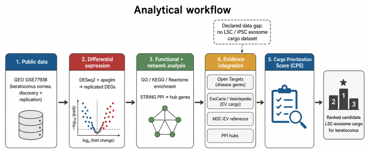
Figure 2.
Volcano plot of differential expression in keratoconus versus control cornea (discovery cohort, GSE77938). Coloured points pass adjusted P < 0.05 and |log2FC| > 1.
Figure 2.
Volcano plot of differential expression in keratoconus versus control cornea (discovery cohort, GSE77938). Coloured points pass adjusted P < 0.05 and |log2FC| > 1.
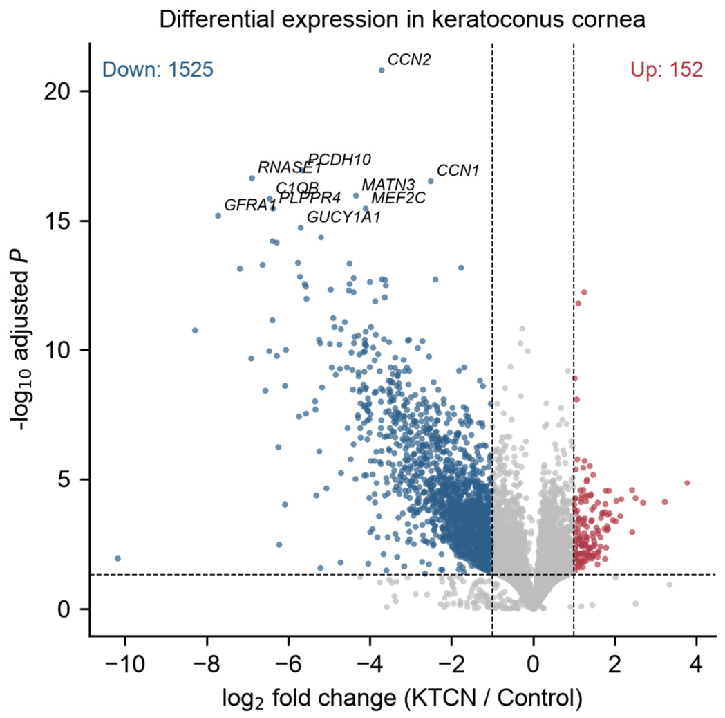
Figure 3.
Heatmap of the top 40 replicated DEGs (variance-stabilised expression, row z-score) showing clear separation of keratoconus and control corneas.
Figure 3.
Heatmap of the top 40 replicated DEGs (variance-stabilised expression, row z-score) showing clear separation of keratoconus and control corneas.
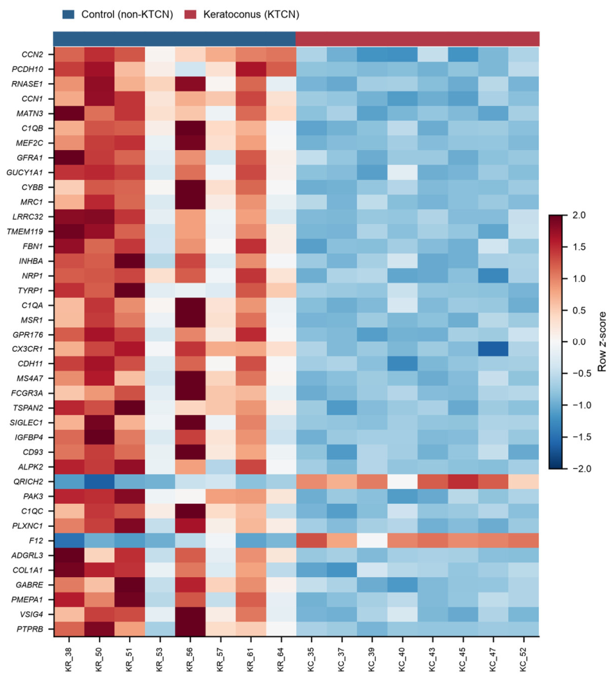
Figure 4.
Top enriched GO biological-process, KEGG and Reactome terms for the replicated DEGs (bar length and marker indicate -log10 adjusted P and gene count).
Figure 4.
Top enriched GO biological-process, KEGG and Reactome terms for the replicated DEGs (bar length and marker indicate -log10 adjusted P and gene count).
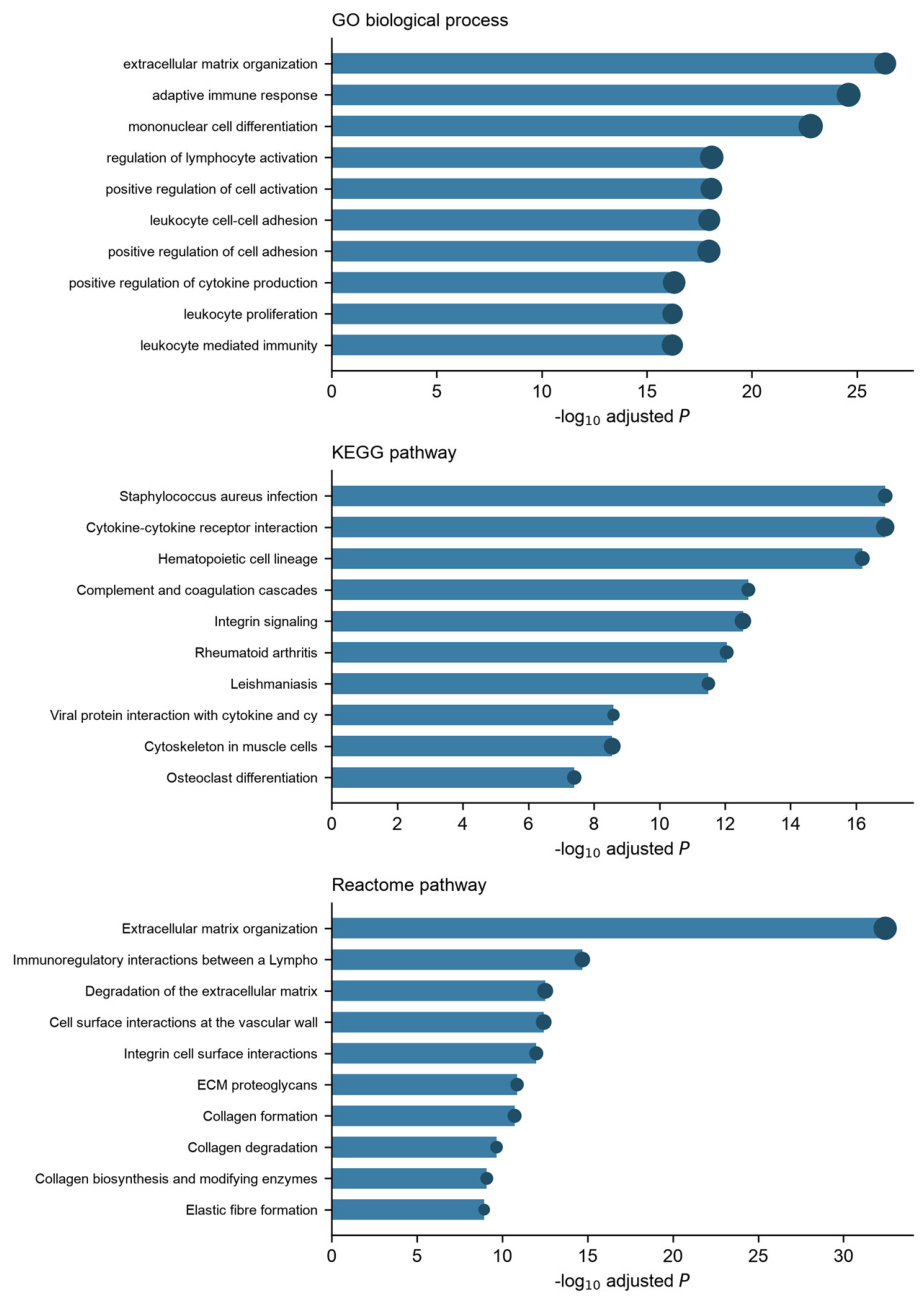
Figure 5.
STRING protein-protein interaction network of replicated keratoconus DEGs (combined score >= 700). Node size, degree centrality; colour, log2 fold change. Top hub genes are labelled.
Figure 5.
STRING protein-protein interaction network of replicated keratoconus DEGs (combined score >= 700). Node size, degree centrality; colour, log2 fold change. Top hub genes are labelled.
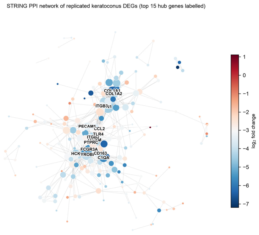
Figure 6.
Cargo Prioritization Score (CPS) of the top 20 candidate LSC-exosome cargo, with the contribution of each weighted evidence component shown as stacked segments.
Figure 6.
Cargo Prioritization Score (CPS) of the top 20 candidate LSC-exosome cargo, with the contribution of each weighted evidence component shown as stacked segments.
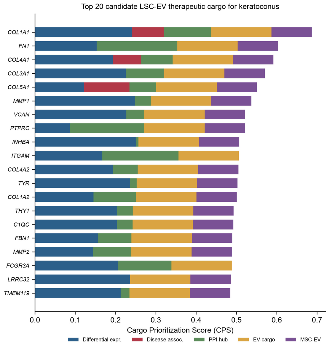
Figure 7.
Proposed mechanism-of-action model (conceptual hypothesis). An engineered limbal stem-cell exosome delivers restoration cargo (ECM/collagen mRNA and protein) and immunomodulatory cargo to the keratoconus cornea, hypothesised to restore extracellular-matrix homeostasis and attenuate low-grade inflammation. This model is hypothetical and was not experimentally validated in the present study.
Figure 7.
Proposed mechanism-of-action model (conceptual hypothesis). An engineered limbal stem-cell exosome delivers restoration cargo (ECM/collagen mRNA and protein) and immunomodulatory cargo to the keratoconus cornea, hypothesised to restore extracellular-matrix homeostasis and attenuate low-grade inflammation. This model is hypothetical and was not experimentally validated in the present study.
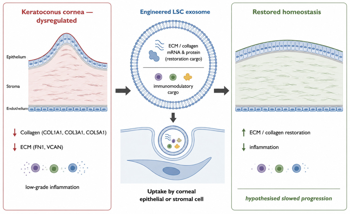
Figure 8.
Translational development roadmap (illustrative). Stage-gate path from the present computational cargo nomination through in-vitro and IND-enabling preclinical validation, FDA interactions (Pre-IND/IND for a cell-free extracellular-vesicle product) and early-phase clinical evaluation in keratoconus. Stages downstream of nomination require experimental validation.
Figure 8.
Translational development roadmap (illustrative). Stage-gate path from the present computational cargo nomination through in-vitro and IND-enabling preclinical validation, FDA interactions (Pre-IND/IND for a cell-free extracellular-vesicle product) and early-phase clinical evaluation in keratoconus. Stages downstream of nomination require experimental validation.
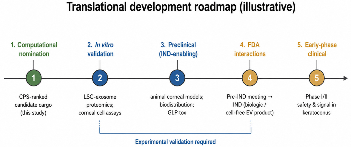
Figure 9.
Future validation strategy. Four complementary workstreams—cargo confirmation in authentic LSC exosomes (MISEV2023-compliant), functional corneal cell assays, ex-vivo/in-vivo disease models, and EV engineering and ocular delivery—required to validate the CPS-nominated candidate cargo.
Figure 9.
Future validation strategy. Four complementary workstreams—cargo confirmation in authentic LSC exosomes (MISEV2023-compliant), functional corneal cell assays, ex-vivo/in-vivo disease models, and EV engineering and ocular delivery—required to validate the CPS-nominated candidate cargo.
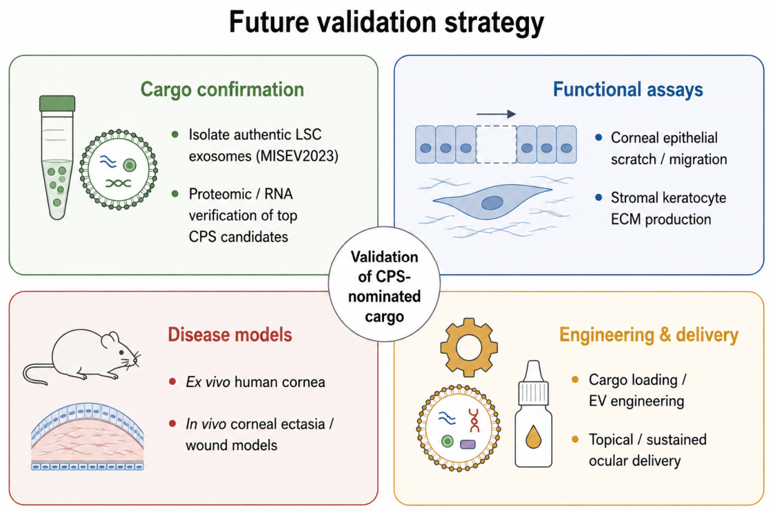
Table 1.
Keratoconus corneal transcriptomic cohorts analysed (GEO accession GSE77938). KTCN, keratoconus; Control, non-keratoconus.
Table 1.
Keratoconus corneal transcriptomic cohorts analysed (GEO accession GSE77938). KTCN, keratoconus; Control, non-keratoconus.
| Cohort | KTCN (n) | Control (n) | Total (n) | Tissue | Assay |
|---|---|---|---|---|---|
| Discovery | 8 | 8 | 16 | Cornea | RNA-seq |
| Replication | 17 | 17 | 34 | Cornea | RNA-seq |
| Total | 25 | 25 | 50 | Cornea | RNA-seq |
Table 2.
Differential-expression summary (GSE77938).
| Metric | Value |
|---|---|
| Genes tested (discovery) | 27830 |
| DEGs (discovery, padj<0.05, |log2FC|>1) | 1677 |
| up-regulated | 152 |
| down-regulated | 1525 |
| Genes tested (replication) | 23008 |
| DEGs (replication) | 3236 |
| Replicated consensus DEGs | 1380 |
Table 4.
Representative enriched terms (top 5 per database).
| Source | Term | adj. P | Count |
|---|---|---|---|
| GO BP | extracellular matrix organization | 4.52e-27 | 85 |
| GO BP | adaptive immune response | 2.49e-25 | 102 |
| GO BP | mononuclear cell differentiation | 1.59e-23 | 106 |
| GO BP | regulation of lymphocyte activation | 8.17e-19 | 98 |
| GO BP | positive regulation of cell activation | 8.47e-19 | 82 |
| KEGG | Staphylococcus aureus infection | 1.3e-17 | 34 |
| KEGG | Cytokine-cytokine receptor interaction | 1.3e-17 | 57 |
| KEGG | Hematopoietic cell lineage | 6.5e-17 | 36 |
| KEGG | Complement and coagulation cascades | 1.93e-13 | 30 |
| KEGG | Integrin signaling | 2.84e-13 | 45 |
| Reactome | Extracellular matrix organization | 3.59e-33 | 96 |
| Reactome | Immunoregulatory interactions between a Lymphoid and a non-Lymphoid cell | 2.03e-15 | 39 |
| Reactome | Degradation of the extracellular matrix | 3.05e-13 | 41 |
| Reactome | Cell surface interactions at the vascular wall | 3.73e-13 | 40 |
| Reactome | Integrin cell surface interactions | 1.02e-12 | 31 |
Table 5.
Top 12 PPI hub genes by degree centrality.
| Source | Term | adj. P | Count |
|---|---|---|---|
| FN1 | 36 | 0.153 | -4.43 |
| ITGAM | 34 | 0.0659 | -4.74 |
| PTPRC | 33 | 0.0955 | -2.96 |
| ITGB2 | 28 | 0.0862 | -4.28 |
| TYROBP | 28 | 0.0518 | -4.09 |
| FCGR3A | 24 | 0.0256 | -5.6 |
| CCL2 | 23 | 0.126 | -2.93 |
| CD163 | 21 | 0.0293 | -5.24 |
| COL1A1 | 21 | 0.018 | -6.39 |
| TLR4 | 20 | 0.023 | -2.51 |
| HCK | 19 | 0.0465 | -4.45 |
| COL1A2 | 19 | 0.0062 | -4.25 |
Table 6.
Top 20 CPS-ranked candidate LSC-exosome cargo for keratoconus.
| Rank | Gene | Dir. | log2FC | Disease | Degree | EV | MSC-EV | CPS |
|---|---|---|---|---|---|---|---|---|
| 1 | COL1A1 | down | -6.39 | 0.318 | 21 | 1 | True | 0.687 |
| 2 | FN1 | down | -4.43 | 0 | 36 | 1 | True | 0.603 |
| 3 | COL4A1 | down | -5.33 | 0.281 | 14 | 1 | True | 0.592 |
| 4 | COL3A1 | down | -6.06 | 0 | 17 | 1 | True | 0.57 |
| 5 | COL5A1 | down | -3.73 | 0.451 | 12 | 1 | True | 0.551 |
| 6 | MMP1 | down | -6.56 | 0 | 7 | 1 | True | 0.537 |
| 7 | VCAN | down | -6.08 | 0 | 8 | 1 | True | 0.521 |
| 8 | PTPRC | down | -2.96 | 0 | 33 | 1 | True | 0.521 |
| 9 | INHBA | down | -6.63 | 0 | 1 | 1 | True | 0.507 |
| 10 | ITGAM | down | -4.74 | 0 | 34 | 1 | False | 0.506 |
| 11 | COL4A2 | down | -5.34 | 0 | 11 | 1 | True | 0.505 |
| 12 | TYR | down | -6.28 | 0 | 3 | 1 | True | 0.502 |
| 13 | COL1A2 | down | -4.25 | 0 | 19 | 1 | True | 0.501 |
| 14 | THY1 | down | -5.57 | 0 | 7 | 1 | True | 0.493 |
| 15 | C1QC | down | -5.55 | 0 | 7 | 1 | True | 0.492 |
| 16 | FBN1 | down | -4.5 | 0 | 15 | 1 | True | 0.49 |
| 17 | MMP2 | down | -4.23 | 0 | 17 | 1 | True | 0.489 |
| 18 | FCGR3A | down | -5.6 | 0 | 24 | 1 | False | 0.489 |
| 19 | LRRC32 | down | -6.28 | 0 | 0 | 1 | True | 0.486 |
| 20 | TMEM119 | down | -5.76 | 0 | 4 | 1 | True | 0.485 |
Disclaimer/Publisher’s Note: The statements, opinions and data contained in all publications are solely those of the individual author(s) and contributor(s) and not of MDPI and/or the editor(s). MDPI and/or the editor(s) disclaim responsibility for any injury to people or property resulting from any ideas, methods, instructions or products referred to in the content. |
© 2026 by the authors. Licensee MDPI, Basel, Switzerland. This article is an open access article distributed under the terms and conditions of the Creative Commons Attribution (CC BY) license (http://creativecommons.org/licenses/by/4.0/).
Copyright: This open access article is published under a Creative Commons CC BY 4.0 license, which permit the free download, distribution, and reuse, provided that the author and preprint are cited in any reuse.


