

Submitted:
01 July 2026
Posted:
02 July 2026
You are already at the latest version
Abstract
As a commonly present organophosphorus flame retardants and persistent organic pollutant 2-ethylhexyl diphenyl phosphate (EHDPP) has been observed to be genotoxic in cultured human hepatoma (HepG2) cells which depended on CYP activities. Yet, its impact on hepatic Cyp expression, hepatotoxicity and genotoxicity in intact mammalians remain unidentified. In this study, adult male C57BL/6J mice received EHDPP by gastric gavage at doses of 50, 100, and 150 mg/kg (b.w.)/d for 7 d, then the hepatic expression of several Cyp proteins, aryl hydrocarbon receptor (AhR) and pregnane X receptor (PXR) was analyzed by Western blotting; hepatoxicity was determined by serum ALT/AST activities and hepatic histological examination, while genotoxicity by comet assay, phosphorylated histone (γ-H2AX) protein, micronucleus test, and Pig-a assay. Micronucleus test in mouse hepatoma (Hepa1-6) cells in vitro was employed to observed the modulating effect of PCB 126 (100 nM)/BAY-218 (700 nM) (Ahr-Cyp1a1 activator/inhibitor). The results indicated that EHDPP induced hepatic Cyp1a1, 2e1, AhR, Cyp1a2, Cyp3a4 and PXR proteins and histologic liver damage at 50 mg/kg/d or higher doses, while at the highest dose (150 mg/kg/d) with hepatic DNA damage and micronucleus formation in bone marrow polychromatic erythrocytes. The result of Pig-a assay (at 14 and 28 d) was negative. In Hepa1-6 cells EHDPP induced micronucleus marginally; however, this effect was enhanced by PCB 126, while abolished by BAY-218. This study suggests that EHDPP may enhance protein expression of hepatic Cyp1a1, Cyp2e1, AhR and PXR, and induce liver damage and DNA/chromosome damage in mouse; Cyp1a1 might be a major activating enzyme.
Keywords:
omet assay
; cytochrome P450 (Cyp) enzymes
; 2-ethylhexyl diphenyl phosphate (EHDPP)
; hepatotoxicity
; micronucleus test
; pig-a assay
1. Introduction
2-Ethylhexyl diphenyl phosphate (EHDPP) is a commonly consumed organophosphorus flame retardant (OPFR). Owing to its highly preferred endurance, flexibility, and heat resistance, EHDPP has been largely used in the production of textiles, homewares, paints, and plastic products [1,2]. Particularly, along with the inclusion of polybrominated diphenyl ethers (PBDEs, previously dominating the flame retardant markets) on the list of persistent organic pollutants as documented in the Stockholm Convention, PBDEs have been gradually phased out, to which EHDPP is one of the OPFRs used as alternative flame retardants. EHDPP and other OPFRs are commonly present in various environmental media, such as surface water, indoor dusts, sedimentations, and biota [3,4], and they have been detected in specimens from humans (breast milk, blood, and urine) and other organisms (such as marine lives and birds) [5,6]. EHDPP has been approved for use in the production of food-packaging materials, and it may enter the human body through ingestion of contaminated food, inhalation of dusts containing EHDPP, and skin contact of relevant materials, among which dietary intake is supposed to be the major exposure mode. Due to human activities and long distance transmission through air flow, EHDPP is distributed in the world very widely. For example, in the soil and sediments in Tibet, China, EHDPP is dominant in various OPFRs [7]. Notably, EHDPP concentrations in processed food appeared to be significantly higher than in unprocessed food, which implies that EHDPP may contaminate food through using packaging materials containing EHDPP [8]. For a population in Beijing, China, the median concentration of EHDPP in whole blood and serum was 1.10 and 0.93 ng/mL, respectively. [9]. Meanwhile, epidemiological and toxicological investigations suggest that EHDPP may have potential toxicity in humans and/or other organisms, which includes reproductive [10], developmental [11], and hepatic toxicity [1,12], as well as participating in chemical carcinogenesis [1]. Although there has been no human population-based cohort study verifying the carcinogenicity of EHDPP, cross-sectional investigations suggest that the blood burden of EHDPP (and some other OPFRs) is significantly associated with several cancers (such as lung cancer and some female-specific cancers) [13,14].
Gene mutation and chromosome damage (major endpoints of mutagenesis) are contributive to chemical carcinogenesis, while DNA damage (in various forms, such as DNA break, formation of DNA adducts, base alkylation, and DNA cross-linking) is the prerequisite for gene mutations and structural chromosome alterations [15,16], as well as some tissue damage [17]. EHDPP by itself is incapable of forming covalent bonds with DNA and proteins, while only after metabolic activation [mostly requiring catalysis by relevant xenobiotic-metabolizing enzymes, such as cytochrome P450s (CYPs)], it could be converted to bio-reactive metabolites. Regarding the metabolism of EHDPP, in an early study the metabolic fate of 14C-EHDPP orally administered to male rats was investigated. The results indicated rapid absorption, and within 24 h most radioactivity was excreted in the urine and feces, all excreted by 7 d; the radioactivity was widely distributed, relatively high in the blood, liver, kidneys and adipose tissue. The major metabolites in the urine were diphenyl phosphate (DPHP) and phenol, with p-hydroxyphenyl DPHP (OH-DPHP) and monophenyl phosphate (MPP) as minor metabolites [18]. In a population of east China, the blood levels of EHDPP and DPHP dominated in various organophosphorus triester and diester compounds, respectively [19]. Further multivariant linear regression analysis of the data indicated that the concentrations of EHDPP and DPHP were significantly correlated with each other, supporting that DPHP is a major metabolite of EHDPP; meanwhile, from the blood samples other metabolites (presumably formed from EHDPP) were also detected, such as 2-ethylhexy monophenyl phosphate (EHMPP) and hydroxylated EHMPP (OH-EHMPP) [19]. These hydroxylated metabolites may not bio-reactive enough to attack DNA and proteins by forming covalent binding, and whether they are further metabolically activated to electrophilic metabolites remains unidentified, especially considering that the higher reactive as an electrophile the shorter half-life and more difficult to be detected. Indeed, we have previously observed that EHDPP may be metabolically activated for DNA- and chromosome-breaking effects in cultured mammalian cells, which depend on several human CYP enzymes, predominantly CYP2E1 and CYP3A4 [20]. However, relevant genotoxic effects in intact mammalians has never been reported.
In this study, EHDPP at varying doses was used to orally expose young adult male C57BL/6J mice for 7 d, then the induction of liver tissue damage, hepatic DNA damage, modulation in hepatic expression of several Cyp enzymes and nuclear receptors, micronucleus formation in the bone marrow, and induction of Pig-a gene mutations in the circulative erythrocytes, were analyzed. In order to explore the Cyp enzyme involved in activating EHDPP for its genotoxicity, an in vitro micronucleus test in mouse hepatoma (Hepa1-6) cell line, with and without modulators of specific Cyp enzymes, was also performed.
2. Materials and Methods
2.1. Chemicals and Materials
EHDPP (purity ≥ 97%) and N-nitrosodiethylamine (NDEA, purity ≥ 99%) were purchased from Shanghai Aladdin Biochem. Technol. Co., Ltd. (Shanghai, China). 3,3′,4′,4′,5-Pentachlorobiphenyl (PCB 126, ≥ 99%) was from AccuStandard Inc. (New Haven, CT, USA). (S)-6-(4-Chlorophenyl)-2-(3-fluorophenyl)-N-(1-hydroxypropan-2-yl)-3-oxo-2 (BAY-218, ≥ 98%) was from Meilunbio (Dalian, China).
The primary antibody raised in rabbit against human CYP3A4 (cross-reacting with mouse Cyp3a11), and the secondary antibody, i.e., horseradish peroxidase (HRP)-conjugated goat anti-rabbit IgG, were both purchased from Abcam (Cambridge, UK). The primary antibodies raised in rabbit against human aryl hydrocarbon receptor (AhR), human pregnane X receptor (PXR), human CYP1A1 (cross-reacting with mouse Cyp1a1), human CYP1A2 (cross-reacting with mouse Cyp1a2), and human phosphorylated histone 2AX (γ-H2AX) were from ABclonal (Wuhan, China). The primary antibody raised in rabbit against human CYP2E1 (cross-reacting with mouse Cyp2e1) was from Proteintech Group, Inc. (Wuhan, China). The primary antibody raised in rabbit against human GAPDH was from Yeasen Biotechnology (Shanghai, China). APC-labelled anti-mouse CD24 antibody (APC anti-mouse CD24) was from Biolegend (San Diego, CA, USA), and SYTO™13 (for staining nucleic acid) was from Thermo Fisher Scientific (Carlsbad, CA, USA).
The assay kits for serum alanine aminotransferase (ALT) and aspartate aminotransferase (AST) activities were purchased from Elabscience Biotechnology Co., Ltd. (Wuhan, China), and the Comet Assay Kit was from Beyotime (Shanghai, China). The Cell Counting Kit-8 (CCK-8) was obtained from Selleck Chemicals (Houston, TX, USA). In the in vitro experiments, each test compound was dissolved in dimethyl sulfoxide (DMSO) before being applied to cell cultures, with the final concentration of DMSO being kept at 0.1% (v/v).
2.2. Animals and Treatments
Five-week old male C57BL/6J mice were purchased from Guangdong Medical Laboratory Animal Center [License No. SCXK (Yue) 2021-0002], then they were acclimatized to the animal (specific pathogen-free) laboratory in Southern Medical University for 1 week, during which they were accessible to standard feeds and tap water ad libitum. The animal room was maintained at 20–26 ◦C and 55–65% humidity with a 12-h light/12 h-dark cycle. All manipulations of the animals were in accordance with the National Institutes of Health guidelines outlined in the Guide for the Care and Use of Laboratory Animals, and were approved by the Experimental Animal Ethics Review Committee of Southern Medical University (Code: SMUL202408036).
Six-week-old (young adult) male C57BL/6J mice were randomly divided into 5 groups, with 5 mice per group, which included solvent (corn oil) control, EHDPP low dose (50 mg/kg b.w./d), EHDPP middle dose (100 mg/kg/d), EHDPP high dose (150 mg/kg/d), and positive control (NDEA, 15 mg/kg/d) group. The treatments lasted consecutively for 7 d, and administration of test compounds was performed by gastric gavage at 8:00 ~ 9:00 in the morning. On 8 d, all mice were subjected to sacrifice, hepatic histology/serum aminotransferase assay, determination of hepatic levels of various proteins, and bone marrow micronucleus test, which are described in the sections thereafter.
A second set of experiment followed the same treatment regimen as described above, while with only 3 mice in each treatment. Pig-a test was performed on 0 d, 14 d, and 28 d by using the blood collected from the orbital venous plexus of each animal.
During the whole experimental process, each mouse was observed for general condition and activity daily, and its bodyweight was measured every 3 days.
2.3. Animal Necropsies and Sample Collection
For the first set of experiment, on 8 d each mouse was euthanized by an overdose of pentobarbital via intraperitoneal injection. Then about 600~700 μL of fresh blood was collected from each mouse from the abdominal aorta by a puncture, for subsequent serum isolation and ALT/AST activity measurement.
From each mouse, the liver was freshly isolated and weighed, then an approximately 5 mm-thick strip of hepatic tissue was cut off and fixed in 4% paraformaldehyde for histopathological analysis. Another block (about 5 mm3 in size) hepatic tissue was excised from the left lateral lobe of the liver, for the preparation of hepatocytes and comet assay. Aliquots of about 15 mg liver tissue were cut off from the liver of each mouse, and immediate frozen in liquid nitrogen, for subsequent preparation of protein lysate and Western blot assays of various hepatic proteins.
For the second set of experiment, on 0 d (immediately after treatment), 14 d and 28 d after the treatment of mice with test compounds, each mouse was anesthetized by intraperitoneal injection of 0.3% sodium phenobarbital at the dose of 40 mg/kg, then 100~200 µL blood was collected from each mouse by a puncture into the orbital venous plexus, followed by cotton ball compression for hemostasis. The fresh blood was anticoagulated by regular treatment with K2-EDTA and subsequently used in a Pig-a assay.
2.4. Serum ALT/AST Activity Measurement
Serum was prepared using the fresh blood sample collected from each mouse through natural clotting (at room temperature), subsequent centrifugation at 12,000×g for 15 min (4 ◦C), and harvest of the supernatant. The serum activity of ALT or AST was measured in accordance with the assay kit manufacturer's instructions. Briefly, after adding sample diluent (40 µL) and serum sample (10 µL) sequentially onto each well of a 96-well cell culture board and mixing them by gentle shaking, 100 µL of enzyme-labelled reagent was further added, followed by closing the wells with parafilm and incubation at 37◦C for 1 h. Then toward each well 300 µL of washing liquid was added, and after standing for 1 min, the liquid mixture was removed. This washing procedure was repeated for totally 5 times. Afterwards, onto each well color developer A and B (50 µL for each) were sequentially added, followed by gentle shaking to mix them, then the culture board was placed in dark at 37◦C for 15 min to permit color development. The reaction was terminated by adding 50 µL of stop solution onto each well, and immediately (within 15 min) the optical density of each well at 515 nm was measured by using a microplate reader (Tecan, Spark 20M, Männedorf, Switzerland). Triplicate measurements were set up for each sample, and serum ALT/AST activity (U/L) was calculated in accordance to the paralleling standard curve.
2.5. Histopathological Examination of the Hepatic Tissue
Following fixing each hepatic sample in 4% paraformaldehyde, further treatment of each sample, section preparation, a series of treatment of the sections, and staining the sections with hematoxylin and eosin (H&E) were in accordance with our recent report [21]. The stained sections mounted on slides were histologically read under the double blind principle by an experienced experimenter using a light microscope (Olympus, CX31, Tokyo, Japan). Histopathological changes in the liver were classified into grade 0, 1, 2, and 3 according to established criteria [22], based on evaluation of 10 fields randomly selected from the slide/slides of each sample. Data are expressed as the distribution of various histopathological grades in each group.
2.6. Western Blot Analyses of AhR, PXR, Cyp1a1, Cyp1a2, Cyp2e1, Cyp3a11, and γ-H2AX
An aliquot of liver tissue from each mouse was taken out from liquid nitrogen tank, and after being defrosted at room temperature, it was cut with ophthalmic scissors into very small pieces, followed by homogenization of each sample, preparation of protein lysate, protein quantification of the lysate, and Western blot assays of various hepatic proteins, according to our recent reports [21]. Particularly, an amount of protein lysate containing 30 μg protein was loaded onto each lane of 10% SDS-PAGE, as appropriate according to pre-experiment results; after the proteins were separated by electrophoresis, they were blotted onto a polyvinylidene fluoride (PVDF) membrane, which was blocked in TBST (tris-buffered saline with 0.1% Tween-20) containing 5% nonfat-milk for 1 h, and then incubated with various primary antibodies, including those raised in rabbit against human AhR (1:2,000 diluted), human PXR (1:3,000), human CYP1A1 (1:2000), human CYP1A2 (1;1,000), human CYP2E1 (1:5,000), human CYP3A4 (1:5,000), human γ-H2AX (1:2,000), and human GAPDH (1:20,000). Each protein was probed with horseradish peroxidase (HRP)-conjugated secondary antibodies for 1 h at room temperature, then they were visualized using an enhanced chemiluminescence (ECL) detection system. Each target protein was quantified with the ImageJ software as relative to that in the solvent control, where GAPDH was used as the loading control. Two or three replicates were set up in each experiment, which was independently repeated for 3 times, in order to verify the data consistency.
2.7. Comet Assay
Each isolated block of hepatic tissue sample was soaked in appropriate volume of ice-cold PBS solution, then it was cut to very small pieces. Separated hepatocytes were prepared, then subjected to a comet assay (alkaline single-cell gel electrophoresis) according to our recent descriptions [21]. In each experiment three independent replicates were set up. The resultant slides were blindly coded, and using a fluorescence microscope (Olympus, model BX53, Tokyo, Japan) the images on each slide were captured, where 10 different fields under 20×object lens (comprising at least 100 cells) per slide were randomly selected for measurement of fluorescence signals. A Comet Assay Software Project (Beijing Biolaunching Technologies Co., Ltd., Beijing, China) was used to calculate the Tail Moment and Tail DNA (%).
2.8. Pig-a Mutagenicity Assay
Pig-a mutagenicity assay was performed according to established conditions [23], with minor modifications. The following descriptions outline the major procedure. An aliquot of 80 µL freshly prepared anticoagulated blood was taken, to which 100 µL of 1% heparin was added. The mixture was gently added to 3 mL of lymphocyte separation medium, which was centrifuged at 1,500 RPM for 20 min at room temperature (25ºC). The pellet (cells) was resuspended in 150 µL of cold (4ºC) PBS. The cells were spun down by centrifugation at 240×g, then resuspended in 5 mL of cold PBS supplemented with 2% fetal bovine serum (FBS). This cell washing procedure was repeated for one more time, then the cells were resuspended in 150 µL of cold PBS (with no FBS), to which appropriate amount of anti-CD24-APC working solution (anti-CD24-APC: PBS as 1:30, v:v) was added; after being mixed, the reaction mixture was placed on ice in dark for 30 min (staining). Subsequently, the mixture was centrifuged at 1,500 RPM for 5 min at room temperature, followed by cell washing treatment for twice (as described above), then the cells were resuspended in 1 mL of SYTOTM13 working solution (final concentration being 157 nM), and subjected to staining in darkness for 30 min. Finally, the sample was kept in ice, and was analyzed for 106 cells per sample by flow cytometry (BD LSRFortessa X-20, SanJose, CA) within 2 h of time. Cells stained for CD24 were regarded as glucose-6-phosphate isomerase (GPI) positive (wild-type), with those negative for the staining recognized as GPI negative (mutants). Data are expressed as the frequency of mutants in each treatment relative to that in the solvent control.
2.9. In Vivo Micronucleus Assay
Each mouse was deeply anesthetized by intraperitoneal injection of 0.3% sodium pentobarbital solution at the dose of 30 mg/kg, followed by cervical dislocation for euthanizing it. Then, the femurs of each mouse were isolated, cut on both upper and lower ends, thus the bone marrow was perfused with appropriate amount of Dulbecco's modification of Eagle's medium (DMEM, with 4.5 g/L glucose) (Gibco, Carlsbad, CA) supplemented with 10% (v:v) FBS (Gibco, Biological Industries, Beit HaEmek, Israel) for collection of bone marrow. The bone marrow sample was then subjected to sampling smear on glass slides, fixed in methanol/acetic acid (3:1), and stained with Giemsa stain (at pH 6.8), as previously described [24]. The slides were blindly scored by an experienced experimenter under light microscopy at 1000× magnification: (1) totally 4000 randomly encountered polychromatic erythrocytes (PCEs) were observed for the presence or absence of micronucleus, from which the frequency of nucleated PCE (‰) was calculated by dividing the total number of observed PCEs (4000) by that of micronucleated PCEs; (2) a total of 1000 randomly encountered erythrocytes were classified into either PCE or normal chromatic erythrocytes (NCEs), from which the percent of PCEs in the combination of PCE and NCE was calculated, indicating the level of cell proliferation in the bone marrow (propagability of hematopoietic cells is essential for the validity of a micronucleus test).
2.10. Cell Line, Cell Culture, and Cytotoxicity Test
Hepa1-6 is a cell line established from mouse hepatocellular carcinoma, with a cell population doubling time as about 24 h. The cells were cultured in DMEM supplemented with 10 % FBS, 100 IU/mL penicillin G and 100 μg/mL streptomycin, at 37◦C in a humidified atmosphere containing 5 % CO2.
The level of cell viability and growth was determined using the CCK-8 assay, in which the cellular content of NADH was measured according to the optical density at 450 nm. The test was performed following previous descriptions [25] with modifications only on the chemical exposure regimen. Briefly, the cells were exposed to EHDPP at concentrations ranging from 5 to 40 μM for 48 h, with DMSO (0.1%, v:v) as the solvent control. As modulators of specific Cyp enzymes, PCB 126 (100 nM, activator of AhR and inducer of Cyp1a1) or BAY-218 (700 nM, antagonist of AhR and inhibitor of Cyp1a1) was added to some cultures from 6 h after cell inoculation (18 h ahead of EHDPP exposure) to the end of regimen (totally for 66 h). Six independent replicates were set up in each treatment. At the termination of regimen, 10 μL of CCK-8 was added to each culture, which was maintained at 37◦C for 2 h, then each culture was subjected to optical density measurement at 450 nm by a microplate reader (Biorad Model 680, Hercules, CA).2.11 In vitro micronucleus test
The micronucleus test in Hepa1-6 cells was performed basically in accordance with previous descriptions [26], with a chemical exposure regimen consistent with that in section 2.10. EMS (2.5 mM, a directly acting genotoxicant) was used as the positive control. Duplicate cultures were set up in each group. The resultant slides were blindly scored by an experienced experimenter under microscopy at 1000× magnification. In each experiment a total of 2000 randomly encountered and structurally integrate interphase cells were observed for the presence of micronucleus in the cytoplasm, thus the frequency of micronucleated cells (‰) was obtained. Data are expressed as means ± half ranges of variation in each group.
2.12. Statistical Analysis
The serum aminotransferase activities, the Tail Moment and Tail DNA%, the frequency of micronucleated PCEs, and the relative level of each hepatic protein and cell viability/growth of cultured cells, are expressed as means ± S.D., and statistically analyzed by using the one-way ANOVA. The duplicate data from the in vitro micronucleus test, however, were combined to form quantal data as valid for a χ2 examination. The distribution of hepatic histopathological grades was of non-parametric data, which were analyzed by a rank sum test.
3. Results
3.1. Effects of EHDPP on the Body Weight, Liver Organ Coefficient, Serum Aminotransferase Activities and Hepatic Histology in Young Adult Male C57/BL6J Mice
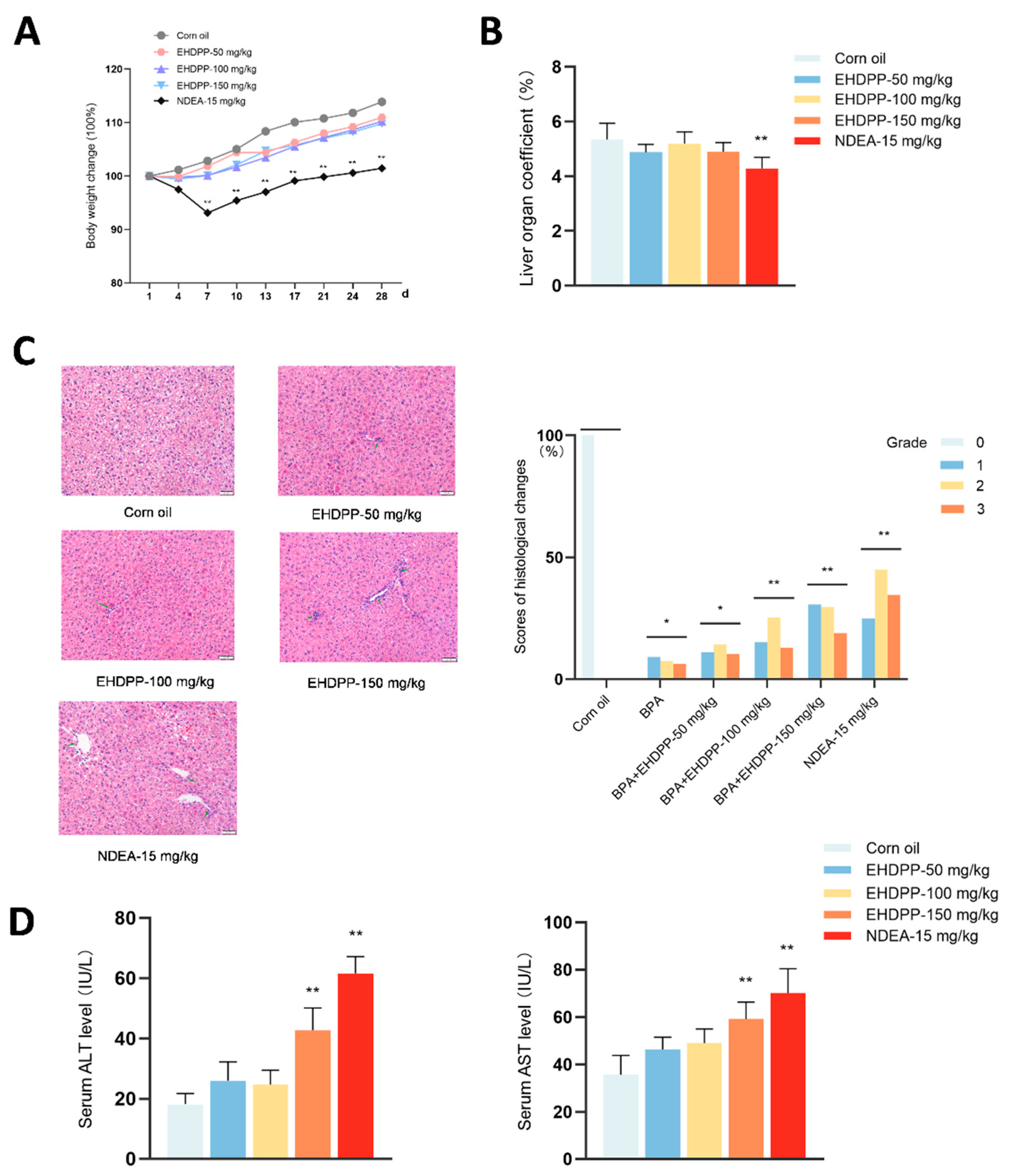
During the whole experimental duration, the bodyweights of mice in the solvent control group increased gradually, and a similar tendency in the groups with EHDPP at various doses (with no statistically significant difference at any time points from the solvent control); however, the bodyweights of mice exposed to NDEA (15 mg/kg/d), the positive control group, were decreased with statistical significance during 7 ~ 28 d as compared with the solvent control, implying a toxic effect of NDEA in the experimental animals (see Fig. 1, panel A). Likewise, the liver organ coefficients in mice exposed to EHDPP at various doses were not significantly different from that in the solvent control (p > 0.05), nevertheless, that in the positive control was significantly lower than in the solvent control, again indicative of some adverse effect on the liver (Fig. 1B). As shown in Fig. 1C, the hepatic histology in the solvent control was 100% in grade 0, while in the mice exposed to increasing doses of EHDPP, and 15 mg/kg/d of NDEA, there appeared increasing frequencies of grade 1, 2, and 3 histologic changes, with significant difference with the solvent control, which may imply some influences of NDEA and EHDPP on the hepatic microstructure. Finally, as shown in Fig. 1D, the serum ALT and AST activities (marker of hepatocyte damage) in mice exposed to 50 and 100 mg/kg/d of EHDPP were not different from those in the solvent control, indicating no significant liver damage with EHDPP at ≤ 100 mg/kg/d for 7 d. On the contrary, in the group with EHDPP at 150 mg/kg/d and NDEA at 15 mg/kg/d, both ALT and AST activities increased significantly (p < 0.01). By an integrated consideration of both histologic and serum aminotransferase activity changes, it is clear that both EHDPP at the highest test dose (150 mg/kg/d) and NDEA as a positive control demonstrated some degree of hepatotoxicity, while the histologic changes in lower doses of EHDPP might just represent minor or subclinical changes.
Figure 1.
Effect of EHDPP on the body weight, liver organ coefficient, activities of serum aminotransferases, and hepatic histologic structure. Six-week aged C57BL/6J mice were exposed to EHDPP by gastric gavage at the doses of 50, 100, and 150 mg/kg/d for 7 days, where corn oil was used as the vehicle, and NDEA (15 mg/kg/d, an indirect genotoxicant requiring metabolic activation primarily by Cyp2e1) served as a positive control. Experimental mice were randomly selected and distributed into various groups, with five mice in each group. The data of bodyweights (panel A) covered the whole experiment duration (28 d), while the liver organ coefficient (B), hepatic histological changes (C), and serum aminotransferase activities (D) were observed at 8 d (24 h within the termination of chemical exposure). Data were statistically analyzed using the one-way ANOVA (A, B, and D), except for the distribution of hepatic histology grades (C) which was statistically analyzed by a rank sum test; *p < 0.05, **p < 0.01, as compared with the solvent control. .
Figure 1.
Effect of EHDPP on the body weight, liver organ coefficient, activities of serum aminotransferases, and hepatic histologic structure. Six-week aged C57BL/6J mice were exposed to EHDPP by gastric gavage at the doses of 50, 100, and 150 mg/kg/d for 7 days, where corn oil was used as the vehicle, and NDEA (15 mg/kg/d, an indirect genotoxicant requiring metabolic activation primarily by Cyp2e1) served as a positive control. Experimental mice were randomly selected and distributed into various groups, with five mice in each group. The data of bodyweights (panel A) covered the whole experiment duration (28 d), while the liver organ coefficient (B), hepatic histological changes (C), and serum aminotransferase activities (D) were observed at 8 d (24 h within the termination of chemical exposure). Data were statistically analyzed using the one-way ANOVA (A, B, and D), except for the distribution of hepatic histology grades (C) which was statistically analyzed by a rank sum test; *p < 0.05, **p < 0.01, as compared with the solvent control. .
3.2. Induction of Hepatic DNA Damage by in Mice Orally Exposed to EHDPP
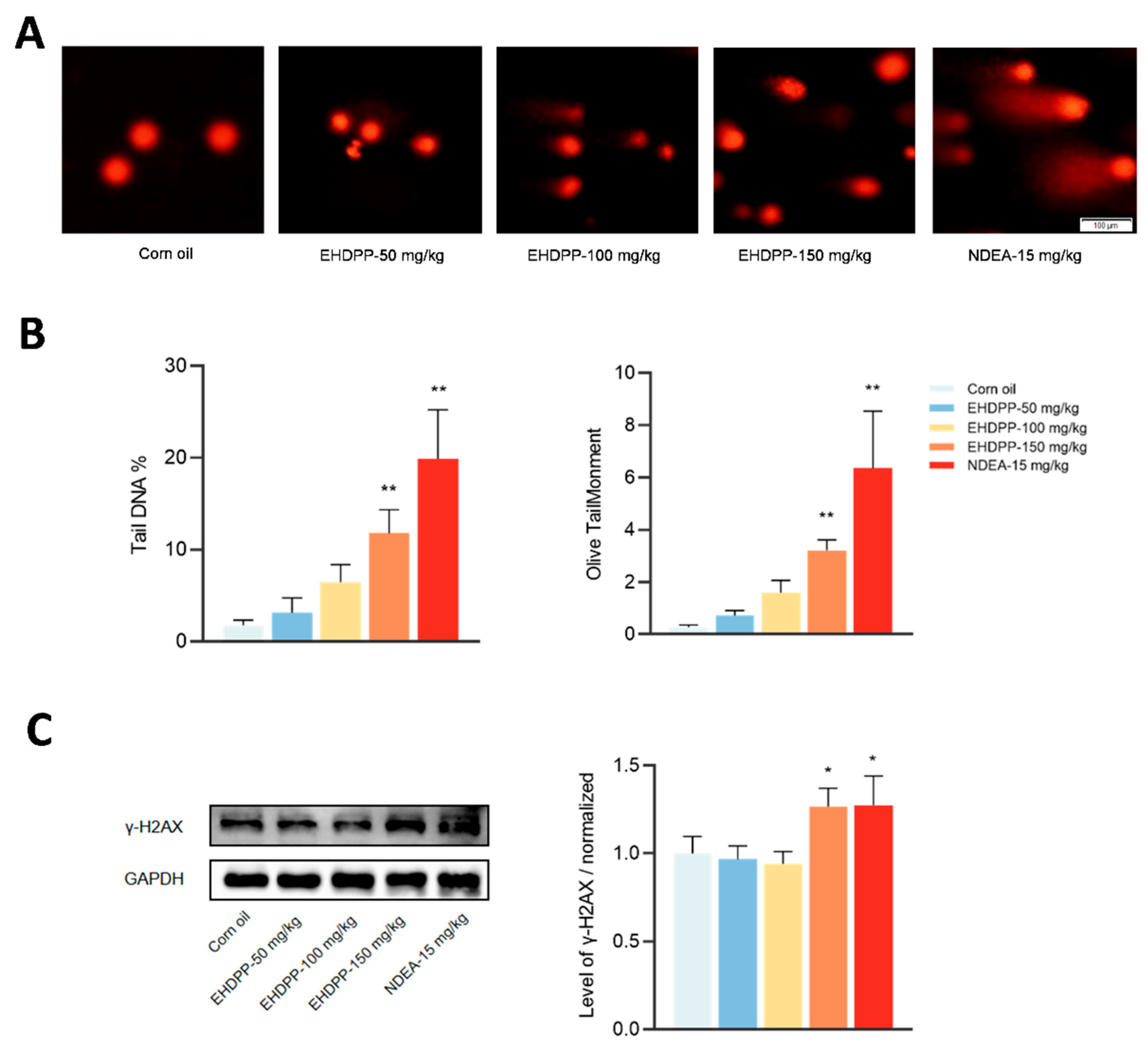
The DNA-damaging effect of test compounds were determined by using comet assay of hepatocytes isolated from each mouse and the level of hepatic γ-H2AX by Western blot assay. As shown in Fig. 2 (panels A & B), orally administered EHDPP at the highest dose (150 mg/kg/d) and NDEA (15 mg/kg/d, the positive control) increased both the Tail DNA % and the Olive TailMoment, indicating a DNA-breaking effect of EHDPP. Meanwhile, the values of both parameters in mice exposed to 50 and 100 mg/kg seemed also increasing from the levels in the solvent control, but with no statistical significance (p > 0.05). Likewise, the hepatic γ-H2AX levels in the groups with 150 mg/kg/d of EHDPP and NDEA (15 mg/kg/d) were statistically increased as compared with that in the solvent control, while in the groups with lower doses of EHDPP they were unchanged.
3.3. Results of Pig-a Gene Mutation Test in Mice Orally Exposed to EHDPP
The levels of CD-24-negative mutants were determined at three different time points, i.e., 0, 14, and 28 d. As shown in Fig. 3, the frequencies of mutants, in both the circulative erythrocytes (upper panel) and reticulocytes (lower panel), from mice orally exposed to NDEA at the dose of 15 mg/kg/d for 7 d were significantly increased as compared with those in the solvent controls (p < 0.01 in both cell types), which supports the validity of the experimental system. On the contrary, the frequencies of mutants at all the three time points in mice orally exposed to EHDPP at 50, 100, and 150 mg/kg/d for 7 d were not significantly different from the relevant controls. Clearly, under the current experimental circumstances EHDPP was incapable of inducing gene mutations at the CD-24 locus in mice.
3.4. Induction of Micronucleus in the Bone Marrow of Mice Orally Exposed to EHDPP
As indicated in Table 1, in mice orally exposed to EHDPP at 150 mg/kg/d for 7 d as well as the positive control (NDEA, 15 mg/kg/d for 7 d) the frequencies of micronucleus formation in bone marrow polychromatic erythrocytes were increased (p < 0.05 and p < 0.01, respectively); while in the groups administered with lower doses of EHDPP (50 and 100 mg/kg/d for 7 d) no significant changes in the frequencies of micronucleus formation were observed, though the absolute values did gradually increase along with the elevation of doses (from 0 to 100 mg/kg/d).

Table 1.
Induction of micronucleus formation in the bone marrow polychromatic erythrocytes (PCEs) in male mice exposed to EHDPP for 1 week.
Table 1.
Induction of micronucleus formation in the bone marrow polychromatic erythrocytes (PCEs) in male mice exposed to EHDPP for 1 week.
| Group | Dose (mg/kg/d) | Frequency of micronucleated PCEs (‰) |
| EHDPP | 0 (Corn oil) | 2.63 ± 0.63 |
| 50 | 3.25 ± 0.96 | |
| 100 | 3.40 ± 1.55 | |
| 150 | 4.40 ± 1.03* | |
| Positive control (NDEA) | 15 | 7.80 ± 1.18** |
In each group, 5 mice were involved, in each of which 2000 randomly encountered PCEs were evaluated microscopically at the magnification of 10,000 (under oil lens). Data are means ± S.D., *p < 0.05, **p < 0.01, as compared with the corn oil control using one-way ANOVA.
Figure 2.
Induction of hepatic DNA damage by EHDPP as indicated by a comet assay (A & B) and hepatic γ-H2AX elevation (C). See the legend of Fig. 1 for the chemical treatment regimen. On 8 d (within 24 h after termination of EHDPP exposure) fresh hepatocytes were isolated from each mouse, using which the comet tail moment and tail DNA content (%) (indicating DNA breaks) were measured; meanwhile, the protein level of hepatic γ-H2AX (its elevation marking double-strand DNA breaks) was determined by Western blot assay. Data are means ± S.D. (n = 5); *p < 0.05, **p < 0.01, by one-way ANOVA, as compared with the solvent control.
Figure 2.
Induction of hepatic DNA damage by EHDPP as indicated by a comet assay (A & B) and hepatic γ-H2AX elevation (C). See the legend of Fig. 1 for the chemical treatment regimen. On 8 d (within 24 h after termination of EHDPP exposure) fresh hepatocytes were isolated from each mouse, using which the comet tail moment and tail DNA content (%) (indicating DNA breaks) were measured; meanwhile, the protein level of hepatic γ-H2AX (its elevation marking double-strand DNA breaks) was determined by Western blot assay. Data are means ± S.D. (n = 5); *p < 0.05, **p < 0.01, by one-way ANOVA, as compared with the solvent control.
3.5. Effect of EHDPP on Hepatic Expression of AhR, PXR, Cyp1a1, Cyp1a2, Cyp2e1, and Cyp3a11
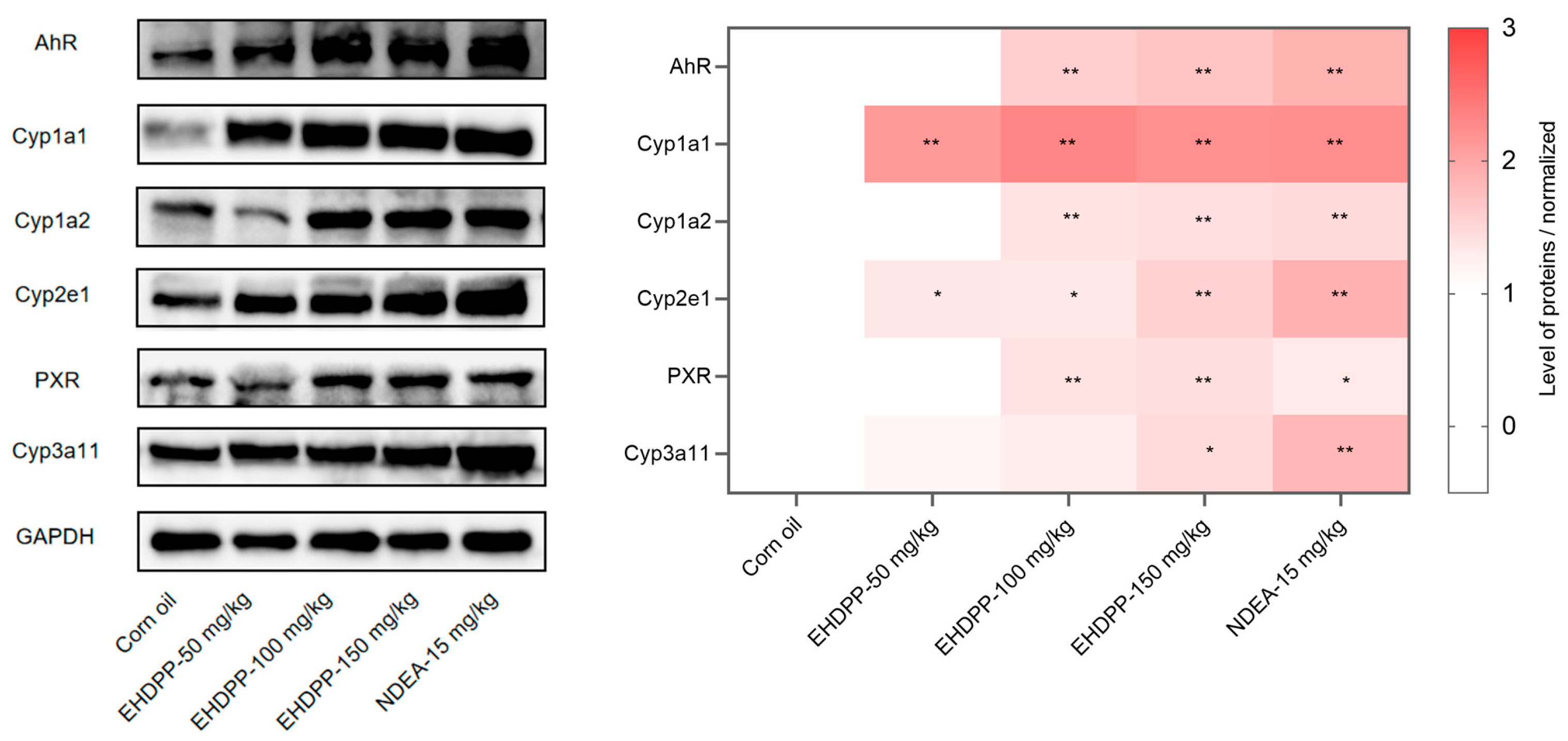
As shown in Fig. 4, in regard to the potency and intensity of EHDPP-induced changes in several hepatic proteins, (1) all the six proteins, i.e., AhR, PXR, Cyp1a1, Cyp1a2, Cyp2e1, and Cyp3a11, increased in expression levels; (2) among different hepatic proteins, the increase in Cyp1a1 protein expression was clearly most outstanding, followed by AhR, Cyp2e1, Cyp1a2, PXR and Cyp3a11 (in a potency/intensity decreasing order); (3) as a paralleling positive control group, NDEA (15 mg/kg/d) also induced Cyp1a1 most efficiently, in a Cyp1a1 > AhR/Cyp2e1/Cyp3a11 > Cyp1a2/PXR order.
Figure 3.
Results of Pig-a gene mutations in circulative normal erythrocytes (upper panel) and reticulocytes (lower panel) in mice orally exposed to EHDPP on 0 d, 14 d, and 28 d. See the legend of Fig. 1 for chemical exposure regimen. The cells isolated from the blood of each mouse experienced reaction with anti-CD24-APC and staining with SYTOTM13, then within 2 h of time 1×10⁶ cells per sample were scanned by flow cytometry, to differentiate CD24-negative from CD-24 positive erythrocytes and reticulocytes. The CD24-negative cells represent glucose-6-phosphate isomerase (GPI) defective mutants. Data are means ± S.D. (n = 3); *p < 0.05, **p < 0.01, by the Student’s t-test, as compared with the solvent control.
Figure 3.
Results of Pig-a gene mutations in circulative normal erythrocytes (upper panel) and reticulocytes (lower panel) in mice orally exposed to EHDPP on 0 d, 14 d, and 28 d. See the legend of Fig. 1 for chemical exposure regimen. The cells isolated from the blood of each mouse experienced reaction with anti-CD24-APC and staining with SYTOTM13, then within 2 h of time 1×10⁶ cells per sample were scanned by flow cytometry, to differentiate CD24-negative from CD-24 positive erythrocytes and reticulocytes. The CD24-negative cells represent glucose-6-phosphate isomerase (GPI) defective mutants. Data are means ± S.D. (n = 3); *p < 0.05, **p < 0.01, by the Student’s t-test, as compared with the solvent control.
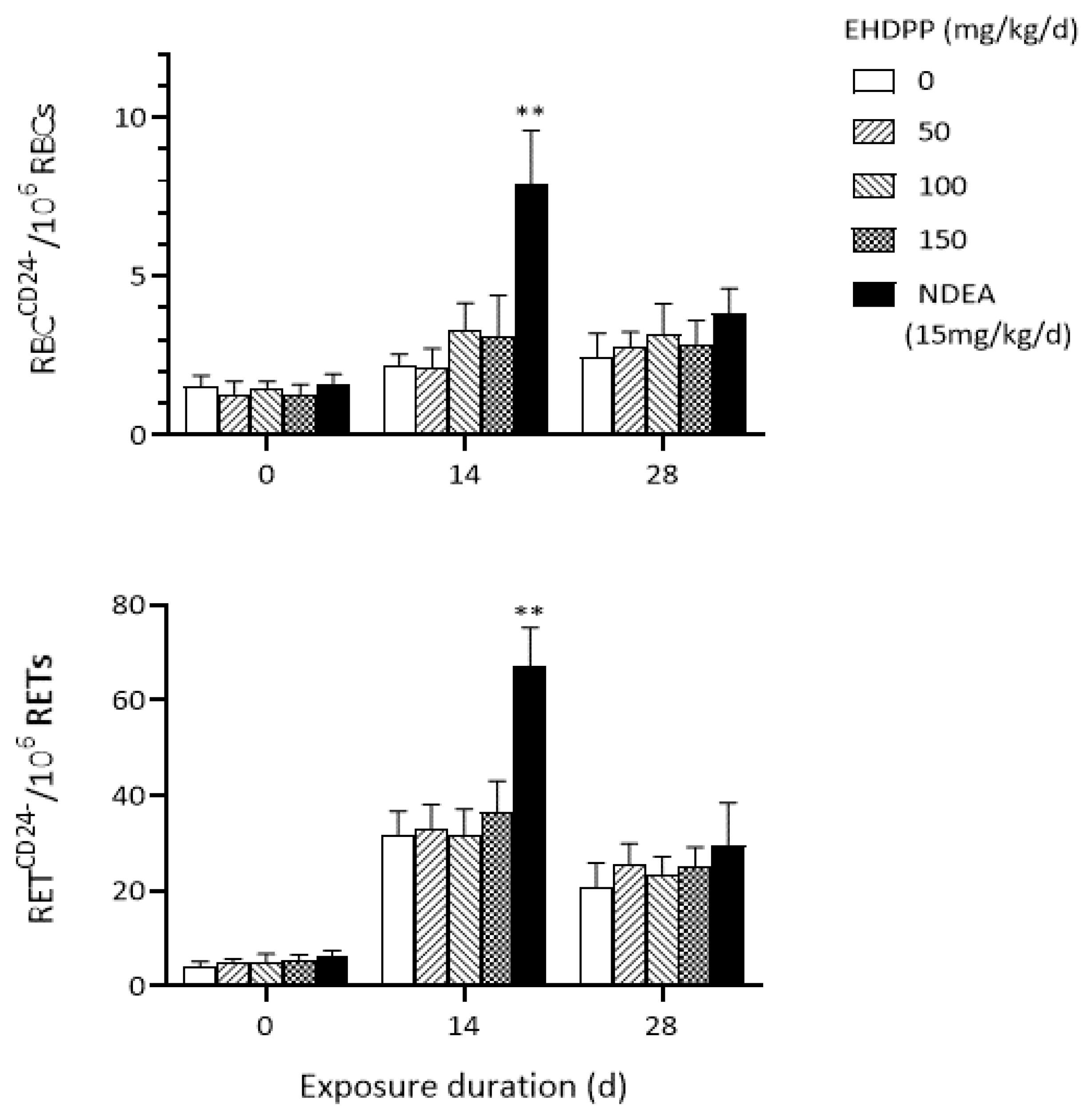
Figure 4.
Levels of hepatic AhR, PXR, Cyp1a1, Cyp1a2, Cyp2e1, and Cyp3a11 proteins (by Western blot assay) in mice orally exposed to DHDPP at the indicated doses for 7 d. See the legend of Fig. 1 for the chemical exposure regimen. From each mouse, a liver tissue sample was used for protein lysate preparation, then 30 μg of each protein lysate was loaded onto 10% SDS-PAGE. After separation of proteins by electrophoresis, the proteins in the gels were blotted onto PVDF membranes, which were then incubated with relevant primary antibodies. Next the membranes were incubated with appropriate secondary antibodies, and finally they were visualized by chemiluminescence. The protein bands of each target protein was semi-quantified for relative levels as referenced to GAPDH. Values are means ± S.D. (n = 5); compared with the solvent control, *p < 0.05, **p < 0.01, by one-way ANOVA.
Figure 4.
Levels of hepatic AhR, PXR, Cyp1a1, Cyp1a2, Cyp2e1, and Cyp3a11 proteins (by Western blot assay) in mice orally exposed to DHDPP at the indicated doses for 7 d. See the legend of Fig. 1 for the chemical exposure regimen. From each mouse, a liver tissue sample was used for protein lysate preparation, then 30 μg of each protein lysate was loaded onto 10% SDS-PAGE. After separation of proteins by electrophoresis, the proteins in the gels were blotted onto PVDF membranes, which were then incubated with relevant primary antibodies. Next the membranes were incubated with appropriate secondary antibodies, and finally they were visualized by chemiluminescence. The protein bands of each target protein was semi-quantified for relative levels as referenced to GAPDH. Values are means ± S.D. (n = 5); compared with the solvent control, *p < 0.05, **p < 0.01, by one-way ANOVA.
3.6. Cytotoxicity and Micronucleus Formation by EHDPP in Hepa1-6 Cells and the Influence of AhR/Cyp1a1 Modulators
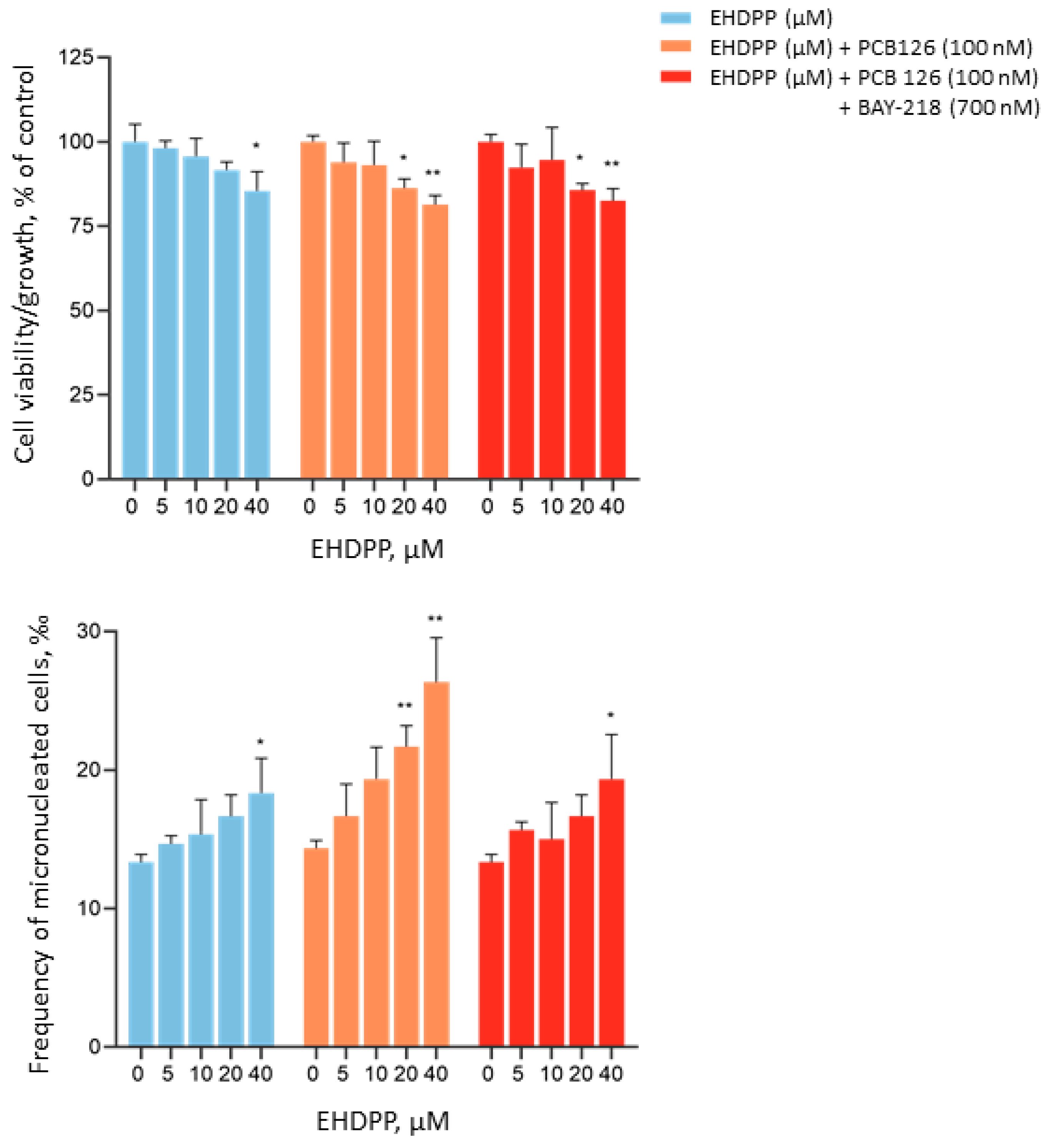
As indicated in Fig. 5 (upper panel), EHDPP alone did not change the cell viability/growth at concentrations from 5 to 20 μM, while at 40 μM a mild reduction in cell viability/growth occurred. In the presence of modulator PCB 126 (100 nM) and the combination of both PCB 126 and BAY-218 (700 nM), the threshold concentration of EHDPP for reduced cell viability/growth became a little lower, i.e., 20 μM, indicating mildly increased potency of cytotoxicity of EHDPP. However, the cytotoxicity of EHDPP with and without modulators stayed mild, as indicated by the relative cell viability/growth levels staying above 80%.
In the micronucleus test, EHDPP alone did not influence the frequency of micronucleated cells, except for the highest test concentration (40 μM) at which the frequency of micronucleated cells was increased with statistical significance (p < 0.05). With PCB 126 (100 nM) as a modulator exposing the cells from 18 h ahead of EHDPP exposure to the end of regimen, EHDPP increased the frequency of micronucleated cells along with the elevation of the concentration, which met statistical significance at 20 and 40 μM, in the presence of concentration dependence. However, further addition of a second modulator, BAY-218 (700 nM, with the same exposure regimen as PCB 126) led to alleviated micronucleus formation, as indicated by the increase of the threshold concentration back to 40 μM (indicating a reduction in the potency of effect).
Figure 5.
Cytotoxicity and micronucleus formation by EHDDP in Hepa1-6 cells and the impact of PCB 126 and BAY-218 as AhR/Cyp1a1 modulators. Hepa1-6 cells were exposed to PHT at the concentrations of 5, 10, 20, and 40 μM for 48 h, with 6 replicates in the CCK-8 assay, while duplicate experiments in the micronucleus test. PCB 126 (0.1 μM, potent activator of AhR and inducer of Cyp1a1) and BAY-218 (0.7 μM, antagonist of AhR and suppressor of Cyp1a1 expression) were present in the cultures from 18 h ahead of EHDPP exposure to the end of test regimen (66 h in total). Data are means ± S.D for the cell viability/growth values, with the one-way ANOVA being used for statistical analysis. The micronucleus test results are expressed as means ± 1/2 ranges of variation, which were converted to quantal data by combining the duplicate data, and then analyzed by using the χ2 examination. As compared with the solvent control, *p < 0.05, **p < 0.01.
Figure 5.
Cytotoxicity and micronucleus formation by EHDDP in Hepa1-6 cells and the impact of PCB 126 and BAY-218 as AhR/Cyp1a1 modulators. Hepa1-6 cells were exposed to PHT at the concentrations of 5, 10, 20, and 40 μM for 48 h, with 6 replicates in the CCK-8 assay, while duplicate experiments in the micronucleus test. PCB 126 (0.1 μM, potent activator of AhR and inducer of Cyp1a1) and BAY-218 (0.7 μM, antagonist of AhR and suppressor of Cyp1a1 expression) were present in the cultures from 18 h ahead of EHDPP exposure to the end of test regimen (66 h in total). Data are means ± S.D for the cell viability/growth values, with the one-way ANOVA being used for statistical analysis. The micronucleus test results are expressed as means ± 1/2 ranges of variation, which were converted to quantal data by combining the duplicate data, and then analyzed by using the χ2 examination. As compared with the solvent control, *p < 0.05, **p < 0.01.
4. Discussion
In this study, treatment of mice with EHDPP at 50 ~ 150 mg/kg/d doses for 7 d did not show obvious influence on the general condition, physical activities, bodyweight growing, or the liver organ coefficient, while a mild hepatotoxic change was observed with EHDPP at the highest test dose, 150 mg/kg/d, as indicated by both elevation of serum aminotransferase activities and histologic changes. Also at this particular dose of EHDPP, both hepatic DNA damage and bone marrow micronucleus formation were induced, though there appeared no Pig-a gene mutations in either circulative erythrocytes or reticulocytes. Compared with the potency of NDEA (15 mg/kg/d), which is commonly recognized as a strong genotoxicant, the effect of EHDPP seems moderate, about 10-fold weaker than NDEA.
Under the EHDPP exposure conditions in this study, the impact of EHDPP on the expression of several hepatic nuclear receptors and Cyp enzymes is featured by most obvious induction of Cyp1a1, and secondly that of Cyp2e1. There is no evidence showing an association of the expression/activation of AhR with the expression of Cyp2e1, however, AhR is an essential and transcriptional regulator of Cyp1a1 expression, and a moderate regulator of Cyp1a2 as well in both the mouse [27,28] and human [29,30]. Truly, in this study EHDPP induced the expression of Cyp1a2 less potently than that of Cyp1a1. PXR was also induced by EHDPP in our study, but of smaller intensity than for AhR. Cyp3a11 (and its human homologue CYP3A4) is downstream to PXR in a transcriptional regulation axis [31,32], and indeed hepatic Cyp3a11 in mice was induced by EHDPP, though only at the highest dose of EHDPP (150 mg/kg/d). In both human hepatocyte-derived cell models and in the liver of intact adult male C57BL/6J mice, PXRs were observed to inhibit the AhR/Cyp1a1 pathway and the relevant genotoxic effect of benzo(a)pyrene [33]. Interestingly, we have previously observed genotoxicity of EHDPP in mammalian cells, which depended on metabolic activation primarily by CYP3A4 and CYP2E1; the effect was almost blocked by ketoconazole, [20] an inhibitor of PXRs and the transcriptional targets, i.e., Cyp3a11 in mice [34] and CYP3A4 in humans [35]. Paradoxically, in this study induction of hepatic Cyp1a1 (and its upstream regulator AhR) by EHDPP apparently prevailed that of Cyp3a11 (and PXR). This prompted us to hypothesize that there might be major difference between the mouse and human in regard to the priority of AhR or PXR to be stimulated for the induction of Cyp1a1 or Cyp3a11 in metabolically activating EHDPP, i.e., possibly the AhR/Cyp1a1 is involved in activating EHDPP for its genotoxic effects.
To test the above hypothesis, we designed an in vitro micronucleus test using the Hepa1-6 cell line. Our results indicated that PCB 126 as an AhR activator/Cyp1a1 inducer (at its non-genotoxic concentration, 100 nM) [36] enhanced the genotoxicity of EHDPP, while this effect was blocked by further addition of BAY-218 (700 nM, a AhR antagonist/Cyp1a1 inhibitor) to the cultures. The results preliminarily indicate that Cyp1a1 might be involved in activating EHDPP to genotoxic metabolite(s). Nevertheless, this suggestion needs to be further clarified by continued, particularly in vivo studies.
It has been reported that EHDPP induced a series of toxicity in a mouse oocyte in vitro culture system, including disrupted oocyte maturation, cell cycle progression, cytoskeletal damage, and elevated levels of reactive oxygen species (ROS) [37]; however, whether these effects of EHDPP were attributed to the proto-compound, the metabolites, or the combination of both remains unclear. In 6-week old male CD-1 mice orally exposed to EHDPP at the doses of 1, 10, and 100 mg/kg/d for 6 weeks, prohibited cell proliferation, enhanced apoptosis and oxidative stress in the testes and spermatocytes were observed, and ROS production appeared to cause DNA damage and mitochondrial dysfunction in the spermatocytes [38]; again, the relationship of metabolism of EHDPP and its effects was not studied. To the best of our knowledge, the present study is the first evidence for EHDPP-induced genotoxicity at the endpoints of both DNA and chromosome damage in intact mammalian animals and the potential relevance of the effects to metabolic activation, primarily by Cyp1a1.
In regard to the hepatotoxicity of EHDPP, in a cultured human hepatocyte (L-02) model EHDPP disturbed the energy homeostasis and cell cycle, and caused endoplasmic reticulum stress, apoptosis, and inflammatory response [12]. In 7-day-old male chicken, decreased liver coefficient, microstructural and ultrastructural hepatocyte (plasma membranes and mitochondria) damage, and hepatic inflammation were observed 14 ~ 28 d after oral administration of EHDPP at the doses of 800 ~ 3200 mg/kg for once, which were partially attributed to EHDPP-induced oxidative stress [39]. The present study provides further evidence for the hepatotoxicity of EHDPP, particularly in a mammalian animal model.
The estimated human exposure of EHDPP is generally far lower than the doses applied in this study. For example, in an investigation of the OPFRs in Chinese foodstuffs and relevant estimation of dietary intake in a population in Jiangsu Province, China, EHDPP demonstrated to be the most abundant OPFR with a mean concentration of 1.12 ng/g wet weight in various food categories, and based on the general Chinese dietary habits and their consumption data the estimated dietary intakes (EDI) of EHDPP were 8.4 ng/kg/d (with the 95th percentile being 18.9 ng/kg.d), the reference doses (RfD) were 600 ng/kg/d, and the Hazard Quotient was 0.014 (95th percentile being 0.032) [40]. In a study performed in Portugal, estimated daily intakes of EHDPP from dust ingestion were averaged 1.5 ng/kg/d for adults while 18 ng/kg/d for children [41]. Taken together, the levels of daily exposure of humans to EHDPP should be of several orders lower than those in this study. This may imply that EHDPP alone is unlikely to induce hepatotoxicity or genotoxicity in humans. However, considering that there are numerous OPFRs other than EHDPP, as well as other kinds of organic pollutants [such as PBDEs, PCBs, and polycyclic aromatic hydrocarbons (PAHs)] which may expose human populations simultaneously [42,43], and the real exposure durations being much longer than the experimental time in this study, there might be combined and long-term effects, to which EHDPP may contribute to some extent.
In conclusion, this study provides evidence for the hepatoxicity and DNA- and chromosome-damaging effects of EHDPP in mammalian animals; meanwhile, EHDPP may predominantly induce hepatic Cyp1a1, Cyp2e1 and AhR, and the AhR/Cyp1a1 pathway might be involved in activating EHDPP for genotoxicity. The toxic effects of EHDPP in the real world, featured by several orders lower exposure levels, may be clarified by further investigation.
Author Contributions
Conceptualization, Y.L. and K.H.; methodology, H.G and Z.Z.; validation, Y.L. and Z.Z.; formal analysis, Z.Z., H.G, S.Z., and L.C.; data curation, Z.Z. and Y.L; writing—original draft preparation, Y.L. and Z.Z.; writing—review and editing, Y.L. and K.H.; supervision, Y.L.; project administration, Y.L.; funding acquisition, Y.L. All authors have read and agreed to the published version of the manuscript.
Funding
This research was supported by the Guangdong Provincial Basic and Applied Basic Research Foundation, China (Y.L., 2023A1515010970), and Talents’ Plan Foundation of Guangdong Second Provincial General Hospital (K. H, 2024F003).
Institutional Review Board Statement
The animal study protocol was approved by the Institutional Review Board (or Ethics Committee) of Southern Medical University (protocol code: SMUL202408036, date of approval: June 15, 2024).
Data Availability Statement
Data will be available upon request.
Acknowledgments
The authors are thankful to MS Manxin Chen and MS Haiting Sun for their excellent technical assistance.
Conflicts of Interest
The authors declare no conflicts of interest.
References
- Wu, Y.; Liu, S.; Dong, H.; Zhang, J.; Lu, R.; Yang, Q.; Jaman, R.; Zhu, J.; Zhang, C.; Zhou, J. Environmental occurrence, human exposure and multidimensional toxicity effects of 2-ethylhexyl diphenyl phosphate (EHDPP): Toxicological mechanism analysis and future research prospects. Ecotoxicol. Environ. Saf. 2026, 310, 119810. [Google Scholar] [CrossRef] [PubMed]
- Moreno, T.; Balasch, A.; Bartrolí, R.; Eljarrat, E. A new look at rubber recycling and recreational surfaces: the inorganic and OPE chemistry of vulcanised elastomers used in playgrounds and sports facilities. Sci. Total Environ. 2023, 868, 161648. [Google Scholar] [CrossRef] [PubMed]
- Shi, H.; Zhao, Y. Bringing the emerging organophosphate flame retardants (eOPFRs) into view: A hidden ecological and human health threat. Aquat. Toxicol. 2024, 267, 106833. [Google Scholar] [CrossRef] [PubMed]
- Tao, F.; Sjöström, Y.; de Wit, C.A.; Hagström, K.; Hagberg, J. Organohalogenated flame retardants and organophosphate esters from home and preschool dust in Sweden: Pollution characteristics, indoor sources and intake assessment. Sci. Total Environ. 2023, 896, 165198. [Google Scholar] [CrossRef] [PubMed]
- Gbadamosi, M.R.; Abdallah, M.A.; Harrad, S. A critical review of human exposure to organophosphate esters with a focus on dietary intake. Sci. Total Environ. 2021, 771, 144752. [Google Scholar] [CrossRef] [PubMed]
- Aminot, Y.; Tao, L.; Héas-Moisan, K.; Pollono, C.; O'Loghlin, M.; Munschy, C. Organophosphate esters (OPEs) in the marine environment: Spatial distribution and profiles in French coastal bivalves. Chemosphere 2023, 330, 138702. [Google Scholar] [CrossRef] [PubMed]
- You, J.; Chen, Z.-M.; Hou, X.-Y.; Guo, J.S.; Wang, C.-C.; Gao, J.M. Occurrence, potential sources and risks of organophosphate esters in the high-elevation region, Tibet, China. Sci. Total Environ. 2022, 806 Pt 3, 151348. [Google Scholar] [CrossRef] [PubMed]
- Chen, X.; Liang, X.; Yang, J.; Yuan, Y.; Xiao, Q.; Su, Z.; Chen, Y.; Lu, S.; Wang, L. High-resolution mass spectrometry-based screening and dietary intake assessment of organophosphate esters in foodstuffs from South China. Sci. Total Environ. 2023, 905, 167169. [Google Scholar] [CrossRef] [PubMed]
- Hou, M.; Shi, Y.; Jin, Q.; Cai, Y. Organophosphate esters and their metabolites in paired human whole blood, serum, and urine as biomarkers of exposure. Env. Int. 2020, 139, 105698. [Google Scholar] [CrossRef] [PubMed]
- Li, Y.; Zhao, M.; Shen, X.; Dong, K.; Zhao, F.; Li, R.; Wang, C.; Li, H. Adverse effects of aryl organophosphate esters on reproduction. Environ. Sci. Technol. 2025, 59, 10122–10135. [Google Scholar] [CrossRef]
- Li, Y.; Ma, H.; Chen, R.; Zhang, H.; Nakanishi, T.; Hu, J. Maternal transfer of 2-ethylhexyl diphenyl phosphate leads to developmental toxicity possibly by blocking the retinoic acid receptor and retinoic X receptor in Japanese medaka (Oryzias latipes). Environ. Sci. Technol. 2021, 55, 5056–5064. [Google Scholar] [CrossRef] [PubMed]
- Zhu, L.; Huang, X.; Li, Z.; Cao, G.; Zhu, X.; She, S.; Huang, T.; Lu, G. Evaluation of hepatotoxicity induced by 2-ethylhexyldiphenyl phosphate based on transcriptomics and its potential metabolism pathway in human hepatocytes. J. Hazard. Mater. 2021, 413, 125281. [Google Scholar] [CrossRef] [PubMed]
- Ding, K.; Bai, L.; Liu, W.; Cai, H.; Lv, M.; Huang, Y.; Chen, Y. Environmental flame retardant EHDPP stabilizes EGFR to accelerate lung cancer progression: Integrated network toxicology, bioinformatics, and in vitro evidence. Ecotoxicol. Environ. Saf. 2026, 311, 119854. [Google Scholar] [CrossRef] [PubMed]
- Liu, Y.; Li, Y.; Dong, S.; Han, L.; Guo, R.; Fu, Y.; Zhang, S.; Chen, J. The risk and impact of organophosphate esters on the development of female-specific cancers: Comparative analysis of patients with benign and malignant tumors. J. Hazard. Mater. 2021, 404 Pt B, 124020. [Google Scholar] [CrossRef] [PubMed]
- Yamanaka, K.; Hasegawa, A.; Sawamura, R.; Okada, S. DNA strand breaks in mammalian tissues induced by methylarsenics. Biol. Trace Elem. Res. 1989, 21, 413–417. [Google Scholar] [CrossRef] [PubMed]
- Downs, T.R.; Arlt, V.M.; Barnett, B.C.; Posgai, R.; Pfuhler, S. Effect of 2-acetylaminofluorene and its genotoxic metabolites on DNA adduct formation and DNA damage in 3D reconstructed human skin tissue models. Mutagenesis 2021, 36(1), 63–74. [Google Scholar] [CrossRef] [PubMed]
- Wang, Y.J.; Ho, Y.S.; Chu, S.W.; Lien, H.J.; Liu, T.H.; Lin, J.K. Induction of glutathione depletion, p53 protein accumulation and cellular transformation by tetrachlorohydroquinone, a toxic metabolite of pentachlorophenol. Chem. Biol. Interact. 1997, 105, 1–16. [Google Scholar] [CrossRef] [PubMed]
- Nishimaki-Mogami, T.; Minegishi, K.; Tanaka, A.; Sato, M. Isolation and identification of metabolites of 2-ethylhexyl diphenyl phosphate in rats. Arch. Toxicol. 1988, 61, 259–264. [Google Scholar] [CrossRef] [PubMed]
- Ya, M.; Yu, N.; Zhang, Y.; Su, H.; Tang, S.; Su, G. Biomonitoring of organophosphate triesters and diesters in human blood in Jiangsu Province, eastern China: Occurrences, associations, and suspect screening of novel metabolites. Environ. Int. 2019, 131, 105056. [Google Scholar] [CrossRef] [PubMed]
- Chen, Z.; Xie, J.; Li, Q.; Hu, K.; Yang, Z.; Yu, H.; Liu, Y. Human CYP enzyme-activated clastogenicity of 2-ethylhexyl diphenyl phosphate (a flame retardant) in mammalian cells. Environ. Pollut. 2021, 285, 117527. [Google Scholar] [CrossRef] [PubMed]
- Chen, M.; Sun, H.; Zhu, S.; Yu, H.; Liu, Y. Potent genotoxicity of phenytoin in adult male C57BL/6J mice, an effect requiring metabolic activation (possibly by Cyp2c enzymes). Chem. Biol. Interact. 2026, 431, 112017. [Google Scholar] [CrossRef] [PubMed]
- Xu, L.L.; Chen, Y.K.; Zhang, Q.Y.; Chen, L.J.; Zhang, K.K.; Li, J.H.; Liu, J.L.; Wang, Q.; Xie, X.L. Gestational exposure to GenX induces hepatic alterations by the gut-liver axis in maternal mice: A similar mechanism as PFOA. Sci. Total. Environ. 2022, 820, 153281. [Google Scholar] [CrossRef] [PubMed]
- Phonethepswath, S.; Avlasevich, S.L.; Torous, D.K.; Mereness, J.; Bemis, J.C.; Macgregor, J.T.; Dertinger, S.D. Flow cytometric analysis of Pig-a gene mutation and chromosomal damage induced by procarbazine hydrochloride in CD-1 mice. Environ. Mol. Mutagen. 2013, 54, 294–298. [Google Scholar] [CrossRef] [PubMed]
- Attia, S.M. The genotoxic and cytotoxic effects of nicotine in the mouse bone marrow. Mutat. Res. 2007, 632, 29–36. [Google Scholar] [CrossRef] [PubMed]
- Jiang, H.; Lai, Y.; Hu, K.; Chen, D.; Liu, B.; Liu, Y. Genotoxicity of 1-methylpyrene and 1-hydroxymethylpyrene in Chinese hamster V79-derived cells expressing both human CYP2E1 and SULT1A1. Environ. Mol. Mutagen. 2015, 56, 404–411. [Google Scholar] [CrossRef] [PubMed]
- Wang, Y.; Chen, Y.; Chen, Y.; Luo, W.; Liu, Y. Induction of clastogenesis and gene mutations by carbamazepine (at its therapeutically effective serum levels) in mammalian cells and the dependence on human CYP2B6 enzyme activity. Arch. Toxicol. 2023, 97, 1753–1764. [Google Scholar] [CrossRef] [PubMed]
- Zaher, H.; Yang, T.J.; Gelboin, H.V.; Fernandez-Salguero, P.; Gonzalez, F.J. Effect of phenobarbital on hepatic CYP1A1 and CYP1A2 in the Ahr-null mouse. Biochem. Pharmacol. 1998, 55, 235–238. [Google Scholar] [CrossRef] [PubMed]
- Ryu, D.Y.; Levi, P.E.; Hodgson, E. Regulation of hepatic CYP1A isozymes by piperonyl butoxide and acenaphthylene in the mouse. Chem. Biol. Interact. 1997, 105, 53–63. [Google Scholar] [CrossRef] [PubMed]
- Zhang, R.; Sun, J.; Ma, L.; Wu, X.; Pan, G.; Hao, H.; Zhou, F.; Jiye, A.; Liu, C.; Ai, H.; Shang, L.; Gao, H.; Peng, Y.; Wan, P.; Wu, H.; Wang, G. Induction of cytochromes P450 1A1 and 1A2 by tanshinones in human HepG2 hepatoma cell line. Toxicol. Appl. Pharmacol. 2011, 252, 18–27. [Google Scholar] [CrossRef] [PubMed]
- Padberg, F.; Hering, H.; Luch, A.; Zellmer, S. Indirect co-cultivation of HepG2 with differentiated THP-1 cells induces AHR signalling and release of pro-inflammatory cytokines. Toxicol. Vitr. 2020, 68, 104957. [Google Scholar] [CrossRef] [PubMed]
- Down, M.J.; Arkle, S.; Mills, J.J. Regulation and induction of CYP3A11, CYP3A13 and CYP3A25 in C57BL/6J mouse liver. Arch. Biochem. Biophys. 2007, 457, 105–10. [Google Scholar] [CrossRef] [PubMed]
- Li, Y.; Ross-Viola, J.S.; Shay, N.F.; Moore, D.D.; Ricketts, M.L. Human CYP3A4 and murine Cyp3A11 are regulated by equol and genistein via the pregnane X receptor in a species-specific manner. J. Nutr. 2009, 139, 898–904. [Google Scholar] [CrossRef] [PubMed]
- Cui, H.; Gu, X.; Chen, J.; Xie, Y.; Ke, S.; Wu, J.; Golovko, A.; Morpurgo, B.; Yan, C.; Phillips, T.D.; Xie, W.; Luo, J.; Zhou, Z.; Tian, Y. Pregnane X receptor regulates the AhR/Cyp1A1 pathway and protects liver cells from benzo-[α]-pyrene-induced DNA damage. Toxicol. Lett. 2017, 275, 67–76. [Google Scholar] [CrossRef] [PubMed]
- Huang, H.; Wang, H.; Sinz, M.; Zoeckler, M.; Staudinger, J.; Redinbo, M.R.; Teotico, D.G.; Locker, J.; Kalpana, G.V.; Mani, S. Inhibition of drug metabolism by blocking the activation of nuclear receptors by ketoconazole. Oncogene 2007, 26, 258–268. [Google Scholar] [CrossRef] [PubMed]
- Lim, Y.P.; Kuo, S.C.; Lai, M.L.; Huang, J.D. Inhibition of CYP3A4 expression by ketoconazole is mediated by the disruption of pregnane X receptor, steroid receptor coactivator-1, and hepatocyte nuclear factor 4alpha interaction. Pharmacogenet. Genom. 2009, 19, 11–24. [Google Scholar] [CrossRef] [PubMed]
- Yu, H.; Song, M.; Hu, K.; Wang, Y.; Fan, R.; Yang, Z.; Glatt, H.; Braeuning, A.; Liu, Y. Influence of bisphenol compounds at nanomolar concentrations on chromosome damage induced by metabolically activated carcinogens in HepG2 cells. Environ. Sci. Technol. 2021, 55, 10001–10011. [Google Scholar] [CrossRef] [PubMed]
- Zhang, Y.; Meng, F.; Zhao, T.; Du, J.; Li, N.; Qiao, X.; Yao, Y.; Wu, D.; Peng, F.; Wang, D.; Yang, S.; Shi, J.; Liu, R.; Zhou, W.; Li, L.; Hao, A. Melatonin improves mouse oocyte quality from 2-ethylhexyl diphenyl phosphate-induced toxicity by enhancing mitochondrial function. Ecotoxicol. Environ. Saf. 2024, 280, 116559. [Google Scholar] [CrossRef] [PubMed]
- Wang, Y.; Guo, A.L.; Xu, Y.; Xu, X.; Yang, L.; Yang, Y.; Chao, L. EHDPP induces proliferation inhibition and apoptosis to spermatocyte: Insights from transcriptomic and metabolomic profiles. Ecotoxicol. Environ. Saf. 2024, 284, 116878. [Google Scholar] [CrossRef] [PubMed]
- Yang, Y.; Wang, X.; Zhang, H.; Li, J.; Chen, J.; Yu, M.; Li, G.; Zhang, R.; Ge, M. Oxidative stress and ferroptosis involved in 2-ethylhexyl diphenyl phosphate -induced hepatotoxicity in chicken. Chem. Biol. Interact. 2022, 368, 110216. [Google Scholar] [CrossRef] [PubMed]
- Zhao, L.; Jian, K.; Su, H.; Zhang, Y.; Li, J.; Letcher, R.J.; Su, G. Organophosphate esters (OPEs) in Chinese foodstuffs: Dietary intake estimation via a market basket method, and suspect screening using high-resolution mass spectrometry. Environ. Int. 2019, 128, 343–352. [Google Scholar] [CrossRef] [PubMed]
- Coelho, S.D.; Sousa, A.C.A.; Isobe, T.; Kim, J.W.; Kunisue, T.; Nogueira, A.J.A.; Tanabe, S. Brominated, chlorinated and phosphate organic contaminants in house dust from Portugal. Sci. Total Environ. 2016, 569-570, 442–449. [Google Scholar] [CrossRef] [PubMed]
- Travis, S.C.; Aga, D.S.; Queirolo, E.I.; Olson, J.R.; Daleiro, M.; Kordas, K. Catching flame retardants and pesticides in silicone wristbands: Evidence of exposure to current and legacy pollutants in Uruguayan children. Sci. Total Environ. 2020, 740, 140136. [Google Scholar] [CrossRef] [PubMed]
- Kou, J.; Li, X.; Zhang, M.; Wang, L.; Hu, L.; Liu, X.; Mei, S.; Xu, G. Accumulative levels, temporal and spatial distribution of common chemical pollutants in the blood of Chinese adults. Environ. Pollut. 2022, 311, 119980. [Google Scholar] [CrossRef] [PubMed]
Disclaimer/Publisher’s Note: The statements, opinions and data contained in all publications are solely those of the individual author(s) and contributor(s) and not of MDPI and/or the editor(s). MDPI and/or the editor(s) disclaim responsibility for any injury to people or property resulting from any ideas, methods, instructions or products referred to in the content. |
© 2026 by the authors. Licensee MDPI, Basel, Switzerland. This article is an open access article distributed under the terms and conditions of the Creative Commons Attribution (CC BY) license (http://creativecommons.org/licenses/by/4.0/).
Copyright: This open access article is published under a Creative Commons CC BY 4.0 license, which permit the free download, distribution, and reuse, provided that the author and preprint are cited in any reuse.


