
Submitted:
30 June 2026
Posted:
01 July 2026
You are already at the latest version
Abstract
Pure and glycine-doped strontium formate dihydrate [Sr(HCOO)₂·2H₂O] single crystals were grown by slow evaporation using glycine concentrations of 0.005 – 0.1 M. The influence of glycine incorporation on structural, vibrational, electronic, and antimicrobial properties was studied experimentally and using DFT (B3LYP/6-311G**). The HOMO – LUMO gap (7.111 eV) indicates stability, while the hyperpolarizability (6973.787 a.u.) suggests potential nonlinear optical behavior. FT-IR and FT-Raman analyses show characteristic modes with slight shifts due to glycine–lattice interactions. Doped crystals exhibit enhanced antimicrobial activity, with a maximum inhibition zone of 36 mm against Escherichia coli at 0.1 M concentration. Results indicate that glycine affects the electronic and biological properties of strontium formate dihydrate, suggesting potential optical and antimicrobial applications.
Keywords:
strontium formate dihydrate
; glycine doping
; density functional theory
; FT-IR spectroscopy
; frontier molecular orbitals
; hyperpolarizability
; antimicrobial activity
; nonlinear optical materials
1. Introduction
Strontium formate dihydrate (SFD), with the chemical formula [Sr(HCOO)₂·2H₂O], is an intriguing crystalline material that crystallizes in the orthorhombic crystal system and belongs to the space group P2₁2₁2₁, containing four formula units per unit cell. The crystal and molecular structure of (SFD) was initially determined by Caligne [1], who reported the lattice parameters as a = 7.332 Å, b = 12.040 Å, and c = 7.144 Å. Subsequent studies by Greena et al. [2,3,4,5] have examined the influence of transition-metal doping-specifically with Cu, Ni, Zn and Mg - on the structural, optical and biocidal properties of (SFD). More recently, Muthupoongodi et al. [6,7] demonstrated that glycine doping significantly enhances the functional properties of (SFD), making it a promising material for photonics and electronic device applications. Furthermore, their thermal studies revealed that pure (SFD) crystals exhibit high thermal stability up to approximately 72 °C and contain less than two molecules of water of crystallization, with an estimated value of 1.66 molecules.
The molecular conformation and frontier molecular orbital (FMO) characteristics are crucial for understanding the functional properties and potential applications of molecular systems; however, obtaining such information through experimental techniques is often challenging. These data can instead be reliably derived using density functional theory (DFT), which explains the electronic structure of atoms, molecules, and materials based on their three-dimensional electron density distribution. DFT is widely regarded as a powerful computational tool for predicting vibrational spectra, molecular geometries, and electronic properties with high accuracy [8].
In recent years, there has been increasing interest in exploring organic and semi-organic materials for nonlinear optical (NLO) applications, primarily due to their large second-order electric susceptibilities, which are directly associated with the first-order hyperpolarizability. Among these, amino acids have attracted considerable attention owing to their crucial role in biochemical processes, particularly in influencing the structural and functional properties of biomolecules such as metaloproteins. Research on amino acid-doped semi-organic crystals has expanded significantly due to their therapeutic importance in applications such as radiopharmaceuticals, chemotherapy, and the treatment of metal toxicity [9,10,11,12,13]. The donor atoms present in amino acids, primarily oxygen (O) and nitrogen (N), from their chelating groups, exert opposite electronic effects [14], which make them highly relevant in biochemistry and pharmacology due to their potential antimicrobial, antineoplastic, and anticancer activities [15,16,17,18,38,39,40].
Although several studies have investigated transition-metal and amino acid-doped strontium formate dihydrate crystals, systematic studies correlating glycine incorporation with the electronic structure, vibrational behavior and antimicrobial activity of the crystal remain limited. In particular, the influence of glycine incorporation on charge distribution, frontier molecular orbitals, nonlinear optical response and biological activity has not been comprehensively explored using combined experimental and DFT approaches. Glycine was selected as a dopant due to its simple molecular structure, biocompatibility, zwitterionic nature and the presence of amino and carboxyl functional groups capable of interacting with the host crystal lattice through hydrogen bonding and electrostatic interactions.
Greena et al. [3] demonstrated the antimicrobial properties of metal-doped (SFD) crystals using Mg, Cu, Ni and Zn ions. However, the antimicrobial potential of amino acid-doped (SFD) crystals has not yet been explored and investigated. Since and then, we have synthesized glycine-doped (SFD) single crystals and aimed to explore their antimicrobial efficacy against selected pathogens. Interestingly, we examined the influence of glycine concentration as well as the incorporation on the molecular geometry, frontier molecular orbitals, electronic properties and antimicrobial properties of strontium formate dihydrate crystals using combined spectroscopic and theoretical investigations.
2. Experimental Section
2.1. Methodology
2.2. Growth of Pure and Glycine-Doped Strontium Formate Dihydrate Crystals
Analytical reagent (AR) grade strontium carbonate (SrCO₃), formic acid (HCOOH) and glycine (NH₂CH₂COOH) with purity greater than 99% were used without further purification. Pure strontium formate dihydrate crystals were synthesized by slowly adding stoichiometric amounts of strontium carbonate to aqueous formic acid under continuous stirring at room temperature according to the reaction:
SrCO₃ + HCOOH → Sr(HCOO)₂ + CO₂ + H₂O
The obtained solution was filtered using Whatman filter paper to remove insoluble impurities and allowed to evaporate slowly at room temperature (30 ± 2 °C). For glycine-doped crystals, glycine was added to the supersaturated solution at concentrations of 0.005 M, 0.05 M and 0.1 M under constant stirring for 3 h to ensure homogeneous mixing. The prepared solutions were filtered and kept undisturbed for slow evaporation. Transparent crystals suitable for characterization were harvested after approximately 10 – 15 days.
2.3. Evaluation of Antimicrobial Activities
The antibacterial and antifungal activities of the synthesized pure and glycine-doped (SFD) single crystals were evaluated using the disc diffusion method. The biological screening was performed by dissolving the test crystals in dimethylformamide (DMF), which served as the solvent for preparing the required concentrations. Fresh microbial suspensions were prepared from 24-hour cultures of bacteria and fungi using sterile peptone water and adjusted to a turbidity equivalent to 0.5 McFarland standard to ensure uniform inoculum density.
Muller-Hinton agar plates (90 mm Petri dishes) were inoculated evenly using the prepared microbial suspensions. Sterile paper discs (6 mm in diameter) were impregnated with 10 μL of the respective test solutions and carefully placed in a circular arrangement on each inoculated plate. Discs loaded with DMF alone were used as negative controls to ensure solvent compatibility. Control experiments confirmed that the concentration of DMF used in the assay did not exhibit any inhibitory effect on microbial growth, validating its suitability for the antimicrobial and antifungal activity tests.
Antimicrobial experiments were performed in triplicate, and the obtained inhibition zone values are presented as mean ± standard deviation. Statistical significance between pure and doped samples was evaluated using one-way ANOVA with p < 0.05 considered statistically significant.
3. Results and Discussion
3.1. Optimized Molecular Structure
The structural optimizations of the (SFD) model systems were carried out using the Gaussian 09 package to determine the lowest-energy conformations. To ensure that the obtained geometries correspond to true minima on the potential energy surface, vibrational frequency analyses were performed. The fully optimized molecular structure of (SFD), along with the corresponding atom numbering scheme, is depicted in Figure 1.
The calculated bond lengths and bond angles are summarized in Table 1, and the obtained structural parameters show good agreement with the previously reported literature values [1].
The optimized molecular geometry also corresponds well with the X-ray crystallographic data of (SFD). In the optimized structure, the Sr atom is coordinated by two water molecules and two formate groups, with all four Sr–O bonds exhibiting nearly identical lengths in the DFT analysis (~2.45 Å). However, in the reported crystal structure [1], the two water molecules appear to interact more weakly with the Sr center compared to the formate groups. This indicates the presence of slight structural defects associated with the water molecules in the crystalline phase. Furthermore, the analysis suggests that the actual crystal contains fewer than two fully coordinated water molecules, which is consistent with the earlier findings reported in the literature [6].
3.2. Frontier Molecular Orbitals (HOMO - LUMO)
The frontier molecular orbitals (HOMO and LUMO) play a crucial role in understanding the chemical reactivity and stability of a molecule. The energy of the (HOMO) is directly associated with the ionization potential, while the (LUMO) energy corresponds to the electron affinity [22,23]. The energy difference between (HOMO) and (LUMO) is widely used as an indicator of kinetic stability and chemical reactivity [24,25]. A larger (HOMO – LUMO) energy gap generally signifies higher kinetic stability and lower chemical reactivity, as it becomes energetically unfavorable to transfer electrons from the low-lying (HOMO) to the high-lying (LUMO) [23]. Variations in the (HOMO – LUMO) gap caused by interaction with noble metals, non-metallic species, semiconductor materials, or other molecular systems can lead to significant changes in charge transfer characteristics, peak intensities, and spectral positions. For (SFD), the calculated (HOMO – LUMO) energy separation is 7.111 eV, and the graphical representation of its frontier molecular orbitals is provided in Figure 2.
3.3. Molecular Electrostatic Potential
The molecular electrostatic potential (MEP) is a powerful tool for visualizing and understanding the distribution of electronic charge on the molecular surface, thereby providing insights into the relative polarity of different regions (positive and negative) [26,27]. It is particularly useful for predicting reactive sites, understanding molecular interactions with the surrounding environment, and studying biological recognition processes. Mapping the electrostatic potential onto an iso-electron density surface offers a clear visual representation of the polarity distribution [28]. On the (MEP) surface, different colors indicate varying electrostatic potential values: red corresponds to regions of highest negative potential, blue represents areas of highest positive potential, and green indicates regions of near-zero potential.
The distinction between nucleophilic and electrophilic regions directly influences the molecule’s ability to donate or accept electrons [29]. The total electron density surface mapped with the electrostatic potential surface (EPS) for the (SFD) crystal is illustrated in Figure 3(a). Regions of negative potential, highlighted in red and yellow, correspond to electron-rich areas that attract protons, whereas positive potential regions, shown in blue, represent electron-deficient zones where protons experience repulsion due to low electron density and incomplete shielding of nuclear charges.
The electrostatic potential contour map, representing both positive and negative potentials, is shown in Figure 3(c). Dense regions indicate slightly electron-rich zones, while sparsely colored areas represent regions of nearly zero potential. As depicted in Figure 3(a), the area around the oxygen atoms in the formate group (HCOO-) exhibits the most negative potential (red) with a minimum value of −0.102 a.u. In contrast, the strontium (Sr) atom shows the highest positive potential (blue) with a maximum value of +0.102 a.u.; Notably, the intense positive region is primarily localized on the strontium center and hydrogen atoms, which is attributed to their bonding with highly electronegative oxygen atoms.
3.4. Mulliken Atomic Charges
The atomic charges play a significant role in determining several molecular properties, including dipole moment and polarizability. The Mulliken population analysis [30] was used to calculate the atomic charges of (SFD), and the results are summarized in Table 2(a). The calculated values reveal that hydrogen atoms 12H, 13H, 14H, and 15H carry comparatively higher positive charges than hydrogen atoms 10H and 11H. This difference arises due to the influence of nearby electronegative oxygen atoms. In contrast, hydrogen atoms 10H and 11H are bonded to carbon atoms 2C and 3C, which are themselves connected to two highly electronegative oxygen atoms each. As a result, the positive charge on these two hydrogen atoms is relatively reduced, although they still retain a slight positive nature.
Similarly, the carbon atoms 2C and 3C exhibit lower positive charges due to their direct bonding with the electronegative oxygen atoms 4O, 5O, 6O, and 7O. Consequently, the hydrogen atoms 10H and 11H, which are attached to these carbon centers, possess lower atomic charges (0.199 and 0.199) compared to other hydrogen atoms in the molecule. Furthermore, all oxygen atoms in (SFD) are found to carry negative charges, confirming their role as electron-donor sites. The relatively large calculated dipole moment of (SFD) (2.058 Debye) suggests the presence of strong intermolecular attractions, primarily governed by dipole – dipole interactions and van der Waals forces within the crystal lattice. The detailed crystallographic parameters obtained from this analysis are presented in Table 2(b).
3.5. Vibrational Spectra
The DFT-simulated FT-infrared (FT-IR) and (FT-Raman) spectra of (SFD) obtained in the present study were compared with the experimental spectra reported in the literature [6], as shown in Figure 4(a) & (b). The assignment of vibrational bands in the solid-state (FT-IR) and (FT-Raman) spectra was carried out using theoretically predicted frequencies and further validated by comparison with previously reported experimental data [6]. Overall, the calculated vibrational frequencies show excellent agreement with the experimental observations Figure 4(a).
The water molecule in the (SFD) crystal exhibits C₂V symmetry, with the normal modes 2a₁ and b₂ being active in both the IR and Raman spectra. The stretching vibrations associated with free H₂O molecules generally appear in the region 3000 - 3800 cm⁻¹ [30]. In the experimental FT-IR spectrum, a broad absorption band is observed in the range 3000 - 3500 cm⁻¹, corresponding to the presence of water molecules within the (SFD) crystal [6]. In contrast, the theoretical spectrum predicts the H₂O stretching modes in the higher wavenumber range of 3500 - 3800 cm⁻¹. The upward shift of these sharp and intense bands in the simulated spectrum can be attributed to strong intermolecular hydrogen bonding between the water molecules and the formate anion. Furthermore, the intensity of these peaks in the theoretical spectrum suggests the presence of two water molecules within the optimized structure.
The Raman spectrum calculated using DFT methods is presented in Figure 4(b). However, certain vibrational bands could not be directly correlated between the experimental and theoretical spectra. This discrepancy arises from the fact that the DFT calculations were performed in the gas phase, which does not fully account for the intermolecular interactions present in the real crystal. Consequently, some vibrational modes associated with hydrogen bonding between water molecules in the crystal, particularly those expected in the 2000 - 2700 cm⁻¹ region, are absent in the theoretical spectrum but appear in the experimental results. On the other hand, water-related vibrational modes are still observed in the theoretical spectrum within the 2700 - 3200 cm⁻¹ range.
Additionally, the theoretical analysis reveals a few stretching vibrations in the 1000 - 500 cm⁻¹ region that are not detected experimentally [6], while previous reports indicate that water molecule vibrations are expected around 500 - 400 cm⁻¹ [31]. The low-frequency region below 200 cm⁻¹ in the Raman spectrum corresponds to lattice vibrations of the crystal.
The correlation between theoretical and experimental vibrational spectra demonstrates that the optimized molecular structure obtained from DFT calculations adequately reproduces the principal vibrational features of the crystal system. Minor deviations in peak positions arise primarily from the fact that theoretical calculations were performed in the gas phase, whereas the experimental spectra correspond to the solid crystalline state where intermolecular hydrogen bonding and lattice interactions significantly influence vibrational behavior. In particular, the broadening and shifting of O-H stretching bands in the experimental spectra indicate strong hydrogen-bond interactions involving water molecules and glycine functional groups within the crystal environment.
Overall, the comparative analysis of experimental and simulated FT-IR and FT-Raman spectra suggests a slight reduction in the number of water molecules within the crystal lattice. The findings from this study indicate that the hydrous strontium formate crystal likely contains fewer than two fully coordinated water molecules.
3.6. Nonlinear Optical Properties
The theoretical evaluation of molecular polarizability and hyperpolarizability, along with their interpretation in terms of structure–property relationships, has become an area of significant research interest due to its crucial role in the design of novel materials for nonlinear optical (NLO) applications and in understanding the interaction between electromagnetic fields and matter [32].
In the present study, the first-order hyperpolarizability (β) and total dipole moment (μ) of (SFD) were computed using the B3LYP functional combined with the 6-311G** basis set, a widely adopted approach for predicting NLO properties. The calculated static dipole moment (μ) and mean first-order hyperpolarizability (β₀) were obtained from the x-, y-, and z- components of the corresponding tensors, as defined by the following relations:
µx = 0.0006, µy = 0.0017 and µz = 2.0578
The dipole moment (μ) was calculated using Gaussian 09 software and it is found to be
µ = 2.0578 Debye. The first order hyperpolarizability (β) was also calculated were determined using the finite field approach theory. The components of first order hyperpolarizability can be obtained using the following equation (Equ. 2):
βi = βijk + ⅓ ∑(βijj + βjij + βjji), (I ≠j)
Using the x, y and z components, and the magnitude of the first hyperpolarizibility tensor can be calculated. The output from Gaussian 09 provides 10 components of this matrix as βxxx, βyxx, βxyy, βyyy, βzxx, βxyz, βzyy, βxzz, βyzz, βzzz, respectively. The components of the first hyperpolarizability can be calculated using the following equations (Equ. 3):
= + +
= + + ,
The above tensor values are presented in Table 3.
The first order hyperpolarizability of the crystal is calculated and it is found to be 6973.787 a.u.; The calculated hyperpolarizability of the crystal is 6973.7 a.u. times that of the standard (NLO) material urea (0.13 × 10⁻³⁰ e.s.u.) [32]. We concluded that the title crystal and its derivatives are potential molecules for future studies of nonlinear optical applications.
3.7. Antimicrobial Activity
3.7.1. Determination of (MIC)
The in vitro antimicrobial activity of the as-grown pure and glycine-doped (SFD) single crystals was evaluated against four bacterial strains: Escherichia coli, Staphylococcus aureus, Klebsiella pneumoniae, and Pseudomonas aeruginosa. Both standard reference drugs and the test samples were dissolved in dimethylformamide (DMF) to obtain a final concentration of 100 μg/mL. The minimum inhibitory concentration (MIC) was determined using the broth microdilution method [33]. Serial dilutions of the test samples and standard compounds were prepared using nutrient broth for bacterial strains and Sabouraud dextrose broth for fungal strains [34]. The inoculated samples were incubated under controlled conditions at 37 °C for 24 h for bacterial cultures and at 25 °C for 48 h for fungal cultures. The (MIC) values were recorded as the lowest concentration of the test compound capable of completely inhibiting microbial growth.
3.7.2. Antibacterial Activity
The glycine-doped (SFD) single crystals synthesized with varying concentrations of glycine (0.005 M, 0.05 M, and 0.1 M) exhibited significant antimicrobial activity against Escherichia coli, Staphylococcus aureus, Klebsiella pneumoniae, and Pseudomonas aeruginosa. The minimum inhibitory concentration (MIC) values obtained for the tested crystals are presented in Table 4.
Among the samples, the crystals doped with a higher concentration of glycine demonstrated enhanced antibacterial efficiency. This improvement can be attributed to the enrichment of heteroatoms such as nitrogen (N) and oxygen (O) within the crystal lattice, which facilitate strong chelating interactions with microbial components. Although all doped crystals exhibited moderate antimicrobial activity, the (SFD) crystals with a higher glycine content showed comparatively better inhibition than undoped (SFD). The significant enhancement in antimicrobial performance is likely due to the presence of amino (-NH₂) and carboxylic (-COOH) functional groups in glycine, which may induce specific biochemical transformation mechanisms within microbial systems.
These functional groups can improve cell permeability by interacting with the lipid membrane, thereby facilitating the transport of bioactive components. Since the lipid membrane preferentially allows the passage of lipid-soluble compounds, lipophilicity plays a critical role in controlling antimicrobial efficiency. However, despite the improved activity of glycine-doped (SFD) crystals, their antimicrobial efficacy remained lower than that of standard drugs, as illustrated in Figure 5.
The observed antimicrobial behavior can be correlated with two key factors: (i) The incorporation of heteroatoms such as O and N enhances the solubility of the crystals (both hydrophilic and hydrophobic) and promotes interactions with trace elements present in microbial cells, thereby inhibiting their growth; (ii) The mode of action involves the ability of the carboxylic acid and amino groups within the doped crystals to form hydrogen bonds with counter-ions, solvent molecules, and active centers of cellular constituents. This interaction disrupts essential cellular metabolism, ultimately leading to microbial growth inhibition [35].
3.7.3. Antifungal Activity
To explore the potential biological applications of glycine-doped (SFD) crystals in the field of bioinorganic chemistry, their antifungal activity was evaluated using the disk diffusion method. Pure (SFD) and glycine-doped strontium formate dihydrate crystals with different glycine concentrations were tested in vitro against five pathogenic fungal strains: Aspergillus niger, Fusarium solani, Curvularia lunata, Rhizoctonia bataticola, and Candida albicans. The results of the antifungal studies are summarized in Table 5, which clearly indicate that the glycine-doped (SFD) crystals exhibit greater antifungal efficacy compared to pure (SFD) crystals against all tested fungal species.
The enhancement in antifungal performance can be attributed to the ability of glycine to interfere with the replication process of fungal cells by blocking metabolically active sites, thereby suppressing their growth. This improved activity may also be explained by an increase in the lipophilic nature of the doped crystals, which facilitates stronger interactions between the glycine molecules and the lipid components of the fungal cell membrane. Such interactions are likely to disrupt membrane permeability and impair cellular respiration, ultimately leading to significant disturbances in normal cellular processes and effective inhibition of fungal proliferation.
The enhanced antimicrobial activity observed for glycine-doped strontium formate dihydrate crystals may be attributed to several synergistic factors. The incorporation of glycine introduces additional amino (-NH₂) and carboxyl (-COOH) functional groups capable of interacting with microbial cell membranes through hydrogen bonding and electrostatic interactions. These interactions can alter membrane permeability, resulting in leakage of intracellular components and disruption of essential metabolic pathways. Furthermore, the presence of oxygen-rich formate groups and polar glycine moieties may facilitate stronger interactions with negatively charged bacterial cell surfaces, particularly in Gram-negative bacteria. The increased polarity and surface activity of the doped crystals may also enhance reactive oxygen species generation and interfere with enzymatic processes required for microbial survival.
The improved antifungal activity may similarly arise from disruption of membrane integrity and inhibition of fungal respiration pathways. However, detailed mechanistic investigations involving membrane permeability assays, ROS measurements and electron microscopy are required to fully establish the antimicrobial mechanism of the glycine-doped crystals.
4. Conclusions
DFT calculations (B3LYP/6-311G**) were used to study the structural and electronic properties of strontium formate dihydrate. HOMO–LUMO analysis indicates a stable electronic structure with moderate charge transfer characteristics. The experimental FT-IR and FT-Raman spectra were well supported by theoretical vibrational calculations. Minor deviations are attributed to intermolecular hydrogen bonding effects in the solid state, which are not fully represented in gas-phase calculations. The calculated first-order hyperpolarizability (β = 6973.787 a.u.) suggests a strong nonlinear optical response compared to standard materials such as urea, indicating potential for (NLO) applications. Antimicrobial studies show that glycine-doped crystals exhibit improved antibacterial and antifungal activity compared to the pure sample. This enhancement is likely due to the presence of amino and carboxyl groups that promote interactions with microbial cell membranes. However, the mechanism of glycine incorporation and its structural influence cannot be conclusively confirmed in the absence of advanced crystallographic and surface analyses such as XRD refinement and XPS. Overall, the results indicate that glycine modification influences both electronic and biological properties of strontium formate dihydrate, making it a promising candidate for further investigation in optical and bioactive material applications.
Funding
Board of Research in Nuclear Science - Department of Atomic Energy (BRNS-DAE), No: 2013/34/1/BRNS/No.0486.
Institutional Review Board Statement
Not applicable.
Informed Consent Statement
Not applicable.
Data Availability
Data will be made available on request.
Acknowledgments
The authors (Dr. J. Angel Mary Greena & Dr. S. Muthupoongodi) gratefully acknowledge the financial support received from the Board of Research in Nuclear Science - Department of Atomic Energy (BRNS-DAE), Mumbai, India, with Sanction No: 2013/34/1/BRNS/No.0486 to carry out this research work.
Declaration of Competing Interest
The authors declare that they have no known competing financial or personal interests. .
References
- Caligene, P.J.L. Affinement de la structure cristalline du formiate de strontium dihydraté, Sr(HCOO)₂·2H₂O. Acta Crystallogr. B 1971, 27, 2429–2431. [Google Scholar]
- Greena, J.A.M.; Shajan, X.S.; Kumaresan, S. Growth and characterization of strontium formate dihydrate [Sr(HCOO)₂·2H₂O] single crystals doped strongly with glycine. Int.J. Mater.Sci. 2010, 5, 209. [Google Scholar]
- Greena, J.A.M.; Karuppasam, K.; Antony, R.; Shajan, X.S.; Kumaresan, S. Growth and characterization of strontium formate dihydrate single crystals. Der Chem. Sin. 2012, 3(5), 1229–1238. [Google Scholar]
- Greena, J.A.M.; Karuppasamy, K.; Antony, R.; Shajan, X.S.; Kumaresan, S. Growth and characterization of semi-organic nonlinear optical crystal: strontium formate dihydrate doped with alanine. Chem.Sci.Trans. 2013, 2(1), 141–146. [Google Scholar]
- Greena, J.A.M.; Karuppasamy, K.; Antony, R.; Shajan, X.S.; Kumaresan, S. Synthesis and characterization of strontium formate dihydrate single crystals doped with urea. IOSR J. Appl. Phys. 2013, 1(4), 25–28. [Google Scholar]
- Muthupoongodi, S.; Manickam, S.T.D.; Mahadevan, C.K.; Greena, J.A.M.; Balakumar, S.; Shajan, X.S. Growth and characterization of pure and L-alanine doped strontium formate dihydrate single crystals. J. Cryst. Growth 2015, 428, 46–53. [Google Scholar] [CrossRef]
- Muthupoongodi, S.; Manickam, S.T.D.; Mahadevan, C.K.; Greena, J.A.M.; Balakumar, S.; Shajan, X.S. Investigation on the third-order nonlinear optical properties of strontium formate dihydrate single crystals. Optik 2016, 127, 4320–4323. [Google Scholar] [CrossRef]
- Li, R.; Lu, H.; Zhang, X.; Liu, P.; Chen, L.; Chen, J.; Zhao, B. Experimental and theoretical studies on the molecular structure and vibrational spectra of 3,5-diamino-1,2,4-triazole. Spectrochim. Acta A 2015, 148, 369–374. [Google Scholar]
- Shi, M.; Cai, Q.; Yao, L.; Mao, Y.; Ming, Y.; Ouyang, G. The effect of curcumin on antigen-induced activation of mast cells. Cell Biol. Int. 2006, 30, 221–226. [Google Scholar] [CrossRef] [PubMed]
- Canadas, M.; Torres, E.L.; Aris, A.M.; Mendrila, M.A.; Sevilla, M.T. Spectroscopic and electrochemical study of the interaction between metal ions and the antioxidant curcumin. Polyhedron 2000, 19, 2059–2064. [Google Scholar]
- Rosenberg, B.; Van Camp, L.; Trosko, J.E.; Mansour, V.H. Platinum compounds: a new class of potent antitumour agents. Nature 1969, 222, 385–386. [Google Scholar] [CrossRef] [PubMed]
- Cummings, S.D. Vibrational spectroscopy of platinum group metal complexes. Coord. Chem. Rev. 2009, 253, 449–478. [Google Scholar] [CrossRef]
- Adams, B.K.; Ferstl, E.M.; Davis, M.C.; Herold, M.; Kurtkaya, S.; Camalier, R.F.; Hollingshead, M.G.; Kaur, G.; Sausville, E.A.; Rickles, F.R.; Snyder, J.P.; Liotta, D.C.; Shoji, M. PTK787/ZK 222584, A novel and potent inhibitor of vascular endothelial growth factor receptor tyrosine kinases. Bioorg. Med. Chem. 2004, 12, 3871–3883. [Google Scholar] [PubMed]
- Jakupec, M.A.; Galanski, M.; Keppler, B.K. Antitumour metal compounds: more than 30 years of academic research. Rev. Physiol. Biochem. Pharmacol. 2003, 146, 1–53. [Google Scholar] [PubMed]
- Yang, F.; Lim, G.P.; Begum, A.N.; Ubeda, O.J.; Simmons, M.R.; Ambegaokar, S.S.; Chen, P.P.; Kayed, R.; Glabe, C.G.; Frautschy, S.A.; Cole, G.M. Curcumin inhibits formation of amyloid β oligomers and fibrils. J.Biol.Chem. 2005, 280, 5892–5901. [Google Scholar] [CrossRef] [PubMed]
- Shahabadi, N.; Falsafi, M.; Moghadam, N.H. Interaction of a water-soluble cobalt(III) Schiff base complex with DNA. J. Photochem. 2013, 122, 45–51. [Google Scholar] [CrossRef]
- Gescher, A. Curcumin: the story so far, J. Chemother. 2004, 4, 3–6. [Google Scholar]
- Vajragupta, O.; Boonchoong, P.; Morris, G.M.; Olson, A. Discovery of curcumin derivatives as inhibitors of HIV-1 integrase. Bioorg. Med. Chem. Lett. 2005, 15, 3364–3368. [Google Scholar] [CrossRef] [PubMed]
- Frisch, M.J.; Trucks, G.W.; Schlegel, H.B.; et al. Gaussian 09; Gaussian Inc.: Wallingford, CT, 2009. [Google Scholar]
- Becke, A.D. Density-functional thermochemistry III: the role of exact exchange. J. Chem. Phys. 1993, 98, 5648–5652. [Google Scholar] [CrossRef]
- Benassi, R.; Ferrari, E.; Lazzari, S.; Spagnolo, F.; Saladini, M. Experimental and theoretical study on the structure and properties of curcumin. J.Mol.Struct. 2008, 892, 168–176. [Google Scholar] [CrossRef]
- Fukui, K. The role of frontier orbitals in chemical reactions. Science 1982, 218, 474–754. [Google Scholar] [CrossRef]
- Gunasekaran, S.; Balaji, R.A.; Kumaresan, S.; Anand, G.; Srinivasan, S. Vibrational spectra and normal coordinate analysis of L-threonine. Can. J. Anal. Sci. Spectrosc. 2008, 53, 149–162. [Google Scholar]
- Kim, K.H.; Han, Y.K.; Jung, J. Theoretical study of the mechanism for the reaction of O(³P) with CH₃CH₂Cl. Org. Chem. Acc. 2005, 113, 233. [Google Scholar]
- Aihara, J. Aromaticity of cyclopropenyl cation and related systems. J.Phys.Chem. A 1999, 103, 7487. [Google Scholar]
- Reed, A.E.; Weinhold, F. Natural atomic orbitals and natural populations. J.Chem.Phys. 1985, 83, 1736. [Google Scholar] [CrossRef]
- Reed, A.E.; Weinstock, R.B.; Weinhold, F. Natural bond orbital analysis of near-Hartree–Fock water dimer. J.Chem.Phys. 1985, 83, 735. [Google Scholar] [CrossRef]
- Panickar, C.Y.; Varghese, H.T.; Nayak, P.S.; Narayana, B.; Saojini, B.K.; Fun, H.K.; War, J.A.; Srivastava, S.K.; Alsenory, C.V. Vibrational spectroscopic and NBO studies of semicarbazide derivative. Spectrochim. Acta A 2015, 148, 18–28. [Google Scholar]
- Mulliken, R.S. Electronic population analysis on LCAO-MO molecular wave functions. J.Chem.Phys. 1955, 23, 1833–1840. [Google Scholar] [CrossRef]
- Arjunan, V.; Kalaivani, M.; Marchewka, M.K.; Mohan, S. Vibrational spectra and thermodynamic analysis of 1-bromo-4-nitrobenzene. J.Mol.Struct. 2013, 1045, 160–170. [Google Scholar] [CrossRef]
- Thangarasu, S.; Suresh Kumar, S.; Athimoolam, S.; Sridhar, B.; Asath Bahadur, S.; Shanmugam, R.; Thamarai Chelvam, A. Nonlinear optical properties of conjugated molecules. J.Mol.Struct. 2004, 892, 168–176. [Google Scholar]
- Adant, M.; Dupuis, I.; Brédas, J.L. Ab initio study of nonlinear optical properties of urea. Int.J.Quantum Chem. 2004, 56, 497–507. [Google Scholar]
- Sonmez, M.; Celebi, M.; Berber, I. Synthesis, characterization and antimicrobial activity of metal complexes. Eur.J.Med.Chem. 2010, 45, 1935–1940. [Google Scholar] [PubMed]
- Senthil Kumar, R.; Arunachalam, S. DNA binding and cleavage studies of ruthenium(II) complexes. Polyhedron 2007, 26, 3255–3262. [Google Scholar] [CrossRef]
- Negma, N.A.; Zakia, M.F.; Salem, M.A.I. Cationic surfactants based on fatty acids and their biological activity. Colloids Surf. B Biointerfaces 2010, 77, 96. [Google Scholar]
- Chandrasekar, T.; Pravin, N.; Raman, N. DNA interaction and antimicrobial studies of metal complexes. J.Mol.Struct. 2015, 1081, 477–485. [Google Scholar]
- Chandrasekar, T.; Pravin, N.; Raman, N. Bio-relevant metal complexes of a pyrazolone-based Schiff base. Inorg. Chem. Commun. 2014, 43, 45–50. [Google Scholar] [CrossRef]
- Ayoob, M.M.; Hawaiz, F.E. Synthesis, crystal structure, DFT calculation and Hirshfeld surface analysis of N-(4-methyl phenyl)-2-(3-nitro-benzamido) benzamide. Bull.Chem.Soc. Ethiop. 2024, 38(1), 229–239. [Google Scholar]
- Mohammed, S.; Ibrahim, M.; Hussen, T. Single crystal X-ray, spectroscopic characterization and DFT study of a Schiff base compound. Bull.Chem.Soc.Ethiop. 2022, 36, 487–498. [Google Scholar]
- Tadesse, A.; Gebremariam, T. Spectroscopic, crystallographic and DFT investigation of an organic molecular crystal. Bull.Chem.Soc.Ethiop. 2019, 33, 411–420. [Google Scholar]
Figure 1.
The optimized molecular structure of (SFD) crystal with atom numbering scheme.
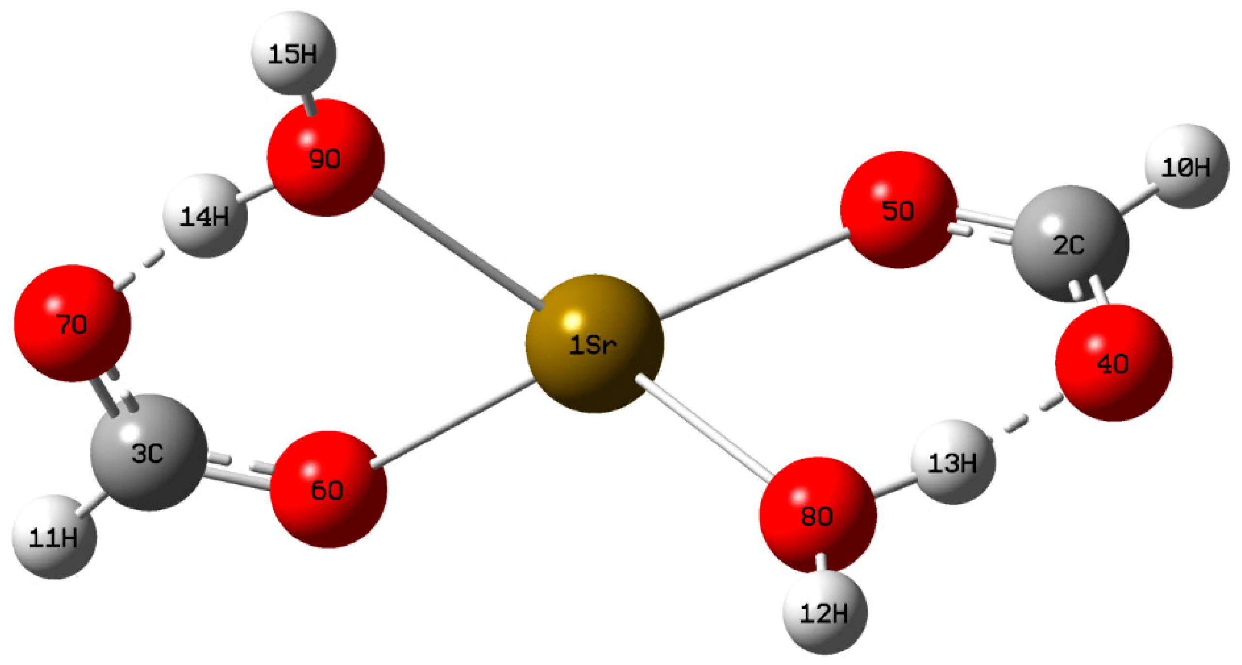
Figure 2.
The pictorial representation of frontier molecular orbitals of (SFD). ELUMO = 0.326, Egap = 7.111 eV, EHOMO = 7.437 eV.
Figure 2.
The pictorial representation of frontier molecular orbitals of (SFD). ELUMO = 0.326, Egap = 7.111 eV, EHOMO = 7.437 eV.
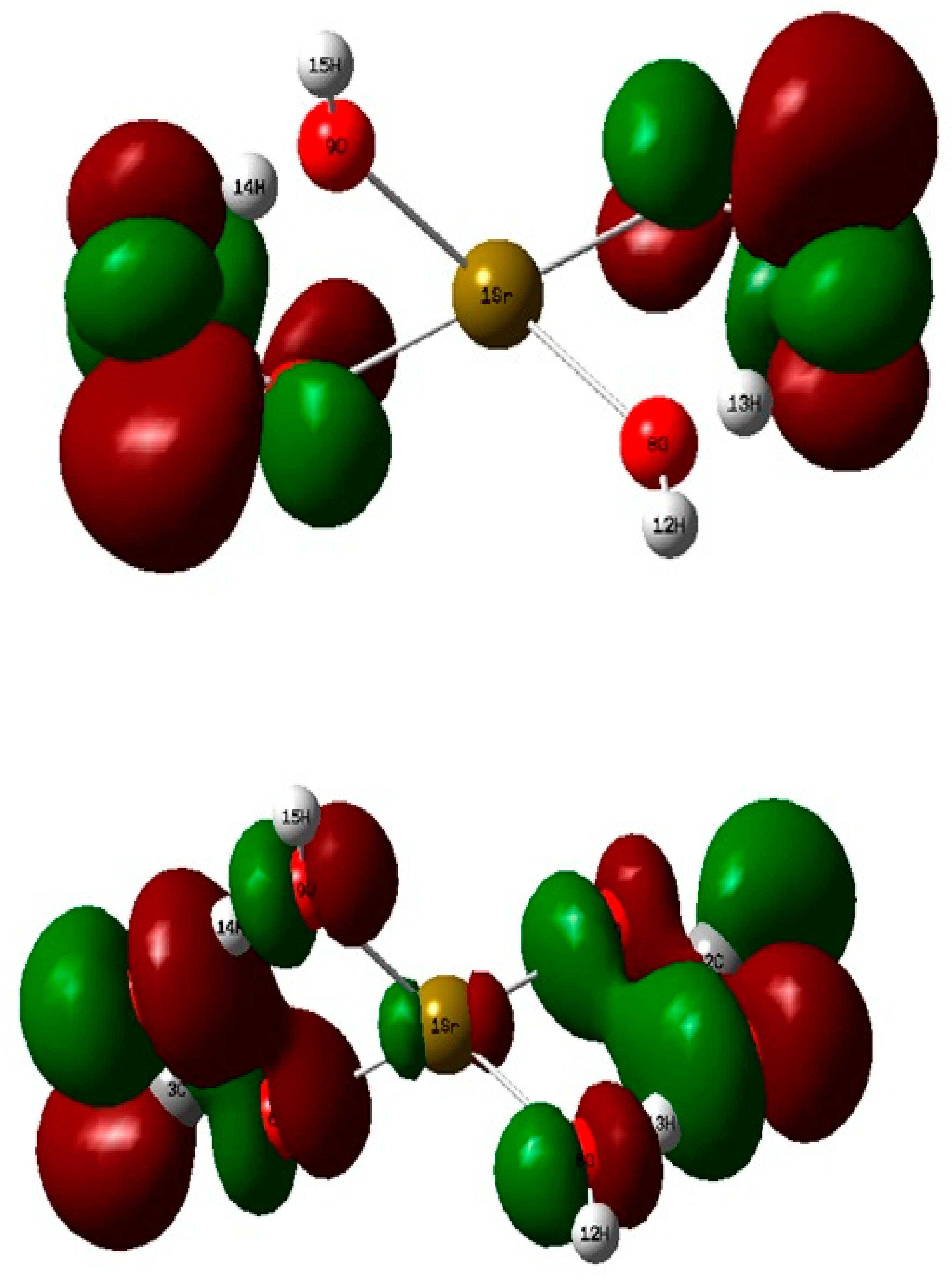
Figure 3.
(a) (MEP) transparent diagram, (b) The total electron density surface mapped with (EPS) of solid (SFD) crystal, (c) The contour map of electrostatic potential of (SFD) crystal.
Figure 3.
(a) (MEP) transparent diagram, (b) The total electron density surface mapped with (EPS) of solid (SFD) crystal, (c) The contour map of electrostatic potential of (SFD) crystal.
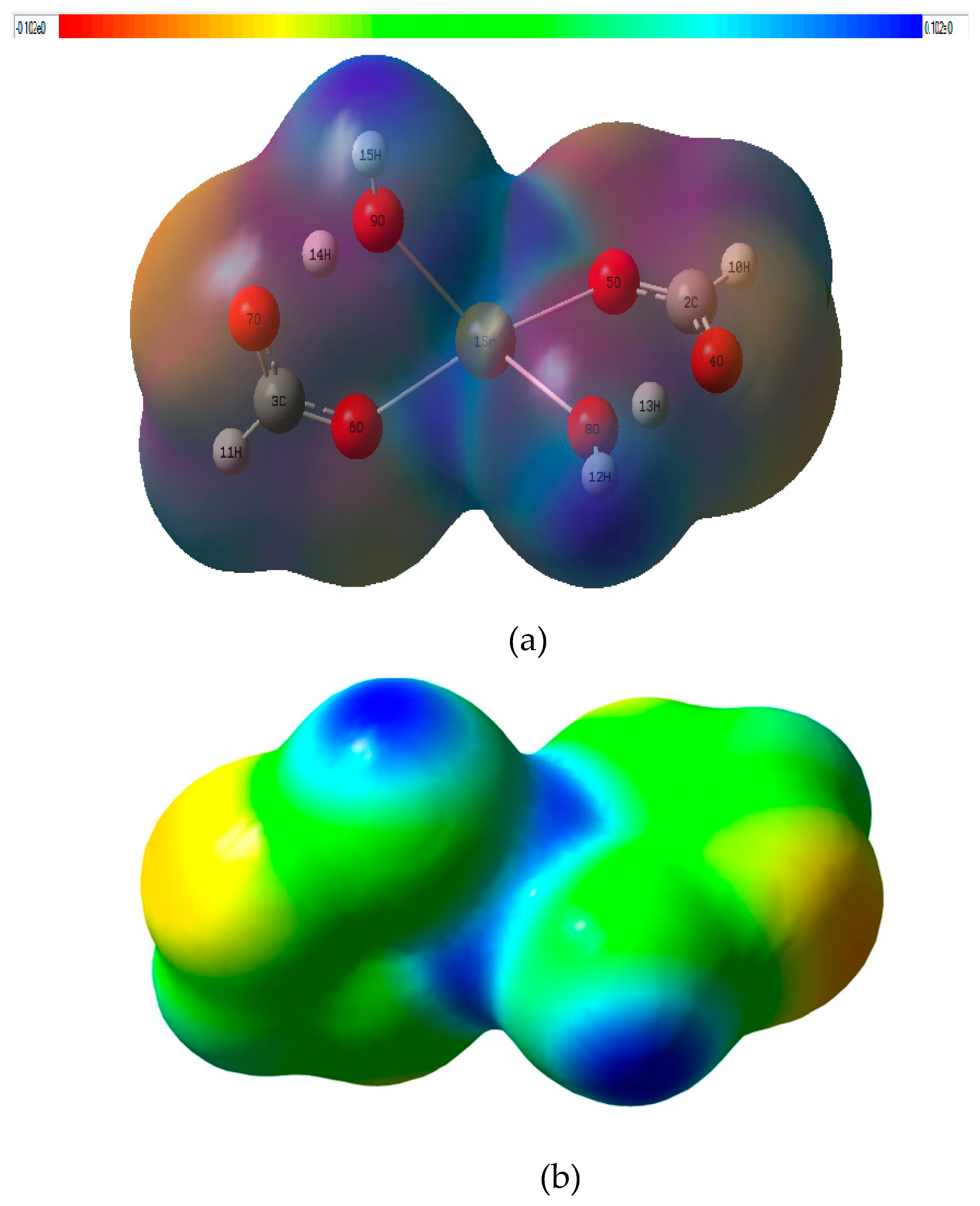
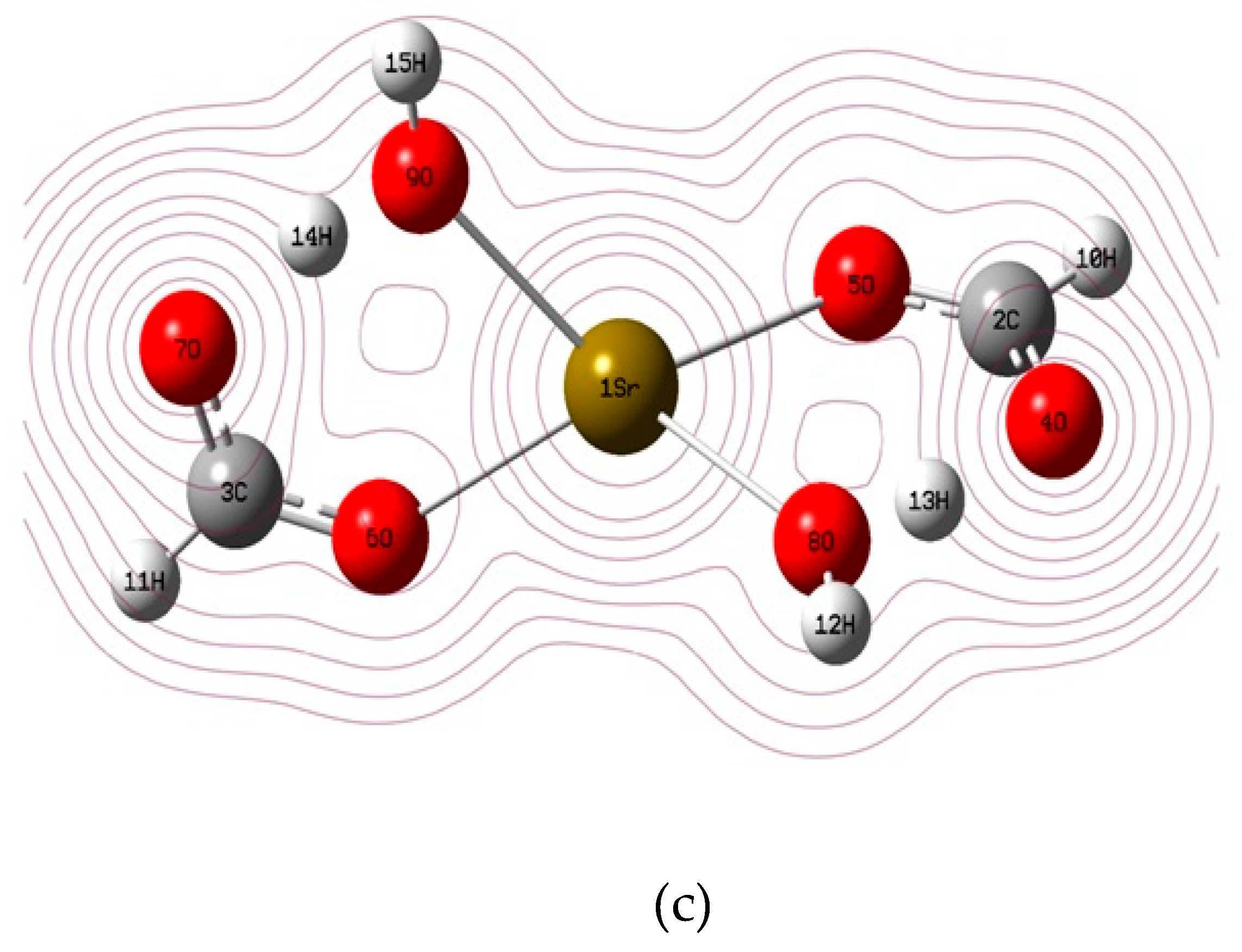
Figure 4.
(a) FT-IR spectra, (b) FT-Raman spectrum (SFD) crystal (Experimental and Theoretical).
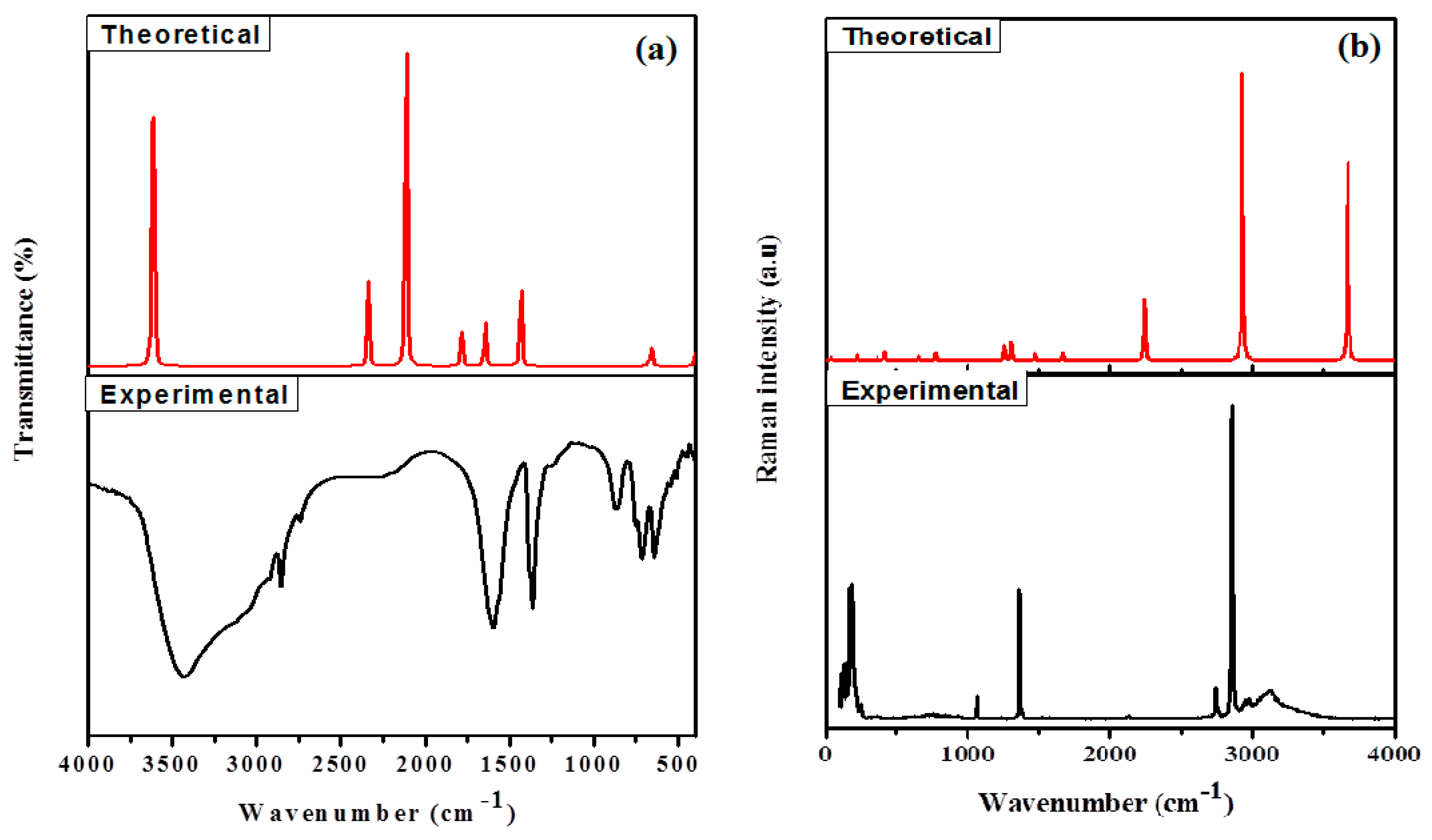
Figure 5.
Antimicrobial efficacy of (a) 0.1 glycine doped (SFD) Crystals, (b) Standard Drug. .
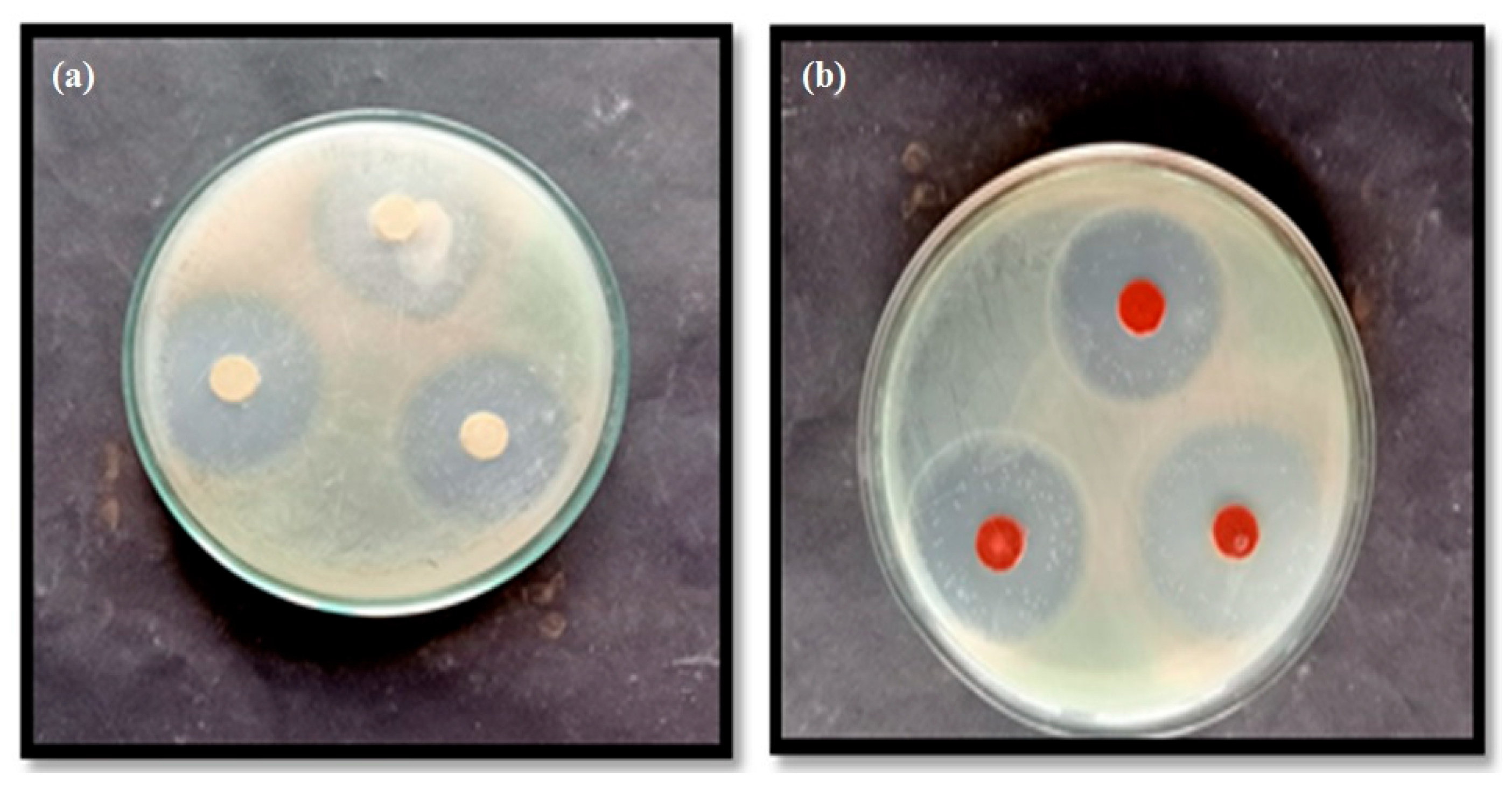

Table 1.
The important optimized bond lengths (Å) and bond angles (°) obtained in the present study compared to that determined X-ray crystallographically (reported in the literature)[1].
Table 1.
The important optimized bond lengths (Å) and bond angles (°) obtained in the present study compared to that determined X-ray crystallographically (reported in the literature)[1].
|
Bond lengths (Å) |
Optimized |
Experimental [1] |
Bond angle (Å) |
Optimized |
Experimental [1] |
|---|---|---|---|---|---|
| 1Sr-5O 1Sr-6O 1Sr-8O 1Sr-9O 2C-5O 3C-6O 2C-4O 3C-6O 2C-10H 3C-11H 8O-12H 9O-15H 8O-13H 9O-14H |
2.448 2.448 2.451 2.451 1.292 1.292 1.287 1.287 1.098 1.098 0.969 0.969 1.063 1.063 |
2.524 2.548 2.649 2.640 1.239 1.233 1.253 1.247 - - 0.75 0.70 0.90 1.05 |
6O-1Sr-8O 5O-1Sr-9O 5O-1Sr-8O 6O-1Sr-9O 12H-8O-13H 14H-9O-15H 1Sr-5O-2C 1Sr-6O-3C 5O-2C-4O 6O-3C-7O |
125.0 125.0 78.7 78.7 112.6 112.6 130.5 130.5 124.6 124.6 |
- - - - 100 100 - - - - |
Table 2.
(a) The Mulliken atomic charges of (SFD) crystal, (b) Crystal parameters obtained from DFT analysis.
Table 2.
(a) The Mulliken atomic charges of (SFD) crystal, (b) Crystal parameters obtained from DFT analysis.
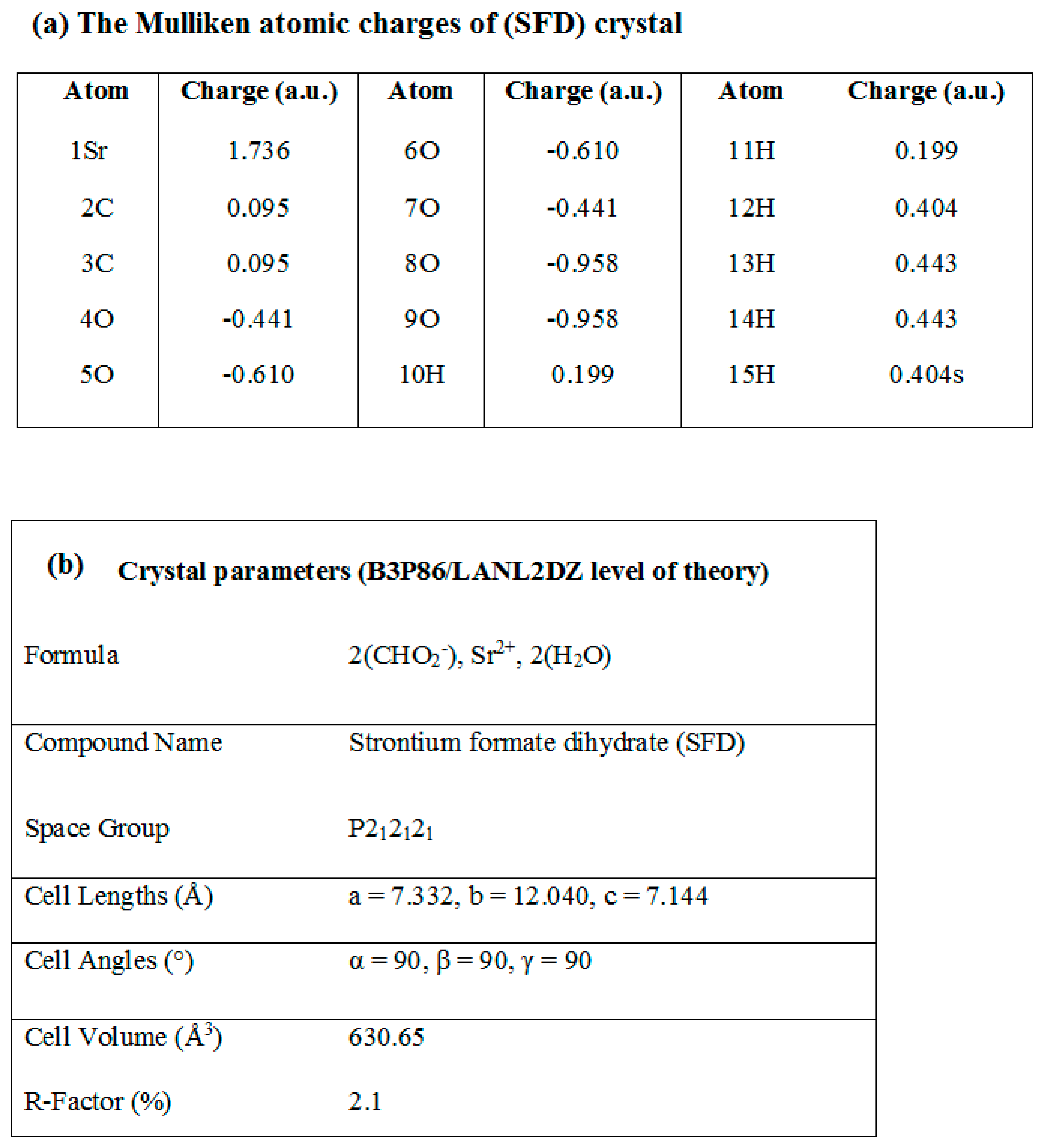 |
Table 3.
Hyperpolarizability tensor quantities.
| β components | (in a.u.) |
|---|---|
| Βxxx | 0.2771421 |
| Βyxx | 1.8381045 |
| Βxyy | 1.0608383 |
| Βyyy | -8.2346295 |
| Βzxx | -1632.0589865 |
| Βxyz | -741.8557439 |
| Βzyy | 2810.9273803 |
| Βxzz | -1.745182 |
| Βyzz | -1.9312431, |
| Βzzz | 5794.924352 |
| Β(total) | 6973.78777 |
Table 4.
Zone of inhibition of Escherichia coli, Staphylococcu aureus, Klebsiella pneumoniae and Pseudomonas aeruginosa at various concentration against Glycine, pure & Glycine doped (SFD) Crystals.
Table 4.
Zone of inhibition of Escherichia coli, Staphylococcu aureus, Klebsiella pneumoniae and Pseudomonas aeruginosa at various concentration against Glycine, pure & Glycine doped (SFD) Crystals.
|
S.No. |
Test organism |
Zone of inhibition | Standard Drug | ||||
|---|---|---|---|---|---|---|---|
| Pure | Doped (SFD) | ||||||
| Glycine | Pure (SFD) | 0.005 g | 0.05 g | 0.1 g | 0.1 g | ||
| 1. | Escherichia coli | 8 mm | 15 mm | 18 mm | 26 mm | 36 mm | 40 mm |
| 2. | Staphylococcus aureus | 10 mm | 17 mm | 23 mm | 25 mm | 28 mm | 45 mm |
| 3. | Klebsiella pneumoniae | 9 mm | 12 mm | 20 mm | 25 mm | 35 mm | 38 mm |
| 4. | Pseudomonas aeruginosa | 13 mm | 16 mm | 20 mm | 23 mm | 30 mm | 42 mm |
Table 5.
Minimum inhibitory concentration of the pure and glycine doped strontium formate dihydrate against the growth of fungi (μM).
Table 5.
Minimum inhibitory concentration of the pure and glycine doped strontium formate dihydrate against the growth of fungi (μM).
| Compound | Minimum inhibitory concentration (MIC) ( × 104 μM) | ||||
|---|---|---|---|---|---|
| Aspergillus niger | Fusarium solani | Curvularia lunata | Rhizoctonia bataticola | Candida albicans | |
| (SFD)-Pure | 23.1 | 22.8 | 24.3 | 24.7 | 24.9 |
| 0.005 M glycine doped (SFD) | 21.3 | 21.6 | 22.3 | 22.1 | 23.6 |
|
0.05 M glycine doped (SFD) |
18.6 | 19.1 | 20.4 | 21.1 | 21.6 |
|
0.1 M glycine doped (SFD) |
20.4 | 19.5 | 20.8 | 19.8 | 22.7 |
| aFluconazole [36,37] | 1.3 | 1.6 | 14 | 1.7 | 1.6 |
a Fluconazole used as the standard. .
Disclaimer/Publisher’s Note: The statements, opinions and data contained in all publications are solely those of the individual author(s) and contributor(s) and not of MDPI and/or the editor(s). MDPI and/or the editor(s) disclaim responsibility for any injury to people or property resulting from any ideas, methods, instructions or products referred to in the content. |
© 2026 by the authors. Licensee MDPI, Basel, Switzerland. This article is an open access article distributed under the terms and conditions of the Creative Commons Attribution (CC BY) license (http://creativecommons.org/licenses/by/4.0/).
Copyright: This open access article is published under a Creative Commons CC BY 4.0 license, which permit the free download, distribution, and reuse, provided that the author and preprint are cited in any reuse.