Preprint
Review

This version is not peer-reviewed.

Acute Myeloid Leukemia with Myelodysplasia-Related Gene Mutations

Submitted:

30 June 2026

Posted:

01 July 2026

You are already at the latest version

Abstract

Background/Objectives: Acute Myeloid Leukemia (AML) with myelodysplasia-related gene mutations (AML-MR), often referred to as secondary AML (s-AML) or AML-MRC, is an aggressive form of leukemia that typically arises from an antecedent myelodysplastic syndrome (MDS) but may originate also de novo. It is characterized by mutations in key genes associated with MDS that include some epigenetic regulators (ASXL1, EZH2), splicing factors (SF3B1, SRSF2, U2AF1, ZRSR2) and transcription factors (BCOR, RUNX1, STAG2). The main objective of this review paper consists in analyzing recent studies that have improved the criteria for characterization, definition and classification of AML-MR. Methods: An extensive search of the most recent literature on the topic was performed, selecting and critically analyzing the most relevant studies. Results. The studies carried out in the last years have provided an extensive molecular characterization of AML-MR, supporting more sound criteria for their identification and for a better definition with respect to other AML subtypes, particularly with respect to TP53-mutant AML. Conclusions: A unifying classification of AML-MR is now possible, allowing its identification as a unique, well-defined and separate entity.

Keywords: 
;  ;  ;  ;  ;  

1. Introduction

The World Health Organization (WHO) classification of myeloid neoplasms was revised several times to provide a progressively more accurate understanding of the cellular and molecular features underlying these tumors. The development of techniques allowing the exploration of human genome have permitted a more accurate definition of the molecular abnormalities occurring in myeloid neoplasms. Particularly, these studies have shown the extensive molecular heterogeneity of acute myeloid leukemias (AMLs), allowing their classification in molecular subtypes that are distinguished for their genetic alterations, prognosis and response to therapy. The wide diffusion of techniques of gene analysis, such as next generation sequencing have permitted the introduction of gene information in the diagnostic criteria for AML. Several recent studies have provided a detailed analysis of the genetic abnormalities occurring in AML and a detailed census of the genes altered by structural changes or mutational events in AML [1,2,3].
The World Health Organization (WHO) classification of AML proposed in 2016 classifies AML into four main subtypes: AML with recurrent genetic abnormalities; AML with myelodysplasia-related changes (AML-MRC); therapy-related AML (t-AML); AML not otherwise specified [4]. The diagnostic criteria for AML-MRC WHO 2016 are the following: history of myelodysplastic syndrome or MDS/myeloproliferative neoplasms; morphologic dysplasia in >50% of cells of at least two lineages; the presence of MDS-defining cytogenetic abnormalities [4]. Since the evaluation of dysplasia appeared subjective, with limited interobserver agreement, its value was questioned and the ontogeny of AML outweighed genomics, new classifications have introduced new criteria for diagnosis of AML-MRC.
Thus, data on gene abnormalities have been introduced into AML diagnosis and have contributed to a more accurate risk stratification. This molecular information has been incorporated into the recent molecular and prognostic classifications of AMLs. Particularly the fifth edition of WHO classification (2022 WHO) and the International Consensus Classification (ICC) of myeloid neoplasms have been published in 2022 [5,6]. Notable changes introduced into these classifications were the lowering of blast threshold that defines AML and renaming myelodysplastic syndrome and myelodysplastic neoplasm. An additional very important change introduced into the WHO/ICC 2022 classifications with respect to the fourth WHO classification (2016 WHO) concerned the criteria for definition and classification of the AML subset associated with myelodysplasia. As above discussed, in the 2016 WHO guidelines, the main criteria for the diagnosis of AML with myelodysplasia-related changes (AML-MRC) are represented by morphological features in the bone marrow, history of MDS, chromosomal abnormalities typical of MDS [4]. In the 2022 WHO, the criteria for diagnosis of AML-MRC changed and morphological dysplasia alone was excluded from the diagnostic criteria, while mutations in at least one of these myelodysplasia-related genes (ASXL1, BCOR, EZH2, SF3B1, SRSF2, STAG2, U2AF1, and ZSZR2) were included [5]. These genes include 3 splicing modulators (SF3B1, SRSF2 and U2AF1), 3 epigenetic regulators (ASXL1, EZH2 and BCOR) and 2 transcription factors (RUNX1 and STAG2). In the 2022 ICC guidelines, the criteria for AML-MRC included also one additional gene, RUNX1 mutations to the myelodysplasia-related genes (MRG) [6].
An additional element of discrepancy between 2022 classifications pertains to the biological boundaries between MDS and AML, with the new MDS-AML category introduced by ICC, defined by 10-19% blasts; this group largely overlaps with MDS with excess blasts 2 [6]. Furthermore, a previous history of MDS was considered among diagnostic criteria for WHO 2022 and a qualifier for ICC 2022.
In the studies on AML-MR, two groups of patients can be distinguished: AML with MRG mutations without MR-chromosome abnormalities and AML with MR chromosome abnormalities, such as chromosome 5 and 7 abnormalities; each of the groups can be subdivided according clinical history, in that some patients have developed AML post a MDS or MDS/MPN. The AML category with MDR cytogenetic abnormalities incorporates cases with a complex karyotype (defined as ≥3 unrelated chromosomal abnormalities) and/or other unbalanced chromosomal changes that previously fell into the prior AML-MRC category but lack a TP53 mutation or MDR gene mutations, chromosome 5, 7, 17 or 20 abnormalities.
In many of the current studies 5 groups of AML patients are defined:
-
De novo AML, newly diagnosed AML without a clinical history of an antecedent hematological disorder; after the establishment of the genome signature, AML patients with class-defining mutations, such as patients with NPM1 mutations, are assigned to the de novo AML irrespective of the presence of MRG mutations.
-
S-AML, clinically defined AML preceded by clinically documented MDS.
-
MDS/AML-MRG, molecularly defined secondary type MDS/AML with at least 1 mutation of ASXL1, BCOR, EZH2, RUNX1, SF3B1, SRSF2, STAG2, U2AF1, and ZSZR2 (10-19% of blasts).
-
AML-MRG, molecularly defined secondary-type AML with at least 1 mutation of ASXL1, BCOR, EZH2, RUNX1, SF3B1, SRSF2, STAG2, U2AF1, and ZSZR2 >20% of blasts).
-
TP53-mutant AML with mutations in TP53 and/or chromosome abnormalities involving chromosome 17p.

2. Molecular and Clinical Features of AML-MR
Several studies have shown that the frequency of MRG mutations greatly increases with age. Hoff and coworkers reported the molecular characterization of 1023 AML patients aged >60 years (median age 72 years) enrolled in the context of the BEAT study. In this group of patients, three MR genes were among the most mutated: RUNX1 (22%), SRFSF2 (22%) and ASXL1 (21%) [7]. Globally, MRGs were mutated in 57% of these patients, which is more than twice the frequency in younger patients [8]. Cusan et al have analyzed the mutational and cytogenetic profiles in AML patients of different ages from 18-10 years up to >75 years; some gene mutations, such as those involving RUNXX1, SRFSF2, ASXL1 and BCOR display a strong age-dependent pattern, thus explaining the marked rise of the frequency of MRG mutations observed in older AML patients [9]. Jahn et al have explored the genomic landscape and leukemogenetic pathways in a cohort of 815 older AML patients treated in the context of ASTRAL-1 trial [10]. NGS studies showed frequent mutations in many MR genes such as ASXL1, SRSF2 and RUNX1 [10]. To delineate leukemia-initiating trajectories, an oncogenetic tree was constructed by inferring the sequence of mutation acquisition, exploring the relationship existing among genetic alterations; the elaborated algorithm defined an oncogenetic tree with five branches with ASXL1, DDX41, DNMT3A, TET2 and TET2 emanating from the root. Importantly, the tree originating from the ASXL1 node gives rise to several individual clones with EZH2, NRAS, RUNX1, SRSF2 and U2AF1; BCOR, JAK2, KRAS, PHF6 and SF3B1 originated from RUNX1; STAG2 from the SRFSF2 node; importantly, the branches originating from the ASXL1 node include 8 of the 9 genes defining the AML-MR category [10]. Clustering by hierarchical Dirichlet processes identified 5 distinct groups of AML; importantly, the largest group, class 1 (49%), was characterized by the presence of the 9 genes defining AML with MRG mutations and by MR cytogenetic abnormalities [10].
A key study by Lindsley and coworkers described the basic features of AML-MRC [11]. These authors showed that the presence of SRSF2, SF3B1, U2AF1, ZRSR2, ASXL1, EZH2, BCOR or STAG2 identified a subgroup of AMLs, defined as secondary AML (s-AML); analysis of serial samples from individual patients showed that these mutations occur early in leukemogenesis and persist in clonal remissions [11]. Patients with MRG mutations were older, leukopenic at diagnosis and overall have a worse outcome [11].
Several studies have explored the prognostic impact of MRG mutations and have contributed to understand their prognostic heterogeneity, in part related to their association or not with favorable co-mutations.
Several retrospective studies involving large cohorts of intensively treated patients have confirmed the unfavorable prognostic impact of MRG mutations [12,13].
Zhou and coworkers reported a real-world retrospective analysis of clinical outcomes in AML with MR; this analysis compared the WHO-2022 criteria for AML-MR with the criteria for ICC-2022 for AML-MR [14]. The clinicopathological features were similar, except for higher rates of complex karyotype, monosomy 17, TP53 mutations, and fewer RUNX1 mutations in the WHO-AML-MR group [14]. AML-MR classified in both classification systems had inferior outcomes compared to AML without MR and better than TP53-mutated AMLs [10]. ICC-AML-MRC had better outcomes than WHO-AML-MRC, but this is due to the classification of TP53-mutant AML as a separate group in ICC-AML classification.
Recent studies have explored the mechanisms responsible for the variable prognostic impact of MRG mutations.
As MRG mutations are acquired at an early stage of disease evolution and further mutations are acquired over the course of disease, clonality of mutations could provide a better understanding of the prognostic impact of MRG mutations. Thus, Mecklenbrauck and coworkers have explored the prognostic impact of clonal representation of MRG mutations in a group of 550 AML-MRC patients [15]. These patients were stratified into three risk groups, according to 2022 ELN: favorable risk (20%), intermediate risk (5%) and adverse risk (75%). Patients with favorable risk profile had similar EFS and OS with or without MRG mutations [15]. Patients with MRG mutations with adverse risk profile had similar EFS but longer OS compared to the rest of adverse-risk AML patients without MRG mutations [15]. Patients with MRG mutations were subdivided into two groups according to variant allele frequency (VAF) of MRG mutant genes: low VAF (<44.%%) and high VAF (>44.5%); high VAF patients were significantly older and had a high WBC count and lower platelet count at diagnosis; ASXL1 mutations were more likely to be mutated in the high VAF group, while SF3B1 and STAG2 mutations were predominantly observed in the low VAF group; high VAF patients showed a significantly shorter EFS and OS as compared to low VAF patients; patients with high VAF had a higher cumulative incidence of relapse compared to the low VAF group [15]. Among patients with favorable risk profile, MRG VAF did not affect prognosis; in contrast, in adverse risk group, patients with high MRG mutation VAF had significantly shorter EFS and OS compared to those with a low VAF [15]. These observations support the hypothesis that MRG mutations drive the more aggressive phenotype of leukemic cells when they are part of the dominant clone [15].
A recent study explored the differential prognostic impact of MRG mutations in a large cohort of 4,978 AML patients molecularly characterized and intensively treated [16]. 1698 of these patients (34.1%) harbor MRG mutations at the level of at least one gene. The MR genes more frequently mutated were RUNX1 (12.8%), ASXL1 (9.9%), SRSF2 (9.2%), STAG2 (5.9%), BCOR (5.4%) and EZH2 (4.1%) [16]. AML-MR cases displayed a lower CR rate (65.7% vs 77%), shorter EFS (6.3 months vs 10.5 months), RFS (14.3 months vs 20.5 months) and OS (16.6 months vs 26.5 months) compared to AMLs without MRG mutations [16]. Outcomes were explored also in a subgroup of patients who underwent allo-HSCT in CR1: compared to patients without MRG mutations, patients with MRG mutations showed a shorted EFS (26.5 months vs 50.5 months), RFS (41.1 months vs 101.9 months) and OS (63.4 months vs not not-reached) [16]. Analysis of patient outcomes with respect to individual MRG mutations showed three groups: ASXL1, RUNX1, SF3B1, SRFSF2 and U2AF1-mutant AML displayed a markedly shortened OS compared to those WT for these genes; the presence of BCOR mutations moderately decreased OS compared to BCOR-WT; the presence of EZH2, STAG2 and ZRSF2 did not affect OS compared to AML WT for these genes [16]. The analysis of the impact of specific MRG mutations showed gene-specific prognostic patterns with ASXL1, RUNX1, SF3B1 and U2AF1 mutations associated with adverse-risk; SRSF2 and STAG2 aligned with intermediate-risk; BCOR, EZH2 and ZRSR2 did not significantly differing from intermediate or adverse risk [16]. It is important to note that the prognostication of various MRG mutations was established on the basis of the response to intensive chemotherapy treatment. The prognostication impact of SF mutations has been evaluated in a group of 994 patients with ND AML, including 266 patients with a SF mutation (SRSF2, U2AF1, SF3B1, ZRSF2) , with a median age of 67 years; in patients treated with IC the median OS was significantly shorter for SF-mutant than for SF-WT patients (15.9 vs 26.7 months); this significance abrogated when evaluating patients who received VEN with IC (mOS 19.6 vs 30.7 months) or VEN with low-intensity therapy (mOS 12.3 vs 8.5 months, respectively) [17]. These observations were supported by experimental studies showing a unique relationship between expression of RNA splicing factors and response to BCL2 inhibitor VEN, as well as to a inhibitor of splicing -dependent kineses which overcomes VEN resistance [18]. Other studies showed the selective efficacy of VEN in unift AML-MR patients under low-intensity treatment regimens [19].
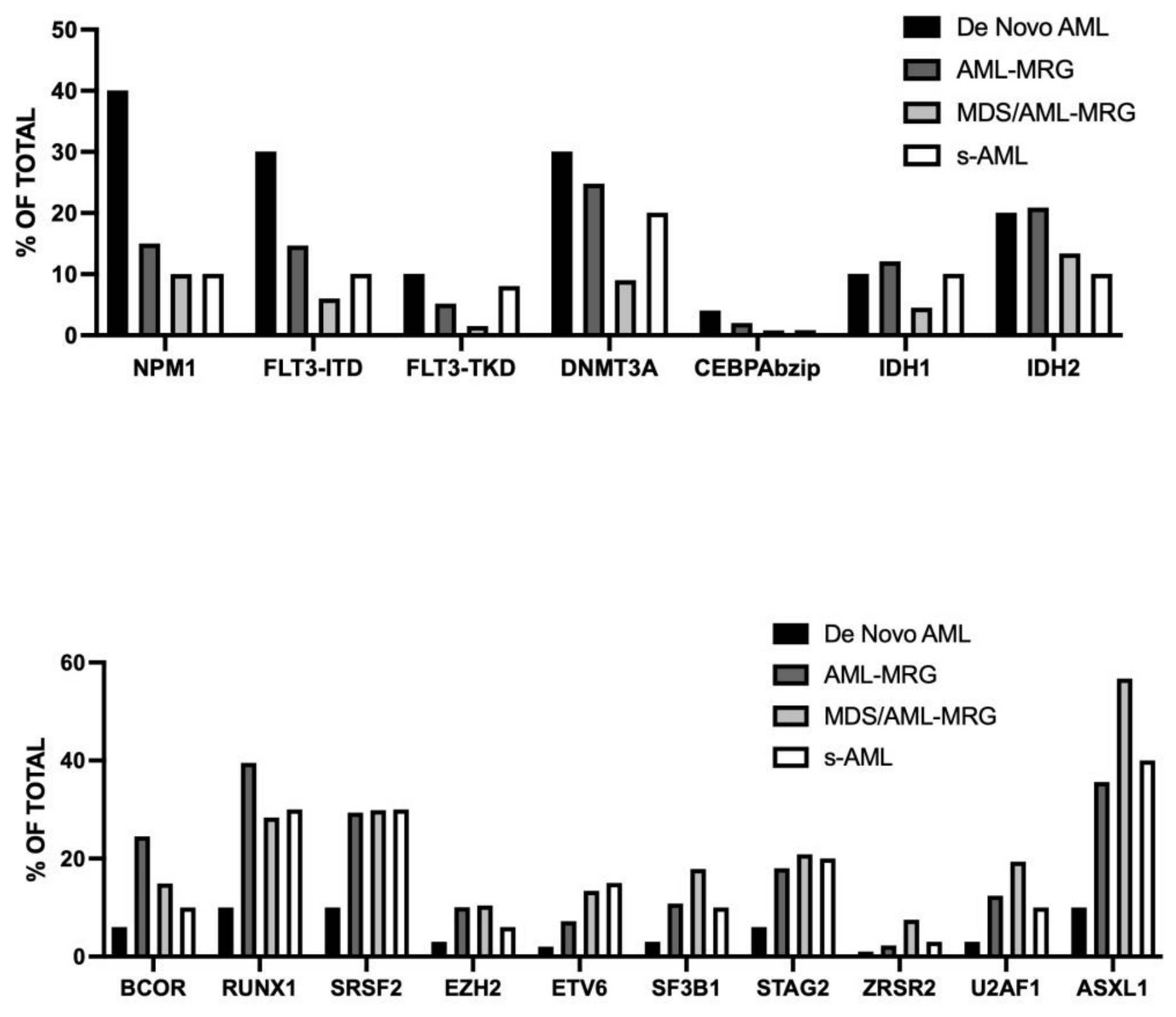
Bortjes et al. have reported an analysis of molecular abnormalities and outcomes of a retrospective cohort of 2684 intensively treated AML patients [20]. In these patients, three subsets of AMLs harboring MRG mutations were analyzed: sAML defined as AMLs preceded by a clinically documented MDS; st-AML molecularly defined as secondary-type (ST-AML) with at least one mutation in ASXL1, BCOR, ETV6, EZH2, SF3B1, STAG2, UAF1, and ZRSR2 (≥20% of leukemic blasts); as-MDS-AML, molecularly defined as secondary type MDS/AML with at least one mutation in ASXL1, BCOR, ETV6, EZH2, SF3B1, STAG2, UAF1, and ZRSR2 (10-19% of leukemic blasts) [20]. The comparison of these groups of AML showed that mutations of NPM1, FLT3-ITD, FLT3-TKD, DNMT3A are associated with de novo AML ontogeny, while mutations in ASXL1, BCOR, ETV6, EZH2, RUNX1, SF3B1, STAG2, UAF1, and ZRSR2 are significantly associated with s-AML, MDS/AML-MRG and AML-MRG [20]. (Fig.1) Among these three groups of AMLs with MRG mutations, sAML had the lowest OS, while st-AML and st-MDS-AML displayed a similar OS [20]. Particularly, s-AML had a 1-year shorter OS compared to MDS/AML (45.7% vs 72.2%) and to st-AML (45.7% vs 65.4%); however, s-AML displayed only a trend toward a lower OS compared to MDS/AML (30.4% vs 44.9%) and st-AML (30.4% vs 40.1%) [20]. Also at molecular level, st-AML and s-MDS-AML are high comparable, except for a higher frequency of ASXL1 mutations in MDS-AML (56.7% vs 35.6%) and a higher occurrence of DNMT3A mutations in st-AML (24.9% vs 8%) [20]. St-AMLs exhibited an OS highly comparable to that observed for adverse-risk AML, as stratified according to ELN2022 [21]. It is important to note that s-AML was not validated in current classifications. In fact, both ELN2022 and ICC2022 recognize AML following MDS as a diagnostic qualifier rather than a separate disease entity [20].
A multicenter retrospective study of a cohort of AML, including 75 MDS-AML patients, confirmed the molecular and clinical convergence between AML-MR and MDS-AML [22]. AML-MR and MDS-AML patients exhibited overlapping molecular features, including frequent ASXL1, TP53 mutations, as well as a high frequency of complex karyotypes; non-MR cases showed significantly lower frequencies of these alterations but were enriched for NPM1 and FLT3 mutations (25.0% and 22.7%, respectively); both AML-MR and MDS-AML had significantly shorter OS than AMLs without MR; OS was similar for AML-MR and MDS-AML (10.3 months vs 13.3 months, respectively) [22]. These observations suggest that MDS-AML and AML-MR form a biological continuum with shared clinical and molecular features.
Figure 1. Frequency of the main gene mutations observed in AML patients subdivided into four groups: de novo AML, AML-MRG, MDS/AML-MRG and s-AML. Top Panel: gene mutations (NPM1, FLT3-ITD, FLT3-TKD, CEBPAbzip, IDH1 and IDH2) preferentially observed in de novo AML. Bottom Panel: gene mutations ASXL1, BCOR, ETV6, EZH2, RUNX1, SF3B1, STAG2, UAF1, and ZRSR2, significantly associated with s-AML, MDS/AML-MRG and AML-MRG. Original data are reported in Boertjes et al. [20].
Preprints 220932 g001
Attardi and coworkers have reported an extensive characterization of 924 patients with MDS/AML (10-19% of blasts) or AML (>20% of blasts), classified according to ICC [23]. This cohort included 109 patients with mutated TP53, 497 with MRG mutations, 93 with MR-cytogenetic abnormalities, 77 with t-AML and 136 control AML [20]. In the group of TP53-muated AML, 74 were classified as AML and 35 as MDS/AML; chromosome 17/17p abnormalities were more frequent in AML than in MDS/AML (27vs 8.6%), as well as DNMT3A mutations (24% vs 0%) [23]. In the group with MR-cytogenetic abnormalities 65 patients and 29 as MDS/AML; chromosome 5 abnormalities were less frequent in AML than in MDS/AML (23% vs 50%), as well as del(20q) )1.5% vs 18%) [23]. In the large group of AML with MRG mutations, 251 were classified as AML and 246 as MDS/AML; FLt3, DNMT3A, CSFR3R, IDH1, IDH2 and BCOR mutations were more frequent in AML than in MDS/AML, as well as median clonal size of STAG2 and RUNX1 mutant alleles [23]. The groups with TP53 mutations and with MR-related cytogenetic abnormalities have a dismal prognosis, comparable for both AML and MDS groups; in the group with MRG mutations, MDS/AML patients had significantly better prognosis than AML [23]. The study in the group of secondary AML (s-AML) based on clinical history (post MDS or post-MDS/MPN) with respect to genetically-defined s-AML showed in all three groups of patients no differences in outcome; the only significant differences were observed for frequencies of ASXL1 and SF3B1 mutations higher in AML post-MDS than in de novo AML (48% vs 25% for ASXL1 and 19.0% vs 7.1% for SF3B1) [23]. The comparison of post-MDS AML with a group of t-AML showed a greater frequency of ASXL1, RUNX1, SF3B1, SRSF2, JAK2 and TET2 gene mutations in the former ones compared to the latter ones [23]. A high proportion of post-MDS AML are classified as AML-MR following WHO 2022 and were classified as adverse-risk according to ELN 2022 [23].
The results of these studies showed a consistent similarity of molecular alterations of MDS/AML-MRG and s-AML and AML-MRG, suggesting a continuum from MDS/AML to AML-MR. MDS and its precursor lesions progress in a highly variable and unpredictable pattern, ranging from a slow, stable condition over many years to a rapid decline. In about 30% of cases, MDS advances into secondary AML (s-AML) which bears some typical molecular features of MDS. A recent longitudinal study evaluated the 6-year rate of progression of MDS and its precursor lesions, showing: 17%, 22%, 52% and 73% of rate of progression for ICUS/IDUS, CCUS, LR-MDS and HR-MDS, respectively [24]. MDS and s-AML share significant genetic homology. This disease progression is a multistep process, where early driver mutations establish the initial MDS clone, and subsequent genetic events drive progression to vert leukemia. The founding early mutations typically involve RNA splicing (SF3B1, SRFSF2), epigenetic regulation (ASXL1, DNMT3A, TET2) or transcription factors such as RUNX1 [25,26]. Evolution to AML is frequently driven by the acquisition of mutations in signal transduction genes, such as FLT3, NRAS, KIT) or transcription factors (RUNX1) [26,27,28,29]. Furthermore, about 50% of MDS transitioning to s-AML develop complex chromosome abnormalities, frequently involving losses or structural changes in chromosomes 5, 7 and 17 [26,27,28,29].
Genomic analyses at single-cell level showed that mutations in transcription factor genes (such as RUNX1 or ETV6) and in signaling genes (such as FLT3, KRAS/NRAS, PTPN11) are typically subclonal in MDS, occurring over a “background” founding clone characterized by epigenetic modified gene (ASXL1, TET2, EZH2) and frequently accompanied by spliceosome gene mutations (SRSF2, SF3B1). Signaling gene mutations are infrequent in MDS at diagnosis as detected by standard NGS studies; however, Guess et al., using sensitive PCR studies showed that these mutations at a VAF <2% are observed in a subset of MDS patients [26,27,28,29]. In many MDS patients, new signaling gene mutations arose concomitantly with progression to s-AML, while in other cases pre-existing subclonal signaling mutations expanded or contracted as the blast count increased [26,27,28,29]. The presence of these subclonal signaling pathway mutations was associated with an increased risk of MDS progression, particularly in low-risk MDS patients [26,27,28,29]. Changes in clonal architecture of progressing MDS were classified as statis or dynamic, with dynamic clonal architecture having a more proliferative phenotype by blast count fold change [26,27,28,29]. Complex patterns of clonal evolution are also observed in some instances, with some clones persisting with the acquisition or loss of signaling mutations in other clone [26,27,28,29].
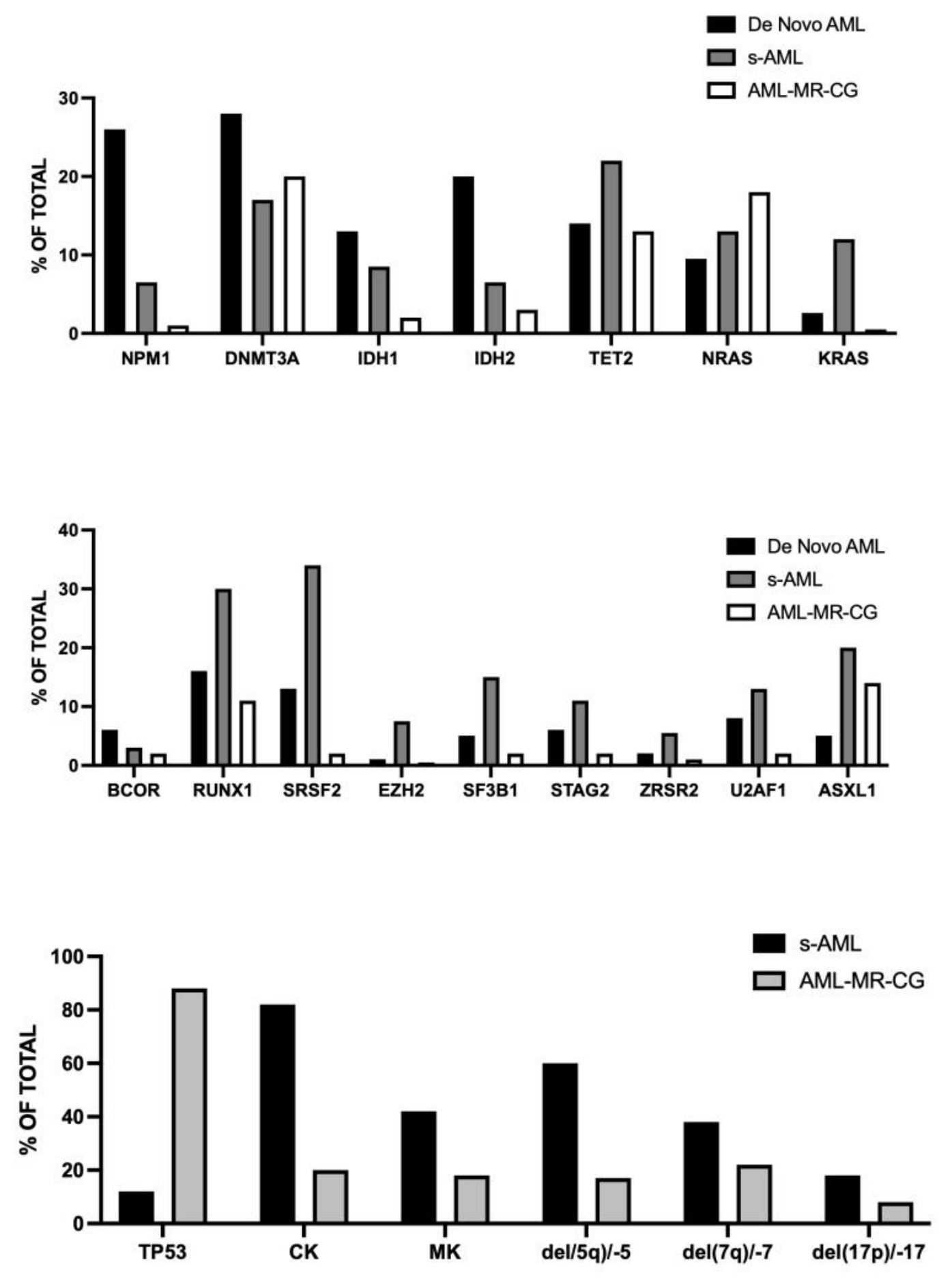
Figure 2. Main gene mutations in de novo AML, s-AML and AML-MR-CG (Top and Middle Panels); main cytogenetic alterations in s-AML and AML-MR-CG (Bottom Panel). Original data are reported in McCarter et al. [30].
Preprints 220932 g002
McCarter et al. have explored the interaction existing between AMLMRC defined on the basis of ontogeny (s-AML) and on the basis of MRG mutations [30]. Thus, they explored a group of 344 ND AML patients carefully explored at molecular and cytogenetic levels, with a detailed clinical history and all treated with intensive chemotherapy induction regimens [21]. First, these investigators carefully reevaluated ontogeny assignments of these patients and according to the WHO 2016 diagnostic criteria they defined two subgroups of AML-MRC patients: one group with a preceding documented history of MDS (MR-H) corresponding to 107 patients; a second group with MDS-related cytogenetic abnormalities (MR-CG), corresponding to 29 patients; a group of 157 patients (defined as de novo AML) do not have MRC according to WHO 2016, and a group of 92 patients with t-AML [30]. The analysis of the mutational profile showed that MRG mutations were observed in 50% of MR-H, 4.5% of MR-CG, 17% of t-AML and 28% of de novo AML [30]. The outcomes of these AML subgroups showed that MR-H as well as MR-CG have a poor prognosis compared to de novo AML; de novo AML with MRG mutations have outcomes comparables to those without MRG mutations and display a survival profile intermediate between favorable and intermediate-risk ELN 2022 [30].
Other recent studies have shown the negative outcome of s-AML patients who developed AML transformed from MDS or MDS-MPN; s-AML was associated with poor outcomes irrespective of AML genomics and treatment, suggesting the need to its inclusion as an independent AML adverse risk subgroup for accurate prognostication and clinical trial reporting [31].
Liu et al. have retrospectively analyzed 619 newly diagnosed AML patients with complete cytogenetics and mutational profiling by targeted NGS [19]. For the analysis of AML-MR subgroup, they subdivided AMLs into four subgroups: RUNX1, with RUNX1 mutations without any other MRG mutation; MR, with any MRG mutation, exclusive of RUNX1; RUNX1-MR, with mutations in RUNX1 and at least one additional MRG gene; MR-GC, with cytogenetic mutations myelodysplasia-related [32]. The OS of these four subgroups of AML-MR was similar; AML-MRC displayed a superior prognosis compared to TP53-mutant group, but inferior to other AML diagnostic categories without MRG mutations [32].

3. Results MRG Mutations in FLT3-Mutant AMLs
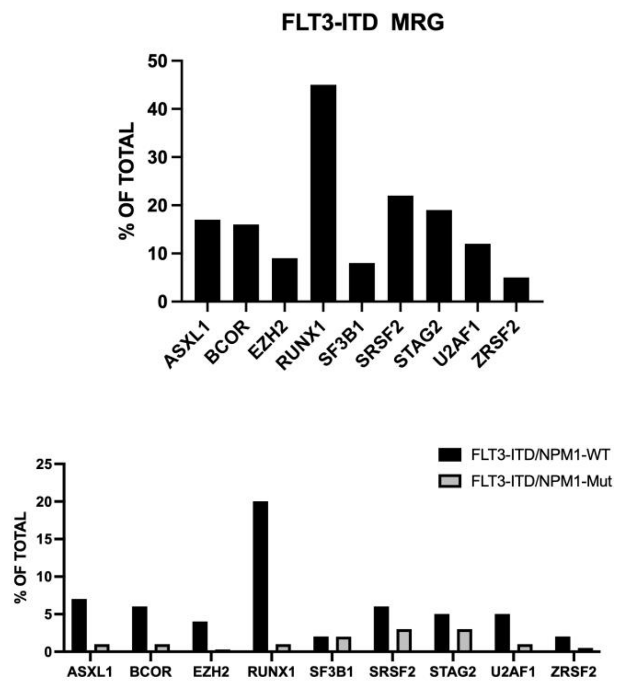
A recent study evaluated the prognostic impact of MRG mutations in a large cohort of 842 AML patients with FLT3-ITD mutations; 171 (20%) of these FLT3-ITD-mutant AMLs display MRG mutations: 63% had only one mutation, 26% had two mutations, 11% had 3 or more mutation; 57% of these patients were NPM1-mutated [33]. In MRG-mutated AMLs, the most common MRG mutations were RUNX1 (45%), SRSF2 (22%), STAG2 (19%) and ASXL1 (17%) [33]. (Fig.3) MRG mutations occurred in 34% of FLT3-ITD/NPM1-WT patients and in 9% of FLT3-ITD/NPM1mut AML; DNMT3A mutations were more common in FLT3-ITD/NPM1mut AML, while NRAS, ASXL1, BCOR, EZH2, RUNX1 and U2AF1 mutations were more common in the FLT3-ITD/NPM1-WT group [24]. (Fig.3) In multivariate analysis, MRG co-mutations in FLT3-ITD-mutant group did not alter outcome and do not confer adverse prognosis in the overall FLT3-ITDpos cohort [20]. Both RFS and OS were significantly shorter in FLT3-ITD/NPM1-WT patients with MRG mutations compared to patients without MRG mutations; in contrast, in FLT3-ITD/NPM1mut AMLs the presence of MRG co-mutations did not affect outcomes [33]. FLT3-ITD allelic ratio did not impact the prognostic impact of MRG mutations [33]. In FLT3-ITD/NPM1-WT patients the number of MRG mutations affect prognosis, since patients with a single MRG mutation had RFS and OS comparable to MRG-negative patients, while patients with 2 or more mutations had significantly worse RFS and OS [33].
Figure 3. Most recurrent MRG mutations observed in FLT3-ITD AML patients. Top Panel: The frequency of MRG mutations was reported as the percentage of the total MRG mutations. Bottom Panel: Frequency of the various MRG mutations observed in FLT3-ITD/NPM1-WT and FLT3-ITD/MPM1mut AMLs. Oriignal data are reported by Mecklenbrauck et al. [33].
Preprints 220932 g003
It is of interest to note that FLT3 mutations are rare in MDS and are acquired at the time of their progression to AML [34,35].

4. MRG Mutations in NPM1-Mutant AMLs

Several studies have explored the prognostic impact of MRG mutations in NPM1mut AMLs. In this context, Eckardt et al. performed a retrospective study on 936 NPM1mut AML patients who received intensive chemotherapy treatment [36]. NPM1mut AML patients with MRG mutations are significantly older and have reduced WBC count at initial diagnosis compared to those without MRG mutations. 13.4% of these patients displayed at least one MRG mutation: SRSF2 (5.1%), STAG2 (3.2%), EZH2 (2.4%), BCOR (1.7%), SF3B1 (1.4%), ASXL1 (1.3%), ZRSR2 (0.5%) and U2AF1 (0.4%) [36]. The rate of FLT3-ITD mutations was similar in both AML-MR and AMLs without MR [36]. The most recurrent mutational correlations were for co-occurring alterations of U2AF1 and RUNX1 and SRSF2 and IDH2 [27]. CR rates were similar in patients with or without MRG mutations (74.4% vs 77.7%); mRFS was 32.9 months and 24.3 months in patients without or with MRG mutations; mOS was 29.1 months and 27.2 months in patients without MRG or with MRG mutations [36]. The rate of allo-HSCT between patients without MRG mutations or with MRG mutations differed significantly regarding HSCT in first CR (15.4% vs 7.2%, respectively) [36]. The rate of HSCT as salvage therapy was similar in the two groups of patients [36].
Othman and coworkers confirmed these findings, reporting a 3-year OS of 67% in NPM1mut AML with MRG mutations compared to 65% in NPM1mut AML without MRG mutations [37].
However, other studies showed that MRG mutations confer a negative prognosis to NPM1mut AMLs. Thus, Chan et al. explored a group of 233 NPM1mut AML, 18.5% with MRG mutations; patients with MRG mutations had worse OS than those without MRG mutations (15.3 months vs 43.7 months) [38]. However, in the group of patients with MRG mutations (86% of patients had 60 or more years, compared to 52% in the patients without MRG mutations; for aged patients OS was significantly worse in AML-MRC patients than in those without MRG mutations (12.6 months vs 26.1 months); however, in patients <60 years there was no difference in OS between patients with or without MRG mutations [38]. MRD-negativity in NPM1mut patients predicts longer OS; mOS of patients NPM1mut with MRG mutations achieving MRD negativity was lower than that observed for patients NPM1mut without MRG mutations achieving MRD negativity [29]. This study suggests that the co-occurrence of MRG mutations in NPM1mut AML confers a significant detrimental effect on survival that is in part related to the older age of these patients.
A more recent study further supported that the adverse effect of MRG mutations in NPM1mut AML patients is mainly related to their older age [30]. In fact, when stratified by age, older NPM1mut AML patients had worse OS than younger NPM1mut patients overall, although presence of MRG mutations trended toward even worse OS in older patients [39]. In younger patients, MRG mutations are not associated with a difference in OS [39].
Cocciardi et al. reported the analysis of 568 NPM1mut AML patients; 18.1% of these patients had MRG mutations [31]. Mutated genes were SRSF2 (7.7%), STAG2 (4.9%), ASXL1 (2.5%), EZH2 (1.2%), U2AF1 (1.2%), ZRSR2 (1.1%), SF3B1 (1.1%), RUNX1 (0.7%) and BCOR (0.4%) [31]. The frequency of MRG mutations was markedly influenced by age being 8% in patients of 18-60 years and 27% in patients >60 years [40]. VAF analysis of MRG mutations showed that these mutations represented the dominant clone over NPM1 mutations in 76% of cases, co-dominant in 16% of cases and only in 8% of cases NPM1 was the dominant clone [40]. Restricting the analysis of 470 of these patients with a 2022 ELN favorable-risk profile, multivariable analysis for EFS identified age, DNMT3AR882 and MRG mutations as unfavorable factors; restricting the analysis to a subset of CR-CRc patients with available data on NPM1mut MRD status, MRG mutations lost their significant effect, whereas DNMT3AR882 mutations maintained their unfavorable effect [40].
A recent meta-analysis evaluated the outcomes of 4,363 NPM1mut AML patients, of whom 655 patients (15%) had co-occurring MRG mutations; the presence of MRG mutations in these patients was associated with significantly reduced mOS (hazard ratio (HR) 1.10, p<0.001), shorter EFS (HR 1.43, p<0.006) and a lower probability of achieving a complete remission (HR 0.94, p0.01) [41]. Subgroup analysis limited to NPM1mut AMLs classified as favorable-risk following ELN2022 confirmed the unfavorable effect of MRG mutations on OS (HR 1.34) [41]. These findings may have important implications for risk stratification of NPM1mut patients; in fact, in the ELN 2022 risk stratification, MRG mutations do not modify the favorable risk profile conferred by NPM1mut in the absence of FLT3-ITD and adverse-risk cytogenetics [41]. Patients with NPM1mut and MRG mutations, not harboring these additional high-risk features, are classified as favorable-risk and are not considered candidate for HSCT in CR1. However, the results of this meta-analysis suggest reconsidering this problem. In the absence of specific clinical data on these patients, the problem of selection for allo-HSCT of individual NPM1mut AML patients with MRG mutations and with favorable-risk profile must be individualized and based on the evaluation of MRD status and on the balance of risk-benefit of an allo-HSCT [42].
Zhou et al. reported the retrospective analysis of 221 adult AML patients with a favorable-risk profile according to the ELN 2022 classification [34]. 21.3% of these patients harbored MRG mutations; this subgroup of AML displayed a higher frequency of TET2 and ETV6 mutations. The presence of MRG mutations did not impact 2-year OS in the whole group of these AMLs; however, the sub-analysis of these patients according to the number of MRG mutations per patient, showed that patients with 2 or more MRG mutations had a significantly reduced OS compared to those without MRG mutations [43]. Thus, a quantification of MRG mutation burden is required for the assessment of risk stratification of MRG-mutant AMLs with a favorable-risk profile [43].

5. Conclusions

WHO5thand ICC2022 both include AML-MR as a new diagnostic entity that incorporates the mutation profile; although, the diagnostic criteria proposed by WHO5thand ICC2022 are overlapping, some notable differences are present, including inclusion of RUNX1 in the AML-MR gene list (only for ICC2022), and the evaluation of AML-MR considering gene mutations or cytogenetic profile as distinct entities and the inclusion or not of AML with TP53 mutations. An additional element of discrepancy is also related to the impact of AML ontogeny and consequently the consideration as a unique entity of AML patients with history of antecedent MDS or MDS/MPN.
The studies carried out in the last years strongly suggest that: RUNX1 should be included in the MR gene list; TP53-mutant AML should be excluded from AML-MR and considered as a separate entity; AML-MR should include in a unique entity AMLs with MRG mutations, with MR-CGAs and with an antecedent history of MDS or MDS/MPN. This unified classification of AML-MR will facilitate the planning of dedicated clinical trials their harmonization and thir comparative evaluation.

Author Contributions

The author of performed all the steps required for the preparation of the rpesent paper.

Funding

This research received no external funding.

Institutional Review Board Statement

Not applicable.

Conflicts of Interest

The authors declare no conflicts of interest.

References

  1. Papaemmanuil, E.; Gerstung, M.; Bullinger, L.; Gaidzik, K.; Paschka, P.; Roberts, N.D.; Potter, N.E.; Hauser, M.; Thol, F.; Bolli, N.; et al. Genomic classification and prognosis in acute myeloid leukemia. N Engl J Med 2016; 374, 2209–2221. [CrossRef]
  2. Bullinger, L.; Dohner, K.; Dohner, H. Genomics of acute myeloid leukemia diagnosis and pathways. J Clin Oncol 2017; 35, 934–946. [CrossRef]
  3. Tazi Y, Arango-Ossa J, Zhou Y, Bernard E, Thomas I, Gilkes A, Freeman S, Pradat Y, Johnson S.J, Hills R, et al. Unified classification and risk-stratification in acute myeloid leukemia. Nat Commun 2022; 13: 4622. [CrossRef]
  4. Arber, D.A.; Orazi, A.; Hasserjian, R.; Theile, J.; Borowitz, M.J.; LeBeau, M.; Bloomfield, C.; Cazzola, M.; Verdiman, J. The 2016 revision of the world health organization classification of myeloid neoplasms and acute leukemia. Blood 2016; 127: 2391-2405. [CrossRef]
  5. Khoury, J.D.; Solary E, Abla O, Akkari Y, Alaggio R, Apperley JF, Bejar R, Berti E, Busque L, Chan J, et al. The 5th edition of the World Health Organization classification of hematolymphoid tumors: myeloid and histiocytic/dendritic neoplasms. Leukemia 2022; 36: 1703-1719. [CrossRef]
  6. Arber, D.A.; Orazi, A.; Hasserjian, R.P.; International Consensus Classification of myeloid neoplasms and acute leukemias: integrating morphologic, clinical and genomic data. Blood 2022; 140: 1220-1228. [CrossRef]
  7. Hoff, F.W.; Huang, Y.; Welkie, R.L.; Swords, R.; Traer, E.; Stein, E.M.; Lin, T.; Patel, P.; Collins, R.H.; Baer, M.; et al. Molecular characterization of newly diagnosed acute myeloid leukemia patients aged 60 years or older: a report from the Beat AML clinical trial. Blood Cancer J 2025, 15, 55. [CrossRef]
  8. Tsai, X.; Sun, K.J.; Lo, M.Y.; Tien, F.M.; Kuo, Y.Y.; Tseng, M.H.; Peng, Y.L.; Chuang, Y.H.; Ko, B.S.; Tang, Y.L.; et al. Poor prognostic implications of myelodysplasia-related mutations in both older and younger patients with de novo AML. Blood Cancer J 2023, 13, 4. [CrossRef]
  9. Cusan, M:; Larkin, K.; Nicolet, D.; Jurinovic, V.; Mrozek, K.; Batcha, A.; Rothenberg-Thirally, M.; Schneider, S.; Sauerland, C.; Gorlich, D.; et al. Multi-dimensional analysis of AML cross-continent reveals age-associated trends in mutational landscape and treatment outcomes (AML Cooperation Group and Alliance for Clinical Trials in Oncology. Leukemia 2025, 39, 2926-2934. [CrossRef]
  10. Jahn, E.; Saadati, M.; Fenaux, P.; Gobbi, M.; Roboz, G.; Bullinger, L.; Lutsik, P.; Riedel, A.; Plass, C.; Jahn, N.; et al. Clinical impact of the genomic landscape and leukemogenic trajectories in non-intensively treated elderly acute myeloid leukemia patients. Leukemia 2023, 37, 2187-2196. [CrossRef]
  11. Lindsley, R.C, Lindsley RC, Mar BG, Mazzola E, Grauman PV, Shareef S, AllenSL, Pigneux A, Wetzler M, Stuart RK, Herba HP, et al. Acute myeloid leukemia ontogeny is defined by distinct somatic mutations. Blood 2015; 125: 1367-1376. [CrossRef]
  12. Mrozek, K.; Kohlschmidt, J.; Blachly, J.S.; Nicolet, D.; Carroll, A.J.; Archer, K.J.; Mims, A.S.; Larkin, K.T.; Orwick, S.; Oakes, C.C.; et al Outcome prediction by the 2022 European Leukemia Net genetic-risk classification for adults with acute myeloid leukemia: and Alliance study. Leukemia 2023; 37: 788-798. [CrossRef]
  13. Rausch, C.; Rothenberg-Thurley, M.; Dufour, A.; Schneider, S.; Gittinger, H.; Sauerland, C.; Gorlich, D.; Krug, U.; Berdel, W.E.; Woermann, B.J., et al. Validation and refinement of the 2022 European Leukemia Net genetic risk stratification of acute myeloid leukemia. Leukemia 2023; 37: 1234-1244. [CrossRef]
  14. Zhou, Q, Zhao, D.; Zarif, M.; Davidson, M.B.; Minden, M.D.; Tierens, A.; Yeung, Y.W.T.; Wei, C.; Chang, H. A real-world analysis of clinical outcomes in AML with myelodysplasia-related changes: a comparison of ICC and WHO-HAEM criteria. Blood Adv 2024, 8, 1760-1766. [CrossRef]
  15. Mecklenbrauck, R.; Borhert, N.; Gabdoulline, R.; Poll, P.; Funke, C.; Brandes, M.; Dallmann, L.K.; Fiedler, W.; Krauter, J.; Trummer. A.; et al. Prognostic impact of clonal representation of myelodysplasia-related gene mutations in acute myeloid leukemia. Leukemia 2025, 39: 1773-1777. [CrossRef]
  16. Bill, M.; Eckardt, J.N.; Dohner, K.; Rohnert, M.A.; Rausch, C.; Metzler, K.H.; Spiekermann, K.; Stasik, S.; Wurm, A.A.; Sauer, T.; et al. Differential prognostic impact of myelodysplasia-related gene mutations in a European cohort of 4978 intensively treated AML patients. Leukemia 2026, 40, 63-71. [CrossRef]
  17. Senapati, J.; Umutia, S.; Loghavi, S.; Short, N.J.; Issa, G.C.; Maiti, A.; Abbas, H.A.; Daver, N.G.; Pemmaraju, N.; Pierce, S.; et al. Venetoclax abrogates the prognostic impact of splicing factor gene mutations in newly diagnosed acute myeloid leukemia. 2023, 142, 1647-1660. [CrossRef]
  18. Wang, E.; Pineda, J.M.B.; Kim, W.J.; Chen, S.; Bourcier, J.; Stahl, M.; Hogg, S.J.; Bewersdorf J.P.; Han, C.; Singer, M.E.; et al. Modulation of RNA splicing enhances responses in BCL2 inhibition in leukemia. Cancer Cell 2023, 41, 164-180. [CrossRef]
  19. Lin, J.; Liu, X.; Ying, S.; Zhu, Y.; Huang, W.; Zheng, W.; Ye, X.; Shu, J.; Luo, Y.; He, J.; et al. Selective efficacy of venetoclax in unfit patients with acute myeloid leukemia with myelodysplasia-related gene mutations under low-intensity therapy. Annals Hematol 2025, 104, 2717-2729. [CrossRef]
  20. Bortjes, E.L,; Grob, T.; Al Hinai, A.; Beverloo, B.; Versluis, J.; Kavelaars, J.; Rijken, M.; Compagne, K.; Eperlinck, C.; Sanders, M.; et al. MDS/AML and AML with myelodysplasia-related gene mutations: clinical and molecular similarities. Blood Adv 2026, 10, 939-950. [CrossRef]
  21. Dohner, H.; Wei, A.H.; Appelbaum, F.R.; et al. Diagnosis and management of AML in adults: 2022 recommendations from an international expert panel on behalf of the ELN. Blood 2022, 14, 13451377. [CrossRef]
  22. Wei, Y.; Yan, X.; Ma, J.; Cai, Z.; Guo, X.; Miao, Z.; Zhang, Z.; Dai, Y.; Gao, X.; Ge, Z. Comparative analysis of ICC and WHO classifications reveal molecular and clinical convergence between AML-MR and MDS/AML. Hematological Oncol 2026, 44, e70174. [CrossRef]
  23. Attardi, E.; Cipriani, M.; Guarnera, L.; Savi, A.; Fabiani, E.; Mallegni, F.; Moretti, F.; Silvestrini, G.; Awada, H.; Durmaz, A.; et al. Validation of ICC hierarchical classification in secondary AML. Blood Adv 2026, in press. [CrossRef]
  24. De Zern, A.; Gillis, N.; Otterstatter, M.; Abel, G.; Padron, E.; Deeg, J.; Baghdadi, T.; Liu, J.; Xie, Z.; Zhang, L.; et al. A novel approach to defining progression in MDS and precursor myeloid conditions in the MDS natural history study. Blood Adv 2026, 10, 2743-2753. [CrossRef]
  25. Meghendorfer, M.; de Albuquerque, A.; Nadaraj, N.; Alpermann, T.; Kern, W.; Steuer, K.; Perglerova, K.; Haferlach, C.; Schnittger, S.; Haferlach, T. Karyotype evolution and acquisition of FLT3 or RAS pathways alterations drive progression of myelodysplastic syndrome to acute myeloid leukemia. Haematologica 2025, 100, e487. [CrossRef]
  26. Jain, A.G.; Ball, S.; Aguirre, L.; Al Ali, N.; Kaldas, D.; Tinsley-Vance, S.; Kuykendall, A.; Chan, O.; Sweet, K.; Lancet, J.E.; et al. Patterns of lower risk myelodysplastic progression: factors predicting progression to high-risk myelodysplastic syndrome and acute myeloid leukemia. Haematologica 2024, 1019, 2157-2163. [CrossRef]
  27. Guess, T.; Potts, C.R.; Bhat, P.; Cartailler, J.A.; Brooks, A.; Holt, C.; Yeanamandra, A.; Wheeler, F.C.; Savona, M.R.; Ferrell, P.B. Distinct patterns of clonal evolution drive myelodysplastic syndrome progression to secondary acute myeloid leukemia. Blood Cancer Discov 2022, 3, 316-329. [CrossRef]
  28. Menssen, A.; Khanna, A.; Miller, C.; Srivatsan, C.N.; Chang, C.S.; Shao, J.; Robinson, J.; O’Laughlin, M.; Fronick, C.; Fulton, R.S.; et al. Convergent clonal evolution is a hallmark of myelodysplastic syndrome progression. Cancer Discov 2022, 3, 3430-3435.
  29. Hasserjian, R.P. The seeds of progression from myelodysplasia syndrome to acute myeloid leukemia are sown early in the of disease. The Hematologist 2002, 19, 1-5. [CrossRef]
  30. McCarter, J.; Nemirovsky, D.; Famulare, C.; Famoud, N.; Mohanty, A.S.; Stone-Molloy, Z.; Chervin, J.; Ball, B.J.; Epstein-Peterson, Z.; Arcila, M:; et al. Interaction between myelodysplasia-related gene mutations and ontogeny in acute myeloid leukemia. Blood Adv 2023, 5, 5000-5012. [CrossRef]
  31. Senapati, J.; Kantarjian H.; Haddad, F.; Short, N.J.; Borthakur, G.; Kanagal-Shamanna, R.; Tang, G.; Jabbour, E.; DiNardo, C.D.; Daver, N.; et al. Outcomes of patients with treated secondary acute myeloid leukemia: a high risk subtype that warrants an independent prognostic designation. Am J Hematol 2025, 100: 249-259. [CrossRef]
  32. Liu, Y.; Loneman, D.; Bready, B.; Nemirosky, D.; Wang, X.; Stein, E, Derkach, A.; Hasserjian, R; Xiao, W. Unifying the classification of acute myeloid leukemia, myelodysplasia-related: a multi-institutional experience. Blood 2025, 146 (suppl.1), 219. [CrossRef]
  33. Mecklenbrauck, R.; Ramiro, A.V.; Strang, E.; Gabdoulline, R.; Elicegui, J.M.; Sobas, M.; Pleyer, L.; Turki, A.; Voso, M.T.; Benner, A.; et al. Prognostic impact of myelodysplasia-related gene mutations in FLT3-ITD-mutated acute myeloid leukemia. Leukemia 2026, 40, 622-629. [CrossRef]
  34. Meggendorfer, M.; De Albuquerque, A.; Nadarajah, N.; Alpermann, T.; Kern, W.; Steuer, K.; Perglerová, K.; Haferlach, C.; Schnittger, S.; Haferlach, T. Karyotype evolution and acquisition of FLT3 or RAS pathway alterations drive progression of myelodysplastic syndrome to acute myeloid leukemia. Haematologica. 2015; 100: e487–e490. [CrossRef]
  35. Badar, T, Patel, K.P.; Thompson, P.A.; DiNardo, C.; Takahashi, K.; Cabrero, M.; Borthakur, G.; Cortes, J:; Konopleva, M.; Kadia, T.; et al. Detectable FLT3-ITD or RAS mutation at the time of transformation from MDS to AML predicts for very poor outcomes. Leuk. Res. 2015; 39:1367–1374. [CrossRef]
  36. Eckardt, J.N.; Bill, M.; Rausch, C.; Metzler, K.; Spiekermann, K.; Stasik, S.; Sauer, T.; Scholl, S.; Hochaus, A.; Crysandt, M.; et al. Secondary-type mutations do not impact outcome in NPM1-mutated acute myeloid leukemia – implications for the European Leukemia Net risk classification. Leukemia 2023, 37, 2282-2285. [CrossRef]
  37. Othman, J.; Potter, N.; Ivey, A.; Tazi, Y.; Jovanovic, J.; Freeman, S.D.; Gilkes, A.; Gale, R.; Rapoz-D’Silva, T.; Runglall, M.; et al. Molecular, clinical, and therapeutic determinants of outcome in NPM1 mutated AML. Blood 2024, 144: 714-724. [CrossRef]
  38. Chan, O.; Al Ali, N.; Tashkandi, H.; Ellis, A.; Bali, S.; Grenet, J.; Hana, C.; Deutsch, Y.; Zhang, L.; Hussaini, M.; et al. Mutations highly specific for secondary AML are associated with poor outcomes in ELN favorable risk NPM1-mutated AML. Blood Adv 2024, 8, 1075-1081. [CrossRef]
  39. Liu, V.; Othus M, Ries R, Naru J, Pogosova-Agadjianyan E, Appelbaum F, Beppu L, Chauncey T, Erba H, Godwin J, et al. impact of MDS-associated mutations and age on outcomes in NPM1-mutated AML. Blood 2025; 146 (suppl.1), 6999-7000. [CrossRef]
  40. Cocciardi, S.; Saadati, M.; Weib, N.; Spath, D.; Kapp-Schwoerer, S.; Schneider, I.; Meid, A.; Galzik, V.; Skambraks, S.; Fiedler, W.; et al. Impact of myelodysplasia-related and additional gene mutations in intensively treated patients with NPM1-mutated AML. Hemapshere 2025, 9, e70060. [CrossRef]
  41. Chang, Y.S.; Lee, Y.W.; Liu, C.Y. Prognostic implications of myelodysplasia-related gene mutations in NPM1-mutated acute myeloid leukemia: a systematic review and meta-analysis. Haematologica 2026, in press,. [CrossRef]
  42. Desai, N.; Mattsson, J. Do myelodysplasia-related gene mutations after transplant decisions in NPM1-mutated acute myeloid leukemia? Comment on “Prognostic implications of myelodysplasia-related gene mutations in NPM1-mutated acute myeloid leukemia: a systematic review and meta-analysis”. Haematologica 2026, in press. [CrossRef]
  43. Zhou, L.; Ying, S.; Fang, F.; Li, Q.; An, F.; Li, J.; Sun, J.; Zheng, W.; Zhai, Z.; Zhu, Y. Prognostic impact of myelodysplasia-related gene mutations in ELN-2022 favorable-risk acute myeloid leukemia subtypes. Ann Med 2026, 58, 2636337. [CrossRef]
Disclaimer/Publisher’s Note: The statements, opinions and data contained in all publications are solely those of the individual author(s) and contributor(s) and not of MDPI and/or the editor(s). MDPI and/or the editor(s) disclaim responsibility for any injury to people or property resulting from any ideas, methods, instructions or products referred to in the content.
Copyright: This open access article is published under a Creative Commons CC BY 4.0 license, which permit the free download, distribution, and reuse, provided that the author and preprint are cited in any reuse.
Prerpints.org logo

Preprints.org is a free preprint server supported by MDPI in Basel, Switzerland.

Subscribe

© 2026 MDPI (Basel, Switzerland) unless otherwise stated

Accessibility

Disclaimer

Terms of Use

Privacy Policy

Privacy Settings