

Submitted:
29 June 2026
Posted:
01 July 2026
You are already at the latest version
Abstract
Background: Idiopathic intrauterine growth restriction (IUGR) is strongly associated with placental dysfunction, impaired spiral uterine artery remodeling, and adverse perinatal outcomes. Although insulin-like growth factor-1 (IGF-1) signaling is essential for placental and fetal development, the role of specific IGF-1 isoforms remains unclear. This study investigated placental IGF-1Eb expression in idiopathic IUGR and its potential as a biomarker of placental dysfunction. Methods: A total of 62 third-trimester human placentas were analyzed, including 47 from pregnancies complicated by idiopathic intrauterine growth restriction (IUGR) and 15 from pregnancies with appropriate-for-gestational-age (AGA) fetal growth, which served as the control group. The mRNA expression levels of the IGF-1Eb isoform were assessed by reverse transcription quantitative PCR in a subset of 28 fresh placental samples. The immunoexpression of IGF-1Eb protein was assessed in paraffin-embedded tissue sections. Histopathological lesions were classified according to the Amsterdam criteria, and correlations with clinical, demographic and pathological parameters were assessed using appropriate statistical analyses. Results: The mRNA expression of the IGF-1Eb isoform in placentas did not differ significantly between the IUGR and AGA groups. In contrast, immunohistochemical analysis revealed statistically significant differences in IGF-1Eb protein expression. The protein localization was cytoplasmic, perimembranous and occasionally nuclear, expressed in the perivillous and extravillous trophoblast, and the endothelium of fetal and maternal vessels. Moderate IGF-1Eb immunoexpression in the perivillous syncytiotrophoblast was observed significantly more frequently in IUGR placentas compared with the AGA group and was associated with histological changes of maternal vascular malperfusion, and with clinical parameters, including gestational age, neonatal birth weight, placental weight, maternal body mass index, and fetal sex. Furthermore, a significant increase in IGF-1Eb immunopositivity was observed in the endothelium of maternal decidual and fetal villous vessels in IUGR placentas. In contrast, no statistically significant differences were recorded in the scores or intensity of IGF-1Eb immunoexpression in the extravillous trophoblast between the two groups. Conclusions: Although total IGF-1Eb mRNA levels remained unchanged, increased protein immunoexpression was associated with idiopathic IUGR in distinct parts of the placenta. In association with histological changes of maternal vascular malperfusion, these findings suggest that IGF-1Eb may serve as a potential marker of placental dysfunction in IUGR pregnancies.
Keywords:
intrauterine growth restriction (IUGR)
; fetal growth restriction (FGR)
; insulin-like growth factor-1 (IGF-1)
; IGF-1Eb isoform
; placental insufficiency
; syncytiotrophoblast
; placental vascular pathology
; spiral uterine artery remodeling
; uteroplacental vascular remodeling
; immunohistochemistry
; maternal vascular malperfusion (MVM)
1. Introduction
Intrauterine growth restriction (IUGR) is a significant obstetric disorder, generally identified by an estimated fetal weight below the 10th percentile for gestational age on prenatal ultrasonography and arising from diverse etiologies, including placental insufficiency, maternal disorders, and fetal abnormalities [1,2]. This condition is associated with increased perinatal morbidity and mortality and has been linked to adverse long-term health consequences in affected offspring, including metabolic, renal, and cardiovascular disorders [3,4]. Pathophysiologically, IUGR reflects the inability of the fetus to attain its genetically determined growth potential, largely due to impaired placental development and function characterized by abnormal angiogenesis and insufficient transfer of oxygen and nutrients during critical stages of fetal growth, particularly in the third trimester of pregnancy [5,6]. Idiopathic IUGR may occur with no identifiable or known medical cause, after ruling out all common underlying risk factors. Despite extensive investigation, the pathophysiological mechanisms underlying idiopathic IUGR are not fully understood [7].
Among its components, insulin-like growth factor-1 (IGF-1) is a key regulator of trophoblast proliferation, survival, and placental vascularization [8,9,10]. Alternative splicing of the IGF-1 gene gives rise to several isoforms with distinct E-peptide extensions, including IGF-1Ea, IGF-1Eb, and IGF-1Ec, which may exert specific autocrine and paracrine effects [11,12,13]. The IGF-1Eb isoform has been associated with processes involved in tissue growth, repair, and adaptive responses to stress, and growing evidence also links IGF signaling to the pathophysiology of gynecological disorders, including endometriosis, endometrial carcinoma, and uterine leiomyomas [11,12,14,15,16]. However, its expression patterns and functional relevance within the placenta remain insufficiently characterized [13]. Given the critical role of IGF-1 signaling in fetal development, altered expression of specific IGF-1 isoforms may contribute to placental insufficiency and the development of IUGR [13]. Characterizing isoform-specific expression patterns may therefore provide valuable insights into disease mechanisms and potential biomarkers of placental dysfunction.
In continuance of our previous research on the IGF-1Ea isoform [13], the present study aims to investigate the expression of the IGF-1Eb isoform in human placentas obtained from third-trimester normal and idiopathic IUGR singleton pregnancies. Furthermore, we seek to explore potential correlations between placental IGF-1Eb expression and relevant clinical outcomes as well as histopathological parameters. Elucidating these relationships may enhance our understanding of the molecular alterations associated with idiopathic IUGR and contribute to improved diagnostic and therapeutic strategies.
2. Material and Methods
2.1. Patient Population
This study included 62 placental samples obtained from singleton pregnancies, consisting of 15 appropriate-for-gestational-age (AGA) and 47 pregnancies complicated by intrauterine growth restriction (IUGR), all previously analyzed for IGF-1Ea expression [13]. Deliveries were performed either vaginally or by cesarean section at the General Maternity Hospital of Athens “Elena Venizelou” (Athens, Greece).
Maternal age ranged from 16 to 46 years, and gestational age at delivery ranged from 26 to 41 weeks. Written informed consent was obtained from all participants. The study protocol was approved by the Scientific Committee of the General Maternity Hospital of Athens “Elena Venizelou” (approval no. 2nd Scientific Committee Meeting/8th agenda/23-1-2018) and the Research and Bioethics Committee of the Medical School of the National and Kapodistrian University of Athens (approval no. 1718016683).
Inclusion criteria for the IUGR group consisted of pregnancies with an estimated fetal weight below the 5th percentile, representing a more severe form of fetal growth restriction. IUGR cases associated with preeclampsia were also included. Control placentas were derived from uncomplicated AGA pregnancies delivering healthy neonates with birth weights between the 5th and 90th percentiles. All control cases were full-term (>37 weeks of gestation) and showed no gross pathological abnormalities. Exclusion criteria for both groups included abnormal glucose tolerance test results between 24 and 28 weeks of gestation, use of nutritional supplements, maternal hormonal therapy, smoking or recreational drug use, pre-existing hypertension, hepatic, cardiovascular, renal, or endocrine disorders, multiple gestations, chorioamnionitis, placental abruption, prolonged rupture of membranes, fetal chromosomal or structural abnormalities, and intrauterine viral infections, as previously described [13].
Gestational age was determined based on the last menstrual period and confirmed by first-trimester crown–rump length measurements. Maternal and neonatal clinical data, including maternal age, body mass index (BMI), gestational age at delivery, neonatal and placental weight, and fetal sex, were recorded for all cases.
2.2. Tissue Sampling
The sampling methodology was identical to that previously described for IGF-1Ea analysis [13]. Placental specimens were collected between February 2018 and August 2021, with all tissues obtained within 15 minutes following delivery. After removal of the fetal membranes and umbilical cord, placentas were weighed prior to further processing.
For RNA analysis, fresh placental tissue was obtained from 28 cases. Samples were excised from central placental regions located approximately 5 cm from the umbilical cord insertion site, while peripheral areas were avoided. Villous tissue spanning from the decidua basalis to the fetal surface was selected, excluding regions exhibiting calcification, infarction, extensive fibrin accumulation, or intervillous thrombosis. Tissue specimens were dissected into small pieces, washed with 0.9% phosphate-buffered saline to eliminate residual blood, rapidly frozen, and stored at −80°C until RNA isolation. The remaining placental tissue was subsequently fixed in 10% buffered formalin at room temperature for one week to allow histological and immunohistochemical examination.
2.3. Histopathology
Formalin-fixed, paraffin-embedded sections were prepared using an automated tissue processor (Donatello, Diapath) and stained with hematoxylin (Biognost) for 5 minutes followed by eosin Y 1% alcoholic solution for 10 seconds at room temperature.
Histopathological evaluation and lesion classification were performed according to the Amsterdam criteria [18]. Placental findings were assessed for major patterns including: (i) maternal vascular malperfusion (MVM), (ii) fetal vascular malperfusion (FVM), (iii) massive perivillous fibrin/fibrinoid deposition (MPFD),(iv) inflammatory lesions such as chronic villitis of unknown etiology (VUE), and (v) delayed villous maturation (DVM). Additional miscellaneous lesions were also recorded when present.
2.4. RNA Isolation and cDNA Synthesis
Frozen placental tissues derived from 13 normal and 15 IUGR-complicated third-trimester pregnancies were included, with all samples having undergone prior analysis of IGF-1Ea expression [13].Total RNA was isolated from the frozen placental tissue using TRItidy G™ reagent (PanReac AppliChem GmbH) following the manufacturer’s recommended protocol. Placental tissue samples were sectioned into approximately 12×8 mm fragments and homogenized in 0.5 ml of TRItidy G reagent. After the addition of 500 µl isopropanol, samples were centrifuged at 13,226 × g for 15 min at room temperature. The resulting RNA pellet was washed with 75% ethanol and subsequently resuspended in 20 µl diethylpyrocarbonate-treated water. Complementary DNA (cDNA) synthesis was carried out by reverse transcription using the ProtoScript® II First Strand cDNA Synthesis Kit (New England BioLabs; cat. no. E6560L) according to the manufacturer’s instructions.
2.5. Reverse transcriptase quantitative PCR (RT-qPCR)
The oligonucleotide primer sequences used for IGF-1Eb amplification were: forward primer, 5′-ATGTCCTCCTCGCATCTCT-3′, and IGF-1Eb reverse primer, 5′-CCTCCTTCTGTTCCCCTC-3′, generating a 411 bp amplicon. Quantitative PCR analysis was performed using the Bio-Rad iCycler IQ5 Multicolor Real-Time PCR Detection System (Bio-Rad Laboratories, Inc.). Each reaction mixture contained 12.5 µl iQ™ SYBR Green Supermix (Bio-Rad Laboratories, Inc.), 50 ng cDNA template, and 0.4 µM of each primer, with ddH₂O added to achieve a final reaction volume of 20 µl. Negative controls lacking template DNA were included in every run to confirm the absence of contamination. The amplification protocol consisted of an initial denaturation step at 95°C for 4 min, followed by 45 cycles of denaturation at 95°C for 12 sec, annealing at 61°C for 30 sec, and extension at 72°C for 30 sec, with a final extension step at 72°C for 5 min. Relative gene expression was calculated using the 2−ΔΔCt method, with 18S ribosomal RNA used for normalization between IUGR and control placental samples (17). All reactions were performed in duplicate.
2.6. Immunohistochemistry
A total of 15 AGA and 47 IUGR formalin-fixed, paraffin-embedded placental tissue samples were included for immunohistochemical analysis, following the same approach used for IGF-1Ea evaluation [13]. Staining was performed using the EnVision FLEX+ Mouse High pH (Link) system (cat. no. K8002; Dako, Agilent Technologies). Sections of 4 µm thickness were cut using a microtome, dried overnight at 37°C, deparaffinized in xylene, and rehydrated through graded ethanol solutions. Antigen retrieval was carried out by heating the sections at 97°C in a PT module using DAKO high-pH buffer (pH 9) for 20 min, followed by cooling to room temperature. Endogenous peroxidase activity was quenched with 3% hydrogen peroxide for 5 min at room temperature in the dark. Slides were then washed with distilled water and DAKO wash buffer. The sections were incubated for 24 h at 4°C with a polyclonal rabbit anti-human IGF-1Eb antibody (1:500 in Dako antibody diluent). After washing, slides were treated with EnVision FLEX+ Rabbit linker (cat. no. K8009; Dako, Agilent Technologies) for 15 min at room temperature, followed by additional washes. Subsequently, the EnVision polymer was applied for 30 min, and the slides were rinsed again. Immunoreactivity was visualized using 3,3′-diaminobenzidine (DAB) for 10 min. Counterstaining was performed with hematoxylin for 5 min at room temperature. The sections were then dehydrated through graded ethanol and xylene and mounted using dibutyl phthalate xylene mounting medium. Adrenal carcinoma tissue, obtained from archived surgical pathology material at Aretaieion University Hospital (Medical School, National and Kapodistrian University of Athens), served as a positive control for IGF-1Eb immunostaining. Negative controls were processed identically, except for omission of the primary antibody. Immunohistochemical assessment was initially performed under light microscopy at 100× magnification. For each case, five randomly selected fields were evaluated, and semi-quantitative analysis was carried out at 200× magnification using a grid system. All evaluations were independently performed by two experienced pathologists, and consensus results were recorded for each sample.
2.6.1. Assessment of Immunostaining in the Perivillous Trophoblast
The methodology applied was consistent with that previously used for IGF-1Ea evaluation [13]. Specifically, a semi-quantitative scoring system was employed to assess IGF-1Eb immunolabeling in the perivillous syncytiotrophoblast. Samples were categorized according to the percentage of positively stained syncytial areas: score 0 represented 0–10% positivity (negative), score 1 corresponded to 11–50% positivity (moderate expression), and score 2 indicated >51% positive areas (high expression). Furthermore, staining intensity was evaluated using a semi-quantitative scale, where 0 denoted absent staining, 1 weak, 2 moderate, and 3 strong immunoreactivity.
2.6.2. Assessment of Immunostaining in the Extravillous Trophoblast
The methodology used was identical to that applied for IGF-1Ea evaluation [13]. Extravillous trophoblastic cells were assessed for IGF-1Eb expression and categorized as negative, indicating the absence of detectable staining, or positive, defined by the presence of stained cells. In addition, staining intensity was graded using a semi-quantitative scale, where 0 indicated no staining, 1 weak immunoreactivity, 2 moderate staining, and 3 strong immunoreactivity.
2.6.3. Assessment of Immunostaining in the Vascular Endothelium
The methodology followed was consistent with that previously applied for IGF-1Ea evaluation [13]. Immunostaining of the vascular endothelium in maternal decidual vessels was assessed and classified as negative when no staining was observed or positive when immunoreactivity was identified in at least one maternal vessel within the basal plate. Similarly, endothelial staining in fetal villous vessels was examined and categorized as negative in the absence of immunoreactivity or positive when staining was detected in at least one fetal vessel.
2.7. Statistical Analysis
All results are expressed as the mean of two independent experimental replicates ± standard deviation (SD). Associations between categorical variables were evaluated using Pearson’s chi-square test or Fisher’s exact test, as appropriate. Differences between continuous variables were analyzed using the Mann–Whitney U test. A P-value <0.05 (two-tailed) was considered statistically significant. Statistical analyses were performed using IBM SPSS software version 28.0 (IBM Corp.).
3. Results
3.1. Clinical and Pathological Findings
The clinicopathological features of the included cases were previously characterized and reported as part of the IGF-1Ea study [13].
3.2. Placental IGF-1Eb mRNA Expression
The placental IGF-1Eb mRNA expression levels were assessed using qPCR analysis. Mean expression levels did not differ significantly between the IUGR and AGA groups (P=0.325; Figure 1).
In addition, placental IGF-1Eb mRNA expression showed no significant correlation with clinical characteristics, including maternal age, BMI, gestational age at delivery, fetal sex, neonatal birth weight, or placental weight. Likewise, no association was identified with histopathological findings such as MVM, MPFD, FVM, VUE, DVM, or other placental lesions. No statistically significant differences in placental IGF-1Eb mRNA expression were observed between IUGR and AGA pregnancies among full-term deliveries (>37 weeks of gestation). Similarly, expression levels remained comparable between the two groups in cases where fetal birth weight exceeded 2,500 g. Finally, among pregnancies with placental weight >400 g, placental IGF-1Eb mRNA expression levels were also similar between the IUGR and AGA groups, without significant variation.
3.3. Immunostaining Localization and Distribution of IGF-1Eb in Placental Tissue
3.3.1. IGF-1Eb Isoform Expression in the Perivillous Syncytiotrophoblast of Human Placentas.
Immunohistochemical analysis demonstrated positive IGF-1Eb protein expression in the syncytiotrophoblast, where staining appeared as granular or diffuse brown coloration localized to both the cytoplasm and the cytoplasmic membrane of perivillous cells. Representative IGF-1Eb immunopositive staining in the perivillous syncytiotrophoblast is illustrated in Figure 2.
In a series of 47 pregnancies complicated by IUGR, high IGF-1Eb expression in the perivillous syncytiotrophoblast was observed in 4 cases (8.5%), while moderate expression was detected in 21 cases (44.7%). The remaining cases showed low or weak expression. The remaining 22 cases (46.8%) showed no detectable expression. By comparison, among the 15 AGA pregnancies, moderate expression was observed in only 1 case (6.7%), while 14 cases (93.3%) were negative for IGF-1Eb expression in the same placental region. A statistically significant difference in IGF-1Eb expression patterns was found between the IUGR and AGA groups (P=0.006), with placentas from IUGR pregnancies demonstrating a greater prevalence of moderate IGF-1Eb expression within the perivillous syncytiotrophoblast than AGA placentas (Figure 3, Table I). In particular, placentas from IUGR pregnancies demonstrated a greater prevalence of moderate IGF-1Eb expression within the perivillous syncytiotrophoblast than normal placentas, suggesting a potential relationship between IGF-1Eb expression in this compartment and the pathogenesis of IUGR placentas (Figure 3, Table I).
Immunohistochemical expression scores of IGF-1Eb in the perivillous syncytiotrophoblast differed significantly between IUGR and AGA pregnancies among mothers younger than 40 years. Specifically, moderate IGF-1Eb expression was observed more frequently in placentas from the IUGR group compared with AGA placentas (Table I). A significant association was also identified between IGF-1Eb immunohistochemical expression and gestational age. In pregnancies delivered at ≥37 weeks of gestation, moderate expression of IGF-1Eb in the perivillous syncytiotrophoblast was more common in IUGR placentas than in AGA placentas (Table I). Similarly, significant differences were found according to neonatal birth weight. Among neonates weighing >2,500 g, placentas from the IUGR group more frequently demonstrated moderate IGF-1Eb expression in the perivillous syncytiotrophoblast compared with placentas from the AGA group (Table I). Placental weight was likewise associated with differences in IGF-1Eb immunohistochemical scores. IUGR placentas weighing >400 g showed moderate IGF-1Eb expression in the perivillous syncytiotrophoblast more often than AGA placentas of similar weight (Table I). In addition, maternal BMI <30 kg/m² was significantly related to IGF-1Eb expression patterns. Moderate expression of the IGF-1Eb isoform was identified in 63.6% of IUGR cases with maternal BMI <30 kg/m², compared with only 8.3% of AGA placentas (Table I). Fetal sex also demonstrated a significant association with IGF-1Eb expression. Most IUGR pregnancies carrying female fetuses exhibited moderate or high immunohistochemical expression scores of IGF-1Eb in the perivillous syncytiotrophoblast, whereas such expression was less common in AGA placentas (Table I). Regarding the histopathological lesions, significant differences in IGF-1Eb immunohistochemical expression were observed between IUGR and AGA placentas in relation to maternal vascular malperfusion (MVM) of the placental bed. Moderate expression of the IGF-1Eb isoform was more strongly associated with IUGR placentas than with AGA placentas in the presence of MVM (Table I). Collectively, these findings support a potential association between increased IGF-1Eb immunohistochemical expression in the perivillous syncytiotrophoblast and the pathophysiology of IUGR placentas.
Within the IUGR group, strong IGF-1Eb staining intensity was identified in 2 cases (4.3%), moderate intensity in 9 cases (19.1%), and weak intensity in 36 cases (76.6%). In the AGA group, no placentas demonstrated strong staining intensity, while moderate intensity was observed in 1 case (6.7%) and weak intensity in 14 cases (93.3%). Comparison between the IUGR and AGA groups revealed no statistically significant difference in IGF-1Eb staining intensity (P=0.381) (Figure 4; Table II). Furthermore, the intensity of IGF-1Eb immunoreactivity was not significantly correlated with maternal age, gestational age greater than 37 weeks, neonatal birth weight above 2,500 g, placental weight exceeding 400 g, maternal BMI, fetal sex, or maternal vascular malperfusion (MVM) (Table II).
3.3.2. IGF-1Eb Isoform Expression in Extravillous Trophoblastic Cells of Human Placentas
Positive IGF-1Eb protein expression was observed in the extravillous trophoblast of the basal plate and in the anchoring columns. Representative immunohistochemical staining demonstrating cytoplasmic and membranous IGF-1Eb localization in extravillous trophoblast is seen in Figure 5.
Immunohistochemical positivity scores and staining intensity for IGF-1Eb in extravillous trophoblastic cells did not differ significantly between IUGR and appropriate-for-gestational-age (AGA) placentas (P = 0.412). Positive IGF-1Eb staining was detected in 73.3% of AGA placentas and in 61.7% of IUGR placentas (Figure 6; Table III). Similarly, there were no significant associations of IGF-1Eb immunoreactivity in the extravillous trophoblast with histopathological findings or with clinical characteristics, including maternal age, neonatal birth weight greater than 2,500 g, placental weight above 400 g, maternal BMI, or fetal sex. (Table III).
The staining intensity of IGF-1Eb in extravillous trophoblastic cells did not show a statistically significant difference between IUGR and AGA pregnancies (Figure 7; Table IV). Moreover, no significant associations were identified between IGF-1Eb staining intensity and clinical parameters such as maternal age, neonatal birth weight greater than 2,500 g, placental weight exceeding 400 g, maternal BMI, fetal sex, or maternal vascular malperfusion (MVM).
3.3.3. IGF-1Eb Expression in the Endothelium of Maternal Decidual and Fetal Villous Blood Vessels in Human Placentas
Positive IGF-1Eb expression was also detected in the endothelium of the fetal vessels within the stem, intermediate and distal chorionic villi, as well as in the endothelium of the maternal decidual vessels. Figure 8 shows IGF-1Eb expression in fetal villous vessels in an IUGR placenta.
A statistically significant difference was identified in positive immunohistochemical IGF-1Eb staining within the endothelium of maternal decidual vessels when comparing IUGR and AGA placentas from third-trimester pregnancies (P<0.001) (Table V). Likewise, IGF-1Eb immunopositivity in the endothelial cells of fetal vessels located in the stem and intermediate villi was significantly higher in IUGR placentas than in AGA placentas (P<0.001) (Table V). In contrast, no statistically significant difference was observed in IGF-1Eb staining within the endothelial cells of fetal vessels in the distal-peripheral villi between the IUGR and AGA groups (P=0.061).
Immunohistochemical positivity of the IGF-1Eb peptide in the endothelial cells of maternal decidual vessels in IUGR placentas demonstrated significant associations with maternal age, gestational age, neonatal birth weight, placental weight, maternal BMI, fetal sex, and histological lesions of maternal vascular malperfusion (MVM) of the placental bed when compared with AGA placentas (Table V). Similarly, positive IGF-1Eb staining in the endothelium of fetal stem-intermediate vessels in IUGR placentas was significantly correlated with maternal age, gestational age, neonatal and placental weight, maternal BMI, fetal sex, and placental bed MVM relative to AGA placentas (Table V). Moreover, IGF-1Eb positivity within the endothelial cells of fetal distal-peripheral vessels in IUGR placentas showed significant associations with gestational age, neonatal birth weight, placental weight, maternal BMI, and female fetal sex in comparison with AGA placentas (Table V).
4. Discussion
The phosphatidylinositol-3-kinase (PI3K)/Akt signaling pathway constitutes one of the principal intracellular cascades activated downstream of the insulin-like growth factor-1 receptor (IGF-1R) and plays a central role in placental development, trophoblast differentiation, angiogenesis, and regulation of fetal nutrient supply [19,20]. Increasing evidence implicates dysregulation of this pathway in placental insufficiency and the pathophysiology of intrauterine growth restriction (IUGR), a condition characterized by impaired fetal growth resulting largely from inadequate placental function [21]. Despite substantial investigation of the broader IGF signaling axis in growth-restricted pregnancies, the expression profile and biological significance of individual IGF-1 splice variants in placental pathology remain insufficiently understood. In this context, the present study provides, to our knowledge, the first characterization of placental IGF-1Eb expression in idiopathic IUGR pregnancies through combined quantitative mRNA analysis and immunohistochemical localization, while simultaneously exploring its relationship with clinicopathological and histopathological variables.
The principal findings of this investigation can be summarized as follows. First, placental IGF-1Eb mRNA expression did not differ significantly between IUGR and appropriate-for-gestational-age (AGA) pregnancies. Second, immunohistochemical analysis demonstrated significantly increased IGF-1Eb protein expression/localization within the perivillous syncytiotrophoblast and the vascular endothelium of maternal decidual and fetal villous vessels in placentas complicated by IUGR. Third, extravillous trophoblastic expression remained largely unchanged between groups. Finally, enhanced endothelial immunoreactivity exhibited strong associations with histopathological evidence of maternal vascular malperfusion (MVM). Collectively, these findings suggest that placental IGF-1Eb regulation in IUGR is compartment-specific and predominantly governed by post-transcriptional mechanisms rather than by altered gene transcription alone.
IGF-1 exerts its biological effects primarily through activation of IGF-1R, a transmembrane tyrosine kinase receptor abundantly expressed in placental trophoblasts and endothelial cells [22,23]. Ligand binding induces receptor autophosphorylation and recruitment of insulin receptor substrate (IRS) proteins, subsequently activating downstream signaling pathways, principally PI3K/Akt and mitogen-activated protein kinase (MAPK) cascades [24]. Among these pathways, PI3K/Akt signaling appears particularly relevant to placental biology because it regulates trophoblast proliferation, resistance to apoptosis, glucose uptake, protein synthesis, angiogenesis, and nutrient transporter activity [23,25]. Experimental evidence indicates that Akt activation promotes trophoblast survival and differentiation while simultaneously enhancing placental nutrient transport through modulation of amino acid and glucose transporter expression [26].
Impairment of this signaling network has been consistently reported in growth-restricted pregnancies. Previous studies have demonstrated reduced placental IGF signaling activity, including diminished IGF-1R phosphorylation and attenuated downstream Akt activation in IUGR placentas [27,28,29]. Such disruption may contribute directly to trophoblastic dysfunction by limiting proliferative capacity and increasing susceptibility to oxidative and hypoxic injury, pathological conditions frequently observed in placental insufficiency [30,31,32]. Reduced PI3K/Akt activity may additionally compromise mTOR signaling and thereby impair nutrient transport and fetal growth [33,34]. Within this biological framework, preservation or selective upregulation of specific IGF-related proteins may constitute a compensatory mechanism aimed at maintaining placental homeostasis despite impaired upstream signaling [35].
The apparent discrepancy between unchanged IGF-1Eb mRNA expression and significantly increased protein immunoreactivity in IUGR placentas strongly supports this interpretation. Divergence between transcript abundance and protein expression is increasingly recognized in placental biology and reflects the complex regulatory landscape governing trophoblast adaptation [36,37]. Protein levels are influenced not only by transcription but also by translational efficiency, post-transcriptional modification, intracellular trafficking, degradation kinetics, and local tissue retention. These mechanisms become particularly relevant under conditions of placental stress, where adaptive responses frequently occur at translational and post-translational levels rather than through changes in gene expression alone [38].
This phenomenon is especially relevant in the context of hypoxia, a hallmark of placental insufficiency and maternal vascular malperfusion. Oxygen tension is a critical determinant of placental development and trophoblast differentiation [39]. Physiological hypoxia during early gestation supports normal placental morphogenesis and trophoblast invasion; however, persistent or pathological hypoxia later in pregnancy activates stress-response pathways capable of disrupting placental structure and function [40,41]. Central to this response is stabilization of hypoxia-inducible factors (HIFs), particularly HIF-1α, which regulate genes involved in angiogenesis, metabolism, proliferation, and cellular survival [42,43,44,45]. Sustained HIF signaling has been documented in placentas complicated by fetal growth restriction and preeclampsia, supporting the concept that chronic hypoxic stress contributes substantially to placental dysfunction [46,47,48].
Under such conditions, regulation of protein synthesis may become uncoupled from transcriptional activity. Hypoxia influences translational control, peptide processing, and protein secretion through both HIF-dependent and independent mechanisms [49,50]. Consequently, the increased IGF-1Eb immunoreactivity identified in IUGR placentas may reflect enhanced translation, altered protein turnover, or localized accumulation rather than increased transcriptional activation. The present findings therefore suggest that IGF-1Eb isoform participates in adaptive stress responses activated within the growth-restricted placenta.
A particularly notable finding was the increased IGF-1Eb expression within the perivillous syncytiotrophoblast. Moderate syncytiotrophoblastic immunoreactivity occurred significantly more frequently in IUGR placentas and demonstrated meaningful associations with gestational age, placental weight, neonatal birth weight, maternal body mass index, fetal sex, and maternal vascular malperfusion. These associations indicate that regulation of this splice variant may be influenced by both maternal and fetal determinants and further support the biological relevance of IGF-1Eb in placental adaptation.
The syncytiotrophoblast constitutes the primary maternal–fetal exchange interface and serves essential transport, endocrine, and immunological functions [51,52,53,54]. Through extensive contact with maternal blood, this multinucleated epithelium regulates transfer of oxygen, glucose, amino acids, and other nutrients necessary for fetal growth. IGF-1 signaling within this compartment has been recognized as a major regulator of trophoblast proliferation, survival, and nutrient transporter expression [28,55]. Activation of IGF-1R stimulates PI3K/Akt signaling and downstream mammalian target of rapamycin (mTOR) pathways, thereby promoting transporter trafficking and metabolic activity [56,57,58]. Reduced Akt and mTOR activity documented in IUGR placentas correlates closely with impaired amino acid transport and reduced fetal growth [59,60].
Accordingly, increased syncytiotrophoblastic IGF-1Eb expression may represent an adaptive attempt to compensate for defective signaling and preserve trophoblast function. Similar compensatory upregulation of growth factor pathways has been reported in placentas exposed to chronic hypoxia and nutrient deprivation [61,62,63,64]. Rather than indicating pathological overexpression, enhanced IGF-1Eb localization may reflect a protective mechanism aimed at sustaining trophoblast viability and maternal–fetal nutrient exchange in the setting of placental insufficiency.
An additional observation of particular interest was the association between syncytiotrophoblastic IGF-1Eb expression and fetal sex. Increased immunoreactivity was more frequently identified in placentas from IUGR pregnancies carrying female fetuses, suggesting that fetal sex may influence regulation of this splice variant and potentially modulate placental adaptive responses to growth restriction. Increasing evidence indicates that placental function exhibits substantial sexual dimorphism, involving differences in endocrine activity, immune regulation, nutrient handling, and stress-response pathways [65,66]. Such differences appear especially relevant in complicated pregnancies, where male and female placentas may adopt distinct adaptive strategies in response to adverse intrauterine conditions [67]. Placental gene expression profiles have been shown to differ significantly according to fetal sex in growth-restricted pregnancies, with female and male placentas exhibiting distinct molecular signatures and pathway enrichment patterns [68,69]. Female placentas have frequently been proposed to adopt a more adaptive or resilient phenotype, prioritizing maintenance of placental function and fetal survival under conditions of environmental stress, whereas male placentas may exhibit comparatively limited compensatory flexibility. Although the mechanisms underlying these differences remain incompletely understood, they likely involve interactions among sex steroids, epigenetic regulation, mitochondrial activity, and immune signaling pathways [65,66,67,68,69,70,71,72]. Within this context, the present findings raise the possibility that IGF-1Eb forms part of a sex-specific adaptive program activated in growth-restricted pregnancies. While this interpretation remains speculative, it highlights an important avenue for future investigation and underscores the necessity of considering fetal sex as a biologically relevant variable in placental research.
The present findings also extend previous observations regarding placental IGF expression in growth-restricted pregnancies. Earlier immunohistochemical studies identified altered IGF localization within placental trophoblast and vascular compartments and proposed a role for IGF signaling in local adaptive responses to fetal compromise. Dalçik et al. reported increased placental IGF immunoreactivity in IUGR placentas, while subsequent work by Özkan and colleagues demonstrated enhanced trophoblastic IGF-I staining in pregnancies complicated by small-for-gestational-age fetuses [73,74]. Similarly, investigations examining the broader IGF axis, including receptor expression and signaling activity, have supported a critical role for IGF-mediated pathways in fetal growth and placental adaptation [75,76,77].
However, those studies primarily evaluated total IGF-I expression and therefore could not distinguish between alternative splice variants. The current study expands this literature by demonstrating isoform-specific alterations involving IGF-1Eb. Such findings are biologically important because alternative splicing of the IGF-1 gene generates distinct E-peptides that may possess functions extending beyond those of mature IGF-1 itself [78]. Although the physiological role of IGF-1Eb in placental tissue remains incompletely characterized, evidence from skeletal muscle biology and regenerative medicine suggests that IGF-1Eb-derived peptides participate in tissue remodeling, cellular proliferation, and stress adaptation [79,80]. These observations support the possibility that individual IGF-1 splice variants may serve specialized and nonredundant functions within the placenta.
This concept is reinforced by the compartment-specific expression/localization patterns identified in the present study. In contrast to the marked increase observed within syncytiotrophoblastic and endothelial compartments, extravillous trophoblast (EVT) expression of IGF-1Eb remained stable across study groups. EVT cells play a central role in uterine spiral artery remodeling and establishment of adequate uteroplacental circulation during early gestation [81,82]. Because these processes are largely completed by mid-pregnancy, the absence of significant differences in third-trimester EVT expression may indicate that IGF-1Eb has a limited role in late EVT physiology or that its activity within this compartment is temporally restricted to earlier stages of placentation.
The stability of EVT immunoreactivity may therefore reflect developmental specificity rather than biological irrelevance. It remains plausible that IGF-1Eb contributes to trophoblast invasion or vascular remodeling during first- or second-trimester placentation, effects that would not necessarily be detectable in late-gestation specimens. Alternatively, differential regulation among trophoblast subtypes may indicate functional specialization of IGF-1 isoforms. The selective increase observed within syncytiotrophoblast and endothelial cells, together with unchanged EVT expression, supports the hypothesis that alternative IGF-1 isoforms participate in distinct and compartment-specific biological processes rather than representing interchangeable molecular products.
Particularly compelling was the strong association between IGF-1Eb expression and maternal vascular malperfusion. MVM represents one of the principal histopathological substrates of placental insufficiency and reflects impaired spiral artery remodeling, defective uteroplacental perfusion, and chronic placental ischemia [83,84,85]. Histological manifestations such as distal villous hypoplasia, accelerated villous maturation, infarction, and decidual arteriopathy collectively reflect sustained vascular compromise and are closely associated with IUGR and adverse perinatal outcomes.
The enhanced endothelial IGF-1Eb immunoreactivity identified in placentas exhibiting MVM therefore warrants particular consideration. Placental angiogenesis and vascular homeostasis are critically regulated by IGF-1R-mediated PI3K/Akt signaling, which supports endothelial survival, nitric oxide production, migration, and vascular remodeling [86,87,88,89]. Akt activation stimulates endothelial nitric oxide synthase (eNOS), increasing nitric oxide availability and supporting vasodilation and uteroplacental perfusion [90,91]. Impairment of endothelial PI3K/Akt signaling has been documented in placentas complicated by IUGR and preeclampsia and contributes to vascular dysfunction and abnormal fetoplacental blood flow [92,93,94].
Within this mechanistic framework, increased endothelial IGF-1Eb expression may represent a compensatory response to chronic placental hypoperfusion. Rather than serving merely as a marker of vascular injury, enhanced endothelial localization may reflect activation of protective signaling pathways designed to preserve vascular integrity and maintain blood flow in compromised placentas. Chronic ischemic and oxidative stress associated with MVM could plausibly stimulate local IGF-1Eb synthesis, retention, or altered processing through HIF-mediated and post-transcriptional mechanisms. The significant association between endothelial positivity and vascular pathology observed in the present study supports this interpretation and suggests that IGF-1Eb may participate directly in vascular adaptation during placental insufficiency. These findings also complement previous work from our group examining placental IGF-1Ea expression in IUGR pregnancies. Prior analysis demonstrated altered IGF-1Ea immunolocalization associated with placental vascular pathology and maternal vascular malperfusion [13]. Interestingly, IGF-1Eb displayed both overlapping and divergent patterns of expression. Similar to IGF-1Ea, increased IGF-1Eb immunoreactivity correlated with vascular dysfunction and histopathological abnormalities. However, IGF-1Eb demonstrated particularly prominent endothelial localization, suggesting potentially distinct biological functions among splice variants. Such observations support the emerging concept that alternative IGF-1 transcripts are not merely redundant byproducts of gene expression but may instead mediate complementary and highly specialized actions within placental tissue.
Several strengths of this study should be acknowledged. Placental lesions were classified according to the Amsterdam consensus criteria, ensuring standardized and internationally accepted pathological assessment. Furthermore, integration of quantitative mRNA analysis with immunohistochemical localization permitted evaluation of both transcriptional and compartment-specific protein expression patterns, providing a more comprehensive characterization of IGF-1Eb regulation than either approach alone. Detailed clinicopathological and histopathological correlations further strengthened interpretation of the findings and allowed assessment of their biological and clinical context.
Nevertheless, certain limitations merit consideration. The relatively limited number of placentas available for mRNA analysis may have reduced statistical power to detect subtle transcriptional differences. In addition, although immunohistochemical assessment was performed independently by experienced pathologists, the semi-quantitative nature of this methodology introduces an inherent degree of observer subjectivity. Because the study was observational and cross-sectional, causal relationships cannot be established. Most importantly, functional experiments were not undertaken to define the precise biological actions of IGF-1Eb within placental tissue or to determine whether increased protein expression exerts protective, compensatory, or potentially maladaptive effects.
Future investigations should therefore incorporate larger cohorts and mechanistic approaches to clarify the functional significance of IGF-1Eb in placental biology. Experimental studies using trophoblast and endothelial cell models, together with analyses of hypoxia-responsive signaling and splice-variant–specific activity, may help determine whether IGF-1Eb directly modulates trophoblast survival, angiogenesis, or nutrient transport. Longitudinal and early gestational studies may further clarify temporal regulation of this isoform and its potential involvement in placental development before establishment of overt growth restriction.
5. Conclusions
In conclusion, the present study identifies distinct and compartment-specific alterations of placental IGF-1Eb isoform expression in idiopathic IUGR pregnancies. Despite unchanged mRNA levels, increased syncytiotrophoblastic and endothelial protein expression, together with strong associations with maternal vascular malperfusion, support the concept that IGF-1Eb regulation occurs predominantly through post-transcriptional mechanisms and forms part of the placental adaptive response to vascular and hypoxic stress. These findings extend current understanding of the placental IGF axis by demonstrating isoform-specific regulation and suggest that alternative IGF-1 splice variants may contribute to specialized biological functions within the growth-restricted placenta. Further mechanistic investigation is warranted to determine whether IGF-1Eb may ultimately serve as a biomarker of placental dysfunction or a potential therapeutic target in pregnancies complicated by placental insufficiency and fetal growth restriction.
Author Contributions
AF, MV, APh and AEK conceptualized and designed the study; AF, MV and AEK were responsible for the collection and assembly of the data. AG performed the data analysis. AF, MV, FNV, VKV, APh, and AEK contributed to the interpretation of the data analysis. AF, MV, FNV, VKV, APh, AG, AP, PS, AP, KP, and AEK contributed to the writing, drafting, revising, editing, reviewing and the conception and design of the study. All authors read and approved the final version of the manuscript.
Funding
This research received no external funding.
Institutional Review Board Statement
The study was conducted in accordance with the Declaration of
Helsinki, and approved by the Scientific Commiee of the General Maternity Hospital of Athens “Elena
Venizelou” (approval no. 2nd Scientific Commiee Meeting/8th agenda/23-1-2018) and the Research and
Bioethics Commiee of the Medical School of the National and Kapodistrian University of Athens (approval no.
1718016683).
Informed Consent Statement
Informed consent was obtained from all subjects involved in the study.
Data Availability Statement
The datasets used and/or analyzed during this study are avail able from the corresponding author on reasonable request.
Acknowledgments
The present study is part of a thesis for a Doctor of Philosophy (PhD) in Obstetrics and Gynecology, Medical School, Kapodistrian University of Athens, Greece for Mr. Apostolos Fasoulopoulos.
Conflicts of Interest
The authors declare no conflicts of interest.
References
- Chew, L.C.; Osuchukwu, O.O.; Reed, D.J.; Verma, R.P. Fetal Growth Restriction. StatPearls [Internet]; StatPearls Publishing: Treasure Island (FL), 2024; Available online: https://www.ncbi.nlm.nih.gov/books/NBK562268/.
- Tsikouras, P.; Antsaklis, P.; Nikolettos, K.; Kotanidou, S.; Kritsotaki, N.; Bothou, A.; Andreou, S.; Nalmpanti, T.; Chalkia, K.; Spanakis, V.; et al. Diagnosis, Prevention, and Management of Fetal Growth Restriction (FGR). J. Pers. Med. 2024, 14, 698. [Google Scholar] [CrossRef]
- Menendez-Castro, C.; Rascher, W.; Hartner, A. Intrauterine growth restriction - impact on cardiovascular diseases later in life. Mol. Cell Pediatr. 2018, 5, 4. [Google Scholar] [CrossRef]
- Darby, J.R.T.; Varcoe, T.J.; Orgeig, S.; Morrison, J.L. Cardiorespiratory consequences of intrauterine growth restriction: Influence of timing, severity and duration of hypoxaemia. Theriogenology 2020, 150, 84–95. [Google Scholar]
- Barut, F.; Barut, A.; Gun, B.D.; Kandemir, N.O.; Harma, M.I.; Harma, M.; et al. Intrauterine growth restriction and placental angiogenesis. Diagn. Pathol. 2010, 5, 24. [Google Scholar] [CrossRef]
- Umapathy, A.; Clark, A.; Sehgal, A.; Karanam, V.; Rajaraman, G.; Kalionis, B.; et al. Molecular regulators of defective placental and cardiovascular development in fetal growth restriction. Clin. Sci. (Lond) 2024, 138(13), 761–775. [Google Scholar] [CrossRef]
- Zhou, H.; Zhao, C.; Wang, P.; Yang, W.; Zhu, H.; Zhang, S. Regulators involved in trophoblast syncytialization in the placenta of intrauterine growth restriction. Front. Endocrinol. 2023, 14, 1107182. [Google Scholar] [CrossRef]
- Starling, S. Fetal orchestration of maternal metabolism via IGF2. Nat. Rev. Endocrinol. 2023, 19, 557. [Google Scholar] [CrossRef]
- Nithya, M.N.; Krishnappa, J.; Sheela, S.R.; Raavi, V. Alteration in the levels of IGF-axis components in small for gestational age neonates. Gene Rep. 2024, 35, 101891. [Google Scholar]
- Soliman, A.T.; Alyafei, F.; Alaaraj, N.; et al. Placental hormones, IGF-1, and early-life growth: endocrine links between birth size and infant metabolic programming—a systematic review. Eur. J. Pediatr. 2026, 185, 313. [Google Scholar] [CrossRef]
- Kasprzak, A.; Szaflaski, W. Role of alternatively spliced messenger RNA (mRNA) isoforms of the insulin-like growth factor 1 (IGF1) in selected human tumors. Int. J. Mol. Sci. 2020, 21, 6995. [Google Scholar]
- Stavropoulos, A.; Varras, M.; Philippou, A.; Vasilakaki, T.; Varra, V.K.; Varra, F.N.; Tsavari, A.; Lazaris, A.C.; Koutsilieris, M. Immunohistochemical expression of insulin-like growth factor-1Ec in primary endometrial carcinoma: Association with PTEN, p53 and survivin expression. Oncol. Lett. 2020, 20, 395. [Google Scholar]
- Fasoulopoulos, A.; Varras, M.; Varra, F.-N.; Philippou, A.; Myoteri, D.; Varra, V.-K.; et al. Expression of the IGF-1Ea isoform in human placentas from third trimester normal and idiopathic intrauterine growth restriction singleton pregnancies: Correlations with clinical and histopathological parameters. Mol. Med. Rep. 2025, 31(3), 69. [Google Scholar] [CrossRef]
- Philippou, A.; Armakolas, A.; Koutsilieris, M. Evidence for the Possible Biological Significance of the igf-1 Gene Alternative Splicing in Prostate Cancer. Front. Endocrinol. 2013, 4, 31. [Google Scholar] [CrossRef]
- Gkioka, E.; Msaouel, P.; Philippou, A.; Vlachogiannis, N.I.; Voskou, C.T.; Margiolis, A.; Koutsilieris, M. Review: The role of insulin-like growth factor-1 signaling pathways in uterine leiomyoma. Vivo 2015, 29, 637-649. [Google Scholar]
- Blontzos, N.; Mavrogianni, D.; Ntzeros, K.; Kathopoulis, N.; Moustogiannis, A.; Philippou, A.; Koutsilieris, M.; Protopapas, A. Differential expression of insulin growth factor 1 (IGF-1) isoforms in different types of endometriosis: Preliminary results of a single-center study. Biomolecules 2023, 14, 7. [Google Scholar]
- Livak, K.J.; Schmittgen, T.D. Analysis of relative gene expression data using realtime quantitative PCR and the 2(-Delta Delta C(T)) method. Methods 2001, 25, 402–408. [Google Scholar]
- Khong, T.Y.; Mooney, E.E.; Ariel, I.; Balmus, N.C. M.; Boyd, T.K.; Brundler, M.A.; Derricott, H.; Evans, M.J.; Faye-Petersen, O.M.; Gillan, J.E. Sampling and definitions of placental lesions: Amsterdam placental workshop group consensus statement. Arch. Pathol. Lab Med. 2016, 140, 698-713. [Google Scholar]
- Hakuno, F.; Takahashi, S.I. IGF1 receptor signaling pathways. J. Mol. Endocrinol. 2018, 61, T69–T86. [Google Scholar] [CrossRef]
- Knöfler, M.; Haider, S.; Saleh, L.; Pollheimer, J.; Gamage, T.K.J.B.; James, J. Human placenta and trophoblast development: key molecular mechanisms and model systems. Cell Mol. Life Sci. 2019, 76, 3479–3496. [Google Scholar] [CrossRef]
- Sferruzzi-Perri, A.N.; Camm, E.J. The programming power of the placenta. Front Physiol. 2016, 7:33, eCollection 2016. [Google Scholar] [CrossRef]
- Han, V.K.; Carter, A.M. Spatial and temporal patterns of expression of messenger RNA for insulin-like growth factors and their binding proteins in the placenta of man and laboratory animals. Placenta 2000, 21(4), 289–305. [Google Scholar] [CrossRef]
- Forbes, K.; Westwood, M. The IGF axis and placental function. A mini review. Horm. Res. 2008, 69(3), 129–137. [Google Scholar] [CrossRef]
- Pollak, M. Insulin and insulin-like growth factor signalling in neoplasia. Nat. Rev. Cancer 2008, 8(12), 915–928. [Google Scholar] [CrossRef]
- Laviola, L.; Natalicchio, A.; Giorgino, F. The IGF-I signaling pathway. Curr. Pharm. Des. 2007, 13(7), 663–669. [Google Scholar] [CrossRef]
- Jansson, T.; Powell, T.L. Human placental transport in altered fetal growth: does the placenta function as a nutrient sensor? Placenta 2006, 27 (Suppl A), S91–S97. [Google Scholar]
- Setia, S.; Sridhar, M.G. Changes in GH/IGF-1 axis axis in intrauterine growth restriction: consequences of fetal programming? Horm. Metab. Res. 2009, 41(11), 791–8. [Google Scholar] [CrossRef]
- Sferruzzi-Perri, A.N.; Sandovici, I.; Constancia, M.; Fowden, A. Placental phenotype and the insulin-like growth factors: resource allocation to fetal growth. J. Physiol. 2017, 595(15), 5057–5093. [Google Scholar] [CrossRef]
- Chassen, S.; Jansson, T. Complex, coordinated and highly regulated changes in placental signaling and nutrient transport capacity in IUGR. Biochim Biophys. Acta Mol. Basis Dis. 2019, 1866(2), 165373. [Google Scholar] [CrossRef]
- Myatt, L. Placental adaptive responses and fetal programming. J. Physiol. Epub. 2006, 572 Pt 1, 25–30. [Google Scholar] [CrossRef]
- Aouache, R.; Biquard, L.; Vaiman, D.; Miralles, F. Oxidative Stress in Preeclampsia and Placental Diseases. Int. J. Mol. Sci. 2018, 19(5), 1496. [Google Scholar] [CrossRef]
- Parapob, N.; Luewan, S.; Kamlungkuea, T.; Tongsong, T. Oxidative Stress in Pathogenesis of Preeclampsia: Mechanistic and Clinical Insights. Antioxidants 2026, 15(3), 387. [Google Scholar] [CrossRef]
- Hart, B.; Morgan, E.; Alejandro, E.U. Nutrient Sensor Signaling Pathways and Cellular Stress in Fetal Growth Restriction. J. Mol. Endocrinol. 2019, 62(2), R155–R165. [Google Scholar] [CrossRef]
- Chen, X.-L.; Gong, X.-X.; Xiong, Z.-L.; Zhou, D.; Yang, J.; Zhang, H.-F.; et al. Syncytin-1 deficiency impairs placental nutrient transport via PI3K/Akt/mTOR signaling. J. Nutr. Biochem Epub. 2025, 146, 110060. [Google Scholar] [CrossRef]
- Gladych-Macioszek, A.; Ozegowska, K.; Radzicka-Mularczyk, S.; Sibiak, R.; Desoye, G.; Wender-Ozegowska, E. Insulin-Like Growth Factors, Binding Proteins and Their Role in Pregnancy in Patients with Diabetes. J. Diabetes Res. 2025, 2025, 3330482. [Google Scholar] [CrossRef]
- Vogel, C.; Marcotte, E.M. Insights into the regulation of protein abundance from proteomic and transcriptomic analyses. Nat. Rev. Genet. 2012, 13(4), 227–232. [Google Scholar] [CrossRef]
- Li, Q., Wei, X., Zhang, Y. Zeng, W.; Lin, Y. Trophoblast adaptation to hypoxia: balance and dysfunction. Cell Commun. Signal 2025, 23, 538. [CrossRef]
- Zhong, Q.; Xiao, X.; Qiu, Y.; Xu, Z.; Chen, C.; Chong, B.; et al. Protein posttranslational modifications in health and diseases: Functions, regulatory mechanisms, and therapeutic implications. MedComm (2020) 2023, 4(3), e261. [Google Scholar] [CrossRef]
- Vornic, I.; Buciu, V.; Furau, C.G.; Gaje, P.N.; Ceausu, R.A.; Dumitru, C.-S.; Barb, A.C.; Novacescu, D.; Cumpanas, A.A.; Latcu, S.C.; et al. Oxidative Stress and Placental Pathogenesis: A Contemporary Overview of Potential Biomarkers and Emerging Therapeutics. Int. J. Mol. Sci. 2024, 25, 12195. [Google Scholar] [CrossRef]
- Caniggia, I.; Winter, J.; Lye, S.J.; Post, M. Oxygen and placental development during the first trimester: implications for the pathophysiology of pre-eclampsia. Placenta 2000, 21 (Suppl A), S25–S30. [Google Scholar] [CrossRef]
- Zhao, H.; Wong, R.J.; Stevenson, D.K. The Impact of Hypoxia in Early Pregnancy on Placental Cells. Int. J. Mol. Sci. 2021, 22(18), 9675. [Google Scholar] [CrossRef]
- Caniggia, I.; Mostachfi, H.; Winter, J.; Gassmann, M.; Lye, S.L.; Kuliszewski, M.; et al. Hypoxia-inducible factor-1 mediates the biological effects of oxygen on human trophoblast differentiation through TGFbeta(3). J. Clin. Invest. 2000, 105, 577–587. [Google Scholar] [CrossRef]
- Cowden Dahl, K.D.; Fryer, B.H.; Mack, F.A.; Compernolle, V.; Maltepe, E.; Adelman, D.M.; et al. Hypoxia-inducible factors 1α and 2α regulate trophoblast differentiation. Mol. Cell Biol. 2005, 25, 10479–10491. [Google Scholar] [CrossRef]
- Pringle, K.G.; Kind, K.L.; Sferruzzi-Perri, A.N.; Thompson, J.G.; Roberts, C.T. Beyond oxygen: complex regulation and activity of hypoxia inducible factors in pregnancy. Hum. Reprod. Update 2009, 16(4), 415–431. [Google Scholar] [CrossRef]
- Li, Q.; Wei, X.; Zhang, Y.; Zeng, W.; Lin, Y. Trophoblast adaptation to hypoxia: balance and dysfunction. Cell Commun. Signal 2025, 23, 538. [Google Scholar] [CrossRef]
- Soleymanlou, N.; Jurisica, I.; Nevo, O.; Ietta, F.; Zhang, X.; Zamudio, S.; et al. Molecular evidence of placental hypoxia in preeclampsia. J. Clin. Endocrinol. Metab. 2005, 90, 4299–4308. [Google Scholar] [CrossRef]
- Tan, C.X.; Yeoh, H.X.Y.; Tazilan, N.A.A.M.; Tan, J.W.D.; Alfian, N.; Zakaria, H.; AzharShah, S.; Rahman, R.A.; Wong, Y.P.; Tan, G.C. HIF-1AExpressioninPlacentaof Pregnancies Complicatedwith Preeclampsia andFetalGrowth Restriction. Diagnostics 2025, 15, 1843. [Google Scholar] [CrossRef]
- You, K.; Tong, Y.; Sun, T.; Wang, D.; Liu, Q.; Xu, X. HIF-1α mediated placental ischemic signaling in the development of early-onset preeclampsia. Redox Biol. 2026, 92, 104110. [Google Scholar] [CrossRef]
- Zamudio, S. The placenta at high altitude. High Alt. Med. Biol. 2003, 4(2), 171–191. [Google Scholar] [CrossRef]
- McCracken, S.A.; Seeho, S.K.M.; Carrodus, T.; Park, J.H.; Woodland, N.; Gallery, E.D.M.; Morris, J.M.; Ashton, A.W. Dysregulation of Oxygen Sensing/Response Pathways in Pregnancies Complicated by Idiopathic Intrauterine Growth Restriction and Early-Onset Preeclampsia. Int. J. Mol. Sci. 2022, 23, 2772. [Google Scholar] [CrossRef]
- Gude, N.M.; Roberts, C.T.; Kalionis, B.; King, R.G. Growth and function of the normal human placenta. Thromb. Res. 2004, 114(5-6), 397–407. [Google Scholar] [CrossRef]
- Burton, G.J.; Jauniaux, E. Development of the Human Placenta and Fetal Heart: Synergic or Independent? Front Physiol. 2018, 9:373. [Google Scholar] [CrossRef]
- Balasundaram, P.; Farhana, A. Immunology at the Maternal-Fetal Interface. In StatPearls [Internet]; StatPearls Publishing: Treasure Island (FL), Jan 2026; Available online: https://www.ncbi.nlm.nih.gov/books/NBK574542/.
- Kareus, E.; Levenson, D.; Lee, S.; Gomez-Lopez, N. The maternal-fetal interface as an immunological barrier: Structure, regulation, and breakdown. Cell Rep. 2026, 45(4), 117164. [Google Scholar] [CrossRef]
- Bowman, C.E.; Arany, Z.; Wolfgang, M.J. Regulation of maternal–fetal metabolic communication. Cell Mol. Life Sci. 2020, 78(4), 1455–1486. [Google Scholar] [CrossRef]
- Roos, S.; Jansson, N.; Palmberg, I.; Säljo, K.; Powel, T.L.; Jansoon, T. Mammalian target of rapamycin in the human placenta regulates leucine transport and is down-regulated in intrauterine growth restriction. J. Physiol. 2007, 582 Pt 1, 449–459. [Google Scholar] [CrossRef]
- Jansson, T.; Powell, T.L. Role of the placenta in fetal programming: underlying mechanisms and potential interventional approaches. Clin. Sci. (Lond) 2007, 113(1), 1–13. [Google Scholar] [CrossRef]
- Khan, M.Z.; Zugaza, J.L.; Aleman, I.T. The signaling landscape of insulin-like growth factor 1. J. Biol. Chem. 2024, 301(1), 108047. [Google Scholar] [CrossRef]
- Roos, S.; Powell, T.L.; Jansson, T. Placental mTOR links maternal nutrient availability to fetal growth. Biochem Soc. Trans. 2009, 37 Pt 1, 295–298. [Google Scholar] [CrossRef]
- Francis, E.C.; Shimada, H.; Powell, T.L.; Boyle, K.E.; Dabelea, D.; Jansson, T.; et al. Normalization of trophoblast mTOR signaling rescues impaired function in primary human trophoblast cells isolated from pregnancies complicated by fetal growth restriction. Cell Death Discov. 2025, 11, 513. [Google Scholar] [CrossRef]
- Burton, G.; Jauniaux, E. Pathophysiology of placental-derived fetal growth restriction. Am. J. Obstet. Gynecol. 2018, 218(2S), S745–S761. [Google Scholar] [CrossRef]
- Kidima, W.B. Syncytiotrophoblast Functions and Fetal Growth Restriction during Placental Malaria: Updates and Implication for Future Interventions. BioMed Res. Int. 2015, 2015, 451735. [Google Scholar] [CrossRef]
- Zhou, H.; Zhao, C.; Wang, P.; Yang, W.; Zhu, H.; Zhang, S. Regulators involved in trophoblast syncytialization in the placenta of intrauterine growth restriction. Front. Endocrinol. 2023, 14, 1107182. [Google Scholar] [CrossRef]
- Oancea, M.; Strilciuc, S.; Zanoaga, O.; Ciocan, C.; Malutan, A.; Pănescu, I.; et al. Trophoblast Function in Preeclampsia: Decoding the Mechanistic Roles of Coding and Non-Coding Genes. Int. J. Mol. Sci. 2025, 26(23), 11709. [Google Scholar] [CrossRef]
- Braun, A.E.; Mitchel, O.R.; Gonzalez, T.L.; Sun, T.; Flowers, A.E.; Pisarska, M.D.; et al. Sex at the interface: the origin and impact of sex differences in the developing human placenta. Biol. Sex. Differ. 2022, 13, 50. [Google Scholar] [CrossRef]
- Meakin, A.S.; Cuffe, J.S.M.; Darby, J.R.T.; Morrison, J.L.; Clifton, V.L. Let’s Talk about Placental Sex, Baby: Understanding Mechanisms That Drive Female- and Male-Specific Fetal Growth and Developmental Outcomes. Int. J. Mol. Sci. 2021, 22(12), 6386. [Google Scholar] [CrossRef]
- O’Callaghan, J.L.; Clifton, V.L.; Prentis, P.; Ewing, A.; Saif, Z.; Plazer, E.S. Sex-dependent differential transcript expression in the placenta of growth restricted infants. Placenta 2022, 128, 1–8. [Google Scholar] [CrossRef]
- Rosenfeld, C.S. Sex-Specific Placental Responses in Fetal Development. Endocrinology 2015, Volume 156(Issue 10), 3422–3434. [Google Scholar] [CrossRef]
- Clifton, V.L. Sex and the Human Placenta: Mediating Differential Strategies of Fetal Growth and Survival. Placenta 2010, 31 (Suppl), S33–S39. [Google Scholar] [CrossRef]
- Kalisch-Smith, J.I.; Simmons, D.G.; Dickinson, H.; Moritz, K.M. Sexual Dimorphism in the Formation, Function and Adaptation of the Placenta . Placenta 2017, 54, 10–16. [Google Scholar] [CrossRef]
- Baines, K.J.; West, R.C. Sex differences in innate and adaptive immunity impact fetal, placental, and maternal health. Biol. Reprod. 2023, 109(3), 256–270. [Google Scholar] [CrossRef]
- Dalçik, H.; Yardimoğlu, M.; Vural, B.; Dalçik, C.; Filiz, S.; Köktürk, S.; et al. Expression of insulin-like growth factor in the placenta of intrauterine growth-retarded human fetuses . Acta Histochem. 2001, 103(2), 195–207. [Google Scholar] [CrossRef]
- Ozkan, S.; Vural, B.; Dalçık, C.; Taş, A.; Dalçık, H. Placental expression of insulin-like growth factor-I, fibroblast growth factor-basic and neural cell adhesion molecule in pregnancies with small for gestational age fetuses . J. Perinatol. 2008, 28(7), 468–474. [Google Scholar] [CrossRef]
- Sferruzzi-Perri, A.N.; Lopez-Tello, J.; Salazar-Petres, E. Placental adaptations supporting fetal growth during normal and adverse gestational environments. Exp. Physiol. 2023, 108(3), 371–397. [Google Scholar] [CrossRef]
- Singh, A.; Pajni, K.; Panigrahi, I.; Dhoat, N.; Senapati, S.; Khetarpal, P. Components of IGF-axis in growth disorders: a systematic review and patent landscape report. Endocrine 2022, 76(3), 509–525. [Google Scholar] [CrossRef]
- Holmes, R.; Porter, H.; Newcomb, P.; Holly, J.M.; Soothill, P. An immunohistochemical study of type I insulin-like growth factor receptors in the placentae of pregnancies with appropriately grown or growth restricted fetuses. Placenta 1999, 20(4), 325–30. [Google Scholar] [CrossRef]
- Philippou, A.; Maridaki, M.; Pneumaticos, S.; Koutsilieris, M. The complexity of the IGF1 gene splicing, posttranslational modification and bioactivity. Mol. Med. 2014, 20, 202–214. [Google Scholar] [CrossRef]
- Goldspink, G. Mechanical signals, IGF-I gene splicing, and muscle adaptation. Physiology 2005, 20, 232–238. [Google Scholar] [CrossRef]
- Philippou, A.; Halapas, A.; Maridaki, M.; Koutsilieris, M. Type I insulin-like growth factor receptor signaling in skeletal muscle regeneration and hypertrophy. J. Musculoskelet. Neuronal Interact. 2007, 7(3), 208–218. [Google Scholar] [PubMed]
- Pijnenborg, R.; Vercruysse, L.; Hanssens, M. The uterine spiral arteries in human pregnancy: facts and controversies. Placenta 2006, 27(9-10), 939–58. [Google Scholar] [CrossRef]
- Albrecht, E.D.; Pepe, G.J. Regulation of uterine spiral artery remodeling: a review . Reprod. Sci. 2020, 27(10), 1932–1942. [Google Scholar] [CrossRef]
- Zang, L.; Liu, J.; Feng, X.; Lash, G.E. Unraveling the mysteries of spiral artery remodeling. Placenta 2023, 141, 51–56. [Google Scholar] [CrossRef]
- Staff, A.C.; Fjeldstad, H.E.; Fosteim, I.K.; Moe, K.; Turowski, G.; Johnsen, G.T.; et al. Failure of physiological transformation and spiral artery atherosis: their roles in preeclampsia. Am. J. Obstet. Gynecol. 2022, 226(2S), S895–S906. [Google Scholar] [CrossRef]
- Hu, X.; Zhang, L. Uteroplacental Circulation in Normal Pregnancy and Preeclampsia: Functional Adaptation and Maladaptation. Int. J. Mol. Sci. 2021, 22, 8622. [Google Scholar] [CrossRef]
- Forbes, K.; Westwood, M. The IGF axis and placental function. a mini review. Horm. Res. 2008, 69(3), 129–37. [Google Scholar] [CrossRef]
- Wang, K.; Zheng, J. Signaling Regulation of Fetoplacental Angiogenesis. J. Endocrinol. 2011, 212(3), 243–255. [Google Scholar] [CrossRef]
- Krause, B.J.; Hanson, M.A.; Casanello, P. Role of nitric oxide in placental vascular development and function. Placenta 2011, 32(11), 797–805. [Google Scholar] [CrossRef]
- Huang, Z.; Huang, S.; Song, T.; Yin, Y.; Tan, C. Placental Angiogenesis in Mammals: A Review of the Regulatory Effects of Signaling Pathways and Functional Nutrients. Adv. Nutr. 2021, 12(6), 2415–2434. [Google Scholar] [CrossRef]
- Nüsken, E.; Appel, S.; Saschin, L.; Kuiper-Makris, C.; Oberholz, L.; Schömig, C.; Tauscher, A.; Dötsch, J.; Kribs, A.; Alejandre Alcazar, M.A.; et al. Intrauterine Growth Restriction: Need to Improve Diagnostic Accuracy and Evidence for a Key Role of Oxidative Stress in Neonatal and Long-Term Sequelae. Cells 2024, 13, 501. [Google Scholar] [CrossRef]
- Jiang, P.Y.; Lin, S.; Liu, J.R.; Liu, Y.; Zheng, L.M.; Hong, Q.; et al. Paternal lipopolysaccharide exposure induced intrauterine growth restriction via the inactivation of placental MEST/PI3K/AKT pathway in mice. Arch. Toxicol. 2023, 97, 2929–2941. [Google Scholar] [CrossRef]
- Scifres, C.M.; Nelson, D.M. Intrauterine growth restriction, human placental development and trophoblast cell death. J. Physiol. 2009, 587 Pt 14, 3453–8. [Google Scholar] [CrossRef]
- Kaitu'u-Lino, T.J.; Hastie, R.; Hannan, N.J.; Brownfoot, F.; De Silva, M.; Cannon, P.; et al. Loss of Akt increases soluble endoglin release from endothelial cells but not placenta. Pregnancy Hypertens. 2016, 6(2), 95–102. [Google Scholar] [CrossRef]
- Chiang, Y.-T.; Seow, K.-M.; Chen, K.-H. The Pathophysiological, Genetic, and Hormonal Changes in Preeclampsia: A Systematic Review of the Molecular Mechanisms. Int. J. Mol. Sci. 2024, 25, 4532. [Google Scholar] [CrossRef]
Figure 1.
Placental IGF-1Eb mRNA expression in pregnancies complicated by IUGR (n=15) and AGA (n=13), expressed as fold change relative to the AGA (control) group. Small circles indicate outlier values. (AGA, appropriate for gestational age; IGF-1Eb, insulin-like growth factor-1Eb; IUGR, intrauterine growth restriction).
Figure 1.
Placental IGF-1Eb mRNA expression in pregnancies complicated by IUGR (n=15) and AGA (n=13), expressed as fold change relative to the AGA (control) group. Small circles indicate outlier values. (AGA, appropriate for gestational age; IGF-1Eb, insulin-like growth factor-1Eb; IUGR, intrauterine growth restriction).
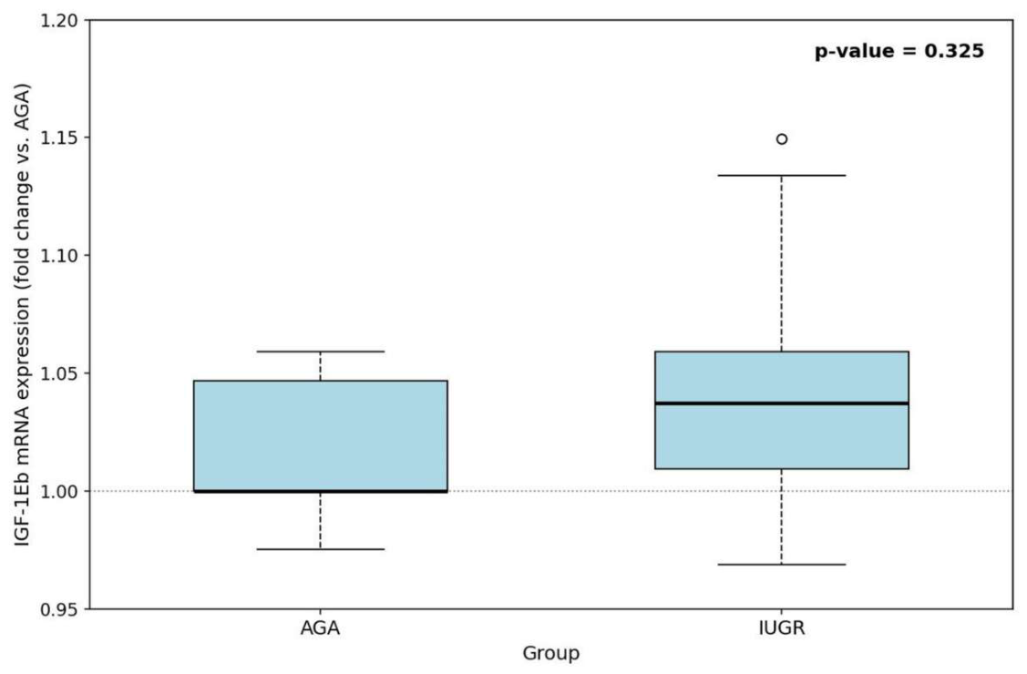
Figure 2.
Enhanced immunohistochemical expression of IGF-1Eb in the perivillous trophoblast.
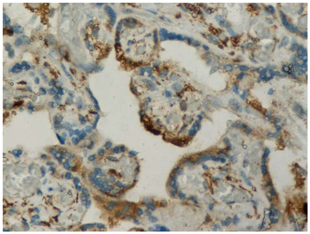
Figure 3.
IGF-1Eb immunoreactivity scores ranging from moderate to high in perivillous syncytiotrophoblasts of third-trimester human placentas.
Figure 3.
IGF-1Eb immunoreactivity scores ranging from moderate to high in perivillous syncytiotrophoblasts of third-trimester human placentas.
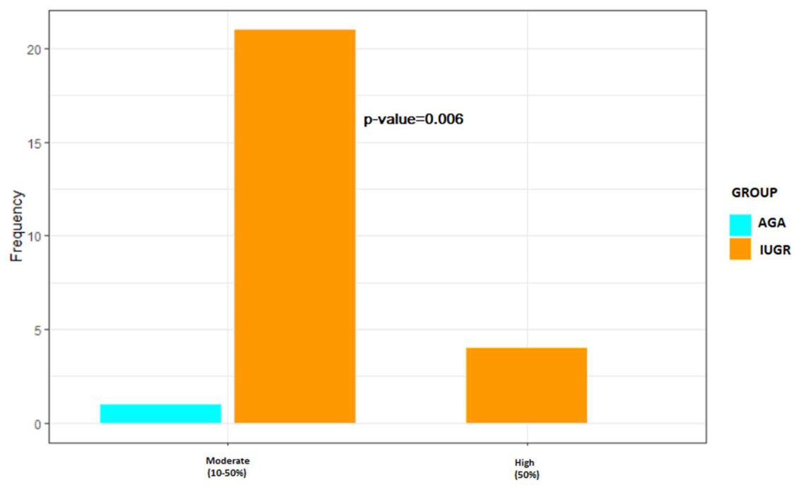
Figure 4.
Representative weak, moderate, and strong IGF-1Eb immunoreactivity in perivillous syncytiotrophoblasts of human third-trimester placentas.
Figure 4.
Representative weak, moderate, and strong IGF-1Eb immunoreactivity in perivillous syncytiotrophoblasts of human third-trimester placentas.
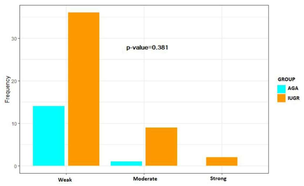
Figure 5.
Immunohistochemical expression of IGF-1Eb in extravillous trophoblastic cells.
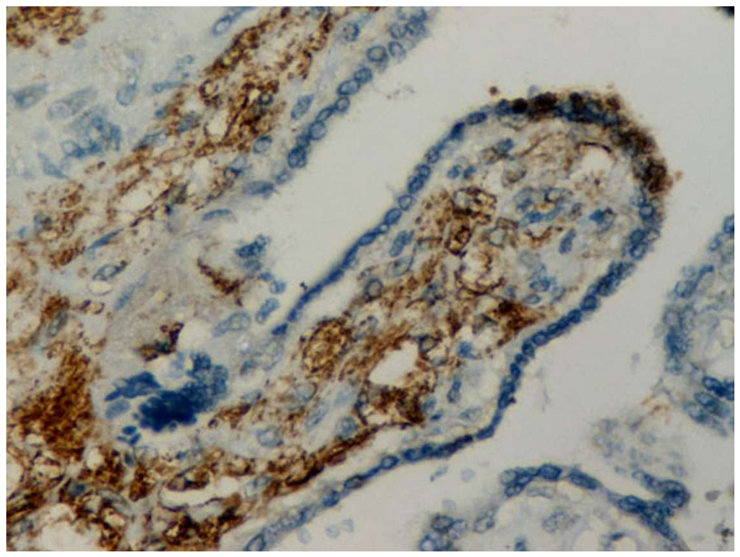
Figure 6.
Positive and negative IGF-1Eb immunohistochemical expression in extravillous cytotrophoblasts of human placentas obtained during third-trimester pregnancies.
Figure 6.
Positive and negative IGF-1Eb immunohistochemical expression in extravillous cytotrophoblasts of human placentas obtained during third-trimester pregnancies.
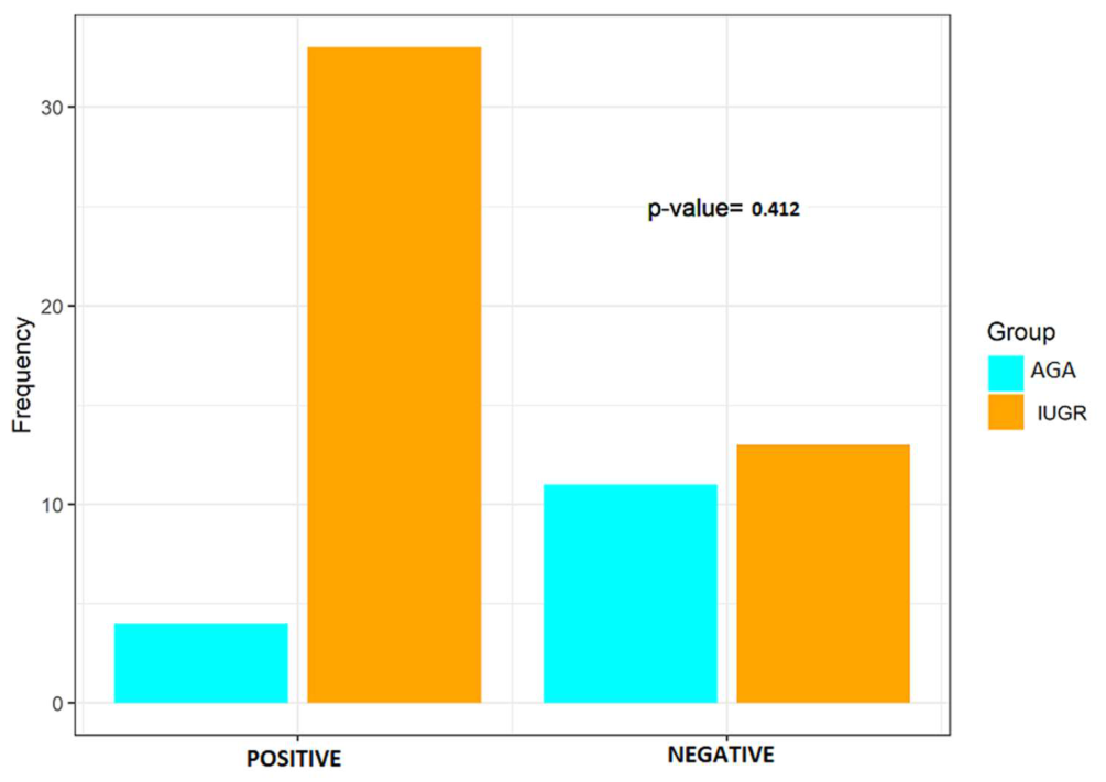
Figure 7.
Immunohistochemically assessed intensity of IGF-1Eb expression.
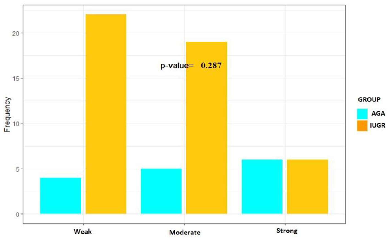
Figure 8.
Immunohistochemical expression of IGF-1Eb in the endothelium of fetal villous vessels in IUGR placenta. Positive perivillous trophoblastic cells are also seen on the tip of the chorionic villous.
Figure 8.
Immunohistochemical expression of IGF-1Eb in the endothelium of fetal villous vessels in IUGR placenta. Positive perivillous trophoblastic cells are also seen on the tip of the chorionic villous.
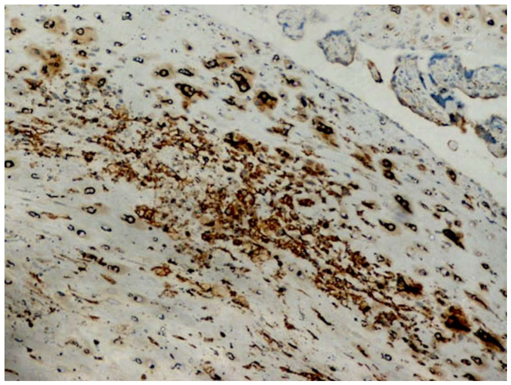

Table I.
Eb expression in the perivillous syncytiotrophoblasts.
| Clinicopathological parameters | AGA (%) | IUGR (%) | p-value | ||||
|---|---|---|---|---|---|---|---|
| Negative | Moderate | High | Negative | Moderate | High | ||
| Total | 14 (93.3) | 1 (6.7) | 0 (0.0) | 22 (46.8) | 21 (44.7) | 4 (8.5) | 0.006 |
| Age, years | |||||||
| <40 | 13 (92.9) | 1 (7.1) | 0 (0.0) | 18 (48.6) | 17 (45.9) | 2 (5.4) | 0.015 |
| >40 | 1 (100.0) | 0 (0.0) | 0 (0.0) | 2 (33.3) | 2 (33.3) | 2 (33.3) | 0.459 |
| Gestational age, weeks | |||||||
| <2,500 | 0 (0.0) | 0 (0.0) | 0 (0.0) | 11 (37.9) | 15 (51.7) | 3 (10.3) | - |
| >2,500 | 14 (93.3) | 1 (6.7) | 0 (0.0) | 5 (50.0) | 4 (40.0) | 1 (10.0) | 0.043 |
| Placental weight, g | |||||||
| <400 | 0 (0.0) | 0 (0.0) | 0 (0.0) | 17 (54.8) | 12 (38.7) | 2 (6.5) | - |
| >400 | 14 (93.3) | 1 (6.7) | 0 (0.0) | 5 (31.2) | 9 (56.2) | 2 (12.5) | 0.002 |
| BMI, Kg/m2 | |||||||
| <30 | 11 (91.7) | 1 (8.3) | 0 (0.0) | 2 (18.2) | 7 (63.6) | 2 (18.2) | 0.002 |
| >30 | 3 (100.0) | 0 (0.0) | 0 (0.0) | 1 (16.7) | 4 (66.7) | 1 (16.7) | 0.060 |
| Fetal sex, % | |||||||
| Male | 6 (85.7) | 1 (14.3) | 0 (0.0) | 3 (37.5) | 4 (50.0) | 1 (12.5) | 0.153 |
| Female | 8 (100.0) | 0 (0.0) | 0 (0.0) | 5 (25.0) | 13 (65.0) | 2 (10.0) | 0.002 |
| Histopathology, % | |||||||
| MVM | 11 (91.7) | 1 (8.3) | 0 (0.0) | 15 (42.9) | 16 (45.7) | 4 (11.4) | 0.015 |
| MPFD | 0 (0.0) | 0 (0.0) | 0 (0.0) | 3 (75.0) | 1 (25.0) | 0 (0.0) | - |
| FVM | 0 (0.0) | 0 (0.0) | 0 (0.0) | 1 (100.0) | 0 (0.0) | 0 (0.0) | - |
| VUE | 1 (100.0) | 0 (0.0) | 0 (0.0) | 4 (80.0) | 1 (20.0) | 0 (0.0) | - |
| DVM | 2 (100.0) | 0 (0.0) | 0 (0.0) | 2 (40.0) | 3 (60.0) | 0 (0.0) | - |
| Other | 0 (0.0) | 0 (0.0) | 0 (0.0) | 3 (75.0) | 1 (25.0) | 0 (0.0) | - |
Table II.
Eb immunohistochemical expression in the perivillous syncytiotrophoblast of AGA and IUGR human placentas obtained from third-trimester pregnancies.
Table II.
Eb immunohistochemical expression in the perivillous syncytiotrophoblast of AGA and IUGR human placentas obtained from third-trimester pregnancies.
| Clinicopathological parameters | AGA (%) | IUGR (%) | p-value | ||||
|---|---|---|---|---|---|---|---|
| Low | Moderate | Strong | Low | Moderate | Strong | ||
| Total | 14 (93.3) | 1 (6.7) | 0 (0.0) | 36 (76.6) | 9 (19.1) | 2(4.3) | 0.381 |
| Age, years | |||||||
| <40 | 13 (92.9) | 1 (7.1) | 0 (0.0) | 29 (78.4) | 6 (16.2) | 2 (5.4) | 0.444 |
| >40 | 1 (100.0) | 0 (0.0) | 0 (0.0) | 4 (66.7) | 2 (33.3) | 0 (0.0) | 0.495 |
| Gestational age, weeks | |||||||
| <37 | 0 (0.0) | 0 (0.0) | 0 (0.0) | 18 (69.2) | 6 (23.1) | 2 (7.7) | - |
| >37 | 14 (93.3) | 1 (6/7) | 0 (0.0) | 18 (85.7) | 3 (14.3) | 0 (0.0) | 0.626 |
| Neonatal weight, g | |||||||
| <2,500 | 0 (0.0) | 0 (0.0) | 0 (0.0) | 21 (72.4) | 6 (20.7) | 2 (6.9) | - |
| >2,500 | 14 (93.3) | 1 (6.7) | 0 (0.0) | 8 (80.0) | 2 (20.0) | 0 (0.0) | 1.000 |
| Placental weight, g | |||||||
| <400 | 0 (0.0) | 0 (0.0) | 0 (0.0) | 22 (71.0) | 7 (22.6) | 2 (6.5) | - |
| >400 | 14 (93.3) | 1 (6.7) | 0 (0.0) | 14 (87.5) | 2 (12.5) | 0 (0.0) | 1.000 |
| BMI, Kg/m2 | |||||||
| <30 | 11 (91.7) | 1 (8.3) | 0 (0.0) | 9 (81.8) | 2 (18.2) | 0 (0.0) | 0.484 |
| >30 | 3 (100.0) | 0 (0.0) | 0 (0.0) | 5 (83.3) | 1 (16.7) | 0 (0.0) | 0.453 |
| Fetal sex, % | |||||||
| Male | 6 (85.7) | 1 (14.3) | 0 (0.0) | 6 (75.0) | 2 (25.0) | 0 (0.0) | 0.605 |
| Female | 8 (100.0) | 0 (0.0) | 0 (0.0) | 15 (75.0) | 3 (15.0) | 0 (0.0) | 0.296 |
| Histopathology, % | |||||||
| MVM | 11 (91.7) | 1 (8.3) | 0 (0.0) | 27 (77.1) | 6 (17.1) | 2 (5.7) | 0.553 |
| MPFD | 0 (0.0) | 0 (0.0) | 0 (0.0) | 2 (50.0) | 2 (50.0) | 0 (0.0) | - |
| FVM | 0 (0.0) | 0 (0.0) | 0 (0.0) | 1 (100.0) | 0 (0.0) | 0 (0.0) | - |
| VUE | 1 (100.0) | 0 (0.0) | 0 (0.0) | 4 (80.0) | 1 (20.0) | 0 (0.0) | - |
| DVM | 2 (100.0) | 0 (0.0) | 0 (0.0) | 4 (80.0) | 1 (20.0) | 0 (0.0) | - |
Table III.
Eb in extravillous trophoblasts of normal and IUGR human third-trimester placentas.
| Clinicopathological parameters | AGA (%) | IUGR (%) | p-value | ||
|---|---|---|---|---|---|
| Negative | Positive | Negative | Positive | ||
| Total | 4 (26.7) | 11 (73.3) | 18 (38.3) | 29 (61.7) | 0.412 |
| Age, years | |||||
| <40 | 4 (28.6) | 10 (71.4) | 14 (37.8) | 23 (62.2) | 0.537 |
| >40 | 0 (0.0) | 1 (100.0) | 3 (50.0) | 3 (50.0) | 0.350 |
| Gestational age, weeks | |||||
| <37 | 0 (0.0) | 0 (0.0) | 7 (26.9) | 19 (73.1) | - |
| >37 | 4 (26.7) | 11 (73.3) | 11 (52.4) | 10(47.6) | 0.123 |
| Neonate weight, g | |||||
| <2,500 | 0 (0.0) | 0 (0.0) | 13 (44.8) | 16 (55.2) | - |
| >2,500 | 4 (26.7) | 11 (73.3) | 5 (50.0) | 5 (50.0) | 0.234 |
| Placental weight, g | |||||
| <400 | 0 (0.0) | 0 (0.0) | 13 (41.9) | 18 (58.1) | - |
| >400 | 4 (26.7) | 11 (73.3) | 5 (31.3) | 11 (68.8) | 0.779 |
| BMI, Kg/m2 | |||||
| <30 | 4 (33.3) | 8 (66.7) | 4 (36.4) | 7 (63.6) | 0.879 |
| >30 | 0 (0.0) | 3 (100.0) | 1 (16.7) | 5 (83.3) | 0.453 |
| Fetal sex, % | |||||
| Male | 1 (14.3) | 6 (85.7) | 2 (25.0) | 6 (75.0) | 0.605 |
| Female | 3 (37.5) | 5 (62.5) | 8 (40.0) | 12 (60.0) | 0.903 |
| Histopathology, % | |||||
| MVM | 2 (16.7) | 10 (83.3) | 13 (34.3) | 23 (65.7) | 0.249 |
| MPFD | 0 (0.0) | 0 (0.0) | 3 (75) | 1 (25) | - |
| FVM | 0 (0.0) | 0 (0.0) | 1 (100.0) | 0 (0.0) | - |
| VUE | 0 (0.0) | 1 (100.0) | 3 (60.0) | 2 (40.0) | 0.273 |
| DVM | 2 (100.0) | 0 (0.0) | 1 (20.0) | 4 (80.0) | 0.053 |
| Other | 0 (0.0) | 0 (0.0) | 2 (50.0) | 2 (50.0) | - |
Table IV.
Eb immunohistochemical staining intensity in extravillous trophoblasts of third-trimester human placentas.
Table IV.
Eb immunohistochemical staining intensity in extravillous trophoblasts of third-trimester human placentas.
| Clinicopathological parameters | AGA (%) | IUGR (%) | p-value | ||||
|---|---|---|---|---|---|---|---|
| Weak | Moderate | Strong | Weak | Moderate | Strong | ||
| Total | 11 (73.3) | 4 (26.7) | 0 (0.0) | 30 (65.2) | 9 (19.6) | 7 (15.2) | 0.287 |
| Age, years | |||||||
| <40 | 10 (71.4) | 4 (28.6) | 0 (0.0) | 19 (70.4) | 4 (14.8) | 4 (14.8) | 0.292 |
| >40 | 1 (100.0) | 0 (0.0) | 0 (0.0) | 10 (6.7) | 4 (26.7) | 1 (6.7) | 1.000 |
| Gestational age, weeks | |||||||
| <37 | 0 (0.0) | 0 (0.0) | 0 (0.0) | 13 (52.0) | 6 (24.0) | 6 (24.0) | - |
| >37 | 1 (73.3) | 4 (26.7) | 0 (0.0) | 17 (81.0) | 3 (14.3) | 1 (4.8) | 0.809 |
| Neonate weight, g | |||||||
| <2,500 | 0 (0.0) | 0 (0.0) | 0 (0.0) | 16 (57.2) | 6 (21.4) | 6 (21.4) | - |
| >2,500 | 11 (73.3) | 4 (26.7) | 0 (0.0) | 9 (90.0) | 1 (10.0) | 0 (0.0) | 0.615 |
| Placental weight, g | |||||||
| <400 | 0 (0.0) | 0 (0.0) | 0 (0.0) | 15 (50.0) | 8 (26.7) | 7 (23.3) | - |
| >400 | 11 (73.3) | 4 (26.7) | 0 (0.0) | 15 (93.8) | 1 (6.2) | 0 (0.0) | 0.172 |
| BMI, Kg/m2 | |||||||
| <30 | 0 (0.0) | 0 (0.0) | 0 (0.0) | 22 (71.0) | 2 (22.6) | 2 (6.5) | - |
| >30 | 14 (93.3) | 1 (6.7) | 0 (0.0) | 14 (87.5) | 2 (12.5) | 0 (0.0) | 1.000 |
| Fetal sex, % | |||||||
| Male | 11 (91.7) | 1 (8.3) | 0 (0.0) | 9 (81.8) | 2 (18.2) | 0 (0.0) | 0.484 |
| Female | 3 (100.0) | 0 (0.0) | 0 (0.0) | 5 (83.3) | 1 (16.7) | 0 (0.0) | 0.453 |
| Histopathology, % | |||||||
| MVM | 10 (83.3) | 2 (16.7) | 0 (0.0) | 25 (73.5) | 4 (11.8) | 5 (14.7) | 0.523 |
| MPFD | 0 (0.0) | 0 (0.0) | 0 (0.0) | 2 (50.0) | 2 (50.0) | 0 (0.0) | - |
| FVM | 0 (0.0) | 0 (0.0) | 0 (0.0) | 1 (100.0) | 0 (0.0) | 0 (0.0) | - |
| VUE | 1 (100.0) | 0 (0.0) | 0 (0.0) | 2 (40.0) | 3 (60.0) | 0 (0.0) | - |
| DVM | 0 (0.0) | 2 (100.0) | 0 (0.0) | 2 (40.0) | 2 (40.0) | 1 (20.0) | - |
| Other | 0 (0.0) | 0 (0.0) | 0 (0.0) | 2 (50.0) | 1 (25.0) | 1 (25.0) | - |
Table V.
b immunopositivity within the endothelial cells of maternal and fetal blood vessels in human placentas obtained from third-trimester pregnancies.
Table V.
b immunopositivity within the endothelial cells of maternal and fetal blood vessels in human placentas obtained from third-trimester pregnancies.
| Clinicopathological parameters | Maternal vessels | Fetal stem-intermediate vessels | Fetal distal-peripheral vessels | ||||||
|---|---|---|---|---|---|---|---|---|---|
| AGA | IUGR | p-value | AGA | IUGR | p-value | AGA | IUGR | p-value | |
| Total | 2 (13.3) | 35 (74.5) | <0.001 | 3 (20.0) | 36 (76.6) | <0.001 | 2 (13.3) | 20 (43.5) | 0.061 |
| Age | |||||||||
| <40 | 2 (14.3) | 27 (73.0) | <0.001 | 3 (21.4) | 28 (75.7) | <0.001 | 2 (14.3) | 16 (43.2) | 0.053 |
| >40 | 0 (0.0) | 5 (83.3) | 0.088 | 0 (0.0) | 5 (83.3) | 0.088 | 0 (0.0) | 3 (50.0) | 0.350 |
| Gestational age, weeks | |||||||||
| <37 | 0 (0.0) | 18 (69.2) | - | 0 (0.0) | 19 (73.1) | - | 0 (0.0) | 10 (38.5) | - |
| >37 | 2 (13.3) | 17 (81.0) | <0.001 | 3 (20.0) | 17 (81.0) | 0.001 | 2 (13.3) | 10 (50.0) | 0.034 |
| Newborn’s weight | |||||||||
| <2,500 | 0 (0.0) | 25 (86.2) | - | 0 (0.0) | 26 (89.7) | - | 0 (0.0) | 13 (46.4) | - |
| >2,500 | 2 (13.3) | 8 (80.0) | 0.002 | 3 (20.0) | 8 (80.0) | 0.005 | 2 (13.3) | 6 (60.0) | 0.028 |
| Placental weight, g | |||||||||
| <400 | 0 (0.0) | 23 (74.2) | - | 0 (0.0) | 22 (71.0) | - | 0 (0.0) | 8 (26.7) | - |
| >400 | 2 (13.3) | 12 (75.0) | 0.001 | 3 (20.0) | 14 (87.5) | <0.001 | 2 (13.3) | 12 (75.0) | 0.001 |
| BMI, Kg/m2 | |||||||||
| <30 | 2 (16.7) | 11 (100.0) | <0.001 | 3 (25.9) | 11 (100.0) | <0.001 | 2 (16.7) | 11 (100.0) | <0.001 |
| >30 | 0 (0.0) | 4 (66.7) | 0.058 | 0 (0.0) | 5 (83.3) | 0.018 | 0 (0.0) | 4 (66.6) | 0.058 |
| Fetal sex, % | |||||||||
| Male | 1 (14.3) | 7 (87.5) | 0.005 | 2 (28.6) | 8 (100.0) | 0.003 | 2 (28.6) | 6 (75.0) | 0.072 |
| Female | 1 (12.5) | 17 (85.0) | <0.01 | 1 (12.5) | 17 (85.0) | <0.001 | 0 (0.0) | 12 (60.0) | 0.004 |
| Histopathology | |||||||||
| MVM | 2 (16.7) | 25 (71.4) | 0.002 | 3 (25.0) | 26 (74.3) | 0.005 | 2 (16.7) | 17 (50.0) | 0.086 |
| MPFD | 0 (0.0) | 2 (66.7) | - | 0 (0.0) | 2 (66.7) | - | 0 (0.0) | 0 (0.0) | - |
| FVM | 0 (0.0) | 1 (100.0) | - | 0 (0.0) | 1 (100.0) | - | 0 (0.0) | 0 (0.0) | - |
| VUE | 0 (0.0) | 4 (80.0) | - | 0 (0.0) | 3 (60.0) | - | 0 (0.0) | 0 (0.0) | - |
| DVM | 0 (0.0) | 3 (60.0) | - | 0 (0.0) | 3 (60.0) | - | 0 (0.0) | 2 (40.0) | - |
| Other | 0 (0.0) | 3 (75.0) | - | 0 (0.0) | 3 (75.0) | - | 0 (0.0) | 1 (25.0) | - |
Disclaimer/Publisher’s Note: The statements, opinions and data contained in all publications are solely those of the individual author(s) and contributor(s) and not of MDPI and/or the editor(s). MDPI and/or the editor(s) disclaim responsibility for any injury to people or property resulting from any ideas, methods, instructions or products referred to in the content. |
© 2026 by the authors. Licensee MDPI, Basel, Switzerland. This article is an open access article distributed under the terms and conditions of the Creative Commons Attribution (CC BY) license.
Copyright: This open access article is published under a Creative Commons CC BY 4.0 license, which permit the free download, distribution, and reuse, provided that the author and preprint are cited in any reuse.