

Submitted:
27 June 2026
Posted:
01 July 2026
You are already at the latest version
Abstract
Background/Objectives: To evaluate whether automated three-dimensional (3D) volumetric body composition metrics derived from the Body and Organ Analysis (BOA) algorithm provide prognostic information beyond body mass index (BMI) for overall survival (OS) in patients with surgically treated head and neck squamous cell carcinoma (HNSCC), and to compare cervical (C1–C7) measurements with thoracic and abdominal benchmarks.
Methods: This single-center retrospective cohort study included 391 surgically treated HNSCC patients at Universitätsmedizin Greifswald (2010–2019). Patients with definitive (chemo)radiation (no surgery) or non-standard adjuvant chemotherapy alone (n=13) were excluded; thus, all included patients had primary surgical management. Tumor staging was retrieved from institutional records for all 391 patients. For patients with documented ongoing follow-up but without a specific last-contact date, survival status was verified by chart review where possible, and patients confirmed alive at the institutional database snapshot date were administratively censored on 28 January 2025. Pretreatment CT scans were processed using the open-source BOA algorithm (v0.1.3) to obtain volumetric tissue measurements of muscle, bone, and adipose compartments across abdominal, thoracic, and cervical (C1–C7) regions. Composite indices — Sarcopenia Index (SI = Muscle/Bone) and Myosteatotic Fat Index (MFI = IMAT/TAT) — were derived. Univariate and multivariate Cox proportional hazards models were used. The primary clinically adjusted model included cervical SI, age, sex, tumor site, UICC stage, treatment modality, the Head and Neck Charlson Comorbidity Index (HN-CCI), and secondary malignancy; a minimally adjusted model (HN-CCI and secondary malignancy) was retained as a supportive analysis. Leukocytes and postoperative CRP were examined in a sensitivity model. Reporting followed the STROBE checklist.
Results: Of 391 evaluable cases (91% male, median follow-up 5.0 years, 141 deaths), BMI showed only a weak univariate association with overall survival (HR 0.84, p=0.052) and was not independently associated with OS after adjustment (multivariate HR 0.88, p=0.15). In contrast, in the minimally adjusted supportive model the cervical Sarcopenia Index (Cerv SI, Muscle/Bone) was associated with overall survival (HR 0.71, 95% CI 0.58–0.88, p=0.002); in the primary clinically adjusted model including age, sex, tumor site, UICC stage, treatment modality, HN-CCI, and secondary malignancy, cervical SI remained independently associated with OS (HR 0.75, 95% CI 0.61–0.92, p=0.007; n=254). Thoracic Myosteatotic Fat Index (Thor MFI) was associated with worse overall survival in the minimally adjusted supportive model (HR 1.35, p=0.002) and remained associated after extended oncologic adjustment (HR 1.26, p=0.013). Harrell's concordance index for overall survival prediction was 0.60 for cervical SI and 0.55 for BMI; the difference was modest. Inter-regional SI correlations were strong between abdomen and thorax (r=0.69) but only moderate for cervical–thoracic (r=0.42). An in-sample comparison showed that a BOA-derived single-level C3 Sarcopenia Index was non-prognostic (C-index 0.52) and weakly correlated with the volumetric C1–C7 SI (r=0.04), suggesting that the volumetric and single-vertebra-level metrics derived from the BOA pipeline capture different information. In paired bootstrap comparisons, cervical SI showed a modest but significant C-index improvement over BMI (ΔC = +0.087, p = 0.038) but did not significantly outperform the BOA-derived C3 SI, ASA classification, or thoracic SI.
Conclusions: BOA-derived volumetric body composition indices, particularly the cervical Sarcopenia Index, are associated with overall survival in surgically treated HNSCC patients and remain prognostic after adjustment for established oncologic confounders, whereas BMI is not. Cervical C1–C7 BOA analysis enables body composition assessment from routine head and neck CT and may provide an objective imaging-derived marker for future risk-stratification studies. Prospective validation and formal comparison with established C3/L3 methods are warranted before routine clinical use.
Keywords:
body composition
; sarcopenia
; head and neck cancer
; BOA algorithm
; computed tomography
; volumetric analysis
; prognosis
; cervical vertebra
Introduction
Head and neck squamous cell carcinoma (HNSCC) represents one of the most common malignancies worldwide [31], with treatment decisions guided primarily by tumor staging, site, and comorbidity burden [25]. Despite advances in surgical techniques and adjuvant therapy, individual prognosis varies substantially, highlighting the need for additional prognostic biomarkers that capture patient-level vulnerability beyond tumor characteristics.
Sarcopenia—the pathological loss of skeletal muscle mass and function—has emerged as a robust independent prognostic factor across multiple malignancies. In HNSCC specifically, a recent multi-level meta-analysis of 63 studies (n=14,804) confirmed that pretreatment radiologically defined sarcopenia is consistently associated with worse survival (log OR 0.81, p<0.001) and increased treatment complications [4]. Earlier meta-analyses by Hua et al. [2] (HR 1.97, 95% CI 1.71–2.26 for OS) and Wong et al. [3] (HR 1.98, 95% CI 1.64–2.39) corroborated these findings across heterogeneous HNSCC populations.
Traditionally, image-based sarcopenia in HNSCC has been assessed using single-slice cross-sectional muscle area at the third lumbar (L3) or third cervical (C3) vertebral level, measured either manually or semi-automatically. This approach, while validated, has limitations: it captures a two-dimensional snapshot of three-dimensional tissue, is subject to inter-observer variability, and requires specific vertebral landmarks that may not be included in routine head and neck imaging. Meanwhile, body mass index (BMI), the most widely used anthropometric measure, fails to distinguish between muscle and fat mass. This gives rise to the “BMI paradox” in HNSCC, whereby overweight patients paradoxically show improved survival compared to normal-weight patients [15].
Recent advances in deep learning have enabled fully automated, three-dimensional volumetric body composition analysis (BCA) from routine clinical CT scans. The Body and Organ Analysis (BOA) algorithm [1], developed at the University Hospital Essen, combines body-composition segmentation with the TotalSegmentator framework to enable automated tissue quantification throughout the entire scanned volume. BOA segments muscle, bone, and multiple adipose tissue compartments (subcutaneous, visceral, intramuscular, epicardial, pericardial) with high accuracy (Dice >0.95) and processes a complete CT in under one minute [1,17,18]. Importantly, BOA provides volumetric measurements—not just single-slice areas — potentially capturing body composition more completely.
Several groups have demonstrated that volumetric BCA may carry additional prognostic value compared to single-slice analysis. In lung cancer, Künnemann et al. [10] showed that a volumetric Sarcopenia Index (Muscle/Bone ratio) outperformed conventional L3-based single-slice measurements in predicting OS. Jung et al. [22] demonstrated in 36,317 UK Biobank participants that volumetric body composition measures predicted mortality independently of BMI, with associations attenuated when using single-slice metrics. However, in a colorectal cancer cohort, Anyene et al. [11] found that single-slice L3 and volumetric metrics had similar prognostic performance, suggesting the advantage may be context-dependent.
For HNSCC patients, in whom abdominal imaging is frequently unavailable, the possibility of deriving prognostic body-composition information from routine neck CT is of particular clinical relevance. Cervical muscle measurements at C3 have been validated against L3 references [5,6,8,19], and recent work by Barajas Ordonez et al. [7] (n=904) confirmed that both skeletal muscle area and muscle radiation attenuation at C3 predict OS in HNSCC. However, these studies used single-slice 2D measurements. Whether volumetric 3D cervical body composition from automated algorithms provides comparable or superior prognostic value to thoracic and abdominal measurements has not been investigated.
The aim of this study was threefold: (1) to evaluate whether BOA-derived 3D volumetric body composition indices provide prognostic information beyond BMI for overall survival (OS) and disease-free survival (DFS) in surgically treated HNSCC; (2) to assess whether cervical C1–C7 volumetric measurements provide prognostic information comparable to thoracic and abdominal measurements; and (3) to evaluate the robustness of the primary finding after adjustment for established oncologic confounders including tumor stage.
Materials and Methods
Study Design and Participants
This retrospective single-center cohort study included consecutive patients with histologically confirmed HNSCC who underwent primary curative surgery ± adjuvant (chemo)radiation at the Department of Otorhinolaryngology, Head and Neck Surgery, Universitätsmedizin Greifswald between 2010 and 2019. Patients were eligible if they had (a) a documented first diagnosis date, (b) at least one pretreatment CT scan obtained before surgery and encompassing the neck and, where available, the thorax and abdomen, and (c) available follow-up data. The study was reported in accordance with the Strengthening the Reporting of Observational Studies in Epidemiology (STROBE) guidelines for cohort studies. Before initiation, the ethics committee approved the study with the registry number BB 099/24 on 5 July 2024. Patients who received definitive (chemo)radiation without surgery or non-standard adjuvant chemotherapy alone (n=13) were excluded because the study was designed as a surgically treated cohort; these cases had been captured in the source database but fell outside the intended surgical inclusion criteria, so that the cohort uniformly reflected surgically treated HNSCC.
Body Composition Analysis
Pretreatment CT scans were processed using the open-source BOA algorithm (v0.1.3; github.com/UMEssen/Body-and-Organ-Analysis) [1]. BOA employs a multiresolution nnU-Net architecture to segment body regions and tissues and combines it with TotalSegmentator [18] for organ identification. The algorithm provides volumetric measurements (in mL) for the following tissue compartments within defined body regions: skeletal muscle, bone, subcutaneous adipose tissue (SAT), visceral adipose tissue (VAT), intramuscular/intermuscular adipose tissue (IMAT), epicardial adipose tissue (EAT), pericardial adipose tissue (PAT), and total adipose tissue (TAT). Measurements were obtained separately for three body regions: the abdominal cavity (excluding extremities), the thoracic cavity (excluding extremities), and the cervical spine (C1–C7, excluding extremities). Quality control was performed using the BOA status output; only measurements with “OK” status were retained.
CT examinations were acquired as part of routine clinical staging. For the primary analysis, the staging CT closest to surgery was used; across the cohort, the staging CT was obtained a median of 17 days before surgery (IQR 10–25 days), with 86% acquired within 30 days before surgery and all scans (100%) obtained before the operative date. Where multiple eligible scans were available, the scan with the most complete cervical/thoracic/abdominal coverage and an acceptable BOA quality status was selected. CT examinations were acquired on a range of scanners in routine clinical use over the 2010–2019 study period. Across all processed CT series, the large majority were obtained on Siemens systems — most frequently the SOMATOM Sensation 16 and SOMATOM Definition Flash — with smaller numbers acquired on Philips and General Electric scanners. Reconstructed slice thickness ranged from 0.625 to 5 mm (median 2 mm). Reconstruction kernel, intravenous contrast phase, and tube voltage were not uniformly retrievable across this retrospective cohort (see Limitations).
In addition to the BOA internal “OK” status filter, a visual quality-control review was performed in a random subset of cervical segmentations and in outlier cases. Segmentations were checked for gross vertebral-level mismatch, incomplete C1–C7 coverage, dental-artifact-related segmentation failure, and erroneous inclusion or exclusion of non-target tissue. Three investigators independently reviewed a random subset of 35 cervical segmentations. No significant segmentation artifacts or errors were identified; specifically, no cases of dental-artifact-related segmentation failure, vertebral-level mismatch, or incomplete C1–C7 coverage were observed, and no cases required exclusion or re-processing on this basis. Representative cervical segmentations from this review are shown in Figure S2.
Derived Composite Indices
Based on prior work in lung cancer [10], two composite indices were calculated for each body region: (1) Sarcopenia Index (SI) = Muscle volume / Bone volume, a measure of relative muscle mass normalized to skeletal frame size; and (2) Myosteatotic Fat Index (MFI) = IMAT volume / TAT volume, a measure of fatty infiltration of muscle relative to total adiposity, reflecting muscle quality. For cervical analysis, tissue volumes from C1 through C7 were summed into a single cervical composite measurement.
Clinical Data and Outcomes
Demographic and clinical variables were extracted from institutional databases: age at diagnosis, sex, BMI (kg/m²), tumor site, TNM staging (UICC 7th and 8th editions, reflecting the editions in use across the 2010–2019 inclusion period), treatment modality, Head and Neck Charlson Comorbidity Index (HN-CCI), ASA physical status classification, preoperative leukocyte count, maximum postoperative C-reactive protein (CRP), secondary malignancy status (“secondary malignancy” as documented in clinical records), p16 status, and Clavien–Dindo graded complications and infections. ASA classification was used as a functional status measure because ECOG performance status data were incompletely and inconsistently documented in these retrospective records. This is a recognized limitation of retrospective HNSCC cohorts, as ECOG assessment is inherently subjective and subject to considerable inter-rater variability [26,27], and ASA has been shown to outperform ECOG as a predictor of overall survival in HNSCC patients treated with adjuvant (chemo)radiation [28]. The primary outcome was overall survival (OS), defined as time from first diagnosis to death from any cause or last follow-up. The secondary outcome was disease-free survival (DFS), defined as the time from diagnosis to the first documented recurrence or death from any cause, whichever occurred first. UICC stage was modeled as an ordinal stage-group variable (I–IV) as documented in the clinical record; this approach harmonized stage-group information across the UICC editions in use during the inclusion period.
Statistical Analysis
Continuous predictors were standardized (z-scored) for Cox regression to enable comparison of hazard ratios (HR) across variables with different scales. To address multiple testing, we predefined a hierarchy of analyses: the primary imaging marker was the cervical C1–C7 SI; secondary markers were the thoracic SI, thoracic MFI, and abdominal SI; exploratory markers were all remaining single-compartment volumes and the single-vertebra C3 comparator. In a sensitivity analysis, treating UICC stage as a categorical rather than ordinal covariate did not materially change the association between cervical SI and overall survival (HR 0.76, p = 0.012).
Univariate Cox proportional hazards models were fitted for each predictor. The primary clinically adjusted model (reported in Table 4) included the cervical SI together with age, sex, tumor site (larynx, oral cavity, oropharynx, and hypopharynx, with larynx as the reference category), UICC stage, treatment modality (surgery alone, surgery plus chemoradiation, and surgery plus radiotherapy, with surgery alone as the reference category), HN-CCI, and secondary malignancy; a parallel specification replaced UICC stage with T and N category separately. A minimally adjusted model including only HN-CCI and secondary malignancy (reported in Table 3) was retained as a supportive analysis. Treatment modality was included to account for broad differences in treatment course, but treatment coefficients were not interpreted causally because treatment selection reflects tumor risk, patient fitness, and multidisciplinary decision-making; the cervical SI association was essentially unchanged in models with and without treatment (HR 0.75 without versus HR 0.73 with treatment). Because maximum postoperative CRP is measured after the imaging biomarker is acquired and may lie on the causal pathway between frailty, surgical morbidity, and survival, leukocytes and CRP were not included in the primary pretreatment model but were examined in a separate sensitivity model (Table 3, Model E). Sensitivity analyses included stratification by imaging availability. The proportional hazards assumption was assessed using Schoenfeld residuals; in the primary clinically adjusted model, the cervical SI satisfied the proportional hazards assumption (p = 0.48), with minor deviations noted for sex and UICC stage that did not affect the cervical SI estimate. The linearity of BMI was assessed by comparing a restricted cubic spline model (four knots) with a linear term using a likelihood-ratio test, which showed no evidence of non-linearity (χ² = 0.67, df = 3, p = 0.88); BMI was therefore modeled as a linear term. Log-rank tests with median-split dichotomization were used for Kaplan–Meier survival curves. Harrell’s concordance index (C-index) was computed for discriminative ability. Pearson correlation coefficients quantified inter-regional agreement. Statistical significance was set at p<0.05 (two-sided). Analyses were performed using Python 3.12 with the lifelines and scipy packages.
Results
Cohort Characteristics
Of 404 patients in the database with valid survival data, 391 surgically treated HNSCC patients were included in the final analysis after exclusion of 13 patients with definitive nonsurgical treatment or non-standard adjuvant chemotherapy alone, using an administrative censoring cutoff of 28 January 2025 for patients confirmed alive (median follow-up: 5.0 years; IQR: 2.6–6.3). There were 356 males (91%) and 35 females (9%), with a mean age of 60.9 years. p16 status was tested in 34 patients (8.7%), of whom 17 (50%) were p16-positive. TNM (UICC 7th and 8th editions) staging was retrievable for all 391 patients (100%). The mean BMI was 26.6 ± 5.3 kg/m². Treatment approaches included surgery alone (n=173, 44%), surgery with radiation (n=84, 21%), and surgery with chemoradiation (n=134, 34%). A total of 141 deaths (36.1%) were recorded. Secondary malignancy was documented in 67 cases (17.1%). BOA-processed CT data were available for 256 patients (cervical, C1–C7), 222 (thoracic), and 159 (abdominal). Single-level C3 segmentation data — used as an in-sample BOA-derived single-vertebra comparator — were available for 248 patients. The overview is displayed in Table 1. Imaging availability was not random and was associated with UICC stage (χ² = 70.29, df = 12, p < 0.001; Supplementary Table S1): patients with multi-region imaging had a higher proportion of UICC III–IV disease than patients with cervical-only imaging, so regional comparisons are interpreted as exploratory. The cohort flow and per-region BOA availability are summarized in Supplementary Figure S1.
Univariate Survival Analysis
In univariate Cox regression with standardized predictors (Table 2), BMI showed no significant association with OS (HR 0.84, p=0.052, C-index 0.55). Among individual tissue volumes, thoracic muscle (HR 0.85, p=0.22) and thoracic SAT (HR 0.79, p=0.05) demonstrated non-significant protective trends. The composite indices performed significantly better: thoracic SI (p=0.002 by log-rank; C-index 0.602), cervical SI (p=0.002, C-index 0.604), and thoracic MFI (p<0.001; C-index 0.621) all showed significant discrimination. Median OS for patients with thoracic SI above the median was 8.9 years, versus 4.2 years for those below the median.
Notably, raw cervical muscle volume alone was not prognostic (p=0.47), whereas the cervical SI (Muscle/Bone ratio) was strongly associated with OS (p<0.001), supporting frame-size normalization using bone volume. In sex-stratified analysis, thoracic SI and cervical SI were significant in males (n=356), but the female subgroup (n=35) was underpowered. ASA classification was a significant univariate predictor of OS (HR 1.55 per SD, 95% CI 1.30–1.85, p<0.001). Patients with ASA 3–4 had a median OS of 6.3 years versus 11.9 years for ASA 1–2 (log-rank p<0.001), with mortality rates of 44.1% and 29.0%, respectively. The BOA-derived single-vertebra C3 Sarcopenia Index was also non-prognostic (HR 1.01, 95% CI 0.83–1.24, p=0.899), with a C-index of 0.520 indicating no discriminative ability over chance (Table 2).
Multivariate Survival Analysis
Minimally adjusted models are presented first for comparability with prior body-composition studies; the predefined primary clinically adjusted models are presented subsequently in Table 4. Table 3 shows the results of the minimally adjusted multivariate Cox regression. In models adjusted for HN-CCI and secondary malignancy:
- Cervical SI was independently associated with longer OS (HR 0.71, 95% CI 0.58–0.88, p=0.002)
- Thoracic MFI was associated with worse OS (HR 1.35, 95% CI 1.11–1.63, p=0.002)
- Thoracic SI was independently associated with OS (HR 0.72, 95% CI 0.57–0.90, p=0.005)
- BMI remained non-significant (HR 0.88, p=0.15)

Table 3.
Minimally adjusted and sensitivity Cox proportional hazards models for overall survival. HRs are per 1-SD increase. These models adjust for HN-CCI and secondary malignancy (with leukocytes and CRP in the sensitivity model, Model E) and are retained as supportive analyses; the primary clinically adjusted models are shown in Table 4.
Table 3.
Minimally adjusted and sensitivity Cox proportional hazards models for overall survival. HRs are per 1-SD increase. These models adjust for HN-CCI and secondary malignancy (with leukocytes and CRP in the sensitivity model, Model E) and are retained as supportive analyses; the primary clinically adjusted models are shown in Table 4.
| Model / Variable | n | HR | 95% CI | p-value |
| Model A: Thor SI + HN-CCI + Secondary Malignancy (n=221, 85 events) | ||||
| Thor Sarc. Index | 221 | 0.716 | 0.57 – 0.90 | 0.005 |
| HN-CCI | 1.145 | 0.92 – 1.42 | 0.225 | |
| Secondary malignancy | 1.382 | 1.15 – 1.66 | <0.001 | |
| Model B: Thor MFI + HN-CCI + Secondary Malignancy (n=221, 85 events) | ||||
| Thor MFI (IMAT/TAT) | 221 | 1.345 | 1.11 – 1.63 | 0.002 |
| HN-CCI | 1.109 | 0.89 – 1.38 | 0.353 | |
| Secondary malignancy | 1.382 | 1.15 – 1.66 | <0.001 | |
| Model C: Cerv SI + HN-CCI + Secondary Malignancy (n=254, 102 events) | ||||
| Cerv Sarc. Index | 254 | 0.714 | 0.58 – 0.88 | 0.002 |
| HN-CCI | 1.109 | 0.91 – 1.35 | 0.305 | |
| Secondary malignancy | 1.313 | 1.11 – 1.55 | 0.002 | |
| Model D: BMI + HN-CCI + Secondary Malignancy (n=376, 133 events) | ||||
| BMI | 376 | 0.876 | 0.73 – 1.05 | 0.154 |
| HN-CCI | 1.233 | 1.04 – 1.46 | 0.016 | |
| Secondary malignancy | 1.488 | 1.29 – 1.71 | <0.001 | |
| Model E: Cerv SI + HN-CCI + Secondary Malignancy + Leuko + CRP (sensitivity) (n=148, 63 events) | ||||
| Cerv Sarc. Index | 148 | 0.701 | 0.54 – 0.91 | 0.008 |
| HN-CCI | 1.202 | 0.94 – 1.54 | 0.149 | |
| Secondary malignancy | 1.296 | 1.04 – 1.61 | 0.021 | |
| Baseline leukocytes | 0.566 | 0.00 – 968.82 | 0.881 | |
| Max CRP postop | 0.770 | 0.15 – 3.92 | 0.753 | |
| Model F: Cerv SI + ASA + HN-CCI + Secondary Malignancy (n=254, 102 events) | ||||
| Cerv Sarc. Index | 254 | 0.757 | 0.61 – 0.94 | 0.011 |
| HN-CCI | 1.023 | 0.83 – 1.26 | 0.833 | |
| Secondary malignancy | 1.317 | 1.11 – 1.56 | 0.001 | |
| ASA | 1.256 | 1.00 – 1.57 | 0.046 | |
| Model G: BMI + ASA + HN-CCI + Secondary Malignancy (n=376, 133 events) | ||||
| BMI | 376 | 0.839 | 0.70 – 1.00 | 0.055 |
| HN-CCI | 1.076 | 0.90 – 1.29 | 0.424 | |
| Secondary malignancy | 1.446 | 1.25 – 1.67 | <0.001 | |
| ASA | 1.506 | 1.24 – 1.83 | <0.001 | |
| Model H: C3 SI + HN-CCI + Secondary Malignancy (n=246, 99 events) | ||||
| C3 Sarc. Index | 246 | 0.966 | 0.79 – 1.19 | 0.742 |
| HN-CCI | 1.167 | 0.96 – 1.42 | 0.126 | |
| Secondary malignancy | 1.364 | 1.15 – 1.61 | <0.001 | |
| Model I: C3 SI + C1–C7 Vol SI + HN-CCI + Secondary Malignancy (n=246, 99 events) | ||||
| C3 Sarc. Index (single-vertebra) | 246 | 0.991 | 0.81 – 1.21 | 0.933 |
| C1–C7 Volumetric SI | 0.722 | 0.58 – 0.89 | 0.002 | |
| HN-CCI | 1.134 | 0.93 – 1.38 | 0.216 | |
| Secondary malignancy | 1.285 | 1.08 – 1.53 | 0.004 | |
Model E is a postoperative inflammatory marker sensitivity model that includes baseline leukocytes and maximum postoperative CRP. Models F and G assess the incremental prognostic value of ASA. In Model F, both cervical SI and ASA remain associated with OS, suggesting that they provide partially complementary prognostic information. In Model G, ASA remains strongly associated with OS, whereas BMI shows only a weak association. Models H and I compare the BOA-derived single-vertebra C3 SI with the volumetric C1–C7 SI: the C3 SI is not prognostic, while the C1–C7 SI remains independently associated with OS. HR < 1 indicates a protective effect. Bold indicates p < 0.05.
Table 4.
Primary clinically adjusted Cox proportional hazards models for overall survival, adjusted for established oncologic confounders (age, sex, tumor site, UICC stage, treatment modality, HN-CCI, and secondary malignancy). HRs are per 1-SD increase for continuous predictors. Reference categories: tumor site = Larynx; treatment modality = Surgery alone. Bold indicates p<0.05.
Table 4.
Primary clinically adjusted Cox proportional hazards models for overall survival, adjusted for established oncologic confounders (age, sex, tumor site, UICC stage, treatment modality, HN-CCI, and secondary malignancy). HRs are per 1-SD increase for continuous predictors. Reference categories: tumor site = Larynx; treatment modality = Surgery alone. Bold indicates p<0.05.
| Model / Variable | n | HR | 95% CI | p-value |
| Model X1: Cerv SI + age + sex + site + UICC + tx + HN-CCI + 2nd malignancy (n=254, 102 events, C=0.67) | ||||
| Cerv Sarc. Index | 254 | 0.753 | 0.61 – 0.93 | 0.007 |
| Age | 1.109 | 0.89 – 1.38 | 0.348 | |
| Sex (male) | 1.094 | 0.88 – 1.37 | 0.429 | |
| HN-CCI | 1.084 | 0.89 – 1.32 | 0.419 | |
| Secondary malignancy | 1.293 | 1.09 – 1.53 | 0.003 | |
| UICC stage | 1.062 | 0.85 – 1.33 | 0.599 | |
| Site: Hypopharynx | 1.040 | 0.56 – 1.92 | 0.900 | |
| Site: Oral cavity | 0.753 | 0.47 – 1.21 | 0.244 | |
| Site: Oropharynx | 0.865 | 0.50 – 1.49 | 0.600 | |
| Treatment: Surgery+CRT | 1.647 | 0.98 – 2.76 | 0.058 | |
| Treatment: Surgery+RT | 1.216 | 0.73 – 2.02 | 0.451 | |
| Model X2: Cerv SI + age + sex + site + T + N + tx + HN-CCI + 2nd malignancy (n=254, 102 events, C=0.70) | ||||
| Cerv Sarc. Index | 254 | 0.785 | 0.64 – 0.97 | 0.022 |
| Age | 1.128 | 0.91 – 1.40 | 0.273 | |
| Sex (male) | 1.093 | 0.87 – 1.37 | 0.435 | |
| HN-CCI | 1.084 | 0.89 – 1.32 | 0.424 | |
| Secondary malignancy | 1.276 | 1.08 – 1.51 | 0.005 | |
| T category | 1.322 | 1.09 – 1.61 | 0.005 | |
| N category | 1.056 | 0.84 – 1.33 | 0.646 | |
| Site: Hypopharynx | 0.949 | 0.52 – 1.74 | 0.866 | |
| Site: Oral cavity | 0.802 | 0.50 – 1.30 | 0.372 | |
| Site: Oropharynx | 0.997 | 0.57 – 1.74 | 0.992 | |
| Treatment: Surgery+CRT | 1.436 | 0.85 – 2.43 | 0.176 | |
| Treatment: Surgery+RT | 1.016 | 0.61 – 1.68 | 0.949 | |
| Model X3: Cerv SI + ASA + age + sex + site + UICC + tx + HN-CCI + 2nd malignancy (n=254, 102 events, C=0.67) | ||||
| Cerv Sarc. Index | 254 | 0.782 | 0.64 – 0.96 | 0.021 |
| ASA | 1.214 | 0.98 – 1.51 | 0.078 | |
| Age | 1.067 | 0.86 – 1.33 | 0.560 | |
| Sex (male) | 1.101 | 0.88 – 1.38 | 0.397 | |
| HN-CCI | 1.029 | 0.84 – 1.26 | 0.784 | |
| Secondary malignancy | 1.292 | 1.09 – 1.53 | 0.003 | |
| UICC stage | 1.051 | 0.84 – 1.32 | 0.673 | |
| Site: Hypopharynx | 1.008 | 0.54 – 1.87 | 0.980 | |
| Site: Oral cavity | 0.758 | 0.47 – 1.22 | 0.258 | |
| Site: Oropharynx | 0.931 | 0.54 – 1.61 | 0.798 | |
| Treatment: Surgery+CRT | 1.663 | 0.99 – 2.80 | 0.055 | |
| Treatment: Surgery+RT | 1.240 | 0.75 – 2.06 | 0.406 | |
| Model X4: Thor SI + age + sex + site + UICC + tx + HN-CCI + 2nd malignancy (n=221, 85 events, C=0.67) | ||||
| Thor Sarc. Index | 221 | 0.773 | 0.61 – 0.98 | 0.030 |
| Age | 1.071 | 0.85 – 1.34 | 0.552 | |
| Sex (male) | 1.086 | 0.83 – 1.41 | 0.542 | |
| HN-CCI | 1.154 | 0.93 – 1.43 | 0.190 | |
| Secondary malignancy | 1.375 | 1.15 – 1.65 | <0.001 | |
| UICC stage | 1.086 | 0.85 – 1.39 | 0.511 | |
| Site: Hypopharynx | 0.840 | 0.45 – 1.58 | 0.590 | |
| Site: Oral cavity | 0.726 | 0.44 – 1.21 | 0.220 | |
| Site: Oropharynx | 0.959 | 0.54 – 1.71 | 0.886 | |
| Treatment: Surgery+CRT | 1.471 | 0.84 – 2.59 | 0.181 | |
| Treatment: Surgery+RT | 1.445 | 0.83 – 2.51 | 0.193 | |
| Model X5: Thor MFI + age + sex + site + UICC + tx + HN-CCI + 2nd malignancy (n=221, 85 events, C=0.68) | ||||
| Thor MFI (IMAT/TAT) | 221 | 1.262 | 1.05 – 1.52 | 0.013 |
| Age | 1.151 | 0.92 – 1.43 | 0.209 | |
| Sex (male) | 1.038 | 0.79 – 1.36 | 0.786 | |
| HN-CCI | 1.120 | 0.91 – 1.38 | 0.293 | |
| Secondary malignancy | 1.364 | 1.14 – 1.64 | <0.001 | |
| UICC stage | 1.096 | 0.86 – 1.40 | 0.469 | |
| Site: Hypopharynx | 0.882 | 0.47 – 1.67 | 0.699 | |
| Site: Oral cavity | 0.739 | 0.44 – 1.24 | 0.248 | |
| Site: Oropharynx | 0.983 | 0.55 – 1.75 | 0.953 | |
| Treatment: Surgery+CRT | 1.399 | 0.79 – 2.47 | 0.249 | |
| Treatment: Surgery+RT | 1.442 | 0.83 – 2.52 | 0.198 | |
| Model X6: BMI + age + sex + site + UICC + tx + HN-CCI + 2nd malignancy (n=376, 133 events, C=0.67) | ||||
| BMI | 376 | 0.915 | 0.77 – 1.09 | 0.315 |
| Age | 1.176 | 0.99 – 1.40 | 0.071 | |
| Sex (male) | 1.026 | 0.85 – 1.24 | 0.784 | |
| HN-CCI | 1.173 | 0.99 – 1.39 | 0.061 | |
| Secondary malignancy | 1.447 | 1.26 – 1.67 | <0.001 | |
| UICC stage | 1.148 | 0.94 – 1.40 | 0.175 | |
| Site: Hypopharynx | 1.245 | 0.72 – 2.15 | 0.431 | |
| Site: Oral cavity | 0.863 | 0.58 – 1.28 | 0.466 | |
| Site: Oropharynx | 1.175 | 0.73 – 1.89 | 0.505 | |
| Treatment: Surgery+CRT | 1.228 | 0.79 – 1.92 | 0.367 | |
| Treatment: Surgery+RT | 1.147 | 0.73 – 1.80 | 0.547 | |
| Model X7: Cerv SI + HN-CCI + 2nd malignancy (TNM-restricted sensitivity) (n=254, 102 events, C=0.63) | ||||
| Cerv Sarc. Index | 254 | 0.739 | 0.61 – 0.90 | 0.002 |
| HN-CCI | 1.098 | 0.91 – 1.32 | 0.325 | |
| Secondary malignancy | 1.291 | 1.10 – 1.52 | 0.002 |
Secondary malignancy was a strong independent confounder across all models (HR 1.31–1.49, p≤0.002). HN-CCI was borderline significant (HR 1.15–1.20, p=0.04–0.16). In a sensitivity model that additionally included preoperative leukocytes and maximum postoperative CRP (Table 3, Model E)—both measured after the imaging biomarker and therefore at risk of mediator bias—the cervical SI remained associated with OS (HR 0.70, 95% CI 0.54–0.91, p=0.008). Neither leukocytes nor maximum postoperative CRP contributed independently in this model (both p>0.7).
ASA Classification: Subanalysis
ASA was a significant univariate predictor (HR 1.55, p<0.001; Table 2). In a multivariate model that included cervical SI, HN-CCI, and secondary malignancy (Table 3, Model F), cervical SI retained its association with OS (HR 0.76, p=0.011). At the same time, ASA remained a contributor (HR 1.26; 95% CI 1.00–1.57; p=0.046). In the extended adjusted model (Table 4, Model X3), the cervical SI remained significant (HR 0.78, p=0.021), whereas ASA was attenuated to borderline significance (HR 1.21, p=0.078). When ASA was included in the BMI-based model (Table 3, Model G), ASA retained strong independent significance (HR 1.51, p<0.001) while BMI was attenuated (p=0.055). This pattern is consistent with cervical SI and ASA capturing partially overlapping yet complementary information about patient vulnerability, whereas BMI does not. ASA correlated moderately with cervical SI (Spearman ρ = –0.36, p<0.001), with HN-CCI (ρ = 0.41, p<0.001), and with age (ρ = 0.24, p<0.001), but not with BMI (ρ = 0.07, p=0.19).
BOA-Derived Single-Vertebra C3 Versus Volumetric C1–C7 Sarcopenia Index
The BOA-derived single-vertebra C3 Sarcopenia Index was also non-prognostic (univariate HR 1.01, 95% CI 0.83–1.24, p=0.899, C-index 0.52) and did not survive multivariate adjustment (Table 3, Model H). The Pearson correlation between the BOA single-vertebra C3 SI and the volumetric C1–C7 SI was near zero (r=0.04, p=0.55, n=248). Because the BOA-derived C3 metric is a single-vertebra volumetric measurement, not the conventional manually segmented 2D cross-sectional area used in most HNSCC studies, this comparison is best interpreted within the BOA-derived feature space rather than as a direct refutation of conventional C3-based assessment. In a multivariate model including both metrics (Table 3, Model I), the volumetric C1–C7 SI retained independent significance (HR 0.72, 95% CI 0.58–0.89, p=0.002), whereas the BOA-derived C3 SI contributed no additional information (HR 0.99, p=0.93). The C3 SI showed weak-to-moderate correlations with thoracic SI (r=0.21, p=0.006) and abdominal SI (r=0.35, p<0.001), and was more strongly correlated with BMI (r=0.40, p<0.001) than with the volumetric C1–C7 SI.
Kaplan–Meier Analysis and Discriminative Ability
Figure 1 presents Kaplan–Meier survival curves stratified by median split for the key predictors. The C-index comparison (Figure 1E) demonstrates that the body composition metrics (thoracic SI 0.60, thoracic MFI 0.62, cervical SI 0.60) had modestly higher discrimination than BMI (0.55); the absolute improvements are small. In paired bootstrap comparisons restricted to patients with both predictors available, cervical SI showed a modest but significant C-index improvement over BMI (ΔC = +0.087, p = 0.038) but did not significantly outperform the BOA-derived single-vertebra C3 SI, ASA classification, or thoracic SI (Supplementary Table S2).
Figure 1.
Kaplan–Meier survival curves and concordance indices. (A) BMI by median split (p=0.16). (B) Thoracic Sarcopenia Index (p=0.002). (C) Cervical Sarcopenia Index (p=0.002). (D) Thoracic Myosteatotic Fat Index (p<0.001). (E) C-index comparison across predictors. (F) Pearson correlation matrix of the Sarcopenia Index between body regions.
Figure 1.
Kaplan–Meier survival curves and concordance indices. (A) BMI by median split (p=0.16). (B) Thoracic Sarcopenia Index (p=0.002). (C) Cervical Sarcopenia Index (p=0.002). (D) Thoracic Myosteatotic Fat Index (p<0.001). (E) C-index comparison across predictors. (F) Pearson correlation matrix of the Sarcopenia Index between body regions.
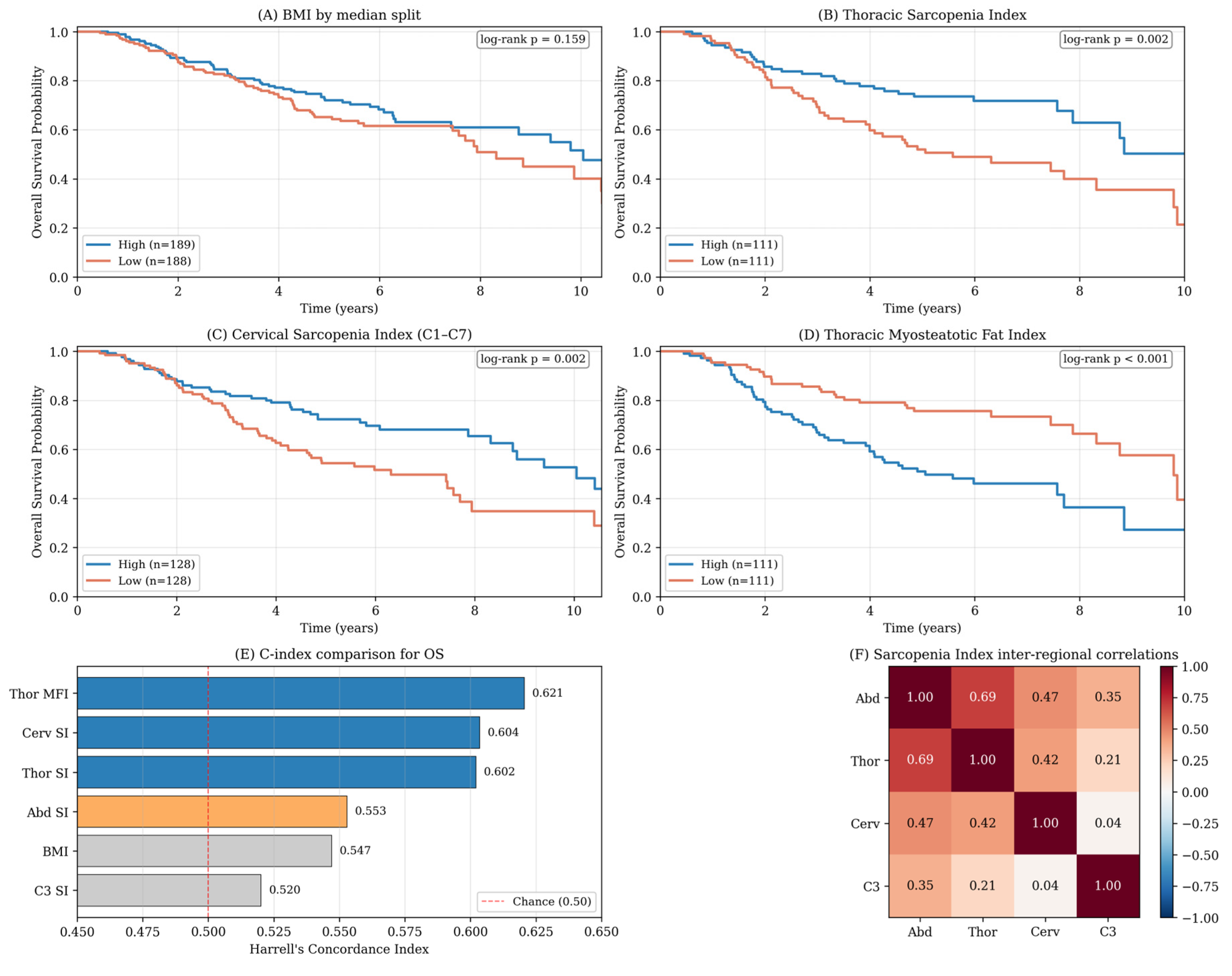
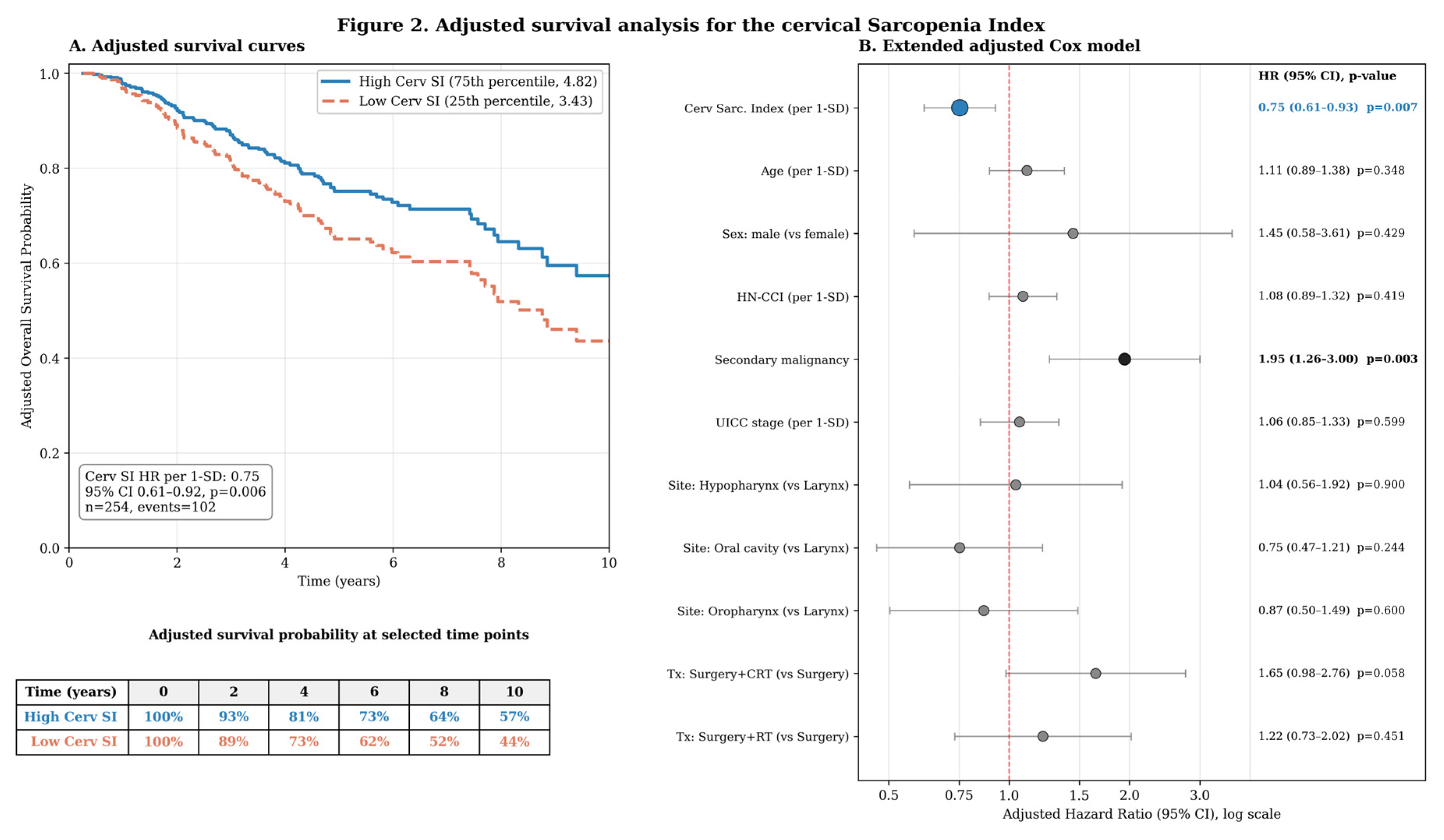
Figure 2.
Adjusted survival analysis for the cervical Sarcopenia Index. A: Standardized Cox-adjusted overall survival curves for cervical Sarcopenia Index values at the 25th and 75th percentiles. Curves were estimated from the extended Cox model (Model X1) adjusted for age, sex, tumor site, UICC stage, treatment modality, HN-CCI, and secondary malignancy. Adjusted survival probabilities at selected time points are shown below the plot. B: Forest plot of the extended adjusted Cox proportional hazards model. Hazard ratios are shown with 95% confidence intervals on a logarithmic scale. HRs for continuous predictors are reported per 1-SD increase. Reference categories: tumor site = Larynx; treatment modality = Surgery alone.
Figure 2.
Adjusted survival analysis for the cervical Sarcopenia Index. A: Standardized Cox-adjusted overall survival curves for cervical Sarcopenia Index values at the 25th and 75th percentiles. Curves were estimated from the extended Cox model (Model X1) adjusted for age, sex, tumor site, UICC stage, treatment modality, HN-CCI, and secondary malignancy. Adjusted survival probabilities at selected time points are shown below the plot. B: Forest plot of the extended adjusted Cox proportional hazards model. Hazard ratios are shown with 95% confidence intervals on a logarithmic scale. HRs for continuous predictors are reported per 1-SD increase. Reference categories: tumor site = Larynx; treatment modality = Surgery alone.
Inter-Regional Correlations
Muscle volumes showed moderate correlations between regions (abdomen–thorax r=0.32, abdomen–cervical r=0.25, thorax–cervical r=0.28). The Sarcopenia Index correlated more strongly between regions: abdomen–thorax r=0.69 (p<0.001), abdomen–cervical r=0.47, thorax–cervical r=0.42. Skeletal Muscle Index (muscle/height²) correlated moderately with BMI across regions, confirming that body composition indices capture information distinct from BMI alone. Notably, the single-vertebra C3 SI was uncorrelated with the volumetric C1–C7 SI (r=0.04, p=0.55), while correlating modestly with BMI (r=0.40) and with thoracic (r=0.21) and abdominal (r=0.35) SI.
Primary Clinically Adjusted Analysis
Because tumor stage, age, sex, primary site, and treatment modality are established prognostic factors in HNSCC, the predefined primary clinically adjusted Cox models included these variables, along with HN-CCI and secondary malignancy, for the four predefined imaging markers; ASA was added in a separate sensitivity variant (Table 4). Surgery alone was used as the reference treatment category and Larynx as the reference tumor site; one patient with a multi-site primary was excluded from the site-adjusted models. Tumor staging was retrieved for all 391 patients; no patients were excluded from extended-adjustment analyses due to missing TNM.
After extended oncologic adjustment (age, sex, tumor site, UICC stage, treatment modality, HN-CCI, secondary malignancy), the cervical Sarcopenia Index remained independently associated with overall survival (Model X1: HR 0.75, 95% CI 0.61–0.92, p=0.007; n=254, events=102; C-index 0.67). The association persisted when T and N were entered separately rather than as a composite UICC stage (Model X2: HR 0.78, p=0.022). When ASA was added to the extended adjusted model (Model X3), the cervical SI remained significant (HR 0.78, p=0.021) and ASA showed a borderline association (HR 1.21, p=0.077).
Thoracic SI (Model X4: HR 0.77, p=0.029) and thoracic MFI (Model X5: HR 1.26, p=0.013) also retained independent associations under extended adjustment, while BMI (Model X6: HR 0.91, p=0.30) did not. The cervical SI hazard ratio remained near 0.71–0.78 across all model specifications, supporting the robustness of the cervical finding.
Disease-Free Survival (Secondary Outcome)
Disease-free survival (DFS) was defined as the time from first diagnosis to first documented recurrence or death from any cause, whichever occurred first; because death from any cause without documented recurrence was counted as an event, the endpoint corresponds to disease-free (rather than strict recurrence-free) survival. DFS data were available for 391 patients, with 170 events (105 recurrences and 65 deaths without documented recurrence). Because recurrence documentation in retrospective records is less robust than vital-status documentation, DFS was treated as a secondary supportive endpoint.
In univariate Cox regression for DFS, the cervical Sarcopenia Index showed a similar protective association as for OS (HR 0.71, 95% CI 0.59–0.86, p<0.001). Thoracic SI was also associated with DFS (HR 0.75, p=0.007); thoracic MFI showed an association with DFS (HR 1.24, p=0.015). BMI was not associated with DFS (HR 0.96, p=0.58), nor was the BOA single-vertebra C3 SI (HR 0.97, p=0.74).
In a multivariate Cox model adjusted for HN-CCI and secondary malignancy, the cervical SI remained associated with DFS (HR 0.76, 95% CI 0.62–0.92, p=0.005; n=254, events=118). Kaplan–Meier median-split analyses paralleled the OS findings (cervical SI, log-rank p=0.019; thoracic SI, p=0.043; thoracic MFI, p=0.007; BMI, p=0.74; C3 SI, p=0.68).
Discussion
This study shows that automated 3D volumetric body composition analysis using the BOA algorithm yields prognostically informative biomarkers in patients with surgically treated HNSCC that, in this cohort, provide information beyond BMI. The principal finding is that the cervical Sarcopenia Index (Muscle/Bone ratio, derived from C1–C7 volumetric data) is associated with overall survival in a minimally adjusted model adjusting for comorbidity burden and secondary malignancy, and that this association remains after extending the adjustment to age, sex, tumor site, UICC stage, and treatment modality (Table 4). The hazard ratio remained near 0.71–0.78 across model specifications, supporting robustness. This is clinically relevant because most HNSCC patients undergo neck CT but not routine abdominal or thoracic imaging.
Volumetric Versus Single-Slice Body Composition Analysis
Our volumetric 3D segmentation approach contrasts with the established single-slice C3 or L3 method. In an exploratory within-cohort comparison, the volumetric C1–C7 BOA Sarcopenia Index was prognostic while the BOA-derived single-vertebra C3 metric was not. Importantly, the BOA C3 metric in this study is a single-vertebra volumetric measurement, not the conventional manually segmented 2D cross-sectional area used in most HNSCC sarcopenia studies, and the comparison should therefore be interpreted as a comparison within the BOA-derived feature space rather than a definitive refutation of conventional 2D C3-based assessment. The BOA C3 SI showed no meaningful discriminative ability (C-index 0.52) and, in the head-to-head model (Table 3, Model I), contributed no additional prognostic information after the inclusion of the volumetric C1–C7 metric. The correlation between the BOA C3 and volumetric C1–C7 metrics was near zero in our data, despite their overlapping anatomical scope. This suggests that, in the BOA-derived feature space, a single-vertebra C3 metric captures different information than the integrated volumetric assessment across C1–C7. Jung et al. [22] demonstrated in 36,317 UK Biobank participants that volumetric body composition measures had stronger mortality associations than single-slice metrics after multivariable adjustment, and Künnemann et al. [10] showed in a two-center lung cancer study (n=4,709) that the volumetric Sarcopenia Index outperformed conventional L3-based measurements. In contrast, Anyene et al. [11] found similar prognostic performance between single-slice L3 and multi-slice T12–sacrum metrics in colorectal cancer. Our in-sample C3 comparison suggests that, within the BOA-derived feature space, volumetric C1–C7 assessment captures prognostic information not reflected by a single-vertebra C3 BOA metric.
The Sarcopenia Index as a Frame-Normalized Metric
A practical finding is that raw cervical muscle volume alone was not associated with OS, whereas the Muscle/Bone ratio was. The denominator (bone volume) effectively controls for body frame size without requiring height, a practical advantage when stature is missing from records. This parallels the approach by Jungbauer et al. [14], who used SM/B and (SM+VAT)/B ratios from thoracic BOA in a recurrent/metastatic HNSCC immunotherapy cohort (n=49) and found significant correlations with inflammatory indices (dNLR r=–0.47). In the lung cancer literature, Künnemann et al. [10] similarly demonstrated that SI = Muscle/Bone outperformed all other volumetric features, including conventional height-indexed metrics.
BMI Paradox and the Case for Body Composition Assessment
The non-prognostic nature of BMI in this cohort (multivariate p=0.15, C-index 0.55), in contrast to the cervical SI association, is consistent with the “BMI paradox” reported in HNSCC. Hobday et al. [15] showed in a meta-analysis that overweight HNSCC patients (BMI 25–30) have lower mortality than normal-weight patients, a paradox explained by BMI’s inability to differentiate muscle from fat. Vangelov et al. [16] demonstrated that sarcopenic obesity—combining depleted muscle with obesity—is associated with a fourfold increased risk of critical weight loss during radiotherapy, further illustrating BMI’s limitations. Our data provide additional quantitative evidence: the moderate correlation between SMI and BMI confirms that these metrics capture overlapping but distinct biological information.
Muscle Quality: The Myosteatotic Fat Index
The thoracic MFI (IMAT/TAT) was associated with OS in the minimally adjusted supportive model (HR 1.35, p=0.002) and remained associated after extended oncologic adjustment (HR 1.26, p=0.013). Muscle quality (myosteatosis) and muscle quantity relative to skeletal frame (cervical SI) thus appear to provide complementary prognostic information. Vangelov et al. [9] showed that IMAT at C3 predicted PFS in HNSCC independently of muscle area. In the broader oncology literature, intramuscular fat infiltration (myosteatosis) has been increasingly recognized as a prognostic marker, sometimes superior to muscle mass alone [9,10]. The MFI—normalizing IMAT to total adipose tissue—may offer a more stable and interpretable metric than absolute IMAT volume.
Cervical Body Composition: Clinical Implications
The identification of cervical C1–C7 volumetric body composition as an independent prognostic factor has possible clinical relevance, but external validation is required before clinical implementation. Most HNSCC patients undergo neck CT but not abdominal imaging. Olson et al. [5] established C3-derived sarcopenia thresholds predicting L3-based sarcopenia in HNSCC, and Barajas Ordonez et al. [7] confirmed the prognostic value of C3 muscle metrics in 904 patients. Our study extends this by demonstrating that a combined C1–C7 volumetric measurement—automatically segmented by BOA—is independently prognostic after controlling for multiple confounders. This eliminates the need for manual segmentation, selection of specific vertebral levels, or conversion formulas.
Integration with Inflammatory Biomarkers
The relationship between body composition and systemic inflammation is increasingly recognized. Jungbauer et al. [14] demonstrated that BOA-derived SM/B ratios are inversely correlated with neutrophil-driven inflammation indices in patients with HNSCC undergoing immunotherapy. Cho et al. [24] showed that sarcopenia combined with elevated NLR carries a worse prognosis than either alone. In our cohort, preoperative leukocyte count and maximum postoperative CRP did not independently contribute to survival prediction, likely because total WBC count is a crude proxy for the neutrophil-specific inflammatory pathway. Future studies incorporating differential blood counts and SIRI calculations may reveal synergistic prognostic models that combine body composition with inflammation.
Body Composition Versus Clinical Functional Status Assessment
ASA classification, a strong univariate predictor (HR 1.55, p<0.001), remained associated with OS when the cervical Sarcopenia Index was included (Model F, ASA HR 1.26, p=0.046; Cerv SI HR 0.76, p=0.011). With extended oncologic adjustment (Model X3), the cervical SI remained significant (HR 0.78, p=0.021), and ASA was attenuated to borderline significance (HR 1.21, p=0.078). Both ASA and the cervical SI thus appear to contribute partially overlapping but complementary information about patient vulnerability, while BMI fails to capture this dimension at all. This is consistent with findings by Marschner et al. [28], who compared ASA, ACE-27, and ECOG in 302 HNSCC patients receiving adjuvant (chemo)radiotherapy and found that ASA and ACE-27 outperformed ECOG-PS in multivariate survival analysis. A systematic review by Pai et al. [29] similarly identified ASA as the most frequently employed comorbidity index in head and neck surgery (represented in 70 of 116 studies), with the CCI and ACE-27 most consistently predicting mortality. Wu et al. [30] confirmed ASA grade III–IV as an independent risk factor for postoperative complications and reduced survival among patients undergoing HNC surgery. The practical implication is that automated volumetric body composition analysis may complement clinical functional status assessment by providing an objective, imaging-based measure of physiologic reserve derived from existing diagnostic CT scans, helping to reduce inter-rater variability and the documentation gaps inherent in retrospective performance status data [26,27].
Limitations
This study has several limitations. First, the retrospective single-center design limits generalizability and is compounded by non-random availability of imaging. As shown in Supplementary Table S1, BOA-evaluable imaging coverage was strongly associated with UICC stage (χ² = 70.29, df = 12, p < 0.001), with higher-stage patients more frequently undergoing thoracic and/or abdominal imaging; regional comparisons should therefore be interpreted as exploratory. Second, detailed CT acquisition parameters were not uniformly retrievable across the 2010–2019 retrospective cohort, potentially introducing technical heterogeneity, and BOA segmentation accuracy was not formally validated against manual segmentation in this HNSCC cohort. However, BOA has been externally validated; cervical use in the presence of dental artifacts and variable scan coverage warrants further validation, and our local visual quality-control review mitigates but does not replace formal manual validation. Third, we lacked neutrophil differential counts for SIRI calculation and serum albumin data; ECOG performance status was incompletely documented, necessitating ASA as a functional-status surrogate; and smoking and alcohol exposure were not uniformly recorded. p16 testing was performed in only 34 of 391 patients, precluding adequate analysis of HPV-associated oropharyngeal disease. Fourth, the female subgroup (n=35) was underpowered for sex-stratified analysis, despite evidence that sarcopenia’s prognostic impact may differ by sex [5,20]. Fifth, the multivariate body composition models had relatively small sample sizes for the thoracic (n=221) and abdominal (n=159) subgroups. Sixth, tumor staging was based on retrieved institutional records and could not be independently re-staged; and for 54 patients with documented ongoing follow-up but without a specific last-contact date, vital status was verified by chart review where possible, with patients confirmed alive administratively censored at the institutional database snapshot date (28 January 2025). Finally, we did not assess longitudinal changes in body composition; Mascarella et al. [21] recently demonstrated that sarcopenia trajectories predict survival in operable HNSCC, suggesting dynamic assessment may add prognostic value.
Conclusions
In this retrospective single-center cohort of 391 surgically treated HNSCC patients, automated 3D volumetric body composition analysis using the BOA algorithm yielded composite indices that were independently associated with overall survival beyond BMI. The cervical C1–C7 Sarcopenia Index was the most robust marker, retaining independent significance after minimal adjustment for HN-CCI and secondary malignancy (HR 0.71, p=0.002), after extended oncologic adjustment including UICC stage, age, sex, tumor site, and treatment modality (HR 0.75, p=0.007), and in a sensitivity model additionally adjusting for inflammatory markers (HR 0.70, p=0.008). These findings support exploration of automated volumetric body composition analysis as a complement to clinical risk assessment in surgically treated HNSCC and suggest that prognostic body composition information can be obtained from routine head and neck CT. Because discrimination remained modest and the study was retrospective and single-center, prospective multicenter validation, formal comparison with established C3 and L3 single-slice methods, and assessment of clinical decision impact (e.g., via decision-curve analysis) are warranted before routine clinical use.
Supplementary Materials
The following supplementary materials are available online: Supplementary Figure S1 — cohort flow and BOA-evaluable imaging availability by region; Supplementary Figure S2 — representative cervical BOA segmentations from the visual quality-control review; Supplementary Table S1 — distribution of UICC stage and imaging availability across regions; Supplementary Table S2 — formal C-index comparison between predictors with bootstrapped 95% confidence intervals.
Author Contributions
Conceptualization, M.B. and V.W.; Methodology, M.B., V.W., and M.E.; Software, V.W. and M.E.; Validation, M.B., M.E., and V.W.; Formal Analysis, M.B. and C.K.; Investigation, C.K., N.H., P.D., C.-J.B., and M.B.; Resources, V.W. and M.B.; Data Curation, C.K. and M.B.; Writing — Original Draft Preparation, C.K. and M.B.; Writing — Review & Editing, all authors; Visualization, M.B. and C.K.; Supervision, M.B..; Project Administration, M.B. All authors have read and agreed to the published version of the manuscript.
Funding
This research received no external funding.
Institutional Review Board Statement
The study was conducted in accordance with the Declaration of Helsinki and approved by the Ethics Committee of Universitätsmedizin Greifswald (protocol code BB 099/24, 5 July 2024).
Informed Consent Statement
Patient consent was waived in accordance with the relevant data protection and hospital legislation of the federal state of Mecklenburg-Vorpommern, Germany (Landeskrankenhausgesetz Mecklenburg-Vorpommern), which permits retrospective analysis of routinely collected, pseudonymized clinical data for scientific purposes without individual informed consent. The waiver was approved by the Ethics Committee of Universitätsmedizin Greifswald (BB 099/24).
Data Availability Statement
The datasets analyzed during the current study are not publicly available because they contain identifying clinical and imaging data from a single-center patient cohort. Anonymized data supporting the conclusions of this article will be made available by the corresponding author on reasonable request, subject to local data-protection regulations.
Acknowledgments
The authors thank the staff of the Department of Diagnostic Radiology and Neuroradiology and of the Department of Otorhinolaryngology, Head and Neck Surgery at Universitätsmedizin Greifswald for clinical and technical support, and the developers of the Body and Organ Analysis (BOA) algorithm (University Hospital Essen) for making the software openly available.
Conflicts of Interest
The authors declare no conflicts of interest.
References
- Haubold J, Baldini G, Parmar V, et al. BOA: A CT-Based Body and Organ Analysis for Radiologists at the Point of Care. Invest Radiol. 2024;59(6):433-441. [CrossRef]
- Hua X, Liu S, Liao JF, et al. When the Loss Costs Too Much: A Systematic Review and Meta-Analysis of Sarcopenia in Head and Neck Cancer. Front Oncol. 2020;9:1561. [CrossRef]
- Wong A, Zhu D, Kraus D, Tham T. Radiologically Defined Sarcopenia Affects Survival in Head and Neck Cancer: A Meta-Analysis. Laryngoscope. 2021;131(2):333-341. [CrossRef]
- van Heusden HC, van Beers MA, Schaeffers AWMA, et al. The predictive and prognostic role of radiologically defined sarcopenia in head and neck cancer: a systematic review and multi-level meta-analysis. Br J Cancer. 2025;133(2):131-143. [CrossRef]
- Olson B, Edwards J, Degnin C, et al. Establishment and Validation of Pre-Therapy Cervical Vertebrae Muscle Quantification as a Prognostic Marker of Sarcopenia in Patients With Head and Neck Cancer. Front Oncol. 2022;12:812159. [CrossRef]
- Chang SW, Tsai YH, Hsu CM, et al. Prognostic Value of Third Cervical Vertebra Skeletal Muscle Index in Oral Cavity Cancer. Laryngoscope. 2021;131(7):E2257-E2265. [CrossRef]
- Barajas Ordonez F, Aghayev A, Hinnerichs M, et al. Skeletal Muscle Radiation Attenuation at C3 Predicts Survival in Head and Neck Cancer. Curr Oncol. 2025;32(10):587. [CrossRef]
- Van den Broeck J, Sealy MJ, Brussaard C, et al. The correlation of muscle quantity and quality between all vertebra levels and level L3, measured with CT: An exploratory study. Front Nutr. 2023;10:1148809. [CrossRef]
- Vangelov B, Bauer J, Kotevski D, Smee RI. Muscle quality and not quantity as a predictor of survival in head and neck squamous cell carcinoma. Oral Oncol. 2023;144:106508. [CrossRef]
- Künnemann MD, Römer C, Helfen A, et al. Exploration of Fully-Automated Body Composition Analysis Using Routine CT-Staging of Lung Cancer Patients for Survival Prognosis. J Cachexia Sarcopenia Muscle. 2025;16(4):e70021. [CrossRef]
- Anyene I, Caan B, Williams GR, et al. Body composition from single versus multi-slice abdominal computed tomography: Concordance and associations with colorectal cancer survival. J Cachexia Sarcopenia Muscle. 2022;13(6):2974-2984. [CrossRef]
- Jayawardena R, Ranasinghe P, Ranathunga T, et al. A systematic review of automated segmentation of 3D CT scans for volumetric body composition analysis. J Cachexia Sarcopenia Muscle. 2023;14(6):2520-2532. [CrossRef]
- Jeong D, Richards AR, Jean-Baptiste E, et al. Comparison of volumetric and single-slice computed tomography body composition metrics for colorectal cancer survival. Eur J Radiol. 2025;190:112241. [CrossRef]
- Jungbauer F, Ludwig S, Huber L, et al. Systemic inflammation plays a key prognostic role in patients with head and neck cancer treated with immunotherapy and is linked to CT-based body composition metrics. Eur Arch Otorhinolaryngol. 2026;283(5):3361-3371. [CrossRef]
- Hobday S, Armache M, Paquin R, et al. The Body Mass Index Paradox in Head and Neck Cancer: A Systematic Review and Meta-Analysis. Nutr Cancer. 2023;75(1):48-60. [CrossRef]
- Vangelov B, Smee RI, Bauer J. Sarcopenic obesity in patients with head and neck cancer is predictive of critical weight loss during radiotherapy. Br J Nutr. 2024;132(5):599-606. [CrossRef]
- Koitka S, Kroll L, Malamutmann E, Oezcelik A, Nensa F. Fully automated body composition analysis in routine CT imaging using 3D semantic segmentation convolutional neural networks. Eur Radiol. 2021;31(4):1795-1804. [CrossRef]
- Wasserthal J, Breit HC, Meyer MT, et al. TotalSegmentator: Robust Segmentation of 104 Anatomic Structures in CT Images. Radiol Artif Intell. 2023;5(5):e230024. [CrossRef]
- Zopfs D, Pinto Dos Santos D, Kottlors J, et al. Two-dimensional CT measurements enable assessment of body composition on head and neck CT. Eur Radiol. 2022;32(9):6427-6434. [CrossRef]
- Morelli C, Formica V, Bossi P, et al. Untailored vs. Gender- and Body-Mass-Index-Tailored Skeletal Muscle Mass Index (SMI) to Assess Sarcopenia in Advanced Head and Neck Squamous Cell Carcinoma (HNSCC). Cancers (Basel). 2023;15(19):4716. [CrossRef]
- Mascarella MA, Patel T, Vendra V, et al. Sarcopenia Trajectories Predict Survival in Operable Head and Neck Cancer. Head Neck. 2025;47(11):2929-2938. [CrossRef]
- Jung M, Raghu VK, Reisert M, et al. Deep learning-based body composition analysis from whole-body magnetic resonance imaging to predict all-cause mortality in a large western population. EBioMedicine. 2024;110:105467. [CrossRef]
- Koh JH, Lim CYJ, Tan LTP, et al. Prevalence and Association of Sarcopenia with Mortality in Patients with Head and Neck Cancer: A Systematic Review and Meta-Analysis. Ann Surg Oncol. 2024;31(9):6049-6064. [CrossRef]
- Cho Y, Kim JW, Keum KC, et al. Prognostic Significance of Sarcopenia With Inflammation in Patients With Head and Neck Cancer Who Underwent Definitive Chemoradiotherapy. Front Oncol. 2018;8:457. [CrossRef]
- Dittmann P, Lehnert B, Ihler F, et al. Structured Early Follow-Up in Head and Neck Squamous Cell Carcinomas: A Retrospective Cohort Study. Biomedicines. 2025;13(5):1246. [CrossRef]
- Datta SS, Ghosal N, Daruvala R, et al. How do clinicians rate patient’s performance status using the ECOG performance scale? A mixed-methods exploration of variability in decision-making in oncology. Ecancermedicalscience. 2019;13:913. [CrossRef]
- Neeman E, Gresham G, Ovasapians N, et al. Comparing Physician and Nurse Eastern Cooperative Oncology Group Performance Status (ECOG-PS) Ratings as Predictors of Clinical Outcomes in Patients with Cancer. Oncologist. 2019;24(12):e1460-e1466. [CrossRef]
- Marschner SN, Maihöfer C, Späth R, et al. Adjuvant (chemo)radiotherapy for patients with head and neck cancer: can comorbidity risk scores predict outcome? Strahlenther Onkol. 2024;200(12):1025-1037. [CrossRef]
- Pai KK, Omiunu A, Peddu DK, et al. The Utility of Comorbidity Indices in Assessing Head and Neck Surgery Outcomes: A Systematic Review. Laryngoscope. 2022;132(7):1388-1402. [CrossRef]
- Wu L, Li Q, Zhu Z, et al. Impact of preoperative comorbidities on postoperative complication rates and survival outcome in patients with head and neck cancer undergoing surgical treatment. Sci Rep. 2025;15(1):38746. [CrossRef]
- Bray F, Laversanne M, Sung H, et al. Global cancer statistics 2022: GLOBOCAN estimates of incidence and mortality worldwide for 36 cancers in 185 countries. CA Cancer J Clin. 2024;74(3):229-263. [CrossRef]
Table 1.
Baseline characteristics of the study cohort.
| Characteristic | Value | % or range |
| Demographics | ||
| Total patients | 391 | |
| Male / Female | 356 / 35 | 91.0% / 9.0% |
| Age at diagnosis (years) | 60.9 ± 9.4 | median 59.9 |
| BMI (kg/m²) | 26.6 ± 5.3 | n = 377 |
| HN-CCI | 0.6 ± 0.8 | n = 378 |
| ASA classification | ||
| ASA 1 / 2 / 3 / 4 | 7 / 210 / 154 / 7 | 1.9% / 55.6% / 40.7% / 1.9% |
| Primary tumor site | ||
| Larynx | 151 | 38.6% |
| Oral cavity | 140 | 35.8% |
| Oropharynx | 61 | 15.6% |
| Hypopharynx | 39 | 10.0% |
| Treatment modality | ||
| Surgery alone | 173 | 44.2% |
| Surgery + radiation | 84 | 21.5% |
| Surgery + chemoradiation | 134 | 34.3% |
| Biomarkers & comorbidity | ||
| p16-positive (of tested) | 17 / 34 | 50% of the tested |
| Secondary malignancy | 67 | 17.1% |
| Outcomes | ||
| Deaths | 141 | 36.1% |
| Median follow-up (years) | 5.0 | IQR 2.6–6.3 |
| BOA data availability | ||
| Cervical (C1–C7) | 256 | 65.5% |
| Thoracic | 222 | 56.8% |
| Abdominal | 159 | 40.7% |
| Body composition (mean ± SD) | ||
| Cerv Muscle volume (mL) | 1086 ± 487 | n = 256 |
| Cerv Sarcopenia Index | 4.1 ± 1.0 | |
| Thor Muscle volume (mL) | 4635 ± 1148 | n = 222 |
| Thor Sarcopenia Index | 2.0 ± 0.3 | |
| Thor MFI (IMAT/TAT) | 0.20 ± 0.05 | |
| Abdominal muscle volume (mL) | 6074 ± 1970 | n = 159 |
| Abd Sarcopenia Index | 2.6 ± 0.4 | |
| C3 Muscle volume (mL) | 320 ± 116 | n = 248 |
| C3 Sarcopenia Index | 3.7 ± 1.0 | |
Values are mean ± SD unless otherwise stated. BOA = Body and Organ Analysis; HN-CCI = Head and Neck Charlson Comorbidity Index; SI = Sarcopenia Index (Muscle/Bone); MFI = Myosteatotic Fat Index (IMAT/TAT); SMI = Skeletal Muscle Index (Muscle/height²).
Table 2.
Univariate Cox proportional hazards analysis for overall survival. HRs are per 1-SD increase in standardized predictors.
Table 2.
Univariate Cox proportional hazards analysis for overall survival. HRs are per 1-SD increase in standardized predictors.
| Variable | n | Ev. | HR | 95% CI | p | C-idx |
| Anthropometric | ||||||
| BMI | 377 | 134 | 0.838 | 0.70–1.00 | 0.052 | 0.547 |
| Abdominal BOA | ||||||
| Abd Muscle | 159 | 68 | 1.008 | 0.79–1.28 | 0.951 | 0.492 |
| Abd SMI | 158 | 67 | 1.330 | 1.07–1.65 | 0.009 | 0.509 |
| Abd SAT | 159 | 68 | 0.909 | 0.70–1.18 | 0.468 | 0.557 |
| Abd VAT | 159 | 68 | 0.826 | 0.64–1.07 | 0.146 | 0.571 |
| Abd IMAT | 159 | 68 | 1.009 | 0.79–1.28 | 0.941 | 0.490 |
| Abd Sarc. Index | 159 | 68 | 0.799 | 0.62–1.02 | 0.077 | 0.553 |
| Thoracic BOA | ||||||
| Thor Muscle | 222 | 86 | 0.855 | 0.67–1.10 | 0.218 | 0.563 |
| Thor SMI | 221 | 85 | 1.194 | 0.96–1.48 | 0.105 | 0.460 |
| Thor SAT | 222 | 86 | 0.794 | 0.63–1.00 | 0.051 | 0.564 |
| Thor VAT | 222 | 86 | 0.809 | 0.64–1.02 | 0.069 | 0.562 |
| Thor IMAT | 222 | 86 | 1.045 | 0.84–1.29 | 0.689 | 0.515 |
| Thor Sarc. Index | 222 | 86 | 0.674 | 0.53–0.86 | 0.001 | 0.602 |
| Thor MFI (IMAT/TAT) | 222 | 86 | 1.382 | 1.15–1.66 | <0.001 | 0.621 |
| Cervical BOA (C1–C7) | ||||||
| Cerv Muscle | 256 | 103 | 1.075 | 0.88–1.31 | 0.469 | 0.531 |
| Cerv SMI | 254 | 102 | 1.178 | 0.96–1.44 | 0.112 | 0.544 |
| Cerv SAT | 256 | 103 | 1.045 | 0.90–1.21 | 0.556 | 0.529 |
| Cerv IMAT | 256 | 103 | 1.073 | 0.89–1.30 | 0.465 | 0.520 |
| Cerv Sarc. Index | 256 | 103 | 0.662 | 0.54–0.81 | <0.001 | 0.604 |
| C3 Muscle (single-vertebra) | 248 | 100 | 1.015 | 0.83–1.23 | 0.881 | 0.525 |
| C3 Sarc. Index (single-vertebra) | 248 | 100 | 1.013 | 0.83–1.24 | 0.899 | 0.520 |
| Clinical confounders | ||||||
| HN-CCI | 378 | 134 | 1.189 | 1.01–1.40 | 0.035 | |
| ASA classification | 378 | 134 | 1.550 | 1.30–1.85 | <0.001 | |
| Secondary malignancy | 391 | 141 | 1.488 | 1.30–1.70 | <0.001 | |
HR = hazard ratio; CI = confidence interval; C-idx = Harrell’s concordance index; Ev. = events (deaths). Bold indicates p < 0.05. HR < 1 indicates the protective effect of higher values. ASA was tested as a continuous variable. The BOA-derived single-vertebra C3 Sarcopenia Index is included for direct comparison with the volumetric C1–C7 SI.
Disclaimer/Publisher’s Note: The statements, opinions and data contained in all publications are solely those of the individual author(s) and contributor(s) and not of MDPI and/or the editor(s). MDPI and/or the editor(s) disclaim responsibility for any injury to people or property resulting from any ideas, methods, instructions or products referred to in the content. |
© 2026 by the authors. Licensee MDPI, Basel, Switzerland. This article is an open access article distributed under the terms and conditions of the Creative Commons Attribution (CC BY) license (http://creativecommons.org/licenses/by/4.0/).
Copyright: This open access article is published under a Creative Commons CC BY 4.0 license, which permit the free download, distribution, and reuse, provided that the author and preprint are cited in any reuse.


