

Submitted:
29 June 2026
Posted:
01 July 2026
You are already at the latest version
Abstract
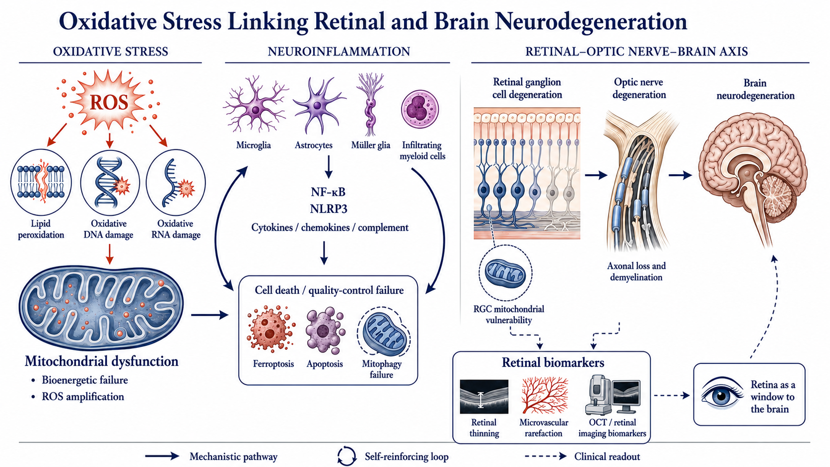
Neurodegenerative diseases represent a major and growing global health burden characterized by progressive neuronal dysfunction, axonal degeneration, and irreversible neural tissue loss. Increasing evidence identifies oxidative stress as a central pathogenic mechanism in Alzheimer’s disease, Parkinson’s disease, amyotrophic lateral sclerosis, and several optic neuropathies. Interest has increasingly focused on the brain-retina axis, as the retina and optic nerve share structural, metabolic, and molecular features with the central nervous system and may provide accessible insights into neurodegeneration.This scoping review mapped current evidence on oxidative stress in neurodegeneration, emphasizing cranial nerve involvement, optic nerve vulnerability, retinal ganglion cell degeneration, visual dysfunction, oxidative biomarkers, and emerging therapeutic strategies. The review followed established methodological frameworks and PRISMA-ScR recommendations.The literature shows that oxidative stress interacts with mitochondrial dysfunction, neuroinflammation, impaired mitophagy, ferroptosis, and altered bioenergetics, contributing to neuronal injury in cerebral and retinal disorders. Retinal ganglion cells appear particularly vulnerable because of their high metabolic demands and reliance on oxidative phosphorylation. Glaucoma and other optic neuropathies share molecular signatures with central neurodegenerative diseases. Retinal imaging and oxidative biomarkers show promise for diagnosis, monitoring, and stratification. Overall, oxidative stress emerges as a convergent mechanism across the brain-retina continuum, supporting biomarker-driven neuroprotection and precision medicine.
Keywords:
neurodegenerative diseases
; oxidative stress
; retina
; optic nerve diseases
; retinal ganglion cells
; mitochondria
; neuroinflammation
; biomarkers
; glaucoma
; precision medicine
Introduction
Neurodegenerative diseases represent one of the greatest biomedical and public health challenges of the twenty-first century. Population aging has been accompanied by a marked increase in disorders characterized by neuronal dysfunction, synaptic failure, axonal degeneration, and irreversible neural tissue loss, including Alzheimer’s disease (AD), Parkinson’s disease (PD), amyotrophic lateral sclerosis (ALS), Huntington’s disease (HD), multiple sclerosis (MS), and several optic neuropathies [1]. Despite their diverse clinical manifestations and pathological hallmarks, these disorders share convergent molecular mechanisms, among which oxidative stress has emerged as a central pathogenic driver [2].
Oxidative stress results from an imbalance between the production of reactive oxygen species (ROS) and reactive nitrogen species (RNS) and the capacity of endogenous antioxidant defenses, leading to lipid peroxidation, protein oxidation, DNA damage, mitochondrial dysfunction, impaired cellular signaling, and neuroinflammation [3,4]. The central nervous system (CNS) is particularly vulnerable because of its high oxygen consumption, abundant polyunsaturated fatty acids, intense mitochondrial activity, and limited antioxidant reserves [5]. Consequently, disturbances in redox homeostasis profoundly affect neuronal survival and function. Current evidence indicates that oxidative stress is both a driver and a consequence of neurodegeneration, participating in self-reinforcing pathogenic loops involving mitochondrial dysfunction, protein aggregation, ferroptosis, impaired autophagy, and chronic neuroinflammation [6,7,8].
Increasing attention has focused on the interplay among oxidative stress, mitochondrial dysfunction, and neuroimmune signaling. Activated microglia and astrocytes generate ROS, cytokines, and inflammatory mediators, creating a vicious cycle in which oxidative injury and neuroinflammation amplify one another [9]. Simultaneously, impaired mitochondrial bioenergetics, defective mitophagy, mitochondrial DNA damage, and electron transport chain dysfunction contribute to pathological ROS generation and neuronal degeneration [10,11]. These mechanisms have stimulated the search for biomarkers capable of identifying neurodegenerative activity before irreversible clinical manifestations develop. Biomarkers such as 8-hydroxy-2′-deoxyguanosine (8-OHdG), malondialdehyde (MDA), 4-hydroxynonenal (4-HNE), protein carbonyls, glutathione-related indices, F2-isoprostanes, and markers of mitochondrial dysfunction have been investigated as indicators of disease onset, progression, and therapeutic response [12,13]. However, methodological heterogeneity and limited standardization continue to restrict their routine application [14].
Although oxidative stress has traditionally been investigated in major cerebral neurodegenerative disorders, growing evidence suggests that cranial nerves may represent particularly vulnerable targets of redox-mediated injury [15]. Their long axonal projections, high energetic demands, dependence on mitochondrial transport, and limited regenerative capacity increase susceptibility to cumulative oxidative damage [16]. Several cranial neuropathies exhibit pathological features overlapping with classical neurodegenerative diseases, including mitochondrial dysfunction, neuroinflammation, impaired axonal transport, and neuronal loss [17].
Among cranial nerves, the optic nerve has attracted particular attention as a unique and accessible model of neurodegeneration. Embryologically derived from the diencephalon and anatomically considered an extension of the CNS, it shares many structural and molecular characteristics with central neuronal pathways [18]. Consequently, degeneration of retinal ganglion cells (RGCs) and their axons is increasingly viewed within the broader framework of CNS neurodegeneration [19,20,21].
The visual system has exceptionally high metabolic demands, and RGCs depend heavily on mitochondrial oxidative phosphorylation to sustain axonal transport, synaptic transmission, and visual processing [22]. This dependence renders them especially vulnerable to redox imbalance, mitochondrial dysfunction, impaired mitophagy, altered nicotinamide adenine dinucleotide (NAD⁺) metabolism, and chronic neuroinflammation, mechanisms that closely resemble those implicated in AD, PD, ALS, and other neurodegenerative disorders [21].
Glaucoma represents the best-characterized model of oxidative stress-related optic neurodegeneration [23,24]. Now recognized as a complex neurodegenerative disorder, it is characterized by progressive RGC loss, optic nerve degeneration, mitochondrial dysfunction, glial activation, vascular dysregulation, and chronic oxidative injury [23,25]. Oxidative stress contributes to RGC apoptosis, lamina cribrosa remodeling, axonal transport impairment, complement activation, and neuroinflammatory responses that collectively drive visual loss [22,26]. Similar mechanisms have been implicated in ischemic optic neuropathy, hereditary mitochondrial optic neuropathies, diabetic retinal neurodegeneration, age-related retinal dysfunction, and other neurodegenerative conditions associated with visual impairment [19,25,27,28,29,30,31]. Emerging evidence further suggests that retinal and optic nerve biomarkers may provide valuable insights into early neurodegenerative changes elsewhere in the CNS [32]. Consequently, the retina has increasingly been proposed as a “window to the brain,” offering unique opportunities for early diagnosis, disease monitoring, and therapeutic intervention [18,20].
Despite rapid advances, important uncertainties remain regarding the interplay among oxidative stress, neurodegeneration, optic nerve damage, retinal degeneration, and visual dysfunction. This scoping review aims to map and synthesize contemporary evidence on the role of oxidative stress across neurodegenerative disorders, with particular emphasis on optic nerve vulnerability, RGC degeneration, retinal biomarkers, and visual dysfunction. By integrating evidence across the brain-retina continuum, the review seeks to provide a unified mechanistic framework linking oxidative stress, mitochondrial dysfunction, neuroinflammation, regulated cell death, retinal imaging biomarkers, and emerging precision-neuroprotective strategies while identifying priorities for future translational research.
Methods
Study Design
This scoping review was designed to provide a structured overview of the available literature on oxidative stress-related mechanisms across neurodegenerative and neuro-ophthalmological conditions, integrating evidence from central nervous system disorders, cranial nerve pathology, optic nerve involvement, retinal degeneration, and visual dysfunction. The objective was to map an extremely heterogeneous body of mechanistic, translational, imaging, experimental and clinical literature rather than evaluate intervention efficacy. Consequently, a scoping review represented the most appropriate methodology.
The review methodology was developed in accordance with the seminal framework for scoping studies proposed by Arksey et al. [33], subsequently refined by Levac et al. [34], and further guided by the recommendations of the Joanna Briggs Institute (JBI) Manual for Evidence Synthesis [35]. Reporting was performed according to the Preferred Reporting Items for Systematic Reviews and Meta-Analyses Extension for Scoping Reviews (PRISMA-ScR) checklist [36].
The objective of the review was not to evaluate treatment efficacy or perform quantitative synthesis, but rather to comprehensively characterize the breadth, nature, and distribution of available evidence, identify key mechanistic pathways, summarize current biomarkers and therapeutic targets, and highlight existing knowledge gaps requiring future investigation.
Review Question
The review was guided by the following overarching research question:
- “What is the current state of evidence regarding the role of oxidative stress in neurodegenerative processes affecting the central nervous system, cranial nerves, optic nerve, retina and visual function?”
To ensure a structured approach, the review question was formulated according to the Population-Concept-Context (PCC) framework recommended by the Joanna Briggs Institute [35].
- Population: Human subjects, experimental animal models, ex vivo tissues, and in vitro systems investigating neurodegenerative processes.
- Concept: Oxidative stress, redox imbalance, ROS, RNS, mitochondrial dysfunction, neuroinflammation, ferroptosis, oxidative biomarkers, and antioxidant therapeutic strategies.
- Context: Neurodegenerative diseases affecting the central nervous system, cranial nerves, optic nerve, retina, and visual pathways.
Eligibility Criteria
Eligibility criteria were defined a priori to identify literature relevant to the role of oxidative stress in neurodegenerative disorders. Particular interest was given to evidence addressing mitochondrial dysfunction, neuroinflammation, RGCs, optic neuropathies and visual impairment within the broader neurodegenerative spectrum.
To provide a comprehensive overview, multiple study designs were included: original research articles, systematic reviews, meta-analyses, scoping reviews, narrative reviews, consensus statements, and clinical practice guidelines. Both preclinical and clinical evidence were considered, including human studies, animal models, translational investigations and in vitro experiments. Only articles published in English between January 1, 2020, and June 1, 2026, were included to capture contemporary developments in the field.
Because the objective of the review was to map conceptual, mechanistic, translational, and clinical developments across a rapidly evolving multidisciplinary field, review articles were retained and charted separately from primary studies. This approach enabled the identification of emerging themes, research trends, areas of convergence, and persisting knowledge gaps that might not be apparent from individual studies alone.
Studies were excluded if they lacked substantive scientific data relevant to the review objectives. Conference abstracts without full-text availability, editorials, commentaries, correspondence and opinion papers were not considered. Publications unrelated to oxidative stress, redox biology, or neurodegenerative mechanisms were excluded, as were studies focused exclusively on acute ocular trauma, infectious diseases, or neoplastic conditions without a neurodegenerative component.
Information Sources
To ensure broad coverage of both biomedical and interdisciplinary literature, four major electronic databases were searched: PubMed/MEDLINE, Embase, Scopus and the Web of Science Core Collection. These databases were selected because of their extensive indexing of clinical, translational, experimental, and basic science research across the fields of neuroscience, ophthalmology, neurobiology and redox biology.
To enhance the sensitivity of the search strategy and minimize the risk of missing relevant publications, additional search techniques were employed. The reference lists of all included studies, as well as those of pertinent review articles, were manually examined to identify further eligible records. Forward and backward citation tracking was also performed to capture influential studies that may not have been retrieved through the primary database searches. This multi-source approach was adopted to maximize the comprehensiveness of the evidence base and ensure the inclusion of the most relevant and up-to-date literature available on the topic.
Search Strategy
The search strategy was designed to achieve a comprehensive retrieval of the literature pertaining to oxidative stress and its role in neurodegeneration, with particular focus on optic nerve pathology, retinal neurodegeneration, and visual dysfunction. In accordance with recommendations from the Joanna Briggs Institute [35], the strategy combined controlled vocabulary terms, including Medical Subject Headings (MeSH) and Emtree descriptors, with free-text keywords to maximize both sensitivity and specificity across the selected databases.
Search terms were developed iteratively around three core domains: oxidative stress and redox biology; neurodegenerative diseases; and ocular, retinal, and optic nerve involvement. The oxidative stress domain included terms such as “oxidative stress”, “reactive oxygen species”, “ROS”, “reactive nitrogen species”, “RNS”, “redox”, “ferroptosis” and “mitochondrial dysfunction”. These were combined with neurodegeneration-related terms including “neurodegeneration”, “Alzheimer disease”, “Parkinson disease”, “amyotrophic lateral sclerosis”, “ALS”, “Huntington disease” and “dementia”. To capture visual system involvement, additional terms included “optic nerve”, “retina”, “retinal ganglion cell”, “glaucoma”, “optic neuropathy”, “vision”, “visual dysfunction” and “neuro-ophthalmology”.
For PubMed/MEDLINE, these concepts were combined using Boolean operators to identify relevant experimental and clinical studies. Equivalent search strategies were adapted for Embase, Scopus, and Web of Science according to their indexing systems, syntax requirements, and controlled vocabularies. Database-specific filters and indexing terms were used when appropriate to optimize retrieval while maintaining methodological consistency. The complete search strategies are provided in the Supplemental Table S1.
The strategy was refined through pilot searches and iterative testing against key known publications. The final search was designed to maximize comprehensiveness and sensitivity while maintaining sufficient precision to minimize retrieval of irrelevant records.
Study Selection
All records identified through database and supplementary searches were imported into reference management software, and duplicate entries were removed before screening. Study selection was conducted in two sequential phases. First, titles and abstracts were screened to identify potentially relevant publications. Second, complete manuscripts were assessed against the predefined eligibility criteria to determine final inclusion.
To ensure methodological rigor and reduce selection bias, screening and eligibility assessments were performed independently by three reviewers (YVT, PVP, and MNE). Disagreements regarding study inclusion were resolved through discussion and consensus. When consensus could not be achieved, a fourth reviewer (MLGL) provided an independent evaluation and final decision. Reasons for exclusion at the full-text stage were systematically recorded to enhance transparency and reproducibility.
The overall process of study identification, screening, eligibility assessment and inclusion was documented using a PRISMA-ScR flow diagram, in accordance with current recommendations for reporting scoping reviews.
Data Extraction
Data extraction was conducted using a standardized charting form developed according to recommendations for scoping reviews. The form ensured systematic and consistent data collection while accommodating the diversity of evidence relevant to the review objectives. For each included study, information was extracted on bibliographic and methodological characteristics, including authors, year of publication, country, study design and population or experimental model. Particular attention was given to the neurodegenerative condition investigated, ocular or visual system involvement and the oxidative stress mechanisms examined.
Additional data included molecular, biochemical, imaging and functional biomarkers of oxidative damage, neurodegeneration, or retinal dysfunction. Information on therapeutic targets, pharmacological interventions, neuroprotective strategies, and other translational approaches was also recorded when available. The principal findings of each study were summarized, together with methodological limitations, research priorities, and knowledge gaps identified by the authors.
To ensure accuracy and reproducibility, data extraction was performed independently by the same three reviewers. Completed charting forms were cross-checked and verified, and any discrepancies were resolved through discussion and consensus. This process minimized extraction errors and ensured a reliable synthesis of the available evidence.
Data Synthesis and Presentation of Results
In accordance with the methodological principles supporting scoping reviews, the present study was designed to provide a comprehensive mapping of the available evidence rather than to generate pooled estimates of effect or determine the comparative effectiveness of specific interventions. Consistent with current Joanna Briggs Institute guidance and PRISMA-ScR recommendations, a formal risk-of-bias assessment and quantitative meta-analysis were not undertaken, as the primary objective of the review was to characterize the breadth, diversity and nature of the existing literature across a complex and heterogeneous field of research. This approach is considered appropriate when the purpose of the evidence synthesis is exploratory and descriptive, particularly in areas characterized by diverse study designs, experimental models, mechanistic investigations, and emerging translational evidence [37].
Following data extraction, the included studies were synthesized descriptively through evidence mapping and thematic analysis. Primary studies and review articles were considered complementary sources of evidence. While primary studies informed mechanistic, experimental, and clinical findings, review articles facilitated the identification of overarching conceptual frameworks, emerging research directions, and cross-disciplinary areas of convergence. Findings were presented using summary tables, evidence matrices, thematic classifications and graphical representations of key biological and mechanistic pathways. This approach provided a comprehensive overview of the literature while illustrating relationships among oxidative stress, neurodegenerative mechanisms, retinal pathology, and visual dysfunction.
To ensure conceptual consistency, the evidence was organized into eight predefined thematic domains: (1) oxidative stress and central nervous system neurodegeneration; (2) mitochondrial dysfunction and redox imbalance; (3) neuroinflammation and oxidative signaling; (4) cranial nerve degeneration; (5) optic nerve and retinal neurodegeneration; (6) oxidative stress biomarkers; (7) the retina as a biomarker of CNS disease; and (8) therapeutic, neuroprotective, and translational implications.
This framework enabled the integration of findings from clinical studies, experimental models, translational investigations, and review literature while preserving the methodological diversity of the field. Beyond summarizing current knowledge, the synthesis identified areas of convergence across research domains, highlighted emerging mechanistic and translational trends, and delineated persistent knowledge gaps that may guide future experimental and clinical investigations [38].
Results
The database searches identified 2,121 records. After removal of 1,217 duplicates, 904 records underwent title and abstract screening, and 236 full-text articles were assessed for eligibility. Following full-text review, 59 reports were excluded, most frequently because they lacked an oxidative stress-related mechanism, a neurodegenerative focus, or relevant retinal, optic nerve, or neuro-ophthalmological outcomes, yielding 175 studies. The 175 included studies comprised different types of evidence. Reviews represented the largest component of the literature, with 69 articles, followed by animal studies and in vitro studies, which accounted for 52 and 46 records, respectively. Clinical evidence was limited, with only 6 clinical studies identified, while 1 in silico study and 1 systematic review were also included. Regarding the disease areas addressed, glaucoma was the most frequently investigated condition, with 53 studies, followed by diabetic retinopathy in 30 studies. Broader neurodegenerative diseases were examined in 24 studies, whereas age-related macular degeneration was represented in 13 studies.
The study identification and selection process is summarized in the PRISMA-ScR flow diagram (Figure 1).
Overview of the Literature
Across the corpus retained for synthesis, oxidative stress emerged not as a peripheral epiphenomenon but as the mechanistic hub around which the contemporary understanding of neurodegeneration is increasingly organized. The mapped literature spanned the full translational continuum (from cell-free redox chemistry and cultured neuroretinal models to transgenic animals, human biofluid studies, and clinical neuro-imaging cohorts) yet converged on a strikingly compact set of recurring motifs: mitochondrial bioenergetic failure, redox-sensitive inflammatory signaling, dysregulated programmed cell death (apoptosis, ferroptosis, pyroptosis and their hybrids), defective autophagy and mitophagy and progressive axonal attrition [16,39,40,41]. A second organizing observation was anatomical. The retina and optic nerve recurred throughout the dataset as the neural compartments in which these motifs could be observed earliest, quantified most precisely and perturbed most readily, by virtue of their exceptional metabolic load, mitochondrial density and surgical accessibility [39,42,43]. Representative studies spanning these mechanistic, anatomical and translational themes are summarized in Table 1.
Building on this representative evidence base, the eight domains below move deliberately from the general to the particular, from pan-central-nervous-system redox biology toward the optic nerve as its most tractable readout, before turning outward again to biomarkers, the retina-as-window paradigm, and therapeutics.
Domain 1. Oxidative Stress and Central Nervous System Neurodegeneration
The pathogenic centrality of oxidative stress in the canonical neurodegenerative diseases was the most uniformly reported finding. Syntheses addressing AD, PD, ALS and HD described a shared sequence in which excess ROS/RNS overwhelm glutathione- and thioredoxin-based defenses, producing lipid peroxidation, protein carbonylation, nucleic-acid oxidation and ultimately synaptic failure and neuronal apoptosis [7,67,68,69]. Mitochondrial dysfunction emerged as a shared mechanistic substrate linking the four classical proteinopathies, a convergence that is also reflected in models of dementia comorbidity [12,67,70,71].
A second, near-unanimous theme was directional: redox imbalance was reframed from a terminal by-product of dying neurons into an upstream initiator and feed-forward amplifier of degeneration [2,72,73]. The strongest form of this claim, mutual potentiation between oxidative stress and neuroinflammation, was documented in dopaminergic and demyelinating disease alike [11,74]. Additionally, the principle generalized beyond the common diseases to orphan neurodegenerative conditions and to the ocular manifestations of systemic disease, where dopaminergic, α-synuclein- and mitochondria-related abnormalities were detected in parkinsonian eyes [75,76]. Notably, no study positioned oxidative stress as merely epiphenomenal, yet the reviews consistently situated it within, rather than above, an interacting pathogenic network.
Domain 2. Mitochondrial Dysfunction and Redox Imbalance
If oxidative stress was the most frequently invoked mechanism, mitochondrial dysfunction was its most frequently invoked source and amplifier. Studies across cerebral and retinal models implicated a shared lesion set (impaired oxidative phosphorylation, electron-transport-chain leak, collapse of membrane potential, mitochondrial DNA damage, and disturbed fission-fusion dynamics) simultaneously as generators of pathological ROS and as their casualties [8,39,41,77,78,79,80]. Within this set, mitochondrial dynamics emerged as a recurrent node of vulnerability, with Drp1-mediated fission linked to ganglion-cell death and pathologically elevated intraocular pressure shown to drive Drp1-dependent fragmentation culminating in a hybrid, PANoptotic death program [44].
Quality-control machinery featured prominently. Axonal mitophagy emerged as a determinant of ganglion-cell fate, with mitophagic failure specifically implicated in glaucomatous degeneration and threshold-dependent ROS handling proposed as a “precision therapeutic window” [41,81,82]. The corollary (that augmenting mitochondrial competence is protective) was supported experimentally. Several studies demonstrated that boosting neuronal mitochondrial activity limits neurodegeneration in a murine model of MS by enhancing mitochondrial biogenesis in stem-cell-derived ganglion cells, augmenting neuronal mitochondrial activity in demyelinating disease [49,52]. Restoration of NAD⁺ homeostasis was a frequent therapeutic correlate of these findings [83,84].
Underlying the domain was a consistent anatomical rationale. The RGC was repeatedly singled out as a sentinel of mitochondrial integrity. Its long, initially unmyelinated intraocular axon and high tonic firing impose an energetic burden that leaves little reserve against bioenergetic insult, which is a vulnerability made concrete by the localization of retinal DNA and RNA oxidation to ganglion-cell mitochondria [22,39,43]. Inherited mitochondrial optic neuropathies, including Leber hereditary optic neuropathy (LHON) and dominant optic atrophy, further illustrated this principle as natural disease models of RGC susceptibility. The reported co-segregation of ALS with LHON suggested that mitochondrial dysfunction may act as a shared disease modifier across motor and visual phenotypes, while broader ocular evidence consolidated mitochondrial failure as a convergent pathogenic mechanism across retinal and optic nerve disease [27,39,51].
Domain 3. Neuroinflammation and Oxidative Signaling
Neuroinflammation and oxidative signaling emerged as a closely coupled pathogenetic axis, rather than as separate processes [12,26,40,68]. Microglia, astrocytes, Müller glia and infiltrating myeloid cells were identified as both sensors and amplifiers of oxidative injury, generating ROS, nitric oxide and pro-inflammatory mediators while themselves being activated by the same signals [26,40,85]. Mechanistically, the recurring downstream mechanisms included NF-κB-driven transcription, NLRP3 inflammasome activation, cytokine and chemokine release, complement activation, and NRF2-related stress-response pathways across cerebral, retinal and optic nerve tissues [86,87,88,89,90].
Within the visual system this axis was richly documented. Within the visual system the same loop was localized repeatedly to glaucomatous and diabetic neurodegeneration, with dysfunctional mitochondria and glial inflammation positioned at its center and exosomal cargo proposed as a vehicle for propagating redox-inflammatory signals between ocular cell populations [91,92,93,94] Therapeutically oriented work refined the cellular target, distinguishing resident microglia from monocyte-derived macrophages as “dual immune armies” and showing that restraining microglial activation (for example by inhibiting the mechanosensitive channel TRPV4, which curbed microglial ferroptosis) attenuated glaucomatous inflammation [95,96,97,98]. A recurrent finding was the existence of a self-perpetuating oxidative stress-neuroinflammation loop, partially restrained by astrocytic antioxidant defenses [73,99].
Domain 4. Oxidative Stress in Specialized Axonal Projections and Cranial Nerve Vulnerability
While the optic nerve represented the principal focus of investigation, several studies widened the concept of redox vulnerability to encompass other long-range, energy-intensive neuronal projections. Shared pathological hallmarks included mitochondrial dysfunction, defective oxidative phosphorylation, axonal transport impairment, calcium dyshomeostasis, and restricted regenerative potential, underscoring common mechanisms of neurodegenerative susceptibility across neural systems [39,100,101,102]. Evidence of distinct cranial- and spinal-nerve involvement in progressive supranuclear palsy further suggested that selective axonal susceptibility is not confined to the optic nerve but may reflect a broader vulnerability of long neural projections to bioenergetic and oxidative stress [17,103].
ALS offered a particularly illustrative example of the multisystem nature of neurodegeneration, as retinal and ocular abnormalities were documented in a disease historically considered a purely motor disorder. The observation of altered superoxide dismutase activity in tear fluid and peripheral blood further suggested that ocular biomarkers may provide a non-invasive window into systemic oxidative imbalance [104,105]. All these observations supported the review’s framing of redox imbalance as a pan-axonal pathogenic pathway, with the optic nerve serving less as an exception than as the best-instrumented example [25,40,78]. A conceptual synthesis of the redox-dependent mechanisms implicated in specialized axonal projection vulnerability is presented in Figure 2.
Domain 5. Optic Nerve and Retinal Neurodegeneration
As previously noted, the optic nerve and retina constituted the most densely represented anatomical domain among the included studies, while glaucoma emerged as the most frequently investigated condition. The literature consolidated a view of glaucoma as a multifactorial neurodegeneration (progressive RGC death, optic-nerve-head remodeling, glial activation, vascular dysregulation and chronic oxidative injury) rather than a disorder of intraocular pressure alone [19,26,106,107,108,109,110]. Enzymatic ROS sources were resolved in detail, with NADPH oxidase, and NOX4 in particular, identified as a principal generator of glaucomatous ROS whose pharmacological inhibition suppressed redox-sensitive transcription and attenuated ocular-hypertensive retinal injury [48,60]. Mechanistic studies increasingly clarified how oxidative injury contributes to RGCs death. Beyond classical apoptosis, oxidative stress was associated with regulated non-apoptotic cell-death pathways, including ferroptosis, pyroptosis and Drp1-related PANoptotic signaling [44,111,112,113,114,115,116,117,118,119]. In parallel, endogenous defenses were mapped: pressure elevation triggered early activation of the NRF2-KEAP1-ARE antioxidant program as a protective response, and normal-tension glaucoma, in which pressure is unremarkable, was used to argue that oxidative stress can act as a primary rather than secondary driver [45,59]. Additionally, Mendelian-randomization analysis provided human genetic evidence consistent with a causal contribution of oxidative stress to glaucoma risk. Glial compartments shared the vulnerability, with optic-nerve-head astrocytes protected from oxidative death by interventions that prevented caspase-3 activation and tau dephosphorylation [58,66]. Representative mechanisms are summarized in Table 2.
Beyond glaucoma, oxidative stress was implicated across a broader optic-neuropathy spectrum, including hereditary mitochondrial, ischemic, inflammatory, traumatic, and toxic/nutritional forms. Across these conditions, mitochondrial dysfunction recurred as a convergent lesion linking redox imbalance to RGCs vulnerability [21,25,61]. Experimental evidence further suggested that NRF2 activation, including with bardoxolone methyl and omaveloxolone, may improve RGCs survival in ischemic optic neuropathy models, while toxic insults such as organophosphate exposure also appeared to injure the optic nerve through oxidative pathways [62,123,124].
Diabetic retinal disease was reframed throughout the studies as a neurodegeneration that precedes overt microvascular change [29,125]. Hyperglycemia-driven ROS converged on ganglion-cell death by several routes: combined endoplasmic-reticulum and oxidative stress, thioredoxin-interacting-protein (TXNIP) signaling, β-catenin/GSK3β and ERK-Akt-mTOR dysregulation, and Nrf2/HO-1-regulated ferroptosis, the last suppressible by dopamine [57,125,126,127,128,129,130,131,132,133,134].
In the outer retina, the retinal pigment epithelium emerged as an oxidative battleground in age-related macular degeneration (AMD), through blood-retinal-barrier breakdown, DJ-1-dependent defenses and ageing and iron-dependent lipid peroxidation driving ferroptosis [47,53,135,136,137,138]. Ferroptosis itself, iron-catalyzed phospholipid peroxidation, was among the fastest-growing threads, spanning diabetic and inherited retinal degenerations and bridging redox chemistry to a defined cell-death modality [53,121]. Models of photoreceptor and rod-cone degeneration reinforced the theme: gene therapy with oxidative-stress-resistance-1 (OXR1) retarded degeneration in the RD1 mouse while graded environmental light or high-fat feeding accelerated oxidative, inflammatory retinal neurodegeneration, while receptor-level interventions (e.g., galanin receptor 3 inhibition) slowed it [54,139,140]. Additionally, autophagy was repeatedly cast as the counter-regulatory arm [122,141].
Two overarching concepts emerged from this body of evidence. First, the exceptionally high metabolic demands of RGCs, which rely heavily on oxidative phosphorylation to sustain axonal transport, neurotransmission, and cellular homeostasis, render these neurons particularly susceptible to even subtle disturbances in bioenergetic balance. This intrinsic vulnerability is further exacerbated by glutamate-mediated excitotoxicity, creating a permissive environment for progressive neuronal injury and degeneration [22,23,142,143,144,145]. Second, the remarkable convergence of molecular and cellular pathways identified in optic neuropathies with those implicated in AD, PD and ALS supports the view that the optic nerve represents more than a simple analogue of CNS pathology. Rather, it has increasingly been proposed as a readily accessible and clinically relevant model for investigating the mechanisms, biomarkers, and progression of neurodegeneration within the broader CNS [25,76,146]
Domain 6. Oxidative Biomarkers
Biomarker discovery was among the most active areas mapped. The studies converged on a recognizable panel of oxidative readouts: 8-hydroxy-2′-deoxyguanosine (8-OHdG) and 8-hydroxyguanosine (8-OHG) for nucleic-acid oxidation; malondialdehyde (MDA), 4-hydroxynonenal (4-HNE) and F2-isoprostanes for lipid peroxidation; protein carbonyls; and glutathione- and superoxide-dismutase-based indices of antioxidant capacity [13,61,147,148,149,150,151,152]. These were assayed across an unusually wide range of matrices (serum, plasma, aqueous and vitreous humor, tear fluid, and cerebrospinal fluid) [105,153,154]. The main oxidative-stress biomarkers mapped in this review are summarized in Table 3.
Disease-specific applications were consistently reported across the literature, including serum oxidative and oxidative-inflammatory biomarkers in ocular hypertension and glaucoma, vitreous proteomic signatures in vitreoretinal disorders, and tear-fluid superoxide dismutase as a potential marker of systemic redox dysregulation in ALS [105,153,155,156]. Increasingly, oxidative biomarkers were integrated with imaging findings in age-related macular degeneration and explored as regulatory biomarkers, including specific microRNA species in primary open-angle glaucoma [154,159,165].
A recurring challenge was the lack of standardization. Substantial heterogeneity in analyte selection, assay platforms, biological matrices, and reporting methods limited comparability across studies. Consequently, the field has progressively shifted toward multimarker approaches combining oxidative, inflammatory, mitochondrial, and neurodegenerative indicators rather than relying on single analytes [40,166]. While oxidative biomarkers show considerable diagnostic and prognostic potential, their routine clinical implementation remains contingent upon further analytical validation, methodological harmonization, and the establishment of standardized reference ranges.
Domain 7. Retina as a Biomarker of CNS Disease
A distinct and rapidly expanding strand reframed the retina not as a target of neurodegeneration but as a reporter of it. The argument rested on shared embryology, laminar architecture, neurovascular organization and molecular apparatus between retina and brain, which together make retinal change a plausible proxy for cerebral pathology accessible without biopsy or contrast administration [18,19,41,163].
The instrument of this paradigm was imaging. Optical coherence tomography and its angiographic extension (OCT/OCTA), retinal-nerve-fiber-layer and ganglion-cell-complex thickness, and retinal microvascular metrics were repeatedly associated with AD, PD, MS, frontotemporal degeneration and ALS [104,160,163]. Several reports indicated that retinal thinning and microvascular rarefaction may parallel, or even precede, detectable cerebral change and the “retina as a window to the brain” became an explicit organizing metaphor, extended even to glaucoma as a brain disease and to diabetic retinopathy as a source of neurodegenerative biomarkers [31,40,160]. Evidence that periodontitis-associated metabolic syndrome was linked to measurable retinal neurodegeneration further suggested that systemic redox-inflammatory burden can be reflected in retinal structure [40,167].
The literature consistently acknowledges important limitations, including the predominance of cross-sectional studies, heterogeneous imaging protocols and limited biomarker specificity. Nevertheless, the integration of artificial intelligence (AI) into ocular imaging and multimodal biomarker analysis represents a promising avenue for accelerating the clinical translation of retinal biomarkers. Recent advances in machine learning and deep learning have enabled increasingly accurate automated interpretation of optical coherence tomography (OCT), OCT angiography (OCTA), and fundus images, facilitating the detection of subtle structural and microvascular alterations that may escape conventional assessment. Moreover, AI algorithms are increasingly capable of integrating retinal imaging with fluid biomarkers, genetic data, clinical variables and other omics datasets to generate multidimensional biomarker signatures supporting patient phenotyping, endotyping, prognostic prediction, and therapeutic stratification. These multimodal analytical frameworks have the potential to advance precision medicine by improving individualized risk assessment, disease monitoring, and the identification of patients most likely to benefit from targeted neuroprotective and redox-modulating therapies. However, their routine clinical implementation will require robust external validation, standardized imaging protocols, transparent algorithm development and appropriate ethical and regulatory oversight. Consequently, retinal biomarkers should currently be regarded as complementary to established cerebral biomarkers within integrated multimodal diagnostic strategies [160,168,169]. The proposed oxidative stress-driven cascade linking mitochondrial dysfunction, neuroinflammation, RGC degeneration, and OCT-detectable retinal thinning is summarized in Figure 3.
Domain 8. Therapeutic and Translational Implications
Therapeutic and translational studies formed the final and one of the most heterogeneous domains, with interventions clustering into recognizable mechanistic families. Mitochondria-directed strategies included coenzyme Q10, shown to protect porcine retina from peroxide injury and, formulated as a vitamin-E-TPGS-conjugated eyedrop, to relieve neurodegeneration and mitochondrial dysfunction in the diabetic retina, alongside NAD⁺-repleting approaches in glaucoma [83,170,171,172]. Activators of the NRF2-ARE axis constituted a second family: bardoxolone methyl and omaveloxolone in ischemic optic neuropathy, pressure-validated NRF2 induction in ocular hypertension, nature-inspired activators protecting retinal explants, and the cysteine pro-drug N-acetyl-L-cysteine ethyl ester (NACET), which induced NRF2 and prevented retinal ageing and diabetic retinopathy [45,46,50,173,174].
Further families included free-radical scavengers and nitroxides (hybrid TEMPOL derivatives advanced specifically for ocular neurodegeneration) molecular hydrogen, manganese-porphyrin redox catalysts, and dietary polyphenols and nutraceuticals targeting the mitochondrion, of which resveratrol is a prototype [41,78,175,176,177,178,179]. Ferroptosis modulation, anti-inflammatory and microglia-directed agents, regenerative secretome-based therapy from mesenchymal stem cells, plasma-rich-in-growth-factors eyedrops, incretin- and dopamine-based metabolic agents, and OXR1 gene therapy rounded out the experimental armamentarium [71,111,120,180,181].
Across glaucoma, hereditary optic neuropathy, retinal degeneration and the cerebral proteinopathies, preclinical reports were strikingly concordant: lowering oxidative load preserved mitochondrial function, dampened neuroinflammation and improved neuronal survival [26,40,141]. Yet the same literature was uniformly sober about translation. Broad-spectrum antioxidant supplementation had largely disappointed in the clinic and reviews attributed the gap to biological heterogeneity, late intervention, incomplete biomarker validation, and the reality that oxidative stress is but one interlocking node of a network also comprising inflammation, mitochondrial failure, vascular compromise and proteostatic collapse [166]. The emerging prescription was precision, matching mechanism-specific redox, mitochondrial and imaging phenotypes to targeted interventions and to the disease stage at which they can still act [41,72]
Discussion
This scoping review mapped a large, methodologically heterogeneous body of contemporary evidence (2020-2026) onto eight pre-specified domains and, in doing so, disclosed a field that has reorganized itself around a single proposition: that oxidative stress is a convergent, causally upstream mechanism of neurodegeneration which operates with particular force in the visual system. Three findings proved robust to this heterogeneity. First, oxidative injury, mitochondrial dysfunction and neuroinflammation were reported together far more often than separately. Second, the RGC and optic nerve recurred as the compartment in which these mechanisms are most measurable and most manipulable. Third, the same molecular vocabulary like NRF2, NAD⁺, Drp1, NLRP3, ferroptosis appeared whether the disease under study was cerebral or ocular, supporting a brain-retina continuum rather than a dichotomy.
The most important conceptual yield of the mapping was the consolidation of an oxidative stress-mitochondria-neuroinflammation triad contributing to the pathology of various neurodegenerative diseases. The directionality reported across studies was circular rather than linear: mitochondrial electron leak generates ROS [67,68]; ROS damage mitochondrial lipids, proteins and DNA, further impairing respiration [67,182]; and oxidatively stressed neurons and glia activate NF-κB- and NLRP3-dependent inflammatory programs that themselves produce additional reactive species [74,92]. This self-potentiation was documented across both cerebral, dopaminergic and demyelinating, diseases and, within the eye, glaucomatous and diabetic neurodegeneration, indicating a mechanism that transcends anatomical boundaries [11,74,91,92,94]. The therapeutic significance of a circular pathology is considerable. Any single-node intervention is liable to be buffered by the others, an argument that may explain the modest clinical returns of isolated antioxidant supplementation and that motivates combination or upstream strategies such as NRF2 restoration and mitochondrial stabilization [83,166].
A second synthesis concerns why the optic nerve recurred so insistently. The anatomical and metabolic case assembled across the studies is coherent. RGCs are CNS neurons whose initially unmyelinated intraocular axons sustain continuous high-frequency conduction, imposing an oxidative-phosphorylation demand met by a dense, anterogradely transported mitochondrial population [21,22,23]. This configuration converts the optic nerve into a biological amplifier of bioenergetic stress and, because it is transparent to light and surgically accessible, into an unusually well-instrumented one. The inherited mitochondrial optic neuropathies provide proof of principle that isolated respiratory-chain dysfunction is sufficient to kill RGCs [22], while the co-occurrence of ALS with LHON and the broader retinal involvement of ALS indicate that the optic nerve also reports systemic axonopathy [51,104,146]. The implication is that the optic nerve should be read not as an ophthalmological special case but as a tractable window onto mechanisms otherwise observable only at autopsy in the brain.
A notable temporal trend was the ascent of regulated cell death, particularly ferroptosis, as an organizing concept. Where earlier accounts of oxidative neuronal death defaulted to apoptosis, the mapped literature increasingly invoked iron-dependent phospholipid peroxidation and hybrid death programs. Drp1-linked PANoptosis, glaucomatous ferroptosis and pyroptosis, iron-mediated peroxidation in the ageing macula, and ferroptotic cascades in diabetic retinopathy [44,53,120,183]. These findings represent a mechanistic refinement with direct therapeutic implications. Ferroptosis is pharmacologically tractable through iron chelation, glutathione-peroxidase-4 support, and lipophilic radical trapping, making it a potentially modifiable form of redox-driven cell death [96,121]. Its association with microglial ferroptosis further links oxidative injury to neuroinflammatory amplification in glaucoma, suggesting that the modality of cell death, not only the magnitude of redox burden, may become a meaningful therapeutic variable [96].
The biomarker and “retina-as-window” domains outline a promising pathway toward clinical translation while highlighting its major challenge. Current evidence indicates that oxidative injury generates measurable molecular signatures in accessible biological matrices and structural alterations detectable by OCT/OCTA [13,165,167]. However, neither fluid nor imaging biomarkers have achieved sufficient standardization for individual-level clinical decision-making because of methodological heterogeneity, predominantly cross-sectional designs, and limited specificity [147,161,166]. The findings suggest that no single biomarker is likely to be adequate. Instead, multimodal approaches integrating oxidative, inflammatory, mitochondrial, and neurofilament biomarkers with retinal structural and vascular imaging may offer greater diagnostic and prognostic value [41,57,153]. Longitudinal studies will be essential to determine whether retinal redox alterations precede, accompany, or follow cerebral neurodegeneration.
The therapeutic literature supports a nuanced interpretation. Although reducing oxidative stress consistently confers neuroprotection in preclinical models, broad-spectrum antioxidant strategies have largely failed to translate into clinical benefit [1,73,147,184]. Three factors may explain this discrepancy. First, mechanism-specific interventions, including NRF2 activators, NACET, coenzyme Q10, NAD⁺ restoration, and mitochondrial biogenesis enhancers, appear better suited to interrupt self-perpetuating redox dysfunction than conventional radical scavengers [22,30,83]. Second, timing is critical, as oxidative injury likely exerts its greatest influence before irreversible neuronal and axonal loss, emphasizing the importance of early detection [25,77,147]. Third, patient stratification is essential, given the marked biological heterogeneity of neurodegenerative disorders.
These considerations also support a broader transition from empirical antioxidant supplementation toward precision neurodegeneration medicine. Rather than treating oxidative stress as a universal therapeutic target, future interventions should be guided by careful patient phenotyping and biological endotyping, integrating molecular, metabolic, imaging, and fluid biomarkers to identify individuals in whom oxidative stress represents a dominant pathogenic mechanism. Biomarker-guided therapeutic selection and longitudinal patient stratification may enable the identification of disease-specific redox signatures, optimize treatment timing, and facilitate personalized multimodal interventions targeting the complex interplay among oxidative stress, mitochondrial dysfunction, neuroinflammation, and regulated cell-death pathways. Such precision-based approaches are increasingly regarded as essential for improving the clinical translation of redox-modulating therapies. Redox-targeted therapies are most likely to benefit phenotypes characterized by elevated oxidative burden, mitochondrial dysfunction, or specific cell-death pathways [13,57]. Notably, emerging molecular targets (NOX/NOX4, Drp1, TXNIP, GPX4/ferroptosis, and complement pathways) and innovative ocular delivery systems, including topical and sustained-release formulations, support the feasibility of precision redox-based therapies, particularly in ophthalmology [41,48,60,65].
Methodological Considerations, Strengths and Limitations
Several limitations should be considered when interpreting the findings of this review. Consistent with the methodological frameworks of Arksey and O’Malley, Levac et al., and the Joanna Briggs Institute, the objective was to map the scope and structure of the evidence rather than quantify effect sizes [33,34,35]. Accordingly, no meta-analysis or formal risk-of-bias assessment was performed, and the observed consistency reflects convergence across studies rather than pooled estimates. Although the eight thematic domains were defined a priori, considerable overlap existed among domains, and study classification inevitably involved some degree of interpretation. Restricting inclusion to English-language publications from 2020-2026 enhanced relevance to current knowledge but may have introduced language and recency bias. Moreover, the predominance of experimental and preclinical studies highlights the persistent gap between mechanistic insights and clinical validation. Nevertheless, the review is strengthened by its comprehensive scope, rigorous PCC-based and PRISMA-ScR-compliant methodology [36], and its integration of neurodegeneration and visual neuroscience, two closely related fields that are often investigated separately.
Research Gaps and Future Directions
A major translational challenge highlighted by the available evidence is the persistent discrepancy between the robust neuroprotective effects of antioxidant therapies observed in experimental models and their generally modest clinical efficacy. Several factors likely contribute to this gap, including inadequate patient stratification, initiation of treatment after irreversible neuronal damage has occurred, limited penetration of therapeutic agents into the CNS and retinal tissues, and the reliance on single-target interventions to modulate a highly interconnected pathogenic network. Indeed, oxidative stress rarely acts in isolation but interacts dynamically with mitochondrial dysfunction, neuroinflammation, impaired proteostasis, vascular dysregulation, and multiple regulated cell-death pathways. These considerations suggest that future therapeutic strategies should move beyond nonspecific antioxidant supplementation toward mechanism-based, biomarker-guided, multimodal interventions administered at the earliest stages of neurodegeneration.
The evidence map highlights several priority areas for future research. Longitudinal studies correlating retinal redox and structural alterations with subsequent cerebral neurodegeneration are needed to validate the retina-as-window hypothesis. Biomarker development requires assay standardization and harmonized reference ranges to enable the validation of multimodal panels. Mechanistic investigations should focus on identifying the specific cell-death pathways and molecular targets modulated by redox-based interventions, as well as the optimal stage for therapeutic intervention. Finally, clinical translation will require biomarker-guided and stage-specific trials, particularly in glaucoma and hereditary optic neuropathies, where disease phenotypes are measurable, target tissues are accessible, and sustained intraocular drug delivery is already feasible [66,83,185].
Conclusions
Contemporary evidence converges on oxidative stress as a shared, upstream and self-amplifying mechanism of neurodegeneration that is expressed with special clarity in the optic nerve and retina. Mapped across eight domains, the literature describes a tightly coupled oxidative-mitochondrial-inflammatory network, an expanding repertoire of regulated cell-death pathways, and a maturing set of fluid and imaging biomarkers that together position the visual system as both a privileged model of central neurodegeneration and a candidate site for its earliest detection. The principal unmet need is no longer mechanistic plausibility but translational discipline: standardized biomarkers, longitudinal validation of the brain-retina link, and precise, appropriately timed redox-modulating therapy matched to mechanistically defined patients. Uniquely accessible among central neural tissues, the eye is well placed to lead that translation.
Supplementary Materials
The following supporting information can be downloaded at: Preprints.Org.
Author Contributions
Conceptualization: GV. Formal analysis: YVT, PVP, MNE, MLGL. Methodology: GV, MLGL. Writing-original draft: GV, MLGL, YVT. Writing-review & editing: MLGL, YVT, PVP, MNE, GF. All authors meet the criteria for authorship, have approved the final version of the manuscript, and agree to be accountable for all aspects of the work.
Funding
This research received no external funding.
Acknowledgments
The authors wish to express their deepest and most sincere gratitude to VIBRA, part of the Fondazione Paolo Procacci, for its generous and sustained scientific support throughout all phases of the publication process. Its persistent commitment to advancing scientific research and promoting academic excellence has been pivotal in enabling the conception, development, refinement, and dissemination of this work.
Conflicts of interest
The authors declare no conflicts of interests.
Ethical approval
Not applicable.
Consent to participate
Not applicable.
Consent to publication
Not applicable.
Availability of data and materials
All the files relating to this research are available from the Corresponding Author, on reasonable request.
Abbreviations
| Abbreviation | Full Term |
| 4-HNE | 4-Hydroxynonenal |
| 8-OHdG | 8-Hydroxy-2′-Deoxyguanosine |
| 8-OHG | 8-Hydroxyguanosine |
| ACSL4 | Acyl-CoA Synthetase Long-chain Family Member 4 |
| AD | Alzheimer’s Disease |
| ALS | Amyotrophic Lateral Sclerosis |
| AMD | Age-related Macular Degeneration |
| ARE | Antioxidant Response Element |
| CNS | Central Nervous System |
| CSF | Cerebrospinal Fluid |
| Drp1 | Dynamin-related Protein 1 |
| FSP1 | Ferroptosis Suppressor Protein 1 |
| GCC | Ganglion Cell Complex |
| GPX4 | Glutathione Peroxidase 4 |
| GSH | Reduced Glutathione |
| GSSG | Oxidised Glutathione |
| HD | Huntington’s Disease |
| IOP | Intraocular Pressure |
| JBI | Joanna Briggs Institute |
| KEAP1 | Kelch-like ECH-associated Protein 1 |
| LHON | Leber Hereditary Optic Neuropathy |
| LPCAT3 | Lysophosphatidylcholine Acyltransferase 3 |
| MCI | Mild Cognitive Impairment |
| MDA | Malondialdehyde |
| MS | Multiple Sclerosis |
| mtDNA | Mitochondrial DNA |
| NAD⁺/ NADH | Nicotinamide Adenine Dinucleotide (Oxidized/Reduced) |
| NF-κB | Nuclear Factor Kappa B |
| NLRP3 | NOD-like Receptor Family Pyrin Domain-containing 3 |
| NOS | Nitric Oxide Synthase |
| NOX/ NOX4 | NADPH Oxidase / NADPH Oxidase 4 |
| NRF2 | Nuclear Factor Erythroid 2-related Factor 2 |
| OCT/ OCTA | Optical Coherence Tomography / Angiography |
| OHT | Ocular Hypertension |
| OPTN | Optineurin |
| OXR1 | Oxidative Stress Resistance 1 |
| OXPHOS | Oxidative Phosphorylation |
| PCC | Population-Concept-Context |
| PD | Parkinson’s Disease |
| POAG | Primary Open-Angle Glaucoma |
| PRISMA-ScR | Preferred Reporting Items for Systematic Reviews and Meta-Analyses Extension for Scoping Reviews |
| PUFA | Polyunsaturated Fatty Acid |
| RGCs | Retinal Ganglion Cells |
| RNFL | Retinal Nerve Fibre Layer |
| ROS / RNS | Reactive Oxygen / Nitrogen Species |
| RPE | Retinal Pigment Epithelium |
| SOD | Superoxide Dismutase |
| TNF-α | Tumour Necrosis Factor alpha |
| TRPV4 | Transient Receptor Potential Vanilloid 4 |
| TXNIP | Thioredoxin-interacting Protein |
References
- Teleanu, R.I.; Niculescu, A.-G.; Roza, E.; Vladâcenco, O.; Grumezescu, A.M.; Teleanu, D.M. Neurotransmitters—Key Factors in Neurological and Neurodegenerative Disorders of the Central Nervous System. Int. J. Mol. Sci. 2022, 23, 5954. [Google Scholar] [CrossRef] [PubMed]
- Rekatsina, M.; Paladini, A.; Piroli, A.; Zis, P.; Pergolizzi, J.V.; Varrassi, G. Pathophysiology and Therapeutic Perspectives of Oxidative Stress and Neurodegenerative Diseases: A Narrative Review. Adv. Ther. 2020, 37, 113–139. [Google Scholar] [CrossRef] [PubMed]
- Jomova, K.; Raptova, R.; Alomar, S.Y.; Alwasel, S.H.; Nepovimova, E.; Kuca, K.; Valko, M. Reactive Oxygen Species, Toxicity, Oxidative Stress, and Antioxidants: Chronic Diseases and Aging. Arch. Toxicol. 2023, 97, 2499–2574. [Google Scholar] [CrossRef] [PubMed]
- Sadasivam, N.; Kim, Y.-J.; Radhakrishnan, K.; Kim, D.-K. Oxidative Stress, Genomic Integrity, and Liver Diseases. Molecules 2022, 27, 3159. [Google Scholar] [CrossRef] [PubMed]
- D’Ascenzo, M.; Colussi, C. Oxidative Stress and the Central Nervous System. Antioxidants 2025, 14, 110. [Google Scholar] [CrossRef] [PubMed]
- Houldsworth, A. Role of Oxidative Stress in Neurodegenerative Disorders: A Review of Reactive Oxygen Species and Prevention by Antioxidants. Brain Commun. 2023, 6, fcad356. [Google Scholar] [CrossRef] [PubMed]
- Teleanu, D.M.; Niculescu, A.-G.; Lungu, I.I.; Radu, C.I.; Vladâcenco, O.; Roza, E.; Costăchescu, B.; Grumezescu, A.M.; Teleanu, R.I. An Overview of Oxidative Stress, Neuroinflammation, and Neurodegenerative Diseases. Int. J. Mol. Sci. 2022, 23, 5938. [Google Scholar] [CrossRef] [PubMed]
- Edwards, G.; Lee, Y.; Kim, M.; Bhanvadia, S.; Kim, K.-Y.; Ju, W.-K. Effect of Ubiquinol on Glaucomatous Neurodegeneration and Oxidative Stress: Studies for Retinal Ganglion Cell Survival and/or Visual Function. Antioxidants 2020, 9, 952. [Google Scholar] [CrossRef] [PubMed]
- Biswas, K. Microglia Mediated Neuroinflammation in Neurodegenerative Diseases: A Review on the Cell Signaling Pathways Involved in Microglial Activation. J. Neuroimmunol. 2023, 383, 578180. [Google Scholar] [CrossRef] [PubMed]
- Palma, F.R.; Gantner, B.N.; Sakiyama, M.J.; Kayzuka, C.; Shukla, S.; Lacchini, R.; Cunniff, B.; Bonini, M.G. ROS Production by Mitochondria: Function or Dysfunction? Oncogene 2024, 43, 295–303. [Google Scholar] [CrossRef] [PubMed]
- He, J.; Zhu, G.; Wang, G.; Zhang, F. Oxidative Stress and Neuroinflammation Potentiate Each Other to Promote Progression of Dopamine Neurodegeneration. Oxid. Med. Cell. Longev. 2020, 2020, 1–12. [Google Scholar] [CrossRef] [PubMed]
- Scarian, E.; Viola, C.; Dragoni, F.; Di Gerlando, R.; Rizzo, B.; Diamanti, L.; Gagliardi, S.; Bordoni, M.; Pansarasa, O. New Insights into Oxidative Stress and Inflammatory Response in Neurodegenerative Diseases. Int. J. Mol. Sci. 2024, 25, 2698. [Google Scholar] [CrossRef] [PubMed]
- Nuñez-Selles, A.J.; Nuñez-Musa, R.A.; Guillen-Marmolejos, R.A. Linking Oxidative Stress Biomarkers to Disease Progression and Antioxidant Therapy in Hypertension and Diabetes Mellitus. Front. Mol. Biosci. 2025, 12, 1611842. [Google Scholar] [CrossRef] [PubMed]
- Ahmad, S.; Yang, W.; Orellana, A.; Frölich, L.; De Rojas, I.; Cano, A.; Boada, M.; Hernández, I.; Hausner, L.; Harms, A.C.; et al. Association of Oxidative Stress and Inflammatory Metabolites with Alzheimer’s Disease Cerebrospinal Fluid Biomarkers in Mild Cognitive Impairment. Alzheimers Res. Ther. 2024, 16, 171. [Google Scholar] [CrossRef] [PubMed]
- Silva, Á.J.C.; De Lavor, M.S.L. Nitroxidative Stress, Cell—Signaling Pathways, and Manganese Porphyrins: Therapeutic Potential in Neuropathic Pain. Int. J. Mol. Sci. 2025, 26, 2050. [Google Scholar] [CrossRef] [PubMed]
- Cheng, X.-T.; Huang, N.; Sheng, Z.-H. Programming Axonal Mitochondrial Maintenance and Bioenergetics in Neurodegeneration and Regeneration. Neuron 2022, 110, 1899–1923. [Google Scholar] [CrossRef] [PubMed]
- Tanaka, H.; Martinez-Valbuena, I.; Forrest, S.L.; Couto, B.; Reyes, N.G.; Morales-Rivero, A.; Lee, S.; Li, J.; Karakani, A.M.; Tang-Wai, D.F.; et al. Distinct Involvement of the Cranial and Spinal Nerves in Progressive Supranuclear Palsy. Brain 2024, 147, 1399–1411. [Google Scholar] [CrossRef] [PubMed]
- London, A.; Benhar, I.; Schwartz, M. The Retina as a Window to the Brain—from Eye Research to CNS Disorders. Nat. Rev. Neurol. 2013, 9, 44–53. [Google Scholar] [CrossRef] [PubMed]
- Marchesi, N.; Fahmideh, F.; Boschi, F.; Pascale, A.; Barbieri, A. Ocular Neurodegenerative Diseases: Interconnection between Retina and Cortical Areas. Cells 2021, 10, 2394. [Google Scholar] [CrossRef] [PubMed]
- Ptito, M.; Bleau, M.; Bouskila, J. The Retina: A Window into the Brain. Cells 2021, 10, 3269. [Google Scholar] [CrossRef] [PubMed]
- Kang, E.Y.-C.; Liu, P.-K.; Wen, Y.-T.; Quinn, P.M.J.; Levi, S.R.; Wang, N.-K.; Tsai, R.-K. Role of Oxidative Stress in Ocular Diseases Associated with Retinal Ganglion Cells Degeneration. Antioxidants 2021, 10, 1948. [Google Scholar] [CrossRef] [PubMed]
- Yang, T.-H.; Kang, E.Y.-C.; Lin, P.-H.; Yu, B.B.-C.; Wang, J.H.-H.; Chen, V.; Wang, N.-K. Mitochondria in Retinal Ganglion Cells: Unraveling the Metabolic Nexus and Oxidative Stress. Int. J. Mol. Sci. 2024, 25, 8626. [Google Scholar] [CrossRef] [PubMed]
- Calkins, D.J. Adaptive Responses to Neurodegenerative Stress in Glaucoma. Prog. Retin. Eye Res. 2021, 84, 100953. [Google Scholar] [CrossRef] [PubMed]
- Xu, Y.; Yu, P.; Xie, Y.; Yang, J.; Wu, J.; Ling, L.; Zhou, W. Oxidative Stress in Glaucomatous Retinal Ganglion Cell Injury: Mechanisms and Neuroprotective Strategies. Exp. Eye Res. 2026, 264, 110837. [Google Scholar] [CrossRef] [PubMed]
- Buonfiglio, F.; Böhm, E.W.; Pfeiffer, N.; Gericke, A. Oxidative Stress: A Suitable Therapeutic Target for Optic Nerve Diseases? Antioxidants 2023, 12, 1465. [Google Scholar] [CrossRef] [PubMed]
- Tezel, G. Molecular Regulation of Neuroinflammation in Glaucoma: Current Knowledge and the Ongoing Search for New Treatment Targets. Prog. Retin. Eye Res. 2022, 87, 100998. [Google Scholar] [CrossRef] [PubMed]
- Yu-Wai-Man, P.; Griffiths, P.G.; Hudson, G.; Chinnery, P.F. Inherited Mitochondrial Optic Neuropathies. J. Med. Genet. 2009, 46, 145–158. [Google Scholar] [CrossRef] [PubMed]
- Datta, S.; Cano, M.; Ebrahimi, K.; Wang, L.; Handa, J.T. The Impact of Oxidative Stress and Inflammation on RPE Degeneration in Non-Neovascular AMD. Prog. Retin. Eye Res. 2017, 60, 201–218. [Google Scholar] [CrossRef] [PubMed]
- Simó, R.; Stitt, A.W.; Gardner, T.W. Neurodegeneration in Diabetic Retinopathy: Does It Really Matter? Diabetologia 2018, 61, 1902–1912. [Google Scholar] [CrossRef] [PubMed]
- Kaarniranta, K.; Uusitalo, H.; Blasiak, J.; Felszeghy, S.; Kannan, R.; Kauppinen, A.; Salminen, A.; Sinha, D.; Ferrington, D. Mechanisms of Mitochondrial Dysfunction and Their Impact on Age-Related Macular Degeneration. Prog. Retin. Eye Res. 2020, 79, 100858. [Google Scholar] [CrossRef] [PubMed]
- Simó, R.; Simó-Servat, O.; Bogdanov, P.; Hernández, C. Diabetic Retinopathy: Role of Neurodegeneration and Therapeutic Perspectives. Asia-Pac. J. Ophthalmol. Phila. Pa 2022, 11, 160–167. [Google Scholar] [CrossRef] [PubMed]
- Banna, H.U.; Slayo, M.; Armitage, J.A.; Del Rosal, B.; Vocale, L.; Spencer, S.J. Imaging the Eye as a Window to Brain Health: Frontier Approaches and Future Directions. J. Neuroinflammation 2024, 21, 309. [Google Scholar] [CrossRef] [PubMed]
- Arksey, H.; O’Malley, L. Scoping Studies: Towards a Methodological Framework. Int. J. Soc. Res. Methodol. 2005, 8, 19–32. [Google Scholar] [CrossRef]
- Levac, D.; Colquhoun, H.; O’Brien, K.K. Scoping Studies: Advancing the Methodology. Implement. Sci. 2010, 5, 69. [Google Scholar] [CrossRef] [PubMed]
- Peters, M.D.J.; Marnie, C.; Tricco, A.C.; Pollock, D.; Munn, Z.; Alexander, L.; McInerney, P.; Godfrey, C.M.; Khalil, H. Updated Methodological Guidance for the Conduct of Scoping Reviews. JBI Evid. Implement. 2021, 19, 3–10. [Google Scholar] [CrossRef] [PubMed]
- Tricco, A.C.; Lillie, E.; Zarin, W.; O’Brien, K.K.; Colquhoun, H.; Levac, D.; Moher, D.; Peters, M.D.J.; Horsley, T.; Weeks, L.; et al. PRISMA Extension for Scoping Reviews (PRISMA-ScR): Checklist and Explanation. Ann. Intern. Med. 2018, 169, 467–473. [Google Scholar] [CrossRef] [PubMed]
- Peters, M.D.J.; Marnie, C.; Colquhoun, H.; Garritty, C.M.; Hempel, S.; Horsley, T.; Langlois, E.V.; Lillie, E.; O’Brien, K.K.; Tunçalp, Ӧzge; et al. Scoping Reviews: Reinforcing and Advancing the Methodology and Application. Syst. Rev. 2021, 10, 263. [Google Scholar] [CrossRef] [PubMed]
- Peters, M.D.J.; Godfrey, C.M.; Khalil, H.; McInerney, P.; Parker, D.; Soares, C.B. Guidance for Conducting Systematic Scoping Reviews. Int. J. Evid. Based Healthc. 2015, 13, 141–146. [Google Scholar] [CrossRef] [PubMed]
- Wu, X.; Jin, H.; Zhang, B.; Yue, M.; He, J.; Shan, J.; Hong, C.; Shen, T. Mitochondrial Dysfunction as a Convergent Pathogenic Mechanism in Ocular Diseases: Emerging Targeted Therapeutic Strategies. Eur. J. Med. Res. 2026, 31, 680. [Google Scholar] [CrossRef]
- Shakaki, N.; Yu, M. Targeting Neuroinflammation and Oxidative Stress to Slow Neurodegeneration in the Visual System. J. Clin. Med. 2026, 15, 3254. [Google Scholar] [CrossRef] [PubMed]
- Kaštelan, S.; Antunica, A.G.; Konjevoda, S.; Tomić, Z.; Sarić, A.; Kulaš, M.; Kulaš, L.; Begović, E.K.; Čanović, S.; Kovačević, P.; et al. Mitochondrial ROS in Retinal Neurodegeneration: Thresholds, Quality Control Failure, and Precision Therapeutic Windows. Biomolecules 2026, 16, 445. [Google Scholar] [CrossRef] [PubMed]
- Zhang, Y.-Y.; Sun, Q.-F.; Bai, W.; Yao, J. Retinal Astrocytes: Key Coordinators of Developmental Angiogenesis and Neurovascular Homeostasis in Health and Disease. Biology 2026, 15, 201. [Google Scholar] [CrossRef] [PubMed]
- Gu, L.; Kwong, J.M.; Caprioli, J.; Piri, N. DNA and RNA Oxidative Damage in the Retina Is Associated with Ganglion Cell Mitochondria. Sci. Rep. 2022, 12, 8705. [Google Scholar] [CrossRef] [PubMed]
- Zeng, Z.; You, M.; Fan, C.; Rong, R.; Li, H.; Xia, X. Pathologically High Intraocular Pressure Induces Mitochondrial Dysfunction through Drp1 and Leads to Retinal Ganglion Cell PANoptosis in Glaucoma. Redox Biol. 2023, 62, 102687. [Google Scholar] [CrossRef] [PubMed]
- Naguib, S.; Backstrom, J.R.; Gil, M.; Calkins, D.J.; Rex, T.S. Retinal Oxidative Stress Activates the NRF2/ARE Pathway: An Early Endogenous Protective Response to Ocular Hypertension. Redox Biol. 2021, 42, 101883. [Google Scholar] [CrossRef] [PubMed]
- Realini, G.; Amato, R.; Rasa, M.; Ceccatelli, R.; Cannavale, A.; Bottoni, L.; Marchini, F.; Minetti, A.; Giustarini, D.; Canovai, A.; et al. N-Acetyl-l-Cysteine Ethyl Ester (NACET) Induces the Transcription Factor NRF2 and Prevents Retinal Aging and Diabetic Retinopathy. Redox Biol. 2025, 88, 103914. [Google Scholar] [CrossRef] [PubMed]
- Upadhyay, M.; Milliner, C.; Bell, B.A.; Bonilha, V.L. Oxidative Stress in the Retina and Retinal Pigment Epithelium (RPE): Role of Aging, and DJ-1. Redox Biol. 2020, 37. [Google Scholar] [CrossRef] [PubMed]
- Liao, J.; Peng, B.; Huang, G.; Diao, C.; Qin, Y.; Hong, Y.; Lin, J.; Lin, Y.; Jiang, L.; Tang, N.; et al. Inhibition of NOX4 with GLX351322 Alleviates Acute Ocular Hypertension-Induced Retinal Inflammation and Injury by Suppressing ROS Mediated Redox-Sensitive Factors Activation. Biomed. Pharmacother. 2023, 165, 115052. [Google Scholar] [CrossRef] [PubMed]
- Rosenkranz, S.C.; Shaposhnykov, A.A.; Träger, S.; Engler, J.B.; Witte, M.E.; Roth, V.; Vieira, V.; Paauw, N.; Bauer, S.; Schwencke-Westphal, C.; et al. Enhancing Mitochondrial Activity in Neurons Protects against Neurodegeneration in a Mouse Model of Multiple Sclerosis. eLife 2021, 10. [Google Scholar] [CrossRef] [PubMed]
- Chen, Y.; Wang, R.; Zhang, N.; Xu, L. Ferroptosis-Mediated Cell-Specific Damage: Molecular Cascades and Therapeutic Breakthroughs in Diabetic Retinopathy. Antioxidants 2025, 15, 1. [Google Scholar] [CrossRef] [PubMed]
- Amore, G.; Vacchiano, V.; La Morgia, C.; Valentino, M.; Caporali, L.; Fiorini, C.; Ormanbekova, D.; Salvi, F.; Bartoletti-Stella, A.; Capellari, S.; et al. Co-Occurrence of Amyotrophic Lateral Sclerosis and Leber’s Hereditary Optic Neuropathy: Is Mitochondrial Dysfunction a Modifier? J. Neurol. 2023, 270, 559–564. [Google Scholar] [CrossRef] [PubMed]
- Surma, M.; Anbarasu, K.; Dutta, S.; Olivera Perez, L.J.; Huang, K.-C.; Meyer, J.S.; Das, A. Enhanced Mitochondrial Biogenesis Promotes Neuroprotection in Human Pluripotent Stem Cell Derived Retinal Ganglion Cells. Commun. Biol. 2023, 6, 218. [Google Scholar] [CrossRef] [PubMed]
- Zhao, T.; Guo, X.; Sun, Y. Iron Accumulation and Lipid Peroxidation in the Aging Retina: Implication of Ferroptosis in Age-Related Macular Degeneration. Aging Dis. 2021, 12, 529–551. [Google Scholar] [CrossRef] [PubMed]
- Sahu, B.; Leon, L.M.; Zhang, W.; Puranik, N.; Periasamy, R.; Khanna, H.; Volkert, M. Oxidative Stress Resistance 1 Gene Therapy Retards Neurodegeneration in the Rd1 Mutant Mouse Model of Retinopathy. Invest. Ophthalmol. Vis. Sci. 2021, 62, 8. [Google Scholar] [CrossRef] [PubMed]
- Kutsyr, O.; Sánchez-Sáez, X.; Martínez-Gil, N.; de Juan, E.; Lax, P.; Maneu, V.; Cuenca, N. Gradual Increase in Environmental Light Intensity Induces Oxidative Stress and Inflammation and Accelerates Retinal Neurodegeneration. Invest. Ophthalmol. Vis. Sci. 2020, 61, 1. [Google Scholar] [CrossRef] [PubMed]
- Usategui-Martin, R.; Puertas-Neyra, K.; Galindo-Cabello, N.; Hernandez-Rodriguez, L.A.; Gonzalez-Perez, F.; Carlos Rodriguez-Cabello, J.; Gonzalez-Sarmiento, R.; Pastor, J.C.; Fernandez-Bueno, I. Retinal Neuroprotective Effect of Mesenchymal Stem Cells Secretome Through Modulation of Oxidative Stress, Autophagy, and Programmed Cell Death. Invest. Ophthalmol. Vis. Sci. 2022, 63, 27. [Google Scholar] [CrossRef] [PubMed]
- Mimura, T.; Noma, H. Oxidative Stress in Diabetic Retinopathy: A Comprehensive Review of Mechanisms, Biomarkers, and Therapeutic Perspectives. Antioxidants 2025, 14, 1204. [Google Scholar] [CrossRef] [PubMed]
- Means, J.C.; Lopez, A.A.; Koulen, P. Resveratrol Protects Optic Nerve Head Astrocytes from Oxidative Stress-Induced Cell Death by Preventing Caspase-3 Activation, Tau Dephosphorylation at Ser422 and Formation of Misfolded Protein Aggregates. Cell. Mol. Neurobiol. 2020, 40, 911–926. [Google Scholar] [CrossRef] [PubMed]
- Harada, C.; Noro, T.; Kimura, A.; Guo, X.; Namekata, K.; Nakano, T.; Harada, T. Suppression of Oxidative Stress as Potential Therapeutic Approach for Normal Tension Glaucoma. Antioxid. Basel Switz. 2020, 9. [Google Scholar] [CrossRef] [PubMed]
- Fan Gaskin, J.C.; Shah, M.H.; Chan, E.C. Oxidative Stress and the Role of NADPH Oxidase in Glaucoma. Antioxid. Basel Switz. 2021, 10. [Google Scholar] [CrossRef] [PubMed]
- Sanz-Morello, B.; Ahmadi, H.; Vohra, R.; Saruhanian, S.; Freude, K.K.; Hamann, S.; Kolko, M. Oxidative Stress in Optic Neuropathies. Antioxidants 2021, 10, 1538. [Google Scholar] [CrossRef] [PubMed]
- Chien, J.-Y.; Chou, Y.-Y.; Ciou, J.-W.; Liu, F.-Y.; Huang, S.-P. The Effects of Two Nrf2 Activators, Bardoxolone Methyl and Omaveloxolone, on Retinal Ganglion Cell Survival during Ischemic Optic Neuropathy. Antioxidants 2021, 10, 1466. [Google Scholar] [CrossRef] [PubMed]
- Catalani, E.; Fanelli, G.; Silvestri, F.; Cherubini, A.; Del Quondam, S.; Bongiorni, S.; Taddei, A.R.; Ceci, M.; De Palma, C.; Perrotta, C.; et al. Nutraceutical Strategy to Counteract Eye Neurodegeneration and Oxidative Stress in Drosophila Melanogaster Fed with High-Sugar Diet. Antioxid. Basel Switz. 2021, 10. [Google Scholar] [CrossRef] [PubMed]
- Korczowska-Łącka, I.; Hurła, M.; Banaszek, N.; Kobylarek, D.; Szymanowicz, O.; Kozubski, W.; Dorszewska, J. Selected Biomarkers of Oxidative Stress and Energy Metabolism Disorders in Neurological Diseases. Mol. Neurobiol. 2023, 60, 4132–4149. [Google Scholar] [CrossRef] [PubMed]
- Gao, M.; Zhou, J.; Shi, N.; Yan, X.; Liang, L. Neuroinflammatory Regulatory Role of Microglia in Optic Nerve Injury: From Pathological Mechanisms to Therapeutic Targets. Front. Immunol. 2026, 17, 1742677. [Google Scholar] [CrossRef] [PubMed]
- Shi, R.; Wu, Y.; Chen, H.; Zhang, Z.; Bao, S.; Qu, J.; Zhou, M. The Causal Effect of Oxidative Stress on the Risk of Glaucoma. Heliyon 2024, 10, e24852. [Google Scholar] [CrossRef] [PubMed]
- Dash, U.C.; Bhol, N.K.; Swain, S.K.; Samal, R.R.; Nayak, P.K.; Raina, V.; Panda, S.K.; Kerry, R.G.; Duttaroy, A.K.; Jena, A.B. Oxidative Stress and Inflammation in the Pathogenesis of Neurological Disorders: Mechanisms and Implications. Acta Pharm. Sin. B 2025, 15, 15–34. [Google Scholar] [CrossRef] [PubMed]
- Nesci, S. Mitochondrial Dysfunction in the Inflammatory Process of Neurodegenerative Diseases. Biomedicines 2026, 14, 682. [Google Scholar] [CrossRef] [PubMed]
- Huang, Y.-H.; Shih, H.-W.; Tsai, Y.-C. Expressing miR-282 Mitigates Aβ42-Induced Neurodegeneration in Alzheimer’s Model in Drosophila. Biochem. Biophys. Res. Commun. 2024, 734, 150768. [Google Scholar] [CrossRef] [PubMed]
- Alqahtani, T.; Deore, S.L.; Kide, A.A.; Shende, B.A.; Sharma, R.; Dadarao Chakole, R.; Nemade, L.S.; Kishor Kale, N.; Borah, S.; Shrikant Deokar, S.; et al. Mitochondrial Dysfunction and Oxidative Stress in Alzheimer’s Disease, and Parkinson’s Disease, Huntington’s Disease and Amyotrophic Lateral Sclerosis -An Updated Review. Mitochondrion 2023, 71, 83–92. [Google Scholar] [CrossRef] [PubMed]
- Zohud, O.; Lone, I.M.; Midlej, K.; Iraqi, F.A. The Complexity of Dementia Development and Its Comorbidities: The Collaborative Cross-Mouse Population for Multivarious Tasks Approach. Anim. Model. Exp. Med. 2026, 9, 72–88. [Google Scholar] [CrossRef] [PubMed]
- Liu, S.; Liu, J.; Wang, Y.; Deng, F.; Deng, Z. Oxidative Stress: Signaling Pathways, Biological Functions, and Disease. MedComm 2025, 6, e70268. [Google Scholar] [CrossRef] [PubMed]
- Zakrzewski, P.K.; Boczek, T. Neuron–Glia Crosstalk in the Regulation of Astrocytic Antioxidative Mechanisms Following CNS Injury. Antioxidants 2025, 14, 1415. [Google Scholar] [CrossRef] [PubMed]
- Yuan, B.; Jin, X.; Li, C.; Shen, Y.; Tian, J.; Duan, S. The Oxidative Stress-Inflammation Self-Perpetuating Cycle in Multiple Sclerosis: From Mechanisms to Emerging Antioxidant Strategies. Front. Pharmacol. 2026, 17, 1806176. [Google Scholar] [CrossRef] [PubMed]
- Espinós, C.; Galindo, M.I.; García-Gimeno, M.A.; Ibáñez-Cabellos, J.S.; Martínez-Rubio, D.; Millán, J.M.; Rodrigo, R.; Sanz, P.; Seco-Cervera, M.; Sevilla, T.; et al. Oxidative Stress, a Crossroad Between Rare Diseases and Neurodegeneration. Antioxid. Basel Switz. 2020, 9. [Google Scholar] [CrossRef] [PubMed]
- Indrieri, A.; Pizzarelli, R.; Franco, B.; De Leonibus, E. Dopamine, Alpha-Synuclein, and Mitochondrial Dysfunctions in Parkinsonian Eyes. Front. Neurosci. 2020, 14, 567129. [Google Scholar] [CrossRef] [PubMed]
- Ryan, A.K.; Rich, W.; Reilly, M.A. Oxidative Stress in the Brain and Retina after Traumatic Injury. Front. Neurosci. 2023, 17, 1021152. [Google Scholar] [CrossRef] [PubMed]
- Huang, C.-P.; Lin, Y.-W.; Huang, Y.-C.; Tsai, F.-J. Mitochondrial Dysfunction as a Novel Target for Neuroprotective Nutraceuticals in Ocular Diseases. Nutrients 2020, 12, 1–23. [Google Scholar] [CrossRef] [PubMed]
- Nebbioso, M.; Lambiase, A.; Armentano, M.; Tucciarone, G.; Sacchetti, M.; Greco, A.; Alisi, L. Diabetic Retinopathy, Oxidative Stress, and Sirtuins: An in Depth Look in Enzymatic Patterns and New Therapeutic Horizons. Surv. Ophthalmol. 2022, 67, 168–183. [Google Scholar] [CrossRef] [PubMed]
- Shosha, E.; Fouda, A.Y.; Lemtalsi, T.; Haigh, S.; Fulton, D.; Ibrahim, A.; Al-Shabrawey, M.; Caldwell, R.W.; Caldwell, R.B. Endothelial Arginase 2 Mediates Retinal Ischemia/Reperfusion Injury by Inducing Mitochondrial Dysfunction. Mol. Metab. 2021, 53, 101273. [Google Scholar] [CrossRef] [PubMed]
- Liang, Y.; Li, Y.; Jiao, Q.; Wei, M.; Wang, Y.; Cui, A.; Li, Z.; Li, G. Axonal Mitophagy in Retinal Ganglion Cells. Cell Commun. Signal. 2024, 22, 382. [Google Scholar] [CrossRef] [PubMed]
- Stavropoulos, D.; Grewal, M.K.; Petriti, B.; Chau, K.-Y.; Hammond, C.J.; Garway-Heath, D.F.; Lascaratos, G. The Role of Mitophagy in Glaucomatous Neurodegeneration. Cells 2023, 12, 1969. [Google Scholar] [CrossRef] [PubMed]
- Visalli, F.; Cappellani, F.; Gagliano, G.; Spinello, A.; Avitabile, A.; Cannizzaro, L.; Capobianco, M.; Gagliano, C.; Zeppieri, M. Mitochondrial Resilience in Glaucoma: Targeting NAD+ Metabolism and Oxidative Stress in Retinal Ganglion Cell Degeneration with Nicotinamide Riboside and Berberine: Preliminary Clinical Evidence. Diseases 2026, 14, 56. [Google Scholar] [CrossRef] [PubMed]
- Petriti, B.; Williams, P.A.; Lascaratos, G.; Chau, K.Y.; Garway-Heath, D.F. Neuroprotection in Glaucoma: NAD(+)/NADH Redox State as a Potential Biomarker and Therapeutic Target. Cells 2021, 10. [Google Scholar] [CrossRef] [PubMed]
- Rosato, C.; Bettegazzi, B.; Intagliata, P.; Balbontin Arenas, M.; Zacchetti, D.; Lanati, A.; Zerbini, G.; Bandello, F.; Grohovaz, F.; Codazzi, F. Redox and Calcium Alterations of a Müller Cell Line Exposed to Diabetic Retinopathy-Like Environment. Front. Cell. Neurosci. 2022, 16, 862325. [Google Scholar] [CrossRef] [PubMed]
- Janžič, L.; Kouter, K. Complement and Inflammasome Crosstalk in Chronic Inflammation. Front. Immunol. 2026, 17, 1778759. [Google Scholar] [CrossRef] [PubMed]
- Michalak, K.P.; Michalak, A.Z. Understanding Chronic Inflammation: Couplings between Cytokines, ROS, NO, Cai2+, HIF-1α, Nrf2 and Autophagy. Front. Immunol. 2025, 16, 1558263. [Google Scholar] [CrossRef] [PubMed]
- Muliyil, S.; Levet, C.; Düsterhöft, S.; Dulloo, I.; Cowley, S.A.; Freeman, M. ADAM17-Triggered TNF Signalling Protects the Ageing Drosophila Retina from Lipid Droplet-Mediated Degeneration. EMBO J. 2020, 39, EMBJ2020104415. [Google Scholar] [CrossRef] [PubMed]
- Xiao, D.; Yang, F.; Li, M.; Lu, Z.; Xing, S.; Wang, S.; Tao, T.; Jiang, Z.; Tao, L. Dihydromyricetin Mitigates Oxidative Damage Induced by Mixed Phthalates via Nrf2 Activation in Experimental Models of Glaucoma. Exp. Eye Res. 2026, 263, 110761. [Google Scholar] [CrossRef] [PubMed]
- Zhang, J.; Zhang, T.; Zeng, S.; Zhang, X.; Zhou, F.; Gillies, M.C.; Zhu, L. The Role of Nrf2/sMAF Signalling in Retina Ageing and Retinal Diseases. Biomedicines 2023, 11, 1512. [Google Scholar] [CrossRef] [PubMed]
- Duarte, J.N. Neuroinflammatory Mechanisms of Mitochondrial Dysfunction and Neurodegeneration in Glaucoma. J. Ophthalmol. 2021, 2021, 1–18. [Google Scholar] [CrossRef] [PubMed]
- Jassim, A.H.; Inman, D.M.; Mitchell, C.H. Crosstalk Between Dysfunctional Mitochondria and Inflammation in Glaucomatous Neurodegeneration. Front. Pharmacol. 2021, 12, 699623. [Google Scholar] [CrossRef] [PubMed]
- Wang, L.; Wei, X. Exosome-Based Crosstalk in Glaucoma Pathogenesis: A Focus on Oxidative Stress and Neuroinflammation. Front. Immunol. 2023, 14, 1202704. [Google Scholar] [CrossRef] [PubMed]
- Mendonca, H.; Carpi-Santos, R.; Da Costa Calaza, K.; Blanco Martinez, A. Neuroinflammation and Oxidative Stress Act in Concert to Promote Neurodegeneration in the Diabetic Retina and Optic Nerve: Galectin-3 Participation. Neural Regen. Res. 2020, 15, 625–635. [Google Scholar] [CrossRef] [PubMed]
- Tan, K.; Zhang, Y.; Jiang, P.; Yu, Y.; Wu, Z.; Peng, J.; Liu, P.; Peng, Q. Dual Immune Armies in Glaucoma: Microglia and Monocyte-Derived Macrophages. Front. Immunol. 2026, 17, 1801497. [Google Scholar] [CrossRef] [PubMed]
- Zhang, X.; Su, Y.; Wang, F. Mechanosensitive Ion Channel TRPV4 Inhibition Alleviates Glaucomatous Inflammation by Regulating Microglial Ferroptosis. Int. Immunopharmacol. 2026, 183, 116863. [Google Scholar] [CrossRef] [PubMed]
- Xu, B.; Velu, P.; Fan, J.; Singaravel, V. Sanggenol L Attenuates Inflammation and Apoptosis via Nrf2/PI3K/Akt Signaling in Retinal Ganglion Cells: An in Vitro and in Silico Study on OGD/R-Induced Retinal Ischemia–Reperfusion Injury. 3 Biotech. 2026, 16, 224. [Google Scholar] [CrossRef] [PubMed]
- Naguib, S.; DeJulius, C.R.; Backstrom, J.R.; Haider, A.A.; Ang, J.M.; Boal, A.M.; Calkins, D.J.; Duvall, C.L.; Rex, T.S. Intraocular Sustained Release of EPO-R76E Mitigates Glaucoma Pathogenesis by Activating the NRF2/ARE Pathway. Antioxidants 2023, 12, 556. [Google Scholar] [CrossRef] [PubMed]
- Picca, A.; Ferri, E.; Calvani, R.; Coelho-Júnior, H.J.; Marzetti, E.; Arosio, B. Age-Associated Glia Remodeling and Mitochondrial Dysfunction in Neurodegeneration: Antioxidant Supplementation as a Possible Intervention. Nutrients 2022, 14, 2406. [Google Scholar] [CrossRef] [PubMed]
- Cullen, P.F.; Sun, D. Astrocytes of the Eye and Optic Nerve: Heterogeneous Populations with Unique Functions Mediate Axonal Resilience and Vulnerability to Glaucoma. Front. Ophthalmol. 2023, 3, 1217137. [Google Scholar] [CrossRef] [PubMed]
- Rusciano, D.; Gagliano, C.; Avitabile, A.; Maya-Vetencourt, J.F. Environmental Enrichment as a Mitochondria-Targeting Systems Strategy across Neurodegenerative Diseases and Retinal Dystrophies. Front. Neurosci. 2026, 20, 1744873. [Google Scholar] [CrossRef] [PubMed]
- D’Esposito, F.; Zeppieri, M.; Cordeiro, M.F.; Capobianco, M.; Avitabile, A.; Gagliano, G.; Musa, M.; Barboni, P.; Gagliano, C. Insights on the Genetic and Phenotypic Complexities of Optic Neuropathies. Genes 2024, 15, 1559. [Google Scholar] [CrossRef] [PubMed]
- Palanivel, V.; Salkar, A.; Shenoy, A.; Eva, T.A.; Perera, R.; Chitranshi, N.; Gupta, V.; You, Y.; Mirzaei, M.; Graham, S.L.; et al. Neuropeptide Y at the Crossroads of Neurodegeneration: Mechanistic Insights and Emerging Therapeutic Strategies. Neuropeptides 2026, 115, 102583. [Google Scholar] [CrossRef] [PubMed]
- Rojas, P.; Ramírez, A.I.; Fernández-Albarral, J.A.; López-Cuenca, I.; Salobrar-García, E.; Cadena, M.; Elvira-Hurtado, L.; Salazar, J.J.; de Hoz, R.; Ramírez, J.M. Amyotrophic Lateral Sclerosis: A Neurodegenerative Motor Neuron Disease With Ocular Involvement. Front. Neurosci. 2020, 14, 566858. [Google Scholar] [CrossRef] [PubMed]
- Pavlenko, T.; Chesnokova, N.; Beznos, O.; Shikareva, N.; Nodel, M.; Shevtsova, K.; Panina, U.; Shteinberg, D.; Kukharskaya, O.; Sukhanova, I.; et al. Superoxide Dismutase Activity in Tear Fluid and Blood of Patients and Mouse Model of Amyotrophic Lateral Sclerosis: A Pilot Study. PEERJ 2025, 13. [Google Scholar] [CrossRef] [PubMed]
- Li, L.; Geng, X.; Tian, L.; Wang, D.; Wang, Q. Grape Seed Proanthocyanidins Protect Retinal Ganglion Cells by Inhibiting Oxidative Stress and Mitochondrial Alteration. Arch. Pharm. Res. 2020, 43, 1056–1066. [Google Scholar] [CrossRef] [PubMed]
- O’Neill, N.; Khan, R.S.; Abd Alhadi, S.; Dine, K.E.; Geisler, J.G.; Chaqour, B.; Ross, A.G.; Shindler, K.S. Mitochondrial Uncoupler, 2,4-Dinitrophenol, Reduces Spinal Cord Paralysis and Retinal Ganglion Cell Loss in the Experimental Autoimmune Encephalomyelitis Model of Multiple Sclerosis. Biomolecules 2025, 15, 189. [Google Scholar] [CrossRef] [PubMed]
- Li, S.; Yang, Q.; Zhou, Z.; Yang, X.; Liu, Y.; Hao, K.; Fu, M. Gastrodin Protects Retinal Ganglion Cells from Ischemic Injury by Activating Phosphatidylinositol 3-Kinase/Protein Kinase B/Nuclear Factor Erythroid 2-Related Factor 2 (PI3K/AKT/Nrf2) Signaling Pathway. Bioengineered 2022, 13, 12625–12636. [Google Scholar] [CrossRef] [PubMed]
- Song, Y.; Wang, M.; Zhao, S.; Tian, Y.; Zhang, C. Matrine Promotes Mitochondrial Biosynthesis and Reduces Oxidative Stress in Experimental Optic Neuritis. Front. Pharmacol. 2022, 13. [Google Scholar] [CrossRef] [PubMed]
- Zhou, X.; Zhao, X. Gastrodin Represses Hydrogen Peroxide-Induced Oxidative Stress in Retinal Pigment Epithelial Cells through p38MAPK/iNOS Pathway. Qual. Assur. Saf. Crops Foods 2021, 13, 24–30. [Google Scholar] [CrossRef]
- Zhou, X.; Rong, R.; Liang, G.; Li, H.; You, M.; Zeng, Z.; Xiao, H.; Ji, D.; Xia, X. Simultaneously Deplete Reactive Oxygen Species and Inhibit Pyroptosis by Dopamine/Thioketal-Containing Polymers Delivering Disulfiram in Combination with Cu(II) for Acute Glaucoma. Nano Today 2022, 47, 101668. [Google Scholar] [CrossRef]
- Tong, Y.; Yam, M.H.; Zhang, J.; Du, L.; Zhou, L.; Yip, Y.W.Y.; Ho, B.M.; Bisnauthsing, H.; Li, J.; Kong, I.; et al. GHRHR Deficiency Enhances Retinal Ganglion Cell Survival and Visual Functions in Experimental Glaucoma by Inhibiting Ferroptosis. Adv. Sci. Weinh. Baden.-Wurtt. Ger. 2026, e22929. [Google Scholar] [CrossRef] [PubMed]
- Bastola, T.; Perkins, G.A.; Huu, V.A.N.; Ju, S.; Kim, K.-Y.; Shen, Z.; Skowronska-Krawczyk, D.; Weinreb, R.N.; Ju, W.-K. Administration of Bicarbonate Protects Mitochondria, Rescues Retinal Ganglion Cells, and Ameliorates Visual Dysfunction Caused by Oxidative Stress. Antioxidants 2024, 13, 743. [Google Scholar] [CrossRef] [PubMed]
- Wu, L.-H.; Cheng, Y.-W.; Lin, F.-L.; Hsu, K.-C.; Wang, M.-H.; Yen, J.-L.; Wang, T.-J.; Lin, T.E.; Liu, Y.-C.; Huang, W.-J.; et al. A Novel HDAC8 Inhibitor H7E Exerts Retinoprotective Effects against Glaucomatous Injury via Ameliorating Aberrant Müller Glia Activation and Oxidative Stress. Biomed. Pharmacother. 2024, 174, 116538. [Google Scholar] [CrossRef] [PubMed]
- Amankwa, C.E.; Kodati, B.; Donkor, N.; Acharya, S. Therapeutic Potential of Antioxidants and Hybrid TEMPOL Derivatives in Ocular Neurodegenerative Diseases: A Glimpse into the Future. Biomedicines 2023, 11, 2959. [Google Scholar] [CrossRef] [PubMed]
- Lam, C.H.; Zou, B.; Chan, H.H.; Tse, D.Y. Functional and Structural Changes in the Neuroretina Are Accompanied by Mitochondrial Dysfunction in a Type 2 Diabetic Mouse Model. Eye Vis. Lond. Engl. 2023, 10, 37. [Google Scholar] [CrossRef] [PubMed]
- Rayner, C.L.; Bottle, S.E.; Martyn, A.P.; Barnett, N.L. Preserving Retinal Structure and Function with the Novel Nitroxide Antioxidant, DCTEIO. Neurochem. Res. 2023, 48, 3402–3419. [Google Scholar] [CrossRef] [PubMed]
- Mulfaul, K.; Ozaki, E.; Fernando, N.; Brennan, K.; Chirco, K.R.; Connolly, E.; Greene, C.; Maminishkis, A.; Salomon, R.G.; Linetsky, M.; et al. Toll-like Receptor 2 Facilitates Oxidative Damage-Induced Retinal Degeneration. Cell Rep. 2020, 30, 2209–2224.e5. [Google Scholar] [CrossRef] [PubMed]
- Garcia-Medina, J.J.; Rubio-Velazquez, E.; Lopez-Bernal, M.D.; Cobo-Martinez, A.; Zanon-Moreno, V.; Pinazo-Duran, M.D.; Del-Rio-Vellosillo, M. Glaucoma and Antioxidants: Review and Update. Antioxid. Basel Switz. 2020, 9. [Google Scholar] [CrossRef] [PubMed]
- Chen, D.; Miao, S.; Chen, X.; Wang, Z.; Lin, P.; Zhang, N.; Yang, N. Regulated Necrosis in Glaucoma: Focus on Ferroptosis and Pyroptosis. Mol. Neurobiol. 2024, 61, 2542–2555. [Google Scholar] [CrossRef] [PubMed]
- Liu, C.; Zhu, L.; Gao, F.; Jin, C.; Tian, H.; Xu, G.-T.; Zhang, L.; Lu, L. Ferroptosis in Diabetic Retinopathy: Cellular Heterogeneity, Progression, and Therapeutic Targets. Mol. Biol. Rep. 2026, 53, 533. [Google Scholar] [CrossRef] [PubMed]
- Chang, K.-C.; Liu, P.-F.; Chang, C.-H.; Lin, Y.-C.; Chen, Y.-J.; Shu, C.-W. The Interplay of Autophagy and Oxidative Stress in the Pathogenesis and Therapy of Retinal Degenerative Diseases. Cell Biosci. 2022, 12, 1. [Google Scholar] [CrossRef] [PubMed]
- Cui, B.; Tao, Y.; Gui, S.; Wang, X.; Sun, X.; Wang, S. Organophosphate Pesticide Exerts Toxic Effect on the Optic Nerve of Glaucoma Rats by Promoting Oxidative Stress and Inflammation. Sci. Rep. 2026. [Google Scholar] [CrossRef] [PubMed]
- Harkin, K.; Augustine, J.; Stitt, A.W.; Xu, H.; Chen, M. Wedelolactone Attenuates N-Methyl-N-Nitrosourea-Induced Retinal Neurodegeneration through Suppression of the AIM2/CASP11 Pathway. Biomedicines 2022, 10. [Google Scholar] [CrossRef] [PubMed]
- Shu, X.-S.; Zhu, H.; Huang, X.; Yang, Y.; Wang, D.; Zhang, Y.; Zhang, W.; Ying, Y. Loss of β-Catenin via Activated GSK3β Causes Diabetic Retinal Neurodegeneration by Instigating a Vicious Cycle of Oxidative Stress-Driven Mitochondrial Impairment. Aging 2020, 12, 13437–13462. [Google Scholar] [CrossRef] [PubMed]
- Rao, S.; Xiao, K.; Chen, X.; Sun, X. Dopamine Alleviated Diabetic Retinal Neurodegeneration through Protecting Retinal Ganglion Cells from Ferroptosis via the Nrf2/HO-1 Signaling Pathway. ACS Omega 2025, 10, 30397–30410. [Google Scholar] [CrossRef] [PubMed]
- Pal, S.; Rao, G.N.; Pal. A High Glucose-Induced ROS Accumulation Is a Critical Regulator of ERK1/2-Akt-Tuberin-mTOR Signalling in RGC-5 Cells. Life Sci. 2020, 256, 117914. [Google Scholar] [CrossRef] [PubMed]
- Khatak, K.; Narayanan, R.; Subramaniam, S.N.; Thirumalazhagan, J.; Sudharshan, S.; Raman, R.; Buch, S.J.; Chatterjee, N. Disturbed Glucose Metabolism and Oxidative Stress through PI3K/AKT Signaling Pathway in HIV-1 TAT Activated Retinal Müller Glia. J. Neuroimmunol. 2026, 418, 578960. [Google Scholar] [CrossRef] [PubMed]
- Koh, T.-Y.; Cho, W.R.; Jeon, H.-Y.; Moon, C.-H.; Yoon, J.-S.; Kim, M.; Ha, K.-S. Sustained Human C-Peptide Protects against Retinal Neurodegeneration via PEDF Restoration and Oxidative Stress Inhibition in a Mouse Model of Age-Related Macular Degeneration. Free Radic. Biol. Med. 2026, 253, 208–220. [Google Scholar] [CrossRef] [PubMed]
- Nanda, A.; Aparna, S.; Sahoo, R.N.; Patri, M. Amlodipine Attenuates Carrageenan-Induced Oxidative Stress Targeting Transsynaptic Neuronal Damage by Promoting Survival of Retinal Ganglion Cells in Adult Zebrafish (Danio Rerio). Ann. Neurosci. 2026, 33, 37–46. [Google Scholar] [CrossRef] [PubMed]
- Gómez-Jiménez, V.; Burggraaf-Sánchez De Las Matas, R.; Ortega, Á.L. Modulation of Oxidative Stress in Diabetic Retinopathy: Therapeutic Role of Natural Polyphenols. Antioxidants 2025, 14, 875. [Google Scholar] [CrossRef] [PubMed]
- Ramos, H.; Bogdanov, P.; Huerta, J.; Deàs-Just, A.; Hernández, C.; Simó, R. Antioxidant Effects of DPP-4 Inhibitors in Early Stages of Experimental Diabetic Retinopathy. ANTIOXIDANTS 2022, 11. [Google Scholar] [CrossRef] [PubMed]
- Chen, H.-Y.; Ho, Y.-J.; Chou, H.-C.; Liao, E.-C.; Tsai, Y.-T.; Wei, Y.-S.; Lin, L.-H.; Lin, M.-W.; Wang, Y.-S.; Ko, M.-L.; et al. The Role of Transforming Growth Factor-Beta in Retinal Ganglion Cells with Hyperglycemia and Oxidative Stress. Int. J. Mol. Sci. 2020, 21, 1–21. [Google Scholar] [CrossRef] [PubMed]
- Ali, A.; Boyne, M.; Othman, A.; Kamli-Salino, S.; Kuffova, L.; Forrester, J.V.; Delibegovic, M. Myeloid Cell Protein Tyrosine Phosphatase 1B Drives Retinal Neurodegeneration in Diabetic Mice. Diabetes 2026. [Google Scholar] [CrossRef] [PubMed]
- Tisi, A.; Feligioni, M.; Passacantando, M.; Ciancaglini, M.; Maccarone, R. The Impact of Oxidative Stress on Blood-Retinal Barrier Physiology in Age-Related Macular Degeneration. Cells 2021, 10, 1–21. [Google Scholar] [CrossRef] [PubMed]
- Lee, J.-J.; Chang-Chien, G.-P.; Lin, S.; Hsiao, Y.-T.; Ke, M.-C.; Chen, A.; Lin, T.-K. 5-Lipoxygenase Inhibition Protects Retinal Pigment Epithelium from Sodium Iodate-Induced Ferroptosis and Prevents Retinal Degeneration. Oxid. Med. Cell. Longev. 2022, 2022, 1792894. [Google Scholar] [CrossRef] [PubMed]
- Yang, M.; Tsui, M.G.; Tsang, J.K.W.; Goit, R.K.; Yao, K.-M.; So, K.-F.; Lam, W.-C.; Lo, A.C.Y. Involvement of FSP1-CoQ10-NADH and GSH-GPx-4 Pathways in Retinal Pigment Epithelium Ferroptosis. Cell Death Dis. 2022, 13. [Google Scholar] [CrossRef] [PubMed]
- Fang, Y.; Taubitz, T.; Tschulakow, A.V.; Heiduschka, P.; Szewczyk, G.; Burnet, M.; Peters, T.; Biesemeier, A.; Sarna, T.; Schraermeyer, U.; et al. Removal of RPE Lipofuscin Results in Rescue from Retinal Degeneration in a Mouse Model of Advanced Stargardt Disease: Role of Reactive Oxygen Species. Free Radic. Biol. Med. 2022, 182, 132–149. [Google Scholar] [CrossRef] [PubMed]
- Kutsyr, O.; Noailles, A.; Martínez-Gil, N.; Maestre-Carballa, L.; Martinez-Garcia, M.; Maneu, V.; Cuenca, N.; Lax, P. Short-Term High-Fat Feeding Exacerbates Degeneration in Retinitis Pigmentosa by Promoting Retinal Oxidative Stress and Inflammation. Proc. Natl. Acad. Sci. 2021, 118, e2100566118. [Google Scholar] [CrossRef] [PubMed]
- Azam, M.; Pashandi, Z.; Liu, M.; Jastrzebska, B. Inhibition of Galanin Receptor 3 Slows down Retina Degeneration in Retinitis Pigmentosa through Modulation of Inflammatory and Oxidative Stress Response. Sci. Rep. 2026, 16. [Google Scholar] [CrossRef] [PubMed]
- Catalani, E.; Brunetti, K.; Del Quondam, S.; Cervia, D. Targeting Mitochondrial Dysfunction and Oxidative Stress to Prevent the Neurodegeneration of Retinal Ganglion Cells. Antioxidants 2023, 12, 2011. [Google Scholar] [CrossRef] [PubMed]
- Boccuni, I.; Fairless, R. Retinal Glutamate Neurotransmission: From Physiology to Pathophysiological Mechanisms of Retinal Ganglion Cell Degeneration. Life 2022, 12, 638. [Google Scholar] [CrossRef] [PubMed]
- Zaman, K.; Kapic, A.; Nguyen, V.; Prokai-Tatrai, K. Impact of Orchiectomy on Oxidative Stress-Induced Neurodegeneration in the Male Rat Retina: A Proteomic Analysis. Antioxidants 2026, 15, 479. [Google Scholar] [CrossRef] [PubMed]
- Telegina, D.V.; Antonenko, A.K.; Fursova, A.Zh.; Kolosova, N.G. The Glutamate/GABA System in the Retina of Male Rats: Effects of Aging, Neurodegeneration, and Supplementation with Melatonin and Antioxidant SkQ1. Biogerontology 2022, 23, 571–585. [Google Scholar] [CrossRef] [PubMed]
- Castelli, V.; Paladini, A.; d’Angelo, M.; Allegretti, M.; Mantelli, F.; Brandolini, L.; Cocchiaro, P.; Cimini, A.; Varrassi, G. Taurine and Oxidative Stress in Retinal Health and Disease. CNS Neurosci. Ther. 2021, 27, 403–412. [Google Scholar] [CrossRef] [PubMed]
- Soldatov, V.O.; Kukharsky, M.S.; Belykh, A.E.; Sobolev, A.M.; Deykin, A.V. Retinal Damage in Amyotrophic Lateral Sclerosis: Underlying Mechanisms. Eye Brain 2021, 13, 131–146. [Google Scholar] [CrossRef] [PubMed]
- Ioannidou, S.; Tsolaki, M.; Ginoudis, A.; Tamvakis, A.; Tsolaki, A.; Makedou, K.; Lymperaki, E. Oxidative Stress Biomarkers as Preclinical Markers of Mild Cognitive Impairment: The Impact of Age and Sex. J. Pers. Med. 2025, 15, 171. [Google Scholar] [CrossRef] [PubMed]
- Karmakar, P.; Bag, J.; Naik, S.; Mishra, M. Oxidative Stress and Retinal Damage Induced by Cobalt Chloride in Drosophila Melanogaster: Insights into Cone Cell Susceptibility and Stress Gene Responses. NeuroToxicology 2025, 111, 103346. [Google Scholar] [CrossRef] [PubMed]
- Aranaz, M.; Costas-Rodríguez, M.; Lobo, L.; García, M.; González-Iglesias, H.; Pereiro, R.; Vanhaecke, F. Homeostatic Alterations Related to Total Antioxidant Capacity, Elemental Concentrations and Isotopic Compositions in Aqueous Humor of Glaucoma Patients. Anal. Bioanal. Chem. 2022, 414, 515–524. [Google Scholar] [CrossRef] [PubMed]
- Shi, X.; Li, P.; Liu, H.; Prokosch, V. Oxidative Stress, Vascular Endothelium, and the Pathology of Neurodegeneration in Retina. Antioxidants 2022, 11, 543. [Google Scholar] [CrossRef] [PubMed]
- Deshpande, P.; Gogia, N.; Chimata, A.V.; Singh, A. Unbiased Automated Quantitation of ROS Signals in Live Retinal Neurons of Drosophila Using Fiji/ImageJ. BioTechniques 2021, 71, 416–424. [Google Scholar] [CrossRef] [PubMed]
- Fresia, D.; Cannizzaro, E.; Borgo, A.; Roduit, R. GSH-Independent Induction of ER Stress during Hypoglycaemia in the Retinal Cells of Mice. J. Clin. Med. 2021, 10. [Google Scholar] [CrossRef] [PubMed]
- Dammak, A.; Sanchez Naves, J.; Huete-Toral, F.; Carracedo, G. New Biomarker Combination Related to Oxidative Stress and Inflammation in Primary Open-Angle Glaucoma. Life 2023, 13, 1455. [Google Scholar] [CrossRef] [PubMed]
- Raga-Cervera, J.; Bolarin, J.M.; Millan, J.M.; Garcia-Medina, J.J.; Pedrola, L.; Abellán-Abenza, J.; Valero-Vello, M.; Sanz-González, S.M.; O’connor, J.E.; Galarreta-Mira, D.; et al. Mirnas and Genes Involved in the Interplay between Ocular Hypertension and Primary Open-Angle Glaucoma. Oxidative Stress, Inflammation, and Apoptosis Networks. J. Clin. Med. 2021, 10. [Google Scholar] [CrossRef] [PubMed]
- Baris, M.E.; Furundaoturan, O.; Kocamanoğlu, M.; Sahin, S.; Akçay, Y.; Yılmaz, S.G. Serum Oxidative Stress-Related Biomarkers in Ocular Hypertension and Glaucoma. J. Ophthalmic Vis. Res. 2024, 19, 433–439. [Google Scholar] [CrossRef] [PubMed]
- Santos, F.M.; Mesquita, J.; Castro-de-Sousa, J.P.; Ciordia, S.; Paradela, A.; Tomaz, C.T. Vitreous Humor Proteome: Targeting Oxidative Stress, Inflammation, and Neurodegeneration in Vitreoretinal Diseases. Antioxidants 2022, 11, 505. [Google Scholar] [CrossRef] [PubMed]
- Zhang, W.; Zheng, H.; Pang, Y.; Wang, Y.; Zhang, X. CD38-NAD(+) Axis: New Insights into Glaucoma Therapy. Mol. Neurobiol. 2025, 62, 15669–15686. [Google Scholar] [CrossRef] [PubMed]
- Liu, C.; Dong, W.; Lv, Z.; Kong, L.; Ren, X. Thioredoxin-Interacting Protein in Diabetic Retinal Neurodegeneration: A Novel Potential Therapeutic Target for Diabetic Retinopathy. Front. Neurosci. 2022, 16, 957667. [Google Scholar] [CrossRef] [PubMed]
- Tabak, S.; Schreiber-Avissar, S.; Beit-Yannai, E. Crosstalk between Microrna and Oxidative Stress in Primary Open-Angle Glaucoma. Int. J. Mol. Sci. 2021, 22, 1–17. [Google Scholar] [CrossRef] [PubMed]
- Kaštelan, S.; Gverović Antunica, A.; Puzović, V.; Didović Pavičić, A.; Čanović, S.; Kovačević, P.; Vučemilović, P.A.F.; Konjevoda, S. Non-Invasive Retinal Biomarkers for Early Diagnosis of Alzheimer’s Disease. Biomedicines 2025, 13, 283. [Google Scholar] [CrossRef] [PubMed]
- Snyder, P.J.; Alber, J.; Alt, C.; Bain, L.J.; Bouma, B.E.; Bouwman, F.H.; DeBuc, D.C.; Campbell, M.C.W.; Carrillo, M.C.; Chew, E.Y.; et al. Retinal Imaging in Alzheimer’s and Neurodegenerative Diseases. Alzheimers Dement. 2021, 17, 103–111. [Google Scholar] [CrossRef] [PubMed]
- Ahn, S.J. Retinal Thickness Analysis Using Optical Coherence Tomography: Diagnostic and Monitoring Applications in Retinal Diseases. Diagnostics 2025, 15, 833. [Google Scholar] [CrossRef] [PubMed]
- Doustar, J.; Torbati, T.; Black, K.L.; Koronyo, Y.; Koronyo-Hamaoui, M. Optical Coherence Tomography in Alzheimer’s Disease and Other Neurodegenerative Diseases. Front. Neurol. 2017, 8, 701. [Google Scholar] [CrossRef] [PubMed]
- Christinaki, E.; Kulenovic, H.; Hadoux, X.; Baldassini, N.; Van Eijgen, J.; De Groef, L.; Stalmans, I.; Van Wijngaarden, P. Retinal Imaging Biomarkers of Neurodegenerative Diseases. Clin. Exp. Optom. 2022, 105, 194–204. [Google Scholar] [CrossRef] [PubMed]
- Iorga, R.; Moraru, A.; Costin, D.; Munteanu-Danulescu, R.; Danielescu, C. CORRELATION BETWEEN OCT MEASUREMENTS AND OXIDATIVE STRESS BIOMARKERS IN PATIENTS WITH AGE-RELATED MACULAR DEGENERATION. Med.-Surg. J.-Rev. Med.-Chir. 2023, 127, 541–549. [Google Scholar] [CrossRef]
- Manful, C.F.; Fordjour, E.; Ikumoinein, E.; Abbey, Lord; Thomas, R. Therapeutic Strategies Targeting Oxidative Stress and Inflammation: A Narrative Review. BioChem 2025, 5, 35. [Google Scholar] [CrossRef]
- Arslan, H.; Yorgancilar, N.; Kose, O.; Aslan, M.G.; Altin, A.; Bayrakdar, S.K.; Yemenoglu, H.; Findik, H.; Yilmaz, A. Periodontitis Provokes Retinal Neurodegenerative Effects of Metabolic Syndrome: A Cross-Sectional Study. Dent. J. 2024, 12, 351. [Google Scholar] [CrossRef] [PubMed]
- Acosta, M.L.; Zhang, S.X.; Palacios, A.G. Blood–Retinal Biomarker Integration for Precision Prevention of Alzheimer’s Disease: Lessons from Retinal Aging and the Unfolded Protein Response. Neural Regen. Res. 2026. [Google Scholar] [CrossRef] [PubMed]
- Jin, K.; Lizrova Preiningerova, J.; Grzybowski, A. Editorial: Retina Imaging in Neurodegenerative Disorders. Front. Neurol. 2025, 16, 1613281. [Google Scholar] [CrossRef] [PubMed]
- Deppe, L.; Mueller-Buehl, A.M.; Tsai, T.; Erb, C.; Dick, H.B.; Joachim, S.C. Protection against Oxidative Stress by Coenzyme Q10 in a Porcine Retinal Degeneration Model. J. Pers. Med. 2024, 14. [Google Scholar] [CrossRef] [PubMed]
- Lam, C.H.-I.; Zuo, B.; Chan, H.H.-L.; Leung, T.-W.; Abokyi, S.; Catral, K.P.C.; Tse, D.Y.-Y. Coenzyme Q10 Eyedrops Conjugated with Vitamin E TPGS Alleviate Neurodegeneration and Mitochondrial Dysfunction in the Diabetic Mouse Retina. Front. Cell. Neurosci. 2024, 18, 1404987. [Google Scholar] [CrossRef] [PubMed]
- Alhadi, S.A.; Camacho, D.; Chaqour, B.; Rossman, J.B.; Pham, J.C.; Shindler, K.S.; Ross, A.G. Neuroprotective Effects of SIRT1 in Human RGCs Derived from iPSCs Following Oxidative Stress Induction at Early and Late Stages of Differentiation. Exp. Eye Res. 2025, 259, 110526. [Google Scholar] [CrossRef] [PubMed]
- Victoria, V.M.; Constanza, P.M.; Victoria, G.M.; Virginia, S.P.; Jose, L.; Gustavo, B.; Cecilia, S.M. Nitro-Oleic Acid Protects from Neovascularization, Oxidative Stress, Gliosis and Neurodegeneration in Oxygen-Induced Retinopathy. Redox Biol. 2025, 83, 103634. [Google Scholar] [CrossRef] [PubMed]
- Rossino, M.G.; Amato, R.; Amadio, M.; Rosini, M.; Basagni, F.; Cammalleri, M.; Dal Monte, M.; Casini, G. A Nature-Inspired Nrf2 Activator Protects Retinal Explants from Oxidative Stress and Neurodegeneration. Antioxidants 2021, 10, 1296. [Google Scholar] [CrossRef] [PubMed]
- Dziedziak, J.; Kasarełło, K.; Cudnoch-Jędrzejewska, A. Dietary Antioxidants in Age-Related Macular Degeneration and Glaucoma. Antioxidants 2021, 10, 1743. [Google Scholar] [CrossRef] [PubMed]
- Tan, F.H.P.; Najimudin, N.; Azzam, G.; Zainuddin, A.; Shamsuddin, S.; Mohd Kasihmuddin, M.S. Geroprotective Effects of Salvianolic Acid A through Redox and Detoxification Pathway Activation in an Aging Drosophila Alzheimer’s Model. Biogerontology 2026, 27, 39. [Google Scholar] [CrossRef] [PubMed]
- Li, P.; Shi, X.; Liu, H.; Feng, Y.; Wang, X.; Herb, M.; Ji, H.; Wagner, S.; Vogt, J.; Prokosch, V. HOCPCA Exerts Neuroprotection on Retinal Ganglion Cells by Binding to CaMKIIα and Modulating Oxidative Stress and Neuroinflammation in Experimental Glaucoma. Neurosci. Bull. 2025, 41, 1329–1346. [Google Scholar] [CrossRef] [PubMed]
- Chang, Y.-Y.; Lee, Y.-J.; Hsu, M.-Y.; Wang, M.; Tsou, S.-C.; Chen, C.-C.; Lin, J.-A.; Hsiao, Y.-P.; Lin, H.-W. Protective Effect of Quercetin on Sodium Iodate-Induced Retinal Apoptosis through the Reactive Oxygen Species-Mediated Mitochondrion-Dependent Pathway. Int. J. Mol. Sci. 2021, 22. [Google Scholar] [CrossRef] [PubMed]
- Huang, C.-T.; Wen, Y.-T.; Desai, T.D.; Tsai, R.-K. Intravitreal Injection of Long-Acting Pegylated Granulocyte Colony-Stimulating Factor Provides Neuroprotective Effects via Antioxidant Response in a Rat Model of Traumatic Optic Neuropathy. Antioxidants 2021, 10, 1934. [Google Scholar] [CrossRef] [PubMed]
- Volkert, M.R.; Crowley, D.J. Preventing Neurodegeneration by Controlling Oxidative Stress: The Role of OXR1. Front. Neurosci. 2020, 14, 611904. [Google Scholar] [CrossRef] [PubMed]
- Robles-Rivera, R.R.; Castellanos-González, J.A.; Olvera-Montaño, C.; Flores-Martin, R.A.; López-Contreras, A.K.; Arevalo-Simental, D.E.; Cardona-Muñoz, E.G.; Roman-Pintos, L.M.; Rodríguez-Carrizalez, A.D. Adjuvant Therapies in Diabetic Retinopathy as an Early Approach to Delay Its Progression: The Importance of Oxidative Stress and Inflammation. Oxid. Med. Cell. Longev. 2020, 2020, 1–23. [Google Scholar] [CrossRef] [PubMed]
- Korovesis, D.; Rubio-Tomás, T.; Tavernarakis, N. Oxidative Stress in Age-Related Neurodegenerative Diseases: An Overview of Recent Tools and Findings. Antioxidants 2023, 12, 131. [Google Scholar] [CrossRef] [PubMed]
- Liu, D.; Zheng, Z.; Chen, Z.; Guo, R.; Weng, J.; Huang, S.; Weng, S. Ferroptosis in Müller Cells under Hyperglycemia: Mechanisms and Therapeutic Implications for Diabetic Retinopathy-Associated Optic Neuroinflammation. Int. Ophthalmol. 2025, 45, 302. [Google Scholar] [CrossRef] [PubMed]
- Dahan, A.; Oz, M.; Yarmolinsky, L.; Zahavi, A.; Goldenberg-Cohen, N.; Khalfin, B.; Ben-Shabat, S.; Lubin, B.C.R. Multi-Target Neuroprotective Effects of Flavonoid-Rich Ficus Benjamina L. Leaf Extracts: Mitochondrial Modulation, Antioxidant Defense, and Retinal Ganglion Cell Survival In Vivo. Int. J. Mol. Sci. 2025, 26, 11746. [Google Scholar] [CrossRef] [PubMed]
- Anitua, E.; de la Fuente, M.; Muruzabal, F.; Reparaz, I.; Del Olmo-Aguado, S.; Merayo-Lloves, J.; Alkhraisat, M.H. PRGF Eye Drops Mitigate Oxidative Stress and Fibrosis in an in Vitro Glaucoma Model: The Role of Platelet and Plasma Enrichment. Eur. J. Pharm. Sci. Off. J. Eur. Fed. Pharm. Sci. 2026, 221, 107531. [Google Scholar] [CrossRef] [PubMed]
Figure 1.
PRISMA-ScR flow diagram for the scoping review.
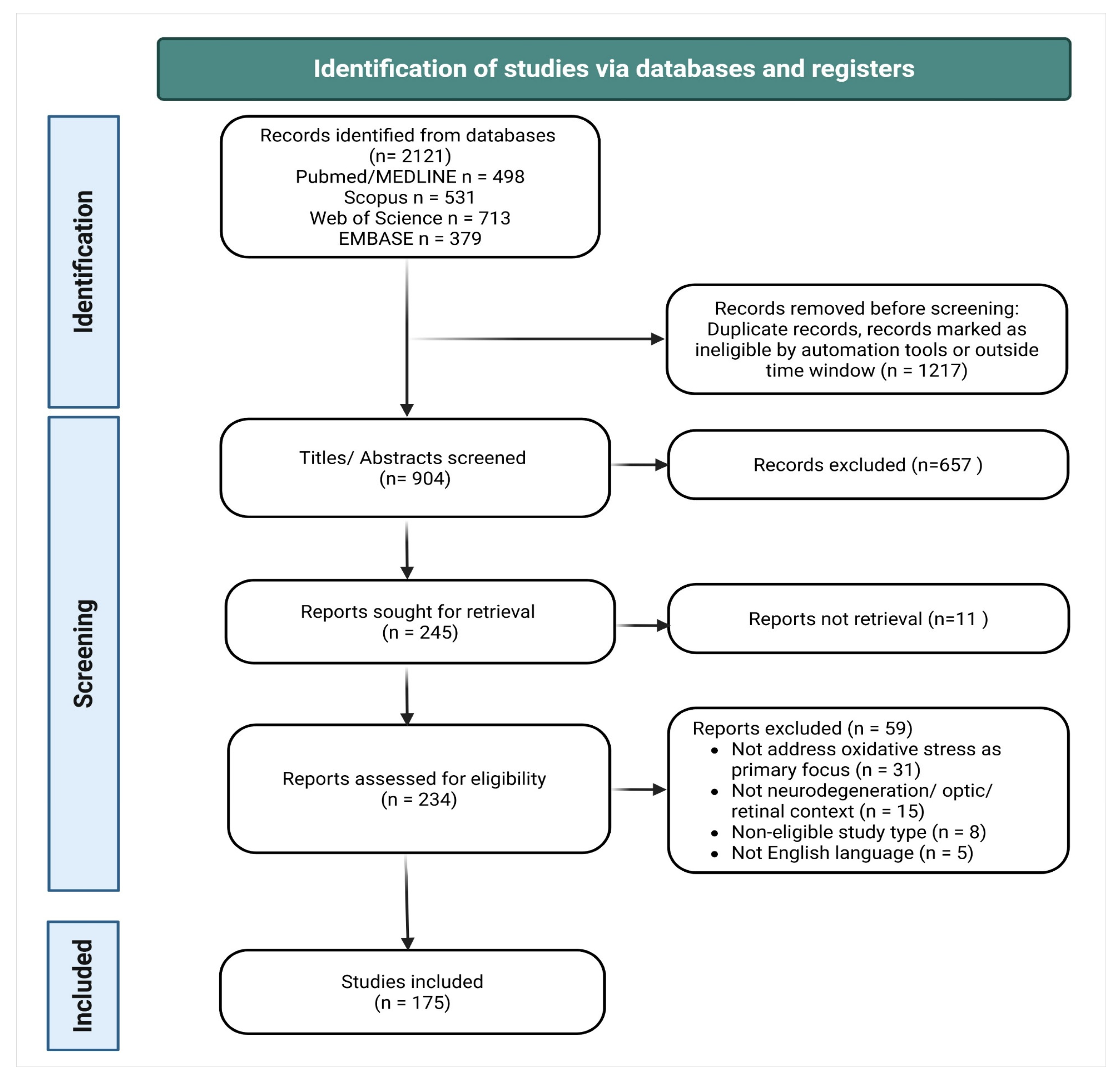
Figure 2.
Oxidative stress in specialized axonal projections and cranial nerve vulnerability. Oxidative stress and redox imbalance contribute to neurodegeneration through interconnected pathological processes, including mitochondrial dysfunction, impaired oxidative phosphorylation, axonal transport disruption, calcium dyshomeostasis, neuroinflammatory activation, defective mitophagy and diminished regenerative capacity. The optic nerve represents the most extensively characterized model of this vulnerability, owing to the exceptional metabolic demands of RGC retinal ganglion cell axons, their dependence on mitochondrial trafficking and oxidative phosphorylation, and the unique accessibility of the visual system to non-invasive structural and functional assessment. Emerging evidence suggests that analogous bioenergetic and redox-dependent mechanisms may also underlie dysfunction in other cranial nerves and long-range neuronal projections, supporting the concept of a common framework of neurodegenerative susceptibility, although direct evidence beyond the visual system remains comparatively limited.
Figure 2.
Oxidative stress in specialized axonal projections and cranial nerve vulnerability. Oxidative stress and redox imbalance contribute to neurodegeneration through interconnected pathological processes, including mitochondrial dysfunction, impaired oxidative phosphorylation, axonal transport disruption, calcium dyshomeostasis, neuroinflammatory activation, defective mitophagy and diminished regenerative capacity. The optic nerve represents the most extensively characterized model of this vulnerability, owing to the exceptional metabolic demands of RGC retinal ganglion cell axons, their dependence on mitochondrial trafficking and oxidative phosphorylation, and the unique accessibility of the visual system to non-invasive structural and functional assessment. Emerging evidence suggests that analogous bioenergetic and redox-dependent mechanisms may also underlie dysfunction in other cranial nerves and long-range neuronal projections, supporting the concept of a common framework of neurodegenerative susceptibility, although direct evidence beyond the visual system remains comparatively limited.
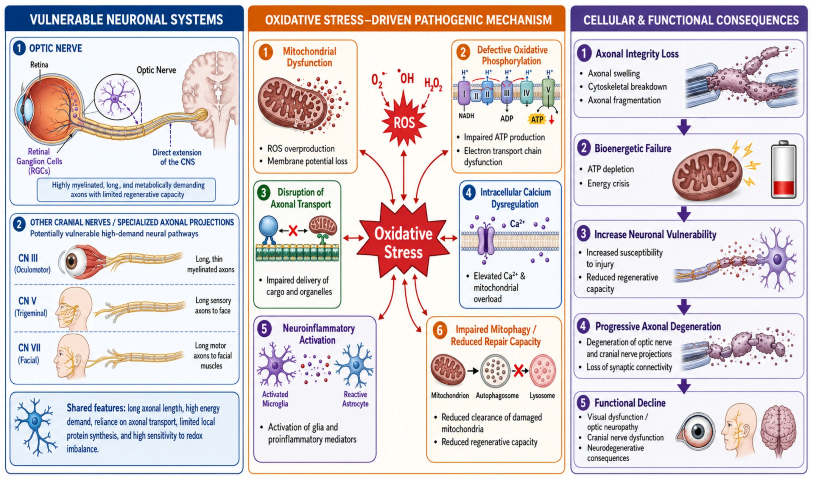
Figure 3.
Conceptual framework of the oxidative stress-driven cascade linking mitochondrial dysfunction to retinal neurodegeneration and clinically detectable retinal biomarkers. Reactive oxygen species promote mitochondrial dysfunction, ATP depletion, mitophagy failure, neuroinflammation, ferroptosis, axonal degeneration, and retinal ganglion cell loss, ultimately leading to OCT-detectable retinal thinning as a potential clinical biomarker.
Figure 3.
Conceptual framework of the oxidative stress-driven cascade linking mitochondrial dysfunction to retinal neurodegeneration and clinically detectable retinal biomarkers. Reactive oxygen species promote mitochondrial dysfunction, ATP depletion, mitophagy failure, neuroinflammation, ferroptosis, axonal degeneration, and retinal ganglion cell loss, ultimately leading to OCT-detectable retinal thinning as a potential clinical biomarker.
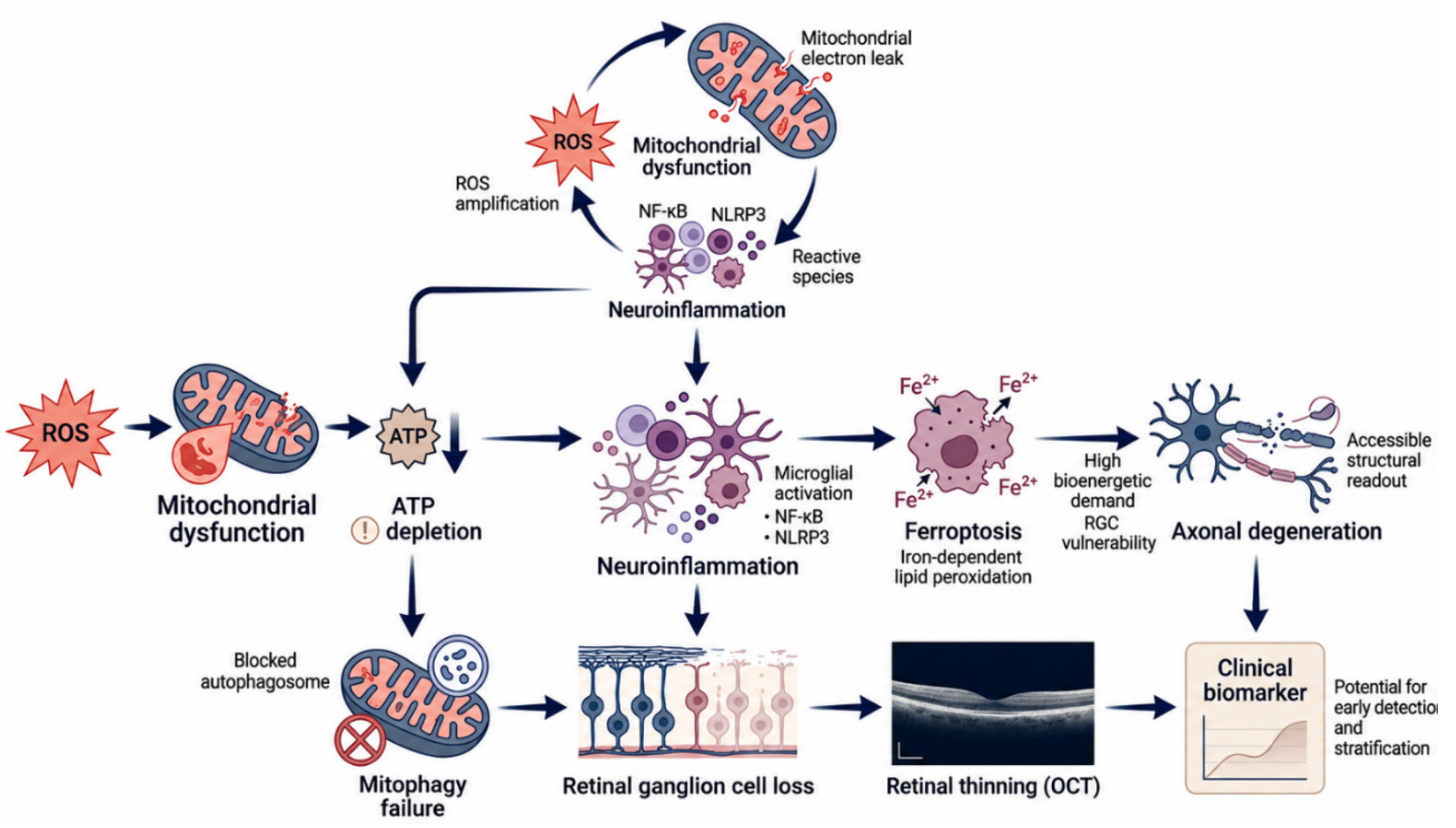

Table 1.
Characteristics of representative included studies.
| Author (Year) | Country | Study Design | Disease/Model | Main Findings |
|---|---|---|---|---|
| Ahmad et al. (2024) [14] | Multinational (Europe) | Clinical (CSF cohort, MCI) | Alzheimer’s disease / MCI | Oxidative-stress and inflammatory metabolites associate with AD cerebrospinal-fluid biomarkers in MCI. |
| Cheng et al. (2022) [16] | USA | Narrative review | Neurodegeneration/axon | Axonal mitochondrial maintenance and bioenergetics determine neuronal vulnerability and regenerative capacity. |
| Calkins et al. (2021) [23] | USA | Narrative review | Glaucoma | Frames glaucoma as a progressive RGC neurodegeneration with adaptive and maladaptive responses to metabolic and oxidative stress along the optic projection. |
| Buonfiglio et al. (2023) [25] | Germany | Narrative review | Optic nerve diseases | Oxidative stress is a suitable therapeutic target across optic nerve diseases. |
| Tezel et al. (2022) [26] | USA | Narrative review | Glaucoma | Synthesizes molecular regulation of neuroinflammation (glia, complement, oxidative signaling) as a treatment target in glaucoma. |
| Kaarniranta et al. (2020) [30] | Finland | Narrative review | AMD | Positions mitochondrial dysfunction (mtDNA damage, impaired mitophagy) as a driver of RPE degeneration in AMD. |
| Banna et al. (2024) [32] | Australia | Narrative review | Eye-brain axis | Reviews retinal imaging as a non-invasive window to brain health. |
| Zeng et al. (2023) [44] | China | Experimental (in vivo + in vitro) | Glaucoma | Pathologically high IOP drives Drp1-dependent mitochondrial dysfunction and RGC PANoptosis; Drp1 inhibition is protective. |
| Naguib et al. (2021) [45] | USA | Animal (microbead mouse) | Ocular hypertension/ glaucoma | IOP elevation triggers an early ROS rise and endogenous NRF2-ARE antioxidant activation as a protective response. |
| Realini et al. (2025) [46] | Italy | Animal (mouse) + in vitro | Retinal aging/ diabetic retinopathy | The cysteine pro-drug NACET induces NRF2 and prevents retinal aging and diabetic retinopathy. |
| Upadhyay et al. (2020) [47] | USA | Experimental (aged mouse / human RPE) | Retina-RPE aging | Aging increases retinal/ RPE oxidative stress; DJ-1 is a key antioxidant defense in the RPE. |
| Liao et al. (2023) [48] | China | Animal + in vitro | Acute ocular hypertension | NOX4 inhibition (GLX351322) suppresses ROS and redox-sensitive inflammation, reducing retinal injury. |
| Rosenkranz et al. (2021) [49] | Germany | Animal (EAE mouse) | Multiple sclerosis | Enhancing neuronal mitochondrial activity protects against inflammatory neurodegeneration. |
| Chen et al. (2024) [50] | China | Narrative review | Glaucoma | Reviews ferroptosis and pyroptosis as regulated-necrosis pathways driving RGC death. |
| Amore et al. (2023) [51] | Italy | Clinical (case report) | ALS + LHON | Co-occurrence of ALS and LHON suggests mitochondrial dysfunction as a shared phenotypic modifier. |
| Surma et al. (2023) [52] | USA | In vitro (hPSC-derived RGCs) | RGC/ optic neuropathy | Enhanced mitochondrial biogenesis promotes RGC neuroprotection against oxidative injury. |
| Zhao et al. (2021) [53] | China | Narrative review | AMD | Iron accumulation and lipid peroxidation implicate ferroptosis in age-related macular degeneration. |
| Sahu et al. (2021) [54] | USA | Animal (rd1 mouse, gene therapy) | Retinitis pigmentosa | OXR1 gene therapy reduces oxidative stress and retards photoreceptor neurodegeneration. |
| Kutsyr et al. (2020) [55] | Spain | Animal (mouse) | Retinal degeneration | Gradually increased environmental light induces oxidative stress/inflammation and accelerates retinal neurodegeneration. |
| Usategui-Martín et al. (2022) [56] | Spain | In vitro/ ex vivo (organotypic retina) | Retinal neuroprotection | MSC secretome modulates oxidative stress, autophagy and programmed cell death to protect the retina. |
| Mimura and Noma (2025) [57] | Japan | Narrative review | Diabetic retinopathy | Comprehensive review of oxidative mechanisms, biomarkers and therapeutics in diabetic retinopathy. |
| Means et al. (2020) [58] | USA | In vitro (ONH astrocytes) | Glaucoma/ optic nerve | Resveratrol protects optic-nerve-head astrocytes from oxidative death (↓caspase-3, ↓tau dephosphorylation, ↓aggregates). |
| Harada et al. (2020) [59] | Japan | Review + model evidence | Normal-tension glaucoma | Oxidative-stress suppression by antioxidants is a potential therapeutic approach in normal-tension glaucoma. |
| Fan Gaskin et al. (2021) [60] | Australia | Narrative review | Glaucoma | NADPH oxidase (NOX) is a principal source of ROS and oxidative stress in glaucoma. |
| Sanz-Morello et al. (2021) [61] | Denmark | Narrative review | Optic neuropathies | Redox imbalance contributes across the optic-neuropathy spectrum. |
| Chien et al. (2021) [62] | Taiwan | Animal (rat NAION) | Ischemic optic neuropathy | Nrf2 activators (bardoxolone methyl, omaveloxolone) improve RGC survival. |
| Catalani et al. (2023) [63] | Italy | Narrative review | RGC degeneration | Targeting mitochondrial dysfunction and oxidative stress to prevent RGC neurodegeneration. |
| Korczowska-Łącka et al. (2023) [64] | Poland | Clinical (patient biomarker study) | Neurological diseases | Selected oxidative-stress and energy-metabolism biomarkers are altered across neurological diseases. |
| Gao et al. (2026) [65] | China | Narrative review | Optic nerve injury | Microglial neuroinflammation in optic nerve injury, from mechanisms to therapeutic targets. |
| Shi et al. (2024) [66] | China | Human genetic (Mendelian randomization) | Glaucoma | Genetic evidence supports a causal effect of oxidative stress on glaucoma risk. |
Table 2.
Core oxidative mechanisms in optic-nerve and retinal neurodegeneration.
| Mechanism Domain | Key Mediators/Molecular Effectors | Role in Optic-Nerve and Retinal Neurodegeneration | Refs |
|---|---|---|---|
| ROS/ RNS and redox imbalance | Superoxide (O2•⁻), H2O2, hydroxyl radical, peroxynitrite (ONOO⁻); NADPH oxidases (NOX1/2/4), xanthine oxidase, uncoupled NOS; lipid peroxidation, protein carbonylation, 8-OHdG; counter-regulated by the NRF2-KEAP1-ARE axis. | Macromolecular damage and RGC apoptosis; acts as both an initiator and a feed-forward amplifier of degeneration. | [3,10,45,60,72] |
| Mitochondrial dysfunction | Electron-transport-chain/OXPHOS deficits, membrane-potential collapse, Drp1-mediated fission, mtDNA damage, NAD⁺ depletion, impaired biogenesis. | Bioenergetic failure of high-demand RGCs; a major ROS source; drives apoptosis/ PANoptosis and axonal degeneration. | [22,39,43,44,52] |
| Neuroinflammation | Microglia, astrocyte and Müller-glia activation; NF-κB, NLRP3 inflammasome, IL-1β/ TNF-α; complement; galectin-3; exosomal signalling; TRPV4. | Establishes a self-reinforcing oxidative-inflammatory loop that amplifies RGC injury. | [7,9,26,86,87,92,94,96] |
| Ferroptosis | Iron overload, ACSL4/ LPCAT3-dependent PUFA-phospholipid peroxidation, system xc⁻/GSH/GPX4 axis, FSP1-CoQ10 pathway. | Iron-dependent death of RGCs, RPE and photoreceptors in glaucoma, diabetic retinopathy and AMD. | [53,120,121] |
| Mitophagy / autophagy (quality control) | PINK1/Parkin, OPTN, BNIP3/NIX-mediated axonal mitophagy; autophagic flux. | Clears damaged mitochondria; failure causes ROS accumulation and glaucomatous degeneration. | [41,81,82,122] |
Abbreviations: ACSL4, acyl-CoA synthetase long-chain family member 4; AMD, age-related macular degeneration; ARE, antioxidant response element; CoQ10, coenzyme Q10; Drp1, dynamin-related protein 1; FSP1, ferroptosis suppressor protein 1; GPX4, glutathione peroxidase 4; GSH, reduced glutathione; 8-OHdG, 8-hydroxy-2′-deoxyguanosine; KEAP1, Kelch-like ECH-associated protein 1; LPCAT3, lysophosphatidylcholine acyltransferase 3; mtDNA, mitochondrial DNA; NAD+, nicotinamide adenine dinucleotide; NF-κB, nuclear factor kappa B; NLRP3, NOD-like receptor family pyrin domain-containing 3; NOS, nitric oxide synthase; NOX, NADPH oxidase; NRF2, nuclear factor erythroid 2-related factor 2; OPTN, optineurin; OXPHOS, oxidative phosphorylation; PINK1, PTEN-induced kinase 1; PUFA, polyunsaturated fatty acid; RGC, retinal ganglion cell; ROS/RNS, reactive oxygen/nitrogen species; RPE, retinal pigment epithelium; TNF-α, tumor necrosis factor alpha; TRPV4, transient receptor potential vanilloid 4.
Table 3.
Oxidative-stress biomarkers across the brain-retina neurodegenerative continuum.
| Biomarker | Matrix | Disease/Context | Clinical Significance | Ref. |
|---|---|---|---|---|
| 8-OHdG/ 8-OHG (oxidative DNA/ RNA damage) | Retina, serum, CSF | Glaucoma, AD, neurodegeneration | Marker of oxidative nucleic-acid damage; localizes to RGC mitochondria; candidate progression marker. | [14,43,64] |
| Malondialdehyde (MDA) | Serum, aqueous humor | Ocular hypertension, POAG | Lipid-peroxidation marker elevated in glaucoma/OHT; component of combined oxidative-stress panels. | [13,153,155] |
| 4-Hydroxynonenal (4-HNE) | Retinal/ vitreous tissue | Vitreoretinal & neurodegenerative disease | Reactive lipid-peroxidation aldehyde; indicator of tissue oxidative damage. | [6,156] |
| F2-isoprostanes | Plasma, CSF | Neurodegeneration | Reference lipid-peroxidation biomarker reflecting systemic oxidative load. | [13,64] |
| Protein carbonyls | Serum, CSF | MCI, neurological disease | Protein-oxidation marker; preclinical indicator of cognitive impairment. | [64,147] |
| Glutathione (GSH/ GSSG ratio) | Blood, aqueous humor | Glaucoma | Reflects antioxidant reserve; a reduced ratio indicates redox imbalance. | [13,153] |
| Superoxide dismutase (SOD) activity | Tear fluid, blood | ALS, glaucoma | Antioxidant-enzyme activity; minimally invasive (tear) readout of systemic redox state. | [105,155] |
| NAD⁺/ NADH redox state | Retina, blood | Glaucoma | Bioenergetic/redox biomarker and therapeutic target. | [83,157] |
| Thioredoxin-interacting protein (TXNIP) | Retinal tissue | Diabetic retinopathy | Links hyperglycemia to oxidative/inflammatory RGC injury; therapeutic target. | [158] |
| MicroRNAs (redox-related miRs) | Aqueous humor, serum | POAG | Regulatory biomarkers of oxidative-stress signaling. | [159] |
| Vitreous oxidative/ inflammatory proteome | Vitreous humor | Vitreoretinal disease | Proteomic signature integrating oxidative, inflammatory and neurodegenerative markers. | [156] |
| Iron/ lipid-peroxidation (ferroptosis) indices | Retina (tissue/imaging) | AMD, diabetic retinopathy | Indicate iron-dependent lipid peroxidation and ferroptotic burden. | [53,121] |
| Retinal structural imaging (RNFL, GCC, OCT/ OCTA) | Retinal imaging (in vivo) | AD, PD, MS, glaucoma | Non-invasive structural/vascular biomarkers paralleling CNS neurodegeneration. | [160,161,162,163,164] |
| Integrated multimarker panels | Serum/CSF + imaging | Neurodegeneration (general) | Combine oxidative, inflammatory, mitochondrial and neurofilament markers for staging and stratification. | [13,32,147] |
Abbreviations: AD, Alzheimer’s disease; ALS, amyotrophic lateral sclerosis; AMD, age-related macular degeneration; CNS, central nervous system; CSF, cerebrospinal fluid; GCC, ganglion cell complex; GSH/GSSG, reduced/oxidized glutathione; 4-HNE, 4-hydroxynonenal; 8-OHdG, 8-hydroxy-2′-deoxyguanosine; 8-OHG, 8-hydroxyguanosine; MCI, mild cognitive impairment; MDA, malondialdehyde; miRs, microRNAs; MS, multiple sclerosis; NAD+/NADH, oxidized/reduced nicotinamide adenine dinucleotide; OCT(A), optical coherence tomography angiography; OHT, ocular hypertension; PD, Parkinson’s disease; POAG, primary open-angle glaucoma; RGC, retinal ganglion cell; RNFL, retinal nerve fibre layer; SOD, superoxide dismutase; TXNIP, thioredoxin-interacting protein.
Disclaimer/Publisher’s Note: The statements, opinions and data contained in all publications are solely those of the individual author(s) and contributor(s) and not of MDPI and/or the editor(s). MDPI and/or the editor(s) disclaim responsibility for any injury to people or property resulting from any ideas, methods, instructions or products referred to in the content. |
© 2026 by the authors. Licensee MDPI, Basel, Switzerland. This article is an open access article distributed under the terms and conditions of the Creative Commons Attribution (CC BY) license.
Copyright: This open access article is published under a Creative Commons CC BY 4.0 license, which permit the free download, distribution, and reuse, provided that the author and preprint are cited in any reuse.


