

Submitted:
30 June 2026
Posted:
30 June 2026
You are already at the latest version
Abstract
Background/Objectives: Nasogastric tube insertion is a very common procedure for enteral feeding, hollow viscera decompression and perioperative management. Although generally safe, both insertion and removal are not without complications. Methods: The aim of the present narrative review is to classify, analyze and make comments on the cases of a gastric tube [usually inserted through the nose but in some cases through the mouth] unexpectedly becoming twisted, coiled or self-knotted within the stomach, entangled with the endotracheal tube, entrapped in the suture line of a hollow viscera, or suddenly broken at its end. Results: This review has the particular characteristic that the articles analyzed are simply case reports, and not clinical studies. The literature search revealed 107 cases since 1977, which we classify into 5 categories of complications instead of the four applied to date. All these relatively rare complications are recognized usually only at the time of gastric tube removal and sometimes require emergency manipulation. After meticulous and documented research, we came to the conclusion that the pathogenesis and etiology of self-knotting seem to be different depending on whether or not a patient is endotracheally intubated. To these reports we have added another eight unpublished personal cases, all successfully managed by means of endoscopy. Conclusions: We argue that these complications must always be considered a possibility by the doctors involved, so that, at the slightest suspicion, there is immediate readiness to treat in order to avoid, as far as possible, a more complicated outcome likely to subject the patient to greater risk and may even require the cooperation of specialties to treat it.
Keywords:
nasogastric tube
; complications
; self-knotting
; entangled with endotracheal tube
; entrapped in the suture line
; tube fracture
1. Introduction
The insertion of a nasogastric tube is a widely used procedure in everyday practice, serving multiple purposes including gastric decompression, enteral feeding, medication administration, and perioperative management. Although generally considered safe, nasogastric tube insertion and removal are not without complications, ranging from minor discomfort to serious and potentially life-threatening events [1,2,3]. Among the most reported complications are gastric tube misplacement, mucosal damage to the nostrils and esophagus, aspiration, esophageal or gastric perforation, and airway-related adverse events; however, there are other complications, less common and, at a first glance, somewhat ‘unexpected’, as illustrated in various cases-reports [3,4,5,6].
These concern deformation or failure of the gastric tube material [usually inserted through the nose but in some cases through the mouth], at an unpredicted time after their insertion, and being recognized as a complication only during the removal process: tube knotting, entrapment in the suture line of a hollow viscera, tube fragmentation and finally high-risk entanglement with the airway device [7]. These incidents, although rare, may result in difficulties or failure of tube removal, prolongation of the withdrawal procedure or the need for endoscopic assistance or even surgical intervention. They are thus of noteworthy clinical importance [8].
Such complications have been consistently described over several decades, indicating the constant and well-recognized risks of nasogastric tube insertion and long-term resting in the stomach, rather than as isolated or outdated concerns. Recent publications continue to report similar adverse events, reaffirming that despite improvements in materials, techniques, and safety protocols, nasogastric tube–related complications remain clinically relevant in modern-day practice [1,9].
The purpose of the present narrative review of the published reports of such complications published over 40-years, along with eight similar unpublished personal cases, is to analyze the different parameters that may be related to the incident and the subsequent handling of the situation to solve it, as well as to critique specific manipulations which likely contributed to, if not actually caused, the complication.
More specifically, we will look at a number of other parameters: relating to the patient - any special anatomical characteristics and their indications for tube placement, the tube - its construction material, diameter, length of tubing inside the stomach, as well as the number of days after insertion, and finally, issues relating to the handling of the procedure by the doctor (or even the nurse in some countries): whether there was a difficulty at insertion (the number of attempts, whether there was any checking of correct placement, whether an endotracheal tube was already in place, the medical specialty of the doctor who initially attempted to remove the tube, whether help from another medical specialty was requested, what the outcome was and whether there was anything that could, with hindsight, have been avoided.
2. Materials and Methods
Study design and objective
We conducted a narrative review to summarize reported cases of mechanical complications associated with gastric tube placement and removal. The review focuses on the four categories of complications described in the literature: self-knotting of the tube, entanglement with the endotracheal tube, entrapment within the suture line of a hollow viscera and tube breakage. In addition to reviewing published series and case reports, we present eight unpublished individual cases from our institution to illustrate management approaches. The objective was to describe patient characteristics, indications for gastric tube placement, tube size, management strategies, and reported procedural difficulties.
Literature research
A comprehensive literature search was performed in PubMed, Scopus, MEDLINE, Google Scholar and the grey literature from 1970 to December 2025, using the keywords: “nasogastric tube” OR “NGT” OR “orogastric tube” OR ‘gastric tube” AND “complications” OR “knotting” OR “entrapment” OR “breakage” OR “fracture” OR “suture” OR “endotracheal”. Additional studies were identified by manual screening of the reference lists of included articles.
Only case reports and case series were included if they reported mechanical complications of gastric tube placement, whether occurring during insertion, manifesting as acute symptomatic events, or detected incidentally during removal or during endoscopic examination of the patient for another reason, and only when patient age or characteristics, tube type, and manipulation details were given. Exclusion criteria encompassed reviews without original case data, and reports lacking sufficient clinical information.
Data extraction was performed independently by two reviewers, and any discrepancies were resolved by consensus. No patient-identifiable information was used in any included case; therefore, ethical approval was not required.
3. Results
A thorough review of the literature from 1977 to 2025 revealed a total of 104 publications including 107 cases, which were either simply presentations of cases or with commentaries on similar cases, as a short review or as a critical analysis. To supplement this overview, we include some similar, unpublished, personal cases from our institution, providing contemporary clinical correlates which underscore the practical implications of gastric tube-related adverse events. As already mentioned, these gastric tube-related complications, recognized mainly during withdrawal, are usually divided into 4 groups, depending on the cause of the complication: tube self-knotting, entanglement with endotracheal tubes, entrapment within an anastomosis or a suture line, and tube breakage.
3.1. Self-Knotted Nasogastric Tubes
A total of 64 published cases of gastric tube knots was identified, the earliest reported in 1977, from the Galveston University of Texas [10]. During the progress of analysis, we realized that 27 out of the 64 cases had already been tracheally intubated, due to an ongoing surgery or for mechanical ventilatory support; meaning that there was a tube in the posterior oro- [or naso]-pharynx toward the trachea. Considering that the friction and interference of the gastric tube on the pre-inserted endotracheal tube within the narrow space of the oropharynx could be, on its own, the reason for the knot formation, we decided to split and separately classify these cases as a discrete, 5th, category; our decision strongly supported by the same etiology of the other category of complications, where the gastric tube was coiled and tightly knotted around the endotracheal tube.
We thus have split these 64 patients into two subgroups: the one, including 37 conscious patients [not intubated and not under mechanical ventilation] to whom a gastric tube had been inserted for feeding or gut decompression purposes, and the other, 27 patients, already intubated when the gastric tube was inserted.
3.1.1. Self-Knotted Nasogastric Tubes Inserted in Non-Intubated Patients
These 37 patients, ranging from newborn [24 days] to 90 years, were mainly adults [29 cases, rate 78.4%], 8 being children, aged between 24 days to 15 years [Table 1]. There was a relative lack of information on gender, but generally males seem to outnumber females [21:16]. No patient was reported as having any special anatomical characteristic, such as nostril stenosis, Zenker diverticulum, esophageal or esophagogastric junction stenosis, gastrectomy of any type, or lumen compression from an extraluminal mass, making tube insertion difficult; apart from one case of caustic esophageal injury [11], two cases of hypopharyngeal and cervical esophagus neoplasia [12,13], the gastric tube was inserted under endoscopic guidance. Four cases were reported as needing a number of attempts for insertion [13,14,15,16,17] while, finally, 15 papers make no mention of either difficulty or non-insertion.
In 23 cases the indication for nasogastric tube placement was for nutritional support or long-term enteral feeding; in 12 cases for bowel decompression after gut obstruction of post-operative or malignant etiology; and in one case for stomach lavage after self-intoxication for suicide.
Information on who placed the gastric tube is also sparse: in 14 cases a doctor, and in 8 cases a nurse is referred to. Regarding the material of which the gastric tubes were constructed, there are only references to 9 Ryles plastic tubes, 5 from PVC, 2 sumps wide bore catheters for decompression, and two silastic with a tungsten tip at its distal end. The depth of insertion was noted in only 10 cases: 4 reported as ‘deep’, or ‘beyond usual length’ and 6 to a normal depth of 50-60 cm depth from the dental border. Tube size, ranging from 8 to 18 FR, was reported in 26 cases: 17 being thinner or equal to 14FR; seven of 16 FR, and two of 18FR.
When the attending physician or the yard nurse realized the difficulty in removal and what it really signified, 14 patients were further treated by the same person applying lubrication, rotation, ‘push and pull’ technique, Magill forceps, or even a forceful removal in 14 cases. In another 13 cases the tube was removed endoscopically and in another three by ENT management, the decision probably based on availability. Five patients were reported as receiving general anesthesia for removal [18,19,20,21,22]. The most applicable technique, regardless of what specialist applied it, was to cut the tube outside the nostril and remove the distal part – with the knot - through the oral cavity [nasal and oral approach]. Finally, regarding the knot configuration itself, while generally reported as “a knot”, there are four descriptions of it as a ‘lariat loop’ [20,23,24,25], one as a ‘figure-of-8 pattern’ [26], one as multiple loops [22], and one as an ‘alpha loop’ [27].
Seven knots were described as impacted — within the nasal cavity [18], the oropharynx [27], the nasopharynx [28], and the posterior pharynx [5]; another one at the cricopharyngeal level [29], and two at the hypopharynx [17,22]; the multiple-loops knot occupied the area from nasopharynx to hypopharynx [22], and the one found entrapped in the upper esophagus [11] revealed the presence of a tracheoesophageal fistula. Another two knots were entrapped in the gastroesophageal junction or a hiatus hernia [12,30].
3.1.2. Self-Knotted Gastric Tubes Inserted in Priorly-Intubated Patients
This category deals with knots occurring in 27 patients, in whom the gastric tube had been inserted [either through the nose or the mouth] after endotracheal intubation, the patient being on mechanical ventilation in order to be operated on for a gastrointestinal disease, acute or chronic, benign or malignant [Table 2]. The first reported complication of this type was reported in 1983.
The 27 intubated patients, ranging in age from a premature female infant [2 days old, operated on for duodenal atresia] to 84 years, were almost all adults [21 cases - rate 78%, 14 of whom were females], while the remaining four were children, the premature-2 days, 6mo, 5 and 7 years of age. There were no specific difficulties in gastric tube insertion reported, except for three [4,7,31] – all placements performed by the anesthesia team. However, there were three references [32,33,34] of surgeons complaining that the stomach remained full of air, obscuring visualization during laparoscopic surgery, leading to the adjustment of the tube position. In almost all cases the gastric tubes had been inserted for gastric decompression, to facilitate laparoscopic vision. There were also three cases of patients with feeding tubes [35,36,37,38] and another one with a tube inserted for technical reasons connected to cervical discectomy surgery [39].
The majority of gastric tubes were removed by the end of surgery except for 6 cases where they remained in place for up to 4 postoperative days. Although there is no systematic reference to tube diameter, anesthesiologists generally prefer wide bore catheters. A 12FR tube was referred to in three cases, and a 6FR Silastic transpyloric catheter in a 6-month premature infant.
Regarding removal attempts, after recognition of tube knotting, most procedures were performed in the operating room, usually by the anesthesia team, or assisted by an endoscopist [36,40,41] or an ENT [42]. The technique usually applied was to cut the tube close to the nostrils, the distal part – with the knot – then being grasped with McGill forceps and removed from the hypopharynx or the posterior nasopharynx. Alternatively, the distal knotted part, visible in the oropharynx, was grasped with McGill forceps, or a snare or forceps, pulled out of the mouth, cut and then removed in two parts. However, there are two cases of hemorrhage [i] after forceful extraction [43], and [ii] probably due to the bulk of the 4-loop knot [2] both at the same hospital.
Finally, the most stressful case was that of a 36-year-old female [40] having undergone liver transplantation. Eight hours after tracheal extubation, the patient suddenly experienced coughing, dyspnea, and cyanosis and a decision was then made to urgently re-intubate. Fortunately, upon insertion of the laryngoscope, the nasogastric tube was found coiled around the epiglottis

Table 1.
Self-knotted nasogastric tubes inserted in non-intubated patients.
| Ref | Age/ Gender | Underlying condition | Use | ∅ in Fr | Material or type | Length of insertion | Difficulty in placement [+/-] | Days in situ |
Who placed? | Management removal |
Comments |
|---|---|---|---|---|---|---|---|---|---|---|---|
| Jindal R et al; 2022 [73] |
55/Μ | Oral Ca | feeding | 15 | PVC | - | [-] | 90 | N/A | Endoscopic failure + laparotomy (PEG) | |
| Kumar A et al; 2017 [91] |
15/F | Neurosurgery | feeding | 14 | PVC | 55 | [-] | 2 | Surgeon | Endoscopic (PEG) | |
| Gulsoy Z et al; 2019 [92] |
67/M | Parkinson -PEG Infection | feeding | 14 | PVC | 60 | [-] | 4 | N/A | Surgical (gastrotomy) | |
| Awe JAA et al; 2014 [14] |
30/M | sigmoid volvulus | de-compression | 16 | Ryles | 60 | [-] | 2 | Surgeon | Manually; Gentle traction | |
| Bernica J et al; 2019 66] |
88/M | Bowel obstruction | de-compression | 18 | Sump tube | - | [-] | 2 | Surgeon | Endoscopic; Cut + removed (nose + mouth) | |
| Delirrad M et al; 2015 [83] |
23/F | Self-intoxicate suicide | gastric lavage | 16 | PVC | - | [-] | 4 h | Nurse (emergency) | Manually; Cut +removed nose + mouth | |
| Mandal NG et al; 2002 [20] |
33/F | Caesarean – ileus |
de-compression | 12 | Ryles | - | [-] | 5 h | Obstetric Operation room |
Manually; Cut + removed nose + mouth | General anesthesia, LARIAT loop knot |
| Santhanam V et al; 2007 [5] |
81/M | Bladder Ca - ileus | de-compression | 14 | Ryles | 40 | [-] | immediately | Anesthesiologist nurse | ENT removal; nasopharyngeal tube over and push | the knot at posterior pharynx |
| Tsandiraki J et al; 2022 [26] |
45mo/F | Acute pneunonia/ tuberculosis | feeding | 12 | PVC | Beyond usual | [-] | 30 | Nurse | Endoscopically | In a figure-of-8 pattern loop |
| Singh M et al; 2014 [23] |
40/M | Bowel obstruction | de-compression | 16 | - | Deep insertion | [-] | 3 | Surgeon | Manually; Cut removed nose + mouth (Magill forceps) | LARIAT loop knot |
| Kumar Sinha A et al; 2020 [24] |
4mo/M | Anorectal malformation | de-compression | 10 | - | - | [-] | 5 | Pediatric Surgeon |
Manually; Cut removed nose + mouth | LARIAT loop knot |
| Khond AD et al; 2026 [18] |
22/F | Intracraneal tube | feeding | 10 | - | - | Ν/A | 90 | Neuro surg ENT |
1st attempt failed, under general anesthesia; endoscopic removal using scissors | |
| Mohsin M et al; 2007 [11] |
25/M | Caustic esophagus injury-suicide | feeding |
- |
- | - | N/A | Long-term | Local MD | Endoscopic; Impacted into upper esophagus | Difficulty in removal Trachea-esophageal fistula left |
| Paul R et al; 2022 [25] |
72/F | coma | feeding | 14 | Ryles | - | Ν/A | Short-term | Anesthe- siologist |
Manually; Magill forceps | LARIAT loop |
| Moscote-Salazar LR et al; 2019 [78] |
86/M | stroke | feeding | 18 | Ryles | - | N/A | Short-term | ICU-nurse | Manually; laryngoscopy -Magill forceps | Successful lubrication + rotation |
| Morris HH et al; 1977 [10] |
5/M | Degenerative disease | feeding | - | - | - | [-] | 3 | Physician | Manually; pull to oropharynx, cut distal end | |
| Tapaiwala SN et al; 2008 [28] |
35/M | Pre-operatively | de-compression | 16 | - | - | N/A | a few hours | N/A | Manually; Cut removed nose + mouth | Immediately post-op impacted nasopharynx |
| McHardy DA et al; 1993 [27] |
child | Bowel obstruction | de-compression | - | - | 25 | [-] | months | N/A | Endoscopic; Cut removed nose + mouth | impacted oropharynx - push and pull” technique |
| Tai CM et al; 2010 [6] |
53/M | Thyroid Ca | feeding | 16 | - | - | N/A | - | Nurse | Endoscopic; Forceful removal | Upper esophagus tangled (4 X 3cm ulceration) |
| Galanopoulos M et al; 2017 [30] |
90/F | stroke | feeding | - | - | - | N/A | 60 | N/A | Endoscopic; forceps | Entrapment in hiatus hernia |
| Te BC et al; 2024 [29] |
75d | Nephrotic syndrome | feeding | 8 | N/A | 76–80 | N/A | 30 | Assistant nursing officer | Manually; Cut removed nose + mouth | At cricopharyngeal level - general anesthesia |
| Shiva S et al; 2024 [93] |
33/M | Post-op Ileus | de-compression | 16 | N/A | 55 | N/A | 4 | - | Manually; laryngoscopy + Magill’s forceps | |
| Narayan KS et al; 2017 [74] |
72/F | stroke | feeding | - | - | 25 | N/A | 4 mo | N/A | Endoscopic; manipulation + push–pull technique | an alpha loop knot |
| Hickey NC et al; 1988 [19] |
21/M | Cerebral palsy - empyema | feeding | 9 | Tungsten- tip tube | - | N/A | Short-term | N/A | General anesthesia | |
| Chavda V et al; 2017 [15] |
75/F | Bowel obstruction/ history of colectomy |
de-compression | 12 | Wide bore | - | [+] 3rd attempt |
- | Nurse | Manually | A knot recognized after tube removal |
| Kim CE et al; 2021 [16] |
66/M | Chronically bedridden | feeding | 12 | - | - | [+] Many attempts |
upon insertion | Attending physician | ENT; Flexible nasal endoscopy |
Nasal and oral approach |
|
Malhotra P et al; 2025 [12] |
55/M | Ca hypopharynx/ upper esophagus | feeding | - | Ryles | N/A | [-] | 6 mo | Endoscopist | Endoscopic | Kinked and stucked at GEJ |
| Wright S et al; 2014 [94] |
- | Post-operative Ileus | de-compression | - | Ryles | - | [-] | Few days | Nurse | - | General anesthesia |
| Awe JAA et al; 2025 [14] |
30/M | Head injury | feeding | 16 | - | 55 | [-] | 15 | Junior staff | Manually; nose and mouth approach - McGill forceps |
Removed at 2nd attempt |
| Munoz NR et al; 2023 [17] |
68/M | HIV_ sepsis |
feeding | - | - | - | [+] | Short-term | pulmo-nologist | Removal by a bronchoscope | Knot at the hypopharynx |
| Abhyankar S et al; 2019 [75] |
8 | burn | feeding | 10 | Ryles | deep | N/A | weeks | N/A | Manually; nose + mouth | |
| 10 | burn | - | 10 | Ryles | deep | N/A | - | N/A | |||
| RAvind R et al; 2015 [13] |
- | Ca cervical esophagus | feeding | 14 | plastic | 60 | [+] | 30 | Clinician Assistant | Endoscopic & manually; nose + mouth | |
| Hirwa KD et al; 2016 [21] |
74/M | Ischemic attack | feeding | 14 | - | - | [-] | - | Emergency doctor | Removed under fibroscopy (artery forceps) | |
| Chang BA et al; 2014 [22] |
24d/M | Sepsis_ urology |
feeding | - | - | - | [-] | - | - | Endoscopic; under general anesthesia | multiple loops |
| Sliwa JA et al; 1989 [86] |
75/Μ | - | feeding | narrow bore | Tungsten- tip tube | - | N/A | - | - | ENT | |
| Hambrick LC et al; 1983 [95] | - | - | de-compression | - | - | - | N/A | - | - | Manually; sinus forceps | Pulling the knot out through the mouth |
3.1.3. Gastric Tube Entanglement Around the Endotracheal Tube
A total of 16 cases of gastric tubes tightly coiled around the endotracheal tube –already in place – have been referred up to date, the oldest published in 1997 and referring to a self-inflicted gunshot wound [Table 3]. Patients age ranged from 25 to 72 years, including two children of 3 months and 6 years. 8 out of the 14 adults were females. Information regarding the gastric tube diameter exists for only 11 out of the 16 cases. Surprisingly, and against the common belief that a thin-bore catheter may become knotted more easily, in the present material we found one case with an 18FR tube, and one with a 16FR.
Three patients were intubated through the nostrils: 2 prior to dental [44], and tongue cancer surgery [45] and the third, a 3-month-old infant, for sepsis and pneumonia, in the ICU [46]. Two of them [45,46] were reported as difficult procedures, as were also four other cases orally intubated [47,48,49,50]; in another two cases of ‘slight resistance’ was reported [51] and a 2nd attempt [52] was also referred to.
The gastric tube had been inserted orally in four cases, in two of which both the endotracheal and the gastric tube had been inserted through the mouth [47,48]; another two adults were already intubated through one nostril so [44,45], the gastric tube was inserted through the other nostril.
The diagnosis of the gastric tube coiling around the endotracheal tube was mainly made at the time the anesthesiologist began the extubation process. When the possibly loose knot became tight, and resistance occurred in the case of the two tubes having been inserted, one through the mouth and the other through the nose. Had they been inserted via the same route, there would have been no problem, and they could have been removed en-block – otherwise the gastric tube needed to be cut at the level of the hypopharynx [from the mouth]. However, there were 4 cases of a dramatic loss of patients’ oxygen saturation [46,52,53,54] with a suction catheter unable to pass through the endotracheal tube lumen, which finally meant that the endotracheal tube lumen was constricted or strangulated by the gastric tube.
3.1.4. Gastric Tube Entrapped in the Suture Line of a Hollow Viscera Operation
A total of 16 cases of gastric tube entrapment by the suture-line of a visceral anastomosis were identified, equally distributed between males and females, with ages ranging from 9 to 88 years [Table 4].
Most cases occurred in the context of upper gastrointestinal surgery; four cases being emergencies for hollow visceral perforation, two during anti-reflux procedures, four in total or partial gastrectomies, and four in bariatric surgeries. One of the fundoplication cases was in 9-year-old child [55] – the common practice of performing percutaneous gastrostomy in children undergoing fundoplication was what saved the young boy. A thin caliber gastroscope passed through the stoma towards the fundus, identified the entrapped nasogastric tube and by the use of a pair of endoscopic scissors the stich was cut, and the gastric tube was released.
Unfortunately, in only two cases of RYGBP lap surgery was the gastric tube entrapment immediately recognized, that is at the time of operation, leading to release of suture-lines and redoing of the anastomoses [56,57]. In all other cases the entrapment was recognized post-operatively, on the day the surgeon decided to remove the tube, that is between day 1 and day 14. Endoscopic procedures were the predominant approach and although difficult was finally successful in all cases, by the employment of every tool that the creativity and the experience of the endoscopist could come up with – from endoscopic scissors, pre-cut papillotomes, needle-knife sphincterotomes and loop wire cutters, to polyp snares, alligator jaw and rat tooth forceps, and coagulation graspers! In one case the tube was left entrapped in the suture-line for 6 weeks for fear of causing anastomotic dehiscence [58], while in another patient operated on for a duodenal perforation repaired by an omental patch on which the gastric tube had been mistakenly stitched, its forceful removal on day 14 day led to tube fracture [59]
Table 2.
Self-knotted gastric tubes inserted in priorly-intubated patients.
| Ref | Age/ Gender | Intubated for | Use | ∅ in Fr | Tube type |
Length of insertion | Difficulty in placement [+/-] | Days in situ |
Management Removal/specialty |
Comments |
|---|---|---|---|---|---|---|---|---|---|---|
| Jain S; 2017 [41] |
60/F | lap Chole-cystectomy | gastric de-compression | 16 | - | 55cm | N/A | End of operation | Endoscopic/physician | Nasopharyngeal laceration |
| Ismail NJ et al; 2014 [35] |
5 | brain injury _ ICU | feeding | - | - | - | N/A | - | Manually/attending physician | Then extubated for tracheostomy |
| Dinsmore RC et all; 1999 [42] |
45/F | hysterectomy | gastric de-compression | 18 | Salem | - | N/A | - | Endoscopic/ENT team | Cut two parts _nose and mouth |
| Abbas GA et al; 2016 [80] |
7/M | lap spleen | gastric de-compression | 12 | - | Mid esophagus | N/A | - | Manually/physician | Cut two parts _nose and mouth |
| Ongom P et al; 2012 [43] |
60/F | bowel obstruction | gastric de-compression | 18 | Ryles | - | [-] | 2 days | Manually/doctor | Forceful extraction - hemorrhage |
| 24/F | adhesiolysis | gastric de-compression | 14 | - | - | N/A | - | - | Bleeding nose _ 4 loops knot | |
| Rookes C et al; 2024 [96] |
43/F | Umbilical hernia | Gastric de-compression | - | - | 70cm | [-] | Short-term | Manually; en-block removal with supraglottic airway device | supraglottic airway device (iGel™ size 4) |
| Nashibi M et al; 2021 [39] |
74/M | cervical discectomy | gastric de-compression | 18 | Ryles | - | [-] | 1 day | Manually/anesthesiologist (bougie) | |
| Aggarwal R et al; 2017 [32] |
54/M | lap Chole-cystectomy | gastric de-compression | 16 | Ryles | 55cm | [-] | 3 days | Manually/doctor | |
| Dubey S.K et al; 2008 [33] |
60/F | lap Chole-cystectomy | gastric de-compression | 14 | Ryles | Stomach | [-] | - | Manually/physician | |
| De U et al; 2007 [65] |
56/F | obstructive jaundice | gastric de-compression | - | Ryles | Stomach | [-] | 4 days | Manually/physician | Bleeding nose |
| Liu W et al; 2013 [36] |
24wk | premature-ICU | feeding | 6 | Silastic | Duodenum | [-] | - | Endoscopic/assistant | Extubated and reintubated |
| Lamba S et al; 2016 [31] |
40/M | pancreatitis gastrocystostomy |
Gastric de-compression | 18 | Wide-bore | stomach | [+] | - | Manually/anesthesiologist Magill forceps | Re-intubated |
| Monib S et al; 2019 [4] |
58/F | diverticulitis | gastric de-compression | 12 | - | Bronchus | [+] | End of operation | Surgical | |
| Malik NW et al; 1999 [37] |
74/F | laryngeal Ca | feeding | 14 | Ryles | Stomach | [-] | - | Manually/physician | |
| Trujillo MH et al; 2006 [67] |
80/M | coronary bypass | gastric de-compression | 14 | - | - | [-] | short-term (ICU) | Manually/nurse | After extubation _difficulty to remove, lariat loop |
| Conroy M et al; 2020 [34] |
74/M | bowel obstruction | gastric de-compression | 14 | PVC | - | [-] | 1 day | Manually;/ENT under sedation | |
| Williams A et al; 2011 [97] |
45/F | cholecystectomy | gastric de-compression | 16 | PVC | - | N/A | End of operation | Laryngoscopy/anesthesiology team - cut and McGill, | obesity |
| Agarwal A et al; 2002 [40] |
36/F | liver transplantation | gastric de-compression | 18 | Ryles | - | N/A | 8 hours | endoscopic; snared | Urgent re-intubation laryngoscopy: NGT coiled around epiglottitis |
| Iftikhar M et al; 2020 [81] |
20/F | adhesiolysis | gastric de-compression | 16 | - | - | N/A | 1 day | Manually; traction under anesthesia | Resistance in removal_ x-ray; knot at nasopharynx_ removal from the nose |
| Gupta SJ et al; 2016 [38] |
84/M | Stroke | Feeding | 14 | - | - | [-] | - | Endoscopic; Snare traction of distal end + simultaneous external pushing | Reverse alpha-loop in fundus; tube stiffened - cricopharyngeal obstruction |
| Hughes M et al; 2026 [53] |
2d/F (premature) |
duodenal atresia | gastric de-compression | - | - | distal esophagus | [-] | short-term (ICU) |
Laryngoscopy/under sedation | knot at proximal esophagus |
| Cappell MS et al; 1992 [8] |
- | obesity surgery | gastric de-compression | - | Tungsten- tip tube | - | N/A | - | Manually/forceps | |
| Tapaiwala SN etl al; 2008 [28] | 35/M | N/A | gastric de-compression | - | - | - | N/A | - | Manually/Magill | |
| Garg S et al; 2015 [98] |
46/F | Lap. Chole-cystectomy | gastric de-compression | 14 | - | - | [-] | End of operation | Manually; en-block removal | Knot performed through i-gel™ |
| Jones M et al; 1988 [7] |
75/F | bowel obstruction adhesions | gastric de-compression | 12 | Wide bore | - | [+] | End of operation | Manually | |
| Hambrick LC et al; 1983 [95] |
N/A | N/A | gastric de-compression | - | - | - | N/A | End of operation | Manually | Forcefully removal |
Table 3.
Gastric tube entanglement around the endotracheal tube.
| Ref | Age/ Gender | Intubated for | ∅ in Fr | Route of insertion | Difficulty in placement [+/-] | Duration of NGT placement | Management - en-block removal |
|---|---|---|---|---|---|---|---|
| Okada et al; 2021 [44] |
25/M | dental surgery |
14 | Naso-pharyngeal | [+] Nasal intubation |
During operation |
pulled through via nostril oral re-intubation |
| Melki I et al; 2010 [46] |
3mo | sepsis intubation | 6 | Ν/A | [+] Nasal intubation |
short-term (ICU) |
Cut + remove through mouth failed; 2nd attempt: naso-fiberoptic endoscopy Hypercapnia |
| Young MJ et al; 2011 [47] |
58/F | gynecological cancer | - | orogastric | [-] | During operation |
after resolving knot |
| Choudhary N et al; 2021 [99] |
40/F | lap. chole-cystectomy | 14 | orogastric | [+] | During operation |
at extubation |
| Pousman RM et al; 1997 [48] |
46/F | gun shot | - | orogastric | [+] | During operation |
reposition of the endotracheal tube (endotracheal tube obstruction) |
| Sood S et al; 2021 [49] |
72/M | lap.chole-cystectomy | 14 | Ν/A | [-] | During operation |
at extubation |
| Smith LE et al; 2019 [108] |
exploratory lap. | - | orogastric | [-] | During operation |
Magill forceps | |
| Davies T et al; 2013 [85] |
42/F | lap. chole-cystectomy | 12 | N/A | [-] | During operation |
at extubation |
| Au-Truong Xl; 1999 [70] |
72/F | exploratory lap. | 12 | Ν/A | [+] | During operation |
at extubation |
| Acharya G et al; 2014 [53] |
60/M | perforation peritonitis | 18 | N/A | [+] | During operation |
at extubation Magill forceps - oral intubation |
| Abe S et al; 2018 [45] |
57/F | tongue cancer |
16 | Ν/A | [+] Nasal intubation |
During operation |
at extubation Cut + excided through mouth |
| Hughes M et al; 2026 [53] |
6/F | exploratory lap. | 8 | N/A | [+] | During operation | during NGT manipulation loss of EtCO₂ waveform - immediate extubation and re-intubation |
| Chaudhary K et al; 2015 [71] | 17/F | gastric pull-up |
- | Ν/A | Ν/A | Ν/A | - |
| Deng J et al; 2022 [51] |
67/F | biliary cancer |
14 | N/A | [+] | During operation |
during extubation |
| Lin CS et al; 2012 [52] |
47/M | perforated peptic ulcer | 14 | N/A | [+] | N/A | during extubation (laryngoscopy) drop of O2 saturation and airway pressure |
| Dunn SA et al; 2019 [54] |
Ν/A | Ν/A | - | Ν/A | N/A | N/A | complete airway obstruction- ventilatory collapse |
Table 4.
Gastric tube entrapped in the suture line of a hollow viscera operation.
| Ref | Type of surgery | Age/ Gender |
Days in situ |
Management-Removal |
|---|---|---|---|---|
| Mahmood A et al; 2007 [100] |
Gastric volvulus perforation | 88/F | 7 | Sutured within a linear stapler and divided within the anastomosis. Three re -operations to restore |
| Reissman P et al; 1994 [55] |
Nissen | 9/M | - | Transoral introduction of a 5 mm endoscope for visualization – another endoscope was proceeded via gastrostomy – traction of the stomach downwards using a grasper to expose the suture line – cutting the suture with endoscopic scissors |
| Shaaban H et al; 2009 [101] |
Lap Nissen | 59/F | - | Endoscopic diathermy to cut the stitch |
| Di Donato G et al; 2023 [88] |
Lap RYGBP | 42/F | 5 | Snare; grasper; scissors; coagulation grasper: all failed – finally released with endoscopic scissors |
| Knežević A et al; 2013 [87] |
B II | 62/M | - | pre-cut papillotome [2 stiches] - alligator foreign body forceps [2 staples] |
| Jain S et al; 2021 [41] |
RYGBP | 37/M | during surgery | Redo stapling of the anastomoses |
| Higa G et al; 2012 [57] |
RYGBP | 47/F | - | Redo stapling of the anastomoses |
| Han HF et al; 1999 [102] |
B II + R-en Y | 66 F | 12 | Endoscopic scissors |
| Kim SY et al; 2016 [16] |
Duodenal perforation | 61/M | 6 | Endoscopic loop cutter |
| Wilkinson MN et al; 2011 [89] |
Ca Vater GJ anastomosis |
72/F | 2 | rat tooth forceps; loop wire cutter; snares; scissors: all failed. Endoscopic scissors within the side hole of the nasogastric tube |
| Chen CN et al; 1997 [84] |
GJ anastomosis | 57/F | 4 | Initial endoscopic removal attempt failed - delayed extraction after two weeks by using biopsy forceps |
| Azzam AZ et al; 2018 [58] |
Total gastrectectomy + R-en-Y |
41/M | - | Early endoscopy avoided due to safety concerns - nasogastric tube transected near the nose – Remainder left in situ and removed at 6 weeks by a rigid scope |
| Kataria H et al; 2015 [103] |
Prepyloric perforation / omental patch |
45/M | Attempt to remove on day 7; failed Second attempt on day 14; successful using needle-knife sphincterotome |
|
| Vardaan A et al; 2013 [59] |
Duodenal perforation / omental patch | 40/M | 14 | Polypectomy snare |
| Kadakal H et al; 2025 [104] |
Sleeve gastrectomy | 53/M | during surgery | Redo of anastomoses |
| Urschel JD et al; 1990 [105] |
B-II | - | 20 | endoscopic snare |
3.1.5. Broken Nasogastric Tubes
Because rupture of the gastric tube, although rarely reported, can lead to obstruction of the intestinal lumen if the tube remnants move forward with peristalsis, these remnants should be endoscopically removed immediately on recognition of the complication.
A total of 11 cases [six males] of broken gastric tubes, with diameters from 8 Fr to 18Fr, were identified, in patients aged from 5 to 88 years. The underlying clinical conditions are presented in [Table 5]. However, special mention should be made of the case of a patient operated on for cervical spinal fusion [60] who, following tracheal extubation and gastric tube removal, and while still in the post-anesthesia care unit, coughed up the broken catheter tip. In another case, a small-bore tube with a tungsten tip was inserted for feeding [Dobhoff tube] but the tip broke off during flushing to unclog it [8]. In another case a chest radiogram found a tube broken into 3 pieces [61] one piece at the level of left clavicle, the middle segment proximal to the gastro-esophageal junction and the tip below the right hemidiaphragm. In yet another case a gastric tube, frozen for easier insertion, leaked in the nostril upon insertion [62]. However, the strangest of all was the case of an 88-year-old female who presented at Emergencies for a head injury after a fall [63]. Head x-rays revealed a coiled tubular foreign body in the nasal cavity and nasopharynx; this turned out to be a broken piece of a gastric tube, inserted during an operation for duodenal perforation 4 years previously.
3.2. Our Eight [Unpublished] Cases
3.2.1. Knot Formation in Intubated ICU Patients
A 35-years old male with multi-trauma, including brain and cervical injury, had been scheduled for percutaneous endoscopic gastrostomy after more than a month in the ICU. He had a history of gut dysmotility, for the alleviation of which he had received erythromycin, while the gastric tube had been placed deep in the antrum, with a big bend along the great curvature, as we retrospectively identified in a thoraco-abdominal x-ray. When the gastroscope was inserted into the stomach, a knotted nasogastric tube was identified. An attempt at removal by traction failed when the knot stuck in the esophagogastric junction. The tip of the catheter was then grasped with a pair of rat-tooth forceps and pulled down to the stomach, where it was cut proximally to the knot with a pair of endoscopic scissors, by inserting the scissor’s one arm through the lateral hole in the tube. The proximal part was then freely removed, while the knotted part was retrieved with a Roth-Net type Retrieval Device for endoscopic polyp removal (Figure 1).
Another, similar, case was an ICU hospitalized, 72-years old male stroke patient. This event happened in the early period of the Covid pandemic, where invasive procedures were limited to only those absolutely necessary. After more than 20 days with the nasogastric tube, a percutaneous endoscopic gastrostomy was decided upon and to reduce the procedural time, the assistant nurse was instructed to remove the nasogastric tube prior to the endoscopic procedure, but, unfortunately, the tube could not be totally removed. The attempt to identify the tip of the tube endoscopically failed because there was no catheter in the esophagus. An attempt to push the tube back presented initial difficulties, until the gastric tube, with a knot at its tip, became visible in the hypopharynx, impacted in the posterior nasal tracks. Having secured the airway tract, since the patient was intubated, we cut the tube in the nostril and removed the remnant by means of a laryngoscope and a Mcgill grasper (Figure 2).
Table 5.
Broken Nasogastric Tubes.
| Ref | Underlying condition | Purpose of insertion |
Age/ Gender |
∅ in Fr | Route of insertion | Days in situ |
Material or type | Comments |
|---|---|---|---|---|---|---|---|---|
| Vardaan A et al; 2013 [59] |
duodenal perforation / peritonitis | feeding | 40/M | - | naso- jejunal |
14 | - | Fractured during removal, found to be sutured in omental patch, Removed endoscopically (polypectomy snare) |
| Cotter TG et al; 2019 [106] |
malnutrition | feeding | 70/F | - | - | 8 | - | Fractured during removal, Removed endoscopically rat-tooth forceps) |
| van Westerloo DJ et al; 2000 [90] |
sepsis - acute appendicitis | feeding | 5/M | 8 | naso-duodenal | 21 | - | Tube coiled and kinking within the gastric remnant - mechanical obstruction |
| Khanal S et al; 2023 [64] | stroke | feeding | 58/M | 16 | - | 5 | - | tube replacement - breakage midway from the tip |
| Bathobakae L et al; 2024 [107] |
pneumonia - delirium | gastric de-compression | 34/M | 16 | - | - | Salem-dual lumen |
Fractured at mid-esophagus Removed endoscopically (polypectomy snare) |
| Kinshuck AJ et al; 2011 [63] |
head injury | de-compression | 88/F | - | - | - | - | Duodenal surgery in the past - unexpected x-ray finding: a coiled tubular foreign body in the nasal cavity and nasopharynx |
| Eldigair H et al; 2021 [61] |
weight loss due to disease | feeding | 54/M | 8 | - | - | - | Chest radiograph: NGT split into three segments: at the levels of left clavicle, of GE junction and of right hemidiaphragm |
| Savoulidou S et al; 2020 [62] |
status epilepticus |
feeding | 63/M | 18 | - | 45 | - | During tube replacement: the new one [frozen] presented a leak at the opening of the nostril immediately upon insertion |
| Ranier G et al; 2013 [60] |
cervical spinal fusion |
de-compression | - | - | orogastric | - | - | patient coughed up the tip of the tube while in post-anesthesia unit, despite referred smooth placement and removal |
| Capell MS et al; 1992 [8] |
gastrectomy | de-compression | - | - | - | - | small bore | |
| - | feeding | - | - | nasogastric | - | Tungsten- tip tube | During water flushing to unclog |
An 80-year-old patient was referred from a rehabilitation center and scheduled for a percutaneous endoscopic gastrostomy for feeding. He had a history of heavy stroke, and had remained intubated for a long period. The endoscope was passed down the esophagus and advanced toward the fundus without difficulty, but the assistant nurse unexpectedly experienced difficulty in retraction after withdrawal of about 30cm. The endoscope was carefully withdrawn to the esophagus, to meet the gastric tube, whereupon it became visible, proximal to the upper esophageal sphincter, self-knotted close to its proximal end, and more or less totally impacted into it. After a bolus intravenous injection of butyl-scopolamine bromide (buscopan), the catheter was removed with no complications simply by applying gentle traction to its other end and under continuous air inflation in the esophagus through the gastroscope. Ultimately, the tube was removed by gentle traction under direct visualization, without further complications.
3.2.2. Gastric Tube Kinking and Formation of a Hair-Pin
Endoscopy to establish or exclude the presence of a tracheo-esophageal fistula was ordered for a 30-year-old multi-trauma male who had been intubated for an extensive period and had suffered repeated pulmonary aspirations. After insertion of an endoscope in the upper esophagus, a few centimeters distal to the upper esophageal sphincter, the tip of the nasogastric tube – used for enteral feeding – was prominent, and causing the aspiration. The endoscope was then advanced into the esophagus towards the stomach, in parallel with “two” tubes: the original tube having been directed towards the stomach but on bending against the gastro-esophageal junction, had turned upwards and terminated just distally to the upper esophageal sphincter (Figure 3). The problem was solved by gently pushing the tube from outside the body, while using the tip of the endoscope to advance the bending part of the catheter into the stomach, until the tip of the tube was released into the stomach. Although, in this case, there was no involvement of the endotracheal tube, the consequences resulting from this complication - pulmonary aspirations of the liquid feeding administered daily - are similar and comparable.
In two similar cases, in an 80 and 83-year-old stoke patients referred from a rehabilitation center for a percutaneous endoscopic gastrostomy for feeding, the endoscope was advanced into the stomach without removal of the nasogastric tube, since we anticipated finding a knotted tube in the stomach, which we could photograph, and, indeed, a nasogastric tube with a double bend was immediately visible in the gastric antrum (Figure 4).
In another case, an 80-year-old severe stoke patient was referred from a rehabilitation center for a percutaneous endoscopic gastrostomy for feeding; upon insertion of the endoscope towards the hypopharynx the nasogastric tube was appeared to be coiled above the epiglottis before advancing through the upper esophageal sphincter (Figure 7).
Moreover, in the first case the tube was found clearly bended at the level of a later hole (Figure 5 and Figure 6).
Figure 7.
NGT coiled above the epiglottis.
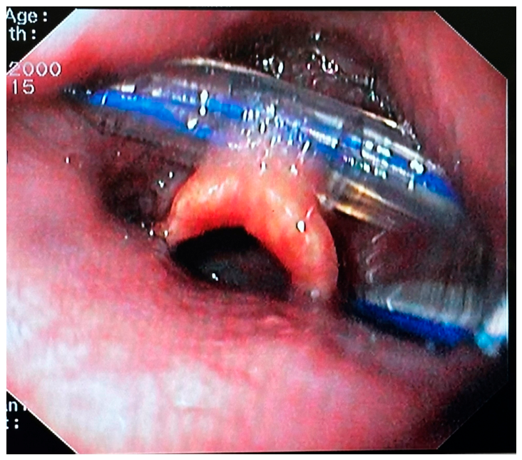
Finally, a 65-year-old female gastric cancer patient had undergone an almost total gastrectomy and a gastro-enteric Roux-en-Y anastomosis, following which a nasogastric tube had been placed in the intestine, far from the distal end-to-side jejuno-jejunal anastomosis. On the 7th post-operative day, the attempted tube removal failed, due to significant resistance in traction. An abdominal X-ray revealed an intense angulation of the tube at two points along its length (Figure 8). We suspected that the tube had been sutured in the distal anastomosis, so endoscopy was decided upon. Following insertion of a thin gastroscope, after the tube had been irrigated with warm water to soften it, a gentle but continuous traction was applied to the tube from the outside under direct endoscopic vision and air inflation, and was successfully removed with no complications. Retrospectively, it was realized that, the previous evening, a junior medical staff member had unjustifiably decided to pull the tube for 15cm, just to bring the tip of the catheter proximal to the jejuno-jejunal anastomosis. When the senior trainee realized this, the junior rushed to push it back, resulting in the crushing and kinking of the tube, the tip of which probably got inserted into the horizontal limb of the end-to-side anastomosis, a conclusion drawn from looking at the x-ray film.
4. Discussion
Nasogastric tube-related complications occurring at an unpredicted time after tube insertion – mostly with no alerting sign – but being found only at the time of the tube removal process, are relatively infrequent and somewhat unexpected. However, they pose important clinical challenges across diverse patient populations and procedural contexts. Overall, more than 100 such incidents have been identified from the global literature, mainly as case reports, with very similar titles which automatically classify them into 4 categories – the authors appearing to have been influenced by similar previously published cases: self-knotting of the tube, entanglement around the endotracheal device, entrapment in the suture line of an upper viscera anastomosis, and fragmentation or breakage of the tube [1,2].
However, when these cases were critically analyzed, despite their classification by title into one of these four categories, we realized that a common denominator was the presence of an already inserted endotracheal tube, placed either through the mouth (orotracheal) – in the majority of cases - or nose (nasotracheal). This common feature suggested to us the interference and friction of the oro- or naso-gastric tube being pushed into the esophagus/stomach against the already properly secured oro- or naso-tracheal device, the pharynx being short but narrow [4,5,65].
Most physicians, and particularly those involved in such procedures, namely anesthesiologists, intensive care specialists, surgeons or emergency department internists, have in their minds the image of a gastric tube being pushed through the patient’s nose or mouth: if resistance is experienced the doctor pulls it back and pushes again, perhaps also handling it with his two fingers or a Magill forceps through the mouth, in order to overcome the resistance caused by friction between the gastric tube and the immovable endotracheal tube. The same sense of friction is experienced by endoscopists when trying to insert a gastroscope in the esophagus in parallel to an endogastric tube. Although the conditions are not exactly the same, since the endoscope can mildly stretch the esophagus by means of air inflation, this can be parallelized with the friction exerted between the gastric and the endotracheal tubes when the former is pushed to more or less blindly pass through the pharynx.
These manipulations, retrospectively considered, are those which seem to be mostly responsible and directly related to the misplacement, the formation of knots at the distal end or to the making of a loop over the tracheal tube. It appears to be the same when, after insertion – or more precisely when it is assumed that the ‘obstacle’ has been passed – repetitive repositioning maneuvers are carried out, by forcefully pushing/pulling to overcome endotracheal tube interference, should the gastric tube appear not to be in place, as in the case of the air auscultation test or the gastric content suction procedure, which confirm proper placement to have failed [14,40].
The most common likely complication is the formation of a knot. The term ‘self-knotting’ refers to the formation of a loose knot around the body of the gastric tube, commonly at its distal end, usually when lying on the gastric antrum – the esophagus [6,11,66] and hiatus hernia [30] not totally excluded – which makes its removal challenging or impossible without some kind of potentially traumatic intervention. The knotting is generally recognized retrospectively during the removal attempt when the loose loop tightens and becomes a knot, which then creates the resistance encountered during withdrawal, since it has almost always already become impacted at the level of the posterior pharynx, and is pressing upon the posterior aspect of the nasal area. According to Mohsin et al [11] and Kumar Sinha et al [24] the mechanism of knot formation is similar to that of supercoiling and concatenate formation. However, it remains unclear what else can occur in the meanwhile, and the knot configuration sometimes becomes more complex, from a simple knot, to a lasso-like lariat loop [20,23,24,25,67], or a 4-loop knot [22,27,43,45]. Paradoxically, based on our knowledge from laparoscopic surgery, the last two types of knots are more likely to occur by a double wrapping of the tube round itself [68,69]; the initial loose knot then tightens when one end is pulled, finally turning the loop into a tight knot, and thus confirming the hypothesis that the previously formed loose knot tightens during withdrawal.
We therefore suggest a reclassification by creating a fifth category relating to knot formation depending on the existence of an endotracheal tube already being in place. In this case the patient is already intubated in readiness for an operation or has been hospitalized, under mechanical ventilation, in an ICU. The effort to insert a gastric tube through the nose or mouth – for gastric decompression in the case of surgery or for feeding when in the ICU – could, in the worst scenario, end up as either a self-knot of the gastric tube or as its entanglement around the endotracheal device, both versions directly related to interference with the endotracheal tube.
Santhanam and Margarson [5] and others [45,48,49,50,70] suggest that factors implicated in the coiling of the tube in the pharynx in anaesthetized individuals are: the supine position, the impaction of the gastric tube on the piriform sinuses and the arytenoid cartilages, the posterior tilting of the intubated larynx and the cuffed endotracheal tube. Additional challenges are also reported as the lack of ability to swallow and the flaccid, medically paralyzed neck muscles, making the insertion of the gastric tube in a sedated patient difficult [39].
On the other side, maneuvers/strategies to facilitate gastric tube placement and thus possibly reduce the chance of self-knotting in the already intubated patient are: the forward displacement of the larynx, lateral neck pressure, adequate lubrication of the tube (preferably chilled) and direct guidance of the tube with two figures in the mouth and use of Magill forceps under laryngoscopic vision [5,23,71]. Additionally, the very careful insertion of a Fogarty catheter through the suction port of the tube to increase its rigidity is also recommended, although it could become dangerous if excessive force were applied [23].
In the present narrative review, we have recognized 27 cases of gastric tubes inserted in intubated patients which had self-knotted somewhere along their length. In the majority of these reports, gastric tube insertion – although mostly reported as laborious [18,72] – was initially found to be uneventful, thus indicating that knotting can occur even when initial insertion appears straightforward. However, there are four cases [46,52,53,54] in which it was immediately obvious that something had gone wrong, indicated by the difficulty in air auscultation to confirm tube placement. It is of interest that almost all cases of knots were revealed at the time of gastric tube traction for removal.
This time-point is considered crucial for three reasons: first, there are some reports of the formation of a loose self-bond at the end of the tube – created by whatever mechanism and at an unknown time – which, at withdrawal, tightens to become a knot, followed by its impaction in an anatomical stricture, such as the posterior pharynx, the nostrils, or even the cricopharyngeal level of the esophageal sphincters; second, the speed of tube withdrawal may seriously affect the degree of the knot impaction in the above mentioned areas and, third, forceful withdrawal to overcome the obstacle might become traumatic, leading to nose bleeding [43,65] or even to posterior pharynx laceration, as occurred in 3 cases [6,11,43].
Early recognition of the self-coiling of the gastric tube while still in the stomach, that is before pulling, is theoretically impossible, since the very few cases recorded are on the occasions of an endoscopy being performed for another indication, mostly a percutaneous endoscopic gastrostomy. But even in the cases reported by Galanopoulos et al [30], the gastric tube had already been removed no later than immediately following the entry of the endoscope into the esophagus. Only in one of our reported cases, were we “lucky” to find a levin catheter with a double kink, when its tip was still in the gastric antrum. This happened because, after starting to write this article, the informal decision was made not to remove the nasogastric tube before advancing the endoscope into the antrum, in the hope of obtaining an endoscopic picture of such a complication. Speaking in numbers, this was after 162 endoscopies for PEG in the previous four months, an indication of the relative rarity of this complication. However, despite the difficulty in early recognition of self-knotting, some indirect signs such as difficulty in air auscultation in the epigastrium or a progressive or sudden stop of gastric content drainage or of liquid feeding [8] should raise serious suspicions of a problem.
On the other hand, given that a gastric tube insertion is a prerequisite in almost all abdominal surgery, especially laparoscopic, and for, among others, gastric decompression to facilitate viscera visualization and instrument movement, it is clear that self-knotting remains the most commonly encountered complication; however, there are many other clinical situations and reasons leading to the creation of a knot, such as with enteral feeding patients, either with a PVC catheter for short-term use or a fine-bore, silastic jejunal for long-term feeding. In general, it is obvious that the more days a gastric tube remains in the stomach, the greater the possibility of complication occurring, given that the tube becomes progressively stiffer due to gastric acidity and food remnants [73,74]. In the cases presenting 64 knotted gastric tubes, there were 7 patients with a PVC catheter, 16 patients with a Ryles [PVC or silicone] tube, 2 Salem types for gastric decompression for surgery, 5 silastic jejunal or weighted enteral feeding tubes, and 3 wide-bore for obstructed bowel disease decompression.
‘Entanglement’ is when the gastric tube makes a loop around the endotracheal tube, and is a more dangerous situation since the loop may strangulate the endotracheal tube and thus compromise airway safety, often requiring coordinated airway and gastrointestinal management [44,46]. In the present search of the literature, we found 9 cases, most reporting difficulties in endotracheal intubation.
In this scenario there are four subcases: in the most common, the patient has been intubated orally and the gastric tube has also been inserted through the mouth - this is the simplest version with the best outcome – both tubes pull out en-block orally; the other three cases involved the patient being intubated orally, with the gastric tube inserted through the nostrils or the opposite, or both tubes inserted through the nose but via different nostrils. In such situations, for removal, the proximal part of the relatively more flexible gastric tube needs to be cut, under direct vision through a laryngoscope, at the level of the pharynx and removed via the nostril, after which the distal part can be removed along with the endotracheal tube.
Although sometimes the situation is urgent because the patient presents a resistance to mechanical ventilation, any careless or hasty movements in attempting to withdraw a coiled or curled gastric tube may create a loop which then tightens into a knot around the endotracheal tube and thus fully obstructs ventilation [48,49], or, in the worse scenario results in an unintentional tracheal extubation [Table 3]. This hypothesis was confirmed by means of a model oral cavity, pharynx, and larynx, constructed by Okada et al [44].
A sudden maximum increase in peak airway pressure and a simultaneous decrease in oxygen saturation, occurring after manipulation of the gastric tube, should immediately alert the anesthesiologist to consider the gastric tube as a strong etiology for tube strangulation and consequently ventilatory obstruction [48,52,53,54]. Thus, necessitating the immediate restoration of ventilation, even if emergency extubation and prompt reintubation is required. As a diagnostic means, direct laryngoscopy confirms the suspicion of tube obstruction, and, when not available, manual bag ventilation might be useful, or passing a suction tube through the tracheal tube, for diagnosis only, could be attempted [48,53,54]. A wide-bore gastric tube is suggested for the safest insertion, especially in cases where it would be used for a short-term only, for intraoperative stomach decompression. However, in the present review there are two cases with a Salem dual-lumen tube, inserted nasally, which coiled around an orotracheal and a nasotracheal tube, respectively [45,70].
A self-knot can also be formed at the distal end of a gastric tube in patients with no intubation. During the literature search we found 37 cases of a tube self-knotting, totally independent of the co-existence of an endotracheal tube more commonly occurring with thin-bore feeding tubes.
According to previous publications, risk factors for self-knotting relate to the characteristics of both the tube and the patient. More precisely, thin, flexible small-bore tubes, particularly in combination with excessive tube length lying in the antrum [23,26,29,75] are more prone to coiling and knotting [76].
Silicone and polyurethane narrow-bore tubes are also generally more prone to knotting [19], in relation to PVC tubes, as their high flexibility and softness allow them to coil easily within the stomach, the body temperature facilitating this [16]. Nonetheless, wide-bore gastric PVC tubes, such as the Salem-sump tube -double lumen- for decompression/drainage and nutritional support are also implicated in knot formation or other complications. There have been similar scenarios with Ryles tubes, being generally thicker and often stiffer, their single purpose being short-term decompression; which is why they were found implicated in complications, after their, mostly orally, insertion immediately after patient intubation and removal immediately [33,49,77] or shortly after extubation [5,20,78,79]. On this point we can add a personal observation, not mentioned by any other author: by observing photos in previous publications [22,43,65,74,80,81], as well as of our cases (Figure 5 and Figure 6) we noticed that the kinking of a gastric tube begins at the point in the tube where there is one of the lateral holes – the wider-bore the catheter the larger the hole, meaning that the tube has PVC material in about half of its perimeter, this being the weakest point.
A longer length of the tube down into the stomach also creates a looping risk, since it allows the tube to twist and curl within the gastric antrum [9,82]. This predisposing factor, in combination with a classic ‘Levin’-type nasogastric tube made from PVC – commonly used as first choice tubes both for decompression and feeding – could lead to the formation of a loose knot [36]. Then, over time, the PVC hardens and the loose knot stiffens.
Thus, there is a trend to believe that the mechanism of knot formation is multifactorial, involving a combination of tube diameter and thus flexibility, excessive intragastric length, as well as the construction material [5,9,14,20,72,76,77,82], and the assistive role of push-pull movements, post-insertion, in order to find the best position. The push-pull movements are also related to patient behavior: coughing, deglutition, retching or swallowing may be considered additional causes of tube misplacement [5,14,83,84,85], including even reduced patient cooperation and excessive head movements [86]. However, tube knotting seems to be independent of age and gender, since it has been identified across a wide age range, from premature neonates and infants to elderly patients, occurring in both sexes, although there seems to be a slight male predominance.
A small-volume stomach, e.g. following subtotal gastrectomy or bariatric surgery, or a reduced-motility-stomach, as after gastric surgery, may increase the possibilities of knotting [5,20]. Indeed, there were 8 cases of knotting after gastric surgery [Table 4]. Further consequences of reduced gut motility, possibly following brain injury [35], improved by means of prokinetics drugs, combined with a small-caliber PVC tube may also lead to the formation of a loose loop, which could tighten into a knot; however, there is no such evidence in the cases analyzed.
In the case studies analyzed, there are very few references as to whether the gastric tube insertion had been conducted according to the proposed guidelines for safe placement [3,18,83]. However, this lack of reference may be of little significance, since knotting at the distal end of the tube is most likely to occur at some time-point after placement. Nevertheless, the guidelines [3,18] also emphasize the importance of early recognition and appropriate manipulation of the knotted tubes, a topic which we have already commented on.
The management of a knotted nasogastric tube varied, depending mainly on the stiffness and location of the impacted knot, as well as the available medical specialties in the hospital. Typically, it begins with gentle traction of the tube, which might prove to be sufficient to retrieve loosely knotted tubes, although imaging techniques such as plain radiographs or fluoroscopy are recommended to confirm the presence and location of a knot to determine further action. Forceful attempts should be totally avoided, to prevent mucosal trauma or epistaxis. Manual removal was successful in 27 cases.
When simple traction fails or resistance to pulling is significant, ENT-assisted removal tools and/or a Magill forceps – often combined with cutting of the distal end of the tube – is employed through the oral cavity, usually under topical anesthesia or even light sedation. With the tube cut, the proximal part is freely removable through the nostril, while the distal part, with the knot, should be removed through the mouth to avoid mucosal damage to the narrow nasal passage mucosa, leading to bleeding [43,65] and possible airway compromise. Implication of an ENT specialty in removing a knotted tube has been reported in 5 patients [Table 1 and Table 2].
If the knot is inaccessible or too tight, or an endoscopist is more readily available than an ENT, endoscopic retrieval can easily be performed [35], as occurred in 23 patients. In such cases endoscopic cutting of the tube close and proximal to the knot is a fairly stress-free procedure, the tube then being freely removeable via the nose or the mouth – depending on the route of insertion, while the distal part, with the knot, can be extracted as a foreign body with either a snare, a basket or alligator or rat-tooth retrieval forceps, depending the preference and experience of the endoscopist and the availability of instruments [87,88,89]. However, there are many reported difficulties in cutting the tube, probably related to its thickness, stiffness and, mainly, on the available cutting tools. A good idea is that the scissors be inserted through one of the peripheral holes, where the tube is thinner and thus more easily cut. However, the use of a diathermy-connected cutter, or polypectomy snare, is not recommended, since these instruments are made for cutting mucosa and not for melting a PVC or similar catheter, well known to create hazard gasses in the stomach.
In rare and severe cases where these measures fail or there is no endoscopic or ENT assistance available, or the tube has caused significant mucosal injury, a surgical intervention should be considered. Such a difficult case is presented by Jindal et al [73]. A similar case, where a catheter had been stitched in a fundoplication procedure, a mini laparotomy and the gastrotomy opening were simultaneously used, through which a grasper was inserted to grab the knot and bring it out of the stomach [55]. These alternatives in management highlight the importance of early recognition and careful handling to prevent complications such as mucosal injury or tube fragmentation; which means all clinicians involved should be alert to the possibility of tube self-knotting and refrain from trying to remove it forcefully, particularly when removal will be via the nostril.
Beyond the difficulties related to knot formation, there are two further complications recognized at the time of removal: the stitching of the nasogastric tube in a suture line during an operation in hollow viscera, and the fracture of the gastric tube within the stomach.
Since the advent and routine adoption of surgical stapling devices, the nasogastric tubes may become inadvertently stitched in a hollow viscera suture line or in an anastomosis line – exclusively associated with upper gastrointestinal surgical procedures – posing a diagnostic and therapeutic challenge when resistance is encountered during tube removal [6]. Early intra-operative recognition of this complication is crucial to avoid anastomotic disruption or even viscera perforation. Despite the clinical relevance of these mechanical complications, the existing literature involves predominantly isolated case reports or small case series, while a comprehensive synthesis of reported cases remains limited.
The main reason this complication occurs is usually the anesthesiologist’s inattention or delay in complying with the surgeon’s request to pull the tube out before the suturing device is used or a stitch inserted, or the surgeon’s omission to make such a request. And of course, if the accident occurs, the most important thing is for the surgeon to realize it before the operation is completed, as happened in 3 out of the 14 cases, in 2 of which the anastomosis was re-constructed. In contrast, in the remaining 11, laborious and time-consuming endoscopic efforts were required to cut the suture or remove the staple in order to release the tube.
Finally, tube fracture or breakage during withdrawal, although the rarest complication, (a total of 11 cases reported, predominantly in adult patients), may lead to retained intraluminal fragments and typically necessitates endoscopic retrieval to prevent further complications [61]. The etiology of rupture seems to be the erosion of the tube material, mainly from remaining in place long-term – in most cases inserted for feeding, in combination with the different acidities within the stomach. The example of the feeding tube which broke while being flushed in the attempt to unclog it is also typical [8,90]. Nevertheless, failure of the material cannot be excluded, as seen in a case in which the gastric tube was found to have ruptured into three pieces [61], and another one in which a frozen tube had broken upon insertion [62]. The practice of using tubes, frozen to stiffen in order to facilitate insertion, seems likely to share the responsibility for breakage, but there is no relative comment in the literature.
Taken together the cases in which the remnant of a broken tube was [i] accidentally found coiled in the nasal cavity and the nasopharynx four years after its removal [63] and [ii] coughed up by the patient while still in the post-anesthesia care unit [60] highlights the need for inspection of the gastric tube in every case on removal, and especially when it has remained in the stomach for an extended period.
5. Conclusions
Nasogastric tube insertion remains a routine yet critical procedure in both emergency and elective clinical settings. Despite its widespread use, clinicians involved must remain vigilant to the rare but potentially serious complications analyzed, which are generally unexpected and only recognized once the gastric tube withdrawal process is begun.
Through a comprehensive analysis of published cases, it is evident that of the risk factors appearing to predispose to gastric tube self-knotting, the most common complication relates to the use of small-bore catheters inserted deep in the stomach. However, the push-pull maneuvers made when tube placement presents difficulties seem to be the main cause of self-knotting, irrespective of whether the patient is endotracheally intubated or not. More dramatic and potentially life-threatening is the complication whereby the gastric tube becomes tied around the endotracheal tube, an event which can lead to sudden airway obstruction. Suturing the gastric tube over a suture line or an anastomosis, if not immediately recognized, can lead to the necessity for reoperation for anastomosis reconstruction. However, this is something that can easily be predicted and avoided if there is good communication between the surgeon and the anesthesiologist, allowing the tracheal tube to be withdrawn slightly prior to the suturing. Finally, the breaking up of the nasogastric tube during removal with a fragment remaining in the stomach, involves the same risks as the presence of a foreign body and must therefore be removed immediately by means of endoscopy. However, this requires that the doctor carrying out the withdrawal must realize that a piece has been left behind, which, in practice, means that every time a tube is removed its integrity must be checked.
We thus emphasize that continuing alertness, reporting, and analysis of similar cases will lead to improvement in patient safety and procedural best practice.
Author Contributions
“Conceptualization, K.K. and AE.M; methodology, G.T. and G.S; software, SC.Z.; and A.K.; validation, L.L.; and T.K.; formal analysis, A.I.; investigation, AE.M.; resources, Z.A. and P.A.; data curation, G.T.; writing—original draft preparation, AE.M.; writing—review and editing, A.Sh and K.K.; visualization, J.V.; supervision, K.K.; project administration, E.S.; All authors have read and agreed to the published version of the manuscript.”.
Funding
“This research received no external funding”.
Institutional Review Board Statement
“Not applicable.”.
Informed Consent Statement
“Not applicable.”.
Data Availability Statement
Data available in a publicly accessible repository.
Acknowledgments
Not applicable.
Conflicts of Interest
“The authors declare no conflicts of interest.”.
Abbreviations
The following abbreviations are used in this manuscript:
| NGT | Nasogastric tube |
| EtCO₂ | End-tidal carbon dioxide |
References
- Vadivelu, N.; Kodumudi, G.; Leffert, L.R.; Pierson, D.C.; Rein, L.K.; Silverman, M.S.; Cornett, E.M.; Kaye, A.D. Evolving Therapeutic Roles of Nasogastric Tubes: Current Concepts in Clinical Practice. Adv. Ther. 2023, 40, 828–843. [Google Scholar] [CrossRef] [PubMed]
- Long, M.; Machan, M.; Tollinche, L. Intraoperative Gastric Tube Intubation: A Summary of Case Studies and Review of the Literature. Open J. Anesthesiol. 2017, 7, 43–62. [Google Scholar] [CrossRef] [PubMed]
- O’Connell, F. Emergency department approach to gastric tube complications and review of the literature. Am. J. Emerg. Med. 2021, 39, e255-259. [Google Scholar] [CrossRef] [PubMed]
- Monib, S.; Elkorety, M.; Jibrin, U.; Dhungana, D.; Thomson, S. Knotted nasogastric tube: a rare, overlooked yet preventable complication. J. Surg. Case Rep. 2019, 2019, rjz290. [Google Scholar] [CrossRef] [PubMed]
- Santhanam, V.; Margarson, M. Removal of self-knotted nasogastric tube: technical note. Int. J. Oral Maxillofac. Surg. 2008, 37, 384–385. [Google Scholar] [CrossRef] [PubMed]
- Tai, C.M.; Wang, H.P.; Lee, C.T.; Chang, C.Y.; Wang, W.L.; Tseng, C.H.; Lin, J.T. Esophageal obstruction by a tangled nasogastric tube. Gastrointest. Endosc. 2010, 72, 1057–1058. [Google Scholar] [CrossRef] [PubMed]
- Jones, M. The knotted nasogastric tube: a simple solution. Br. J. Clin. Pract. 1989, 43, 183–184. [Google Scholar] [CrossRef] [PubMed]
- Cappell, M.S.; Scarpa, P.J.; Nadler, S.; Miller, S.H. Complications of nasoenteral tubes. Intragastric tube knotting and intragastric tube breakage. J. Clin. Gastroenterol. 1992, 14, 144–147. [Google Scholar] [CrossRef] [PubMed]
- Watkin, D.; Farrell, C.; McGee, M.; Welters, I. Complications of nasogastric tube insertion in critical care: A retrospective analysis of radiographs performed for determination of tube position. Radiography (Lond) 2025, 31, 102941. [Google Scholar] [CrossRef] [PubMed]
- Morris, H.H., III. Nasogastric Intubation: A Potentially Knotty Problem. JAMA 1977, 237, 1432–1432. [Google Scholar] [CrossRef]
- Mohsin, M.; Saleem Mir, I.; Hanief Beg, M.; Nazir Shah, N.; Arjumand Farooq, S.; Altaf Bachh, A.; Quadir, A. Nasogastric tube knotting with tracheoesophageal fistula - a rare association. Interact. Cardiovasc Thorac. Surg. 2007, 6, 508–510. [Google Scholar] [CrossRef] [PubMed]
- Malhotra, P.; Sharma, A.; Malhotra, P.; Singh, H.; Kumar, S.; Singh, V.; Khan, A. Ryles tube-commonly forgotten entity after insertion. Indones. J. Appl. Res. (IJAR) 2025, 13, 1660–1663. [Google Scholar] [CrossRef] [PubMed]
- Ravind, R.; Prameela, C.G.; Gurram, B.C.; Dinesh, M. Naughty knot: a case of nasogastric tube knotting. BMJ Case Rep. 2015. [Google Scholar] [CrossRef] [PubMed]
- Awe, J. Self-knotting of Nasogastric tube: an unusual and rare complication. Glob. Adv. Res. J. Microbiol. 2014, 3, 064–067. [Google Scholar]
- Chavda, V.; Alhammali, T.; Farrant, J.; Naidu, L.; El-Rabaa, S. Nasogastric tube knotting: a rare and potentially overlooked complication among healthcare professionals. BMJ Case Rep. 2017. [Google Scholar] [CrossRef] [PubMed]
- Kim, C.E.; Hwang, I.G.; Min, H.J. Removal of Knotted Nasogastric Tube: A Literature Review and Lessons From Our Case. J. Rhinol. 2021, 28, 186–188. [Google Scholar] [CrossRef]
- Munoz, N.R.; Hojjat, A.; Dhaliwal, G.; Adial, A. Knotted Nasogastric Tube Removed With a Bronchoscope. Cureus 2023, 15, e40896. [Google Scholar] [CrossRef] [PubMed]
- Khond, A.; Mendonca, E.; Verma, B.; Borewad, G. A looped nasogastric tube impacted in nasal cavity: a case report. Int. J. Otorhinolaryngol. Head. Neck Surg. 2024, 10, 138–141. [Google Scholar] [CrossRef]
- Hickey, N.C.; Watson, D.C. An unusual complication of removal of a nasogastric tube. Br. Med. J. (Clin Res Ed) 1988, 296, 1038–1039. [Google Scholar] [CrossRef] [PubMed]
- Mandal, N.G.; Foxell, R. Knotting of a nasogastric tube. Anaesthesia 2000, 55, 99. [Google Scholar] [CrossRef] [PubMed]
- Hirwa, K.D.; Toshniwal, N. Knotted nasogastric tube in the posterior nasopharynx: A case report. Qatar Med. J. 2016, 2016, 11. [Google Scholar] [CrossRef] [PubMed]
- Chang, B.A.; Cheung, V.W.; Lea, J. An unusual instance of stridor: airway obstruction from a nasogastric tube knot in a 1-month-old infant. Am. J. Otolaryngol. 2014, 35, 59–61. [Google Scholar] [CrossRef] [PubMed]
- Singh, M.; Gupta, A.; Rao, A.; Prashanth, U.; Kumar, A. Spontaneous “lariat loop” knotting of a nasogastric tube: prevention and management. Trop. Gastroenterol. Off. J. Dig. Dis. Found. 2014, 35, 131–132. [Google Scholar] [CrossRef] [PubMed]
- Kumar Sinha, A.; Ahmad, S.; Rashi, R.; Kumar, A.; Kumar, B. Self-knotting of distal end of nasogastric tube-Not an uncommon possibility. Pediatr. Investig. 2020, 4, 145–147. [Google Scholar] [CrossRef] [PubMed]
- Paul, R.; Lariat Loop, D.B.D. J. Indian Acad. Clin. Med. 2022, 23, 77–78.
- Tsandiraki, J.; Tendai, J.; Likiliwike, A.D.; Moirana, E.N.; Msuya, D.; Tarmohamed, M. A stubborn nasogastric tube: self-knotting of a nasogastric tube. J. Surg. Case Rep. 2022, 2022, rjac338. [Google Scholar] [CrossRef] [PubMed]
- McHardy, D.A.; Sadiq, Y.; Ephraim, M.E. Nasogastric tube knotting. Trop. Doct 1993, 23, 179. [Google Scholar] [CrossRef] [PubMed]
- Tapaiwala, S.N.; Al Riyami, D.; Cole, E. A painful and knotted nasogastric tube. Cmaj 2008, 178, 568. [Google Scholar] [CrossRef] [PubMed]
- Te, B.C.; Mohammad Nazer, N.S.; Aini, N.R. Twisted Fate: Unraveling the Mystery of a Stuck Nasogastric Tube in a Toddler. Ear Nose Throat J. 2024, 1455613241302571. [Google Scholar] [CrossRef] [PubMed]
- Galanopoulos, M.; Tsigaridas, A.; Varytimiadis, L.; Papaefthymiou, A.; Kalampalikis, L.; Ladias, S.; Mantzaris, G.J. A forgotten nasogastric feeding tube that refuses to get out. Clin. Case Rep. 2017, 5, 205–207. [Google Scholar] [CrossRef] [PubMed]
- Lamba, S.; Sethi, S.K.; Khare, A.; Saini, S. Intraoperative wide bore nasogastric tube knotting: A rare incidence. Anesth. Essays Res. 2016, 10, 104–106. [Google Scholar] [CrossRef] [PubMed]
- Aggarwal, R. Self knotting of a nasogastric tube — a rare but preventable complication. J. Clin. Imaging Sci. 2018, 8. [Google Scholar] [CrossRef]
- Dubey, S.K.; Mahendru, V.; Sadhu, S.; Sarkar, S.; Verma, A.K.; Roy, M.K. True knot in Ryles tube: a case report. Indian J. Surg. 2008, 70, 142–143. [Google Scholar] [CrossRef] [PubMed]
- Conroy, M.; Wichmann, K.; Farkas, N.; Di-Nicola, V. Challenging removal of a knotted nasogastric tube following insertion under general anaesthetic. BMJ Case Rep. 2020, 13. [Google Scholar] [CrossRef] [PubMed]
- Ismail, N.J.; Bot, G.M.; Hassan, I.; Shilong, D.J.; Obande, J.O.; Aliu, S.A.; Dung, E.D.; Shehu, B.B. Knotting of a nasogastric feeding tube in a child with head injury: A case report and review of literature. Afr. J. Paediatr. Surg. 2014, 11, 273–275. [Google Scholar] [CrossRef] [PubMed]
- Liu, W.; Faisal, M.; Singh, K. Knotted nasoenteric feeding tube in an infant. J. Neonatal Perinat. Med. 2013, 6, 93–96. [Google Scholar] [CrossRef] [PubMed]
- Malik, N.W.; Timon, C.I.; Russel, J. A unique complication of primary tracheoesophageal puncture: knotting of the nasogastric tube. Otolaryngol. Head. Neck Surg. 1999, 120, 528–529. [Google Scholar] [CrossRef] [PubMed]
- Gupta, S.J.; Sawalakhe, N.R.; Samarth, A.R.; Gaikwad, N.R. Nasogastric Feeding Tube: Not So Innocuous. Trop. Gastroenterol. 2016, 37, 61–63. [Google Scholar] [CrossRef] [PubMed]
- Nashibi, M.; Safari, F.; Sezari, P.; Naderi, A.; Mottaghi, K. Hard and Granny Tie, Nasogastric Tube Knotted in Nasopharynx: A Case Report. Turk. J. Anaesthesiol. Reanim. 2021, 49, 263–264. [Google Scholar] [CrossRef] [PubMed]
- Agarwal, A.; Gaur, A.; Sahu, D.; Singh, P.K.; Pandey, C.K. Nasogastric tube knotting over the epiglottis: a cause of respiratory distress. Anesth. Analg. 2002, 94, 1659–1660, table of contents. [Google Scholar] [CrossRef] [PubMed]
- Jain, S.; Nazir, N. Self-knotting of a nasogastric tube. Anaesth. Pain Intensive Care 2017, 21, 280–281. [Google Scholar]
- Dinsmore, R.C.; Benson, J.F. Endoscopic removal of a knotted nasogastric tube lodged in the posterior nasopharynx. South Med. J. 1999, 92, 1005–1007. [Google Scholar] [CrossRef] [PubMed]
- P. Ongom, G.N.; Kaggwa, S.; Nakavuma, L. Nasogastric Tube Knotting: Two Case Reports from Kampala, Uganda. East Cent. Afr. J. Surg. 2012, 17, 106–109. [Google Scholar]
- Okada, Y.; Ohke, H.; Yoshimoto, H.; Kobashi, M.; Saitoh, M.; Terumitsu, M. Nasogastric Tube Knotted Around a Nasal Endotracheal Tube in the Nasopharynx: Possible Cause. Anesth. Prog. 2021, 68, 90–93. [Google Scholar] [CrossRef] [PubMed]
- Abe, S.; Osaka, Y.; Morita, Y. Difficult Removal of a Nasogastric Tube Because of Knot Formation Around a Nasotracheal Tube: A Case Report. A A Pract. 2018, 11, 184–185. [Google Scholar] [CrossRef] [PubMed]
- Melki, I.; Matar, N.; Maalouf, S.; Rassi, S. Knotting of nasogastric tube around a nasotracheal tube: An unusual cause of hypercapnia in a 3-month-old infant. Am. J. Crit. Care 2010, 19, 198–197. [Google Scholar] [CrossRef] [PubMed]
- Young, M.J.; Ehrenfeld, J.M. Knotting of an orogastric tube around an endotracheal tube. J. Clin. Anesth. 2012, 24, 254–255. [Google Scholar] [CrossRef] [PubMed]
- Pousman, R.M.; Koch, S.M. Endotracheal tube obstruction after orogastric tube placement. Anesthesiology 1997, 87, 1247–1248. [Google Scholar] [CrossRef] [PubMed]
- Sood, S.; Aggarwal, Y.; Kumar, A. Rare Complication of Nasogastric Tube Knotting around Endotracheal Tube in an Anesthetized Patient Undergoing Surgery. Ann. Natl. Acad. Med. Sci. 2021, 57. [Google Scholar] [CrossRef]
- Acharya, G.; Arora, K.K.; Kumar, D. Nasogastric tube coiled around endotracheal tube. J. Anaesthesiol. Clin. Pharmacol. 2014, 30, 584–586. [Google Scholar] [CrossRef] [PubMed]
- Deng, J.; Pan, T.; Li, H.; Lin, Q.; Zhang, Y. Knotting together of gastric and tracheal tubes inserted under general anesthesia: A CARE-compliant case report. Medicine 2022, 101, e31203. [Google Scholar] [CrossRef] [PubMed]
- Lin, C.S.; Hung, C.J.; Lin, F.S.; Chan, W.H. Constriction of an endotracheal tube by a nasogastric tube during general anesthesia resulting in pulmonary edema. J. Cardiothorac. Vasc. Anesth. 2012, 26, e37-38. [Google Scholar] [CrossRef] [PubMed]
- Hughes, M.; Holland, K.; Tobias, J.D. Misplaced and Knotted Nasogastric Tubes in Infants and Children: Report of Two Cases. J. Med. Cases 2026, 17, 128–132. [Google Scholar] [CrossRef] [PubMed]
- Dunn, S.A.; Ashrafi, B.; Candiotti, K.; Rodriguez-Blanco, Y.F. Ventilatory Collapse: Endotracheal Tube Strangulationfrom a Nasogastric Tube Knot. Anesthesiology 2019, 131, 148. [Google Scholar] [CrossRef] [PubMed]
- Reissman, P.; Udassin, R.; Goldin, E.; Durst, A.L. Management of an inadvertently sutured nasogastric tube after Nissen fundoplication. Gastrointest. Endosc. 1994, 40, 260–261. [Google Scholar] [CrossRef] [PubMed]
- Jain, S.; Baksi, A.; Kumar, A.; Aggarwal, S. Inclusion of Orogastric Tube in the Staple Line During Laparoscopic Roux-en-Y Gastric Bypass: an Avoidable Complication. Obes. Surg. 2021, 31, 2835–2836. [Google Scholar] [CrossRef] [PubMed]
- Higa, G.; Szomstein, S.; Rosenthal, R. Stapling of orogastric tube during gastrojejunal anastomosis: an unusual complication after conversion of sleeve gastrectomy to laparoscopic Roux-en-Y gastric bypass. Surg. Obes. Relat. Dis. 2012, 8, 116–118. [Google Scholar] [CrossRef] [PubMed]
- Azzam, A.Z.; Azzam, K.A.; Amin, T. Removal of Nasogastric Tube Accidentally Stitched to Roux-en-Y Oesophagojejunostomy Following a Radical Gastrectomy for Stomach Cancer: Case report and review of the literature. Sultan Qaboos Univ. Med. J. 2018, 18, e110–e111. [Google Scholar] [CrossRef] [PubMed]
- Vardaan, A.; Ahmad, A.; Singh, D.; Misra, R. Endoscopic removal of a fractured nasojejunal tube in the duodenum. Endoscopy 2013, 45 Suppl 2 UCTN, E347-348. [Google Scholar] [CrossRef] [PubMed]
- Ranier, G.; Costello, K. Fractured OG tip: a case report. W V. Med. J. 2013, 109, 38–39. [Google Scholar] [PubMed]
- Eldigair, H.; Khurram, R.; Bennell, J.; Mir, N. Comminuted nasogastric tube fracture: a rare complication. BJR Case Rep. 2021, 7, 20210049. [Google Scholar] [CrossRef] [PubMed]
- Savoulidou, S.; Aslanidis, T. Fracture of nasogastric tube: A case report. 2020, 19, 32–34. [Google Scholar]
- Kinshuck, A.; William, C.; Reddy, C.E.E. A Forgotten Nasogastric Tube. Clin. Rhinol. An. Int. J. 2011, 4, 47–49. [Google Scholar] [CrossRef]
- Khanal, S.; Shrestha, S.; Khadayat, R.; Adhikari, A.; Poddar, E.; Kharel, S.; Regmi, P.R. Breakage of an orogastric tube in a critically Ill patient: a case report. Ann. Med. Surg. (Lond) 2023, 85, 3714–3716. [Google Scholar] [CrossRef] [PubMed]
- De, U.; Agarwal, a.; Singh, V. Spontaneous knotting of ryles tube in a post operative patient. Pak. J. Med. Sci. Pak. J. Med. Sci. 2007, 23. [Google Scholar] [CrossRef]
- Bernica, J.; Deng, N.; Cole, R.A.; Ketwaroo, G.A.; Hair, C. 3086 ”Knot” Your Average Nasogastric Tube. Off. J. Am. Coll. Gastroenterol. ACG 2019, 114, S1659–S1660. [Google Scholar] [CrossRef]
- MH. Trujillo, C.F.; Tortoledo, F.; Ceballos, F. “Lariat loop” knotting of a nasogastric tube: an ounce of prevention. Am. Assoc. Crit.-Care 2006, 15, 413–414. [Google Scholar] [CrossRef]
- Ishikawa, N.; Watanabe, G. Figure 4 Knot: Simple Tying Technique for Robotic and Endoscopic Sutures. Innovations (Phila) 2017, 12, 152–153. [Google Scholar] [CrossRef] [PubMed]
- Ishikawa, N.; Watanabe, G.; Yoshiyama, D.; Koakutsu, T.; Horikawa, T.; Tomita, S.; Ohtsuka, T. Lariat knot: simple tying technique for robotic and endoscopic sutures. J. Robot Surg. 2025, 19, 251. [Google Scholar] [CrossRef] [PubMed]
- Au-Truong, X.; Lopez, G.; Joseph, N.J.; Salem, M.R. A case of a nasogastric tube knotting around a tracheal tube: detection and management. Anesth. Analg. 1999, 89, 1583–1584. [Google Scholar] [CrossRef] [PubMed]
- Chaudhary, K.; Bagharwal, P.; Uppal, R.; Maheshwari, C. Entanglement of Nasogastric Tube and Nasopharyngeal Temperature Probe During Surgery. MAMC J. Med. Sci. 2015, 1, 105. [Google Scholar] [CrossRef]
- Kumar, A.; Kulshreshtha, V.; Kumar, P.; Jaiswal, G.; Gupta, T. Kinked and retained nasogastric tube in polytrauma patient; a case report. Romanian Neurosurg. 2017, 31. [Google Scholar] [CrossRef]
- Jindal, R.; Sahni, M.; Patel, P.; Singh, S.; Hussain, A.; Gulati, C.; Garg, L. Entrapment of a long dwelling nasogastric tube after radiotherapy in an oral cancer patient: a rare case report. Int. Surg. J. 2022, 9, 1653. [Google Scholar] [CrossRef]
- Kumar Shwetanshu Narayan, G.K.G.; Nijhawan, Sandeep; Puri, Shekhar. Unique Removal of a Kinked Nasogastric Tube. J. Dig. Endosc. 2019, 8, 97–99. [Google Scholar]
- Abhyankar, S.; Agarwal, A.; Juneja, M.; Vartak, A. Self knotting of Naso gastric tube in paediatric Burns patient. Int. J. Burn. Trauma 2019, 9, 49–51. [Google Scholar]
- Otsuji, E.; Yamaguchi, T.; Sawai, K.; Hagiwara, A.; Shirasu, M.; Koide, K.; Nakao, E.; Takahashi, T.; Yamagishi, H. Knot formation in a long tube used in the treatment of a post-operative adhesive small bowel obstruction. Hepatogastroenterology 1999, 46, 3172–3174. [Google Scholar] [PubMed]
- Aggarwal, R. The knotted complication of a nasogastric tube. Med. J. Dr. D.Y. Patil. Vidyapeeth 2017, 10, 320–321. [Google Scholar] [CrossRef]
- Moscote-Salazar, L.; Barciela, E.; Ramos Villegas, Y.; Quintana Pájaro, L.; Agrawal, A. Self-Knotting of a Nasogastric Tube. J. Transl. Crit. Care Med. 2019, 1, 111. [Google Scholar] [CrossRef] [PubMed]
- Paul, Rudrajit; Loop, D.B. Lariat. J. Indian Acad. Clin. Med. 2022, 23, 77–78.
- Abbas, G.A.; Tawfeeq, M.A.; Arain, Z.B. Nasogastric tube self-knotting. Ann. Pediatr. Surg. 2016, 12, 162–163. [Google Scholar] [CrossRef]
- Muzna Iftikhar, M.I. To Be Or Knot To Be: A Rare Case Of Nasogastric Tube Knotting. J. Rawalpindi Med. Coll. 2020, 24. [Google Scholar] [CrossRef]
- Matsushita, M.; Mori, S.; Tahashi, Y.; Wakamatsu, T.; Okazaki, K. Excessive length of a tube in the stomach: a risk factor for a tangled or knotted tube. Gastrointest. Endosc. 2011, 74, 237–238, author reply 238. [Google Scholar] [CrossRef] [PubMed]
- Delirrad, M.; Majidi, M. Self-knotted Nasogastric tube: A Case report. 2015, 150–153. [Google Scholar] [CrossRef]
- Chen, C.N.; Lee, W.J.; Cheng, T.J.; Chang, K.J. Endoscopic removal of nasogastric tube sutured unintentionally to gastrojejunostomy. Surg. Laparosc. Endosc. 1997, 7, 359–360. [Google Scholar] [PubMed]
- Davies, T.; Man, A. All tied up in knots. BMJ Case Rep. 2013. [Google Scholar] [CrossRef] [PubMed]
- Sliwa, J.A.; Marciniak, C. A complication of nasogastric tube removal. Arch. Phys. Med. Rehabil. 1989, 70, 702–704. [Google Scholar] [PubMed]
- Knežević, A.; Knežević, V.; Jovanović, I.; Gvozdenović, L. Successful endoscopic management of an entrapped nasogastric tube after a surgical procedure. Surg. Laparosc. Endosc. Percutan Tech 2013, 23, 233. [Google Scholar] [CrossRef] [PubMed]
- Giada Di Donato, A.C.; Valvano, Marco; Cianca, Giovanni; Lombardi, Loreto. Endoscopic removal of a nasogastric tube accidentally stitched during bariatric surgery: A case report and review of the literature. Sci. Repos. 2023, 6, 2–4. [Google Scholar] [CrossRef]
- Wilkinson, M.N.; Jayaraman, V.; Watkins, K.; Bao, P.; Buscaglia, J.M. Newly developed flexible endoscopic scissors for removal of a nasogastric tube trapped within a gastrojejunal anastomotic staple line. Endoscopy 2011, 43, E234–E235. [Google Scholar] [CrossRef] [PubMed]
- van Westerloo, D.J.; Rotteveel, J.; Kneepkens, F. Breakage of a nasoenteral feeding tube in a 5-year-old child. Gastrointest. Endosc. 2000, 52, 806–807. [Google Scholar] [CrossRef] [PubMed]
- Kumar, A.; Kulshreshtha, V.; Kumar, P.; Jaiswal, G.; Gupta, T. K. Kinked and retained nasogastric tube in polytrauma patient: A rare case report. Romanian Neurosurg. 2017, 31(2), 244–247. [Google Scholar] [CrossRef]
- Gülsoy, Z.; Karagözoğlu, S.; Özdemir Kol, I. Knot Formed in Nasogastric Tube: A Case Report. Turk. J. Intensive Care 2019, 17, 172–5. [Google Scholar] [CrossRef]
- Shiva, S.; Lalhruaitluanga, J.; Singh, J.; Karnik, A.; Kumar, A. A Rare Entanglement: Self-Knotting of a Nasogastric Tube. Cureus 2024, 16(3), e55833. [Google Scholar] [CrossRef] [PubMed] [PubMed Central]
- Wright, S.; Warusavitarne, J.; Shaikh, I. Safe removal of knotted nasogastric tubes. Nurs. Times 2014, 110: 43, 16–17. [Google Scholar]
- Hambrick, L.C. An unusual complication of nasogastric intubation. J. Med. Assoc. State Ala. 1983, 52, 10–12. [Google Scholar]
- Rookes, C.; Dimber, R.; Belliappa, U. Don’t Get It Twisted: Self-knotting Gastric Tube used with a Supraglottic Airway Device. Int. J. Clin. Anesthesiol. 2024, 12(1), 1129. [Google Scholar]
- Williams, A.; Liddle, D.; Singh, A.K. A knotted nasogastric tube. Anesth. Essays Res. 2011, 5(1), 109–10. [Google Scholar] [CrossRef] [PubMed] [PubMed Central]
- Garg, S.; Kapoor, M.C. Self-knotting of a nasogastric tube passed through i-gel™. Indian J. Anaesth. 2015, 59(5), 331–2. [Google Scholar] [CrossRef] [PubMed] [PubMed Central]
- Choudhary, Nitin; Gupta, Nishkarsh1; Gupta, Anju; Magoon, Rohan2. Orogastric intubation: A simple procedure gone twisted! Indian Anaesth.’ Forum 2024, 25(1), 49–50. [Google Scholar] [CrossRef] [PubMed]
- Mahmood, A.; Joseph, E.; Robinson, R.B.; Akhras, J.; McKany, M.; Gordon, W. The role of endoscopy in nasogastric tube removal following esophageal surgery: a case report. Int. J. Surg. 2007, 5(5), 342–4. [Google Scholar] [CrossRef] [PubMed]
- Shaaban, H.; Armstrong, C. Nasogastric tube accidentally stitched to the stomach during laparoscopic antireflux surgery. Endoscopy 2009, 41 Suppl 2, E61. [Google Scholar] [CrossRef] [PubMed]
- Han, H.F.; Perng, C.L.; Hung, H.C.; Lin, H.J.; Chang, F.Y.; Lee, S.D. Iatrogenic entrapped nasogastric tube with endoscopic removal. J. Clin. Gastroenterol. 1999, 29(1), 103–4. [Google Scholar] [CrossRef] [PubMed]
- Kataria, H.; Sharma, R.; Bansiwal, R.; Jindal, A.; Attri, A.K. Endoscopic Needle Knife Sphincterotome-an Alternative for Retrieval of an Entrapped Nasogastric Tube. Indian J. Surg. 2015, 77 (Suppl 3), 1473–5. [Google Scholar] [CrossRef] [PubMed] [PubMed Central]
- 104.; Kadakal, H.; Güler, S.A.; Cantürk, N.Z.; Ecenur Varol, T.S.; Tatar, O.C.; Kilci, M.B. Entrapment of a nasogastric tube in the stapler line during laparoscopic sleeve gastrectomy: A case report. Laparosc. Endosc. Surg. Sci. 2025, 32(3), 181–183. [Google Scholar] [CrossRef]
- Urschel, J.D.; Stockburger, H.J. Endoscopic extraction of an entrapped nasogastric tube. Am. Surg. 1990, 56(11), 730–2. [Google Scholar] [PubMed]
- Cotter, T.G.; Stier, M.W.; Rao, V.L. Transected nasogastric tube: a rare complication of nasogastric feeding. Intern Emerg. Med. 2019, 14(1), 179–180. [Google Scholar] [CrossRef] [PubMed]
- Bathobakae, L.; Bashir, R.; Venero, S.; Wilkinson, T.; Yuridullah, R.; Cavanagh, Y.; Baddoura, W. Acute Gastric Dilatation: A Retrospective Case Series from a Single Institution. Case Rep. Gastroenterol. 2024, 18(1), 439–448, Erratum in: Case Rep Gastroenterol. 2025 Aug 26;19(1):596. https://doi.org/10.1159/000547613. [Google Scholar] [CrossRef] [PubMed] [PubMed Central]
- Smith, L.E.; Heath, D.R.; Riess, M.L. Sounds Impossible, but It’s Knot. Anesthesiology 2019, 130(5), 824. [Google Scholar] [CrossRef] [PubMed] [PubMed Central]
Figure 1.
knotted NGT at the EGJ level.
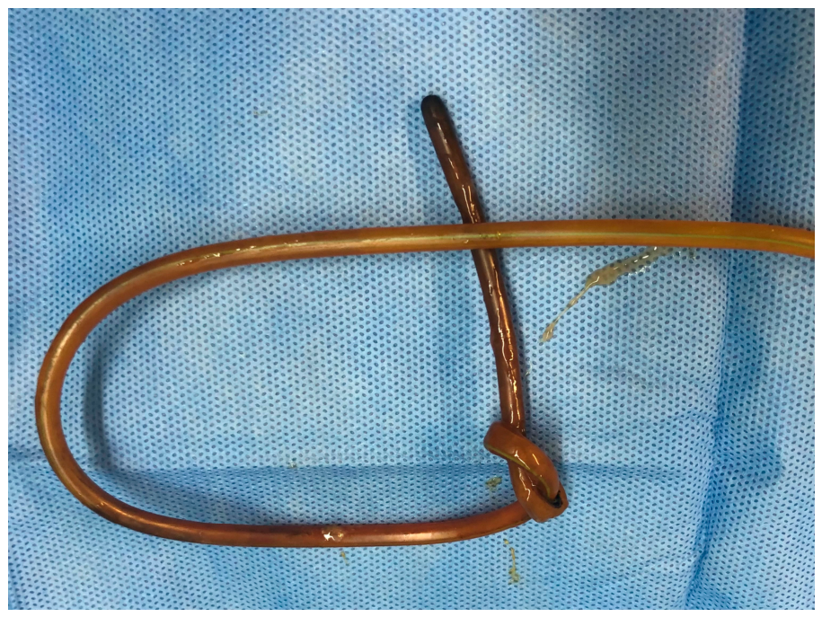
Figure 2.
Self-knotting NGT.
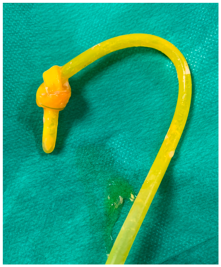
Figure 3.
NGT turned upwards inside esophagus.
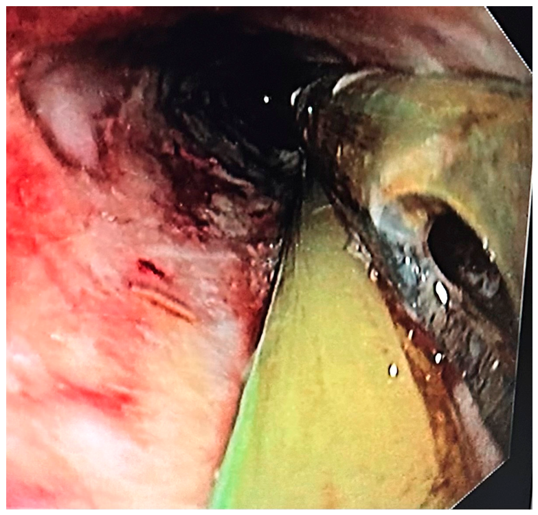
Figure 4.
NGT with double bend inside the gastric antrum.
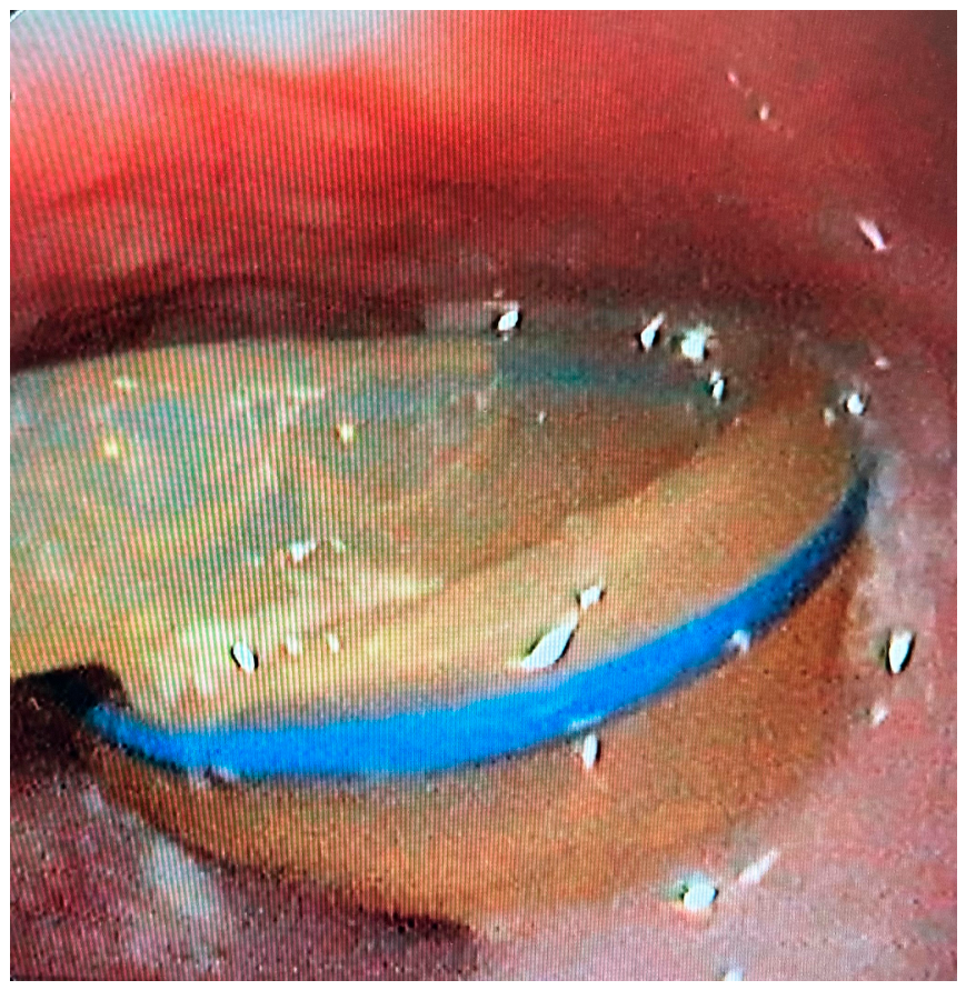
Figure 5.
NGT bended at later hole.
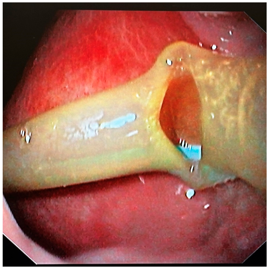
Figure 6.
NGT bended at later hole.
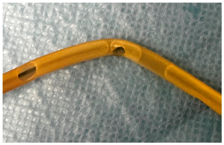
Figure 8.
X-ray revealed an intense angulation of the NGT at two points along its length.
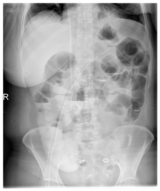
Disclaimer/Publisher’s Note: The statements, opinions and data contained in all publications are solely those of the individual author(s) and contributor(s) and not of MDPI and/or the editor(s). MDPI and/or the editor(s) disclaim responsibility for any injury to people or property resulting from any ideas, methods, instructions or products referred to in the content. |
© 2026 by the authors. Licensee MDPI, Basel, Switzerland. This article is an open access article distributed under the terms and conditions of the Creative Commons Attribution (CC BY) license (http://creativecommons.org/licenses/by/4.0/).
Copyright: This open access article is published under a Creative Commons CC BY 4.0 license, which permit the free download, distribution, and reuse, provided that the author and preprint are cited in any reuse.


