

Submitted:
29 June 2026
Posted:
30 June 2026
You are already at the latest version
Abstract
Preterm labor is a leading cause of neonatal morbidity and mortality, with inflammation playing a central role in its pathogenesis. This structured literature review summarizes the role of the nuclear factor kappa B (NF-κB) signaling pathway in term and preterm labor and critically evaluates current evidence on natural and synthetic NF-κB modulators as potential therapeutic approaches. To ensure comprehensive identification of relevant studies, a structured literature search informed by PRISMA 2020 reporting recommendations was performed, including studies published through August 2025. Experimental, translational, and review studies were synthesized narratively. NF-κB integrates hormonal, mechanical, infectious, and sterile inflammatory signals in gestational tissues, promoting the expression of pro-inflammatory cytokines, cyclooxygenase-2, prostaglandins, chemokines, and contraction-associated proteins that drive uterine activation. While tightly regulated NF-κB activation is essential for physiological term labor, its premature or excessive activation contributes to inflammation-associated preterm labor. Natural compounds and synthetic agents consistently attenuate excessive NF-κB signaling and downstream inflammatory responses in preclinical models, supporting their potential as candidate therapeutic strategies. However, clinical evidence remains limited. Future research should prioritize tissue-specific, context-dependent modulation of NF-κB and establish the safety, optimal therapeutic windows, and clinical efficacy of NF-κB-targeted interventions during pregnancy.
Keywords:
preterm birth
; NF-κB
; inflammation
; myometrium
; tocolysis
; NF-κB inhibitors
1. Introduction
Preterm birth (PTB) is a prevalent condition and represents a major global public health concern. The incidence of PTB varies between countries, ranging from 4% to 16% of all live births. In 2020, approximately 13.4 million infants were born preterm, while the annual global estimate reaches around 15 million, accounting for nearly 11% of all live births. PTB contributes to approximately 18% of deaths in children under five years of age and up to 35% of all neonatal deaths (within the first 28 days of life). Beyond increased mortality, PTB remains a leading cause of long-term morbidity, including hearing loss, blindness, chronic respiratory disorders, and central nervous system injuries.
Recent evidence indicates that the initiation of labor—both at term and preterm—is closely associated with the activation of inflammatory pathways. The transcription factor NF-κB acts as a central molecular integrator of immune, mechanical, and inflammatory signals in gestational tissues. NF-κB signaling is not inherently pathological in pregnancy: basal and tightly regulated NF-κB activity is required for placental development, maternal–fetal immune tolerance, and the initiation of normal parturition. However, excessive, dysregulated, or prematurely activated NF-κB signaling can trigger pathological inflammatory cascades and induce myometrial contractility independently of the physiological timing of labor.
Despite the availability of tocolytic therapies, which primarily target downstream uterine contractions, their effectiveness in preventing PTB remains limited. This underscores the need for novel therapeutic strategies targeting upstream inflammatory mechanisms driving preterm labor [1,2,3].
The aim of this study is to summarize the role of the NF-κB pathway in the regulation of term and preterm labor and to discuss potential therapeutic strategies for selective modulation of NF-κB signaling. The goal of such modulation would be to inhibit premature myometrial activation while preserving the essential physiological functions of this pathway during pregnancy.
2. Immunological Basis of Labor
2.1. Labor as an Inflammatory Process
Labor, both at term and preterm, is a physiological process closely associated with activation of inflammatory pathways. During normal pregnancy, the myometrium (myometrial smooth muscle cells, mSMCs) remains in a state of functional quiescence for most of gestation due to dominant anti-inflammatory signaling and the action of steroid hormones, primarily progesterone.
As term approaches (≥37 weeks of gestation), a coordinated and tightly regulated increase in pro-inflammatory signaling occurs. This includes activation of NF-κB–dependent pathways, which gradually overcomes pregnancy-maintaining mechanisms and initiates the transition of the uterus toward a contractile phenotype [4,5] (Figure 1).
This transition converts the myometrium from a quiescent to a contractile state, representing a key step in labor initiation. Concurrently, the immunological balance shifts toward a pro-inflammatory environment that facilitates uterine contractions, cervical ripening, and fetal membrane rupture, reflecting a physiological inflammatory program of parturition [6,7].
In the final stages of pregnancy, the expression of pro-inflammatory cytokines and chemokines increases, triggering a coordinated cascade of events required for labor onset. The NF-κB signaling pathway is central to integrating these inflammatory and stress-related signals and regulating the expression of genes responsible for initiating both physiological term labor and, when dysregulated, preterm labor [8].
2.2. Key Cytokines and Immune Cells in Labor Regulation
As term approaches, immune cells including neutrophils, macrophages, NK cells, and T lymphocytes become increasingly activated within gestational tissues. Through NF-κB–dependent signaling, these cells induce the expression of pro-inflammatory mediators (IL-1β, IL-6, IL-8, TNF-α, COX-2), which regulate contraction-associated proteins (GJA1, OXTR, COX-2) and promote prostaglandin synthesis.
These changes enhance myometrial excitability and increase calcium influx into mSMCs, thereby promoting coordinated uterine contractility. COX-2 additionally contributes to the induction of matrix metalloproteinases (MMPs), particularly MMP-9, which remodel the extracellular matrix and weaken fetal membranes, supporting membrane rupture and progression of labor [4,9,10].
Elevated levels of cytokines and chemokines (CXCL1, CXCL8, CCL2) promote leukocyte recruitment—mainly macrophages and neutrophils—to the myometrium, cervix, and fetal membranes, establishing a positive feedback loop that sustains and intensifies the local inflammatory response during labor [6,8].
Corticotropin-releasing hormone (CRH), primarily produced by the placenta, plays a pivotal role in regulating gestational length. Elevated CRH levels are associated with placental dysfunction and fetal maturation, while maternal stress further activates the hypothalamic–pituitary–adrenal (HPA) axis, increasing cortisol and CRH production. This, in turn, stimulates the release of pro-inflammatory cytokines, including IL-6, IL-1β, and TNF-α, contributing to activation of the inflammatory pathways involved in parturition [11] (Table 1).
2.3. Immunological Balance During Pregnancy
Normal pregnancy depends on the maintenance of immunological balance, with an-ti-inflammatory mechanisms predominating throughout most of gestation. Progesterone suppresses pro-inflammatory pathways and maintains myometrial quiescence until late gestation, when functional progesterone withdrawal gradually reduces its anti-inflammatory influence.
The resulting shift toward a pro-inflammatory state initiates the physiological program of labor, with NF-κB activation serving as a central regulatory node. Studies have demonstrated that NF-κB activity varies in dimer composition depending on physiological status. In non-pregnant women, p50 homodimers predominate, whereas during pregnancy and especially during labor, p65:p50 heterodimers become more prevalent.
2.4. Role of Immunological Disturbances in Labor
Premature disruption of immunological balance can shift the tightly regulated inflammatory milieu of pregnancy toward an enhanced pro-inflammatory state, increasing the intensity or advancing the timing of NF-κB activation along a physiological–pathological continuum and thereby promoting inappropriate initiation of the labor program.
Such dysregulation does not necessarily reflect a qualitatively distinct process but rather an amplification or temporal misalignment of pathways that normally participate in term parturition, ultimately increasing the risk of PTB.
Triggers include intrauterine infections, dysbiosis, oxidative stress, tissue ischemia, excessive NF-κB activation, and uterine overdistension in cases of polyhydramnios or multiple gestation [4,9,10].
In the chorioamniotic membranes, HMGB1 regulates embryo implantation, fetal development, and the timing of labor onset. Its overexpression has been associated with miscarriage, PTB, and preeclampsia. Acting as a damage-associated molecular pattern (DAMP), HMGB1 binds to RAGE and Toll-like receptors, activating NF-κB and inducing pro-inflammatory cytokine production (IL-1β, IL-18, TNF-α).
This further amplifies inflammation through inflammasome activation (NLRP3, AIM2, NOD1/2), caspase-1, MMP-9, and transcriptional upregulation of inflammatory genes.
Mechanical stress, such as amniotic stretching, also promotes the release of DAMPs, including HMGB1 and mitochondrial DNA, generating reactive oxygen species (ROS) and activating NF-κB.
Together, these pathways illustrate how both mechanical and molecular danger signals converge on NF-κB to modulate inflammatory activation in the amnion, depending on their magnitude and duration [12,13,14].
When this inflammatory cascade is prematurely initiated or excessively amplified, NF-κB drives a transcriptional program that closely resembles that of physiological term labor, including leukocyte infiltration and increased expression of IL-1α, IL-1β, IL-6, TNF-α, chemokines (CCL2, CXCL8), and COX-2.
3. Description of the NF-κB Pathway
3.1. Structure and Activation of NF-κB
The NF-κB pathway is a key regulator of inflammation and is implicated in numerous diseases, including autoimmune disorders, respiratory diseases, cancer, and diabetes. In pregnancy, it also plays a critical and tightly regulated role in coordinating immune, mechanical, and endocrine signals required for parturition. Understanding its structure and activation mechanisms is therefore essential for elucidating both physiological labor and inflammation-driven preterm birth.
In gestational tissues, NF-κB signaling operates within a unique immunological context characterized by the need to balance pro-inflammatory activation with maternal–fetal tolerance, distinguishing it from classical inflammatory responses observed in non-pregnant tissues.
Nuclear factor kappa B (NF-κB) comprises a family of five transcription factors: NF-κB1 (p105/p50), NF-κB2 (p100/p52), p65 (RelA), Rel homologue B (RelB), and c-Rel.
NF-κB regulates inflammatory responses, immune function, cell survival, and proliferation through the expression of genes such as cyclin A, cyclin D1, and CDK6. Activation of NF-κB also protects cells from TNF-α–induced apoptosis, supporting tissue integrity and cell survival; however, when dysregulated, it may contribute to chronic inflammation and oncogenesis.
The pathway is most commonly activated via the classical p65/p50 heterodimer. Under resting conditions, NF-κB is sequestered in the cytoplasm by the inhibitor IκB, which masks its nuclear localization signal (NLS) and prevents gene transcription.
Upon stimulation by cytokines (TNF-α, IL-1β), DNA damage, oxidative stress, Toll-like receptor (TLR) activation (via LPS, pathogen-associated molecular patterns [PAMPs], or damage-associated molecular patterns [DAMPs]), or antigen receptor signals (TCR/BCR), this inhibitory complex is disrupted.
Activation begins with stimulation of the IκB kinase (IKK) complex, composed of two catalytic subunits (IKKα and IKKβ) and a regulatory subunit (IKKγ/NEMO). IKK phosphorylates IκB, leading to its ubiquitination and proteasomal degradation. This exposes the NLS, allowing NF-κB to translocate to the nucleus, where it binds specific DNA sequences to initiate transcription of genes encoding pro-inflammatory cytokines (IL-6, IL-8, TNF-α), cyclooxygenase-2 (COX-2), immunoregulatory molecules (ICAM-1, MHC), oxytocin receptors (OXTR), and cell-survival proteins (Bcl-2, Bcl-xL).
In the context of pregnancy, the downstream transcriptional targets of NF-κB extend beyond classical inflammatory mediators to include genes directly involved in uterine activation, highlighting its dual role as both an immune regulator and a key driver of parturition-specific processes.
A single pro-inflammatory stimulus such as IL-1β can trigger additional cytokine production (e.g., IL-6) and upregulate COX-2 expression, forming a positive inflammatory amplification loop.
At the same time, NF-κB induces transcription of IκB, which re-sequesters NF-κB in the cytoplasm, providing an intrinsic negative feedback mechanism that limits excessive pathway activation [16,17,18,19,20,21].
Despite extensive characterization of NF-κB signaling, the precise mechanisms that determine the transition from physiological to pathological activation in pregnancy remain incompletely understood, representing an important area for future research.
3.2. Activation Pathways
Two main NF-κB activation pathways are recognized:
- Canonical pathway: Activated by TNF-α, IL-1β, LPS, oxidative stress, PAMPs, and DAMPs via receptors such as TNFR, IL-1R, and TLRs (mainly TLR4). TLR expression is highest in monocytes, macrophages, and neutrophils, which serve as major cytokine sources during early inflammatory responses. This pathway involves phosphorylation of the IKK complex, predominantly IKKβ and IKKγ, and primarily employs the p65/p50 heterodimer, producing a rapid and transient inflammatory response [9,17,18,19,20,21,22].
In gestational tissues, activation of the canonical pathway is most commonly associated with acute inflammatory stimuli, including infection and sterile inflammatory signals, and is therefore considered a principal driver of inflammation-induced preterm labor.
- 2.
- Non-canonical pathway: Activated by BAFF, CD40L, and lymphotoxin-β (LT-β) through BAFF-R, CD40, and LTβR. These signals prevent ubiquitin-mediated degradation of NF-κB–inducing kinase (NIK), which subsequently activates IKKα independently of IKKγ. The RelB/p52 dimer predominates in this pathway, producing a slower and more sustained transcriptional response compared with the canonical pathway [17,18,19,20,21].
Although less extensively studied in the context of pregnancy, the non-canonical pathway may contribute to immune regulation and tissue remodeling processes that support gestational maintenance, highlighting a potential functional divergence between NF-κB activation pathways in parturition.
3.3. NF-κB Activation at Term and in Preterm Labor
Studies have demonstrated a marked increase in NF-κB activity in the uterine decidua and placenta preceding and during labor, highlighting its fundamental role in parturition. NF-κB acts as a central integrator of multiple signaling pathways that coordinate the transition of the myometrium from a quiescent to a contractile state.
In this context, NF-κB can be viewed not only as an inflammatory mediator but also as a key regulatory hub that integrates endocrine, mechanical, and immune signals to determine the timing of labor onset.
In the uterus, NF-κB activation is driven by several overlapping and partly physiological mechanisms:
- TLR4 activation: Classically associated with infection via LPS, but also detected in preterm labor without overt infection, likely reflecting responses to sterile inflammatory stimuli such as mechanical stretch, hypoxia, or ischemic injury, which promote the release of endogenous danger-associated molecular patterns (DAMPs).
- Pro-inflammatory cytokines: IL-1β and TNF-α, which are involved in both physiological labor at term and inflammation-associated preterm labor.
- Mechanical signals: Uterine stretching activates NF-κB and MAPK pathways, contributing to the normal preparation of the uterus for labor.
The convergence of these distinct upstream signals on a common NF-κB–dependent transcriptional program underscores the role of this pathway as a central integrator of both physiological and pathological triggers of labor.
These converging signals induce the expression of contraction-associated proteins (CAPs), including:
- increased oxytocin receptor (OXTR) expression, enhancing myometrial sensitivity to oxytocin,
- enhanced COX-2 expression, leading to increased prostaglandin synthesis (PGF2α via FP receptors, PGE2 via EP1/EP3 receptors),
- upregulation of connexin-43 (CX-43), facilitating electrical coupling and synchronized myometrial contractions,
- chemokine production (CCL2, CXCL8/IL-8), promoting leukocyte recruitment and local immune activation.
These processes support the physiological onset and progression of labor. NF-κB activation is observed in all pregnancies prior to labor onset and is considered a normal component of the parturition program.
The distinction between term and preterm labor appears to depend not on the presence or absence of NF-κB activation, but rather on its timing, magnitude, and regulatory context.
Under certain conditions—such as intrauterine infection or exaggerated inflammatory signaling—this tightly regulated pathway may become activated earlier or more robustly, contributing to the premature initiation of uterine contractions and inflammation characteristic of preterm labor [9,16,22,23,24].
4. NF-κB Pathway Modulators as Potential Tocolytic Agents
4.1. Natural Inhibitors
4.1.1. Curcumin
Curcumin, the main polyphenol in Curcuma longa, is a natural modulator of NF-κB signaling with well-documented anti-inflammatory properties relevant to inflammation-associated preterm birth. In vitro and ex vivo studies demonstrate that curcumin attenuates NF-κB activation in gestational tissues by inhibiting IKKα/β, stabilizing IκB, and reducing p50/p65 nuclear translocation, leading to decreased IL-6 expression and gp130/STAT3 signaling [25]. Similar effects have been observed in placental, fetal membrane, and myometrial explants, including reduced p65 DNA binding, oxidative stress, COX-2 expression, cytokine production, and prostaglandin release [26].
In preclinical models of LPS-induced PTB, curcumin reduced NF-κB activation, cytokine levels, and the incidence of preterm birth, supporting its role in modulating excessive rather than basal inflammatory signaling [27]. Additional studies indicate systemic anti-inflammatory effects and inhibition of TLR4/NF-κB signaling in vivo [28,29]. Curcumin has also been shown to reduce myometrial contractility in vitro, suggesting potential tocolytic properties [30]. Systematic reviews identify curcumin as a potential candidate for further investigation, particularly in the context of TLR4–NF-κB modulation [31].
4.1.2. Resveratrol
Resveratrol is a natural polyphenol that modulates NF-κB signaling and reduces pro-inflammatory cytokine production in trophoblast cells [32]. In preclinical PTB models, it decreases iNOS and COX-2 expression, limits prostaglandin synthesis, and reduces preterm birth incidence [33,34,35]. Additionally, ex vivo studies demonstrate direct myometrial relaxant effects, indicating potential dual anti-inflammatory and tocolytic activity [36].
4.1.3. Omega-3 Fatty Acids
Omega-3 fatty acids (DHA, EPA) exert anti-inflammatory effects partly through NF-κB modulation. In gestational tissues, they reduce cytokine production and attenuate infection-induced inflammation by decreasing IκB phosphorylation and IKK activity [37,38]. Clinical evidence remains limited, although some studies suggest reduced placental inflammation with supplementation [39]. Reviews further indicate inhibition of TLR–NF-κB signaling and downstream mediators such as COX-2 and prostaglandins [40].
4.1.4. Other Natural Compounds
Additional natural compounds—including apigenin, quercetin, EGCG, ginger-derived compounds, and luteolin—demonstrate NF-κB inhibitory activity and reduction of pro-inflammatory mediators in gestational tissues [43,44,45,46,47]. Some also exhibit direct effects on myometrial contractility.
Natural compounds appear to modulate excessive NF-κB activation rather than fully suppress the pathway, although evidence remains largely preclinical and requires further validation.
Figure 2 illustrates the potential mechanisms by which natural NF-κB modulators interfere with inflammatory activation in preterm labor.
4.2. Synthetic Inhibitors
4.2.1. IKK Kinase Inhibitors
Selective IKKβ inhibitors, such as SC-514 and TPCA-1, attenuate NF-κB activation and reduce pro-inflammatory mediators in gestational tissues and animal models, supporting their role in limiting inflammation-driven uterine activation [48,49]. Reviews suggest that these agents may offer a more targeted approach compared with nonspecific anti-inflammatory therapies [50,51].
4.2.2. Sulfasalazine
Sulfasalazine reduces NF-κB activation and cytokine expression in gestational tissues [50]; however, paradoxical pro-inflammatory effects and increased apoptosis have also been reported [52]. Clinical observations do not indicate increased PTB risk, but preclinical data remain inconsistent, limiting its therapeutic potential [53,54].
4.2.3. TLR4 Inhibitors
TLR4 antagonists, including (+)-naloxone and (+)-naltrexone, reduce cytokine production and prevent inflammation-induced preterm labor in animal models, highlighting the potential of targeting upstream NF-κB activation [55,56]. Reviews support TLR4 as a relevant therapeutic target in inflammation-associated PTB [50,57,58].
4.2.4. Modern Synthetic Modulators
Recent approaches focus on more selective modulation of inflammatory signaling. The BET inhibitor JQ1 reduces inflammatory gene expression and delays labor onset [59]. The NLRP3 inhibitor MCC950 suppresses inflammasome activation, reduces inflammation, and prevents PTB in experimental models [60,61].
Additional compounds, including aprotic amides, inhibit NF-κB activation and cytokine expression [62], while exosome-based delivery systems may improve tissue-specific targeting and preserve physiological NF-κB functions [63].
Table 2 summarizes the classification of NF-κB modulators according to mechanism of action and their effects in preterm birth.
Overall, natural and synthetic NF-κB modulators differ primarily in pharmacological profile rather than ultimate biological target. Natural compounds generally exert multi-target, pleiotropic anti-inflammatory effects with relatively modest potency, which may confer safety advantages but limits precise pathway control. In contrast, synthetic inhibitors provide more selective and robust targeting of specific components of the NF-κB signaling cascade, enabling stronger suppression of inflammation-associated pathways implicated in preterm labor, albeit with a potentially narrower therapeutic window. Taken together, these differences underscore that future translational strategies should prioritize balanced, context-dependent modulation of NF-κB activity rather than complete pathway inhibition.
5. Materials and Methods
This study was conducted as a structured literature review to evaluate the role of inflammatory processes and nuclear factor kappa B (NF-κB) signaling in the initiation of term and preterm labor, as well as the therapeutic potential of NF-κB modulation. To enhance transparency and reproducibility, the literature search and study selection process was performed using a structured approach informed by the PRISMA 2020 reporting recommendations. Owing to the heterogeneity of the included studies with respect to experimental models, study designs, and reported outcomes, the evidence was synthesized narratively rather than quantitatively.
A comprehensive literature search was performed using the PubMed and Scopus databases. The primary search included articles published between January 2015 and August 2025. Earlier landmark publications were additionally included where necessary to provide historical context and mechanistic background. The final database search was conducted in August 2025.
The PubMed search combined Medical Subject Headings (MeSH) and free-text terms, whereas equivalent free-text terms were used in Scopus. The search strategy included combinations of the following terms using Boolean operators: “preterm birth,” “preterm labor,” “NF-κB,” “inflammation,” “myometrium,” “TLR4,” “cytokines,” “tocolytics,” and “NF-κB inhibitors.” Duplicate records identified across databases were removed before the screening process.
Eligible publications comprised original in vitro, ex vivo, animal, translational, and clinical studies, as well as relevant review articles published in peer-reviewed English-language journals. Studies were included if they investigated NF-κB signaling in the context of labor, inflammatory mechanisms involved in parturition, or pharmacological modulation of these pathways. Review articles were primarily used to identify additional relevant primary studies and to provide contextual background. Conference abstracts, duplicate publications, non-English articles, and studies not directly relevant to the review topic were excluded.
Titles and abstracts were independently screened by two reviewers. Full-text articles of potentially eligible studies were subsequently assessed by the same reviewers. Disagreements regarding study eligibility were resolved through discussion and consensus. When necessary, additional consultation with the senior author was sought.
Given the methodological heterogeneity of the studies included in the review, no formal risk-of-bias assessment or quantitative meta-analysis was performed. Instead, findings were synthesized using a qualitative narrative approach, focusing on the molecular mechanisms linking inflammation and NF-κB signaling to the onset of labor, as well as the current evidence regarding pharmacological modulation of these pathways.
The study selection process is summarized in the PRISMA 2020 flow diagram (Figure 3).
6. Conclusions and Future Perspectives
6.1. Key Conclusions
Inflammation is a fundamental driver of both physiological and pathological labor, with NF-κB acting as a central molecular hub that integrates hormonal, mechanical, infectious, and sterile inflammatory signals across gestational tissues. Appropriate activation of NF-κB is essential for normal term parturition, whereas its premature or excessive activation contributes to inflammation-driven preterm labor through induction of cytokines, prostaglandins, chemokines, and contraction-associated proteins.
Current evidence identifies NF-κB as one of the most promising mechanistic targets for limiting pathological uterine inflammation while preserving the physiological signaling required for timely labor.
6.2. Limitations of Current Evidence
Despite substantial progress in understanding NF-κB biology during pregnancy, several important limitations remain. Most available evidence is derived from in vitro studies and animal models, whereas well-designed clinical studies evaluating NF-κB-targeted interventions during pregnancy are scarce. Furthermore, considerable heterogeneity exists among experimental models, inflammatory stimuli, gestational tissues, dosing regimens, and outcome measures, making direct comparison between studies difficult.
In addition, because NF-κB is a pleiotropic transcription factor involved in immune homeostasis, fetal development, and normal parturition, complete inhibition of this pathway is unlikely to be clinically desirable. Consequently, the translational relevance of many experimental findings remains uncertain.
6.3. Future Perspectives
Future research should move beyond demonstrating anti-inflammatory activity toward identifying tissue-specific, cell-specific, and context-dependent mechanisms of NF-κB regulation during pregnancy. Particular emphasis should be placed on defining therapeutic windows, developing targeted drug-delivery systems that minimize fetal exposure, and integrating transcriptomic, proteomic, and single-cell approaches to characterize NF-κB signaling within individual gestational compartments.
Equally important is the need for well-designed translational studies and randomized clinical trials to establish the pharmacokinetics, safety, and efficacy of NF-κB modulators in pregnant women.
6.4. Final Perspective
Rather than global suppression of NF-κB signaling, selective modulation of excessive pathological activation appears to represent the most biologically plausible therapeutic strategy. Natural compounds and emerging synthetic modulators provide encouraging preclinical evidence that excessive inflammation can be attenuated without abolishing the physiological functions of NF-κB required for normal pregnancy and term labor.
Although these findings support NF-κB as a compelling therapeutic target, clinical translation remains at an early stage. Future advances will depend on the development of selective, pregnancy-compatible interventions capable of restoring inflammatory homeostasis while maintaining maternal and fetal safety.
Author Contributions
Conceptualization, Ż.K.-T.; data curation, M.M., D.M., Al.M. and An.M.; writing—original draft preparation, M.M., D.M., Al.M., An.M. and Ż.K.-T.; writing—review and editing, M.M., D.M., Al.M., An.M. and Ż.K.-T.; visualization, M.M., D.M., Al.M., An.M. and Ż.K.-T.; supervision, Ż.K.-T.; project administration, M.M., D.M., Al.M., An.M. and Ż.K.-T.; funding acquisition, Ż.K.-T. All authors have read and agreed to the published version of the manuscript.
Funding
This research was funded by the Medical University of Lublin, grant number 336.
Institutional Review Board Statement
Not applicable.
Informed Consent Statement
Not applicable.
Data Availability Statement
No new data were created or analyzed in this study. Data sharing is not applicable to this article.
Conflicts of Interest
The authors declare no conflicts of interest.
Abbreviations
The following abbreviations are used in this manuscript:
| BAFF | B-cell Activating Factor |
| BAFF-R | B-cell Activating Factor Receptor |
| BET | Bromodomain and Extra-Terminal proteins |
| BCR | B-cell receptor |
| BRD | Bromodomain-containing protein |
| CAPs | Contraction-Associated Proteins |
| CCL2 | C-C motif chemokine ligand 2 |
| CD40L | CD40 ligand |
| CDK6 | Cyclin-dependent kinase 6 |
| COX-2 | Cyclooxygenase-2 |
| CRH | Corticotropin-Releasing Hormone |
| CX-43 | Connexin-43 |
| CXCL1 | C-X-C motif chemokine ligand 1 |
| CXCL8 (IL-8) | C-X-C motif chemokine ligand 8 |
| DAMPs | Damage-Associated Molecular Patterns |
| DHA | Docosahexaenoic acid |
| EGCG | Epigallocatechin gallate |
| EPA | Eicosapentaenoic acid |
| EP | Prostaglandin E receptor |
| FP | Prostaglandin F receptor |
| gp130 | Glycoprotein 130 |
| HMGB1 | High Mobility Group Box 1 |
| HPA axis | Hypothalamic–Pituitary–Adrenal axis |
| IKK | IκB kinase |
| IKKα | IκB kinase alpha |
| IKKβ | IκB kinase beta |
| IKKγ (NEMO) | IκB kinase gamma / NF-κB Essential Modulator |
| IL-1α | Interleukin 1 alpha |
| IL-1β | Interleukin 1 beta |
| IL-6 | Interleukin 6 |
| IL-8 | Interleukin 8 |
| iNOS | Inducible Nitric Oxide Synthase |
| JQ1 | BET bromodomain inhibitor JQ1 |
| LTβ | Lymphotoxin beta |
| LTβR | Lymphotoxin beta receptor |
| LPS | Lipopolysaccharide |
| MAPK | Mitogen-Activated Protein Kinases |
| MHC | Major Histocompatibility Complex |
| MMP-9 | Matrix metalloproteinase-9 |
| mSMC | Myometrial smooth muscle cells |
| NF-κB | Nuclear Factor kappa B |
| NIK | NF-κB–inducing kinase |
| NK cells | Natural killer cells |
| NLRP3 | NOD-like receptor family pyrin domain containing 3 |
| NOD1/2 | Nucleotide-binding oligomerization domain-containing protein 1/2 |
| OXTR | Oxytocin receptor |
| PAMPs | Pathogen-Associated Molecular Patterns |
| PGF2α | Prostaglandin F2 alpha |
| PGE2 | Prostaglandin E2 |
| PTB | Preterm birth |
| RCT | Randomized Controlled Trial |
| RelA (p65) | v-rel reticuloendotheliosis viral oncogene homolog A |
| RelB | v-rel reticuloendotheliosis viral oncogene homolog B |
| ROS | Reactive Oxygen Species |
| SPMs | Specialized pro-resolving mediators |
| STAT3 | Signal Transducer and Activator of Transcription 3 |
| TLR4 | Toll-Like Receptor 4 |
| TNF-α | Tumor necrosis factor alpha |
| TCR | T-cell receptor |
References
- Khezri, R.; Askari, S.; Jahanian, S.; Ashayeri, N. Preterm birth and public health challenges: Incidence and risk factors. Public Health 2025, 242, 186–191. [Google Scholar] [CrossRef] [PubMed]
- Sultana, S.; Horiuchi, S.; Homer, C.S.; Baqui, A.H.; Vogel, J.P. The prevalence of long-term neurodevelopmental outcomes in preterm-born children in low- and middle-income countries: a systematic review and meta-analysis of developmental outcomes in 72 974 preterm-born children. J. Glob. Health 2025, 15, 04106. [Google Scholar] [CrossRef] [PubMed]
- Hirvonen, M.; Ojala, R.; Korhonen, P.; Haataja, P.; Eriksson, K.; Gissler, M.; Luukkaala, T.; Tammela, O. Visual and Hearing Impairments After Preterm Birth. Pediatrics 2018, 142, e20173888. [Google Scholar] [CrossRef] [PubMed]
- Li, J.K.H.; Lai, P.F.; Tribe, R.M.; Johnson, M.R. Transcription factors regulated by cAMP in smooth muscle of the myometrium at human parturition. Biochem Soc. Trans. 2021, 49, 997–1011. [Google Scholar] [CrossRef] [PubMed]
- Sheller-Miller, S.; Radnaa, E.; Yoo, J.K.; Kim, E.; Choi, K.; Kim, Y.; Kim, Y.N.; Richardson, L.; Choi, C.; Menon, R. Exosomal delivery of NF-κB inhibitor delays LPS-induced preterm birth and modulates fetal immune cell profile in mouse models. Sci. Adv. 2021, 7, eabd3865. [Google Scholar] [CrossRef] [PubMed]
- Lim, R.; Lappas, M. Novel anti-inflammatory actions of TIPE2 in human primary amnion and myometrial cells. Reproduction 2019, 158, 95–107. [Google Scholar] [CrossRef] [PubMed]
- Lv, M.; Jia, Y.; Dong, J.; Wu, S.; Ying, H. The landscape of decidual immune cells at the maternal-fetal interface in parturition and preterm birth. Inflamm. Res. 2025, 74, 44. [Google Scholar] [CrossRef] [PubMed]
- Lappas, M. RKIP is decreased in laboring myometrium and modulates inflammation-induced pro-labor mediators. Reproduction 2017, 153, 545–553. [Google Scholar] [CrossRef] [PubMed]
- Chen, Z.; Li, J.; Xu, W.; Wu, X.; Xiang, F.; Li, X.; Zhang, M.; Zheng, J.; Kang, X.; Wu, R. Elevated expression of Toll-like receptor 4 and cytokines in both serum and myometrium at term may serve as promising biomarkers for uterine activation preceding labor. Front Endocrinol. 2023, 14, 1255925. [Google Scholar] [CrossRef] [PubMed]
- Goradia, P.; Lim, R.; Lappas, M. DREAM is Involved in the Genesis of Inflammation-Induced Prolabour Mediators in Human Myometrial and Amnion Cells. BioMed Res. Int. 2018, 22, 8237087. [Google Scholar] [CrossRef] [PubMed]
- Sehgal, N.; Li, L.; Goin, D.E.; Chen, J.; Jigmeddagva, U.; Morello-Frosch, R.; Woodruff, T.J.; Gaw, S.L.; Robinson, J.F.; Eick, S.M. Psychosocial stress and associations with inflammation in mid-gestation maternal, fetal, and placental tissue. Reprod. Toxicol. 2025, 135, 108922. [Google Scholar] [CrossRef] [PubMed]
- Ren, Y.; Zhu, D.; Han, X.; Zhang, Q.; Chen, B.; Zhou, P.; Wei, Z.; Zhang, Z.; Cao, Y.; Zou, H. HMGB1: a double-edged sword and therapeutic target in the female reproductive system. Front Immunol. 2023, 14, 1238785. [Google Scholar] [CrossRef] [PubMed]
- Plazyo, O.; Romero, R.; Unkel, R.; Balancio, A.; Mial, T.N.; Xu, Y.; Dong, Z.; Hassan, S.S.; Gomez-Lopez, N. HMGB1 Induces an Inflammatory Response in the Chorioamniotic Membranes That Is Partially Mediated by the Inflammasome. Biol. Reprod. 2016, 95, 130. [Google Scholar] [CrossRef] [PubMed]
- Padron, J.G.; Saito Reis, C.A.; Ng, P.K.; Norman Ing, N.D.; Baker, H.; Davis, K.; Kurashima, C.; Kendal-Wright, C.E. Stretch Causes cffDNA and HMGB1-Mediated Inflammation and Cellular Stress in Human Fetal Membranes. Int. J. Mol. Sci. 2024, 25, 5161. [Google Scholar] [PubMed]
- Habelrih, T.; Augustin, T.L.; Mauffette-Whyte, F.; Ferri, B.; Sawaya, K.; Côté, F.; Gallant, M.; Olson, D.M.; Chemtob, S. Inflammatory mechanisms of preterm labor and emerging anti-inflammatory interventions. Cytokine Growth Factor Rev. 2024, 78, 50–63. [Google Scholar] [CrossRef] [PubMed]
- Leimert, K.B.; Verstraeten, B.S.E.; Messer, A.; Nemati, R.; Blackadar, K.; Fang, X.; Robertson, S.A.; Chemtob, S.; Olson, D.M. Cooperative effects of sequential PGF2α and IL-1β on IL-6 and COX-2 expression in human myometrial cells. Biol. Reprod. 2019, 100, 1370–1385. [Google Scholar] [CrossRef] [PubMed]
- Kannan, G.; Paul, B.M.; Thangaraj, P. Stimulation, regulation, and inflammaging interventions of natural compounds on nuclear factor kappa B (NF-κB) pathway: a comprehensive review. Inflammopharmacology 2025, 33, 145–162. [Google Scholar] [CrossRef] [PubMed]
- Zinatizadeh, M.R.; Schock, B.; Chalbatani, G.M.; Zarandi, P.K.; Jalali, S.A.; Miri, S.R. The nuclear factor kappa B (NF-κB) signaling in cancer development and immune diseases. Genes Dis. 2020, 8, 287–297. [Google Scholar] [CrossRef] [PubMed]
- Zhang, T.; Ma, C.; Zhang, Z.; Zhang, H.; Hu, H. NF-κB signaling in inflammation and cancer. MedComm 2021, 2, 618–653. [Google Scholar] [CrossRef] [PubMed]
- Mao, H.; Zhao, X.; Sun, S.C. NF-κB in inflammation and cancer. Cell Mol. Immunol. 2025, 22, 811–839. [Google Scholar] [CrossRef] [PubMed]
- Guo, Q.; Jin, Y.; Chen, X.; Ye, X.; Shen, X.; Lin, M.; Zeng, C.; Zhou, T.; Zhang, J. NF-κB in biology and targeted therapy: new insights and translational implications. Signal Transduct. Target Ther. 2024, 9, 53. [Google Scholar] [CrossRef] [PubMed]
- Chen, Z.; Liu, Q.; Zhu, Z.; Xiang, F.; Wu, R.; Kang, X. Toll-like receptor 4 contributes to uterine activation by upregulating pro-inflammatory cytokine and CAP expression via the NF-κB/P38MAPK signaling pathway during pregnancy. J. Cell Physiol. 2020, 235, 513–525. [Google Scholar] [CrossRef] [PubMed]
- Wen, B.; Zheng, Z.; Wang, L.; Qian, X.; Wang, X.; Chen, Y.; Bao, J.; Jiang, Y.; Ji, K.; Liu, H. HIF-1α is essential for the augmentation of myometrial contractility during labor†. Biol. Reprod. 2022, 107, 1540–1550. [Google Scholar] [CrossRef] [PubMed]
- Li, H.; Li, Y.; Luo, S.; Zhang, Y.; Feng, Z.; Li, S. The roles and mechanisms of the NF-κB signaling pathway in tendon disorders. Front Vet. Sci. 2024, 11, 1382239. [Google Scholar] [CrossRef] [PubMed]
- Devi, Y.S.; Halici, M.; Chapman, J.; Yong, H.; Keelan, J.A.; Mitchell, M.D. Curcumin inhibits IL-1β–induced inflammatory signaling in human decidual cells via suppression of NF-κB and STAT3 pathways. PLoS ONE 2015, 10, e0120091. [Google Scholar] [PubMed]
- Lim, R.; Barker, G.; Lappas, M. Curcumin reduces infection-induced inflammation in human fetal membranes by suppressing the NF-κB pathway. Mol. Hum. Reprod. 2013, 19, 451–62. [Google Scholar] [PubMed]
- Guo, Y.Z.; He, P.; Feng, A.M. Effect of curcumin on expressions of NF-κBp65, TNF-α and IL-8 in placental tissue of premature birth of infected mice. Asian Pac. J. Trop. Med. 2017, 10, 175–178. [Google Scholar] [CrossRef] [PubMed]
- Chen, H.; Tang, Y.; Wang, H.; Chen, W.; Jiang, H. Curcumin alleviates lipopolysaccharide-induced neuroinflammation in fetal mouse brain. Restor. Neurol. Neurosci. 2018, 36, 583–592. [Google Scholar] [CrossRef] [PubMed]
- doi. [CrossRef] [PubMed]
- Gong, P.; Liu, M.; Hong, G.; Li, Y.; Xue, P.; Zheng, M.; Wu, M.; Shen, L.; Yang, M.; Diao, Z.; Hu, Y. Curcumin improves LPS-induced preeclampsia-like phenotype in rat by inhibiting the TLR4 signaling pathway. Placenta 2016, 41, 45–52. [Google Scholar] [CrossRef] [PubMed]
- El-Sayed, E.M. Relaxant effect of curcumin on uterine smooth muscle: an in vitro study. J. Basic Appl. Sci. 2008, 4, 45–48. [Google Scholar]
- Tossetta, G.; Fantone, S.; Giannubilo, S.R.; Marzioni, D. The Multifaced Actions of Curcumin in Pregnancy Outcome. Antioxidants 2021, 10, 126. [Google Scholar] [CrossRef] [PubMed]
- Hannan, N.J.; Brownfoot, F.C.; Cannon, P.; Deo, M.; Beard, S.; Nguyen, T.V.; Palmer, K.R.; Tong, S.; Kaitu’u-Lino, T.J. Resveratrol inhibits release of soluble fms-like tyrosine kinase (sFlt-1) and soluble endoglin and improves vascular dysfunction—implications as a preeclampsia treatment. Sci. Rep. 2017, 7, 1819. [Google Scholar] [CrossRef] [PubMed]
- Ramli, I.; Posadino, A.M.; Giordo, R.; Fenu, G.; Fardoun, M.; Iratni, R.; Eid, A.H.; Zayed, H.; Pintus, G. Effect of Resveratrol on Pregnancy, Prenatal Complications and Pregnancy-Associated Structure Alterations. Antioxidants 2023, 12, 341. [Google Scholar] [CrossRef] [PubMed]
- Bariani, M.V.; Correa, F.; Leishman, E.; Domínguez; Rubio, A.P.; Arias, A.; Stern, A.; Bradshaw, H.B.; Franchi, A.M. Resveratrol protects from lipopolysaccharide-induced inflammation in the uterus and prevents experimental preterm birth. Mol. Hum. Reprod. 2017, 23, 571–581. [Google Scholar] [CrossRef] [PubMed]
- Furuya, H.; Taguchi, A.; Kawana, K.; Yamashita, A.; Inoue, E.; Yoshida, M.; Nakamura, H.; Fujimoto, A.; Inoue, T.; Sato, M.; Nishida, H.; Nagasaka, K.; Adachi, K.; Hoya, M.; Nagamatsu, T.; Wada-Hiraike, O.; Yamashita, T.; Osuga, Y.; Fujii, T. Resveratrol Protects Against Pathological Preterm Birth by Suppression of Macrophage-Mediated Inflammation. Reprod. Sci. 2015, 22, 1561–1568. [Google Scholar] [CrossRef] [PubMed]
- Novaković, R.; Radunović, N.; Marković-Lipkovski, J.; Ćirović, S.; Beleslin-Čokić, B.; Ilić, B.; Ivković, B.; Heinle, H.; Živanović, V.; Gojković-Bukarica, L.J. Effects of the polyphenol resveratrol on contractility of human term pregnant myometrium. Mol. Hum. Reprod. 2015, 21, 545–551. [Google Scholar] [CrossRef] [PubMed]
- Garcia-So, J.; Zhang, X.; Yang, X.; Rubinstein; Mao, D.Y.; Kitajewski, J.; Tenenbaum, H.C.; Genco, C.A.; Han, Y.W. Omega-3 fatty acids suppress Fusobacterium nucleatum–induced placental inflammation originating from maternal endothelial cells. JCI Insight 2019, 4, e125436. [Google Scholar] [CrossRef] [PubMed]
- Yang, Y.C.; Lii, C.K.; Wei, Y.L.; Li, C.C.; Lu, C.Y.; Liu, K.L.; Chen, H.W. Docosahexaenoic acid inhibition of inflammation is partially via cross-talk between Nrf2/heme oxygenase 1 and IKK/NF-κB pathways. J. Nutr. Biochem 2013, 24, 204–212. [Google Scholar] [CrossRef] [PubMed]
- Haghiac, M.; Yang, X.H.; Presley, L.; Smith, S.; Dettelback, S.; Minium, J.; Belury, M.A.; Catalano, P.M.; Hauguel-de; Mouzon, S. Dietary Omega-3 Fatty Acid Supplementation Reduces Inflammation in Obese Pregnant Women: A Randomized Double-Blind Controlled Clinical Trial. PLoS ONE 2015, 10, e0137309. [Google Scholar] [CrossRef] [PubMed]
- Regidor, P.A.; Eiblwieser, J.; Steeb, T.; Rizo, J.M. Omega-3 long chain fatty acids and their metabolites in pregnancy outcomes for the modulation of maternal inflammatory- associated causes of preterm delivery, chorioamnionitis and preeclampsia. F1000Res 2024, 13, 882. [Google Scholar] [CrossRef] [PubMed]
- Nordgren, T.M.; Anderson; Berry, A.; Van; Ormer, M.; Zoucha, S.; Elliott, E.; Johnson, R.; McGinn, E.; Cave, C.; Rilett, K.; Weishaar, K.; Maddipati, S.S.; Appeah, H.; Hanson, C. Omega-3 Fatty Acid Supplementation, Pro-Resolving Mediators, and Clinical Outcomes in Maternal-Infant Pairs. Nutrients 2019, 11, 98. [Google Scholar] [CrossRef] [PubMed]
- Sousa, L.G.; Correia-da-Silva, G.; Teixeira, N.; Fonseca, B.M. Specialized pro-resolving mediators: key regulators in placental function and pregnancy complications. J. Mol. Med. (Berl) 2025, 103, 885–897. [Google Scholar] [CrossRef] [PubMed]
- Lim, R.; Barker, G.; Wall, C.A.; Lappas, M. Dietary phytophenols curcumin, naringenin and apigenin reduce infection-induced inflammatory and contractile pathways in human placenta, foetal membranes and myometrium. Mol. Hum. Reprod. 2013, 19, 451–462. [Google Scholar] [CrossRef] [PubMed]
- Zygmuntowicz, A.; Markiewicz, W.; Grabowski, T.; Burmańczuk, A.; Vyniarska, A.; Jaroszewski, J.J. Quercetin affects uterine smooth muscle contractile activity in gilts. PLoS ONE 2021, 16, e0252438. [Google Scholar] [CrossRef] [PubMed]
- Shih, L.-J.; Chen, T.-F.; Lin, C.-K.; Liu, H.-S.; Kao, Y.-H. Green tea (−)-epigallocatechin gallate inhibits the growth of human villous trophoblasts via ERK, p38, AMPK and Akt signaling pathways. Am. J. Physiol. Cell Physiol. 2016, 311, C308–C321. [Google Scholar] [CrossRef] [PubMed]
- Buddhakala, N.; Talubmook, C.; Sriyotha, P.; Wray, S.; Kupittayanant, S. Inhibitory effects of ginger oil on spontaneous and PGF2α-induced contraction of rat myometrium. Planta Med. 2008, 74, 385–391. [Google Scholar] [CrossRef] [PubMed]
- Wall, C.; Lim, R.; Poljak, M.; Lappas, M. Dietary flavonoids as therapeutics for preterm birth: Luteolin and kaempferol suppress inflammation in human gestational tissues in vitro. Oxid. Med. Cell. Longev. 2013, 2013, 485201. [Google Scholar] [CrossRef] [PubMed]
- Gurney, E.P.; Nguyen, T.; Herndon, C.N.; Harley, I.T.W.; O’Brien, B.; Giudice, L.C. Inhibition of IKK/NF-κB signaling reduces pro-labour gene expression in human myometrial cells. Reproduction 2018, 156, 1–10. [Google Scholar] [CrossRef] [PubMed]
- Ireland, D.J.; Kemp, M.W.; Miura, Y.; Saito, M.; Newnham, J.P.; Keelan, J.A. Intra-amniotic administration of the IKKβ inhibitor TPCA-1 suppresses fetal membrane inflammation in a sheep model of LPS-induced chorioamnionitis. Am. J. Obstet. Gynecol. 2015, 212, 98.e1–98.e9. [Google Scholar] [CrossRef] [PubMed]
- Pavlidis, I.; Stock, S.J. Preterm birth therapies to target inflammation. J. Clin. Pharmacol. 2022, 62, S79–S93. 51. Ireland, D.J.; Kemp, M.W.; Kannan, P.S.; Saito, M.; Newnham, J.P.; Keelan, J.A. Inhibition of inflammatory mediators in human fetal membranes and preterm birth models: Comparative efficacy of anti-inflammatory agents targeting NF-κB. Biol. Reprod. 2017, 97, 88–97. doi: 10.1095/biolreprod.117.150862. [CrossRef]
- Sykes, L.; MacIntyre, D.A.; Yap, X.; Teoh, T.G.; Bennett, P.R. Sulfasalazine induces pro-inflammatory and pro-apoptotic responses in human amniocytes and myometrial cells. Immunology 2015, 144, 388–396. [Google Scholar] [CrossRef] [PubMed]
- Miller, J.E.; Peña, E.; Burd, I. The role of anti-inflammatory therapies in the prevention of preterm birth: A systematic review of preclinical evidence. Reprod. Sci. 2023, 30, 3–15. [Google Scholar] [CrossRef]
- Habelrih, T.; Augustin, T.-L.; Mauffette-Whyte, F.; Ferri, B.; Sawaya, K.; Côté, F.; Gallant, M.; Olson, D.M.; Chemtob, S. Inflammatory mechanisms of preterm labor and emerging anti-inflammatory interventions. Cytokine Growth Factor Rev. 2024, 78, 50–63. [Google Scholar] [CrossRef] [PubMed]
- Chin, P.Y.; MacIntyre, D.A.; Lee, Y.; Kannan, P.S.; Kallapur, S.G.; Jobe, A.H.; Keelan, J.A. Toll-like receptor-4 antagonist (+)-naloxone protects against inflammation-induced fetal programming and preterm birth in mice. Endocrinology 2019, 160, 2646–2662. [Google Scholar] [CrossRef] [PubMed]
- Wahid, H.H.; Tiwari, A.; Keelan, J.A.; Kannan, P.S.; Kallapur, S.G.; Jobe, A.H. (+)-Naltrexone, a selective TLR4 antagonist, prevents cPAF-induced preterm labor and perinatal mortality in mice. Am. J. Pathol. 2020, 190, 1206–1221. [Google Scholar] [CrossRef] [PubMed]
- Robertson, S.A.; Care, A.S.; Moldenhauer, L.M. Targeting Toll-like receptor-4 to tackle preterm birth and fetal inflammatory injury. Clin. Transl. Immunol. 2020, 9, e1121. [Google Scholar] [CrossRef] [PubMed]
- Raia-Barjat, T.; Digonnet, M.; Giraud, A.; Ayash, T.; Vancolen, S.; Benharouga, M.; Chauleur, C.; Alfaidy, N.; Sébire, G. Animal models of chorioamnionitis: Considerations for translational studies. Front. Immunol. 2022, 13, 842194. [Google Scholar] [CrossRef]
- Chin, P.Y.; MacIntyre, D.A.; Lee, Y.; Kannan, P.S.; Kallapur, S.G.; Jobe, A.H.; Keelan, J.A. JQ1 suppresses intrauterine inflammation and delays preterm birth in mice. Clin. Sci. 2019, 133, 2379–2390. [Google Scholar] [CrossRef]
- Gomez-Lopez, N.; Romero, R.; Garcia-Flores, V.; Xu, Y.; Leng, Y.; Alhousseini, A.; Hassan, S.S.; Miller, D.; Winters, A.D.; Munoz, H.; Raghupathy, R.; Berry, M.; Yeo, L.; Santolaya-Forgas, J.; Pacora, P.; Maymon, E.; Hassan, S.S. Inhibition of the NLRP3 inflammasome with MCC950 prevents sterile intra-amniotic inflammation, preterm labor and neonatal mortality in mice. Cell Rep. 2018, 25, 2940–2951. [Google Scholar] [CrossRef] [PubMed]
- Chen, Z.; Shan, Y.; You, X.; Gu, H.; Xu, C.; Long, J.; Ni, X. NLRP3 inflammasome is involved in uterine activation for labor at term and preterm. Reproduction 2021, 162, 449–460. [Google Scholar] [CrossRef] [PubMed]
- Ajgaonkar, S.; Koya, J.; Munnangi, S.; Pekson, R.; Ashby, C.R., Jr.; Reznik, S.E. Novel aprotic amides inhibit NF-κB and prevent inflammation-induced preterm birth in mice. Reprod. Sci. 2024, 31, 145–158. [Google Scholar] [CrossRef]
- Sheller-Miller, S.; Radnaa, E.; Yoo, J.-K.; Kim, E.; Choi, K.; Kim, Y.; Kim, Y.; Richardson, L.; Choi, C.; Menon, R. Exosome-mediated delivery of NF-κB inhibitors attenuates intrauterine inflammation and delays preterm birth in murine models. J. Nanobiotechnol. 2023, 21, 112. [Google Scholar] [CrossRef]
- Gilmore, T.D.; Herscovitch, M. Targeting NF-κB for therapy: Pros and cons. Curr. Opin. Pharmacol. 2006, 6, 357–363. [Google Scholar] [CrossRef] [PubMed]
Figure 1.
Transition from pregnancy maintenance to labor. Pregnancy is maintained by a progesterone-dominated anti-inflammatory environment with restrained NF-κB activity. Near term, physiological activation of NF-κB initiates labor, whereas premature or excessive activation of the same pathways promotes inflammation-associated preterm labor.
Figure 1.
Transition from pregnancy maintenance to labor. Pregnancy is maintained by a progesterone-dominated anti-inflammatory environment with restrained NF-κB activity. Near term, physiological activation of NF-κB initiates labor, whereas premature or excessive activation of the same pathways promotes inflammation-associated preterm labor.
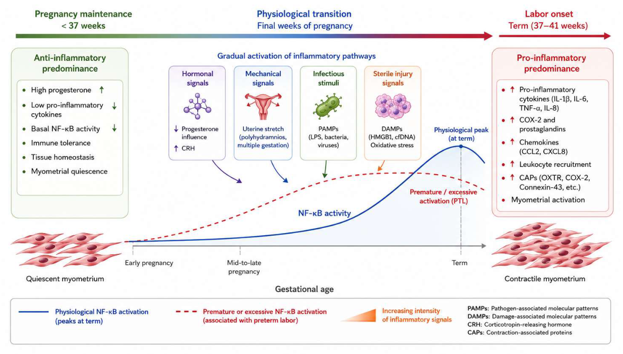
Figure 2.
Schematic overview of NF-κB pathway activation in preterm labor and modulation by natural compounds.
Figure 2.
Schematic overview of NF-κB pathway activation in preterm labor and modulation by natural compounds.
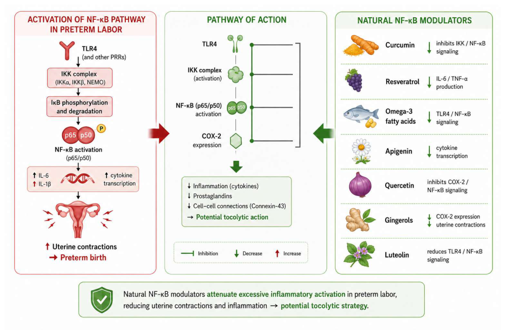
Figure 3.
PRISMA 2020 flow diagram of study selection process.
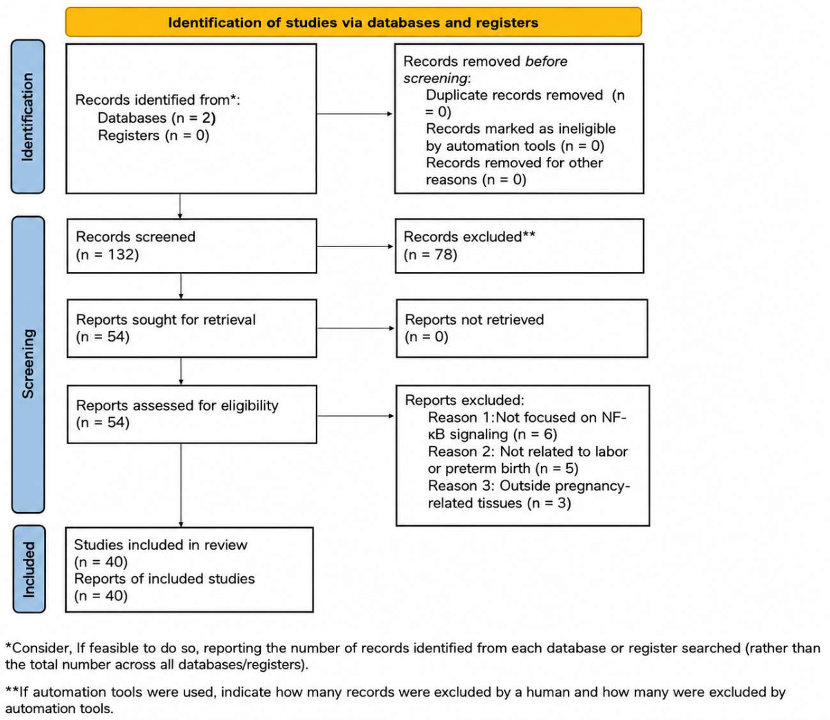

Table 1.
Key Cytokines, Chemokines, and Contraction-Associated Proteins (CAPs) in the Regulation of Labor.
Table 1.
Key Cytokines, Chemokines, and Contraction-Associated Proteins (CAPs) in the Regulation of Labor.
| Category | Key Molecules | Role in Labor |
| Proinflammatory Cytokines | IL-1β, IL-6, TNF-α | Promote myometrial contractions; induce COX-2 expression and prostaglandin synthesis |
| Chemokines | CXCL1, CXCL8, CCL2 | Mediate leukocyte recruitment to the myometrium and fetal membranes, amplifying the inflammatory response |
| Contraction-Associated Proteins (CAPs) | OXTR, Connexin-43, COX-2 | Enhance myometrial contractility and synchronize uterine contractions |
| Extracellular Matrix (ECM) Enzymes | MMP-9 | Remodel the extracellular matrix and weaken fetal membranes, facilitating labor onset |
Table 2.
Classification of NF-κB Modulators According to Mechanism of Action and Effects in Preterm Birth (PTB).
Table 2.
Classification of NF-κB Modulators According to Mechanism of Action and Effects in Preterm Birth (PTB).
| Category | Natural modulators | Synthetic modulators |
| Examples | Curcumin, Resveratrol, Omega-3 fatty acids, Apigenin, Quercetin, EGCG | SC-514, TPCA-1, Sulfasalazine, JQ1, MCC950 |
| Mechanism of action | Inhibition of NF-κB activation via suppression of IKK activity and TLR4/NF-κB signaling; reduction of pro-inflammatory cytokine production | Selective inhibition of IKKβ, NLRP3 inflammasome, and BET proteins; suppression of p65 phosphorylation and NF-κB transcriptional activity |
| Effects in PTB | Decreased expression of IL-1β, IL-6, TNF-α, and COX-2; attenuation of myometrial contractility | Reduced prostaglandin and cytokine production; delay or prevention of inflammation-induced preterm labor |
Table 3.
Classification of modulators according to their primary targets within the NF-κB signaling pathway.
Table 3.
Classification of modulators according to their primary targets within the NF-κB signaling pathway.
| Class of NF-κB Modulators | Mechanism of Action | Representative Compounds |
| IKK inhibitors | Inhibit IKKα/IKKβ-mediated phosphorylation of IκB, preventing its degradation and downstream NF-κB activation | TPCA-1, SC-514, BMS-345541, IMD-0354, Sulfasalazine, BAY 11-7082 |
| IκB stabilizers | Prevent proteasomal degradation of IκB, thereby retaining NF-κB in the cytoplasm | MG-132, Bortezomib, NBD peptide, aprotic amides (DEA, DPA, DMA) |
| NF-κB nuclear translocation inhibitors | Block nuclear import of p65/p50 complex or interfere with DNA binding to promoters | SN50 peptide, Parthenolide, CAPE, Curcumin, Resveratrol, MCC950 |
| TLR4 / inflammasome-related NF-κB modulators | Reduce upstream TLR4-mediated activation of NF-κB signaling | (+)-Naltrexone, (+)-Naloxone |
Disclaimer/Publisher’s Note: The statements, opinions and data contained in all publications are solely those of the individual author(s) and contributor(s) and not of MDPI and/or the editor(s). MDPI and/or the editor(s) disclaim responsibility for any injury to people or property resulting from any ideas, methods, instructions or products referred to in the content. |
© 2026 by the authors. Licensee MDPI, Basel, Switzerland. This article is an open access article distributed under the terms and conditions of the Creative Commons Attribution (CC BY) license (http://creativecommons.org/licenses/by/4.0/).
Copyright: This open access article is published under a Creative Commons CC BY 4.0 license, which permit the free download, distribution, and reuse, provided that the author and preprint are cited in any reuse.