

Submitted:
29 June 2026
Posted:
30 June 2026
You are already at the latest version
Abstract
Bacterial infections are a common occurrence in dental practice, often resulting in the formation of abscesses where anaerobic bacteria are predominant. Both oxygen and metronidazole are considered as effective agents for eliminating anaerobes. However, regardless of the dominance of anaerobes, abscesses will always be polymicrobial, also including facultative anaerobes such as Enterococcus faecalis. The aim of this study is to evaluate the efficacy of metronidazole in managing abscesses and to assess the potential of oxygen exposure to control E. faecalis activity. This study combines two complementary research approaches: (1) a study of medical records following metronidazole monotherapy for abscess treatment, and (2) an in-vitro analysis examining the impact of oxygen circulation on E. faecalis. Following metronidazole monotherapy, 93.33% of abscess cases demonstrated significant clinical improvement upon follow-up assessment. In vitro analysis indicated the absence of gas or odor production under oxygenated treatment conditions, whereas anaerobic conditions within the E. faecalis growth medium resulted in gas and odor formation. Enterococcus faecalis is a prevalent commensal bacterium commonly found in the human microbiota. Due to its inherent resilience, total elimination of E. faecalis is exceedingly challenging, and potentially unachievable. The complex anatomy of the root canal system, characterized by accessory canals and apical deltas, offers a protected niche for E. faecalis, making it inaccessible to conventional therapeutic interventions. Consequently, a paradigm shift may be necessary, transitioning from an eradication-based approach to a strategy centered on controlling the microorganism's proliferation and utilizing its commensal properties that also interact with the immune system to mitigate potential harm, rather than striving for absolute sterility.
Keywords:
metronidazole
; trepanation
; dental abscess
; enterococcus faecalis
; bacterial infection
1. Introduction
Dental infections in non-vital teeth tend to lead to pain due to pressure from gas and fluid on the periodontal tissues surrounding the infected teeth [1]. In cases of abscesses resulting from dental infections in non-vital teeth, obligate anaerobes dominate the polymicrobial environment of the abscess [2,3]. The antibiotic treatments employed to manage these infections are primarily aimed at eradicating anaerobic bacteria [4]. Metronidazole, with its efficacy in eliminating anaerobes, can serve as a therapeutic agent in managing the abscess [5,6]. Although clinicians often co-prescribe an additional antibiotic to enhance the efficacy and to broaden the spectrum, it is still rational to use metronidazole as monotherapy in a disease primarily caused by anaerobic bacteria [7,8]. Trepanation represents another strategy to deal with this kind of infection. A procedure of creating an access cavity through the tooth crown to connect the root canal system to the external environment of the tooth can establish drainage for the abscess as well as introduce oxygen to inhibit the proliferation of anaerobes [9]. Both the use of metronidazole and the trepanation technique are directed at the management of dental abscesses by specifically targeting anaerobic microorganisms [10,11,12]
Enterococcus faecalis is a bacterium frequently encountered in dentistry, recognized for its notorious resilience within the root canals of non-vital teeth [13,14]. As a facultative anaerobe, it possesses the ability to endure in the presence of oxygen [15,16]. While metronidazole and oxygen circulation demonstrate high efficacy against obligate anaerobes, their effectiveness in suppressing the growth of facultative anaerobes remains an area of ongoing investigation [17,18]. Numerous strategies in dental practice aim to eliminating E. faecalis to prevent and manage infections in non-vital teeth, including the use of adequate irrigants [19,20]. However, the hard-to-kill E. faecalis often persists, particularly colonizing the apical third of the root canal system [21,22]. On the other hand, E. faecalis is one of the most prevalent commensal species in the human body and considered non harmful, making its presence seem natural [23]. This study seeks to evaluate the standalone efficacy of metronidazole in managing abscesses and to assess the potential of oxygen exposure to control E. faecalis activity. A comparison and investigation into the differences regarding the condition of E. faecalis within the root canals of non-vital teeth, contrasting oxygen-deprived environments with those possessing oxygen circulation, has not been previously discussed in any scientific article. Therefore, this research is expected to provide novel information.
2. Materials and Methods
This study combines two complementary research approaches to evaluate the impact of an anaerobe-targeted therapy on facultative anaerobes. Specifically, the study comprises: (a) a study of dental patient records following metronidazole monotherapy for abscess treatment, and (b) an in-vitro analysis examining the impact of oxygen circulation on E. faecalis.
- a.
- Medical records study
This research did not conduct any treatment or intervention involving human subjects. The source of the data was medical records of patients in a healthcare facility in Indonesia. A total of 1,492 medical records of patients visiting for dental treatment before 2025 were evaluated. After careful examination, 45 cases from 45 patients were identified that fulfil the following criteria: (1) having an abscess/swelling problem, (2) prescribed with Metronidazole as monotherapy, and (3) completing a follow-up visit. Total sampling techniques were used in the analysis because the use of Metronidazole as monotherapy is relatively uncommon in dentistry and significantly lower than the use of other broad-spectrum antibiotics in treating dental diseases. The analysis focused on documented data within the medical records, specifically changes in swelling and patient-reported symptoms.
- b.
- In-vitro study
A total of 10 extracted human anterior teeth were measured to find relatively similar root lengths, resulting in 5 teeth with root lengths of approximately 17 mm. Four of these teeth were then cleaned to ensure the root canals were cleared up to the apex without any obstruction. The openings created in the crowns of the teeth during the cleaning were left open, and the cleaned root canals remained unfilled. These four teeth underwent three different treatments, where sterilization was performed prior to any treatments using an autoclave at 121 degrees Celsius with a pressure of 15 lbs psi for 20 minutes. The rationale for utilizing the same dry and sterile teeth for three different treatments was to ensure that each procedure was performed under identical tooth conditions, including the dimensions and length of the root canals. One tooth was used as a control and was only sterilized using the autoclave along with the other four teeth, without creating an opening in its crown for canal cleaning. Nutrient agar was prepared in petri dishes, which were subsequently sealed by wrapping them in plastic and subjected to vacuuming to create an anaerobic environment.
Group 1: Sterilized teeth were prepared by briefly heating the root portions over a spirit lamp to facilitate insertion into vacuum-sealed nutrient agar until at least 50% of the root length was embedded. The interface between the plastic and the embedded tooth root was then sealed with flowable composite resins to prevent oxygen diffusion into the agar through the interface (refer to Figure 1a). Subsequently, each tooth was inoculated with 10–20 μL of nutrient broth containing E. faecalis (refer to Figure 1b). Two teeth (A and B) were placed in a sealed, airtight container, relying solely on the initial oxygen concentration within the container (refer to Figure 1c). The remaining two teeth (C and D) were placed in a container equipped with a filtered vent (surgical mask material) to allow for restricted air exchange (refer to Figure 1d).
Group 2: The procedure described for Group 1 was replicated, with the exception that teeth A and B were transposed with teeth C and D.
Group 3: Following inoculation with nutrient broth, a spatula was used to gently tap the cervical regions of teeth A, B, C, and D. This action was performed to remove any residual liquid at the coronal aspect prior to sealing, and to ensure sufficient space for composite resins application (refer to Figure 1e). Subsequently, all teeth were placed within sealed containers containing oxygen absorbers (refer to Figure 1f). The containers were carefully closed to eliminate any air gaps and prevent air circulation. The inclusion of oxygen absorbers was intended to scavenge any remaining oxygen and thereby promote a more anaerobic environment.
An unmodified tooth with an intact crown served as a control and was included in each group. This control tooth was affixed within the agar and sealed at the interface between the plastic and the root wall using composite resins. The portion of the tooth not embedded in the agar was enclosed by plastic walls, allowing for immersion in nutrient broth. The control tooth was placed within a sealed, non-ventilated container (refer to Figure 1g and Figure 1h). This procedure was implemented to maintain sterility within the agar throughout the monitoring period for the sterilized control tooth, which was not subjected to nutrient broth inoculation into its root canal. The nutrient broth surrounding the cervical portion of the control tooth served to verify the integrity of the composite resins seal, confirming the maintenance of a sealed environment within the petri dishes throughout the study.
3. Results
A study of the use of Metronidazole as monotherapy, and in vitro investigations of E. faecalis under oxygen exposure, reveals two concordant findings.
- a.
- Medical records study
Following monotherapy with metronidazole in 45 abscess cases attending follow-up, 93.33% (42 cases) exhibited significant clinical improvement. Conversely, 6.67% (3 cases) demonstrated persistent swelling without noticeable improvement and continued to report pre-treatment symptoms. Among the 42 cases demonstrating significant improvement at follow-up, 95.24% (40 cases) reported complete resolution of symptoms, including the absence of discomfort upon percussion or pressure on the affected tooth. However, 4.76% (2 cases) reported improved conditions, but some discomfort still remained (refer to Table 1).
- b.
- In-vitro study
Following 72 hours of incubation, Groups 1 and 2 exhibited comparable results. The vacuum-sealed plastic wrapping the petri dishes displayed no appreciable alteration from its pre-incubation state. Upon breaching the seals, all agar plates, including control samples, were found to be desiccated, and no discrete white colonies suggestive of bacterial proliferation were observed (refer to Figure 3a and Figure 3b).
In Group 3, the vacuum-formed plastic demonstrated a noticeable deviation from the control group. Specifically, the plastic exhibited reduced tension and evidence of entrapped gas or vapor pockets (see Figure 4a–4d). This contrasted with the relatively stable and consistent appearance of the vacuum-formed plastic observed on the control tooth (see Figure 4e). After the removal of the plastic, an odor unique to Group 3 specimens (excluding the control tooth), was detected by the operator. This phenomenon did not occur during the initial opening of boxes and plastic in Groups 1 and 2.
To determine the presence of viable bacteria in the moistened region, samples were obtained using sterile cotton swabs and subsequently transferred to fresh nutrient agar (refer to Figure 5a). Following a 24-hour incubation period, bacterial growth was observed on all agar plates inoculated from the four plates in Group 3 (refer to Figure 5bFigure –Figure 5e).
4. Discussion
Metronidazole’s efficacy against anaerobic bacteria is well-established, with a long history of clinical use. However, its effectiveness against facultative anaerobic organisms is less certain [18,24,25,26]. Facultative anaerobic bacteria, including E. faecalis, are implicated in the pathogenesis of dental abscesses [27], and are consistently present in non-vital tooth root canals [28]. Infected non-vital teeth exhibit a combination of aerobic and anaerobic infections, characterized by mixed bacterial populations [29]. Anaerobic bacteria are the predominant microorganisms isolated from both odontogenic abscesses (including those of endodontic origin) and non-odontogenic abscesses unrelated to dental pathology. However, facultative anaerobes that are consistently present reflect the polymicrobial etiology of these infections.
A medical records study detailed in this article demonstrated the sustained efficacy of metronidazole in treating over 90% of abscess cases, even in the absence of adjunctive broad-spectrum antibiotics typically preferred for facultative anaerobes. Metronidazole, a nitroimidazole prodrug, shows its efficacy against anaerobic bacteria through reductive activation under hypoxic conditions and induces oxidative stress within the cell [30,31]. Its recognized efficacy in the treatment of dental abscesses stems from the predominance of obligate anaerobic species within these infectious lesions [3,32]. Clinical data support the capacity of metronidazole to alleviate abscess-related symptomatology, such as pain, edema, and purulent exudate, via the elimination of these anaerobes, which thrive in the oxygen-deprived milieu of non-vital dental pulp [6,33]. The observed success of metronidazole monotherapy, as evidenced by the medical records study presented herein, suggests that selectively targeting the anaerobic component may be sufficient to achieve resolution, even in the presence of persistent facultative anaerobes such as E. faecalis. This phenomenon is well-recognized within the realm of general medical knowledge, given that E. faecalis is among the most prevalent commensal organisms in the human body and is capable of coexisting with the host without inducing adverse effects under normal physiological conditions [23].
Oxygen, like metronidazole, is effective in controlling anaerobic organisms but is tolerated by facultative anaerobes. Dental trepanation can also be used to allow oxygen to circulate within the root canal of a non-vital tooth, in addition to serving as an exit point for abscesses if present around the non-vital tooth [10]. The presence of oxygen disturbs anaerobic organisms that thrive in the absence of oxygen. Oxygen exposure can disrupt the normal physiological processes within these anaerobic cells, leading to impaired functionality. It can also cause cell lysis where the membranes of anaerobes rupture and disintegrate [34].
The in vitro study described in this article is not designed as a preliminary investigation for clinical application in humans but rather as an effort to elucidate phenomena already observed in dental practice by replicating them under controlled laboratory conditions. A case report on the clinical application of dental trepanation demonstrated positive outcomes in the prevention of odontogenic abscesses, which frequently arise in non-vital teeth. Notably, these results were achieved despite the treatment’s primary focus being limited to anaerobic bacteria, without being hindered by the inherent challenges of dental procedures, where the complete eradication of E. faecalis is notoriously challenging, if not unachievable [9,10]. The detection of gas and odor, exclusively observed in in vitro analyses of Group 3 and absent in Groups 1 and 2 under oxygenated conditions, acts as the focal point during the observation. Gas and odor are clear signs indicating the extensive growth of bacteria [35]. This observation also aligns with clinical data documented in the case study on dental trepanation, which reported the absence of abscess formation for a minimum of two years as a result of oxygen exposure to the root canal facilitated by the trepanation procedure [9]. The root canal’s anatomy—featuring accessory canals and apical deltas—provides a sanctuary for E. faecalis, inaccessible to treatment [36,37]. This reality can shift the focus from eradication to control, leveraging its commensal nature to minimize harm rather than pursuing sterility.
5. Conclusions
Metronidazole and oxygen are both effective in inhibiting and eliminating the growth of anaerobes. While complete eradication of facultative anaerobes, such as E. faecalis, is challenging, oxygen exposure is expected to reduce its pathogenicity, promoting coexistence under aerobic conditions. The formation of gas and odor in the group subjected to anaerobic conditions, serves as the focal point during the observation in this research. The efficacy of Metronidazole monotherapy in treating dental abscess also indicates that targeting anaerobes in dental infections cases where obligate anaerobes predominating the environment can be the first concern of clinicians and scientists. This perspective can support a new strategy, shifting the direction from complete eradication of E. faecalis to controlling its commensal behavior.
Further studies are required to understand why distinct white spots, which are clear indicators of bacterial growth, were not observed on any medium across all groups. To make further research more aligned with clinical conditions, it should include not only facultative anaerobes, but also anaerobes which tend to predominate in dental abscesses. Hypothetically, incorporating anaerobes into further research would likely provide even more significant positive results regarding oxygen as a controller of bacterial growth compared to using only one type of facultative anaerobic bacteria. Future studies on Metronidazole as oral monotherapy in treating dental abscess should be explored as well. Clinicians tend to use broader spectrum antibiotics or to co-prescribe another antibiotic in the management of dental infections, making the available data regarding the Metronidazole monotherapy limited. Clinical trials are necessary to assess the efficacy of Metronidazole monotherapy compared to other antibiotic regimens, as also suggested by other researchers [8].
Author Contributions
All authors contributed equally to this work.
Funding
This work received no external funding.
Institutional Review Board Statement
Not applicable.
Data Availability Statement
No new data were created.
Acknowledgments
The authors express their sincere gratitude to the Head of the Microbiology Laboratory, Faculty of Pharmacy, Bhakti Kencana University, for insightful discussions related to the in-vitro study. The authors also acknowledge Dr. Dordia Anindita Rotinsulu, School of Veterinary Medicine and Biomedical Sciences, IPB University, for providing valuable technical assistance in the preparation of the in-vitro study. Finally, the authors extend their appreciation to Prof. Reidar K. Lie (University of Bergen, Bergen, Norway) for his support in enriching the discussion regarding trepanation.
Conflicts of Interest
The authors declare no conflict of interest.
References
- NAIR; Ramanchandran, P.N. Pathogenesis of apical periodontitis and the causes of endodontic failures. Crit. Rev. Oral Biol. Med. 2004, 15.6, 348–381. [Google Scholar] [CrossRef] [PubMed]
- SIQUEIRA JR; José, F.; RÔÇAS; Isabela, N. Clinical implications and microbiology of bacterial persistence after treatment procedures. J. Endod. 2008, 34.11, 1291–1301. e3. [Google Scholar] [CrossRef] [PubMed]
- BROOK, Itzhak. Microbiology and management of endodontic infections in children. J. Clin. Pediatr. Dent. 2004, 28.1, 13–17. Available online: https://oss.jocpd.com/files/article/20220907-1322/pdf/JOCPD28.1.13.pdf. [CrossRef]
- SLOTS, Jørgen. Selection of antimicrobial agents in periodontal therapy. J. Periodontal Res. 2002, 37.5, 389–398. [Google Scholar] [CrossRef] [PubMed]
- TAJ, Shifa; et al. Metronidazole Induced Cutaneous Adverse Drug Reaction-A Systematic Review of Descriptive Studies. Curr. Rev. Clin. Exp. Pharmacol. Former. Curr. Clin. Pharmacol. 2024, 19.3, 269–284. [Google Scholar] [CrossRef] [PubMed]
- LÖFMARK, Sonja; EDLUND, Charlotta; NORD, Carl Erik. Metronidazole is still the drug of choice for treatment of anaerobic infections. Clin. Infect. Dis. 2010, 50. Supplement_1, S16–S23. [Google Scholar] [CrossRef] [PubMed]
- KURIYAMA, T.; et al. Antimicrobial susceptibility of 800 anaerobic isolates from patients with dentoalveolar infection to 13 oral antibiotics. Oral Microbiol. Immunol. 2007, 22.4, 285–288. [Google Scholar] [CrossRef] [PubMed]
- DAR-ODEH, Najla; et al. Evaluation of metronidazole oral monotherapy in anaerobic oral infections. Int. J. Clin. Pharmacol. Ther. 2024, 62.11, 517. [Google Scholar] [CrossRef] [PubMed]
- WAHYUDI, Didi; LUBIS, Leonardo. Replantation of an avulsed-tooth by using trepanation: A Case Report. J. Clin. Cas. Rep. Med. Imag. Heal. Sci. 2024, 8, 1340. [Google Scholar] [CrossRef]
- WAHYUDI, Didi; KUSUMASARI, Citra. Oxygen as obturation biomaterial in endodontic treatment: development of novel membranous dental restoration system. F1000Research 2024, 12, 380. [Google Scholar] [CrossRef] [PubMed]
- KOMINE, Chiaki; et al. Application of reactive oxygen species in dental treatment. J. Pers. Med. 2022, 12.9, 1531. [Google Scholar] [CrossRef] [PubMed]
- IMLAY, James A. The molecular mechanisms and physiological consequences of oxidative stress: lessons from a model bacterium. Nat. Rev. Microbiol. 2013, 11.7, 443–454. [Google Scholar] [CrossRef] [PubMed]
- YANG, Shipeng; et al. Strategies and mechanisms targeting Enterococcus faecalis biofilms associated with endodontic infections: a comprehensive review. Front. Cell. Infect. Microbiol. 2024, 14, 1433313. [Google Scholar] [CrossRef] [PubMed]
- RÔÇAS, I. N.; SIQUEIRA JR, J. F. Root canal microbiota of teeth with chronic apical periodontitis. J. Clin. Microbiol. 2008, 46.11, 3599–3606. [Google Scholar] [CrossRef] [PubMed]
- FISHER, Katie; PHILLIPS, Carol. The ecology, epidemiology and virulence of Enterococcus. Microbiology 2009, 155.6, 1749–1757. [Google Scholar] [CrossRef] [PubMed]
- LOKITHA, R.; et al. Antimicrobial therapeutic strategies for Enterococcus Faecalis in dental infections-past, present and future. Anti-Infect. Agents 2023, 21.3, 1–11. [Google Scholar] [CrossRef]
- DINGSDAG, Simon A.; HUNTER, Neil. Metronidazole: an update on metabolism, structure–cytotoxicity and resistance mechanisms. J. Antimicrob. Chemother. 2018, 73.2, 265–279. [Google Scholar] [CrossRef] [PubMed]
- GAETTI-JARDIM JÚNIOR, Elerson; et al. Susceptibility of strict and facultative anaerobes Isolated from endodontic infections to metronidazole and beta-lactams. J. Appl. Oral Sci. 2007, 15, 539–545. [Google Scholar] [CrossRef] [PubMed]
- OROZCO-GALLEGO, Manuel J.; et al. Effectiveness of irrigation protocols in endodontic therapy: An umbrella review. Dent. J. 2025, 13.6, 273. [Google Scholar] [CrossRef] [PubMed]
- GENC SEN, Ozgur; ERDEMIR, Ali. The effectiveness of three different irrigant activation methods in the elimination of Enterococcus faecalis from root canals. Photobiomodulation Photomed. Laser Surg. 2024, 42.10, 628–633. [Google Scholar] [CrossRef] [PubMed]
- DE LIMA, Francine Santos Fernandes; FACURY, Analia Gabriella Borges Ferraz. Enterococcus faecalis and Endodontic Treatment Failure: A Qualitative Systematic Review of Its Role and Elimination Strategies. Aust. Endod. J. 2026. [Google Scholar] [CrossRef] [PubMed]
- SUNDQVIST, Göran; et al. Microbiologic analysis of teeth with failed endodontic treatment and the outcome of conservative re-treatment. Oral Surgery, Oral Medicine, Oral Pathology, Oral Radiology, and Endodontology 1998, 85.1, 86–93. [Google Scholar] [CrossRef] [PubMed]
- KRISHNA; Kondapalli, Vamsi; et al. Enterococcus species and their probiotic potential: Current status and future prospects. J. Appl. Biol. Biotechnol. 2022, 11, 36–44. [Google Scholar] [CrossRef]
- NIX, Ned Leonard; ZUSMAN, Nicholas T.; DÜZGÜNEŞ, Nejat. Odontogenic Infections Resistant to Empiric Therapy, Opportunistic Prevotella, and Metronidazole: A Clinical Case Series and Review of the Literature. Oral 2025, 5.1: 17. [Google Scholar] [CrossRef]
- SHAKYA, Neelam; et al. Epidemiology, microbiology and antibiotic sensitivity of odontogenic space infections in Central India. J. Maxillofac. Oral Surg. 2018, 17, 324–331. [Google Scholar] [CrossRef] [PubMed]
- CHAMORRO-PETRONACCI; Cintia, Micaela; et al. Efficacy of ciprofloxacin, metronidazole and minocycline in ordered mesoporous silica against Enterococcus faecalis for dental pulp revascularization: an in-vitro study. Materials 2022, 15.6, 2266. [Google Scholar] [CrossRef] [PubMed]
- RAMADAN, Asmaa; et al. Fully characterized effective bacteriophages specific against antibiotic-resistant Enterococcus faecalis, the Causative Agent of Dental Abscess. Medicina 2024, 60.3, 501. [Google Scholar] [CrossRef] [PubMed]
- WONG, Jasmine; et al. Microbiological aspects of root canal infections and disinfection strategies: an update review on the current knowledge and challenges. Front. Oral Health 2021, 2, 672887. [Google Scholar] [CrossRef] [PubMed]
- OLSEN, Ingar; DAHLÉN, Gunnar. Salient virulence factors in anaerobic bacteria, with emphasis on their importance in endodontic infections. Endod. Top. 2004, 9.1, 15–26. [Google Scholar] [CrossRef]
- FREEMAN, Collin D.; KLUTMAN, Neil E.; LAMP, Kenneth C. Metronidazole: a therapeutic review and update. Drugs 1997, 54, 679–708. [Google Scholar] [CrossRef] [PubMed]
- FIEDLER, Michaela K.; et al. Metronidazole and ether derivatives target Helicobacter pylori via simultaneous stress induction and inhibition. Nat. Microbiol. 2026, 1–15. [Google Scholar] [CrossRef] [PubMed]
- BROOK, Itzhak. Microbiology of polymicrobial abscesses and implications for therapy. J. Antimicrob. Chemother. 2002, 50.6, 805–810. [Google Scholar] [CrossRef] [PubMed]
- KURIYAMA, Tomoari; et al. Bacteriologic features and antimicrobial susceptibility in isolates from orofacial odontogenic infections. Oral Surgery, Oral Medicine, Oral Pathology, Oral Radiology, and Endodontology 2000, 90.5, 600–608. [Google Scholar] [CrossRef] [PubMed]
- PEDIZZI, Chiara; et al. Effect of oxygen on the microbial activities of thermophilic anaerobic biomass. Bioresour. Technol. 2016, 211, 765–768. [Google Scholar] [CrossRef] [PubMed]
- YAMADA, Yoko; et al. Association of odor from infected root canal analyzed by an electronic nose with isolated bacteria. J. Endod. 2007, 33.9, 1106–1109. [Google Scholar] [CrossRef] [PubMed]
- AHMED, H. M. A.; NEELAKANTAN, P.; DUMMER, P. M. H. A new system for classifying accessory canal morphology. Int. Endod. J. 2018, 51.2, 164–176. [Google Scholar] [CrossRef] [PubMed]
- DZANKOVIC, Aida; et al. Endodontic Challenges Arising from Root Canal Morphology. In Human Teeth-The Intersection of Science, Disease, and Clinical Practice; IntechOpen, 2024. [Google Scholar] [CrossRef]
Figure 1.
1a. Composite resins bonded to the interface between the plastic and the root of the tooth. 1b. Tooth root canal exhibiting contamination with E. faecalis. 1c. Hermetically sealed container lacking air circulation. 1d. Container featuring a cavity covered with a surgical mask to allow for air circulation. 1e. Composite resins utilized for sealing the access cavity within the tooth crown. 1f. Oxygen absorber employed to remove oxygen within the container. 1g. Plastic barriers enclosing the coronal aspect of the tooth to contain fluids. 1h. A hermetically sealed container serving as a control.
Figure 1.
1a. Composite resins bonded to the interface between the plastic and the root of the tooth. 1b. Tooth root canal exhibiting contamination with E. faecalis. 1c. Hermetically sealed container lacking air circulation. 1d. Container featuring a cavity covered with a surgical mask to allow for air circulation. 1e. Composite resins utilized for sealing the access cavity within the tooth crown. 1f. Oxygen absorber employed to remove oxygen within the container. 1g. Plastic barriers enclosing the coronal aspect of the tooth to contain fluids. 1h. A hermetically sealed container serving as a control.
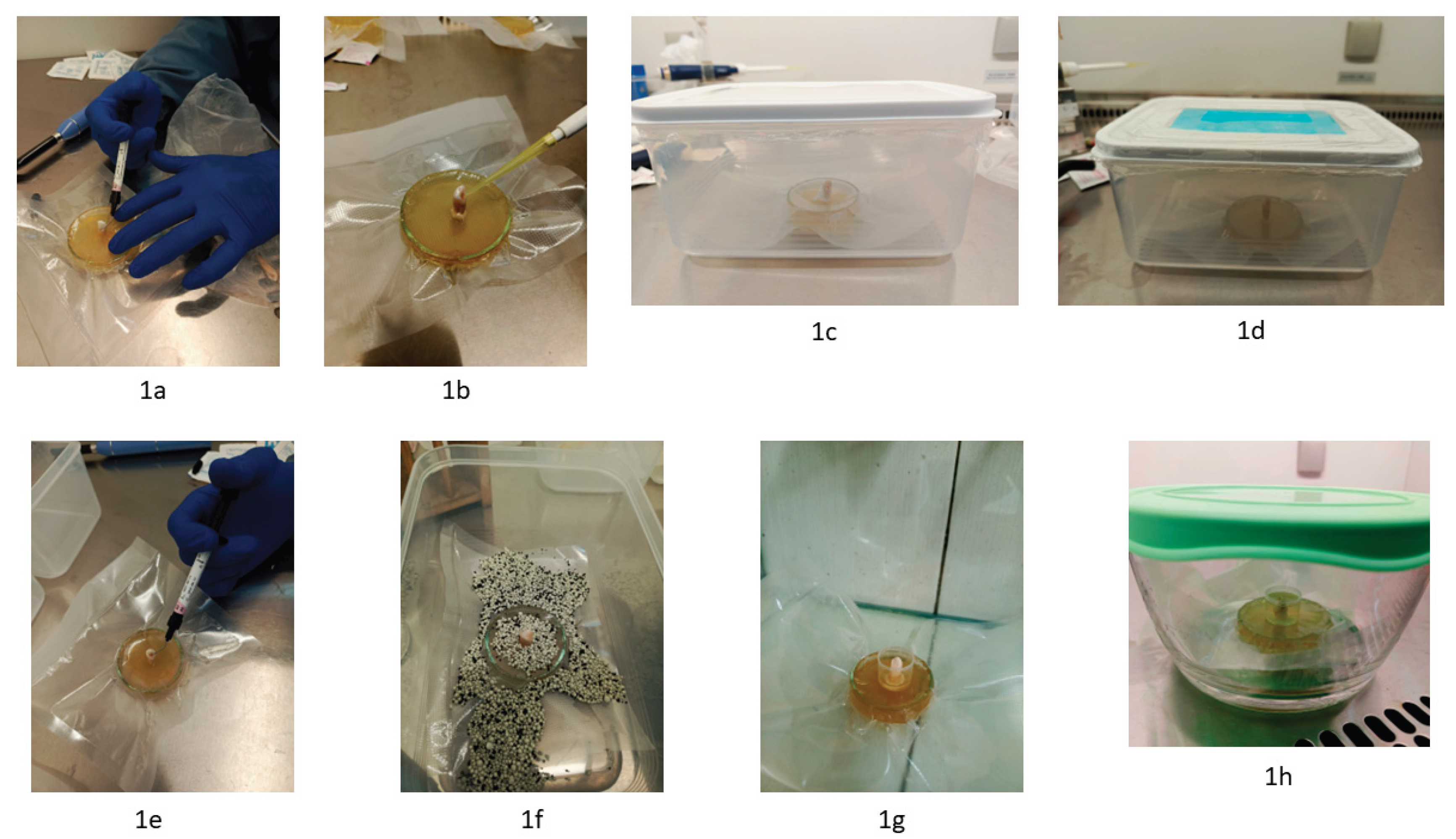
Figure 2.
2a. The incubator contained two teeth maintained in a static air environment within a sealed container, two teeth in a container with forced air circulation, and a single control tooth. 2b. The incubator contained four teeth maintained in a static air environment within a sealed container supplemented with an oxygen absorber to reduce internal oxygen levels, and a single control tooth.
Figure 2.
2a. The incubator contained two teeth maintained in a static air environment within a sealed container, two teeth in a container with forced air circulation, and a single control tooth. 2b. The incubator contained four teeth maintained in a static air environment within a sealed container supplemented with an oxygen absorber to reduce internal oxygen levels, and a single control tooth.
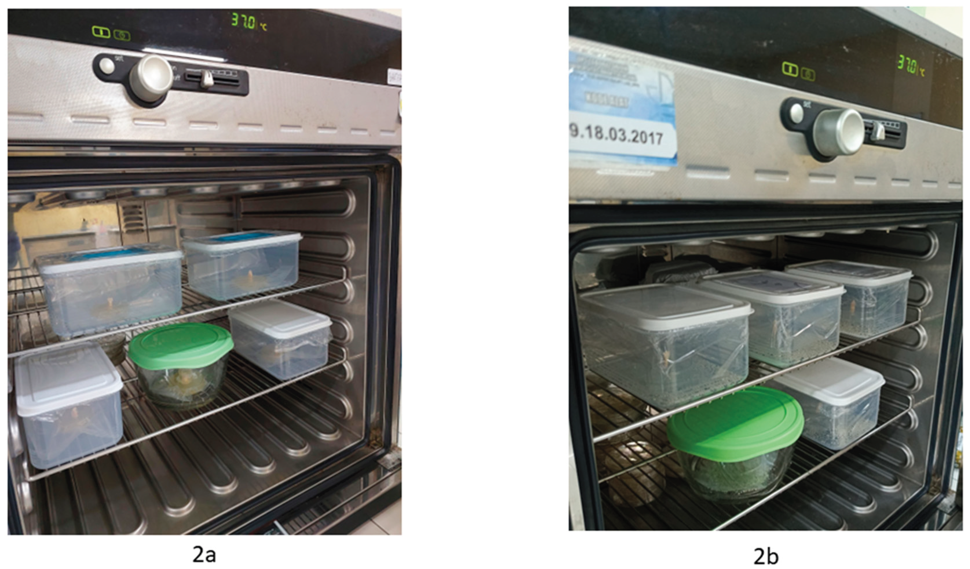
Figure 3.
3a. The agar plates in Group 1 exhibited relatively consistent characteristics, and a representative plate was selected for imaging. 3b. The agar plates in Group 2 exhibited relatively consistent characteristics, and a representative plate was selected for imaging.
Figure 3.
3a. The agar plates in Group 1 exhibited relatively consistent characteristics, and a representative plate was selected for imaging. 3b. The agar plates in Group 2 exhibited relatively consistent characteristics, and a representative plate was selected for imaging.
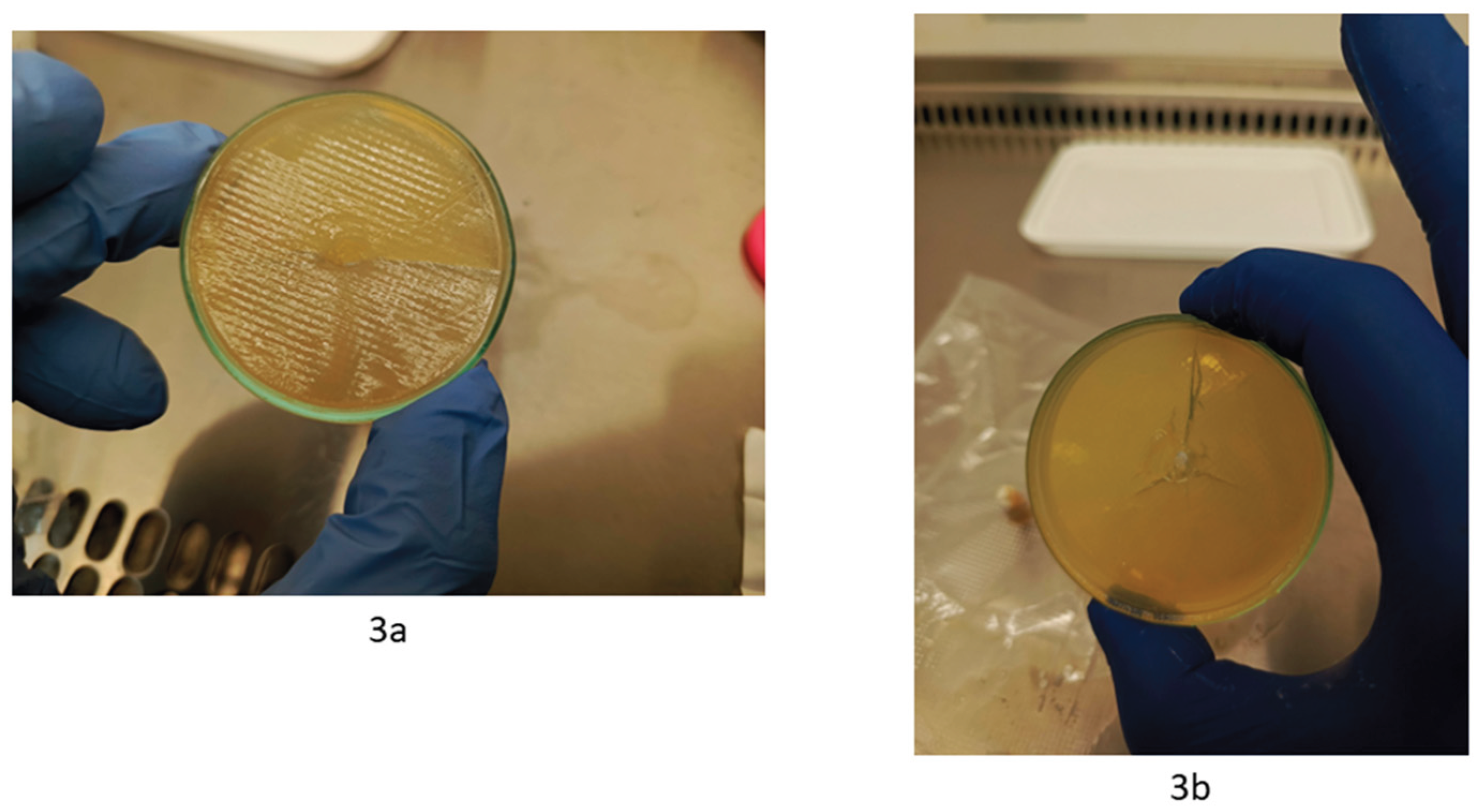
Figure 4.
4a–4d. The plastic demonstrates reduced rigidity and exhibits indications of vapor condensation. 4e. The plastic in the control sample remains unaffected, retaining its structural integrity with negligible changes compared to its pre-incubation state.
Figure 4.
4a–4d. The plastic demonstrates reduced rigidity and exhibits indications of vapor condensation. 4e. The plastic in the control sample remains unaffected, retaining its structural integrity with negligible changes compared to its pre-incubation state.
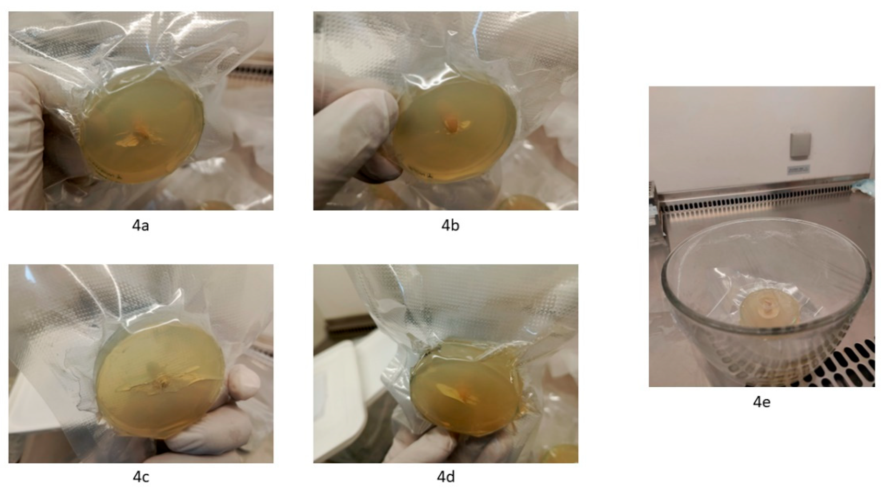
Figure 5.
5a. Nutrient agar exhibiting a more moist appearance was subsequently swabbed. 5b–5e. Bacterial growth was observed consistently across all swabbed moist agar plates.
Figure 5.
5a. Nutrient agar exhibiting a more moist appearance was subsequently swabbed. 5b–5e. Bacterial growth was observed consistently across all swabbed moist agar plates.
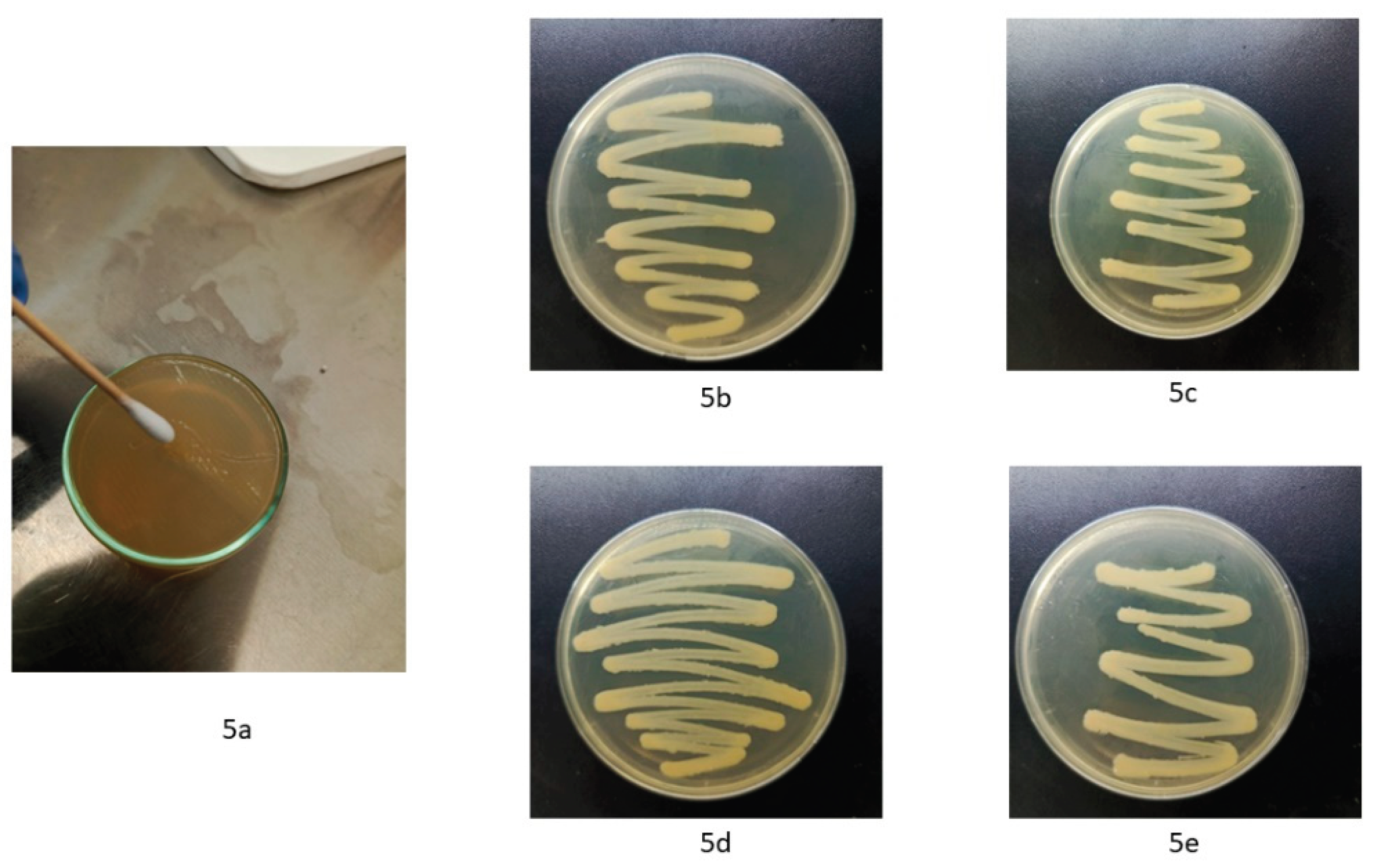

Table 1.
Evaluation of metronidazole use in abscess cases.
| Case | Age | Sex | Follow-up record |
|---|---|---|---|
| 1 | 80 | M | The condition has improved, with some complaints recorded |
| 2 | 27 | M | The condition has clearly improved, with zero complaints recorded |
| 3 | 45 | F | The condition has clearly improved, with zero complaints recorded |
| 4 | 42 | M | The condition has clearly improved, with zero complaints recorded |
| 5 | 28 | F | The condition has clearly improved, with zero complaints recorded |
| 6 | 59 | M | The condition has clearly improved, with zero complaints recorded |
| 7 | 55 | F | The condition has clearly improved, with zero complaints recorded |
| 8 | 52 | F | The condition has clearly improved, with zero complaints recorded |
| 9 | 44 | F | The condition has clearly improved, with zero complaints recorded |
| 10 | 55 | F | The condition has improved, with some complaints recorded |
| 11 | 27 | F | The condition has clearly improved, with zero complaints recorded |
| 12 | 45 | M | The condition has clearly improved, with zero complaints recorded |
| 13 | 45 | M | The condition has clearly improved, with zero complaints recorded |
| 14 | 56 | M | The condition has not significantly improved, with complaints largely remaining the same as the previous visit |
| 15 | 8 | M | The condition has clearly improved, with zero complaints recorded |
| 16 | 9 | M | The condition has clearly improved, with zero complaints recorded |
| 17 | 57 | M | The condition has clearly improved, with zero complaints recorded |
| 18 | 26 | F | The condition has clearly improved, with zero complaints recorded |
| 19 | 34 | M | The condition has clearly improved, with zero complaints recorded |
| 20 | 26 | F | The condition has clearly improved, with zero complaints recorded |
| 21 | 63 | F | The condition has clearly improved, with zero complaints recorded |
| 22 | 33 | F | The condition has clearly improved, with zero complaints recorded |
| 23 | 8 | F | The condition has clearly improved, with zero complaints recorded |
| 24 | 34 | F | The condition has clearly improved, with zero complaints recorded |
| 25 | 69 | F | The condition has clearly improved, with zero complaints recorded |
| 26 | 42 | F | The condition has clearly improved, with zero complaints recorded |
| 27 | 34 | F | The condition has not significantly improved, with complaints largely remaining the same as the previous visit |
| 28 | 28 | F | The condition has clearly improved, with zero complaints recorded |
| 29 | 35 | F | The condition has clearly improved, with zero complaints recorded |
| 30 | 40 | F | The condition has clearly improved, with zero complaints recorded |
| 31 | 48 | F | The condition has clearly improved, with zero complaints recorded |
| 32 | 27 | M | The condition has clearly improved, with zero complaints recorded |
| 33 | 61 | F | The condition has clearly improved, with zero complaints recorded |
| 34 | 30 | M | The condition has clearly improved, with zero complaints recorded |
| 35 | 54 | F | The condition has clearly improved, with zero complaints recorded |
| 36 | 63 | M | The condition has not significantly improved, with complaints largely remaining the same as the previous visit |
| 37 | 3 | F | The condition has clearly improved, with zero complaints recorded |
| 38 | 46 | M | The condition has clearly improved, with zero complaints recorded |
| 39 | 66 | F | The condition has clearly improved, with zero complaints recorded |
| 40 | 51 | M | The condition has clearly improved, with zero complaints recorded |
| 41 | 40 | F | The condition has clearly improved, with zero complaints recorded |
| 42 | 21 | M | The condition has clearly improved, with zero complaints recorded |
| 43 | 33 | M | The condition has clearly improved, with zero complaints recorded |
| 44 | 50 | F | The condition has clearly improved, with zero complaints recorded |
| 45 | 43 | M | The condition has clearly improved, with zero complaints recorded |
Disclaimer/Publisher’s Note: The statements, opinions and data contained in all publications are solely those of the individual author(s) and contributor(s) and not of MDPI and/or the editor(s). MDPI and/or the editor(s) disclaim responsibility for any injury to people or property resulting from any ideas, methods, instructions or products referred to in the content. |
© 2026 by the authors. Licensee MDPI, Basel, Switzerland. This article is an open access article distributed under the terms and conditions of the Creative Commons Attribution (CC BY) license (http://creativecommons.org/licenses/by/4.0/).
Copyright: This open access article is published under a Creative Commons CC BY 4.0 license, which permit the free download, distribution, and reuse, provided that the author and preprint are cited in any reuse.