

Submitted:
29 June 2026
Posted:
30 June 2026
You are already at the latest version
Abstract
Bacterial and fungal skin infection are common in companion animals and often require antimicrobial therapy, contributing to antimicrobial resistance. This study developed shampoos containing red palm oil (RPO) and extracts of Rhinacanthus nasutus (RN), Curcuma longa (CL), Zingiber montanum (ZM), and Zingiber officinale (ZO), which provide antibacterial, antifungal, antioxidant, and anti-inflammatory activities. Extracts were obtained using MAE and were evaluate for phytochemical composition, total phenolic content, antioxidant activity (DPPH assay), anti-inflammatory activity (COX-2 inhibition), and antimicrobial activity. RPO contained high level of vitamin E and β-carotene. Rhinacanthin, curcumin, terpinen-4-ol, and 6-gingerol were the major phytochemical constituents of RN, CL, ZM, and ZO extracts, respectively. All extracts significantly reduced COX-2 expression (p< 0.05) while RN exhibited the strongest antioxidant activity (DPPH IC₅₀ = 15.23 ± 0.53 µg/mL). RN and ZM demonstrated the strongest antibacterial activity against Staphylococcus aureus and methicillin-resistant Staphylococcus aureus (MIC = 8 µg/mL, MBC = 8–16 µg/mL). ZO exhibited the strongest antifungal activity against Malassezia pachydermatis and Microsporum gypseum (MIC = 2 µg/mL, MFC = 2 - 16 µg/mL). The developed shampoos, containing RPO and four herbs’ extracts showed suitable pH values, stable viscosity, antibacterial inhibition and antifungal inhibition (p < 0.05).
Keywords:
red palm oil
; Rhinacanthus nasutus
; Curcuma longa
; Zingiber montanum
; Zingiber officinale
; antimicrobial activity
; COX-2 inhibition
; veterinary dermatology
1. Introduction
Recently, the number of pets, in particular dogs and cats, have steady increases [1] because of the positive effects on human mental health [2] and regard pets as family members [3,4,5]. However, pets are often associated with skin diseases [6,7,8] caused by bacteria, fungi, and yeasts for example Staphylococcus spp., Microsporum spp., and Malassezia spp. [9,10]. These microorganisms can be transmitted from animals to humans and may also spread in the environment [11,12,13,14]. To treat these skin diseases, veterinarians commonly use antibiotics and antifungal drugs on systemic or topical antimicrobial agents as shampoo for long term [15,16]. However, the use of these medications may lead to drug toxicity and antimicrobial resistance (AMR) [17,18], which has become a major global concern in both human and veterinary medicine [19]. Therefore, alternative approaches for the management of skin diseases in companion animals are needed. Natural products that are safe and effective may help reduce the use of conventional antibiotics and antifungal drugs [20,21,22]. Although herbal shampoos are commercially available, most formulations are primarily intended for ectoparasite control rather than for the management of microbial skin infections [23,24]. A shampoo formulated with red palm oil (RPO) and Thai herbal extracts may be a promising alternative veterinary skin-care products. Red palm oil, the main ingredient of the shampoo, contains high levels of β-carotene, vitamin A, and vitamin E, which have antioxidant and skin-protective properties [25,26,27,28]. In addition, RPO has antimicrobial activity against Gram-positive bacteria, which are important causes of skin diseases in pets [25]. Furthermore, the palm oil industry produces by-products such as kernel cake and palm shell, which are currently underutilized [29]. The use of palm oil-derived products in veterinary applications may help reduce waste and promote sustainable production. Therefore, the development of pet shampoo containing RPO and Thai herbal extracts may provide a natural, safe, and sustainable option for veterinary dermatological care.
Rhinacanthus nasutus; RN (Thong phan chang), Curcuma longa; CL (Turmeric), Zingiber montanum; ZM (Cassumunar ginger), and Zingiber officinale; ZO (Ginger) are Thai medicinal plants that contain bioactive compounds with established antimicrobial, antioxidant, and anti-inflammatory activities [30,31,32,33]. Previous investigations have largely focused on individual herbal extracts, primarily in human medicine or as oral supplements in animals [34,35,36,37]. To our knowledge, no study has reported the incorporation the best of these four Thai herbal extracts in combination with RPO into a shampoo formulation intended for companion animal skin care. The incorporation of Thai herbal extracts into shampoo formulations may improve the effectiveness of the shampoo for the treatment of skin diseases in companion animals. In addition, it may help reduce the use of antibiotics and antifungal drugs, thereby reducing the risk of drug-related side effects.
Microwave-assisted extraction (MAE) is a suitable technique for extracting bioactive compounds from medicinal plants and lipid-based materials [38,39]. Compared with conventional extraction methods, such as maceration and Soxhlet extraction, MAE method provides high extraction yields, requires less solvent, and helps protect heat-sensitive compounds [40,41,42]. In addition, MAE is an environmentally friendly extraction technique with extraction technology with applications in pharmaceutical, cosmetic, and veterinary product development [43,44,45,46,47]
Therefore, this study hypothesized that the combination of RPO and selected Thai herbal extracts would provide antimicrobial, antifungal, antioxidant, and anti-inflammatory activities suitable for topical application in companion animals to develop plant-based, environmentally friendly, and sustainable alternatives for veterinary dermatological care. This study aimed to (1) extract bioactive compounds from RPO and selected Thai medicinal plants using MAE; (2) characterize the chemical and biological properties of the extracts and determine their appropriate concentrations for shampoo formulation and (3) develop and evaluate an RPO-based herbal shampoo with potential applications for companion animal skin care.
2. Materials and Methods
2.1. Ethical Considerations
All experiments were conducted entirely in vitro accordance with institutional biosafety regulations and standard laboratory practice guidelines and did not involve any live animals subjects or clinical samples. The microbial strains used in this study were established and previously identified laboratory isolates. Therefore, approval from an animal ethics and biosafety committee was not required for this study.
2.2. Study Period and Location
Palm fruit residues were obtained from the Tha Sa Ba Oil Palm Enterprise Group, Trang Province, Thailand. Thai medicinal plants, including Rhinacanthus nasutus (RN), Curcuma longa (CL), Zingiber montanum (ZM), and Zingiber officinale (ZO), were collected from local sources in southern Thailand and authenticated according to Thai Herbal Pharmacopoeia standards.
The study was conducted from August 2024 to May 2026. All laboratory work, including extraction, phytochemical characterization, antioxidant and anti-inflammatory assays, antimicrobial susceptibility testing, shampoo formulation, and physicochemical stability evaluation, was carried out at the Faculty of Veterinary Science, Rajamangala University of Technology Srivijaya, Thung Yai District, Nakhon Si Thammarat 80240, Thailand (8.306°N, 99.731°E), under controlled laboratory conditions.
2.3. Plant Authentication
Fresh plant materials of RN, CL, ZM, and ZO were collected from cultivated sources in southern Thailand between August and September 2024. Botanical identification followed the Thai Herbal Pharmacopoeia and was performed by a qualified plant taxonomist at the Faculty of Veterinary Science, Rajamangala University of Technology Srivijaya.
Voucher specimens were deposited in the Herbarium of the same faculty under accession numbers RN-VET-001 (R. nasutus), CL-VET-002 (C. longa), ZM-VET-003 (Z. montanum), and ZO-VET-004 (Z. officinale) and retained for future reference.
2.4. Preparation of RPO from Palm Fruit Residue
Red palm oil (RPO) was extracted from dried oil palm mesocarp residue by microwave-assisted extraction (MAE). Dried mesocarp residue (100 g) was mixed with 100 mL absolute ethanol (1:1, w/v) in a microwave-resistant glass container and extracted in a domestic microwave oven (Samsung MS23K3513AW/ST, Korea) at 360 W for three 5-min cycles [48], with cooling intervals between cycles to prevent overheating. The mixture was filtered through Whatman No. 1 filter paper, and the filtrate was concentrated under reduced pressure using a rotary evaporator (Buchi R100, Switzerland) at 40 °C to remove residual solvent. The resulting RPO was weighed and stored in amber glass bottles at 4 °C until analysis. Extractions were performed in triplicate (n = 3 independent extractions), and yield was calculated as:
Yield (%) = (Weight of dried extract / Weight of dried raw material) × 100
2.5. Preparation of Herbal Extraction
Authenticated RN, CL, ZM, and ZO materials were washed with distilled water, oven-dried at 60 °C for 48 h, and ground into fine powder. For each extraction, 100 g of dried powder was mixed with 1,000 mL absolute ethanol (1:10, w/v) and subjected to MAE using the same microwave oven at 360 W for three 5-min cycles. Mixtures were filtered through Whatman No. 1 filter paper, and filtrates were concentrated under reduced pressure (Buchi R100, 40 °C) to dryness. Crude extracts were weighed and stored in amber glass bottles at 4 °C until analysis. Extractions were conducted in triplicate (n = 3), and yield was calculated as:
Yield (%) = (Weight of dried extract / Weight of dried plant material) × 100
Different solid-to-solvent ratios were used because RPO extraction targeted lipid-rich palm mesocarp residue, whereas herbal extraction targeted polar phytochemicals in dried plant material.
2.6. Vitamin A Analysis
Vitamin A content in RPO was determined spectrophotometrically using a method modified from Subramanyam and Parrish [49]. A 0.5-g sample was saponified with 5 mL of 10% (w/v) KOH at 65 °C for 30 min. After cooling, the mixture was extracted with hexane (2:1, hexane:sample solution) and vortex-mixed. The organic phase was collected and evaporated to dryness, and the residue was reconstituted in 2 mL dichloromethane (DCM) and mixed with 2 mL trichloroacetic acid (TCA) reagent. Absorbance was measured at 620 nm within 2 min using a UV–Vis spectrophotometer.
A calibration curve was prepared with vitamin A standards over 0–10 ppm and showed good linearity (R² > 0.999). The limit of detection (LOD) and limit of quantification (LOQ) were 0.02 and 0.05 ppm, respectively. Vitamin A content was calculated from the calibration curve and expressed as µg/g sample. Analyses were performed in triplicate (n = 3), with results reported as mean ± standard deviation (SD).
2.7. Vitamin E Analysis
Vitamin E content was determined according to AOAC Official Method 971.30 with minor modifications. A 0.5-g sample was saponified with 5 mL of 10% (w/v) KOH at 65 °C for 30 min, then extracted with hexane at twice the sample volume. The organic layer was evaporated to dryness and reconstituted in 5 mL ethanol.
For color development, 1 mL bathophenanthroline solution, 1 mL ferric chloride (FeCl₃) solution, 1 mL concentrated phosphoric acid, and 2 mL ethanol were added sequentially. The reaction mixture was protected from light, and absorbance was measured at 534 nm against an ethanol blank.
Quantification used an α-tocopherol calibration curve (0–10 ppm), which showed excellent linearity (R² > 0.999). LOD and LOQ were 0.02 and 0.05 ppm, respectively. Vitamin E content was expressed as µg/g sample. Measurements were performed in triplicate (n = 3), with data reported as mean ± SD.
2.8. Fatty Acid Composition Analysis
Fatty acid composition of RPO was analyzed by gas chromatography–mass spectrometry (GC–MS) (Agilent 7890B GC coupled with a 5977B Mass Selective Detector, Agilent Technologies, Santa Clara, CA, USA). Crude lipids were converted to fatty acid methyl esters (FAMEs) by acid-catalyzed transesterification using 2% methanolic sulfuric acid at 60 °C for 60 min, following standard procedures [50,51]. After cooling, FAMEs were extracted with hexane, washed with saturated sodium chloride solution, and evaporated under a gentle nitrogen stream.
Separation was performed on a DB-5MS capillary column (30 m × 0.25 mm i.d. × 0.25 µm film thickness; Agilent Technologies, USA) with helium as carrier gas at a constant flow of 1.0 mL/min. The injector temperature was 250 °C with a 1:10 split ratio. The oven program began at 60 °C (held 2 min), increased to 200 °C at 10 °C/min, then to 250 °C, where it was held for 10 min to ensure complete elution of long-chain fatty acids. Mass spectrometric detection used electron impact (EI) ionization at 70 eV, with the ion source at 230 °C and the transfer line at 280 °C. Data were acquired in full-scan mode (m/z 40–450).
Fatty acids were identified by comparing retention times and mass spectra with certified FAME standards and the NIST mass spectral library [52]. Quantification was based on relative peak area normalization and expressed as percentage (%) of total fatty acids. Analyses were performed in triplicate (n = 3), with results reported as mean ± SD.
2.9. Determination of Total Phenolic Content
Total phenolic content (TPC) of the herbal extracts was determined using the Folin–Ciocalteu colorimetric method, which quantifies the reducing capacity of phenolic compounds through electron-transfer reactions [49] and is well suited to evaluating phenolic constituents in plant-based veterinary formulations. This method was applied consistently across all extracts. Briefly, 0.1 mL of each extract was diluted with 0.4 mL ethanol, followed by addition of 2 mL sodium carbonate solution (75 g/L) to create an alkaline environment for reduction of the Folin–Ciocalteu reagent. After mixing, 0.1 mL Folin–Ciocalteu reagent (2.0 M) was added to initiate chromophore formation. The mixture was incubated at room temperature in the dark for 30 min to prevent light-induced degradation, and absorbance was measured at 750 nm.
A calibration curve was constructed using gallic acid standards (0–200 mg/L), and TPC was expressed as milligrams of gallic acid equivalents per gram of dry extract (mg GAE/g), following established protocols [50]. Samples were analyzed in triplicate, and results were presented as mean ± SD.
2.10. Antioxidant Activity (DPPH Assay)
Antioxidant activity of RPO and herbal extracts was evaluated using the 2,2-diphenyl-1-picrylhydrazyl (DPPH) radical scavenging assay, following previously described methods [53,54]. A 0.2 mM DPPH solution was prepared in ethanol and stored in the dark at 4 °C until use.
Each extract was dissolved in ethanol to obtain a stock solution and diluted to the desired concentrations. For the assay, 0.1 mL sample solution was mixed with 3.0 mL DPPH solution and incubated at room temperature in the dark for 30 min. Absorbance was measured at 517 nm. Ethanol mixed with DPPH solution served as the negative control, and ascorbic acid served as the positive control. DPPH radical scavenging activity was calculated as:
where A₀ is the absorbance of the control reaction and A₁ is the absorbance of the sample reaction.
% Radical scavenging activity = [(A₀ − A₁) / A₀] × 100
Measurements were performed in triplicate (n = 3) and expressed as mean ± SD. The concentration required to inhibit 50% of DPPH radicals (IC₅₀) was determined by nonlinear regression using GraphPad Prism (Version 10.0, GraphPad Software Inc., San Diego, CA, USA). Lower IC₅₀ values indicate higher antioxidant activity.
2.11. Antibacterial Activity
Antibacterial activities of RPO and herbal extracts were evaluated against five clinically relevant strains: Staphylococcus aureus ATCC 25923, methicillin-resistant S. aureus (MRSA), Staphylococcus epidermidis, Escherichia coli ATCC 25922, and Pseudomonas aeruginosa ATCC 10145. Strains were cultured on Tryptic Soy Agar (TSA) at 37 °C for 24 h, then transferred to Mueller–Hinton Broth (MHB) for overnight incubation. Bacterial suspensions were adjusted to a 0.5 McFarland standard (approximately 1 × 10⁸ CFU/mL) and diluted to approximately 1 × 10⁶ CFU/mL according to CLSI guidelines [55].
Antibacterial activity was determined by the broth microdilution method following CLSI guideline M07-A10. Serial two-fold dilutions of each extract were prepared in sterile 96-well microplates. Vancomycin served as the positive control for Gram-positive bacteria and gentamicin for Gram-negative bacteria; 1% (v/v) DMSO served as the negative control. The final DMSO concentration in all wells did not exceed 1% and showed no inhibitory effect on bacterial growth.
Each well received 100 µL bacterial suspension and was incubated at 37 °C for 18 h, followed by addition of 20 µL resazurin solution (20 mg/mL) and a further 5-h incubation. Bacterial growth was assessed by absorbance at 750 nm. Minimum inhibitory concentration (MIC) was defined as the lowest concentration inhibiting bacterial growth by at least 90% relative to the untreated control.
For minimum bactericidal concentration (MBC), aliquots from wells showing no visible growth were subcultured onto TSA plates and incubated at 37 °C for 24 h; MBC was defined as the lowest concentration yielding no visible colony growth. All experiments were performed in triplicate (n = 3).
2.12. Antifungal Activity
Antifungal activities of RPO and herbal extracts were evaluated against Malassezia pachydermatis, Microsporum canis, and Microsporum gypseum, common fungal pathogens associated with dermatological disease in companion animals [13,18]. Strains were cultured in Brain Heart Infusion (BHI) broth to logarithmic phase, then adjusted to a 0.5 McFarland standard (approximately 1 × 10⁶ CFU/mL) and diluted to approximately 1 × 10⁵ CFU/mL.
Antifungal activity was evaluated by broth microdilution according to CLSI guideline M38-A2 [56]. Serial two-fold dilutions of each extract were prepared in sterile 96-well microplates. Clotrimazole served as the positive control and 1% (v/v) DMSO as the negative control; DMSO concentration did not exceed 1% in any well and had no inhibitory effect on fungal growth.
Each well received 100 µL fungal suspension and was incubated at 37 °C for 18–24 h, followed by addition of 20 µL resazurin solution (20 mg/mL) and a further 5-h incubation. Absorbance was measured at 750 nm. MIC was defined as the lowest concentration inhibiting fungal growth by at least 90% relative to the untreated control.
For minimum fungicidal concentration (MFC), aliquots from wells showing no visible growth were subcultured onto Sabouraud Dextrose Agar (SDA) plates and incubated at 37 °C for 48 h; MFC was defined as the lowest concentration yielding no visible fungal colony growth. All experiments were performed in triplicate (n = 3).
2.13. Shampoo Formulation
Based on the antimicrobial, antifungal, and anti-inflammatory screening results, two topical shampoo formulations were developed for companion animals: a dog shampoo and a cat shampoo. Both contained the same active ingredients—RPO, RN, CL, ZM, and ZO. RN and ZM served as the principal antibacterial agents, ZO as the primary antifungal ingredient, and CL provided anti-inflammatory activity. RPO contributed carotenoids, vitamin A, vitamin E, and additional antioxidant compounds.
The shampoo base was prepared by dissolving sodium lauryl ether sulfate (SLES) and cocamidopropyl betaine in purified water under continuous stirring at room temperature, followed by addition of glycerin as a humectant. Herbal extracts and RPO were dissolved separately and gradually incorporated into the base with continuous mixing until homogeneous. Potassium sorbate was added as a preservative.
The two formulations differed slightly in surfactant composition and pH to accommodate species-specific skin characteristics: the dog shampoo was adjusted to approximately pH 7.7 and the cat shampoo to approximately pH 7.0 using citric acid solution. Final volumes were adjusted with purified water and mixed until uniform. Compositions are presented in Table A1.
The formulated shampoos were transferred into opaque polyethylene containers and stored at room temperature (25 ± 2 °C) pending physicochemical characterization, antimicrobial and antifungal testing, and stability evaluation.
2.14. Evaluation of Shampoo Formulations
The shampoo formulations were evaluated for physicochemical stability and biological activity. Stability testing was conducted under real-time (25 ± 2 °C) and accelerated (40 ± 2 °C) storage conditions for 3 months. Appearance, color, odor, pH, viscosity, and phase separation were evaluated at predetermined intervals throughout storage.
Physical stability was additionally assessed by centrifugation: 10 mL of each formulation was centrifuged at 3,000 rpm for 30 min at room temperature, then visually inspected for phase separation, precipitation, or creaming. pH was measured using a calibrated digital pH meter, and viscosity was determined using a Brookfield viscometer. Stability was considered acceptable when no significant changes in appearance, pH, viscosity, or phase separation occurred during storage.
Biological activity of the final formulations was re-evaluated after formulation to confirm that antimicrobial efficacy was retained following incorporation into the surfactant matrix. Antibacterial activity was determined by broth microdilution according to CLSI guidelines [56,57], and antifungal activity was evaluated against M. canis, M. gypseum, and M. pachydermatis using the procedures described above. All experiments were performed in triplicate (n = 3).
2.15. Statistical Analysis
All experiments were performed independently in triplicate (n = 3), and results were expressed as mean ± SD. Statistical analyses were conducted using IBM SPSS Statistics (Version 29.0; IBM Corp., Armonk, NY, USA). Normality of data distribution was assessed using the Shapiro–Wilk test prior to analysis. Differences among treatment groups were evaluated by one-way analysis of variance (ANOVA), with significant differences further compared using Tukey’s multiple comparison post hoc test. For gene expression analysis, relative mRNA expression levels were calculated using the 2⁻ΔΔCt method before statistical evaluation. Differences were considered significant at p < 0.05.
3. Results
3.1. Extraction Yield and Phytochemical Analysis
Microwave-assisted extraction successfully recovered red palm oil (RPO) and bioactive compounds from the selected Thai medicinal plants. The extraction yield of RPO from oil palm mesocarp residue was 42.02 ± 1.77% (w/w), giving a characteristic dark-red oil rich in carotenoids and lipid-soluble antioxidants. Extraction yields of the medicinal plants were 27.40 ± 1.77% for Rhinacanthus nasutus (RN), 29.10 ± 8.91% for Curcuma longa (CL), 34.00 ± 2.83% for Zingiber montanum (ZM), and 14.60 ± 0.57% for Zingiber officinale (ZO) (mean ± SD, n = 3).
High-performance liquid chromatography (HPLC) analysis confirmed the presence of major phytochemical marker compounds in all extracts(Table 1). CL showed the highest concentration of marker compound among the extracts, while RN and ZM contained moderate levels of their respective bioactive constituents.
Spectrophotometric analysis showed that RPO contained 1.88 ± 0.05 ppm vitamin A, 788.94 ± 12.45 ppm β-carotene, and 810.00 ± 15.32 ppm vitamin E, indicating that RPO is a rich source of natural antioxidants with potential application in topical formulations.
The fatty acid composition of mesocarp-derived RPO was comparable to that of commercial crude RPO (Table 2). Palmitic acid was the predominant fatty acid, accounting for 54.36 ± 0.82% of total fatty acids in mesocarp-derived RPO versus 40.17 ± 0.64% in crude RPO. Other major fatty acids included oleic acid (29.74 ± 0.67% and 40.08 ± 0.81%, respectively), linoleic acid (6.60 ± 0.19% and 10.27 ± 0.25%), stearic acid (4.08 ± 0.11% and 4.85 ± 0.13%), linolenic acid (0.17 ± 0.01% and 0.35 ± 0.02%), arachidic acid (0.33 ± 0.02% and 0.38 ± 0.03%), and eicosanoic acid (0.10 ± 0.01% and 0.14 ± 0.01%). Overall, the fatty acid profile of mesocarp-derived RPO closely resembled that of commercial crude RPO, suggesting that the extraction process effectively preserved the lipid composition of the oil.
3.2. Bioactive Properties of Herbal Extracts
Bioactive properties of the selected Thai medicinal plant extracts were evaluated through total phenolic content (TPC), antioxidant activity, and anti-inflammatory activity assays.
TPC values differed significantly among the extracts (p < 0.05). CL exhibited the highest TPC (242.00 ± 5.12 mg GAE/g DW), followed by RN (121.94 ± 7.69 mg GAE/g DW), ZM (117.34 ± 4.23 mg GAE/g DW), and ZO (89.45 ± 4.53 mg GAE/g DW). The high phenolic content of CL suggests a substantial contribution of phenolic compounds to its biological activities.
Antioxidant activity, determined by the DPPH radical scavenging assay, revealed strong free radical scavenging capacity for all extracts, with IC₅₀ values ranging from 15.23 to 19.31 µg/mL. RN showed the strongest antioxidant activity, with the lowest IC₅₀ value (15.23 ± 0.53 µg/mL), followed by CL (16.10 ± 1.03 µg/mL), ZM (17.00 ± 0.90 µg/mL), and ZO (19.31 ± 1.42 µg/mL). These results suggest that all four extracts possess potent antioxidant properties that may contribute to their therapeutic potential in topical application.
Anti-inflammatory activity was evaluated by measuring relative COX-2 mRNA expression in LPS-stimulated RAW264.7 macrophages (Figure 1). All extracts significantly reduced COX-2 expression compared with the LPS-treated control (p < 0.05), with greater inhibition generally observed at higher concentrations (25–50 µg/mL). CL showed the strongest anti-inflammatory activity among the extracts, reducing COX-2 expression to approximately 0.15-fold of the control level at 50 µg/mL, comparable to or lower than that of the positive control, indomethacin. RN also showed pronounced inhibitory activity, reducing COX-2 expression to approximately 0.20-fold at 50 µg/mL. ZM and ZO showed concentration-dependent suppression of COX-2 expression, with the greatest inhibition observed at 50 µg/mL. Taken together, these results indicate that the selected Thai medicinal plants possess substantial antioxidant and anti-inflammatory activities and may serve as promising functional ingredients for veterinary topical formulations.
3.3. Antimicrobial Activity
Antibacterial activities of the selected Thai medicinal plant extracts were evaluated against five common skin-associated bacterial pathogens: Staphylococcus aureus, methicillin-resistant S. aureus (MRSA), Staphylococcus epidermidis, Escherichia coli, and Pseudomonas aeruginosa. MIC values are summarized in Table 4.

Table 3.
Total phenolic content and antioxidant activity of Thai medicinal plant extracts.
| Extract | Total phenolic content (mg GAE/g DW) |
DPPH IC₅₀ (µg/mL) |
|---|---|---|
| Rhinacanthus nasutus (RN) | 121.94 ± 7.69ᵇ | 15.23 ± 0.53ᶜ |
| Curcuma longa (CL) | 242.00 ± 5.12ᵃ | 16.10 ± 1.03ᵇᶜ |
| Zingiber montanum (ZM) | 117.34 ± 4.23ᵇ | 17.00 ± 0.90ᵇ |
| Zingiber officinale (ZO) | 89.45 ± 4.53ᶜ | 19.31 ± 1.42ᵃ |
Values are expressed as mean ± SD (n = 3). Different superscript letters within the same column indicate significant differences (p < 0.05).
Table 4.
Physicochemical properties and biological activities of dog and cat shampoo formulations.
| Parameter | Dog shampoo | Cat shampoo |
|---|---|---|
| Appearance | Homogeneous orange-red gel | Homogeneous light-orange gel |
| Phase separation | Not observed | Not observed |
| pH | 7.75 ± 0.03 | 7.07 ± 0.04 |
| Viscosity (cP) | 2,850 ± 55 | 2,620 ± 60 |
| Antibacterial inhibition (%) | 92.4 ± 1.3 | 90.8 ± 1.5 |
| Antifungal inhibition (%) | 88.6 ± 1.9 | 87.3 ± 2.1 |
| MIC against S. aureus (µg/mL) | 8 | 8 |
| MIC against S. epidermitis (µg/mL) | 8 | 8 |
| MIC against E. coli (µg/mL) | 16 | 16 |
| MIC against P. aeruginosa (µg/mL) | 32 | 32 |
| MIC against M. pachydermatis (µg/mL) | 16 | 16 |
| MIC against M. canis(µg/mL) | 32 | 32 |
| MIC against M. gypseum (µg/mL) | 64 | 64 |
Values are expressed as mean ± SD (n = 3).
Among the tested extracts, RN and ZM exhibited the strongest antibacterial activity. Both extracts effectively inhibited S. aureus and MRSA, with MIC and MBC values of 8 µg/mL. Against S. epidermidis, RN and ZM showed MIC values of 8 µg/mL and MBC values of 16 µg/mL. RN showed moderate activity against Gram-negative bacteria, with MIC values of 64 µg/mL against both E. coli and P. aeruginosa and MBC values exceeding 64 µg/mL. ZM showed comparatively stronger activity against Gram-negative bacteria, with MIC values of 16 µg/mL against E. coli and 32 µg/mL against P. aeruginosa. In contrast, CL exhibited limited antibacterial activity, active only against MRSA and S. epidermidis, with MIC values of 64 µg/mL; no detectable activity was observed against S. aureus, E. coli, or P. aeruginosa at the tested concentrations.
The positive control antibiotics showed substantially greater antibacterial potency than the plant extracts. Vancomycin exhibited MIC values of 0.5–1 µg/mL against S. aureus, MRSA, and S. epidermidis, while gentamicin showed MIC and MBC values of 0.5 µg/mL against both S. epidermidis and P. aeruginosa. Overall, RN and ZM showed the most promising antibacterial activity and were therefore selected as the key antibacterial components for shampoo formulation.
Antifungal activity of the herbal extracts was evaluated against Malassezia pachydermatis and Microsporum gypseum, two common fungal pathogens associated with dermatological disease in companion animals. MIC values are presented in Table 4.
Among all tested extracts, ZO exhibited the strongest antifungal activity. Against M. pachydermatis, ZO showed MIC and MFC values of 2 µg/mL, while against M. gypseum the MIC and MFC values were 2 and 16 µg/mL, respectively. Moderate antifungal activity was observed for CL and ZM, with MIC values ranging from 16 to 32 µg/mL. RN showed the weakest antifungal activity, particularly against M. gypseum, where both MIC and MFC values reached 64 µg/mL.
The reference antifungal agent clotrimazole exhibited MIC values of 4 µg/mL against M. pachydermatis and 1 µg/mL against M. gypseum, confirming the validity of the assay. Collectively, these findings indicate that RN and ZM are promising antibacterial agents, whereas ZO possesses superior anti-fungal activity. These biological activities supported the selection of RN, ZM, and ZO as active ingredients for the final shampoo formulation. Based on the antimicrobial screening results, RN and ZM were selected as the primary antibacterial ingredients, while ZO was incorporated as the principal antifungal component of the shampoo formulation.
3.4. Evaluation and Formulation of Shampoo
The developed dog and cat shampoo formulations exhibited desirable physicochemical characteristics and retained their biological activities after incorporation of the selected herbal extracts and RPO. Both formulations appeared as homogeneous gels, with no visible phase separation, precipitation, or creaming throughout the evaluation period.
The dog shampoo appeared as an orange-red gel, whereas the cat shampoo had a lighter orange appearance. The pH values of dog and cat shampoos were 7.75 ± 0.03 and 7.07 ± 0.04, respectively, within the acceptable range for companion animal skin care. Viscosity values were 2,850 ± 55 cP for the dog shampoo and 2,620 ± 60 cP for the cat shampoo, indicating suitable rheological properties for topical application.
Antibacterial activity of both formulations was retained after formulation (Table 4). MIC values against S. aureus and S. epidermitis were 8 µg/mL for both shampoos. Against Gram-negative bacteria, MIC values of 16 µg/mL and 32 µg/mL were observed against EC and PA, respectively, with no significant difference between the two formulations, indicating that incorporation into the shampoo base did not adversely affect antibacterial efficacy.
Both formulations also retained antifungal activity against dermatological fungal pathogens. MIC values against M. canis, M. gypseum and M. pachydermatis were 32, 64, and 16 µg/mL, respectively. Antifungal activity of the formulated products was comparable to that of the corresponding crude extracts, suggesting good compatibility between the active ingredients and the shampoo matrix.
Antibacterial inhibition percentages of dog and cat shampoo formulations were 92.4 ± 1.3% and 90.8 ± 1.5%, respectively, while antifungal inhibition percentages were 88.6 ± 1.9% and 87.3 ± 2.1%, respectively. Overall, both formulations showed favorable physicochemical stability and retained substantial antimicrobial and antifungal activity, supporting their potential as topical products for companion animal skin care.
4. Discussion
This study provides a proof-of-concept for shampoo formulations that integrate red palm oil (RPO) recovered from oil palm mesocarp residue with extracts of Rhinacanthus nasutus (RN), Curcuma longa (CL), Zingiber montanum (ZM), and Zingiber officinale (ZO) for companion-animal skin care. The extracts showed antioxidant, anti-inflammatory, antibacterial, and antifungal activities, and the final dog and cat shampoo prototypes retained measurable antimicrobial activity after incorporation into the surfactant base. The principal novelty of this work is the combined use of residue-derived RPO and selected Thai medicinal plants in species-adjusted shampoo prototypes. Nevertheless, the findings represent an in vitro formulation study and should not yet be interpreted as evidence of clinical efficacy or dermal safety in dogs and cats.
The relatively high extraction yields of RPO (42.02%) and ZM (34.00%) indicate that microwave-assisted extraction (MAE) was suitable for recovering extractable constituents under the conditions used. However, extraction yield was not directly associated with biological activity, because the activity of a botanical extract depends on both the abundance and chemical characteristics of its constituents [58,59,60]. CL had the highest curcumin concentration and total phenolic content, whereas RN produced the lowest DPPH IC₅₀ value. This difference suggests that antioxidant performance was influenced by qualitative phytochemical composition rather than total phenolic concentration alone. Because extract combinations were not evaluated separately in the antioxidant assay, the present results do not establish synergistic interactions among the phytochemicals.
RPO contained substantial β-carotene and vitamin E concentrations and was dominated by palmitic and oleic acids. These constituents may support topical antioxidant and emollient functions by limiting lipid oxidation and contributing to the lipid phase of the formulation [25,28]. However, the statement that MAE preserved fatty-acid integrity should be interpreted cautiously because the study did not directly compare the same raw material before and after extraction under alternative processing conditions. The use of palm fruit residue represents a potentially valuable by-product valorization strategy, but environmental sustainability was not quantified in this study. Future assessments should report the mass of residue diverted per unit of RPO produced, solvent and energy consumption, extraction waste, carbon footprint, and production cost to determine whether the process provides a measurable environmental and economic benefit.
All four herbal extracts reduced COX-2 mRNA expression, with stronger suppression generally observed at higher concentrations and particularly pronounced activity for CL. This result is consistent with reports that curcumin- and ginger-derived compounds can modulate inflammatory signaling involving reactive oxygen species, NF-κB, and COX-2 [61,62,63]. Nevertheless, reduced COX-2 transcript abundance alone does not confirm inhibition of the complete inflammatory pathway. Measurement of COX-2 protein, prostaglandin E₂ production, additional inflammatory mediators, and cell viability would be needed to distinguish a specific anti-inflammatory effect from nonspecific cellular toxicity.
Assuming that the intended MIC unit is micrograms per milliliter, the antibacterial findings can be compared with previous reports. The RN MIC of 8 µg/mL against S. aureus and S. epidermidis is consistent with the 8-16 µg/mL range reported for a standardized rhinacanthin-rich RN extract [64]. The ZM MIC of 8 µg/mL against S. aureus and MRSA was lower than the 64-256 µg/mL values previously reported for nonpolar ZM crude extracts and the 32-128 µg/mL range reported for isolated terpenes [65]. Such differences may reflect plant origin, extract composition, solvent, marker-compound concentration, strain susceptibility, and assay conditions. Importantly, Table 2 indicates that ZO had the lowest MIC against S. aureus, MRSA, and S. epidermidis (2 µg/mL), whereas RN and ZM had MIC values of 8 µg/mL. Therefore, ZO should also be recognized as the most active extract against the tested Gram-positive bacteria. The weaker activity against E. coli and P. aeruginosa is compatible with the permeability barrier provided by the Gram-negative outer membrane [66,67].
ZO also showed the strongest antifungal activity, with an MIC of 2 µg/mL against M. pachydermatis and M. gypseum. Published values for ginger preparations vary widely. For example, Z. officinale essential oils from different geographical sources showed an MIC of 313 µg/mL against M. gypseum [68], whereas another study reported an MIC of 0.06 µL/mL for ginger essential oil against the same dermatophyte [69]. These values are not directly interchangeable because the studies used different extract types, concentration units, fungal isolates, and susceptibility protocols. The low MIC obtained in the present study is therefore promising but should be confirmed using independently prepared extract batches, multiple clinical isolates, and standardized organism-specific methods. In particular, M. pachydermatis is a yeast, whereas M. canis and M. gypseum are dermatophytes, and their culture and susceptibility requirements should be reported separately.
The final formulations were homogeneous and had pH and viscosity values suitable for preliminary topical-product development. Incorporation into the shampoo base retained antimicrobial activity, but the data does not support the statement that potency remained unchanged for every organism. Relative to the most active individual extract, the formulation MIC increased from 2 to 8 µg/mL against S. aureus and S. epidermidis, from 2 to 16 µg/mL against M. pachydermatis, and from 2 to 64 µg/mL against M. gypseum. These shifts may reflect dilution of the active extract, interactions with surfactants or other ingredients, or differences in the amount of active marker compounds delivered during testing. A shampoo-base-only control, active-equivalent concentration calculations, contact-time assays, and time-kill studies are needed to determine how the formulation matrix affects activity. Moreover, measurements obtained at a single or incompletely described storage interval demonstrate initial physicochemical compatibility rather than long-term stability.
The antimicrobial activity of a multi-extract shampoo does not by itself demonstrate synergism. Because the individual extracts and their combinations were not examined using checkerboard assays, fractional inhibitory concentration indices, or time-kill analysis, the observed activity could be additive, indifferent, antagonistic, or largely attributable to the most active component. Claims of synergistic activity, reduced resistance development, or suitability for long-term use should therefore be avoided until they are experimentally demonstrated.
Several limitations should be considered. The study was restricted to in vitro assays and did not evaluate cytotoxicity, skin irritation, sensitization, or barrier effects in canine- or feline-derived keratinocytes, fibroblasts, reconstructed skin, or ex vivo skin. Long-term and accelerated stability, oxidation of RPO carotenoids and tocopherols, preservative efficacy, repeated-batch chemical standardization, packaging compatibility, and cost-effectiveness were also not assessed. Future work should include checkerboard and time-kill studies of the extract combinations; cytotoxicity and irritation testing in pet-derived cells and ex vivo skin; standardized marker-compound and batch-variation analyses; real-time and accelerated stability studies; and controlled in vivo studies in dogs and cats with bacterial pyoderma, Malassezia-associated dermatitis, or dermatophytosis. Clinical trials should assess lesion severity, microbial burden, recurrence, tolerability, owner-reported outcomes, and the effect of repeated use on the normal skin microbiota.
5. Conclusions
This study provides a proof-of-concept for shampoo containing RPO and Thai herbal for treating skin infection in pets. RN, ZM, and ZO showed antimicrobial activity, while CL showed prominent antioxidant and COX-2-suppressive effects. The formulated shampoos had acceptable initial appearance, pH, and viscosity and retained measurable antibacterial and antifungal activity after incorporation of the active ingredients. This suggests that the RPO and multiple Thai medicinal-plant shampoo can be used for alternative treatment of skin infection. However, standardized combination testing, safety evaluation in canine and feline skin models, stability and batch-consistency studies, and controlled clinical trials are required before the formulations can be recommended for routine veterinary use.
Author Contributions
Conceptualization, N.R., S.N., and S.K.; methodology, N.R. and S.K.; formal analysis, N.R., S.N., and S.K.; resources, N.R. and S.K.; writing—original draft preparation, N.R., S.N., S.K., B.C., and R.R; writing—review and editing, S.N., and B.C.; investigation, N.R., S.N., and S.K. All authors have read and agreed to the published version of the manuscript.
Funding
This research was funded by Rajamangala University of Technology Srivijaya, Fiscal Year 2023, Research Project No.181011.
Data Availability Statement
The datasets generated or analyzed during this study are available from the corresponding author (S.N.) upon reasonable request.
Acknowledgments
The authors would like to sincerely thank the students and staff of the Faculty of Veterinary Science, Rajamangala University of Technology Srivijaya, Nakhon Si Thammarat, Thailand, for their support and valuable contributions during this study.
Conflicts of Interest
The authors declare no conflicts of interest.
Abbreviations
The following abbreviations are used in this manuscript:
| RPO | Red Palm Oil |
| RN | Rhinacanthus nasutus |
| CL | Curcuma longa |
| ZM | Zingiber montanum |
| ZO | Zingiber officinale |
| MAE | Microwave-assisted extraction |
| TPC | Total phenolic content |
| DPPH | 2,2-diphenyl-1-picrylhydrazyl |
| HPLC | High-performance liquid chromatography |
| GC–MS | Gas chromatography–mass spectrometry |
| FAMEs | Fatty acid methyl esters |
| AMR | Antimicrobial resistance |
| COX-2 | Cyclooxygenase-2 |
| MIC | Minimum inhibitory concentration |
| MBC | Minimum bactericidal concentration |
| MFC | Minimum fungicidal concentration |
| CFU | Colony-forming unit |
| LOD | Limit of detection |
| LOQ | Limit of quantification |
| SD | Standard deviation |
Appendix A
Table A1.
Composition of dog and cat shampoo formulations.
| Ingredient | Dog shampoo (% w/w) | Cat shampoo (% w/w) |
|---|---|---|
| Red palm oil (RPO) | 5.0 | 5.0 |
| RN extract | 1.0 | 1.0 |
| CL extract | 0.5 | 0.5 |
| ZM extract | 1.0 | 1.0 |
| ZO extract | 1.0 | 1.0 |
| Sodium lauryl ether sulfate (SLES) | 15.0 | 12.0 |
| Cocamidopropyl betaine | 8.0 | 10.0 |
| Glycerin | 3.0 | 3.0 |
| Potassium sorbate | 0.20 | 0.20 |
| Citric acid | q.s. to pH 7.75 | q.s. to pH 7.07 |
| Purified water | q.s. to 100 | q.s. to 100 |
Table A2.
Stability testing conditions for the shampoo formulation.
| Test parameter | Condition |
|---|---|
| Real-time storage | 25 ± 2 °C |
| Accelerated storage | 40 ± 2 °C |
| Storage duration | 3 months |
| Centrifugation test | 3,000 rpm, 30 min |
| Parameters evaluated | Appearance, color, odor, pH, viscosity, phase separation |
| Replicates | n = 3 |
References
- Knight, A. The relative benefits for environmental sustainability of vegan diets for dogs, cats and people. PLoS ONE 2023, 18, e0291791. [Google Scholar] [CrossRef] [PubMed]
- Ellis, A.; Stanton, S.C.; Hawkins, R.D.; Loughnan, S. The link between the nature of the human–companion animal relationship and well-being outcomes in companion animal owners. Animals 2024, 14, 441. [Google Scholar] [CrossRef] [PubMed]
- De Silva, L. Cat vs. dog people: perceived well-being of companion animals as a function of species preferences and documented interest; University of Prince Edward Island, 2025. [Google Scholar]
- Tu, A.Y.; Springer, C.M.; Albright, J.D. Evaluation of Characteristics Associated with Self-Identified Cat or Dog Preference in Pet Owners and Correlation of Preference with Pet Interactions and Care: An Exploratory Study. Animals 2024, 14, 2534. [Google Scholar] [CrossRef] [PubMed]
- Rand, J.; Ahmadabadi, Z.; Norris, J.; Franklin, M. Attitudes and beliefs of a sample of Australian dog and cat owners towards pet confinement. Animals 2023, 13, 1067. [Google Scholar] [CrossRef] [PubMed]
- Alabbody, H. The Control and Preventative Measures for the Health Problems of Pets. J. Anim. Health Prod. 2024. [Google Scholar] [CrossRef]
- Miller, J.; Simpson, A.; Bloom, P.; Diesel, A.; Friedeck, A.; Paterson, T.; Wisecup, M.; Yu, C.-M. 2023 AAHA management of allergic skin diseases in dogs and cats guidelines. J. Am. Anim. Hosp. Assoc. 2023, 59, 255–284. [Google Scholar] [CrossRef] [PubMed]
- Araújo, D.; Silva, A.R.; Fernandes, R.; Serra, P.; Barros, M.M.; Campos, A.M.; Oliveira, R.; Silva, S.; Almeida, C.; Castro, J. Emerging approaches for mitigating biofilm-formation-associated infections in farm, wild, and companion animals. Pathogens 2024, 13, 320. [Google Scholar] [CrossRef] [PubMed]
- Yılmaz, N.K.; Baş, B. Superficial pyoderma in cats and dogs: A retrospective clinical study. Ank. ÜNiversitesi Vet. Fakültesi Derg. 2024, 71, 207–213. [Google Scholar] [CrossRef]
- Alautaish, H.H.; Naji, H.A.; Saud, Z.A.H. Clinical study of common bacterial, fungal and parasitic skin diseases in cats. Adv. Life Sci. 2024, 11, 580–584. [Google Scholar] [CrossRef]
- Hobi, S.; Cafarchia, C.; Romano, V.; Barrs, V.R. Malassezia: zoonotic implications, parallels and differences in colonization and disease in humans and animals. J. Fungi 2022, 8, 708. [Google Scholar] [CrossRef]
- Kundu, R.; Bansal, Y.; Singla, N. The zoonotic potential of fungal pathogens: another dimension of the one health approach. Diagnostics 2024, 14, 2050. [Google Scholar] [CrossRef] [PubMed]
- Yurayart, C.; Niae, S.; Limsivilai, O.; Thengchaisri, N.; Sattasathuchana, P. Comparative analysis of the distribution and antifungal susceptibility of yeast species in cat facial hair and human nails. Sci. Rep. 2024, 14, 14726. [Google Scholar] [CrossRef] [PubMed]
- Sattasathuchana, P.; Bumrungpun, C.; Thengchaisri, N. Comparison of subclinical dermatophyte infection in short-and long-haired cats. Vet. World 2020, 13, 2798. [Google Scholar] [PubMed]
- Sykes, J.E.; Damborg, P. Antimicrobial Therapy in Dogs and Cats. Antimicrob. Ther. Vet. Med. 2024, 577–609. [CrossRef]
- Malinovská, Z.; Fekeová, L. Dermatological diseases in dogs: A survey in veterinary facilities. Folia Vet. 2023, 67, 34–40. [Google Scholar] [CrossRef]
- Robbins, S.N.; Goggs, R.; Kraus-Malett, S.; Goodman, L. Effect of institutional antimicrobial stewardship guidelines on prescription of critically important antimicrobials for dogs and cats. J. Vet. Intern. Med. 2024, 38, 1706–1717. [Google Scholar] [CrossRef] [PubMed]
- Niae, S.; Yurayart, C.; Thengchaisri, N.; Sattasathuchana, P. Prevalence and in vitro antifungal susceptibility of commensal yeasts in the external ear canal of cats. BMC Vet. Res. 2021, 17, 288. [Google Scholar] [CrossRef] [PubMed]
- Ortiz-Millán, G. One Health in a globalized world: challenges and responses to zoonotic threats. Glob. Bioeth. 2025, 36, 2550805. [Google Scholar] [CrossRef] [PubMed]
- Khan, M.F. Novel Approaches in Treating Skin Infections in Cats and Dogs: Exploring Antibiotic Resistance and Alternative Therapies. Agric. Biotechnol. Reflect. 2025, 3, 29–40. [Google Scholar] [CrossRef]
- Marco-Fuertes, A.; Marin, C.; Lorenzo-Rebenaque, L.; Vega, S.; Montoro-Dasi, L. Antimicrobial resistance in companion animals: a new challenge for the One Health approach in the European Union. Vet. Sci. 2022, 9, 208. [Google Scholar] [CrossRef] [PubMed]
- Stefanetti, V.; Passamonti, F.; Rampacci, E. Antimicrobial strategies proposed for the treatment of S. pseudintermedius and other dermato-pathogenic Staphylococcus spp. in companion animals: A narrative review. Vet. Sci. 2024, 11, 311. [Google Scholar] [CrossRef] [PubMed]
- Tadee, P.; Chansakaow, S.; Tipduangta, P.; Tadee, P.; Khaodang, P.; Chukiatsiri, K. Essential oil pharmaceuticals for killing ectoparasites on dogs. J. Vet. Sci. 2023, 25, e5. [Google Scholar]
- Bava, R.; Bulotta, R.M.; Castagna, F.; Ruga, S.; Lupia, C.; Conforti, F.; Statti, G.; Crupi, R.; Musella, V.; Palma, E. Essential Oils for Flea and Tick Control in Companion Animals: A Critical Review of Efficacy, Safety, Resistance Mitigation and Integrated Pest Management. Antibiotics 2026, 15, 312. [Google Scholar] [CrossRef] [PubMed]
- Madoromae, H.; Lertcanawanichakul, M. Red Palm Oil: Nutritional Composition, Bioactive Properties, and Potential Applications in Health and Cosmetics: A Narrative Review. Molecules 2025, 30, 4402. [Google Scholar] [CrossRef] [PubMed]
- Lertcanawanichakul, M.; Sahabuddeen, T.; Madoromae, H.; Jankhlay, S. Red Palm Oil as a Bioactive Ingredient for Cosmeceutical Applications: Antioxidant, Antimicrobial, and Skin-Protective Properties. 2025.
- Papadopoulou, S.N.A.; Adamantidi, T.; Kranas, D.; Cholidis, P.; Anastasiadou, C.; Tsoupras, A. A comprehensive review on the valorization of bioactives from marine animal by-products for health-promoting, biofunctional cosmetics. Mar. Drugs 2025, 23, 299. [Google Scholar] [PubMed]
- Stanescu, C.; Chiscop, I.; Mihalache, D.; Popa, F.; Tamas, C.; Stoleriu, G. Skin aging and carotenoids: A systematic review of their multifaceted protective mechanisms. Nutrients 2025, 17, 2596. [Google Scholar] [CrossRef] [PubMed]
- John Martin, J.J.; Wang, Q.; Hou, M.; Li, X.; Liu, X.; Fang, Z.; Sun, C.; Cao, H. Palm oil: a review on nutritional composition, processing, contaminants, and sustainability frameworks in the food system. Front. Plant Sci. 2026, 17, 1798946. [Google Scholar] [CrossRef] [PubMed]
- Brimson, J.M.; Prasanth, M.I.; Malar, D.S.; Brimson, S.; Tencomnao, T. Rhinacanthus nasutus “tea” infusions and the medicinal benefits of the constituent phytochemicals. Nutrients 2020, 12, 3776. [Google Scholar] [CrossRef] [PubMed]
- Khatun, M.; Nur, M.A.; Biswas, S.; Khan, M.; Amin, M.Z. Assessment of the anti-oxidant, anti-inflammatory and anti-bacterial activities of different types of turmeric (Curcuma longa) powder in Bangladesh. J. Agric. Food Res. 2021, 6, 100201. [Google Scholar]
- Devkota, H.P.; Paudel, K.R.; Hassan, M.M.; Dirar, A.I.; Das, N.; Adhikari-Devkota, A.; Echeverría, J.; Logesh, R.; Jha, N.K.; Singh, S.K. Bioactive compounds from Zingiber montanum and their pharmacological activities with focus on zerumbone. Appl. Sci. 2021, 11, 10205. [Google Scholar] [CrossRef]
- Ayustaningwarno, F.; Anjani, G.; Ayu, A.M.; Fogliano, V. A critical review of Ginger’s (Zingiber officinale) antioxidant, anti-inflammatory, and immunomodulatory activities. Front. Nutr. 2024, 11, 1364836. [Google Scholar] [PubMed]
- Alem, W.T. Effect of herbal extracts in animal nutrition as feed additives. Heliyon 2024, 10, e24973. [Google Scholar] [CrossRef] [PubMed]
- Chandra, S.; Saklani, S.; Kumar, P.; Kim, B.; Coutinho, H.D. Nutraceuticals: Pharmacologically active potent dietary supplements. BioMed Res. Int. 2022, 2022, 2051017. [Google Scholar] [CrossRef] [PubMed]
- Saeed, M.; Munawar, M.; Bi, J.B.; Ahmed, S.; Ahmad, M.Z.; Kamboh, A.A.; Arain, M.A.; Naveed, M.; Chen, H. Promising phytopharmacology, nutritional potential, health benefits, and traditional usage of Tribulus terrestris L. herb. Heliyon 2024, 10. [Google Scholar] [CrossRef] [PubMed]
- Wang, E.; Wu, P.; Xu, J.; Hong, X.; Zhao, N.; Sun, T.; Zhang, T.; Xu, Z.; Yang, C. Effects of Traditional Chinese Herbal Extracts on Tear Staining, Iron Status, Immune Function, and Antioxidant Capacity in Dogs. Animals 2026, 16, 1596. [Google Scholar] [CrossRef] [PubMed]
- Ayele, G.; Admassu, H.; Mosisa, G.; Desalegn, A.; Abeje, M. Emerging techniques for catechin extraction from green tea (Camellia sinensis): extraction technologies, functional potential, Toxicology, and food-industry applications: a systematic review. Cogent Food Agric. 2025, 11, 2598723. [Google Scholar]
- Singh, J.; Rasane, P.; Kaur, R.; Kaur, H.; Garg, R.; Kaur, S.; Ercisli, S.; Choudhary, R.; Skrovankova, S.; Mlcek, J. Valorization of grape (Vitis vinifera) leaves for bioactive compounds: Novel green extraction technologies and food-pharma applications. Front. Chem. 2023, 11, 1290619. [Google Scholar] [CrossRef] [PubMed]
- Nonglait, D.L.; Gokhale, J.S. Review insights on the demand for natural pigments and their recovery by emerging microwave-assisted extraction (MAE). Food Bioprocess Technol. 2024, 17, 1681–1705. [Google Scholar]
- López-Salazar, H.; Camacho-Díaz, B.H.; Ocampo, M.L.A.; Jiménez-Aparicio, A.R. Microwave-assisted extraction of functional compounds from plants: A Review. BioResources 2023, 18, 6614–6638. [Google Scholar] [CrossRef]
- Andry, M.; Ligo, A.; Anggi, R.D.; Pradita, D.; Luthvia, L.; Nasution, M.A.; Pertiwi, N.N.; Arifin, A. The Effect of Different Methods of Maceration and Microwave Assisted Extraction (MAE) on Determining Flavonoid Contents of Total Figs (Ficus racemosa L). J. Pharm. Sci. 2025, 8, 799–809. [Google Scholar] [CrossRef]
- Imane, G.; Hemmami, H.; Ben Amor, I.; Zeghoud, S.; Ben Seghir, B.; Hammoudi, R. Different methods of extraction of bioactive compounds and their effect on biological activity: A review. Int. J. Second. Metab. 2023, 10, 469–494. [Google Scholar] [CrossRef]
- Chemat, F.; Cravotto, G. Microwave-assisted Extraction for Bioactive Compounds. 2013. [Google Scholar] [CrossRef]
- Dhotre, I. A comprehensive review on progression and innovations in microwave assisted extraction technology for essential oils. J. Chem. Technol. Biotechnol. 2025, 100, 894–907. [Google Scholar] [CrossRef]
- Ferrara, D.; Beccaria, M.; Cordero, C.E.; Purcaro, G. Microwave-assisted extraction in closed vessel in food analysis. J. Sep. Sci. 2023, 46, 2300390. [Google Scholar] [CrossRef] [PubMed]
- Nithya, S.; Krishnan, R.R.; Rao, N.R.; Naik, K.; Praveen, N.; Vasantha, V.L. Microwave-Assisted Extraction of Phytochemicals. In Drug Discovery and Design Using Natural Products; Cruz, J.N., Ed.; Springer Nature Switzerland: Cham, 2023; pp. 209–238. [Google Scholar]
- Yuan, Y.; Zhang, J.; Fan, J.; Clark, J.; Shen, P.; Li, Y.; Zhang, C. Microwave assisted extraction of phenolic compounds from four economic brown macroalgae species and evaluation of their antioxidant activities and inhibitory effects on α-amylase, α-glucosidase, pancreatic lipase and tyrosinase. Food Res. Int. 2018, 113, 288–297. [Google Scholar] [CrossRef] [PubMed]
- Subramanyam, G.B.; Parrish, D.B. Colorimetric reagents for determining vitamin A in feeds and foods. J. Assoc. Off. Anal. Chem. 1976, 59, 1125–1130. [Google Scholar] [CrossRef] [PubMed]
- Christie, W.W. GAS CHROMATOGRAPHY AND LIPIDS THE OILY PRESS BRIDGWATER; SOMERSET, 1989. [Google Scholar]
- (AOCS), A.O.C.S. Official Method Ce 2-66: Preparation of Methyl Esters of Fatty Acids. In Official Methods and Recommended Practices of the AOCS, 7th ed.; AOCS Press: Urbana, IL, 2017. [Google Scholar]
- International, A. Official Method 963.22: Methyl Esters of Fatty Acids in Oils and Fats—Gas Chromatographic Method. In Official Methods of Analysis of AOAC International, 16th ed.; AOAC International: Arlington, VA, 1995. [Google Scholar]
- Brand-Williams, W.; Cuvelier, M.E.; Berset, C. Use of a free radical method to evaluate antioxidant activity. LWT-Food Sci. Technol. 1995, 28, 25–30. [Google Scholar] [CrossRef]
- Sirivibulkovit, K.; Nouanthavong, S.; Sameenoi, Y. Paper-based DPPH Assay for Antioxidant Activity Analysis. Anal. Sci. 2018, 34, 795–800. [Google Scholar] [CrossRef] [PubMed]
- CLSI. Methods for Dilution Antimicrobial Susceptibility Tests for Bacteria That Grow Aerobically. In CLSI guideline M07-A10; Clinical and Laboratory Standards Institute: Wayne, PA, 2020. [Google Scholar]
- CLSI. M38-A2: Reference Method for Broth Dilution Antifungal Susceptibility Testing of Filamentous Fungi; Clinical and Laboratory Standards Institute: Wayne, PA, 2017. [Google Scholar]
- Sarker, S.D.; Nahar, L.; Kumarasamy, Y. Microtitre plate-based antibacterial assay incorporating resazurin as an indicator of cell growth, and its application in the in vitro antibacterial screening of phytochemicals. Methods 2007, 42, 321–324. [Google Scholar] [CrossRef] [PubMed]
- Gil-Martín, E.; Forbes-Hernández, T.; Romero, A.; Cianciosi, D.; Giampieri, F.; Battino, M. Influence of the extraction method on the recovery of bioactive phenolic compounds from food industry by-products. Food Chem. 2022, 378, 131918. [Google Scholar] [CrossRef] [PubMed]
- Rice-Evans, C.A.; Miller, N.J.; Paganga, G. Structure-antioxidant activity relationships of flavonoids and phenolic acids. Free Radic. Biol. Med. 1996, 20, 933–956. [Google Scholar] [CrossRef] [PubMed]
- Platzer, M.; Kiese, S.; Tybussek, T.; Herfellner, T.; Schneider, F.; Schweiggert-Weisz, U.; Eisner, P. Radical scavenging mechanisms of phenolic compounds: A quantitative structure-property relationship (QSPR) study. Front. Nutr. 2022, 9, 882458. [Google Scholar] [CrossRef] [PubMed]
- Leclercq, I.A.; Farrell, G.C.; Sempoux, C.; dela Peña, A.; Horsmans, Y. Curcumin inhibits NF-κB activation and reduces the severity of experimental steatohepatitis in mice. J. Hepatol. 2004, 41, 926–934. [Google Scholar] [PubMed]
- Li, X.-H.; McGrath, K.C.; Tran, V.H.; Li, Y.-M.; Duke, C.C.; Roufogalis, B.D.; Heather, A.K. Attenuation of proinflammatory responses by S-[6]-Gingerol via inhibition of ROS/NF-Kappa B/COX2 activation in HuH7 cells. Evid.-Based Complement. Altern. Med. 2013, 2013, 146142. [Google Scholar]
- Yuandani; Jantan, I.; Haque, M.A.; Rohani, A.S.; Nugraha, S.E.; Salim, E.; Septama, A.W.; Juwita, N.A.; Khairunnisa, N.A.; Nasution, H.R. Immunomodulatory effects and mechanisms of the extracts and secondary compounds of Zingiber and Alpinia species: a review. Front. Pharmacol. 2023, 14, 1222195. [Google Scholar] [CrossRef] [PubMed]
- Puttarak, P.; Charoonratana, T.; Panichayupakaranant, P. Antimicrobial activity and stability of rhinacanthins-rich Rhinacanthus nasutus extract. Phytomedicine 2010, 17, 323–327. [Google Scholar] [CrossRef] [PubMed]
- Siddique, H.; Pendry, B.; Rahman, M.M. Terpenes from Zingiber montanum and their screening against multi-drug resistant and methicillin resistant Staphylococcus aureus. Molecules 2019, 24, 385. [Google Scholar] [CrossRef] [PubMed]
- Jubair, N.; Rajagopal, M.; Chinnappan, S.; Abdullah, N.B.; Fatima, A. Review on the antibacterial mechanism of plant-derived compounds against multidrug-resistant bacteria (MDR). Evid.-Based Complement. Altern. Med. 2021, 2021, 3663315. [Google Scholar] [CrossRef]
- Arip, M.; Selvaraja, M.; R, M.; Tan, L.F.; Leong, M.Y.; Tan, P.L.; Yap, V.L.; Chinnapan, S.; Tat, N.C.; Abdullah, M. Review on plant-based management in combating antimicrobial resistance-mechanistic perspective. Front. Pharmacol. 2022, 13, 879495. [Google Scholar] [CrossRef] [PubMed]
- Kumar Poudel, D.; Dangol, S.; Rokaya, A.; Maharjan, S.; Kumar Ojha, P.; Rana, J.; Dahal, S.; Timsina, S.; Dosoky, N.S.; Satyal, P. Quality assessment of Zingiber officinale Roscoe essential oil from Nepal. Nat. Product. Commun. 2022, 17, 1934578X221080322. [Google Scholar] [CrossRef]
- Sharma, M.; Sharma, R. Synergistic antifungal activity of Curcuma longa (turmeric) and Zingiber officinale (ginger) essential oils against dermatophyte infections. J. Essent. Oil Bear. Plants 2011, 14, 38–47. [Google Scholar] [CrossRef]
Figure 1.
Effects of herbal extracts on the relative mRNA expression of pro-inflammatory cytokines (COX-2) in stimulated calls determined by qRT-PCR. Data are presented as mean ± SD (n=3). Statistically significant was analyzed using one-way ANOVA followed by Tukey’s multiple comparison test. Different were considered statistically significant at p<0.05*. RN; Rhinacanthus nasutus, CL; Curcuma longa, ZM; Zingiber montanum, ZO; Zingiber officinale.
Figure 1.
Effects of herbal extracts on the relative mRNA expression of pro-inflammatory cytokines (COX-2) in stimulated calls determined by qRT-PCR. Data are presented as mean ± SD (n=3). Statistically significant was analyzed using one-way ANOVA followed by Tukey’s multiple comparison test. Different were considered statistically significant at p<0.05*. RN; Rhinacanthus nasutus, CL; Curcuma longa, ZM; Zingiber montanum, ZO; Zingiber officinale.
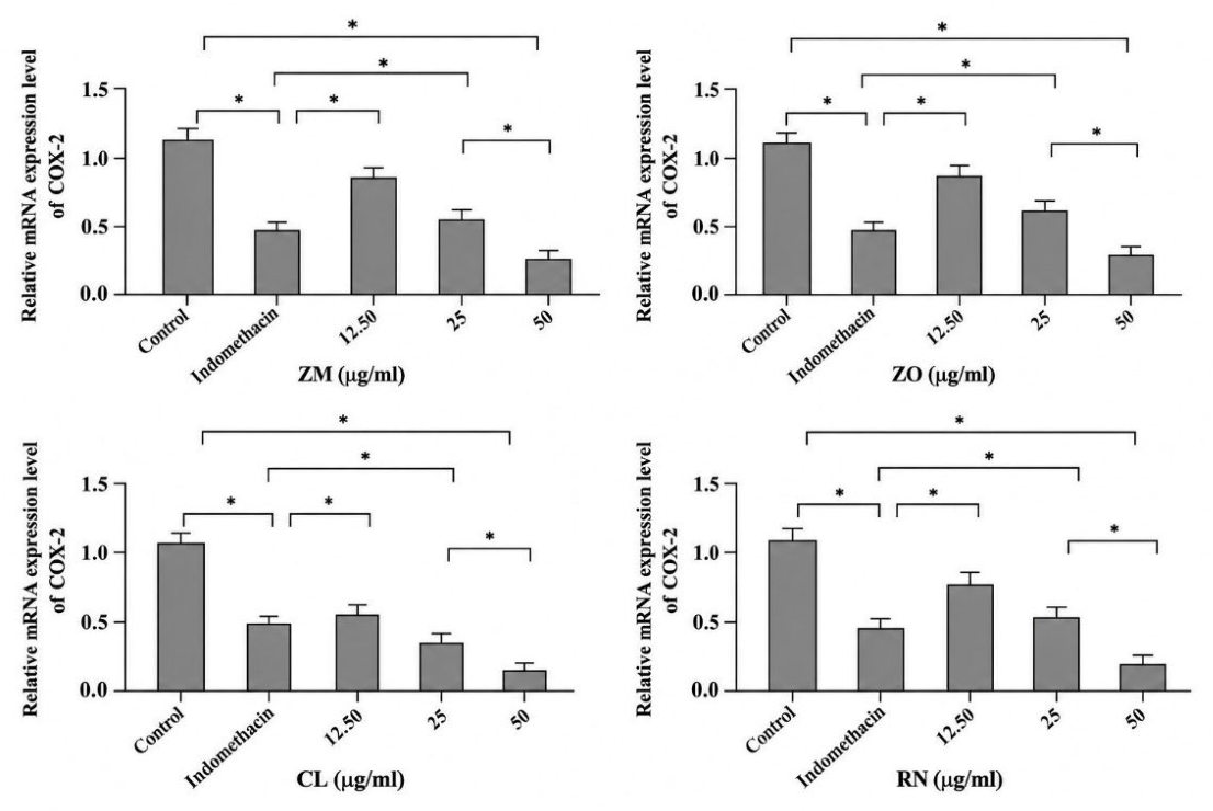
Table 1.
Quantification of major phytochemical marker compounds in Thai medicinal plant extracts.
| Extract | Marker compound | Concentration (mg/mL) |
|---|---|---|
| Rhinacanthus nasutus (RN) | Rhinacanthin | 1.78 ± 0.26 |
| Curcuma longa (CL) | Curcumin | 5.34 ± 0.71 |
| Zingiber montanum (ZM) | Terpinen-4-ol | 2.60 ± 1.67 |
| Zingiber officinale (ZO) | 6-Gingerol | 1.30 ± 1.85 |
Values are expressed as mean ± SD (n = 3).
Table 2.
Fatty acid composition of mesocarp-derived red palm oil (RPO) compared with commercial crude red palm oil.
Table 2.
Fatty acid composition of mesocarp-derived red palm oil (RPO) compared with commercial crude red palm oil.
| Fatty acid | Mesocarp-derived RPO (% total fatty acids) |
Commercial crude RPO (% total fatty acids) |
| Palmitic acid (C16:0) | 54.36 ± 0.82ᵃ | 40.17 ± 0.64ᵇ |
| Stearic acid (C18:0) | 4.08 ± 0.11ᵃ | 4.85 ± 0.13ᵇ |
| Oleic acid (C18:1) | 29.74 ± 0.67ᵇ | 40.08 ± 0.81ᵃ |
| Linoleic acid (C18:2) | 6.60 ± 0.19ᵇ | 10.27 ± 0.25ᵃ |
| Linolenic acid (C18:3) | 0.17 ± 0.01ᵇ | 0.35 ± 0.02ᵃ |
| Arachidic acid (C20:0) | 0.33 ± 0.02ᵃ | 0.38 ± 0.03ᵃ |
| Eicosenoic acid (C20:1) | 0.10 ± 0.01ᵇ | 0.14 ± 0.01ᵃ |
| Palmitic acid (C16:0) | 54.36 ± 0.82ᵃ | 40.17 ± 0.64ᵇ |
| Stearic acid (C18:0) | 4.08 ± 0.11ᵃ | 4.85 ± 0.13ᵇ |
| Oleic acid (C18:1) | 29.74 ± 0.67ᵇ | 40.08 ± 0.81ᵃ |
Values are expressed as mean ± SD (n = 3). Different superscript letters within the same row indicate significant differences between mesocarp-derived RPO and commercial crude RPO (p < 0.05).
Disclaimer/Publisher’s Note: The statements, opinions and data contained in all publications are solely those of the individual author(s) and contributor(s) and not of MDPI and/or the editor(s). MDPI and/or the editor(s) disclaim responsibility for any injury to people or property resulting from any ideas, methods, instructions or products referred to in the content. |
© 2026 by the authors. Licensee MDPI, Basel, Switzerland. This article is an open access article distributed under the terms and conditions of the Creative Commons Attribution (CC BY) license (http://creativecommons.org/licenses/by/4.0/).
Copyright: This open access article is published under a Creative Commons CC BY 4.0 license, which permit the free download, distribution, and reuse, provided that the author and preprint are cited in any reuse.


