

Submitted:
28 June 2026
Posted:
30 June 2026
You are already at the latest version
Abstract
Subungual tumors are rare and often misdiagnosed, leading to unnecessary procedures or delayed treatment; thus, accurate identification of benign neoplasms such as fibrolipoma is essential to guide appropriate management. We report a 59-year-old man who presented with a several-year history of a painless subungual mass of the right great toe with progressive nail thinning and onychodystrophy. Imaging demonstrated soft-tissue swelling without bone involvement. Surgical exploration revealed two well-demarcated, lobulated subungual and periungual masses that were completely excised. Histopathology showed mature adipocytes with fibrous septa, and fluorescence in situ hybridization confirmed HMGA2 translocation without MDM2 or CDK4 amplification, establishing the diagnosis of fibrolipoma. Complete excision resulted in satisfactory nail regrowth at 1-year follow-up with no recurrence. This case highlights the importance of including fibrolipoma in the differential diagnosis of subungual masses and supports the value of histopathological and molecular analysis in achieving precise diagnosis and optimal surgical outcomes.
Keywords:
surgery
; fibrolipoma
; subungual lipoma
; periungual lipoma
; adipose tissue
1. Introduction
Fibrolipoma is classified by the World Health Organization as a rare histologic subtype of traditional lipomas composed of mature adipocytes with fibrous connective tissue [1]. Lipomas of the nail unit are exceedingly rare because the onychodermis is fibrovascular and lacks a subcutaneous adipose layer [2]. Simultaneous subungual and periungual fibrolipomas have not been previously reported.
Preoperative diagnosis of nail unit lipomas is challenging because they mimic other nail tumors, including glomus tumor, subungual exostosis, and squamous cell carcinoma [3]. Imaging and histopathologic examination with immunohistochemical and molecular studies are essential for definitive diagnosis. We describe a case of simultaneous subungual and periungual fibrolipomas in a 59-year-old man, with emphasis on clinical, radiographic, and pathologic findings relevant to the podiatric physician.
2. Detailed Case Description
A 59-year-old man presented with a several-year history of a painless, nodular mass in the subungual and periungual regions of the right great toe. The affected nail plate showed marked thinning and onychodystrophy (Figure 1a). He reported no prior nail trauma or family history of similar conditions. Physical examination revealed swelling of the nail bed and distal phalanx with partial nail plate loss; the remaining nails were normal.
Radiography showed no phalangeal bone abnormality, although periarticular soft-tissue swelling was present without bone erosion. Ultrasound identified several nodules in the superficial soft tissue; the largest measured approximately 16 × 9 mm, with indistinct borders and irregular contour. Color Doppler imaging showed no significant internal vascularity. Surgical exploration revealed two well-demarcated, lobulated, yellowish, soft-tissue masses in the subungual and periungual areas; the lesions were not interconnected (Figure 1b-c).
Histologic examination showed mature adipocytes admixed with fibrous bundles and scattered plump spindle cells (Figure 2). Immunohistochemistry demonstrated partial S-100 positivity, focal p53 positivity, and a Ki-67 proliferative index of 2%; CD34, desmin, SMA, MDM2, and CDK4 were negative. Fluorescence in situ hybridization confirmed HMGA2 gene translocation without CDK4 or MDM2 amplification, establishing the diagnosis of fibrolipoma.
At 1-year follow-up after complete surgical excision, the surgical site had healed well with satisfactory nail plate regrowth from the proximal nail fold (Figure 1d). No recurrence was observed, and both function and cosmesis of the right toe were excellent.
3. Discussion
Fibrolipoma is an uncommon lipoma variant characterized by mature adipocytes with fibrous connective tissue [1]. Lipomas of the nail unit are exceedingly rare because the onychodermis lacks a subcutaneous adipose layer [2]. The simultaneous occurrence of subungual and periungual fibrolipomas in the same digit, as seen in our patient, has not been previously described.
A review of the literature identified 14 previously reported cases of nail unit lipoma, involving subungual (9 cases) and periungual (5 cases) locations, as summarized in Table 1 [2,5,6,7,8,9,10,11,12,13]. The mean patient age was 54.5 years, with a male predominance. Most lesions were painless, although some caused discomfort with footwear or mild movement restriction. All reported cases exhibited nail changes, including plate elevation, thinning, splitting, and dystrophy. Hao et al. [4] recently reported an eight-case series of toe lipomas, including one fibrolipoma, underscoring the relevance of this entity to podiatric practice.
Cytogenetic investigations have identified rearrangements involving the 12q14-15 region harboring the HMGA2 gene in a subset of lipomas [14]. An HMGA2 translocation was detected by FISH in our case, confirming a clonal chromosomal abnormality characteristic of benign lipomatous tumors.
Radiography is essential to exclude underlying bone lesions such as subungual exostosis [11]. In our case, plain radiographs showed no bone destruction. High-frequency ultrasound can define the soft-tissue architecture of the nail unit, with lipomas typically appearing as well-defined, homogeneously hyperechoic masses [6]. MRI is particularly valuable because adipose tissue exhibits characteristic high signal on T1- and T2-weighted images with signal suppression on fat-saturation sequences [15].
Definitive diagnosis requires histopathologic examination. Fibrolipoma is characterized by well-differentiated mature adipocytes partitioned by fibrous septa into a lobular architecture; myxoid stroma and lipoblasts are absent [1,15]. Immunohistochemistry typically shows partial S-100 positivity with negative CD34, desmin, SMA, MDM2, and CDK4 [15]. HMGA2 rearrangement and lack of MDM2 or CDK4 amplification, demonstrated by FISH, help exclude atypical lipomatous tumor or well-differentiated liposarcoma [15,16]. The differential diagnosis includes glomus tumor, subungual exostosis, epidermoid cyst, and squamous cell carcinoma [12].
Complete surgical excision is the treatment of choice for nail apparatus lipomas. Preservation of the nail matrix and germinal epithelium facilitates postoperative nail regrowth. All reported patients, including ours, have remained recurrence-free after excision, underscoring the excellent prognosis.
5. Conclusions
Subungual and periungual fibrolipomas are rare benign tumors that warrant inclusion in the differential diagnosis of painless subungual nodules with nail dystrophy. Accurate diagnosis requires clinical correlation, imaging, and histopathologic examination with immunohistochemical and molecular studies. Complete surgical excision offers excellent functional and cosmetic outcomes with minimal recurrence risk.
Author Contributions
XYT: Writing – original draft, Data curation, Methodology, Visualization. ZYZ: Writing – original draft, Data curation, Investigation. TW: Writing – review & editing, Supervision. HJL: Writing – review & editing. SLX: Supervision, Writing – review & editing, Validation. All authors have read and agreed to the published version of the manuscript.
Funding
This research received no external funding.
Institutional Review Board Statement
This case report was prepared in accordance with the Declaration of Helsinki. The Ethics Committee of West China Hospital, Sichuan University reviewed the protocol and determined that formal ethical approval was not required for this retrospective case report.
Informed Consent Statement
Written informed consent was obtained from the patient’s legal guardian for publication of this case report and accompanying images.
Data Availability Statement
The datasets presented in this study are not publicly available due to patient privacy concerns but are available from the corresponding author upon reasonable request.
Acknowledgments
The authors affirm that no generative artificial intelligence tools were utilized at any stage of this work, including the research design, data collection and analysis, manuscript preparation, or image editing.
Conflicts of Interest
The authors declare no conflicts of interest.
Abbreviations
The following abbreviations are used in this manuscript:
| CD34 | Cluster of Differentiation 34 |
| CDk41 | Cyclin-Dependent Kinase 4 |
| HMGA2 | High-Mobility Group AT-Hook 2 |
| MDM2 | Mouse Double Minute 2 homolog |
| SMA | Smooth Muscle Actin |
References
- Tandon, A.; Srivastava, A.; Jaiswal, R.; Gaur, A. Oral fibrolipoma: A rare clinicopathological entity. J. Oral Maxillofac. Pathol. 2023, 27, 537–539. [Google Scholar] [CrossRef]
- Velez, N.F.; Telang, G.H.; Robinson-Bostom, L.; Jellinek, N.J. Subungual lipoma: Presenting as longitudinal nail splitting. Dermatol. Surg. 2014, 40, 1435–1437. [Google Scholar] [CrossRef] [PubMed]
- Richert, B.; Andre, J.; Choffray, A.; Rahier, S.; de la Brassinne, M. Periungual lipoma: About three cases. J. Am. Acad. Dermatol. 2004, 51, S91–S93. [Google Scholar] [CrossRef] [PubMed]
- Hao, X.; Mirkin, G.; Freedman, D.J.; Beiser, I.; Firestone, L.; Grant, D. Lipomas of the toes: An eight-case series. J. Am. Podiatr. Med. Assoc. 2023, 113, 20285. [Google Scholar] [CrossRef] [PubMed]
- Baran, R. Periungual lipoma at an unusual site. J. Dermatol. Surg. Oncol. 1984, 10, 32–33. [Google Scholar] [CrossRef] [PubMed]
- Gallouj, S.; Aqil, N.; Zahra Mernissi, F. Periungual lipoma: An unusual site. Ann. Dermatol. Venereol. 2019, 146, 121–124. [Google Scholar] [CrossRef] [PubMed]
- Failla, J.M. Subungual lipoma, squamous carcinoma of the nail bed, and secondary chronic infection. J. Hand Surg. Am. 1996, 21, 512–514. [Google Scholar] [CrossRef] [PubMed]
- Bardazzi, F.; Savoia, F.; Fanti, P.A. Subungual lipoma. Br. J. Dermatol. 2003, 149, 418. [Google Scholar] [CrossRef] [PubMed]
- Sanchez Sambucety, P.; Alonso Alonso, T.; Gil Agapito, P.; Gonzalez Moran, A.; Rodriguez Prieto, M.A. Subungual angiomyxolipoma. Dermatol. Surg. 2007, 33, 508–509. [Google Scholar] [CrossRef] [PubMed]
- Kitagawa, Y.; Tamai, K.; Kim, Y.; Hayashi, M.; Makino, A.; Takai, S. Lipoma of the finger with bone erosion. J. Nippon Med. Sch. 2012, 79, 307–311. [Google Scholar] [CrossRef] [PubMed]
- Kwon, N.H.; Kim, H.S.; Kang, H.; Lee, J.Y.; Kim, H.O.; Kang, S.J.; Park, Y.M. Subungual spindle cell lipoma. Int. J. Dermatol. 2013, 52, 636–638. [Google Scholar] [CrossRef] [PubMed]
- Nakamura, R.; Valgas, N.; Bolzani, F.; Bonatto, D.; Kac, B.; de Brito, F. Subungual spindle cell lipoma: Case report: P6700. J. Am. Acad. Dermatol. 2013, 68, AB109. [Google Scholar] [CrossRef]
- Cunha, N.; Sousa, V.C.; Pinheiro, R.; Lencastre, A. And nextadnexa: Subungual lipoma. Eur. J. Dermatol. 2017, 27, 448–449. [Google Scholar] [CrossRef] [PubMed]
- Panagopoulos, I.; Gorunova, L.; Bjerkehagen, B.; Lobmaier, I.; Heim, S. The recurrent chromosomal translocation t(12;18)(q1415;q1221) causes the fusion gene HMGA2-SETBP1 and HMGA2 expression in lipoma and osteochondrolipoma. Int. J. Oncol. 2015, 47, 884–890. [Google Scholar] [CrossRef] [PubMed]
- Nishio, J.; Ideta, S.; Aoki, M.; Hamasaki, M.; Nabeshima, K.; Iwasaki, H.; Naito, M. Fibrolipoma of the ring finger: MR imaging and histological correlation. Vivo 2013, 27, 541–544. [Google Scholar]
- Thway, K.; Flora, R.; Shah, C.; Olmos, D.; Fisher, C. Diagnostic utility of p16, CDK4, and MDM2 as an immunohistochemical panel in distinguishing well-differentiated and dedifferentiated liposarcomas from other adipocytic tumors. Am. J. Surg. Pathol. 2012, 36, 462–469. [Google Scholar] [CrossRef] [PubMed]
Figure 1.
Clinical course from preoperative evaluation to 1-year postoperative follow-up. (a), Preoperative view of the right great toe showing marked nail thinning and onychodystrophy. (b-c), Intraoperative exposure after incising the nail bed and overlying skin; 2 well-demarcated, lobulated soft-tissue masses are evident. (d), One year after complete excision, the nail plate has regrown and covers most of the nail bed without tumor recurrence.
Figure 1.
Clinical course from preoperative evaluation to 1-year postoperative follow-up. (a), Preoperative view of the right great toe showing marked nail thinning and onychodystrophy. (b-c), Intraoperative exposure after incising the nail bed and overlying skin; 2 well-demarcated, lobulated soft-tissue masses are evident. (d), One year after complete excision, the nail plate has regrown and covers most of the nail bed without tumor recurrence.
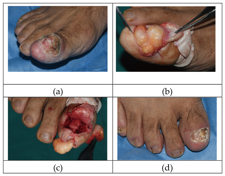
Figure 2.
Histopathologic appearance of the excised specimen (H&E, original magnification ×4). Mature adipocytes are interspersed with variably prominent fibrous septa.
Figure 2.
Histopathologic appearance of the excised specimen (H&E, original magnification ×4). Mature adipocytes are interspersed with variably prominent fibrous septa.
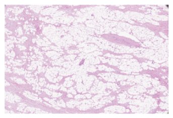

Table 1.
summary of subungual lipoma cases in scientific literature.
| Case | Site | Reference | Age /Sex |
History(Years) |
Location | Symptoms | Nail changes |
|---|---|---|---|---|---|---|---|
| 1 | Periungual | Baran R. (1984) [5] | 41/M | 3 - 4 | R3, (finger) | Painless | Enlarged lateral nail fold |
| 2 | Richert B, et al. (2004) [3] | 55/M | Several | R1, (toe) | Discomfort with footwear,painless | Thickened nail plate, onychorrhexis | |
| 3 | 67/F | Several | R3, (finger) | Painless, slow growing | Not reported | ||
| 4 | 48/M | N/A | R1, (toe) | Gradual, painful on palpation/ contact with footwear |
Not reported | ||
| 5 | Gallouj S, et al. (2019) [6] | 65/F | N/A | R4, (finger) | Slight movement restriction, painless | Normal | |
| 6 | Subungual | Failla JM. (1996) [7] | 73/F | 10 | R1, (finger) | Painless, slow growing | Proximal nail convex, purplish tender area at lunula |
| 7 | Bardazzi F, et al. (2003) [8] | 40/F | Several | L2, (finger) | Painless, progressively grown | Thinning, dystrophy, Hyperconvexity of nail plate |
|
| 8 | Sánchez Sambucety P, et al. (2007) [9] | 43/M | 1 | R1, (finger) | Slow growing, painful on pressure | Not reported | |
| 9 | Kitagawa Y, et al. (2012) [10] | 38/M | 18 | L2 , finger) | Slightly painful, slow growing | Nail plate elevation, convex, nail bed pallor | |
| 10 | Kwon NH, et al. (2013) [11] | 28/M | 1 | R1, (finger) | Painful with pressure | Red lunula, central bulge, distal onycholysis of nail plate | |
| 11 | Nakamura R, et al. (2013) [12] | 64/F | 7 | L4, (finger) | Painless | Loss of the nail plate | |
| 12 | Vélez NF, et al. (2014) [2] | 78/F | Several | R1, (finger) | No pain or swelling | Longitudinal erythronychia and distal splitting of nail plate | |
| 13 | 69/M | 2 | L1, (toe) | Mild swelling | Redness of the proximal lunula and nail splitting | ||
| 14 | Cunha N, et al.(2017) [13] | 49/M | 2 | L3, (finger) | Slightly painful, slow growing | Thinning, hyperconvexity, dystrophy of the nail plate, and central dorsal pterygium |
|
| 15 | Subungual and Periungual | Present case | 59/M | Several | R1, (toe) | Painless, slow growing | Thinning, dystrophy and hyperconvexity of the nail bed |
Disclaimer/Publisher’s Note: The statements, opinions and data contained in all publications are solely those of the individual author(s) and contributor(s) and not of MDPI and/or the editor(s). MDPI and/or the editor(s) disclaim responsibility for any injury to people or property resulting from any ideas, methods, instructions or products referred to in the content. |
© 2026 by the authors. Licensee MDPI, Basel, Switzerland. This article is an open access article distributed under the terms and conditions of the Creative Commons Attribution (CC BY) license (http://creativecommons.org/licenses/by/4.0/).
Copyright: This open access article is published under a Creative Commons CC BY 4.0 license, which permit the free download, distribution, and reuse, provided that the author and preprint are cited in any reuse.


