Preprint
Article

This version is not peer-reviewed.

Management of an Acute Lead Intoxication in a Group of Bottlenose Dolphins (Tursiops truncatus, Montagu, 1821)

Submitted:

26 June 2026

Posted:

29 June 2026

You are already at the latest version

Abstract

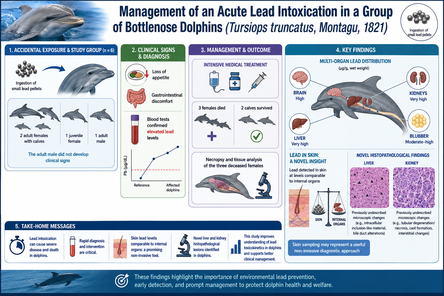
Acute lead intoxication is rarely reported in bottlenose dolphins (Tursiops truncatus) in human care, and information on tissue distribution during acute exposure is limited. This study describes the clinical presentation, management, and tissue distribution of lead (Pb) following accidental ingestion of Pb pellets in a group of six dolphins (two lactating adult females, their male calves, one juvenile female, and one adult male). The adult male did not develop clinical signs nor evidence of pellet ingestion and was therefore not included among the clinically affected cases. Clinical evaluation, hematology, serum biochemistry, heavy metal analysis, diagnostic imaging, and endoscopy were performed. Elevated blood Pb levels were detected in all clinically affected animals. Pellets were identified in the stomach of the three females and one of the calves. The calf without detectable pellets had the highest blood Pb concentration, suggesting lactational transfer. Despite intensive supportive and chelation therapy, the three adult females died, and complete necropsies were conducted. Pb concentrations were measured in liver, kidney, brain (three cerebral regions), blubber, muscle, and skin. Post-mortem analyses demonstrated high Pb concentrations in liver (1.14 mg/kg), kidney (0.814 mg/kg), and brain (0.554 mg/kg), as well as detectable levels in blubber (1.166 mg/kg). Skin Pb concentrations (1.254 mg/kg) were comparable to those of liver and kidney. Histopathology revealed intranuclear inclusion bodies in renal and hepatic cells. These findings characterize systemic Pb distribution in acute intoxication and identify skin as a potential diagnostic matrix in dolphins.
Keywords: 
;  ;  ;  ;  ;  ;  

1. Introduction

Common bottlenose dolphins (Tursiops truncatus, BD) are widely distributed across the globe, except for the Arctic and Antarctic regions. The IUCN considers this species as “Least Concern” given its global conservation status and noted that the main threats for this species in the wild are related to all human activities such as residential and commercial development, transportation and service corridors, biological resource use, intrusions and disturbance, and pollution [1]. More recently, microplastics and pharmaceuticals have been found in blow samples and blubber of wild BD and their effect still needs to be addressed [2,3]. BD is the most common species of cetacean kept in marine parks and dolphinaria around the world [4] not only because of their charismatic behavior but also because of their adaptability [5]. They have been studied for many decades, but data on heavy metal intoxication in this species in professional settings are scarce [6,7] due to the rarity and low probability such events occurring.
Lead intoxication in animals poses a significant health concern, presenting with a range of clinical manifestations that can severely impact their well-being. This condition primarily affects mammalian and avian species, leading to neurologic disturbances, gastrointestinal issues, hematologic changes, immunosuppression, infertility, and renal diseases [8,9]. The severity and types of symptoms observed can vary based on the level of lead exposure and the duration of contact. Dogs and cattle are the species most frequently affected by lead poisoning, with young animals being particularly vulnerable due to behavioral factors such as pica—the consumption of non-food items—and increased exposure opportunities. Acute lead poisoning is more prevalent among younger animals, manifesting prominently with gastrointestinal and neurological signs. In cattle, symptoms may emerge rapidly within 24 to 48 hours post-exposure, revealing a spectrum of clinical signs including ataxia, blindness, excessive salivation, and muscle tremors [10].
Urban environments, especially those undergoing renovations of older buildings painted with lead-based paint, have been identified as significant risk areas for lead exposure, affecting both small animals and children. The findings in backyard poultry flocks indicate rising lead concentrations in eggs, which are linked to the ingestion of paint particles. This presents an unexpected public health risk given that poultry demonstrate a higher resistance to the acute effects of lead poisoning compared to mammals [11]. Additionally, domestic cats may inadvertently consume lead-laden dust through grooming, and improper disposal of lead-poisoned animal remains can lead to secondary poisoning in scavenging wildlife [12].
Lead toxicity is less common in certain species, which often have reduced access to lead sources or possess dietary habits that limit their exposure [13,14,15]. However, fatal cases of lead poisoning in zoo animals have historically been reported affecting different species of birds and mammals and is mostly associated with the interaction with leaded paint [13]. Nowadays, wild birds are still affected by the ingestion of spent lead shots from hunters’ ammunition [16]. This is particularly troubling for endangered species, such as the California condor, which may ingest contaminated carrion kell [17]. Only three reports exist on adult BD in human care [6,7] and one a wild harbour seal [18] affected by the ingestion of either lead pellets or a lead fishing sinker, respectively. Signs and symptoms varied, but animals mostly showed a decreased appetite, gastrointestinal discomfort, neurological impairment, and clear altered liver enzymes. Despite the treatment, one of the dolphins reported, and the seal, succumbed to the toxicosis
This paper describes an episode of acute lead intoxication affecting a pod of BDs of different ages. Special attention is given to the clinical presentation and evaluation, as well as the therapies administered to the animals to address the toxicosis. Additionally, data regarding the distribution of lead in different tissues and the results of a digestion assay are provided, offering insights into the bioavailability of ingested lead particles.

2. Materials and Methods

2.1. Animals Involved and Onset of the Toxicosis

2.1.1. Dolphins

A dolphin pod, living in professional care in a tank containing 2.6 million liters of natural seawater, and consisting of six individuals of different ages, was involved in an acute lead intoxication event beginning in August 2021. The affected individuals included two adult dolphins, both wild-born nursing females; a juvenile female; and two dolphin calves (Table 1). The pod also included the alpha male, the sire of the three youngest dolphins; however, he did not exhibit clinical signs of lead intoxication nor evidence of ingesting lead pellets during the event.

2.1.2. Signs and Symptoms

Female 1 was the first one exhibiting nonspecific signs of disease such as intermittent attention and appetite, followed by low activity and clear signs of pain (closed eyes, buoying, resting in the bottom of the tank), abdominal discomfort (arching, vomiting/regurgitation) and mucoid feces.
Female 3 presented with a sudden loss of appetite, which was initially associated with social distress and pain, only 48 hours after Female 1.
One to two days after the start of the symptomatic phase, the other three dolphins began to exhibit similar symptoms. Female 2 stopped eating and showed signs of pain (closed eyes) and the two calves lost their appetite although they were observed still nursing.

2.2. Clinical Diagnosis

Full physical exams (PEs) were performed on all animals in the group as the signs and symptoms of toxicosis appeared. PEs included behavioral observations, assessment of body condition, and a thorough physical examination of the skin, flippers, flukes, eyes, oral cavity, genital slit, and mammary glands. The respiratory system was evaluated, including blowhole cytology and the collection of swabs for culture. Gastrointestinal tract was also assessed through the analysis of gastric juice and fecal samples. Additionally, blood samples were collected, and imaging studies such as ultrasound, radiography, and gastroscopy were conducted.
As part of their routine husbandry, the dolphins were trained for voluntary participation in their medical checks. Thus, all initial samples were collected voluntarily and processed in-house immediately, as previously described and reported [19,20,21]. Follow-up samples were collected during medical handling, which required physical restraint due to the animals’ increasing discomfort and unwillingness to cooperate.
After each collection, blood serum and remaining whole blood were refrigerated and sent to an external laboratory (IDEXX Vet Med Labor GmbH, Kornwestheim, Germany) for comprehensive analysis, including hematology, blood chemistry, and heavy metal screening. Samples were submitted to a different laboratory (AbLab, Sarzana, Italy) for bacterial and fungal culture.
Ultrasonographic exams were performed regularly on all dolphins to evaluate internal organs, monitor the progression of damage, and identify the presence of foreign bodies (ie. lead pellets), using a Logiq V2 ultrasound system (GE Healthcare Technologies Inc., Chicago, Illinois) equipped with a convex probe (GE 4c, 2 - 5 MHz).
Radiographic examinations were performed using an Econet Vet-20BT Lite portable X-ray system (medical ECONET GmbH, Oberhausen, Germany) paired with an AGFA DR 14e-C digital direct wireless portable detector (Agfa-Gevaert NV, Mortsel, Belgium). The system provided high-resolution images of the head, thorax, and abdominal regions of the dolphins, enabling the determination of the location and quantity of lead particles within their bodies. Exposure parameters, including kVp and mAs, were adjusted according to the region of interest and the body size of each dolphin.
Gastroscopies of the upper gastrointestinal (GI) tract were performed to evaluate mucosal health and retrieve ingested lead pellets from the first two gastric chambers. A Karl Storz videoendoscope (7.8 mm diameter, 140 cm length) was used, attached to a Karl Storz Endoskope Tele pack X LED TP100 light source (Karl Storz SE & Co. KG, Tuttlingen, Germany).
Both radiographic and endoscopic procedures were carried out outside of the water, with the animals in sternal recumbency. All necessary precautions were taken to ensure minimal stress and maximal safety during the intervention. Only Female 1 received 20mg of diazepam (Laboratorios Normon, S.A., Madrid, Spain) 30 mins before the gastroscopic procedures.

2.3. Additional Diagnostics and Assays

2.3.1. Urine

Urine samples were primarily collected from the three females during medical handling. The dolphins were conditioned to urinate on command on a sterile cup when gentle pressure was applied over the area corresponding to the urinary bladder. Samples were stored at -20°C until submitted for lead quantification to the Department of Veterinary Medical Sciences of the University of Bologna (Italy). The calves had not yet been trained for this husbandry behavior. However, samples were opportunistically collected during physical restraint.

2.3.2. Milk

Milk samples were collected from Females 1 and 2 during medical handling, and submitted to the Department of Veterinary Medical Sciences of the University of Bologna (Italy) together with control samples from a previous pregnancy. The animals were positioned on their side to expose the mammary slit above the water surface. Approximately 50 mL of milk were collected using modified syringes [22], which were adapted to create a vacuum. Negative pressure was generated by pulling back the syringe plunger, allowing the milk to be drawn into the syringe.

2.3.3. Skin

Skin samples from the three females and Male 2 were obtained by gently scratching the dorsal aspect of dolphins with a microscope slide. Samples from Male 1 were not collected. Post-mortem skin samples were collected at necropsy. All samples were stored at -20°C until analyzed.

2.3.4. Digestion Assay

To evaluate the bioavailability of the lead, a trial was planned at the Department of Veterinary Medical Sciences of the University of Bologna, Italy to assess the dissolution rate of the pellets in the gastric juice (Figure 1). For this, a digestion assay was conducted using gastric juice from 7 healthy BD not exposed to the lead event. The gastric juice was collected during the routine clinical exams, through voluntary behavior and positive reinforcement, and pH was recorded. At least 70 ml of gastric juice from each BD was used in this assay. The gastric fluid was divided into two 25 ml aliquots (sample A and B), and the remaining amounts were kept to top up Sample A during the assay. In Sample A the total volume of gastric fluid was maintained throughout the study by adding 1 ml of the remaining sample, while no refilling was done for sample B. To each aliquot sample, 0.5 ± 0,001 mg lead pellets were added. All samples and remaining fluids were kept at 37°C to mimic dolphins’ body temperature. At regular intervals (1, 2, 4, 8, 12, 24, and 48 hours) samples A and B were gently homogenized, the pH was measured to verify any possible variations, and 1 ml of gastric fluid was retrieved from each of them and kept frozen at -20°C until lead quantification. The lead pellets in each sample were weighed after 24 and 48 hours of digestion, in order to evaluate the dissolution rate.

2.3.4. Necropsy and Histopathology

Necropsies were performed in all deceased dolphins immediately after death, following previously published guides [23].
Tissue samples were submitted for histopathological evaluation to the School of Biosciences and Veterinary Medicine of the University of Camerino, Italy.

2.3.5. Toxicological Analysis

Toxicological analysis of urine, milk, skin, and internal organs collected during the necropsy were carried out, using microwave digestion and ICP-EOS analysis, following an internal protocol (SOP SPASA PROIT 005 and 010) at the Department of Veterinary Medical Sciences of the University of Bologna, Italy [24].

2.4. Medical Treatments

Medical therapy was implemented in the five dolphins as soon as the first symptoms of disease appeared and based on the resources available. The treatments focused on addressing different goals. Calcium EDTA (CaNa2EDTA), dimercaptosuccinic acid (DMSA), garlic, vitamin C, and active charcoal were administered to chelate the metal in the blood and reduce its absorption in the GI tract. Omeprazol was provided to increase gastric pH and minimize the interaction of gastric acids with the lead pellets.
Up to five milking per day of the nursing females and weaning of the calves were implemented to promote lead excretion through milk production and to prevent further ingestion of contaminated milk by the calves, respectively. Vitamins such as B Complex, B1, and K, along with folic acid; stanozolol, an anabolic steroid; and Yunnan Baiyao, a hemostatic agent, were included in the treatment to counteract the haemolytic effects and bleeding induced by lead toxicity.
Damage to the GI tract and the associated reduction in motility were treated with sucralfate and metoclopramide, while maropitant was added to reduce vomiting and regurgitation. Paraffin oil was included in the therapy for dolphins retaining lead pellets in the third gastric chamber to facilitate passage and elimination through the feces. Neuropathic and visceral pain were managed using tramadol and hyoscine butylbromide (Buscopan).
Silymarin and pancreatic enzymes, along with a lean diet, were administered to protect and support the metabolic functions of the damaged liver and pancreas. Dexamethasone, prednisolone, and furosemide were used to address lung and brain edema caused by disruptions in homeostasis. Fluid therapy and nutritional support were provided to all the animals throughout the course of the toxicosis.
Dosages are described in Table 2.

3. Results

Immediate physical exams performed in all animals, and the in-house diagnostics revealed no significant abnormalities, with unremarkable cytology. Cultures from blowhole swabs, gastric juice and fecal samples were also unremarkable. However, blood tests showed mild leukopenia in Female 1 and slight to moderate yellowish discoloration of the serum in both Female 1 and 3.
Since the only symptoms common to the first two cases were loss of appetite and potential indications of pain, it was challenging to entirely rule out an infectious etiology, determine whether the cases were necessarily related, or assess if the prolapse and the social event played a role in the situation. However, the alteration in serum coloration suggested that both cases involved a potential hepatic process, raising the possibility of a toxic component, as confirmed by the analysis performed by the external lab.
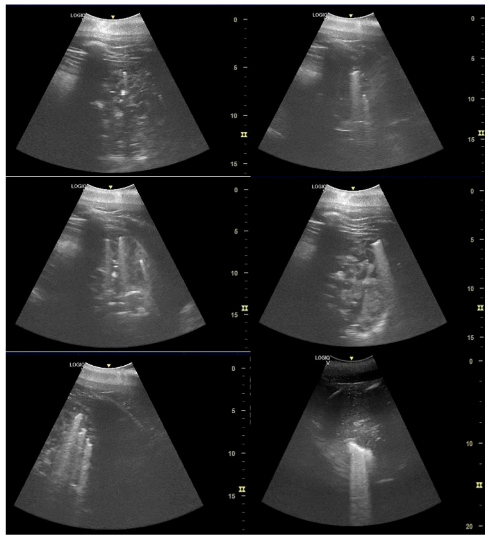
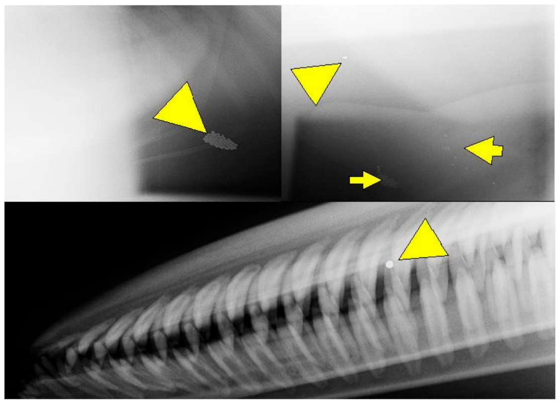
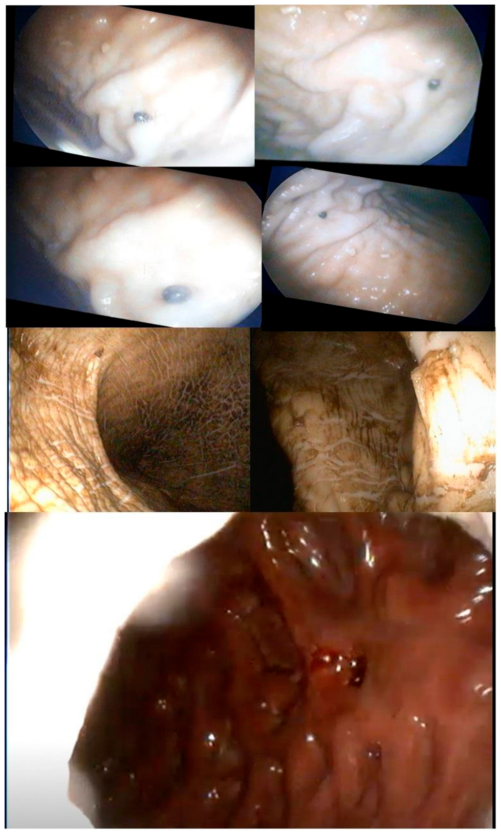
The three diagnostic imaging techniques performed in the 5 affected dolphins provided valuable insights. Abdominal ultrasonography revealed moderate to severe reductions in gastrointestinal motility, hyperechogenicity of the gastric mucosa, and the presence of highly echogenic (hyperechoic) objects (2.2-2.4 mm in diameter) that produced intense acoustic shadows (Figure 2). These metal-like objects were found scattered throughout the three gastric compartments in four of the five dolphins. Radiographs confirmed the presence of radiopaque spherical objects not only in the stomach chambers of four dolphins but also embedded within the gums of some specimens (Figure 3). Finally, gastroscopy allowed the retrieval of multiple pellets from the first and second stomach chambers. The presence of these metal pellets caused irritation of the fundic mucosa and evident inflammation in the glandular stomach (Figure 4). The pellets were identified as components of a broken lead bag of a diving set.
The youngest calf (Male 2) was confirmed to have no pellets or other foreign bodies in his GI tract through all three diagnostic imaging techniques. Furthermore, no alterations of its gastric mucosa were observed during gastroscopy.
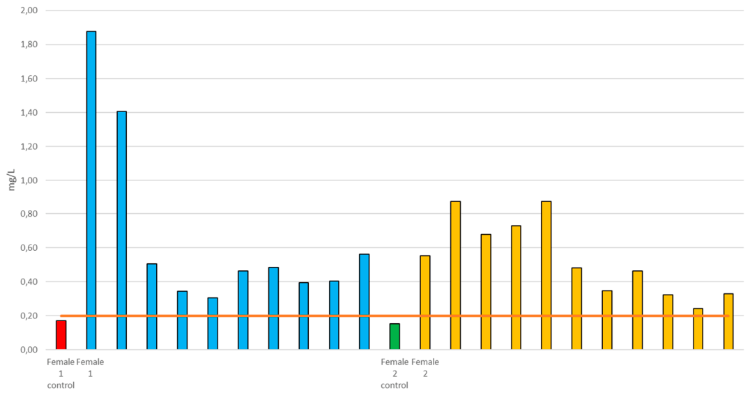
The results from the initial quantification of heavy metals in the blood of the animals confirmed the suspicion of a lead toxicosis (Table 4). Similarly, considerable levels of lead were found in the urine (Table 5) and milk samples tested in Italy, which confirmed that the nursing females were excreting the lead in this way (Graph 1).
Graph 1. Results of the analyses performed on the milk collected from the nursing females during the toxic episode. Orange line corresponds to the Maximum Residual Limit (MRL) defined for milk (0.20 mg/L, [25,26].).
Preprints 220454 g005
During the active phase of the poisoning, the three females developed pulmonary rales and neurological signs, including vomiting and regurgitation. The two calves also experienced episodes of vomiting and regurgitation, though to a lesser extent. Additionally, Female 1 developed corneal edema. Pleural effusion was observed during ultrasonographic examinations in Females 2 and 3.
Follow up blood tests revealed significant tissue and organ alterations. Female 1 exhibited a severe normocytic, normochromic anemia (likely haemolytic), which was mildly regenerative. She also presented moderate anisocytosis and polychromasia, along with an elevated nucleated red blood cell (NRBC) count (4%). Notably, basophilic stippling of erythrocytes was not observed on any of the blood smears. Platelet (PLT) counts were normal across all individuals.
The white blood cell (WBC) count was within reference limits [27] for all the animals, except for Female 3 and Male 1, who showed neutrophilic leukocytosis. Band neutrophils were present in Female 3.
Furthermore, Female 1 displayed clear signs of hepatic and (pre-)renal failure, while the other two females also showed evident hepatic dysfunction. Hyperlipasemia was consistently observed in the follow-up blood analyses of all the five dolphins, indicating that the poisoning was also impairing pancreatic function. In the two calves cortisol levels were occasionally undetectable.
The implementation of chelation treatment led to rapid improvements in blood lead levels. The progression of anemia was slowed in two of the three females (Female 1 and 2), likely due to the chelation therapy as well as the additional supportive treatments. Pulmonary rales and neurological signs were successfully managed. However, despite the treatment and around-the-clock efforts to provide care, the three females ultimately succumbed to the intoxication or to the extensive damage caused by it.
Female 1 passed away 18 days after the onset of symptoms. Immediately before her death, she exhibited lethargy and was floating laterally on her right side. Her glucose level was critically low (<40 mmol/L), and her oral mucosa appeared pale. Emergency care was administered to address hypoglycemic shock; however, the animal reached a point where she was no longer able to keep herself afloat without assistance. Her vital signs gradually declined, with a heart rate dropping below 20 bpm and apneas lasting over 2 minutes before losing reflexes and passing away.
Female 2, who initially appeared to have a better prognosis due to milder symptoms of lead poisoning, began exhibiting lateralized breathing and eventually became static on the surface, 28 days after her first symptoms appeared. Upon examination, she showed reduced reflexes, erratic breathing and worsening of the pulmonary sounds. Given her condition, the decision was made to sedate and humanely euthanize her.
Finally, the youngest female, Female 3, who had exhibited earlier neurological signs and more severe weight loss, passed away 35 days after the initial onset of intoxication. In her last hours, she displayed reduced movement, regurgitation and dyspnea.
Male 1 and 2 continue to thrive to this day. However, the long-term effects of lead intoxication remain uncertain and will require ongoing monitoring.
Necropsy of the three dolphins were performed immediately after their deaths. While Female 2 and 3 displayed poor body condition and evidence of fat degeneration, Female 1 had an adequate body condition. All three animals exhibited yellowish free fluid in the thoracic and abdominal cavities, consistent with pleural effusion and ascites, respectively. However, generalised discoloration of tissues and internal organs was observed only in Female 1.
Pulmonary abnormalities were noted in all the three dolphins, including foam in the airways, indicative of pulmonary edema, and alterations in the lung parenchyma texture. Specific findings included a spongy texture in Female 1, areas of consolidation in Female 2, and congestion in Female 3. Liver abnormalities were also consistent across all necropsies, such as organ enlargement, discoloration, textural changes, and fluid accumulation.
Examination of the spleens revealed reduced size and pale coloration in all three dolphins. Additionally, the spleens of Female 1 and 2 were depleted of blood. The kidneys also showed significant changes, including alterations in coloration and consistency. The renal parenchyma of the reniculi appeared yellow in all cases. The adrenal glands of Female 1 were darker than normal, while a small lesion was found in the left adrenal gland of Female 3.
Gastric mucosal alterations, including ulcers, were present in all the three dolphins. Lead pellets were recovered from the third gastric chamber of two dolphins: 12 pellets in Female 1 and 37 pellets in Female 3. Female 1’s pancreas appeared fluidified, whereas Female 3’s pancreas was hardened and difficult to cut. Notably, air bubbles were found in the vessels of the central nervous system (CNS) in Female 3.
Female 1 and 2 had evident signs of ovarian activity while Female 3 had an ovarian cyst on the left ovary that was already diagnosed long before the plumbism.
No significant results were obtained from the submitted cultures. However, histopathological analysis revealed findings consistent with multiorgan failure due to hypoxia related damage in all three dolphins, characterized by systemic organ dysfunction and respiratory compromise. Although comprehensive histopathological findings will be presented in a separate manuscript, the most relevant observations included:
  • lead-induced intranuclear inclusion bodies in approximately 20% of the injured hepatocytes and renal proximal convoluted lining cells;
  • iron accumulation in hepatocytes;
  • degenerative changes in Purkinje cells, associated with small hemorrhages, edema, gliosis, swelling/proliferation of capillary endothelium, interspersed perivascular cuffing, and cerebral neuronal degeneration;
  • total depletion of red marrow with severe myelophthisis.
Blubber, brain, kidney, liver, muscle, and skin (ante and post-mortem) samples were submitted for analysis at the University of Bologna. Results showed that Pb was present in the blubber of intoxicated animals, with Female 1 exhibiting the highest concentration (2,881 mg/kg). Elevated Pb levels were also found in the brain (0,031 - 1,063 mg/kg), kidney (0,291 - 1.159 mg/kg), and liver (1,006 - 1,340) samples. Interestingly, skin samples contained Pb concentrations comparable or higher to those observed in the liver and kidney. Male 2 exhibited higher skin lead concentrations than any other individual in the group, mirroring the pattern observed in blood.
Pb concentrations in brain samples highlighted differences among the three animals, which could correlate with the severity of neurological symptoms observed during the intoxication (0,031 - 1,063 mg/kg). Table 7 summarizes the concentrations of lead found in the various tissues analyzed.
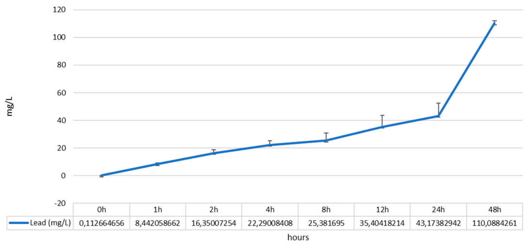
The digestion study showed that Pb pellets were rapidly dissolved at the measured dolphin gastric pH (1.0–1.5), and that high concentrations of Pb (>20 mg/L) were reached in the gastric fluid within 4 hours of the start of the test (Graph 2).
Graph 2. Lead concentration ± s.d. in gastric content of the digestion assay at pH 1.5.
Preprints 220454 g006
Notably, the concentration of lead in the gastric assay doubled between the 24th and 48th hours of testing, indicating how rapidly the dolphins may have been exposed to extremely high concentrations (110 mg/L) of bioavailable lead over a short period.

4. Discussion

The clinical presentation observed in this group of BD is consistent with an acute toxic exposure and closely parallels previously reported cases of lead intoxication in marine mammals. Although the initial signs were nonspecific, including intermittent appetite and changes in attention, the rapid progression toward gastrointestinal and behavioral abnormalities, together with the near-simultaneous involvement of multiple individuals, strongly supports a common-source intoxication rather than an infectious or primary inflammatory process. Gastrointestinal signs were prominent and appeared early in the course of the disease. Behavioral indicators of pain and discomfort were consistently observed across affected individuals. Notably, calves exhibited decreased appetite while continuing to nurse, suggesting a partial preservation of suckling behavior despite systemic illness. This pattern likely reflects both age-related differences in exposure dose and the nonspecific nature of early lead toxicosis.
Comparable clinical features have been reported in previous cases of lead intoxication in odontocetes. Shlosberg et al. [6] described a case of a BD that developed anorexia, lethargy, and gastrointestinal disturbances following ingestion of air gun pellets, with clinical signs initially vague and nonspecific before progressing. Similarly, Stetter et al. [7] reported anorexia, depression, and signs of discomfort in a dolphin successfully treated for lead toxicosis. Although detailed clinical timelines were not fully documented in that case, the early predominance of gastrointestinal and behavioral signs closely mirrors the presentation observed in the present report. Similarities also extend to pinnipeds. Zabka et al. [18] documented acute lead toxicosis in a harbor seal following ingestion of a lead fishing sinker, characterized by anorexia, lethargy, and gastrointestinal abnormalities, with subsequent neurologic involvement. While neurologic signs were more pronounced in that case, the overlap in early clinical features highlights a potential shared pathophysiological response to lead exposure across marine mammal taxa. Importantly, in both pinnipeds and odontocetes, acute intoxication following ingestion of metallic lead objects appears to result in a rapid onset of clinical disease, likely due to sustained release and absorption of lead within the gastrointestinal tract.
The clinical pattern observed in these dolphins is also consistent with lead intoxication described in terrestrial mammals and birds. In domestic animals, acute lead exposure commonly manifests initially as anorexia, abdominal pain, vomiting, and altered behavior, with neurologic signs often developing later or remaining subtle in early stages [28,29]. In avian species, particularly waterfowl exposed to lead shot or fishing sinkers, early clinical signs frequently include lethargy, anorexia, and gastrointestinal dysfunction prior to the onset of overt neurologic impairment [30]. These cross-species similarities underscore the conserved toxicodynamic effects of lead, despite substantial differences in digestive physiology and feeding behavior.
The predominance of gastrointestinal and behavioral signs, with limited overt neurologic manifestations during the initial phase, is particularly noteworthy. This pattern aligns with acute intoxication scenarios in which metallic lead objects remain within the gastrointestinal tract, resulting in continuous exposure rather than a single bolus absorption. In the present case, the temporal clustering of clinical signs across multiple dolphins further supports ingestion of a shared source of lead pellets as the most plausible etiology. Overall, the clinical presentation described here reinforces existing evidence that lead intoxication in dolphins may initially present as a nonspecific, rapidly progressive gastrointestinal and behavioral syndrome. Given the potential for delayed or subtle neurologic involvement, lead exposure should be considered in the differential diagnosis of acute, multisystemic illness affecting multiple individuals in managed marine mammal populations, particularly when access to lead-containing materials cannot be definitively excluded.
The diagnostic approach in this case was necessarily broad during the initial phase, given the nonspecific nature of the presenting clinical signs. Comprehensive physical examinations, behavioral assessments, hematologic and biochemical analyses, microbiological testing, and multimodal imaging were performed in all individuals as signs progressed. This wide diagnostic scope was essential to rule out infectious, inflammatory, gastrointestinal, and respiratory conditions commonly associated with acute anorexia, lethargy, and discomfort in dolphins [19,21].
Despite the extensive diagnostic workup, the finding that most strongly redirected the clinical reasoning toward a toxic etiology was the abnormal coloration of the serum observed shortly after blood collection and processing. The serum exhibited a marked discoloration inconsistent with hemolysis, lipemia, or icterus, prompting immediate concern for heavy metal intoxication. This visual alteration was considered clinically significant, as it could not be readily explained by dehydration, nutritional status, or routine metabolic disturbances, and it was observed consistently in multiple affected individuals.
Alterations in serum appearance have been previously described in cases of lead intoxication across species and are thought to reflect the presence of circulating lead and associated biochemical disturbances, including interference with heme synthesis and erythrocyte integrity [28,29]. While serum discoloration is not pathognomonic, its recognition in the context of acute gastrointestinal and behavioral signs raised early suspicion of lead exposure and justified prioritizing heavy metal screening over other less likely differentials. In marine mammals, diagnostic confirmation of lead intoxication is often delayed due to the nonspecific nature of clinical signs and the logistical challenges associated with advanced diagnostics. In the dolphin described by Shlosberg et al. [6], diagnosis was ultimately achieved during the necropsy through identification of metallic foreign bodies and heavy metal analysis of both pellets and the tissues collected, but early clinical signs were indistinguishable from other causes of systemic illness. Similarly, Zabka et al. [18] reported that in a harbor seal with acute lead toxicosis, the diagnosis relied on a combination of clinical deterioration, and toxicologic confirmation rather than a single defining clinical sign.
In the present case, the abnormal serum coloration served as an early and practical diagnostic clue, allowing clinicians to narrow the differential diagnosis before imaging confirmed the presence of metallic foreign bodies within the gastrointestinal tract. Radiography proved particularly valuable, as lead pellets were readily visualized due to their radiopacity, allowing both localization and estimation of burden, as previously emphasized in marine mammal and wildlife cases [6,18,30]. Ultrasonography was useful for monitoring organ involvement and assessing secondary effects but was less sensitive for direct detection of metallic lead objects. Endoscopic evaluation further supported the diagnosis by enabling direct visualization and retrieval of lead pellets from the forestomach compartments, confirming ongoing gastrointestinal exposure. The combined use of radiography and gastroscopy has been advocated as a complementary approach in suspected cases of lead ingestion in both marine mammals and terrestrial wildlife, particularly when removal of the source is feasible [28,30].
The quantification of heavy metals in blood confirmed lead as the primary toxic agent, with markedly elevated blood lead concentrations detected in all five dolphins. Although arsenic levels were measurable, the concentrations were considered normal for BD [31]. Lead levels were substantially higher than those reported as background or incidental exposure in marine mammals and are consistent with acute, clinically significant intoxication following ingestion of metallic lead objects [6,18]. The absence of relevant elevations in other heavy metals further supports lead as the sole driver of the observed clinical syndrome. The initial concentration of lead in the blood was significantly higher than the toxic limits established for humans (50 ug/l), cows (200 ug/l), dogs and cats (100 ug/l) [32]. When comparing the results with data available from wild dolphins [31,33,34], the affected group showed levels of lead of up to 350 times more than their wild counterparts. Given the paucity of studies concerning lead intoxication in dolphins, we compared analytical results with those reported by Sheffler et al. [35] in a dairy herd. The reported haematic concentration of lead in cows is comparable, or up to 1.6 times lower, than the concentrations found in the blood of the studied dolphins. The affected bottlenose dolphin calves showed concentrations up to 13 times higher than bovine calves.
Of particular significance was the detection of lead in milk samples from the nursing female dolphins, 2.5-5.5 times higher than those in the studied cows. The calves in the present study presented high levels of lead, comparable to or higher than those of their mothers, while the calf analysed in Sheffler’s report showed levels 46 times lower than those detected in the mother. This finding confirms lactational excretion of lead, a phenomenon well documented in humans and terrestrial mammals but not reported in marine mammals. Lead transfer into milk has been associated with mobilization of lead from maternal bone and soft tissues, especially during periods of metabolic stress such as lactation [36,37]. In the present case, the presence of lead in milk provides a plausible route of secondary exposure for the calves and may partially explain the gastrointestinal signs observed in these animals despite their lower overall clinical severity. This finding has important implications for risk assessment and management of intoxications in social groups containing dependent offspring. The data that calves recovered once they were separated by the mothers and precociously weaned confirms the main role of milk as source of intoxication in calves.
The detection of considerable lead concentrations in urine samples provided additional confirmation of systemic absorption and active excretion. Urinary lead excretion has been described as a useful indicator of ongoing exposure and mobilization of lead from tissues, particularly during acute intoxication or following initiation of chelation therapy [28,29]. The variability observed among individuals is consistent with differences in exposure dose, gastrointestinal retention of lead pellets, metabolic status, and renal handling of the metal.
It is noteworthy that the skin (from live animals) seems to serve as an important excretion pathway for Pb, as it shows concentrations similar to those found in the liver. In Male 2, skin concentrations were even higher than in the other dolphins. Additionally, post-mortem analysis of the skin from Female 1 revealed Pb levels of 46.58 mg/kg w.w. Pb, whereas Female 2, which had the lowest tissue concentrations overall, exhibited just 0.78 mg/kg w.w..
Female 2 displayed lower Pb concentrations in the brain, skin, blubber and muscle compared to the other animals. This raises the question of whether these levels correlate with the severity of symptoms observed.
As intoxication progressed, the development of pulmonary rales, pleural effusion, and neurological signs in the adult females reflects the multisystemic nature of acute lead toxicosis. Pulmonary involvement, including edema and effusion, has been described in both marine mammals and terrestrial species and is thought to result from vascular injury, inflammatory responses, and secondary cardiac or renal dysfunction [18,28]. Neurological signs such as vomiting and regurgitation are consistent with lead’s known effects on the central and peripheral nervous systems, although overt seizures or severe neurologic deficits were not a prominent feature in this case.
Hematologic abnormalities were most pronounced in Female 1, who developed a severe normocytic, normochromic anemia with features consistent with a hemolytic process. Lead-induced anemia is classically associated with impaired heme synthesis and reduced erythrocyte lifespan [29]. While basophilic stippling is often cited as a hallmark of lead intoxication, its absence in this case does not preclude lead toxicity, as this finding is inconsistently reported in acute exposures and varies across species [28,38]. The presence of anisocytosis, polychromasia, and elevated nucleated red blood cells indicates a regenerative response, albeit insufficient to counteract ongoing erythrocyte destruction or impaired production.
White blood cell responses were variable, with neutrophilic leukocytosis and left shift observed in some individuals. Such changes are consistent with systemic inflammation, stress responses, or secondary tissue injury rather than a primary infectious process [21]. Normal platelet counts across all dolphins are notable, as thrombocytopenia has been inconsistently reported in lead intoxication and is not considered a defining feature [28].
Biochemical alterations further highlighted the extent of organ damage. Clear evidence of hepatic and renal dysfunction in the adult females aligns with the known accumulation of lead in the liver and kidneys and its direct nephrotoxic and hepatotoxic effects [29,37]. Hyperlipasemia observed consistently in all five dolphins suggests pancreatic involvement, a less frequently emphasized but biologically plausible consequence of systemic lead toxicity. Pancreatic enzyme elevation has been reported in lead-exposed terrestrial mammals and may reflect direct cellular toxicity or secondary ischemic and inflammatory mechanisms [28].
The observation of markedly low or undetectable cortisol concentrations in the calves is particularly intriguing. Although data on adrenal effects of lead in marine mammals are scarce, experimental and clinical studies in other species have demonstrated that lead can interfere with hypothalamic–pituitary–adrenal axis function, potentially impairing stress hormone synthesis or release [37,39,40,41]. Whether this finding contributed to altered stress responses or disease progression in the calves warrants further investigation.
Chelation therapy resulted in a rapid reduction in blood lead concentrations, confirming effective mobilization and elimination of circulating lead. Stabilization of anemia in two of the three females suggests that chelation, combined with supportive care, partially mitigated ongoing toxic effects. Clinical improvement in respiratory and neurological signs further supports the therapeutic benefit of intervention. However, despite aggressive treatment and intensive supportive care, all three adult females ultimately died, underscoring the severity of exposure and the extent of irreversible organ damage sustained prior to or during treatment.
The progressive clinical deterioration and eventual death of the three adult females, despite intensive supportive care and chelation therapy, underscore the severity and systemic nature of the lead intoxication in this case. The staggered timeline to death, ranging from 18 to 35 days after the onset of clinical signs, suggests individual differences in exposure dose, gastrointestinal retention of lead pellets, metabolic status, and extent of irreversible organ damage at the time of diagnosis.
Female 1 experienced the most rapid clinical decline, culminating in hypoglycemic shock, cardiovascular collapse, and respiratory failure. Severe hypoglycemia, pallor of the oral mucosa, bradycardia, and prolonged apneic episodes are consistent with terminal metabolic failure and impaired hepatic gluconeogenesis, likely compounded by adrenal dysfunction and systemic hypoxia. Hypoglycemia has been reported in advanced cases of lead intoxication in other species and may reflect a combination of hepatic failure, endocrine disruption, and overwhelming metabolic stress [28,37]. The inability to maintain buoyancy prior to death reflects profound neuromuscular and cardiopulmonary compromise, a recognized terminal feature in severely ill cetaceans [21].
Female 2 initially appeared to have a more favorable prognosis, yet her later deterioration highlights the insidious and progressive nature of lead toxicosis. The development of lateralized breathing, worsening pulmonary sounds, and reduced reflexes suggest progressive respiratory compromise, likely due to pulmonary edema, effusion, and declining cardiac function. The decision to humanely euthanize this animal was consistent with established welfare guidelines, given the poor prognosis and evidence of irreversible organ failure.
Female 3 survived the longest but exhibited earlier neurological involvement and more severe weight loss, culminating in dyspnea, regurgitation, and reduced movement prior to death. The prolonged course in this individual may reflect a different balance between exposure magnitude and physiological resilience. However, the persistence of lead pellets within the gastrointestinal tract and progressive tissue accumulation ultimately resulted in fatal multiorgan failure.
In contrast, both male calves survived and continue to thrive. Their survival may be attributed to a lower direct exposure to ingested pellets, partial reliance on milk rather than solid food, and possibly a greater capacity for recovery following chelation. Nevertheless, the elevated lead concentrations detected in their tissues, particularly in the skin, raise concerns regarding long-term subclinical effects and justify continued monitoring. As already stated, high concentrations of Pb observed in these calves might originate from nursing, as milk can ease the transport and absorption of Pb, which is known to be uptaken in the GI tract mimicking that of Ca [42]. Additionally, young animals, as well as children, have a higher absorption capacity [43] compared to that in adults (40-50% vs 10% respectively) and this can contribute to the higher Pb levels observed [42]. Chronic or delayed consequences of lead exposure, including neurobehavioral and endocrine alterations, have been described in other species and cannot be excluded in these animals [37,39,40,41]. It is known that 50 ug/l of Pb in humans decrease kidney function in adults, and reduce fetal growth. In children, these levels are associated with lower academic achievement, decreased IQ, and decreases in specific cognitive measures, increased incidence of attention-related behaviors and problem behaviors [44]. The effect of the toxicosis on the cognitive development and behavior of the surviving dolphin calves is unpredictable and presents challenges for its evaluation.
Necropsy findings across the three females were consistent with severe, multisystemic disease dominated by respiratory, hepatic, renal, and hematopoietic failure. The presence of pleural effusion and ascites, along with pulmonary edema and parenchymal alterations, reflects terminal cardiorespiratory compromise and systemic hypoxia. Similar pulmonary findings have been reported in marine mammals and terrestrial species suffering from acute lead toxicosis and are thought to result from vascular injury, inflammatory responses, and secondary cardiac or renal dysfunction [18,28].
Hepatic enlargement, discoloration, textural changes, and iron accumulation are consistent with lead-induced hepatocellular injury and disrupted iron metabolism. Iron accumulation in hepatocytes has been associated with impaired heme synthesis and altered erythrocyte turnover in lead toxicity [29]. Renal changes, including yellow discoloration of the reniculi and altered parenchymal consistency, align with the kidney’s role as a primary target organ for lead accumulation and toxicity.
The reduced size and pallor of the spleens, together with blood depletion, likely reflect severe anemia and terminal circulatory redistribution. Histopathological evidence of total red marrow depletion with severe myelophthisis provides a compelling explanation for the progressive anemia observed clinically and confirms profound disruption of hematopoiesis. Bone marrow suppression and replacement have been well documented in chronic and severe lead exposure and represent a key mechanism underlying refractory anemia [38].
Neuropathological findings, including degenerative changes in Purkinje cells, hemorrhages, edema, gliosis, and neuronal degeneration, are consistent with lead neurotoxicity. The presence of air bubbles within CNS vessels in Female 3 suggests terminal vascular dysfunction and severe hypoxia. Lead-induced neuronal damage, particularly affecting cerebellar Purkinje cells, has been described in multiple species and correlates with both acute neurological signs and longer-term deficits [37,39,40,41].
The identification of lead-induced intranuclear inclusion bodies in hepatocytes and renal tubular epithelial cells is particularly noteworthy, as these inclusions are considered a histopathological hallmark of lead intoxication across species [18,28]. Their presence in a substantial proportion of affected cells provides definitive pathological confirmation of lead toxicity as the primary cause of organ failure.
Recovery of lead pellets from the gastrointestinal tract of Female 1 and Female 3 confirms ongoing exposure until death and helps explain the limited efficacy of chelation once severe tissue damage had occurred. The high number of pellets retrieved from Female 3 supports the hypothesis of a higher or more prolonged exposure in this individual. Gastric mucosal lesions further suggest local corrosive effects and sustained release of bioavailable lead.
Tissue lead distribution analysis revealed widespread accumulation, with particularly high concentrations in blubber, liver, kidney, brain, and skin. While the liver and kidney are well-established target organs for lead accumulation, the detection of substantial lead concentrations in blubber and skin is of special interest. Blubber may act as a reservoir for lipophilic or protein-bound toxins, potentially contributing to delayed redistribution during periods of metabolic stress [45]. Elevated skin concentrations, particularly in Male 2, mirrored blood lead patterns and suggest that skin biopsies may represent a useful, minimally invasive matrix for monitoring lead exposure in live dolphins, a concept previously proposed for other contaminants but less explored for heavy metals.
Differences in brain lead concentrations among the three females correlated with the severity of neurological signs observed ante-mortem, supporting a dose–effect relationship at the individual level. This finding is consistent with experimental and clinical data in other species demonstrating that brain lead burden is a key determinant of neurological dysfunction [39].
Finally, the digestion assay demonstrated that very high lead concentrations can be reached in the gastric fluid within hours of exposure (>20mg/L in 4h), supporting the hypothesis that metallic lead pellets retained in the forestomach act as a continuous source of bioavailable lead. This mechanism provides a plausible explanation for the rapid progression of clinical signs, sustained tissue accumulation, and poor prognosis once exposure is established, even when chelation therapy is initiated. Assuming 10% absorption of the Pb dissolved in the gastric juice [46], concentrations comparable to those observed in the blood at the time of the first analysis may have been reached within the first few hours after the ingestion of the pellets.
Acute toxicity thresholds for lead in mammalian tissues vary depending on the species. No such thresholds have been established for bottlenose dolphins or cetaceans in general, as lead intoxication is extremely rare in this species and LD50 test could not be defined in these protected animals. However, the mean Pb concentrations detected in the brains of Female 1 and Female 3 exceeded the known threshold for cattle (0.7 mg/kg w.w.) but remained below that threshold defined for horses (3 mg/kg w.w.). In contrast, Female 2 exhibited very low cerebral concentrations.
The Pb concentrations in liver and kidneys of all dolphins were below the thresholds defined for cattle, dogs, and horses (> 5 mg/kg w.w. for both organs).

5. Conclusions

This study contributes to a better understanding of Pb distribution and excretion pathways during acute intoxication in dolphins in a managed setting, expanding current knowledge of tissue dissemination patterns in this species. Skin is identified as a potential diagnostic matrix for Pb exposure, which may be particularly valuable for assessing similar cases in wild populations. In addition, the presence of inclusion bodies within renal and hepatic cells is described for the first time in dolphins and may represent a useful histopathological indicator of lead intoxication in this species.
The combination of diagnostic imaging techniques proved highly effective for the localization, approximate quantification, and identification of foreign bodies as lead pellets, as well as for evaluating associated tissue damage. While definitive diagnosis requires toxicological confirmation, this case also highlights the diagnostic value of careful gross evaluation of blood samples, particularly serum appearance, as an early indicator of potential heavy metal exposure. In managed marine mammal populations, where rapid clinical decision-making is essential, recognition of atypical serum coloration may prompt timely imaging and targeted laboratory testing, thereby facilitating earlier diagnosis and intervention.
Taken together, these findings emphasize that although blood, urine, skin, and milk lead quantification are essential for confirming exposure and guiding therapy, prognosis in acute lead intoxication is strongly influenced by exposure magnitude, duration of gastrointestinal retention of metallic lead, and the extent of organ damage at the time of diagnosis. Notably, this case also highlights lactational transfer as a potentially underrecognized pathway for lead redistribution and exposure within dolphin social groups.
Overall, the clinical course, pathological findings, and tissue distribution patterns observed in this case highlight the devastating consequences of acute lead ingestion in dolphins and emphasize the critical importance of early detection, rapid source removal, and prevention of access to lead-containing materials in managed marine mammal environments. Timely management may significantly improve clinical outcomes. Addressing lead intoxication requires continued vigilance and proactive measures to prevent exposure, thereby safeguarding both animal welfare and public health.

Author Contributions

Conceptualization, G.J.S-C., B.B., and A.Z.; methodology, G.J.S-C., B.B., and A.Z.; validation, G.J.S-C. and A.Z.; formal analysis, G.J.S-C., A.G. and A.Z.; investigation, G.J.S-C., B.B., L.G., G.R. and A.Z.; resources, G.J.S-C., B.B., L.G., G.R., A.G. and A.Z.; data curation, G.J.S-C. and A.Z.; writing—original draft preparation, G.J.S-C., and A.Z.; writing—review and editing, G.J.S-C., B.B., L.G., G.R. and A.Z.; visualization, G.J.S-C. and A.Z;. project administration, G.J.S-C. and A.Z. All authors have read and agreed to the published version of the manuscript.

Funding

This research received no external funding.

Institutional Review Board Statement

Ethical review and approval were waived for this study because it is a retrospective descriptive case based on an acute lead intoxication episode that occurred during routine veterinary care. All diagnostic procedures, treatments, sampling, and clinical management were performed as part of necessary medical intervention for the affected animals, in accordance with standard veterinary practice and under established institutional animal care protocols. No experimental procedures were conducted, and no interventions were administered for research purposes. Data were collected and analyzed retrospectively from existing medical records. The study did not involve any additional handling, manipulation, or procedures beyond those required for the diagnosis and treatment of the animals, and therefore did not alter the welfare or clinical management of the individuals involved. As such, the work falls outside the scope of prospective animal experimentation requiring formal ethical approval, and ethical review was therefore waived.

Data Availability Statement

Data is contained within the article.

Acknowledgments

The authors would first like to thank the entire animal care team and staff who worked tirelessly to care for the affected animals. We would also like to acknowledge Dr. Kerstin Ternes, whose assistance was instrumental in securing part of the medication required for the treatment of the intoxicated animals in a timely manner. We are also grateful to Drs. Jenny Meegan and Forrest Gomez of the National Marine Mammal Foundation and the U.S. Navy Marine Mammal Program for their support and professional advice throughout the case management process. The authors would like to thank Dr. Lucia Biagini for her technical support, and Erika Eusebio for her assistance during laboratory analyses conducted as part of this investigation.

Conflicts of Interest

The authors declare no conflicts of interest.

Abbreviations

The following abbreviations are used in this manuscript:
BD Bottlenose Dolphin
PEs Physical Exams
GI Gastrointestinal
CNS Central Nervous System
NRBC Nucleated Red Blood Cells
PLT Platelet
WBC White Blood Cell
PO Per Os - Oral administration
BID Bis In Die - Twice a day
IM Intramuscular
SID Semel In Die - Once a day
Pb Lead
CaNa2EDTA Calcium EDTA
DMSA Dimercaptosuccinic Acid
Buscopan Hyoscine Butylbromide
MRL Maximum Residual Limit

References

  1. Wells, R.S.; Natoli, A.; Braulik, G. Tursiops truncatus (errata version published in 2019). IUCN Red. List Threat. Species 2019, 2019–2011. [Google Scholar]
  2. Dziobak, M.K.; Fahlman, A.; Wells, R.S.; Takeshita, R.; Smith, C.; Gray, A.; Weinstein, J.; Hart, L.B. First evidence of microplastic inhalation among free-ranging small cetaceans. PLoS ONE 2024, 19, e0309377. [Google Scholar] [CrossRef] [PubMed]
  3. Ocampos, A.I.; Guinn, M.A.; Elliott, J.; Wittmaack, C.; Sinclair, C.; Abdulla, H.; Orbach, D.N. Pharmaceuticals in the blubber of live free-swimming common bottlenose dolphins (Tursiops truncatus). Iscience 2024, 27. [Google Scholar] [CrossRef] [PubMed]
  4. Mancia, A. On the revolution of cetacean evolution. Mar. Genom. 2018, 41, 1–5. [Google Scholar] [CrossRef]
  5. Grigg, E.; Markowitz, H. Habitat use by bottlenose dolphins (Tursiops truncatus) at Turneffe Atoll, Belize. Aquat. Mamm. 1997, 23, 163–170. [Google Scholar]
  6. Shlosberg, A.; Bellaiche, M.; Regev, S.; Gal, R.; Brizzi, M.; Hanji, V.; Zaidel, L.; Nyska, A. Lead toxicosis in a captive bottlenose dolphin (Tursiops truncatus) consequent to ingestion of air gun pellets. J. Wildl. Dis. 1997, 33, 135–139. [Google Scholar] [CrossRef] [PubMed]
  7. Stetter, M.; Mangold, B.; Miller, M.; Weber, M.; Capobianco, J. Successful treatment of lead toxicosis in a bottlenose dolphin (Tursiops truncatus). In Proceedings of the Proceedings of the International Association for Aquatic Animal Medicine, 1999; pp. 146–147. [Google Scholar]
  8. Martel, A.K.; Doss, G.A.; Mans, C. Suspected peripheral neuropathy secondary to lead intoxication in three psittacine birds. J. Exot. Pet. Med. 2020, 32, 13–17. [Google Scholar] [CrossRef]
  9. Chiverton, L.; Cromie, R.; Kock, R. European mammal exposure to lead from ammunition and fishing weight sources. Heliyon 2022, 8. [Google Scholar] [CrossRef] [PubMed]
  10. Cowan, V.; Blakley, B. Acute lead poisoning in western Canadian cattle—A 16-year retrospective study of diagnostic case records. Can. Vet. J. 2016, 57, 421. [Google Scholar] [PubMed]
  11. Bautista, A.C.; Puschner, B.; Poppenga, R.H. Lead exposure from backyard chicken eggs: a public health risk? J. Med. Toxicol. 2014, 10, 311–315. [Google Scholar] [CrossRef] [PubMed]
  12. Legagneux, P.; Suffice, P.; Messier, J.-S.; Lelievre, F.; Tremblay, J.A.; Maisonneuve, C.; Saint-Louis, R.; Bêty, J. High risk of lead contamination for scavengers in an area with high moose hunting success. PLoS ONE 2014, 9, e111546. [Google Scholar] [CrossRef] [PubMed]
  13. Zook, B.; Sauer, R.; Garner, F. Lead poisoning in captive wild animals. J. Wildl. Dis. 1972, 8, 264–272. [Google Scholar] [CrossRef] [PubMed]
  14. Blakley, B.R. Lead Poisoning in Animals. Available online: https://www.msdvetmanual.com/toxicology/lead-poisoning/lead-poisoning-in-animals. (accessed on 6 October 2024).
  15. California Department of Food and Agriculture. Lead toxicity and its effects on animals and animal products. Available online: https://www.cdfa.ca.gov/ahfss/Animal_Health/pdfs/leadtoxicity.pdf (accessed on 15 march 2026).
  16. Pain, D.J.; Fisher, I.; Thomas, V.G. A global update of lead poisoning in terrestrial birds from ammunition sources. Ingestion Lead. From Spent Ammunit. Implic. Wildl. Hum. 2009, 99–118. [Google Scholar]
  17. Kelly, T.R.; Grantham, J.; George, D.; Welch, A.; Brandt, J.; Burnett, L.J.; Sorenson, K.J.; Johnson, M.; Poppenga, R.; Moen, D. Spatiotemporal patterns and risk factors for lead exposure in endangered California condors during 15 years of reintroduction. Conserv. Biol. 2014, 28, 1721–1730. [Google Scholar] [CrossRef] [PubMed]
  18. Zabka, T.S.; Haulena, M.; Puschner, B.; Gulland, F.M.; Conrad, P.A.; Lowenstine, L. Acute lead toxicosis in a harbor seal (Phoca vitulina richardsi) consequent to ingestion of a lead fishing sinker. J. Wildl. Dis. 2006, 42, 651–657. [Google Scholar] [CrossRef] [PubMed]
  19. Sweeney, J.C.; Reddy, M.L.; Lipscomb, T.P.; Bjorneby, J.M.; Ridgway, S.H. Handbook of cetacean cytology. In USA: Dolphin Quest Inc; 1999. [Google Scholar]
  20. Corrons, J.-L.V.; Albarède, S.; Flandrin, G.; Heller, S.; Horvath, K.; Houwen, B.; Nordin, G.; Sarkani, E.; Skitek, M.; Van Blerk, M. Guidelines for blood smear preparation and staining procedure for setting up an external quality assessment scheme for blood smear interpretation. Part I: Control material. Clin. Chem. Lab. Med. 2004, 42. [Google Scholar] [CrossRef]
  21. Geraci, J.R.; Lounsbury, V.J. Marine mammals ashore: a field guide for strandings; National Aquarium in Baltimore, 2005. [Google Scholar]
  22. Bryan, C.E.; Ragland, J.M. Dolphins in human care sampling protocol and sample entry assistant (SEA) for tissue archival and Analyte analysis. Natl. Inst. Stand. Technol. Interag. Intern. Rep. 2020, 8281, 1–27. [Google Scholar] [CrossRef]
  23. Pugliares, K.R.; Bogomolni, A.; Touhey, K.M.; Herzig, S.M.; Harry, C.T.; Moore, M.J. Marine mammal necropsy: an introductory guide for stranding responders and field biologists; Woods Hole Oceanographic Institution: Woods Hole, MA, 2007. [Google Scholar]
  24. Zaccaroni, A.; Corteggio, A.; Altamura, G.; Silvi, M.; Di Vaia, R.; Formigaro, C.; Borzacchiello, G. Elements levels in dogs from “triangle of death” and different areas of Campania region (Italy). Chemosphere 2014, 108, 62–69. [Google Scholar] [CrossRef] [PubMed]
  25. European Commission. Regulation of 25 April 2023 on maximum levels for certain contaminants in food and repealing Regulation (EC). Off. J. 5/05/2023 2023, L 119/103 No 1881/2006, 915/2023/EU. [Google Scholar]
  26. Codex Alimentarius. Code of practice for the prevention and reduction of lead contamination in foods. CAC/RCP 2004, 56–2004. [Google Scholar]
  27. Stacy, N.I.; Nollens, H.H. Hematology of marine mammals. Schalm's Vet. Hematol. 2022, 1104–1113. [Google Scholar] [CrossRef]
  28. Osweiler, G. Metals and minerals. Toxicology 1996, 179–211. [Google Scholar]
  29. Goyer, R.A. Toxic and essential metal interactions. Annu. Rev. Nutr. 1997, 17, 37–50. [Google Scholar] [CrossRef] [PubMed]
  30. Friend, M.; Franson, J.C.; Ciganovich, E.A. Field manual of wildlife diseases: general field procedures and diseases of birds; Geological Survey (USGS), 1999. [Google Scholar]
  31. Bryan, C.E.; Christopher, S.J.; Balmer, B.C.; Wells, R.S. Establishing baseline levels of trace elements in blood and skin of bottlenose dolphins in Sarasota Bay, Florida: implications for non-invasive monitoring. Sci. Total Environ. 2007, 388, 325–342. [Google Scholar] [CrossRef] [PubMed]
  32. Puls, R. Mineral levels in animal health: diagnostic data; 1994. [Google Scholar]
  33. Meador, J.; Ernest, D.; Hohn, A.; Tilbury, K.; Gorzelany, J.; Worthy, G.; Stein, J. Comparison of elements in bottlenose dolphins stranded on the beaches of Texas and Florida in the Gulf of Mexico over a one-year period. Arch. Environ. Contam. Toxicol. 1999, 36, 87–98. [Google Scholar] [CrossRef] [PubMed]
  34. Stein, J.E.; Tilbury, K.L. Ecotoxicological investigations of bottlenose dolphin (Tursiops truncatus) strandings: accumulation of persistent organic chemicals and metals. In Toxicology of marine mammals; CRC Press, 2002; pp. 470–500. [Google Scholar]
  35. Sheffler, R.; Rebolloso, S.; Scott, I.; Buchweitz, J.P.; Puschner, B. Milk and Whole Blood Surveillance Following Lethal and Sublethal Lead Intoxication in a Michigan Dairy Herd. Toxics 2025, 13, 445. [Google Scholar] [CrossRef] [PubMed]
  36. Gulson, B.L.; Mizon, K.J.; Korsch, M.J.; Palmer, J.M.; Donnelly, J.B. Mobilization of lead from human bone tissue during pregnancy and lactation—a summary of long-term research. Sci. Total Environ. 2003, 303, 79–104. [Google Scholar] [PubMed]
  37. (ATSDR), A.f.T.S.a.D.R. Toxicological profile for Lead; U.S. Department of Health and Human Services, Public Health Service: Atlanta, GA, 2020. [Google Scholar]
  38. Stockham, S.L.; Scott, M.A. Fundamentals of veterinary clinical pathology; John Wiley & Sons, 2024. [Google Scholar]
  39. Canfield, R.L.; Gendle, M.H.; Cory-Slechta, D.A. Impaired neuropsychological functioning in lead-exposed children. Dev. Neuropsychol. 2004, 26, 513–540. [Google Scholar] [CrossRef] [PubMed]
  40. Cory-Slechta, D.A. Developmental Exposure to Lead: Overview and Integration of Neurobehavioral Consequences and Mediation. Environ. Factors Neurodev. Neurodegener. Disord. 2015, 139–165. [Google Scholar]
  41. Virgolini, M.; Rossi-George, A.; Lisek, R.; Weston, D.; Thiruchelvam, M.; Cory-Slechta, D. CNS effects of developmental Pb exposure are enhanced by combined maternal and offspring stress. Neurotoxicology 2008, 29, 812–827. [Google Scholar] [CrossRef] [PubMed]
  42. Heath, L.; Soole, K.L.; McLaughlin, M.L.; McEwan, G.T.A.; Edwards, J. Toxicity of environmental lead and the influence of intestinal absorption in children. Rev. Environ. Health 2003, 18, 20. [Google Scholar] [CrossRef]
  43. Ziegler, E.E.; Edwards, B.B.; Jensen, R.L.; Mahaffey, K.R.; Fomon, S.J. Absorption and retention of lead by infants. Pediatr. Res. 1978, 12, 29–34. [Google Scholar] [CrossRef] [PubMed]
  44. National Institute of Environmental Health Sciences. Lead. Available online: https://www.niehs.nih.gov/health/topics/agents/lead (accessed on 15 march 2026).
  45. Borrell, A.; Cantos, G.; Pastor, T.; Aguilar, A. Organochlorine compounds in common dolphins (Delphinus delphis) from the Atlantic and Mediterranean waters of Spain. Environ. Pollut. 2001, 114, 265–274. [Google Scholar] [CrossRef] [PubMed]
  46. Rabinowitz, M.B.; Wetherill, G.W.; Kopple, J.D. Kinetic analysis of lead metabolism in healthy humans. J. Clin. Investig. 1976, 58, 260–270. [Google Scholar] [CrossRef] [PubMed]
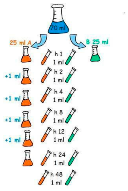
Figure 1. Diagram representing digestion protocol.
Preprints 220454 g001
Figure 2. Ultrasound images illustrating highly echogenic lead pellets (2.2–2.4 mm in diameter) with intense acoustic shadows, observed in the first, second, and third gastric chambers of affected dolphins.
Preprints 220454 g002
Figure 3. Radiographic images demonstrate radiopaque spherical objects (lead pellets) located within the gastric chambers of Female 1 (A) and 3 (B), and embedded in the gums of Female 2 (C).
Preprints 220454 g003
Figure 4. Gastroscopic images highlighting lead pellets retrieved from the first and second stomach chambers of affected females, along with visible irritation and damage to the gastric mucosa caused by the foreign objects.
Preprints 220454 g004
Table 1. Bottlenose dolphins affected by the lead toxicosis.
INDIVIDUAL AGE (y) ORIGIN RELATIONSHIP CLINICAL HISTORY
Female 1 24 Wild-born Mother of Male 1 Urethral prolapse
Female 2 22 Wild-born Mother of Female 3 and Male 1 Respiratory mycosis
Female 3 6 Captive-born Daughter of Female 2 Lower rank in the pod’s hierarchy. Rake marks from recent interaction with another dolphin. Ovarian cyst on the left ovary.
Male 1 3 Captive-born Son of Female 2 and brother of Female 3 Nursing calf
Male 2 2 Captive-born Son of Female 1 Nursing calf
Table 2. Name, dosage, and route of administration of the medication and supplements included in the treatment of the five animals affected by the lead poisoning.
Name Dosage Comment
Calcium EDTA (CaNa2 EDTA) 4500 mg PO BID Given in cycles of one week on, one week off.
Dimercapto succinic acid (DMSA) 600 mg PO BID Initially given in cycles of one week on, one week off. Later decided to maintain it daily.
Garlic 1 clove PO SID
Vitamin C 300 - 500 mg/Kg PO SID
Active charcoal 1 - 5 g/Kg PP SID-BID
Omeprazol 40 mg PO SID
Vitamin B complex 2 - 5 tablets PO BID Bécozyme Forte. Manufacturer: Dragenopharm Apotheker
Püschl GmbH, Göllstrasse 1, D-84529 Tittmoning (Germany)
Vitamin B1 300 mg PO SID
Vitamin K 3 mg PO BID
Folic acid 50 mg PO SID
Stanozolol 4 mg PO SID
Yunnan Baiyao 2 capsules PO SID
Sucralfate 2 g PO SID
Metoclopramide 0.1 mg/Kg IM BID
Maropitant 20 mg IM SID
Paraffin oil 100 ml PO SID-BID
Tramadol 0.66 mg/Kg PO BID
Hyoscine butylbromide (Buscopan) 10 mg PO BID
Silimarin 240 mg PO BID
Pancreatic enzymes 8 g PO Each feeding
Dexamethasone 0.06 mg/Kg IM BID
Prednisolone 0.5 - 1 mg/Kg PO SID
Furosemide 3 mg/Kg IM SID / 40 mg PO SID
Table 4. Results of the initial quantification of heavy metals in the blood collected from the five affected dolphins.
HEAVY METALS Female 1 Female 2 Female 3 Male 1 Male 2
ARSENIC ug/l 53,4 54,2 25,1 11,3 19.6
THALLIUM ug/l <1 <1 <1 <1 <1
CHROME ug/l <5 <5 <5 <5 <5
CADMIUM ug/l <1 <1 <1 <1 <1
NICKEL ug/l <1 <1 <1 <1 <1
LEAD ug/l 1096,9 666,8 876,3 785 1726.7
Table 5. Results of the urine samples (N) analysed.
Valid N Mean Minimum Maximum Std.Dev.
Female 1 10 0,042433 0,014880 0,090520 0,030528
Female 2 13 0,046260 0,015050 0,172190 0,048342
Female 3 6 0,131697 0,021020 0,529560 0,197773
Table 7. Concentrations of Pb (mg/kg) found in the various tissues analyzed.
[Pb] mg/kg Female 1 Female 2 Female 3 Male 1 Male 2
BLUBBER 2,881 0,213 0,405 - -
BRAIN 0,569 0,031 1,063 - -
KIDNEY 0,291 0,991 1,159 - -
LIVER 1,072 1,006 1,340 - -
MUSCLE 0,551 0,213 0,904 - -
SKIN Ante-mortem 1,131 0,598 1,124 - 2,163
SKIN Post-mortem 46,58 0,781 1,328 - -
Disclaimer/Publisher’s Note: The statements, opinions and data contained in all publications are solely those of the individual author(s) and contributor(s) and not of MDPI and/or the editor(s). MDPI and/or the editor(s) disclaim responsibility for any injury to people or property resulting from any ideas, methods, instructions or products referred to in the content.
Copyright: This open access article is published under a Creative Commons CC BY 4.0 license, which permit the free download, distribution, and reuse, provided that the author and preprint are cited in any reuse.
Prerpints.org logo

Preprints.org is a free preprint server supported by MDPI in Basel, Switzerland.

Subscribe

© 2026 MDPI (Basel, Switzerland) unless otherwise stated

Accessibility

Disclaimer

Terms of Use

Privacy Policy

Privacy Settings