

Submitted:
26 June 2026
Posted:
29 June 2026
You are already at the latest version
Preprints on COVID-19 and SARS-CoV-2
Abstract
The intersection of COVID-19 and neurological complications has highlighted post-COVID anti-N-methyl-D-aspartate receptor (NMDAR) encephalitis, a severe autoimmune disorder mediated by antibodies targeting NMDA receptors. This systematic review and institutional case series aimed to synthesize existing literature and present eight new cases from Tehran, Iran, to clarify the pathophysiology, prevalence, clinical manifestations, diagnostic challenges, and therapeutic strategies of this emerging condition. A total of 23 adult patients were analyzed, including 15 cases from the literature and 8 new institutional cases. The mean age was 35.6 years, with a female predominance (60.9%). SARS-CoV-2 infection was confirmed in 91.3% of patients by RT-PCR. The median latency from COVID-19 diagnosis to encephalitis onset was 10 days, with 91.3% occurring within the first month. Neuropsychiatric manifestations were nearly universal (91.3%), including psychosis/hallucinations (56.5%), behavioral or cognitive disturbances (52.2%), seizures (52.2%), movement disorders (34.8%), and altered consciousness (43.5%). MRI revealed abnormalities in 59.1% (most frequently hippocampal involvement), and EEG abnormalities were present in 76.2%, with diffuse slowing, epileptiform activity, and delta brush patterns. Anti-NMDAR antibodies were detected in CSF or serum in 60.9% of cases, with pleocytosis in 34.8%. First-line immunotherapies included intravenous methylprednisolone (87.0%), intravenous immunoglobulin (56.5%), and plasmapheresis (34.8%), while rituximab was used in 30.4% as second-line therapy. At a median 3-month follow-up, 52.2% achieved full recovery, 39.1% partial recovery, and 8.7% died. This study underscores the temporal association between COVID-19 and anti-NMDAR encephalitis, with a consistent clinical phenotype and treatment response resembling classical cases. Early recognition, timely antibody testing, and initiation of immunotherapy are critical to improving outcomes. Larger, prospective studies are warranted to confirm causality, refine diagnostic algorithms, and optimize management strategies.
Keywords:
anti-NMDA receptor encephalitis
; COVID-19
; SARS-CoV-2
; autoimmune encephalitis
; neurological complication
1. Introduction
The Severe Acute Respiratory Syndrome Coronavirus 2 (SARS-CoV-2), first identified in Wuhan, China, in December 2019, is responsible for the global outbreak of Coronavirus Disease 2019 (COVID-19), which has since posed major public health challenges worldwide [1,2].
While the acute infection has been the primary focus of clinical and research attention, increasing evidence highlights that 10–30% of patients experience persistent, long-term effects collectively known as post-COVID syndrome (PCS) [3]. The National Institute for Health and Care Excellence (NICE) defines PCS as symptoms that develop during or after a COVID-19 infection that last for more than 12 weeks, and cannot be explained by an alternative diagnosis [3]. PCS encompasses a wide range of manifestations, including chronic fatigue, musculoskeletal pain, respiratory difficulties, cardiovascular irregularities, and, notably, neurological and neuropsychiatric conditions [3,4].
Among these neurological sequelae, encephalitis has emerged as a significant but poorly understood complication of COVID-19. The proposed pathogenesis of COVID-19-related encephalitis is multifactorial, with three main mechanisms suggested: (i) direct invasion of the central nervous system (CNS), (ii) systemic inflammation, and (iii) molecular mimicry. Direct viral invasion may occur through either transsynaptic propagation or hematogenous spread, facilitated by SARS-CoV-2 binding to angiotensin-converting enzyme 2 (ACE2) receptors on neuronal and endothelial cells [5,6]. However, as cerebrospinal fluid (CSF) polymerase chain reaction tests are frequently negative in COVID-19 encephalitis, direct invasion is considered less likely [7,8,9,10]. More plausibly, systemic inflammation and cytokine dysregulation, particularly involving IL-6 and Th17 cell pathways, can disrupt the blood–brain barrier and contribute to neurological damage [11,12,13,14,15,16]. Additionally, molecular mimicry may trigger autoimmune responses, with antibodies generated against SARS-CoV-2 antigens cross-reacting with neuronal epitopes, potentially leading to autoimmune encephalitis such as anti-N-methyl-D-aspartate receptor (NMDAR) encephalitis [6,17,18,19,20,21].
Anti-NMDAR encephalitis is an autoimmune disorder characterized by IgG antibodies against the NR1 subunit of the NMDA receptor, resulting in neuropsychiatric and neurological symptoms [22]. Although relatively rare—approximately one case per 1.5 million people annually—it is one of the most recognized subtypes of autoimmune encephalitis, affecting predominantly young women [23].
The disease typically evolves in stages, beginning with viral-like prodromes, followed by acute psychiatric symptoms (e.g., psychosis, agitation), then progressing to seizures, movement disorders, dysautonomia, and, in severe cases, coma [24]. Recovery is often prolonged, with persistent cognitive and behavioral impairments even after inflammation resolves [24].
The diagnostic criteria for anti-NMDAR encephalitis are categorized as probable or definite [24]. A probable diagnosis involves the rapid onset (within three months) of at least four out of six major symptom groups: abnormal psychiatric or cognitive behavior, speech dysfunction (e.g., pressured speech, verbal reduction, or mutism), seizures, movement disorders (such as dyskinesias or rigidity), decreased consciousness, and autonomic dysfunction or central hypoventilation [24]. Supporting findings may include an abnormal EEG, showing focal or diffuse slow or disorganized activity, epileptic activity, or an extreme delta brush pattern, along with CSF abnormalities like pleocytosis or oligoclonal bands [24]. Alternatively, three major symptoms plus a systemic teratoma can support a probable diagnosis [24]. For a definite diagnosis, one major symptom group along with IgG GluN1 antibodies is required; if only serum is available, additional confirmatory tests, such as immunohistochemistry or cell-based assays, are recommended [24].
Before the COVID-19 pandemic, viral infections (e.g., herpes simplex, varicella zoster) and paraneoplastic conditions such as ovarian teratoma were known triggers of anti-NMDAR encephalitis [25,26]. However, the potential association between SARS-CoV-2 and the development of this autoimmune encephalitis remains underexplored. Given the global burden of COVID-19 and growing case reports of post-COVID encephalitis, there is an urgent need to synthesize the emerging evidence.
The objective of this systematic review is to systematically map and analyze the literature on anti-NMDAR encephalitis following COVID-19 infection. Specifically, this review aims to describe the clinical manifestations and diagnostic challenges, summarize current therapeutic approaches, enhance early recognition to improve patient outcomes, and highlight gaps in knowledge to guide future research. Additionally, we present eight post-COVID-19 anti-NMDAR encephalitis cases to contribute to the growing body of evidence.
2. Methods
Design: This study consisted of two components, a sysytematic review of published literature and a case series of eight new patients diagnosed with post-COVID-19 anti-NMDAR encephalitis at Loghman Hakim Hospital, Tehran, Iran. The systenatic review was conducted following the PRISMA guidelines. We additionally followed the methodological framework established by Arksey and O’Malley, which is commonly recommended for systematic reviews.
2.1. Search Strategy
We searched five databases of PubMed, MEDLINE, Scopus, Embase, and Web of Science to extract case report and case series articles on autoimmune encephalitis associated with SARS-COV-2 published from the inception to September 23, 2024. The search strategy consisted of keywords including: ([SARS-CoV-2] OR COVID OR COVID-19 OR coronavirus) AND (Anti-NMDR Encephalitis OR Non paraneoplastic Anti-NMDR Encephalitis) OR ([Anti-N-Methyl-D-Aspartate Receptor Encephalitis] OR [NMDA] OR [Autoimmune encephalitis] OR [anti-NMDA receptor autoantibody] OR [Receptors, N-Methyl-D-Aspartate]). The search field was limited to ‘title/abstract’. No language restriction was applied. complete search strategy table presented in the supplementary file (Table S1).
Eligibility criteria: We included studies that reported cases of anti-NMDA receptor encephalitis associated with COVID-19 infection. We excluded studies involving other types of encephalitis, as well as cases of autoimmune encephalitis without confirmed anti-NMDA receptor antibodies in CSF or serum, and cases involving individuals under 18 years old. Excluding patients under 18 avoids heterogeneity due to distinct pathophysiology, syndromes, and clinical data, ensuring a focused and consistent analysis of adult post-COVID encephalitis.
Study Selection: The selection of articles followed a three-step process. First, duplicate articles were omitted. The titles and abstracts of the remaining articles were screened by two reviewers independently. Next, the full texts of the selected studies were reviewed in accordance with the eligibility criteria. Any disagreements were resolved by a third reviewer. Data extraction was then performed independently by two reviewers. A PRISMA flow chart of the search process is shown in Figure 1.
- Data Extraction: The following data points were systematically collected from each eligible study: Study ID, patient demographics (age, sex, past medical history), COVID-19 test results, interval between COVID-19 presentation and onset of encephalitis, neurological and psychiatric symptoms, neuroimaging and EEG findings, serum and CSF analysis, treatment modalities, and patient outcomes. Two reviewers independently extracted the data using a standardized form. Discrepancies were resolved through discussion or consultation with a third reviewer. The data were then compiled into a comprehensive dataset for further analysis.
Data Synthesis and Analysis: We synthesized the extracted data to categorize qualitative and quantitative data points for comprehensive analysis. Using IBM SPSS statistics 27 (IBM Corp., Armonk, NY, USA), we applied the following methodologies:
Qualitative Data: Summarized as percentages to identify trends and patterns.
Quantitative Data: Summarized as mean ± standard deviation (SD) to understand data distribution.
This combined approach provided an overview of the clinical features, diagnostic approaches, treatments, and outcomes of anti-NMDAR encephalitis following SARS-CoV-2 infection.
2.2. Case Series
Setting and Patients: Eight patients with post-COVID-19 anti-1NMDAR encephalitis were identified and treated at Loghman Hakim Hospital, Tehran, Iran. All patients were ≥18 years old with confirmed anti-NMDAR antibodies in CSF and/or serum.
Data Collection: For each patient, the following clinical data were collected from medical records: Demographics and past medical history, COVID-19 diagnostic confirmation, Interval between COVID-19 presentation and onset of encephalitis, Neurological and psychiatric manifestations, neuroimaging and EEG findings, CSF and serum findings, treatment modalities and patient outcomes.
Ethical Considerations: The case series component was conducted in compliance with institutional ethical standards. Ethical approval was obtained from the Shahid Beheshti University of Medical Sciences Research Ethics Committee (approval number: IR.SBMU.RETECH.REC.1403.864). Written informed consent was obtained from patients or their legal guardians for inclusion in the study and publication of anonymized clinical data.
3. Results
3.1. Study Selection
A systematic search of PubMed, Scopus, Web of Science, and Embase initially identified 817 potentially relevant records. After removing 261 duplicate records, 556 articles were screened based on titles and abstracts. Of these, 489 records were excluded for not meeting the eligibility criteria, leaving 67 reports sought for full-text retrieval. One report could not be retrieved, resulting in 66 full-text articles assessed for eligibility. Following detailed evaluation against the inclusion and exclusion criteria, 51 reports were excluded for reasons including: pediatric population (under 18 years), absence of confirmed anti-NMDAR antibodies in CSF or serum, non-SARS-CoV-2-associated encephalitis, or insufficient clinical data for extraction. Ultimately, 15 studies met all eligibility criteria and were included in the final scoping review and pooled analysis. A PRISMA flow diagram summarizing the study selection process is presented in Figure 1.
3.2. Demographic Characteristics
The study included 23 adult patients with anti-NMDAR encephalitis temporally associated with SARS-CoV-2 infection, comprising 15 cases identified from the literature (65.2%) and eight newly reported cases from Loghman Hospital, Tehran, Iran (34.8%). Ages ranged from 18 to 76 years, with a mean of 35.6 ± 16.4 years and a median of 30 years (IQR: 22.5–47.0), with a female predominance (60.9%, n = 14) compared with males (39.1%, n = 9).
Thirteen patients (56.5%) had at least one comorbidity, with the remainder having no significant past medical history. The most frequent comorbid conditions were psychiatric or substance-use disorders (5/23, 21.7%), cardiovascular disease such as hypertension or prior myocardial infarction (3/23, 13.0%), autoimmune diseases such as systemic lupus erythematosus and p-ANCA vasculitis (2/23, 8.7%), and prior neurologic conditions such as epilepsy or stroke (2/23, 8.7%). Other reported conditions included oncologic/paraneoplastic history (e.g., ovarian teratoma, breast cancer in remission) (2/23, 8.7%), chronic infections (e.g., hepatitis C, prior varicella zoster virus) (8.7%), metabolic disorders such as diabetes mellitus (1/23, 4.3%), renal disease such as nephrotic syndrome (1/23, 4.3%), and pregnancy-related complications (1/23, 4.3%). A specific paraneoplastic trigger, ovarian teratoma, was identified in one case (4.3%) (See Table 3).

Table 1.
Characteristics of the Cases sourced from the literature.
| Study ID | Age, Sex | Relevant PMH | COVID test | Onset | Key neuro/psychiatric features | Imaging | EEG | Serum and CSF analysis | Treatment | outcome |
|---|---|---|---|---|---|---|---|---|---|---|
| Allahyari, F. et al. [27] | 18, F | — | RT-PCR + | Simultaneous | AMS, GTC seizures, meningismus | CT/MRI: generalized edema | - | CSF + anti-NMDAR; SARS-CoV-2 PCR + | IVIG, IVMP, oral steroids | Full recovery |
| Álvarez Bravo et al. [28] | 30, F | Left ovarian teratoma | RT-PCR + | 3 days | Psychomotor agitation, hallucinations, focal → GTC seizures, | MRI: Lt hippocampal hyperintensity | Lt frontotemporal epileptic discharges; delta brush | CSF + anti-NMDAR | IVIG, IVMP, AEDs, rituximab | Partial recovery |
| O.V. Ulyanova et al. [29] | 22, F | Hepatitis C | RT-PCR + | 9 days | Psychosis, catatonia → GTC seizures | CT/MRI: normal | Diffuse abnormal potentials | Serum & CSF + anti-NMDAR | IVMP, plasmapheresis, IVIG, AEDs | Death (cerebral edema, pneumonia) |
| Gabriele Melegari et al. [30] | 31, F | — | COVID IgG + | 20 days | Anosmia and Ageusia | MRI: olfactory nucleus hyperintensity | — | Serum + MAG and NMDA Abs | Supportive, vitamin C | Partial recovery |
| T. Hainmueller et al. [31] | 18, M | — | Rapid antigen + | 5 weeks | Seizure, confusion, psychosis, catatonia | CT/MRI: normal | Diffuse polymorphic slowing | CSF + anti-NMDAR | AEDs, IVMP, IVIG, rituximab | Full recovery |
| Caitlin N et al. [32] | 27, M | Mood/psychotic disorder | RT-PCR + | simultaneous | Racing thoughts, hallucinations, suicidal ideation, seizures | MRI: normal | Normal | CSF + anti-NMDAR | IVMP, plasmapheresis, rituximab | Full recovery |
| Lee H. et al. [33] | 21, F | — | RT-PCR + | 3 days | Short-term memory loss, abnormal behavior | MRI: cerebellar & hippocampal lesions | Diffuse beta ± sharp waves temporal | CSF & serum + anti-NMDAR; lymphocytic pleocytosis | Acyclovir, IVMP, IVIG | Partial recovery |
| McHattie et al. [34] | 53, F | Breast Ca (remission), depression | RT-PCR + | 2 weeks | Palilalia, echolalia, dysautonomia, focal seizures | MRI: Lt amygdala & putamen hyperintensity | Slow activity, no epileptiform discharges | CSF + anti-NMDAR, lymphocytic pleocytosis | HCQ, IVIG, tocilizumab, AEDs | Partial recovery |
| Monti et al. [35] | 50, M | HTN | RT-PCR + | 3 days | Refractory status epilepticus, orofacial dyskinesia | MRI: Normal | Delta brush; periodic theta | CSF: pleocytosis, elevated IL-6, oligoclonal bands; + anti-NMDAR | IVMP, IVIG, plasmapheresis, AEDs | Full recovery |
| Naidu k. et al. [36] | 50, F | — | RT-PCR + | 2 weeks | Progressive weakness, confusion, seizures (two admissions) | MRI: multifocal white-matter & corticospinal hyperintensities | — | CSF + anti-NMDAR; elevated CSF IgG | IVMP, IVIG, plasmapheresis, oral steroids | Partial recovery |
| Panariello et al. [37] | 23, M | Drug abuse (THC, cocaine, PCP) | RT-PCR + | simultaneous | Agitation, psychosis, dyskinesia, autonomic failure | CT: normal | Theta activity 6 Hz | CSF: pleocytosis, IL-6 ↑; + anti-NMDAR | IVMP, IVIG | Full recovery |
| Sanchez-Larsen [38] | 22, F | Focal epilepsy (non-lesional) | RT-PCR + | 5 days | Seizure → aphasia, psychosis, insomnia | MRI: normal | Frontal intermittent rhythmic delta activity | CSF & serum + anti-NMDAR | Benzodiazepines, antipsychotics, IVMP, IVIG, rituximab | Full recovery |
| João Moura et al. [39] | 76, M | CVA, MI, pANCA vasculitis, prior VZV | RT-PCR + | simultaneous | Altered mental status, receptive aphasia, meningismus | MRI: old ischemic lesions + recent infarct | — | CSF & serum + anti-NMDAR | IVIG, IVMP, acyclovir | Death (resp. failure) |
| Mestre Fusco et al. [40] | 30, F | — | RT-PCR + | 3 days | Agitation, dysarthria, hallucinations | MRI: left hemispheric/hippocampal hyperintensity | Lt frontotemporal epileptiform discharges, delta brush | CSF & serum + anti-NMDAR | IVMP, IVIG, rituximab, AEDs | partial recovery |
| Valadez-Calderon et al. [41] | 28, M | — | RT-PCR + | 2 weeks | Catatonia, status epilepticus, hallucinations | MRI: bilateral anterior cingulate cortex & temporal hyperintensities | Subcortical dysfunction on EEG | CSF + anti-NMDAR & GAD65 | IVIG, IVMP | partial recovery |
Table notes: *Onset = interval from COVID diagnosis to encephalitis presentation (as reported). Abbreviations: AMS = altered mental state; GTC = generalized tonic-clonic; AEDs = anti-epileptic drugs; IVMP = intravenous methylprednisolone; IVIG = intravenous immunoglobulin; CT = computed tomography; MRI = magnetic resonance imaging.
Table 2.
Characteristics of the eight new cases from Loghman hospital, Tehran, Iran.
| Age and Sex | Past Medical History | COVID-19 Test | Onset of Encephalitis from COVID-19 Presentation | Neurological and Psychiatric Symptoms | Imaging | EEG | Serum and CSF Analysis | Treatment | Outcome |
|---|---|---|---|---|---|---|---|---|---|
| 58, F | DM; bipolar | RT-PCR + | 1 week | Fluctuating consciousness, delirium | MRI: normal | Focal Lt epileptic discharge; moderate encephalopathy | Serum + NMDAR Ab | IVMP, plasmapheresis, rituximab | Partial recovery |
| 66, F | HTN | RT-PCR + | 10 days | Recurrent transient Rt hemiparesis, Aphasia | MRI: nonspecific periventricular foci; MRA: left fetal PCA | Focal Lt epileptic discharge | Serum + NMDAR Ab | IVMP; levetiracetam | Full recovery |
| 32, F | — | RT-PCR + | 3 weeks | Disorientation, delirium, frontal headache | MRI: bilateral hippocampal signal changes; limbic encephalitis pattern | Focal Rt temporal discharge | Serum + NMDAR Ab | Acyclovir, IVMP, levetiracetam | Full recovery |
| 23, M | Nephrotic syndrome; SLE | RT-PCR + | 4 weeks | Ataxia, dysarthria, vertigo | MRI/MRA: normal | Normal | Serum + NMDAR Ab | IVMP, rituximab |
Full recovery |
| 42, F | Pregnant; molar pregnancy | RT-PCR + | 4 weeks | Parkinsonism signs, oromandibular dyskinesia, mood fluctuation | MRI: bilateral sphenoid sinusitis | Mild diffuse encephalopathy | Serum + NMDAR Ab | IVMP, levodopa | Partial recovery |
| 18, F | Bipolar disorder | RT-PCR + | 2 weeks | GTC seizures, myoclonus, mood disorder | MRI: Rt hippocampus atrophy | Focal Rt epileptic discharge | Serum + NMDAR Ab (2x) | IVMP, valproate, levetiracetam, quetiapine, lithium | Full recovery |
| 37, M | — | RT-PCR + | 2 months | Progressive rigidity, spasticity | MRI: normal | Mild diffuse encephalopathy | Serum + NMDAR Ab and GAD65 | Clonazepam |
Partial recovery |
| 44, M | — | RT-PCR + | 3 weeks | Ataxia, choreiform movements (Rt UL) | MRI: multiple high T2/FLAIR WM lesions (supratentorial & infratentorial) | Mild diffused encephalopathy | Serum + NMDAR Ab | IVMP | Partial recovery |
Table 3.
Demographic and baseline characteristics of included anti-NMDAR encephalitis cases following COVID-19 (n = 23).
Table 3.
Demographic and baseline characteristics of included anti-NMDAR encephalitis cases following COVID-19 (n = 23).
| Characteristic | Summary |
|---|---|
| Age, years | Mean ± SD: 35.6 ± 16.4; Median (IQR): 30 (22.5–47.0); Range: 18–76 |
| Sex | Female: 14/23 (60.9%); Male: 9/23 (39.1%) |
| Source of cases | Literature cases: 15/23 (65.2%); New institutional cases (Loghman): 8/23 (34.8%) |
| Any comorbidity* | 13/23 (56.5%) |
| • Psychiatric or substance-use disorder | 5/23 (21.7%) |
| • Cardiovascular (e.g., hypertension, prior MI) | 3/23 (13.0%) |
| • Autoimmune disease (e.g., SLE, p-ANCA vasculitis)** | 2/23 (8.7%) |
| • Neurologic history (e.g., epilepsy, prior stroke)** | 2/23 (8.7%) |
| • Oncologic/paraneoplastic history (e.g., ovarian teratoma, breast cancer remission)** | 2/23 (8.7%) |
| • Metabolic (e.g., diabetes) | 1/23 (4.3%) |
| • Renal (e.g., nephrotic syndrome) | 1/23 (4.3%) |
| • Pregnancy-related (e.g., molar pregnancy) | 1/23 (4.3%) |
| • Chronic infectious (e.g., hepatitis C, prior VZV) | 2/23 (8.7%) |
| Specific paraneoplastic trigger (e.g, Ovarian teratoma) | 1/23 (4.3%) |
Notes: Percentages use n = 23 as the denominator. Categories are not mutually exclusive; one case may contribute to multiple rows, so subtotals exceed 100%. *Examples shown in parentheses; full details appear in the individual case tables.
3.3. COVID-19 Diagnosis and Onset of Neurological Symptoms
Regarding COVID-19 diagnostic testing, the majority of patients (91.3%, n = 21) were confirmed positive by RT-PCR, while one patient (4.3%) was diagnosed via COVID-19 IgG serology and another (4.3%) by rapid antigen testing (RAT). The interval from COVID-19 diagnosis to encephalitis onset had a median of 10 days (IQR 3–21 days; range 0–60 days). Four patients (17.4%) presented with encephalitic symptoms simultaneous with their COVID-19 presentation. Twenty-one patients (91.3%) developed encephalitis within the first month after COVID-19 diagnosis; two patients (8.7%) had onset beyond 1 month (Figure 2; Table 4).
3.4. Neurological and Psychiatric Presentations
Neurological and neuropsychiatric manifestations were highly prevalent among the patients. Overall, 91.3% (n=21/23) experienced at least one neuropsychiatric symptom. Psychosis and hallucinations were reported in 56.5% (n=13), while agitation or catatonia occurred in 43.5% (n=10). Behavioral or cognitive disturbances, including memory and executive function impairment, were present in 52.2% (n=12). Seizures were documented in 52.2% (n=12), most commonly generalized tonic–clonic events, although focal seizures were also observed. Movement disorders, such as dyskinesias, rigidity, chorea, or parkinsonism, were identified in 34.8% (n=8). Speech dysfunction, including mutism, dysarthria, or broader language impairments, was noted in 26.1% (n=6). Autonomic dysfunction, primarily dysautonomia and hypoventilation, was less frequent 17.4% ( n=4). Altered levels of consciousness, ranging from confusion and delirium to coma, were observed in 43.5% (n=10). Additional neurological features, including ataxia, headache, weakness, and sensory changes, were present in 30.4% (n=7) (Table 5).
3.5. Neuroimaging and EEG Findings
Neuroimaging (brain MRI or CT) was performed in 22 patients (92%), of whom 59.1% (n=13) demonstrated abnormalities, while 40.9% (n=9) had normal or nonspecific findings. The most common abnormalities were temporal/hippocampal involvement in 27.3% (n=6), followed by multifocal white-matter lesions in 9.1% (n=2), with other findings including generalized edema, hippocampal atrophy, and atypical changes such as sinusitis or amygdala involvement. In contrast, 40.9% (n=9) of cases had normal or nonspecific findings (Table 6). Electroencephalography (EEG) was performed in 21 patients, revealing abnormalities in 76.2% (n=16), while 23.8% (n=5) had normal recordings. The most frequent abnormal pattern was diffuse slowing/encephalopathy in 42.9% (n=9), followed by epileptiform discharges in 38.1% (n=8), followed by a smaller subset (14.3%, n=3) exhibited the characteristic delta brush pattern, while polymorphic or focal slowing was less commonly observed.
3.6. CSF and Serum Analysis
Anti-NMDAR antibodies were detected in CSF in 14/23 patients (60.9%) and in serum in 14/23 patients (60.9%); both CSF and serum were positive in 5/23 (21.7%). Among additional CSF parameters, pleocytosis was observed in 34.8% (n=8), typically with a lymphocytic predominance. Elevated protein levels were found in 26.1% (n=6), and oligoclonal bands (OCBs) were present in 13.0% (n=3). Glucose abnormalities were not consistently detected. (See Table 6). Two patients showed concurrent presence of anti-NMDA-R and anti-GAD65 antibodies: one in cerebrospinal fluid (CSF) and one in serum ([41] and one of our new cases). Additionally, one patient exhibited co-occurrence of anti-NMDA-R and anti-MAG antibodies in CSF [30].
3.7. Treatment Approaches
Most patients received first-line immunotherapy, most commonly intravenous methylprednisolone (IVMP) in 87.0% (n=20/23), followed by intravenous immunoglobulin (IVIG) in 56.5% (n=13/23), and plasmapheresis (PLEX) in 34.8% (n=8/23). Among those receiving IVMP, the average dosage and duration were approximately 770.83 ± 254.49 mg/day over 4.08 ± 1.02 days, while IVIG was administered at roughly 2 g/kg over 4.54 ± 0.51 days. PLEX sessions averaged 2.46 ± 0.51 liters per session over 4.46 ± 0.51 sessions (reported where available).
Second-line therapy with rituximab was used in 30.4% (n=7/23) of patients. Cyclophosphamide was not used in any cases, and tocilizumab was administered in 4.3% (n=1/23).
Adjunctive therapies included antiepileptic drugs (AEDs) in 47.8% (n=11/23), psychotropic medications in 26.1% (n=6/23), antiviral or antibiotic therapy (e.g., acyclovir, hydroxychloroquine) in 17.4% (n=4/23), and tumor removal (teratoma) in 4.3% (n=1/23) (See Table 7).
3.8. Clinical Outcomes
Among the patients, 52.2% (n=12/23) achieved full recovery, defined as complete resolution of neurological and psychiatric symptoms with no residual deficits. Partial recovery was observed in 39.1% (n=9/23), characterized by substantial improvement but with lingering symptoms such as mild cognitive impairment, mood disturbances, or ongoing rehabilitation needs. Death occurred in 8.7% (n=2/23) of patients, resulting from cerebral edema and respiratory failure in the context of viral pneumonia [29,39]. The median follow-up duration was 3 months, ranging from hospital discharge to 12 months. Most patients with partial recovery required ongoing neurorehabilitation or medication adjustments to manage residual neurological or psychiatric symptoms (See Table 8).
4. Discussion
4.1. Summary of Key Findings
In this systematic review plus case series of 23 adult patients with anti-NMDAR encephalitis temporally associated with SARS-CoV-2 infection (15 literature cases + 8 new cases from Loghman our center), we identified a consistent clinical phenotype and several reproducible epidemiologic and diagnostic patterns. Our study had a mean age of 35.6 ± 16.4 years (median 30 years) and a female predominance (60.9%). SARS-CoV-2 infection was confirmed by RT-PCR in the large majority (91.3%). The median interval between COVID-19 diagnosis and encephalitis onset was 10 days (IQR 3–21), and 91.3% of patients developed encephalitis within one month of infection. Neuropsychiatric features were nearly universal (91.3%), with psychosis/hallucinations (56.5%), behavioral/cognitive changes (52.2%), seizures (52.2%), movement disorders (34.8%) and altered consciousness (43.5%) commonly observed. MRI was abnormal in 59.1% of cases (most commonly temporal/hippocampal changes), while EEG showed abnormalities in 76.2% (diffuse slowing, epileptiform activity and, less commonly, delta brush). Anti-NMDAR antibodies were detected in CSF and serum in 60.9% each (both positive in 21.7%); pleocytosis and elevated protein were present in a minority. First-line immunotherapies (IV methylprednisolone 87.0%, IVIG 56.5%, plasmapheresis 34.8%) were commonly used and rituximab was employed in 30.4% as second-line therapy. At a median follow-up of 3 months, full recovery occurred in 52.2% of patients, partial recovery in 39.1%, and death in 8.7%.
4.2. Pathophysiological Mechanisms
Several pathophysiological mechanisms have been implicated in the development of COVID-19-associated anti-NMDAR encephalitis, including molecular mimicry, systemic inflammation leading to cytokine storm, blood-brain barrier (BBB) disruption, and dysregulation of microRNAs (miRNAs) [42]. In molecular mimicry, SARS-CoV-2 non-structural proteins such as NSP8 and NSP9 exhibit structural similarities to NMDA receptor (NMDAR) subunits GluN1 (NR1) and GluN2A (NR2A), respectively [12,42]. This resemblance can trigger an immune response where T cells recognize these viral proteins, activating B cells to produce cross-reactive immunoglobulin G (IgG) antibodies that bind to NMDARs. Consequently, these antibodies induce receptor crosslinking and internalization, reducing synaptic NMDAR density and impairing glutamatergic neurotransmission, which manifests as neuropsychiatric symptoms such as altered mental status, seizures, and cognitive deficits [12,42]. Additionally, this process involves bystander T cell activation, particularly IL-6-dependent Th17 cell differentiation, which secretes IL-17 to further promote inflammation and BBB permeability [42].
Systemic inflammation in severe COVID-19 often escalates into a cytokine storm, characterized by elevated levels of pro-inflammatory mediators such as IL-6, IL-1β, tumor necrosis factor-alpha (TNF-α), and interferon-gamma (IFN-γ) [42]. These cytokines can cross the BBB and initiate a positive feedback loop, activating resident microglia and astrocytes, leading to reactive gliosis, neuronal damage, and axonal degeneration [42]. IL-6, in particular, plays a central role by altering neuronal and glial function, correlating with disease severity, and exacerbating BBB disruption through endothelial activation, tight junction degradation, and increased vascular permeability [42]. This compromised BBB integrity facilitates the translocation of peripherally generated autoantibodies and immune cells into the central nervous system (CNS), perpetuating autoimmune encephalitis [42].
Furthermore, alterations in miRNA expression provide additional insights into the overlapping pathophysiology [33,43]. Several miRNAs upregulated in COVID-19, such as miR-107 (linked to neuroendocrine pathways), miR-26b (associated with immune regulation in ovarian teratoma-related encephalitis), and miR-155 (involved in inflammatory JAK/STAT and RAS signaling), have also been implicated in classical anti-NMDAR encephalitis, where they modulate gene expression, immune responses, and neuronal homeostasis [43]. Other shared miRNAs include miR-29b, let-7a, let-7f, and miR-21, which may contribute to autoimmune triggers by influencing pathways like molecular mimicry or cytokine production [43]. However, the notable absence of let-7b—a primary downregulated biomarker in idiopathic or paraneoplastic anti-NMDAR encephalitis, known for its role in neuronal differentiation and immune suppression—suggests that COVID-associated cases may involve distinct or incomplete mechanisms [43]. This discrepancy could arise from SARS-CoV-2’s unique ability to hijack host miRNAs or encode viral miRNA-like molecules (e.g., CoV2-miR-O7a), leading to atypical immune dysregulation and a potentially lower incidence of full-blown encephalitis compared to non-COVID forms [43].
4.3. Comparison with Prior (Non-COVID and COVID) Anti-NMDAR Literature
4.3.1. Demographics
Our study’s mean age (35.6 years) and female predominance (60.9%) align broadly with adult anti-NMDAR literature, which generally reports a young adult skew and strong female predominance [44,45]. However, heterogeneity exists: some COVID-focused collections reported older median ages [46], while others documented younger mean ages and higher female percentages [47,48]. These differences likely reflect variable case ascertainment (case reports vs. institutional series), geographic sampling, and inclusion criteria (adult vs. mixed age cohorts).
4.3.2. COVID-19 Diagnosis and Timing of Encephalitis Onset
The short median latency in our series (10 days; 91.3% within 1 month) is consistent with other COVID-associated case reports and small series, which describe neurologic symptom onset typically within days to a few weeks after SARS-CoV-2 infection [21,48]. This relatively tight temporal clustering supports — but does not prove — a biologically plausible post-infectious or para-infectious relationship that differs from paraneoplastic or delayed post-infectious presentations seen in non-COVID studies.
4.3.3. Neurological and Psychiatric Presentations
The clinical phenotype in our sample (psychiatric disturbance, seizures, movement disorder, altered consciousness) mirrors the classical anti-NMDAR syndrome reported across pre-pandemic and pandemic-era series [44,47,49]. The prevalence of psychosis and behavioral disturbance in our cohort (56.5% and 52.2%, respectively) is comparable to many series, though some COVID case collections documented even higher rates of isolated psychiatric presentations. Seizure frequency in our study (52.2%) is within the range seen in larger meta-analyses [44,50,51], although seizure rates vary across studies depending on age structure and case severity.
4.3.4. EEG Findings
EEG abnormalities were common (76.2%) and were dominated by diffuse slowing/encephalopathy and epileptiform discharges — a pattern reported repeatedly [49,52]. EDB, proposed as a relatively specific marker of anti-NMDAR encephalitis, was identified in 14.3% of our cases; prior estimates of EDB prevalence range widely (≈11–31% in different series — [44,49,50], reflecting differences in disease severity, EEG timing, and interpretative criteria. Importantly, several groups have identified quantitative EEG markers (e.g., beta:delta ratio, BDR) that may enhance specificity [53]. Taken together, our data confirm EEG’s high sensitivity as an early objective test, but they also highlight EDB’s variable occurrence and the need for standardized EEG timing and analytic approaches in COVID-associated cohorts.
4.3.5. Neuroimaging
Approximately 59% of our patients had MRI abnormalities, most frequently involving the limbic system (temporal/hippocampal regions). Additional findings included generalized brain edema, hippocampal atrophy, sinusitis, and multifocal white-matter lesions. This frequency is consistent with prior reports showing a substantial proportion of normal MRIs in anti-NMDAR encephalitis [44,54], and that when present, focal medial temporal and frontal T2/FLAIR hyperintensities are common. The takeaway for clinicians is clear: a normal brain MRI is common and should not delay serologic/CSF testing or empiric therapy when the clinical picture is suggestive.
4.3.6. CSF and Serologic Testing
CSF and serum anti-NMDAR detection rates in our pooled sample (~61% each) appear lower than some larger aggregated datasets reporting higher CSF positivity [44,49]. This discrepancy likely reflects methodological heterogeneity (different cell-based assays or laboratory platforms), timing of sampling relative to disease onset, and incomplete reporting. Pleocytosis and elevated protein were present in roughly a third and a quarter of cases, respectively — again reflecting substantial inter-case variability. A few reports documented concurrent autoantibodies (anti-GAD65, anti-MAG), consistent with episodic broader autoimmune activation in a minority of patients. Notably, a small number of COVID cases had detectable SARS-CoV-2 in CSF [21], but this was uncommon; the significance of CSF viral detection for pathogenesis is unresolved.
4.3.7. Treatment Approaches
Treatment strategies in our cohort paralleled standard non-COVID anti-NMDAR practice: high rates of first-line immunotherapy (IVMP, IVIG, PLEX) and use of rituximab for refractory cases. This resembles patterns reported in [44,48,55], though differences in second-line use and access (e.g., cyclophosphamide, tocilizumab, bortezomib) reflect temporal trends and regional practice variation. Available data do not yet clearly indicate that COVID-associated cases respond differently to standard immunotherapies.
4.3.8. Clinical Outcomes
Outcomes in our cohort (52.2% full recovery, 39.1% partial recovery, 8.7% mortality) fall within the wide outcome range reported pre-pandemic and during COVID reporting; mortality (8.7%) is notable and may be influenced by small numbers and case selection (publication bias toward severe/complicated cases). Several prior analyses have identified predictors of poorer outcome — older age, central hypoventilation, ICU support, and delayed immunotherapy — which remain relevant in interpreting our results [44,49,55]. The short median follow-up (3 months) in our series limits conclusions about long-term recovery and relapse risk.
4.4. Emerging Trends Specific to COVID-Associated Anti-NMDAR Encephalitis
From synthesis of the literature plus our cases, several trends emerge that may help clinicians recognize and manage this entity:
- Tight temporal clustering. Most COVID-associated cases occur within days to weeks of infection (median 10 days; 91.3% within 1 month), supporting at least a temporal link that is plausibly biologically meaningful.
- Preservation of classic phenotype. The hallmark neuropsychiatric syndrome (early psychiatric disturbance → seizures → movement disorder/autonomic features) is preserved, meaning clinicians should apply the same diagnostic vigilance in COVID-exposed patients.
- Diagnostics. CSF and serum anti-NMDAR testing remains central; however, sensitivity varied across reports. EEG abnormalities are common and sometimes the earliest objective clue; MRI is frequently normal or nonspecific. Co-existence of other autoantibodies (e.g., GAD65, MAG) was documented in a minority, suggesting broader autoimmune activation in some patients.
- Therapeutics. First-line immunotherapies (IVMP, IVIG, PLEX) remain the cornerstone; rituximab is used for refractory disease. Treatment patterns broadly mirror non-COVID practice but resource access and timing may influence outcomes.
- Prognosis. While many patients improve, a substantial proportion have residual deficits and a measurable early mortality. Short median follow-up in most reports limits conclusions about long-term cognitive and functional recovery.
4.5. Strengths and Limitations
This work integrates a systematic review with a contemporaneous institutional case series, offering both breadth (literature) and depth (detailed clinical data from eight new cases). The methods followed PRISMA-ScR guidance and used standardized data extraction, allowing quantitative summaries of timing, phenotype, investigations and management.
Several limitations affect interpretation, first, the overall sample is small (n=23), limiting statistical inference and generalizability. Second, the dataset pools heterogeneous sources (case reports/series and a single-center cohort), introducing reporting and selection biases: severe or atypical cases are more likely to be published. Third, diagnostic approaches varied (CSF vs serum testing, assay types) and not all reports included comprehensive inflammatory biomarkers (e.g., IL-6), limiting mechanistic insight. Fourth, follow-up was short (median 3 months), preventing robust conclusions about long-term cognitive and functional outcomes. Finally, confounding by alternative triggers (occult teratoma, other infections) and the possibility of coincidental temporal association cannot be completely excluded in individual reports.
4.6. Alternative Interpretations and Potential Confounders
A cautious interpretation recognizes that temporal association does not prove causation. Anti-NMDAR encephalitis may arise coincidentally in patients who happen to acquire SARS-CoV-2, particularly when community prevalence is high. Conversely, severe systemic illness, ICU admission and hypoxia could non-specifically predispose to neuropsychiatric complications. Variability in antibody assay sensitivity and specificity also raises the possibility of false positives or cross-reactivity. Publication bias (over-reporting of dramatic cases) likely inflates estimates of severity and unusual features.
4.7. Clinical and Public-Health Implications, and Future Directions
Clinically, our synthesis supports maintaining a low threshold to evaluate for anti-NMDAR encephalitis in adults who develop new psychiatric symptoms, seizures, movement disorders or unexplained altered consciousness in close temporal proximity to SARS-CoV-2 infection. Early EEG and CSF analysis (including cell-based antibody assays) and prompt initiation of first-line immunotherapy when clinical suspicion is high may improve outcomes. Tumor screening should still be performed according to standard protocols.
For research and public health, priority next steps include: (1) prospective, multicenter registries to estimate incidence and natural history after COVID-19; (2) standardized case definitions and assay harmonization to reduce measurement heterogeneity; (3) longitudinal follow-up studies to quantify cognitive and psychiatric sequelae; and (4) mechanistic studies (cytokine profiles, molecular mimicry, BBB integrity, miRNA signatures) to clarify causality and identify biomarker-guided treatment strategies. Finally, comparative studies contrasting COVID-associated versus non-COVID anti-NMDAR encephalitis with matched controls would help isolate the unique contribution of SARS-CoV-2.
5. Conclusions
Anti-NMDAR encephalitis occurring after SARS-CoV-2 infection appears to present with the classical neuropsychiatric syndrome and to occur most often within the first month after infection. EEG and CSF antibody testing remain central to diagnosis, although sensitivity varies and MRI may be normal. Most patients respond to established immunotherapies, but a substantial minority have residual deficits or life-threatening complications. Larger, prospective and mechanistic studies are needed to confirm causality, define risk factors, refine diagnostic algorithms (including EEG biomarkers), and optimize treatment strategies for this emerging post-infectious neurologic complication.
Supplementary Materials
The following supporting information can be downloaded at the website of this paper posted on Preprints.org, Table S1: Search strategy details.
Author Contributions
Conceptualization: Mehrdad Roozbeh, Pegah Rasoulian; Methodology: Mehrdad Behboodi, Maryam Moghbel Baerz, Anahita Zoghi; Investigation: Zahra Bahrevar, Mahrooz Roozbeh, Hossein Pakdaman; Data curation: Mehrdad Behboodi, Maryam Moghbel Baerz, Anahita Zoghi; Writing – original draft preparation: Mehrdad Behboodi, Maryam Moghbel Baerz; Writing – review and editing: Mehrdad Roozbeh, Pegah Rasoulian, Hossein Pakdaman; Supervision: Mehrdad Roozbeh, Pegah Rasoulian; Project administration: Mehrdad Roozbeh. All authors have read and approved the final version of the manuscript and agree to be accountable for all aspects of the work. Mehrdad Behboodi and Maryam Moghbel Baerz contributed equally to this work and share first authorship.
Funding
This research received no external funding.
Institutional Review Board Statement
This study was conducted in accordance with the Declaration of Helsinki (revised in 2013). Ethical approval for the case series component was obtained from the Research Ethics Committee of Shahid Beheshti University of Medical Sciences, Tehran, Iran (approval number: IR.SBMU.RETECH.REC.1403.864). Written informed consent was obtained from all patients or their legal guardians for participation in the study.
Informed Consent Statement
Written informed consent for publication of anonymized clinical data was obtained from all patients or their legal guardians. Patient details have been anonymized to protect privacy, and no personally identifiable information (including names, initials, or specific identifying details) is included in this manuscript.
Data Availability Statement
The data supporting the findings of this study are available from the corresponding author upon reasonable request. Restrictions may apply to the availability of patient-level clinical data due to privacy and ethical considerations. All data extracted from the published literature are available in the tables and references of this manuscript.
Acknowledgments
Additionally, we present eight post-COVID-19 anti-NMDAR encephalitis cases to contribute to the growing body of evidence. This article is a revised and expanded version of a paper entitled ‘Autoimmune encephalitis following COVID-19: A clinical and neuroimaging study’, which was presented at the World Congress of Neurology (WCN 2025) took place in Seoul, and published as an abstract in the Journal of the Neurological Sciences (2025;480(Suppl):124612)[56].
Conflicts of Interest
The authors declare no conflicts of interest. This research received no external funding that could have influenced its design, conduct, or reporting.
Abbreviations
| NMDA | N-Methyl-D-Aspartate |
| SARS-CoV-2 | Severe Acute Respiratory Syndrome Coronavirus 2 |
| COVID-19 | Coronavirus Disease 2019 |
| PCS | Post-COVID Syndrome |
| NICE | National Institute for Health and Care Excellence |
| CNS | Central Nervous System |
| ACE2 | Angiotensin-Converting Enzyme 2 |
| BBB | Blood-Brain Barrier |
| CSF | Cerebrospinal Fluid |
| IFNα | Interferon Alpha |
| IFNγ | Interferon Gamma |
| IL-1β | Interleukin 1 Beta |
| IL-6 | Interleukin 6 |
| IL-17 | Interleukin 17 |
| Th17 | T Helper 17 |
| IgG | Immunoglobulin G |
| anti-NMDAR | Anti-N-Methyl-D-Aspartate Receptor |
| MRI | Magnetic Resonance Imaging |
| EEG | Electroencephalogram |
| PRISMA-ScR | Preferred Reporting Items for Systematic Reviews and Meta-Analyses extension for Systematic Reviews |
| RT-PCR | Reverse Transcription Polymerase Chain Reaction |
| IVIG | Intravenous Immunoglobulin |
| AEDs | Anti-Epileptic Drugs |
| CRP | C-Reactive Protein |
| ESR | Erythrocyte Sedimentation Rate |
| CA-125 | Cancer Antigen 125 |
| GAD65 | Glutamic Acid Decarboxylase 65 |
| IVMP | Intravenous Methylprednisolone |
| SD | Standard Deviation |
| ANCA | Anti-Neutrophil Cytoplasmic Antibody |
| SLE | Systemic Lupus Erythematosus |
| HTN | Hypertension |
| CVA | Cerebrovascular Accident |
| MI | Myocardial Infarction |
| VZV | Varicella Zoster Virus |
| Lt | Left |
| Rt | Right |
| WBC | White Blood Cell |
| RBC | Red Blood Cell |
| PMN | Polymorphonuclear Leukocytes |
| HSV | Herpes Simplex Virus |
| DNA | Deoxyribonucleic Acid |
| PCR | Polymerase Chain Reaction |
| GAD | Glutamic Acid Decarboxylase |
| IgM | Immunoglobulin M |
| IgA | Immunoglobulin A |
| THC | Tetrahydrocannabinol |
| SSRIs | Selective Serotonin Reuptake Inhibitors |
| BIRADS | Breast Imaging Reporting and Data System |
| CA 19-9 | Cancer Antigen 19-9 |
| FLAIR | Fluid-Attenuated Inversion Recovery |
| MRA | Magnetic Resonance Angiography |
| PCA | Posterior Cerebral Artery |
| TDS | Three Times a Day |
| BD | Twice a Day |
| qhs | Every Night at Bedtime |
References
- Li Q, Guan X, Wu P, Wang X, Zhou L, Tong Y, et al. Early Transmission Dynamics in Wuhan, China, of Novel Coronavirus-Infected Pneumonia. N Engl J Med. 2020;382(13):1199–207.
- Zhu N, Zhang D, Wang W, Li X, Yang B, Song J, et al. A Novel Coronavirus from Patients with Pneumonia in China, 2019. N Engl J Med. 2020;382(8):727–33. [CrossRef]
- National Institute for Health and Care Excellence: Clinical Guidelines. COVID-19 rapid guideline: managing the long-term effects of COVID-19. London: National Institute for Health and Care Excellence (NICE).
- Copyright © NICE 2020.; 2020.
- Payus AO, Liew Sat Lin C, Mohd Noh M, Jeffree MS, Ali RA. SARS-CoV-2 infection of the nervous system: A review of the literature on neurological involvement in novel coronavirus disease-(COVID-19). Bosn J Basic Med Sci. 2020;20(3):283–92.
- Pennisi M, Lanza G, Falzone L, Fisicaro F, Ferri R, Bella R. SARS-CoV-2 and the Nervous System: From Clinical Features to Molecular Mechanisms. Int J Mol Sci. 2020;21(15).
- Scoppettuolo P, Borrelli S, Naeije G. Neurological involvement in SARS-CoV-2 infection: A clinical systematic review. Brain Behav Immun Health. 2020;5:100094.
- Siahaan YMT, Puspitasari V, Pangestu A. COVID-19-Associated Encephalopathy: Systematic Review of Case Reports. J Clin Neurol. 2022;18(2):194–206. [CrossRef]
- Paterson RW, Brown RL, Benjamin L, Nortley R, Wiethoff S, Bharucha T, et al. The emerging spectrum of COVID-19 neurology: clinical, radiological and laboratory findings. Brain. 2020;143(10):3104–20. [CrossRef]
- Rifino N, Censori B, Agazzi E, Alimonti D, Bonito V, Camera G, et al. Neurologic manifestations in 1760 COVID-19 patients admitted to Papa Giovanni XXIII Hospital, Bergamo, Italy. J Neurol. 2021;268(7):2331–8.
- Radmard S, Epstein SE, Roeder HJ, Michalak AJ, Shapiro SD, Boehme A, et al. Inpatient Neurology Consultations During the Onset of the SARS-CoV-2 New York City Pandemic: A Single Center Case Series. Front Neurol. 2020;11:805. [CrossRef]
- Pezzini A, Padovani A. Lifting the mask on neurological manifestations of COVID-19. Nat Rev Neurol. 2020;16(11):636–44. [CrossRef]
- Vasilevska V, Guest PC, Bernstein HG, Schroeter ML, Geis C, Steiner J. Molecular mimicry of NMDA receptors may contribute to neuropsychiatric symptoms in severe COVID-19 cases. J Neuroinflammation. 2021;18(1):245. [CrossRef]
- Pacheco-Herrero M S-RL, Harrington CR, Flores-Martinez YM, Villegas-Rojas MM, Leon-Aguilar AM, Martinez-Gomez PA, Campa-Cordoba BB, Apatiga-Perez R, Corniel-Taveras CN, et al. Elucidating the neuropathologic mechanisms of SARS-CoV-2 infection. Front Neurol. 2021.
- Cazzolla AP, Lovero R, Lo Muzio L, Testa NF, Schirinzi A, Palmieri G, et al. Taste and Smell Disorders in COVID-19 Patients: Role of Interleukin-6. ACS Chem Neurosci. 2020;11(17):2774–81. [CrossRef]
- Gubernatorova EO, Gorshkova EA, Polinova AI, Drutskaya MS. IL-6: Relevance for immunopathology of SARS-CoV-2. Cytokine Growth Factor Rev. 2020;53:13–24. [CrossRef]
- Halpert G, Shoenfeld Y. SARS-CoV-2, the autoimmune virus. Autoimmun Rev. 2020;19(12):102695.
- Dogan L, Kaya D, Sarikaya T, Zengin R, Dincer A, Akinci IO, et al. Plasmapheresis treatment in COVID-19-related autoimmune meningoencephalitis: Case series. Brain Behav Immun. 2020;87:155–8.
- Cao A, Rohaut B, Le Guennec L, Saheb S, Marois C, Altmayer V, et al. Severe COVID-19-related encephalitis can respond to immunotherapy. Brain. 2020;143(12):e102. [CrossRef]
- Kihira S, Delman BN, Belani P, Stein L, Aggarwal A, Rigney B, et al. Imaging Features of Acute Encephalopathy in Patients with COVID-19: A Case Series. AJNR Am J Neuroradiol. 2020;41(10):1804–8. [CrossRef]
- Siow I, Lee KS, Zhang JJY, Saffari SE, Ng A. Encephalitis as a neurological complication of COVID-19: A systematic review and meta-analysis of incidence, outcomes, and predictors. Eur J Neurol. 2021;28(10):3491–502. [CrossRef]
- Vasilevska V, Guest PC, Szardenings M, Benros ME, Steiner J. Possible temporal relationship between SARS-CoV-2 infection and anti-NMDA receptor encephalitis: a meta-analysis. Transl Psychiatry. 2024;14(1):139.
- Kayser MS, Dalmau J. Anti-NMDA receptor encephalitis, autoimmunity, and psychosis. Schizophr Res. 2016;176(1):36–40. [CrossRef]
- Gable MS, Sheriff H, Dalmau J, Tilley DH, Glaser CA. The frequency of autoimmune N-methyl-D-aspartate receptor encephalitis surpasses that of individual viral etiologies in young individuals enrolled in the California Encephalitis Project. Clin Infect Dis. 2012;54(7):899–904.
- Dalmau J, Armangué T, Planagumà J, Radosevic M, Mannara F, Leypoldt F, et al. An update on anti-NMDA receptor encephalitis for neurologists and psychiatrists: mechanisms and models. Lancet Neurol. 2019;18(11):1045–57. [CrossRef]
- Dalmau J, Tüzün E, Wu H-y, Masjuan J, Rossi JE, Voloschin A, et al. Paraneoplastic anti–N-methyl-D-aspartate receptor encephalitis associated with ovarian teratoma. Annals of Neurology. 2007;61(1):25–36.
- Nosadini M, Mohammad SS, Corazza F, Ruga EM, Kothur K, Perilongo G, et al. Herpes simplex virus-induced anti-N-methyl-d-aspartate receptor encephalitis: a systematic literature review with analysis of 43 cases. Dev Med Child Neurol. 2017;59(8):796–805.
- Allahyari F, Hosseinzadeh R, Nejad JH, Heiat M, Ranjbar R. A case report of simultaneous autoimmune and COVID-19 encephalitis. J Neurovirol. 2021;27(3):504–6.
- Álvarez Bravo G, Ramió ITL. Anti-NMDA receptor encephalitis secondary to SARS-CoV-2 infection. Neurologia (Engl Ed). 2020;35(9):699–700. [CrossRef]
- Ulyanova OV, Ermolenko NA, Dudina AA, Belinskaya VV, Dutova TI, Kulikov AV, et al. Autoimmune anti-NMDA receptor encephalitis on the background of COVID-19: a clinical case. J Clin Pract. 2023;14(2):112–9. [CrossRef]
- Melegari G, Rivi V, Zelent G, Nasillo V, De Santis E, Melegari A, et al. Mild to Severe Neurological Manifestations of COVID-19: Cases Reports. Int J Environ Res Public Health. 2021;18(7). [CrossRef]
- Hainmueller T, Lewis L, Furer T. Case report: Anti N-methyl-D-aspartate autoimmune encephalitis following a mildly symptomatic COVID-19 infection in an adolescent male. Front Psychiatry. 2023;14:1270572.
- Adams CN SS, Logaraj R, Kumar S. . A case of anti-NMDA receptor encephalitis amid the COVID-19 pandemic. The Journal of Neuropsychiatry and Clinical Neurosciences. 2021;33.
- Lee H, Jeon JH, Choi H, Koh SH, Lee KY, Lee YJ, et al. Anti-N-methyl-D-aspartate receptor encephalitis after coronavirus disease 2019: A case report and literature review. Medicine (Baltimore). 2022;101(35):e30464.
- McHattie AW, Coebergh J, Khan F, Morgante F. Palilalia as a prominent feature of anti-NMDA receptor encephalitis in a woman with COVID-19. J Neurol. 2021;268(11):3995–7. [CrossRef]
- Monti G, Giovannini G, Marudi A, Bedin R, Melegari A, Simone AM, et al. Anti-NMDA receptor encephalitis presenting as new onset refractory status epilepticus in COVID-19. Seizure. 2020;81:18–20. [CrossRef]
- Naidu K, Tayler R. Anti N-Methyl-D-Aspartate receptor antibody associated Acute Demyelinating Encephalomyelitis in a patient with COVID-19: a case report. J Med Case Rep. 2023;17(1):247.
- Panariello A, Bassetti R, Radice A, Rossotti R, Puoti M, Corradin M, et al. Anti-NMDA receptor encephalitis in a psychiatric Covid-19 patient: A case report. Brain Behav Immun. 2020;87:179–81. [CrossRef]
- Sanchez-Larsen A, Rojas-Bartolomé L, Fernández-Valiente M, Sopelana D. Anti-NMDA-R encephalitis post-COVID-19: Case report and proposed physiopathologic mechanism. Neurologia (Engl Ed). 2023;38(7):513–6.
- Moura J, Duarte S, Sardoeira A, Neves-Maia J, Damásio J, Taipa R, et al. Anti-NMDAr Encephalitis and COVID-19 in a Patient With Systemic pANCA-Vasculitis and Recurrent Varicella Zoster Infection. Neurohospitalist. 2022;12(2):383–7.
- Mestre Fusco A, Beltrán Mármol B, Álvarez Bravo G, Ferran Sureda N, Negre Busó M, Rubió Rodríguez A. PET co-registered with MRI imaging of anti-NMDAR encephalitis patient with SARS-CoV-2 infection. Rev Esp Med Nucl Imagen Mol (Engl Ed). 2022;41 Suppl 1:S46–s7.
- Valadez-Calderon J, Ordinola Navarro A, Rodriguez-Chavez E, Vera-Lastra O. Co-expression of anti-NMDAR and anti-GAD65 antibodies. A case of autoimmune encephalitis in a post-COVID-19 patient. Neurologia (Engl Ed). 2022;37(6):503–4.
- Vengalil A, Nizamutdinov D, Su M, Huang JH. Mechanisms of SARS-CoV-2-induced Encephalopathy and Encephalitis in COVID-19 Cases. Neurosci Insights. 2023;18:26331055231172522.
- Wang H. COVID− 19, anti-NMDA receptor encephalitis and microRNA. Frontiers in Immunology. 2022;13:825103.
- Zhao X, Teng Y, Ni J, Li T, Shi J, Wei M. Systematic review: clinical characteristics of anti-N-methyl-D-aspartate receptor encephalitis. Front Hum Neurosci. 2023;17:1261638.
- Al-Diwani A, Handel A, Townsend L, Pollak T, Leite MI, Harrison PJ, et al. The psychopathology of NMDAR-antibody encephalitis in adults: a systematic review and phenotypic analysis of individual patient data. Lancet Psychiatry. 2019;6(3):235–46. [CrossRef]
- Giné-Servén E, Serra-Mestres J, Martinez-Ramirez M, Boix-Quintana E, Davi-Loscos E, Guanyabens N, et al. Anti-NMDA receptor encephalitis in older adults: A systematic review of case reports. Gen Hosp Psychiatry. 2022;74:71–7. [CrossRef]
- Warren N, Grote V, O’Gorman C, Siskind D. Electroconvulsive therapy for anti-N-methyl-d-aspartate (NMDA) receptor encephalitis: A systematic review of cases. Brain Stimul. 2019;12(2):329–34.
- Sawalha A, Alkilani H, Abdelaziz R. The association between autoimmune encephalitis mediated by N-methyl-ᴅ-aspartate receptor autoantibodies and COVID-19: a systematic review. Encephalitis. 2024;4(1):3–10.
- Gillinder L, Warren N, Hartel G, Dionisio S, O’Gorman C. EEG findings in NMDA encephalitis - A systematic review. Seizure. 2019;65:20–4.
- Parwani J, Ortiz JF, Alli A, Lalwani A, Ruxmohan S, Tamton H, et al. Understanding Seizures and Prognosis of the Extreme Delta Brush Pattern in Anti-N-Methyl-D-Aspartate (NMDA) Receptor Encephalitis: A Systematic Review. Cureus. 2021;13(9):e18154.
- Giri YR, Parrill A, Damodar S, Fogel J, Ayed N, Syed M, et al. Anti-N-methyl-D-aspartate receptor encephalitis in adults: a systematic review and analysis. Neuropsychiatr. 2024;38(2):92–101.
- Freund B, Ritzl EK. A review of EEG in anti-NMDA receptor encephalitis. J Neuroimmunol. 2019;332:64–8. [CrossRef]
- Foff EP, Taplinger D, Suski J, Lopes MBS, Quigg M. EEG findings may serve as a potential biomarker for anti–NMDA receptor encephalitis. Clinical EEG and Neuroscience. 2017;48(1):48–.
- Bacchi S, Franke K, Wewegama D, Needham E, Patel S, Menon D. Magnetic resonance imaging and positron emission tomography in anti-NMDA receptor encephalitis: A systematic review. J Clin Neurosci. 2018;52:54–9. [CrossRef]
- Hoshina Y, Smith TL, Mstat AD, Wong KH, Peterson LK, Zekeridou A, et al. An updated, comprehensive meta-analysis of the treatment of anti-NMDAR encephalitis: Analysis, equipoise, and the urgent need for evidence over anecdote. J Neuroimmunol. 2025;405:578651.
- Roozbeh M, Behboodi M, Moghbel M, Zoghi A, Rasoulian P, Roozbeh M. Autoimmune encephalitis following COVID-19: A clinical and neuroimaging study. Journal of the Neurological Sciences. 2025;480(Suppl):124612. [CrossRef]
Figure 1.
PRISMA flow chart of the search process of systematic review.
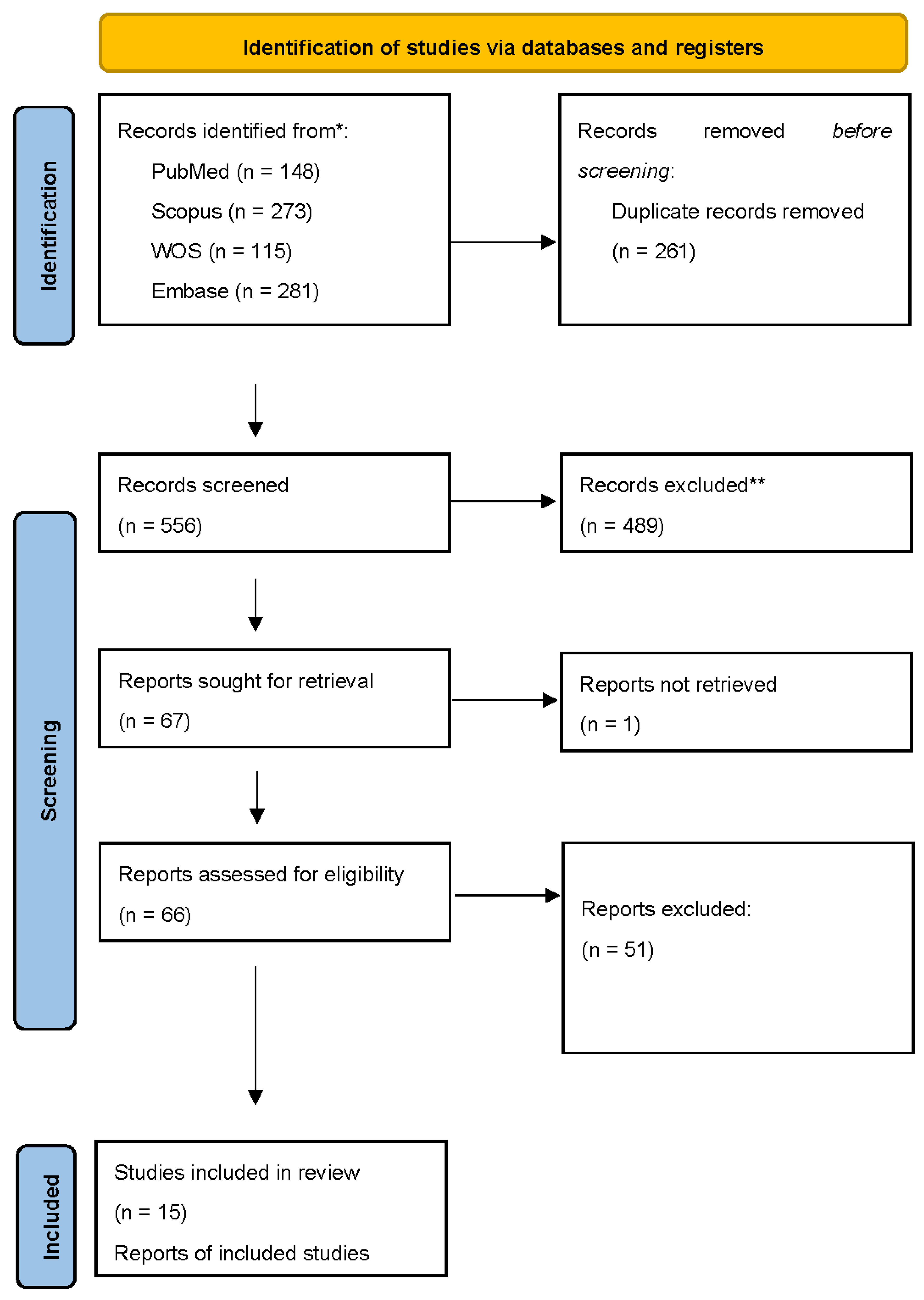
Figure 2.
The box plot representing the timing between the onset of encephalitis and COVID-19 presentation. The box spans from Q1 (3 days) to Q3 (21 days), with the median at 10 days and interquartile range at 18 days. The whiskers extend from the minimum (0 days) to the maximum (60 days).
Figure 2.
The box plot representing the timing between the onset of encephalitis and COVID-19 presentation. The box spans from Q1 (3 days) to Q3 (21 days), with the median at 10 days and interquartile range at 18 days. The whiskers extend from the minimum (0 days) to the maximum (60 days).
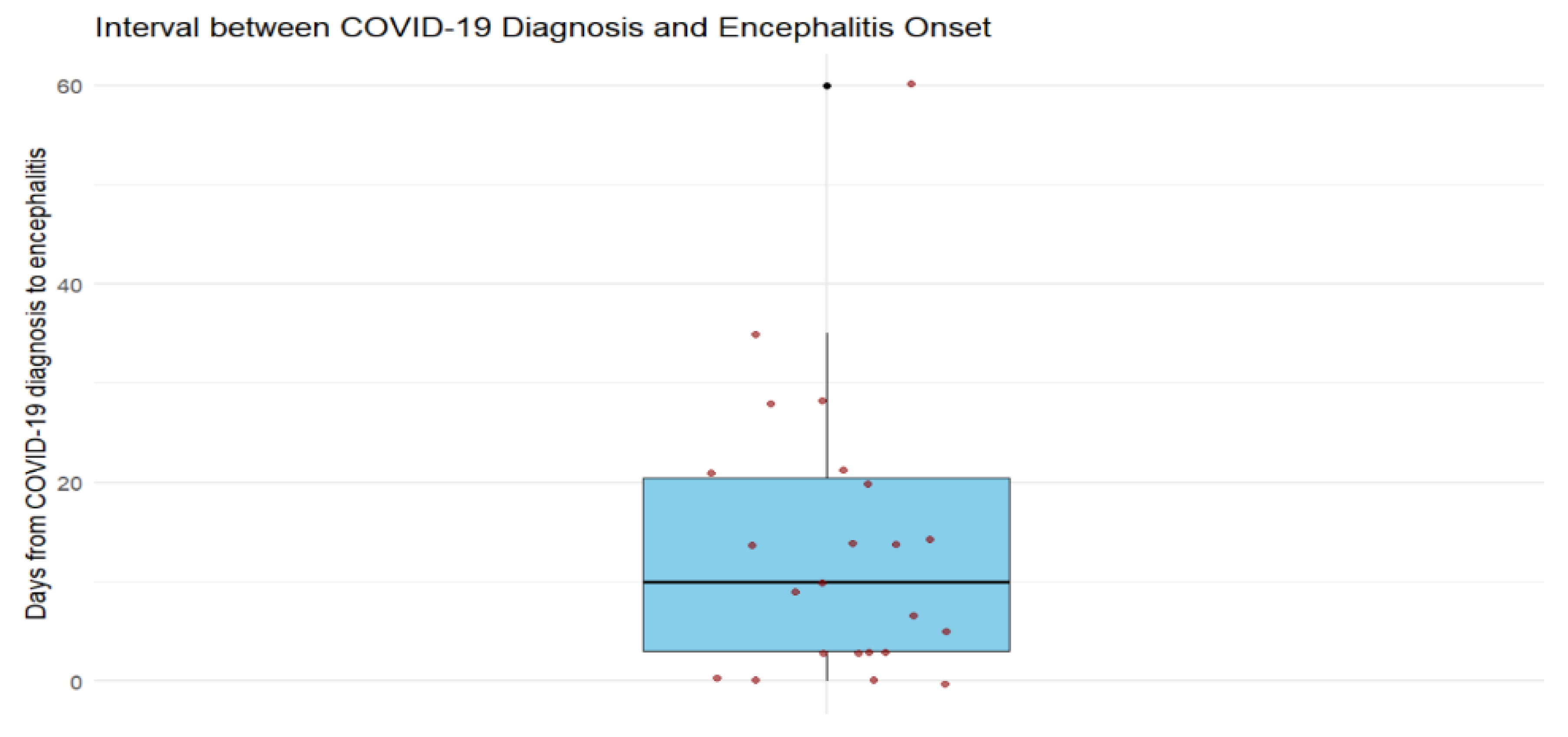
Table 4.
COVID-19 infection characteristics.
| Characteristic | Summary |
|---|---|
| COVID-19 diagnostic test | RT-PCR positive: 21/23 (91.3%); COVID IgG positive: 1/23 (4.3%); RAT positive: 1/23 (4.3%) |
| Interval from COVID-19 to encephalitis onset | Median: 10 days (IQR 3–21); Range: 0 (simultaneous) – 60 days |
| Cases with encephalitis onset simultaneously with COVID symptoms | 4/23 (17.4%) |
| Cases with onset < 1 month | 21/23 (91.3%) |
| Cases with onset > 1 month | 2/23 (8.7%) |
Notes: Abbreviations: Reverse Transcription Polymerase Chain Reaction (RT-PCR), Rapid Antigen Test (RAT).
Table 5.
Clinical manifestations of anti-NMDAR encephalitis following COVID-19.
| Symptom domain | Frequency (%) |
| Neuropsychiatric symptoms | 21/23 (91.3%) |
| • Psychosis / hallucinations | 13/23 (56.5%) |
| • Agitation / catatonia | 10/23 (43.5%) |
| • Behavioral or cognitive changes | 12/23 (52.2%) |
| Seizures (e.g., GTC, focal) | 12/23 (52.2%) |
| Movement disorders (dyskinesias, rigidity, chorea, parkinsonism) | 8/23 (34.8%) |
| Speech dysfunction (mutism, dysarthria, language impairment) | 6/23 (26.1%) |
| Autonomic dysfunction (dysautonomia, hypoventilation) | 4/23 (17.4%) |
| Altered consciousness (coma, delirium, confusion) | 10/23 (43.5%) |
| Other neurological features (ataxia, headache, weakness, sensory changes) | 7/23 (30.4%) |
Notes: Percentages use n = 23 as the denominator. Categories are not mutually exclusive; one case may contribute to multiple rows, so subtotals exceed 100%.
Table 6.
Investigations (EEG, MRI, CSF, serum).
| Investigation | Key findings |
|---|---|
| EEG (n = 21) | Abnormal: 16/21 (76.2%); Normal: 5/21 (23.8%) |
| • Diffuse slowing / encephalopathy | 9/21 (42.9%) |
| • Epileptiform discharges | 8/21 (38.1%) |
| • Delta brush pattern | 3/21 (14.3%) |
| MRI (n = 22) | Abnormal: 13/22 (59.1%); Normal: 9/22 (40.9%) |
| • Temporal/hippocampal involvement | 6/22 (27.3%) |
| • Multifocal white-matter lesions | 2/22 (9.1%) |
| • Other (edema, atrophy, sinusitis, etc.) | 5/22 (22.7%) |
| CSF antibody testing | Positive: 14/23 (60.9%) |
| Serum antibody testing | Positive: 14/23 (60.9%) |
| Other CSF findings | Pleocytosis: 8/23 (34.8%); Elevated protein: 6/23 (26.1%); OCB present: 3/23 (13.0%) |
Table 7.
Treatments administered.
| Treatment modality | Frequency (%) |
| First-line immunotherapy | |
| • IV methylprednisolone (IVMP) | 20/23 (87.0%) |
| • IV immunoglobulin (IVIG) | 13/23 (56.5%) |
| • Plasmapheresis (PLEX) | 8/23 (34.8%) |
| Second-line immunotherapy | |
| • Rituximab | 7/23 (30.4%) |
| • Cyclophosphamide | 0/23 (0%) |
| • Tocilizumab | 1/23 (4.3%) |
| Antiviral / antibiotic therapy (e.g., acyclovir, HCQ) | 4/23 (17.4%) |
| Tumor removal (teratoma) | 1/23 (4.3%) |
| AEDs (antiepileptic drugs) | 11/23 (47.8%) |
| Psychotropic medications (antipsychotics, mood stabilizers) | 6/23 (26.1%) |
Table 8.
Outcomes.
| Outcome | n (%) |
| Full recovery | 12/23 (52.2%) |
| Partial recovery | 9/23 (39.1%) |
| Death | 2/23 (8.7%) |
| Median follow-up duration | 3 months (range: discharge – 12 months) |
Disclaimer/Publisher’s Note: The statements, opinions and data contained in all publications are solely those of the individual author(s) and contributor(s) and not of MDPI and/or the editor(s). MDPI and/or the editor(s) disclaim responsibility for any injury to people or property resulting from any ideas, methods, instructions or products referred to in the content. |
© 2026 by the authors. Licensee MDPI, Basel, Switzerland. This article is an open access article distributed under the terms and conditions of the Creative Commons Attribution (CC BY) license (http://creativecommons.org/licenses/by/4.0/).
Copyright: This open access article is published under a Creative Commons CC BY 4.0 license, which permit the free download, distribution, and reuse, provided that the author and preprint are cited in any reuse.


