

Submitted:
27 June 2026
Posted:
29 June 2026
You are already at the latest version
Abstract
Replicative senescence links telomere shortening to irreversible proliferative arrest and is widely recognized as a major tumor-suppressive mechanism. At the organismal level, however, progressive accumulation of senescent cells contributes to tissue dysfunction, chronic inflammation, and aging. Accumulating evidence suggests that telomere shortening influences chromatin organization and subtelomeric transcription through mechanisms that remain incompletely understood. Building upon the telomere position effect model of cellular senescence and the subtelomere-telomere theory of aging, this review proposes a chromatin-sensing telomere framework in which telomere shortening induces senescence through epigenetic and transcriptional changes at chromosome ends. Central to this model is TERRA, a long non-coding RNA that regulates telomeric chromatin structure, telomere maintenance, and DNA-damage responses. The H3K4 methyltransferase MLL/KMT2A is a key regulator of TERRA transcription. MLL associates with telomeric chromatin in a telomere length-dependent manner and promotes TERRA transcription through H3K4 methylation. Shelterin components, particularly TRF2, function as repressors of TERRA, and we propose that telomere shortening progressively alters shelterin organization and telomeric chromatin topology, thereby reducing access of MLL and RNA polymerase II to subtelomeric promoters. A biphasic model of TERRA regulation emerges in which early TERRA upregulation observed in aging tissues transiently promotes telomere protection, while later critical shortening of TERRA-dominant telomeres drives global TERRA repression and contributes to telomere dysfunction and senescence. This framework provides a mechanistic link between telomere shortening, chromatin regulation, and quasi-programmed aging.
Keywords:
aging
; telomeres
; TERRA
; KMT2A
; replicative senescence
; shelterin
; epigenetics
; chromatin remodeling
; gene regulation
Independent Research Scientist, Tampa, Florida, USA; ccaslini@gmail.com
1. Introduction
The biology of aging has long been debated between two opposing paradigms: one viewing aging as a stochastic accumulation of cellular and molecular damage involving oxidative stress, DNA mutations, telomere shortening, and genomic instability, and another proposing that aging is a programmed, adaptive process encoded in the genome and epigenetically regulated through chromatin-based transcriptional switches [1,2]. Within the programmed framework, aging can be described as a form of phenoptosis, the programmed death of the individual, a process detrimental at the organismal level yet potentially advantageous in terms of supra-individual selection (species or population) [3]. In addition, an alternative view has emerged in which aging is not strictly programmed, but arises from regulated biological processes that, while not evolved to cause aging, progressively drive it through their continued or altered activity, consistent with a quasi-programmed model [4,5]. Although this model has been predominantly developed in the context of persistent mTOR/TOR signaling and cellular hyperfunction, the same logic can be extended to telomere-driven replicative senescence as an adaptive/tumor suppressive mechanism whose cumulative effect becomes deleterious over time.
The discovery of replicative senescence, first described by Hayflick and Moorhead in 1961, provided a pivotal foundation for this debate [6]. Their demonstration that normal human fibroblasts undergo a finite number of divisions before entering senescence, the Hayflick limit, implied the existence of an intrinsic cellular “clock” regulating proliferative capacity. A mechanistic explanation was later provided by Olovnikov’s description of the end-replication problem, which established that DNA polymerase cannot fully replicate the 3′ termini of linear chromosomes, resulting in progressive telomere shortening with each cell division [7]. The subsequent identification of telomerase by Blackburn and colleagues revealed a ribonucleoprotein reverse transcriptase capable of elongating telomeric DNA in germline, stem, and cancer cells, thereby counteracting this attrition [8]. Together, these findings established telomere shortening as a molecular timer that limits somatic cell proliferation, and replicative senescence as a tumor-suppressive response that links progressive telomere shortening to enforced limitation of proliferative capacity in response to oncogenic hyperproliferation [9]. Senescent cells stop dividing yet remain metabolically active, persist within renewing tissues, and contribute to progressive functional decline in chronic degenerative diseases, cancer, and organismal aging through DNA-damage signaling, senescence-associated secretory phenotype (SASP) activation, stem cell exhaustion, and tissue attrition [10,11,12]. Thus, while replicative senescence is a genetically encoded and epigenetically regulated mechanism that initially functions as an adaptive tumor-suppressive program, the persistence and accumulation of metabolically active senescent cells, along with their tissue-level effects, extend far beyond its original selective purpose, aligning with a regulated quasi-program model linking telomere dysfunction to organismal aging.
Attention subsequently shifted from telomere length to the chromatin architecture of telomeric and subtelomeric regions. In 2004, Fossel proposed that replicative aging is epigenetically programmed through progressive changes in telomeric heterochromatin, conceptualized as a protective “hood” of fixed size [13]. As telomeres shorten, this heterochromatic domain has been hypothesized to slide inward along the subtelomere, modulating transcriptional repression in a telomere-length-dependent manner. This model builds upon the concept of telomere position effect (TPE), originally inspired by position-effect variegation in Drosophila melanogaster [14] and later demonstrated at yeast telomeres [15]. Wright and Shay extended these principles to human cells, proposing that telomere length and chromatin organization regulate subtelomeric gene expression during replicative aging [16]. Subsequent evidence of TPE in mammalian systems reinforced the concept that telomere shortening can influence transcriptional programs before terminal telomere dysfunction [17,18]. In this context, the fixed size of a shifting heterochromatin “hood” proportional to the original telomeric structure was proposed as a mechanism to maintain relative repression despite intercellular variation in telomere length [16,19]. Telomere shortening thus becomes not only a passive erosion of DNA repeats but a structural perturbation of chromatin boundaries that can reshape subtelomeric transcriptional landscapes. This means cells are not only “counting divisions,” but interpreting telomere state, which depends on both length and chromatin configuration [20,21].
Collectively, these findings form the basis of the subtelomere-telomere theory of aging, in which TPE-mediated repression of subtelomeric regulatory sequences transcribed into telomeric repeat-containing RNA (TERRA) is a pivotal component of the aging mechanism [16,19,22]. In the present work, we extend this framework under the designation of the chromatin-sensing telomere theory of aging, proposing that aging is not simply the cumulative outcome of random molecular damage nor an evolutionarily pre-determined genetic program, but rather an epigenetically regulated quasi-program. Within this framework, progressive telomere shortening acts as the timed initiator of senescence, while telomere epigenetic regulation and TERRA dynamics constitute the chromatin-mediated mechanisms that execute replicative senescence, to serve primarily as a tumor-suppressive barrier. The resulting accumulation of senescent cells then functions as a quasi-programmed driver of late tissue degeneration and organismal aging.
2. The Chromatin-Sensing Telomere Theory of Aging
The chromatin-sensing telomere theory of aging expands the classical telomere attrition model by proposing that replicative aging is driven not solely by progressive loss of telomeric DNA repeats, culminating in telomere uncapping, but by an epigenetic reprogramming event initiated as telomeres shorten. In this framework, telomere shortening progressively alters the structural and regulatory organization of telomeric chromatin and its associated protective complexes, thereby modulating subtelomeric transcription of coding and non-coding genes involved in regulatory functions.
The conceptual origins of this model trace back to the TPE model of cellular senescence proposed by Wright and Shay [16], and later reformulated by Libertini as the subtelomere-telomere theory of aging [23]. An intermediate formulation by Fossel introduced the idea of a protective “heterochromatin hood” of fixed length that progressively shifts inward over subtelomeric regions as telomeres shorten [13]. Evidence supporting progressive inward spread of TPE repression comes from studies of subtelomeric gene expression in fibroblasts derived from ring chromosome 17 syndrome patients, characterized by shortened telomeres at both chromosome 17 ends and significantly reduced expression of adjacent genes [24]. In addition, in isogenic human diploid fibroblasts (HDFs) approaching senescence, progressive telomere shortening is accompanied by reduced expression of genes proximal to telomeric repeats, consistent with inward spread of TPE repression, whereas more distal genes (within ~10 Mb) become upregulated, consistent with resolution of chromosome-end looping [25]. These findings support a dynamic relationship between telomere length, chromatin state, and transcriptional output of subtelomeric genes.
Within this framework, telomere shortening alters the recruitment of chromatin modifiers and transcriptional regulators to chromosome ends. A chromatin modifier and transcriptional regulator shown to associate with telomeric chromatin in a telomere length-dependent manner was lysine methyltransferase 2A (MLL/KMT2A). MLL binds telomeres preferentially when they are long and catalyzes H3K4 di- and trimethylation (H3K4me2/3) associated with transcriptional activation, thereby facilitating transcription of TERRA [26].
TERRA, first described by Azzalin and colleagues [27], consists of long non-coding RNAs transcribed primarily from subtelomeric promoters located within CpG-islands into telomeric G-rich repeats [28]. Evidence supports a conserved role for TERRA in telomere biology across eukaryotes, including regulation of telomeric chromatin structure, modulation of telomerase activity, and participation in the recombination-based alternative lengthening of telomeres (ALT) mechanism of telomere maintenance in cancer cells. TERRA promotes the formation of telomeric RNA:DNA hybrid structures (R-loops) between TERRA UUAGGG repeats and the complementary C-rich telomeric DNA strand, which facilitate homology-directed repair (HDR) at dysfunctional telomeres [29,30]. Additional findings indicate that TERRA participates in broader regulatory and signaling processes, including modulation of extratelomeric gene expression and signaling of dysfunctional telomeres to the cytoplasm and the extracellular environment [31,32].
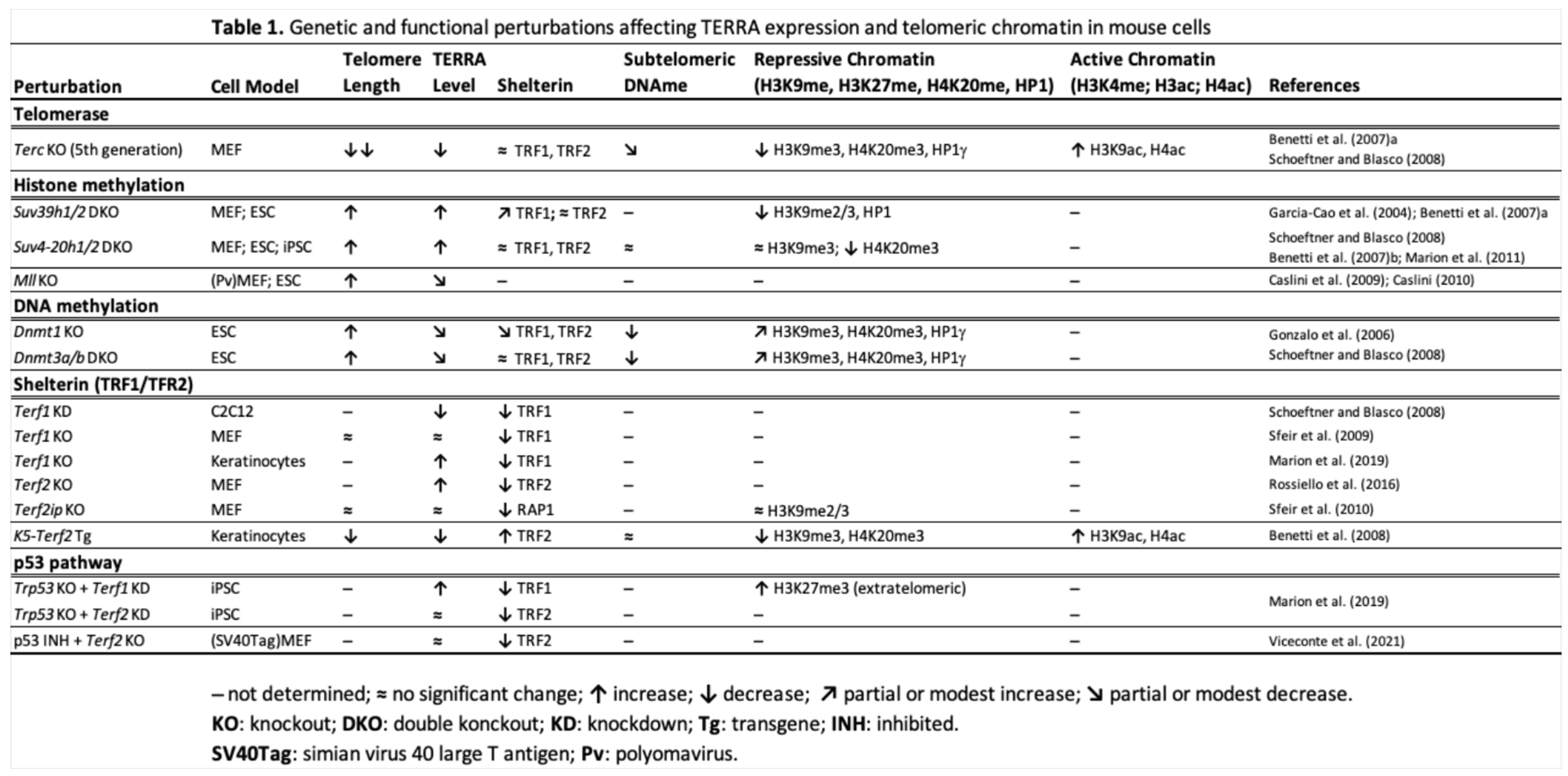
Progressive telomere shortening in senescent HDFs is associated with reduced MLL occupancy at telomeres and concomitant decline in TERRA levels [26]. Together with prior demonstrations that TERRA expression is directly influenced by chromatin-modifying enzymes [29], these findings provide a mechanistic link between telomere length, chromatin state, and transcriptional regulation. In this context, chromatin regulatory complexes such as MLL/H3K4 and Polycomb repressive complex 2 (PRC2), together with components of the shelterin complex and the tumor suppressor p53, emerge as telomere length-sensitive regulators of TERRA transcription that integrate telomere state with chromatin remodeling, transcriptional control, and checkpoint activation in both cancer and aging [26,27,29,33,34,35]. Notably, the conceptual basis for a telomere length-dependent, MLL-regulated control of TERRA transcription as a determinant of chromatin state was enunciated in earlier work, where progressive telomere shortening was proposed to impair TERRA-mediated epigenetic protection and thereby contribute to the onset of replicative senescence [22]. Importantly, studies in telomerase RNA-knockout (Terc-KO) mice at the fifth generation, and in transgenic mice overexpressing TRF2 (Table 1), demonstrated loss of TERRA transcription along with significant chromatin remodeling at shortened telomeres, including reduced telomeric and subtelomeric heterochromatin marks and increased histone acetylation [29,36,37]. Therefore, evidence does not support a strictly heterochromatin-based nature of the proposed protective “hood”. Instead, structural and biochemical data suggest the shelterin complex, the principal structural and regulatory domain at chromosome ends [38,39], as the primary driver of TERRA repression following telomere shortening.
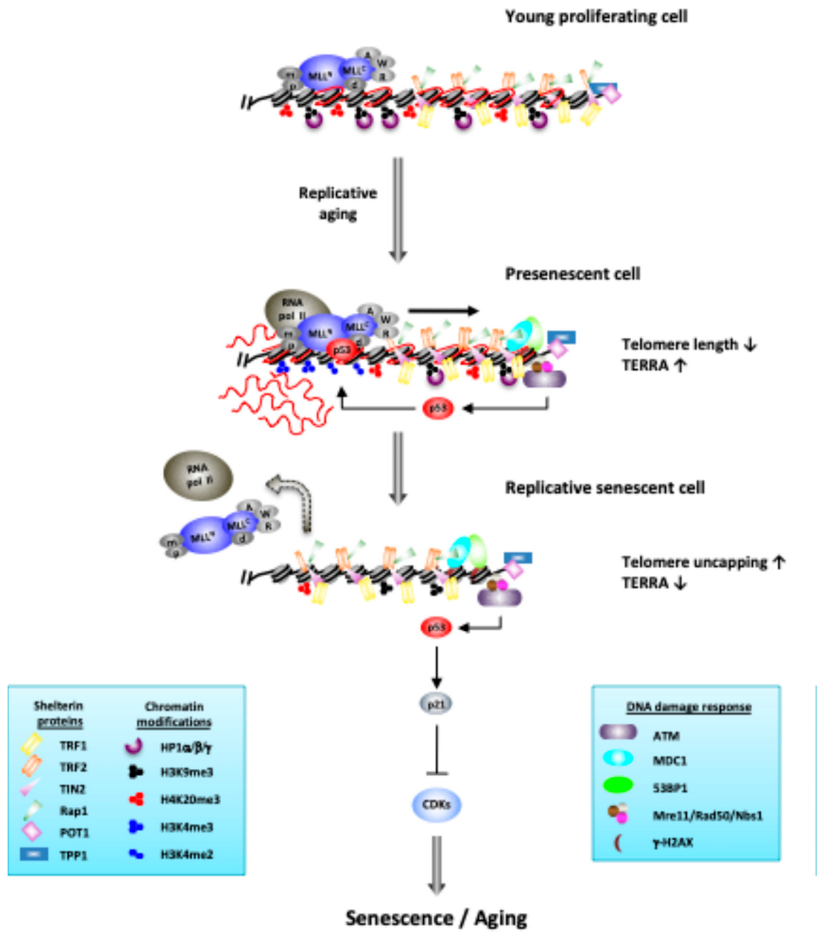
The chromatin-sensing telomere theory of aging operates as a length-dependent chromatin-sensing mechanism in which a shelterin-dependent protective “hood” is proposed to reposition over subtelomeric regions by telomere shortening, progressively reducing MLL and RNA polymerase II (RNA Pol II) accessibility to TERRA promoters, thereby repressing the transcription of TERRA. In this framework, TERRA serves as a regulatory hub that integrates telomere structure with chromatin regulation, and its progressive reduction contributes to the loss of epigenetic stability. This chromatin remodeling promotes transcriptional reprogramming, for instance by chromosome-end looping resolution, resulting in replicative senescence, whose progressive accumulation over time serves as a quasi-programmed mechanism linking chromosome-end dynamics to organismal aging.
2.1. TERRA as a Regulatory Hub in Aging
TERRA has emerged as a central regulator linking telomere biology, chromatin organization, and aging. In mammalian cells, TERRA transcripts have been shown to originate predominantly from specific loci, including the human 20q and Xp subtelomeres, the murine 18q and 9q chromosome ends, distal XqYq pseudoautosomal regions, and 2q and Xq intrachromosomal regions, which mainly contribute to the cellular pool of TERRA [40,41,42,43]. This non-uniform distribution suggests that individual chromosome ends may contribute disproportionately to global TERRA levels and therefore to telomere-dependent chromatin regulation.
TERRA expression is regulated by a network of shelterin proteins, transcription factors, chromatin remodelers, and epigenetic modifiers that couple telomere length and telomeric stress to subtelomeric transcriptional responsiveness. These include TRF1 and TRF2, the tumor suppressor p53 [26,29,33,44], the chromatin remodeler ATRX [45,46], DNA methyltransferases DNMT1 and DNMT3a/b, and the histone methyltransferases SUV39H1/2 and SUV4-20H1/2, MLL/KMT2A, and the PRC2 component EZH2 [26,29,34].
Evidence from mouse embryonic stem cells (ESCs), induced pluripotent stem cells (iPSCs), mouse embryonic fibroblasts (MEFs), and human cells demonstrates that disruption of major heterochromatin regulators profoundly alters TERRA expression and telomere homeostasis [29,47,48,49,50,51,52]. Loss of SUV39H1/2 or SUV4-20H1/2 activity results in elevated TERRA levels, reduced H3K9me2/3 or H4K20me3 heterochromatin marks, decreased heterochromatin protein 1 (HP1) recruitment, increased telomere recombination, and telomere elongation (Table 1) [29,47,48,49]. Likewise, human cells from individuals with immunodeficiency, centromeric instability, and facial anomalies (ICF) syndrome carrying DNMT3B mutations exhibit subtelomeric DNA hypomethylation, increased H3K4me2, reduced H3K9me3, elevated TERRA expression, and short telomeres (Table 2) [51,52]. Collectively, these studies establish TERRA as a sensitive indicator of subtelomeric chromatin organization.
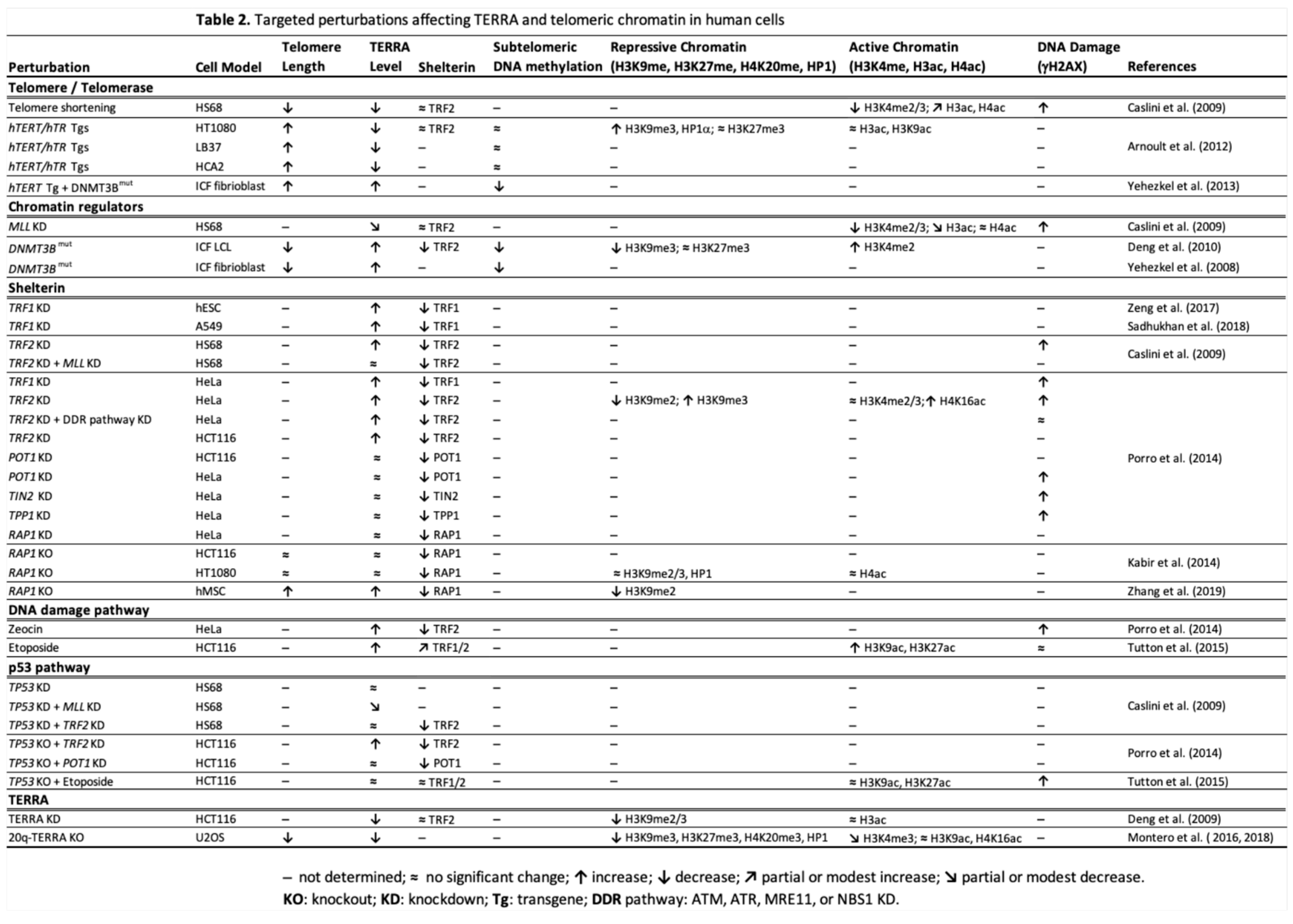
A bona fide TPE also regulates TERRA expression. Early studies demonstrated that genes placed adjacent to newly formed telomeres become transcriptionally repressed [17]. Subsequent analyses in human cancer cells and primary fibroblasts transgenic for telomerase showed that telomere elongation represses TERRA transcription through increased H3K9me3 levels and HP1α activity at chromosome ends (Table 2), establishing a length-dependent chromatin environment that restricts telomeric transcription [53]. These findings provide direct evidence that TERRA expression responds to telomere length and local heterochromatin density.
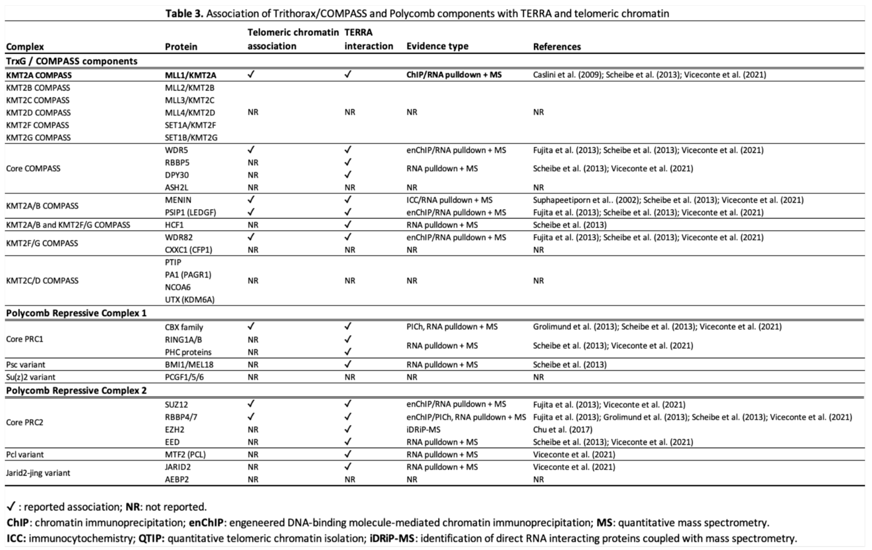
Beyond its transcriptional regulation, TERRA functions as a molecular scaffold that coordinates recruitment of chromatin modifiers to telomeres. RNA affinity purification, RNA immunoprecipitation, and quantitative proteomic studies identified SUV39H1, SETDB1, DNMT1, MLL, and PRC2 core components EZH2, EED, SUZ12, and RBBP4/7 among TERRA-associated proteins (Table 3) [31,34,42,44,54]. These interactions support a model in which TERRA links active transcription to the subsequent establishment and maintenance of telomeric heterochromatin. A particularly informative example comes from ALT-positive U2OS osteosarcoma cells (Table 2), where the subtelomeric 20q locus contributes approximately 20–50% of total cellular TERRA [34,40]. Deletion of this locus (20q-TERRA KO) reduces H3K9me3, H3K27me3, H4K20me3, and HP1 occupancy at telomeres, increases the abundance of critically short telomeres, and impairs telomere maintenance [34]. These findings are consistent with a co-transcriptional gene-silencing model [55], in which TERRA transcription establishes a negative feedback loop that sustains telomeric heterochromatin, thereby reinforcing a bona fide TPE. Collectively, they also indicate that TERRA production is not equivalent across all chromosome ends and that specific subtelomeres can contribute disproportionately to total cellular TERRA levels. A testable hypothesis arising from these observations is that critical shortening of these TERRA-dominant telomeres may therefore exert a global influence on telomeric chromatin regulation and the onset of replicative senescence.
Additional evidence supporting a role for TERRA in aging comes from telomerase-deficient systems. In Terc-KO MEFs from fifth-generation mice (Table 1), progressive telomere shortening is accompanied by a reduction in TERRA expression despite concomitant heterochromatin relaxation, as evidenced by reduced H3K9me3, H4K20me3, and DNA methylation levels, and increased histone acetylation [29,37]. A similar phenotype is observed in HDFs approaching replicative senescence (Table 2), where progressive telomere shortening leads to reduced MLL binding, decreased H3K4me2/3 levels, and downregulation of TERRA, a phenotype recapitulated by MLL depletion [26]. Together, these findings identify TERRA as a central regulatory hub linking telomere length, chromatin organization, and cellular aging. Importantly, telomere length-dependent regulation of TERRA cannot be explained solely by heterochromatin density. Rather, TERRA occupies a central regulatory position in which telomere length, activating chromatin marks, and negative feedback mechanisms are dynamically integrated.
Beyond its local functions at chromosome ends, TERRA can also exert regulatory activity in trans at extratelomeric loci. In mouse ESCs, TERRA-binding sites are enriched within gene bodies, a subset of which are also occupied by ATRX, with TERRA generally associated with gene activation and ATRX with transcriptional repression [31]. In Trp53-KO mouse iPSCs, TERRA transcripts occupy Polycomb target genes genome-wide and modulate their transcriptional landscape through TRF1-dependent recruitment of PRC2 and deposition of H3K27me3 marks (Table 1) [56]. These findings extend TERRA’s role beyond a telomere-restricted chromatin scaffold and support a broader function in epigenetic regulation of gene expression.
Within the chromatin-sensing telomere theory of aging, reduced TERRA levels from progressive telomere shortening are proposed to impair the recruitment of chromatin modifiers, potentially promoting a self-reinforcing cycle of chromatin destabilization, telomere instability, and accelerated shortening. Telomere shortening may progressively reprogram not only telomeric chromatin but also distal transcriptional networks through TERRA-dependent epigenetic signaling. Although no direct evidence currently links these extratelomeric functions of TERRA to the induction of replicative senescence, they suggest that progressive TERRA dysregulation during telomere shortening may contribute to broader transcriptional reprogramming beyond chromosome ends.
2.2. MLL as a Chromatin Sensor at Telomeres
MLL, also known as MLL1/KMT2A (Table 3), originally identified as the mammalian ortholog of the Drosophila melanogaster’s Trithorax (Trx) protein, is the founding member of a family of H3K4 methyltransferases (MLL1-4/KMT2A-D, SET1A-B/KMT2F-G) all of which bind to a multiprotein complex evolutionarily conserved in yeast Saccharomyces cerevisiae termed COMPASS, for complex of proteins associated with Set1, with Set1 representing the only H3K4 methyltransferase in yeast [57]. MLL/COMPASS was initially recognized for maintaining transcriptional activation of HOX and developmental genes [58], thereby antagonizing Polycomb repressive complexes such as PRC1 and PRC2. PRC2-catalyzed H3K27me3 and the resulting chromatin compaction ensure stable propagation of repressive states through cell divisions [59]. Thus, the balance between H3K4me3 and H3K27me3 represents a fundamental chromatin regulatory axis.
Evidence from our group and others extended the function of MLL/COMPASS to telomeric and subtelomeric chromatin, positioning MLL as a chromatin sensor capable of translating telomere shortening into transcriptional and checkpoint responses. Chromatin immunoprecipitation analyses in HDFs demonstrated that MLL binds telomeric chromatin in a telomere length-dependent manner, promotes H3K4 methylation, and cooperates with p53 in the transcriptional upregulation of TERRA at TRF2-depleted dysfunctional telomeres, as part of the DNA damage response (DDR) [26]. MLL/COMPASS physically interacts with p53 and enhances p53-dependent transcription in vitro [60]. Within the COMPASS core subunits, WDR5 positively regulates p53 stability by inhibiting p53 ubiquitination [61]. These findings link MLL-dependent chromatin modification to checkpoint activation pathways. In MEFs, Mll KO prevents TERRA induction and abrogates DNA-damage-triggered p53 phosphorylation on Ser15 [p-p53 (Ser15)] under telomeric stress, while also causing telomere elongation [22,26]. These results indicate that MLL-dependent TERRA transcription couples chromatin remodeling to both DDR signaling and control of telomerase activity.
Proteomic analyses further reinforce MLL integration into telomeric regulatory networks (Table 3). RNA affinity purification and quantitative mass spectrometry studies in human and murine cells identify MLL, COMPASS core subunits RbBP5, WDR5, and DPY30, and complex-specific partners menin, PSIP1, HCF1, and WDR82 among TERRA-associated proteins [42,54]. COMPASS subunits WDR5, menin, PSIP1, and WDR82 are also among proteins associated with mammalian telomeric chromatin, with menin initially localized at meiotic telomeres [62,63]. Among the KMT2 family members, only MLL shows association with telomeric chromatin and the TERRA interactome. Other paralogs (MLL2-4/KMT2B-D and SET1A-B/KMT2F-G) broadly regulate gene and enhancer activity but lack direct evidence of association with TERRA or telomeric localization (Table 3). This distinction underscores MLL’s unique role in coupling telomere structure to transcriptional control.
Evolutionary conservation of this principle is evident in yeast. In Saccharomyces cerevisiae, deletion of Set1 causes loss of H3K4 methylation, derepression of subtelomeric genes, and telomere shortening [64,65]. Deletion of COMPASS components Spp1 and Sdc1 similarly reduces H3K4 methylation and disrupts subtelomeric repression, confirming that intact COMPASS function is required for telomeric chromatin maintenance and TPE [65,66]. Domain analyses demonstrate that the SET catalytic domain is essential for these effects, whereas deletion of RNA recognition motifs RRM1 and RRM2 partially reduces H3K4 methylation and causes mild derepression. Importantly, yeast cells lacking both Set1 and telomerase activity senesce more rapidly than telomerase single mutants despite comparable telomere length, indicating that Set1 may contribute to telomere stability independently of bulk telomere length. In human-like Schizosaccharomyces pombe, deletion of SpSet1 results in telomeric derepression and telomere elongation [67], paralleling the extended telomeres observed in Mll-KO MEFs. These findings support an evolutionarily conserved role for H3K4 methyltransferases at telomeric boundaries. Although the precise mechanism by which H3K4 methylation enforces telomeric silencing in yeast remains unclear, subtelomeric and telomeric RNAs, including TERRA, have been identified in budding and fission yeast, where they interact with telomerase and chromatin regulators, and form R-loops to influence telomere stability [68,69,70]. While direct Set1 regulation of TERRA-like transcripts has not been demonstrated, Set1 localization at telomeres and its control over TPE suggest an evolutionary precursor to the mammalian MLL-TERRA axis.
In Drosophila melanogaster, the MLL ortholog Trx further links these H3K4 methyltransferases to lifespan regulation. Trx acts as an antagonist of Polycomb-group protein silencing. In flies heterozygous for mutations in the PRC2 core subunits E(z) and Esc, lifespan is significantly extended. Mutations in Trx suppress this lifespan extension by restoring high H3K27me3 levels in PRC2 mutants [71]. These findings demonstrate that the balance between Trx-catalyzed H3K4me3 and E(z)-catalyzed H3K27me3 influences organismal longevity.
Taken together, evidence from yeast Set1, fly Trx, and mammalian MLL reveals H3K4 methyltransferases as chromatin boundary regulators that integrate transcriptional activation with repressive chromatin control at genomic edges, including telomeres. In mammals, MLL uniquely links telomere length, TERRA transcription, and p53-mediated checkpoint activation. These evolutionary parallels support the interpretation that MLL functions as a chromatin sensor, translating telomere structural state into transcriptional and aging-related outcomes.
2.3. Shelterin-Mediated Repression of TERRA
Wright and Shay proposed that TPE shifts inward as telomeres shorten [16]. However, the concurrent loss of TERRA transcription and telomeric chromatin marks observed in Terc-KO MEFs at the fifth generation, and in late-passage HDFs, along with their unchanged telomere-associated TRF1 and TRF2 levels despite significant telomere shortening, suggests that a protective “hood” repositioning over subtelomeric regions cannot be due only to heterochromatin [26,37]. Instead, the shelterin complex is proposed as a plausible structural mediator of this shift, a specialized six-protein assembly composed of TRF1, TRF2, RAP1, POT1, TIN2, and TPP1 that binds telomeric DNA to preserve chromosome-end capping [38]. These observations suggest that telomere shortening may alter shelterin organization and telomeric chromatin topology, progressively modifying the accessibility of subtelomeric promoters to transcriptional regulators such as MLL and RNA pol II. However, the precise mechanistic coupling between telomere shortening, shelterin organization, and MLL recruitment remains to be fully elucidated.
TRF1 and TRF2 interact with TERRA, with TRF2 binding via its amino-terminal GAR and carboxy-terminal Myb domains, promoting proper TERRA localization to telomeres [35]. Functional studies show increased TERRA transcription after TRF2 knockdown (KD) in HDFs and cancer cell lines (Table 2), and Terf2 KO in MEFs (Table 1) [26,44,72]. Mechanistically, TRF2 represses TERRA transcription via its TFRH homodimerization domain, which promotes chromatin compaction and reduces telomere accessibility to RNA pol II [44,73]. Terf2 KO in SV40-immortalized MEFs, TRF2 KD in Trp53-KO mouse iPSCs (Table 1), and TRF2/p53 double-KD in HDFs (Table 2) fail to induce TERRA upregulation, indicating that p53 transactivation activity is required for dysfunctional telomere transcription in normal or pluripotent cells [26,42,56]. By contrast, TRF2 depletion induces TERRA upregulation in both wild-type and Trp53-KO HCT116 (Table 2), indicating that this requirement is partially bypassed in cancer cells [33,44]. Collectively, these findings indicate that TRF2-depleted telomeres induce TERRA transcription across multiple cellular contexts, whereas p53 transcriptional activity is specifically required in normal or pluripotent cells but partially dispensable in cancer cells.
Transgenic studies further clarify the transcriptional regulatory role of TRF2 at telomeres. In HDFs, TRF2 overexpression accelerates telomere shortening without altering the timing of replicative senescence, and protects critically short telomeres from end-to-end fusions in presenescent cells [74]. In transgenic mouse keratinocytes (Table 1), increased TRF2 levels accelerate telomere shortening and induce marked repression of TERRA together with significantly reduced telomeric and subtelomeric heterochromatin marks, increased histone acetylation, lower nucleosome density, and greater nucleosomal spacing [36]. In contrast, in ICF lymphoblastoid cell lines and fibroblasts carrying DNMT3b mutations (Table 2), increased TERRA levels are accompanied by decreased TRF2 binding at subtelomeres, shortened telomeres and premature senescence at a low population doubling [51,52,75]. Collectively, these findings support a role for TRF2 in coordinating TERRA transcription, telomeric chromatin organization, and telomere stability beyond its canonical capping function.
TRF1 also plays a context-dependent role in TERRA regulation (Table 1). In mouse C2C12 myoblasts, TRF1 depletion reduces TERRA levels without altering RNA pol II recruitment, suggesting regulation at the level of elongation or transcript stability [29]. No change in global TERRA levels is observed in Terf1-KO MEFs, whereas Terf1-KO mouse keratinocytes show increased TERRA [56,76]. Unlike TRF2, TRF1 depletion increases TERRA levels in Trp53-KO mouse iPSCs, supporting a DDR-independent mechanism [56]. Similarly, in human ESCs, HeLa and A549 cancer cells, TRF1 loss results in TERRA upregulation (Table 2), indicating a repressive role in all these contexts [44,77,78].
Among the other shelterin components, RAP1, POT1, and TPP1 do not robustly immunoprecipitate TERRA, yet RAP1 and POT1 bind to TERRA in RNA affinity purification and iDRiP assays [31,35]. RAP1 deletion produces species- and cell-type-specific effects. In MEFs and human cancer cells, RAP1 KO minimally affects TERRA, H3K9 methylation, or HP1 binding at telomeres [79,80]. In contrast, RAP1-deficient Saccharomyces cerevisiae and human mesenchymal stem cells (hMSCs) exhibit increased TERRA levels, with RAP1-KO hMSCs showing reduced H3K9me2 at telomeres, telomere elongation, and delayed replicative senescence [81,82]. POT1 depletion does not alter TERRA levels in cancer cell lines [44], yet in Caenorhabditis elegans, POT1 functions as a negative regulator of TERRA, where its depletion increases telomere-specific TERRA expression [83].
Additional evidence for shelterin-mediated transcriptional restraint comes from herpes simplex virus 1 infection of HDFs, where TERRA expression increases sharply alongside proteolytic degradation of TPP1 and the dissociation of TRF1, TRF2, and histone H3 from telomeric DNA [84]. Although direct TPP1 KD was not tested, coordinated shelterin stripping correlates with transcriptional derepression, supporting a dynamic shelterin-TERRA regulatory axis responsive to telomere stress. These findings indicate that TRF1, TRF2, POT1, and RAP1 exert context-dependent repression of TERRA and influence telomeric chromatin integrity across species, cell types, telomere states, and epigenetic environments.
Within the chromatin-sensing telomere framework, these findings refine the proposed TPE inward-shift concept. Shelterin occupancy at telomeric repeats remains largely preserved during telomere shortening, yet TERRA expression ultimately declines as telomeres approach critical length. Together, evidence suggests that progressive telomere shortening may alter shelterin organization and telomeric chromatin topology in ways that progressively restrict access of MLL and RNA pol II to subtelomeric TERRA promoters. Reduced TERRA levels are predicted to impair recruitment of chromatin modifiers required for maintenance of telomeric chromatin, thereby accelerating telomere dysfunction. In this proposed model, shelterin mechanistically links telomere shortening to chromatin reprogramming and the onset of replicative senescence.
2.4. A Biphasic Model of TERRA Regulation in Aging
Work in telomerase-negative yeast has highlighted a protective function for TERRA in somatic cells undergoing telomere shortening, where moderate TERRA and the resulting R-loop accumulation promote HDR of dysfunctional telomeres, thereby facilitating telomere maintenance and delaying senescence [85,86]. Kyriacou and Lingner, along with others, review this duality, emphasizing that TERRA accumulation can counteract telomere loss by recruiting repair machinery and mitigating the deleterious effects of progressive shortening [87,88].
Increased TERRA levels have been documented in non-pathological human aging tissues, including human blood cells, brain tissue, fibroblasts, and alveolar type II epithelial cells, along with increased R-loop formation, DNA damage, and chronic activation of cGAS-STING signaling, in inverse correlation with telomere length [89,90]. This presenescent upregulation suggests an adaptive response to progressive telomere shortening, in which elevated TERRA levels contribute to chromatin stabilization and repair, thereby buffering the effects of replicative events accumulation and delaying the onset of senescence. In this regard, persistent DNA damage signaling, chronic innate immune activation, and SASP, as reported in aging tissues, appear to arise primarily from the accumulation of senescent cells, the release of their cytoplasmic DNA, and chronic activation of cGAS-STING signaling [10,91], rather than directly from TERRA-dependent R-loop accumulation.
Previous studies have shown that a small number of critically short telomeres is sufficient to trigger replicative senescence [92,93]. Telomere length is highly heterogeneous, and individual chromosome ends exhibit distinct shortening kinetics and marked allelic heterogeneity, as reported for XpYp telomeres, showing progressive as well as abrupt shortening events during replicative aging [94]. Subsequently, TERRA transcription in mammalian cells has been shown to originate preferentially from specific loci, like the human telomeres 20q and Xp [40,41]. Therefore, within the chromatin-sensing telomere framework, the telomeres most likely to trigger senescence are those that both reach critical shortening and primarily contribute to TERRA expression.
In support of this selective TERRA-dominant telomere-shortening model, recent work has revealed specific length distributions of human telomeres, with 20q consistently among the shorter chromosome ends in the aging population [95]. As telomeres progressively shorten, critical erosion of TERRA-dominant chromosome ends leads to transcriptional repression of TERRA. Because these telomeres account for a major fraction of total TERRA expression, as shown for 20q in the ALT-positive U2OS osteosarcoma cell line [34], their repression results in a global decrease in TERRA levels rather than a localized effect restricted to individual chromosome ends.
These findings support a biphasic model of TERRA regulation in aging, with MLL identified as a core effector of this transition. In presenescent cells, MLL maintains subtelomeric chromatin in a transcriptionally permissive state, enabling TERRA upregulation in cooperation with p53 as an adaptive response to progressive telomere shortening, thereby supporting telomere maintenance and genome stability, likely through R-loop accumulation. Elevated TERRA in aging human tissues, therefore, may reflect a protective phase rather than a primary cause of chronic inflammation. In senescent cells, critical shortening of TERRA-dominant telomeres subsequently triggers a global repression of TERRA transcription by MLL/RNA pol II displacement from subtelomeric promoters, compatible with a model of altered shelterin organization and telomeric chromatin topology. Reduced TERRA levels remove the RNA scaffold required for recruitment of chromatin modifiers at telomeres. Failure to re-establish heterochromatin marks accelerates chromatin relaxation, telomere shortening, and permanent DDR activation, contributing to the establishment of p53-dependent replicative senescence. Replicative senescence is therefore initiated not by isolated dysfunction at random chromosome ends, which tend to be protected by R-loop accumulation, but by the critical shortening of a subset of telomeres with prevalent TERRA production that function as critical nodes in telomere biology.
3. Future Directions
Despite significant advances in understanding the interplay among telomeres, subtelomeres, shelterin, and chromatin regulators, several key mechanistic questions remain important to validate the chromatin-sensing telomere framework. The most important challenge is to define how progressive telomere shortening is converted into loss of MLL occupancy at subtelomeric TERRA promoters. A central prediction of this model is that shortening-induced changes in shelterin organization and/or telomeric chromatin architecture progressively restrict access of MLL and RNA pol II to subtelomeric promoters, ultimately leading to TERRA repression and replicative senescence. Direct experimental testing of this proposed shelterin-mediated remodeling process, together with analyses of higher-order telomeric chromatin structure and MLL recruitment dynamics during replicative aging, will be essential to establish this mechanistic link between telomere shortening and senescence.
A second major question concerns the existence and functional importance of TERRA-dominant telomeres. A limited subset of chromosome ends has been shown to contribute disproportionately to total cellular TERRA levels in mice and humans [40,41,42,43]. This review advances the testable hypothesis that critical shortening or epigenetic repression of these loci may exert a global influence on telomeric chromatin regulation and the onset of replicative senescence. Future studies that combine locus-specific telomere editing, chromatin profiling, and TERRA quantification will be required to determine whether specific chromosome ends serve as critical nodes in telomere biology.
Another priority will be to define how TERRA dynamics are generated across different stages of aging. While replicative senescence is associated with TERRA repression, several aging tissues exhibit elevated TERRA levels. Determining whether these changes arise from altered transcription, RNA stability, or R-loop persistence, as compensatory responses to telomere shortening, will be critical for understanding how TERRA participates in aging. Longitudinal studies integrating telomere length measurements, chromatin-state analyses, TERRA expression, and senescence markers across tissues and species will be particularly valuable for evaluating the proposed biphasic model of TERRA regulation during aging.
Finally, the MLL-TERRA axis represents a potential target for interventions aimed at promoting healthy longevity. An unconventional experimental approach arises from observations in MLL-rearranged acute lymphoblastic leukemias, where TERRA levels remain elevated independently of telomere length and ploidy [96]. These findings raise the possibility that oncogenic MLL fusion proteins may bypass the telomere-length-dependent repression of TERRA. Engineered recruitment of MLL-rearranged transcriptional complexes to TERRA-dominant subtelomeres could therefore provide a useful experimental strategy to evaluate whether sustained TERRA expression delays replicative senescence. Although direct therapeutic application of MLL fusion proteins would be precluded by their oncogenic activity, dissecting the molecular basis of their sustained activation of TERRA may reveal strategies for preserving subtelomeric transcriptional competence during aging.
4. Conclusions
By integrating the telomere position effect model of cellular senescence with the subtelomere-telomere theory of aging and experimental evidence linking MLL to TERRA regulation, this review proposes a chromatin-sensing telomere framework in which progressive telomere shortening is translated into replicative senescence through epigenetic and transcriptional mechanisms operating at chromosome ends. Within this model, telomere shortening functions as a replication-dependent timing mechanism that initiates a regulated chromatin response involving shelterin components, chromatin modifiers, and TERRA. Evidence from mammalian cells supports a biphasic view of TERRA regulation during aging, whereby early TERRA upregulation may transiently stabilize shortening telomeres through R-loop formation and HDR, whereas continued telomere erosion ultimately leads to TERRA repression, chromatin remodeling, and execution of the senescence program (Figure 1).
This framework suggests that replicative senescence is not simply a direct consequence of critically short telomeres, but the outcome of progressive changes in telomere-associated chromatin and transcriptional regulation. We propose that late-stage critical telomere shortening may alter shelterin organization and telomeric chromatin topology, progressively restricting access of transcriptional regulators such as MLL and RNA pol II to subtelomeric promoters. A further testable hypothesis, arising from the integration of current evidence, is that a restricted subset of TERRA-dominant telomeres overly contributes to global TERRA output and may therefore be the critical determinant of senescence initiation.
Because senescent cells can persist for prolonged periods within tissues, repeated activation of replicative senescence throughout life results in their progressive accumulation. The accumulation of senescent cells during aging [97], together with the progressive telomere shortening documented in multiple human and animal tissues [98], supports the view that replicative senescence contributes to organismal aging beyond its established tumor-suppressive function. Within this framework, telomere shortening serves as the timed initiator of replicative senescence, whereas the resulting accumulation of senescent cells drives a quasi-programmed decline in tissue function, further amplified by stochastic molecular damage and chronic inflammatory signaling.
Further studies aimed at elucidating the mechanisms governing MLL displacement, evaluating the proposed shelterin-mediated model of TERRA repression, and confirming that a restricted subset of TERRA-dominant telomeres is the critical determinant of senescence initiation will be essential for assessing this framework and determining whether modulation of the MLL-TERRA axis can be harnessed to promote healthy longevity.
Figure 1 Proposed chromatin-sensing telomere model linking telomere shortening, MLL-dependent TERRA regulation, and replicative senescence. Progressive telomere shortening is proposed to alter telomeric chromatin and shelterin organization while maintaining chromosome-end capping. In presenescent cells, telomere dysfunction activates p53-dependent TERRA transcription, potentially promoting protective telomeric R-loops and telomere maintenance. As shortening proceeds, telomeres with predominant TERRA output become critically short, with reduced accessibility of MLL and RNA pol II to subtelomeric promoters, leading to global TERRA repression, impaired recruitment of chromatin-modifying complexes, persistent DNA damage signaling, activation of the p53-p21 pathway, and replicative senescence. The proposed shelterin-mediated mechanism leading to this transition remains to be experimentally validated. At the organismal level, the progressive accumulation of senescent cells subsequently contributes to chronic inflammation, tissue dysfunction, and aging.
Supplementary Materials
The following supporting information can be downloaded at the website of this paper posted on Preprints.org.
Acknowledgments
During the preparation of this work, the author used an AI-based language model to assist with text refinement, language, and manuscript organization. After using this tool, the author critically reviewed, revised, and verified all content and takes full responsibility for the final manuscript.
Conflicts of Interest
The author declares no conflict of interest.
Abbreviations
The following abbreviations are used in this manuscript:
| TERRA | telomeric repeat-containing RNA |
| TPE | telomere position effect |
| KMT2A | lysine methyltransferase 2A |
| HDR | homology-directed repair |
| DDR | DNA damage response |
| SASP | senescence-associated secretory phenotype |
| HDF | human diploid fibroblast |
| ESC | embryonic stem cell |
| iPSC | induced pluripotent stem cell |
| MEF | mouse embryonic fibroblast |
| COMPASS | complex of proteins associated with Set1 |
| PRC2 | Polycomb repressive complex 2 |
References
- López-Otín, C.; Blasco, M.A.; Partridge, L.; Serrano, M.; Kroemer, G. Hallmarks of aging: An expanding universe. Cell 2023, 186, 243–78. [Google Scholar] [CrossRef] [PubMed]
- Longo, V.D.; Mitteldorf, J.; Skulachev, V.P. Programmed and altruistic ageing. Nat. Rev. Genet. 2005, 6, 866–72. [Google Scholar] [CrossRef]
- Skulachev, V.P. What is “phenoptosis” and how to fight it? Biochemistry 2012, 77, 689–706. [Google Scholar] [CrossRef] [PubMed]
- Blagosklonny, M. V. Aging and Immortality: Quasi-Programmed Senescence and Its Pharmacologic Inhibition. Cell Cycle 2006, 5, 2087–102. [Google Scholar] [CrossRef] [PubMed]
- Blagosklonny, M. V. Aging is not programmed. Cell Cycle 2013, 12, 3736–42. [Google Scholar] [CrossRef] [PubMed]
- Hayflick, L.; Moorhead, P.S. The serial cultivation of human diploid cell strains. Exp. Cell Res. 1961, 25, 585–621. [Google Scholar] [CrossRef] [PubMed]
- Olovnikov, A.M. A theory of marginotomy. J. Theor. Biol. 1973, 41, 181–90. [Google Scholar] [CrossRef] [PubMed]
- Greider, C.W.; Blackburn, E.H. Identification of a specific telomere terminal transferase activity in tetrahymena extracts. Cell 1985, 43, 405–13. [Google Scholar] [CrossRef] [PubMed]
- Shay, J.W.; Wright, W.E. Hayflick, his limit, and cellular ageing. Nat. Rev. Mol. Cell Biol. 2000, 1, 72–6. [Google Scholar] [CrossRef] [PubMed]
- Campisi, J. Aging, Cellular Senescence, and Cancer. Annu. Rev. Physiol. 2013, 75, 685–705. [Google Scholar] [CrossRef] [PubMed]
- Khosla, S.; Farr, J.N.; Tchkonia, T.; Kirkland, J.L. The role of cellular senescence in ageing and endocrine disease. Nat. Rev. Endocrinol. 2020, 16, 263–75. [Google Scholar] [CrossRef] [PubMed]
- Kumari, R.; Jat, P. Mechanisms of Cellular Senescence: Cell Cycle Arrest and Senescence Associated Secretory Phenotype. Front. Cell Dev. Biol. 2021, 9, 645593. [Google Scholar] [CrossRef] [PubMed]
- Fossel, M.B. Cell Senescence in Aging. In Cells, Aging, and Human Disease; Oxford University PressNew York, NY, 2004; pp. 38–65. [Google Scholar] [CrossRef]
- Hazelrigg, T.; Levis, R.; Rubin, G.M. Transformation of white locus DNA in Drosophila: Dosage compensation, zeste interaction, and position effects. Cell 1984, 36, 469–81. [Google Scholar] [CrossRef] [PubMed]
- Gottschling, D.E.; Aparicio, O.M.; Billington, B.L.; Zakian, V.A. Position effect at S. cerevisiae telomeres: Reversible repression of Pol II transcription. Cell 1990, 63, 751–62. [Google Scholar] [CrossRef] [PubMed]
- Wright, W.E.; Shay, J.W. Telomere positional effects and the regulation of cellular senescence. Trends Genet. TIG 1992, 8, 193–7. [Google Scholar] [CrossRef] [PubMed]
- Baur, J.A.; Zou, Y.; Shay, J.W.; Wright, W.E. Telomere position effect in human cells. Science 2001, 292, 2075–7. [Google Scholar] [CrossRef] [PubMed]
- Pedram, M.; Sprung, C.N.; Gao, Q.; Lo, A.W.I.; Reynolds, G.E.; Murnane, J.P. Telomere Position Effect and Silencing of Transgenes near Telomeres in the Mouse. Mol. Cell. Biol. 2006, 26, 1865–78. [Google Scholar] [CrossRef] [PubMed]
- Libertini, G.; Corbi, G.; Nicola, F. Importance and Meaning of TERRA Sequences for Aging Mechanisms. Biochemistry 2020, 85, 1505–17. [Google Scholar] [CrossRef] [PubMed]
- Blasco, M.A. The epigenetic regulation of mammalian telomeres. Nat. Rev. Genet. 2007, 8, 299–309. [Google Scholar] [CrossRef] [PubMed]
- de Lange, T. Shelterin: the protein complex that shapes and safeguards human telomeres. Genes Dev. 2005, 19, 2100–10. [Google Scholar] [CrossRef] [PubMed]
- Caslini, C. Transcriptional regulation of telomeric non-coding RNA: Implications on telomere biology, replicative senescence and cancer. RNA Biol. 2010, 7, 18–22. [Google Scholar] [CrossRef] [PubMed]
- Libertini, G.; Ferrara, N.; Rengo, G.; Corbi, G. Elimination of Senescent Cells: Prospects According to the Subtelomere-Telomere Theory. Biochemistry 2018, 83, 1477–88. [Google Scholar] [CrossRef] [PubMed]
- Surace, C.; Berardinelli, F.; Masotti, A.; Roberti, M.C.; Da Sacco, L.; D’Elia, G.; et al. Telomere shortening and telomere position effect in mild ring 17 syndrome. Epigenet. Chromatin 2014, 7, 1. [Google Scholar] [CrossRef] [PubMed]
- Robin, J.D.; Ludlow, A.T.; Batten, K.; Magdinier, F.; Stadler, G.; Wagner, K.R.; et al. Telomere position effect: regulation of gene expression with progressive telomere shortening over long distances. Genes Dev. 2014, 28, 2464–76. [Google Scholar] [CrossRef] [PubMed]
- Caslini, C.; Connelly, J a; Serna, A.; Broccoli, D.; Hess, J.L. MLL Associates with Telomeres and Regulates Telomeric Repeat-Containing RNA Transcription. Mol. Cell. Biol. 2009, 29, 4519–26. [Google Scholar] [CrossRef] [PubMed]
- Azzalin, C.M.; Reichenbach, P.; Khoriauli, L.; Giulotto, E.; Lingner, J. Telomeric repeat containing RNA and RNA surveillance factors at mammalian chromosome ends. Science 2007, 318, 798–801. [Google Scholar] [CrossRef] [PubMed]
- Nergadze, S.G.; Farnung, B.O.; Wischnewski, H.; Khoriauli, L.; Vitelli, V.; Chawla, R.; et al. CpG-island promoters drive transcription of human telomeres. RNA 2009, 15, 2186–94. [Google Scholar] [CrossRef] [PubMed]
- Schoeftner, S.; Blasco, M.A. Developmentally regulated transcription of mammalian telomeres by DNA-dependent RNA polymerase II. Nat. Cell Biol. 2008, 10, 228–36. [Google Scholar] [CrossRef] [PubMed]
- Arora, R.; Lee, Y.; Wischnewski, H.; Brun, C.M.; Schwarz, T.; Azzalin, C.M. RNaseH1 regulates TERRA-telomeric DNA hybrids and telomere maintenance in ALT tumour cells. Nat. Commun. 2014, 5, 5220. [Google Scholar] [CrossRef] [PubMed]
- Chu, H.-P.; Cifuentes-Rojas, C.; Kesner, B.; Aeby, E.; Lee, H.; Wei, C.; et al. TERRA RNA Antagonizes ATRX and Protects Telomeres. Cell 2017, 170, 86–101.e16. [Google Scholar] [CrossRef] [PubMed]
- Abreu, P.L.; Riva, V.; Zardoni, L.; Azzalin, C.M. Telomeric Repeat-Containing RNA: Biogenesis, Regulation, and Functions. Cold Spring Harb. Perspect. Biol. 2025, 17, a041683. [Google Scholar] [CrossRef] [PubMed]
- Tutton, S.; Azzam, G.A.; Stong, N.; Vladimirova, O.; Wiedmer, A.; Monteith, J.A.; et al. Subtelomeric p53 binding prevents accumulation of DNA damage at human telomeres. EMBO J. 2016, 35, 193–207. [Google Scholar] [CrossRef] [PubMed]
- Montero, J.J.; López-Silanes, I.; Megías, D.; F Fraga, M.; Castells-García, Á.; Blasco, M.A. TERRA recruitment of polycomb to telomeres is essential for histone trymethylation marks at telomeric heterochromatin. Nat. Commun. 2018, 9, 1548. [Google Scholar] [CrossRef] [PubMed]
- Deng, Z.; Norseen, J.; Wiedmer, A.; Riethman, H.; Lieberman, P.M. TERRA RNA Binding to TRF2 Facilitates Heterochromatin Formation and ORC Recruitment at Telomeres. Mol. Cell 2009, 35, 403–13. [Google Scholar] [CrossRef] [PubMed]
- Benetti, R.; Schoeftner, S.; Muñoz, P.; Blasco, M.A. Role of TRF2 in the assembly of telomeric chromatin. Cell Cycle 2008, 7, 3461–8. [Google Scholar] [CrossRef] [PubMed]
- Benetti, R.; García-Cao, M.; Blasco, M.A. Telomere length regulates the epigenetic status of mammalian telomeres and subtelomeres. Nat. Genet. 2007, 39, 243–50. [Google Scholar] [CrossRef] [PubMed]
- De Lange, T. Shelterin-mediated telomere protection. Annu. Rev. Genet. 2018, 52, 223–47. [Google Scholar] [CrossRef] [PubMed]
- Palm, W.; de Lange, T. How Shelterin Protects Mammalian Telomeres. Annu. Rev. Genet. 2008, 42, 301–34. [Google Scholar] [CrossRef] [PubMed]
- Montero, J.J.; López de Silanes, I.; Graña, O.; Blasco, M.A. Telomeric RNAs are essential to maintain telomeres. Nat. Commun. 2016, 7, 12534. [Google Scholar] [CrossRef] [PubMed]
- de Silanes, I.L.; Graña, O.; De Bonis, M.L.; Dominguez, O.; Pisano, D.G.; Blasco, M a. Identification of TERRA locus unveils a telomere protection role through association to nearly all chromosomes. Nat. Commun. 2014, 5, 4723. [Google Scholar] [CrossRef] [PubMed]
- Viceconte, N.; Loriot, A.; Lona Abreu, P.; Scheibe, M.; Fradera Sola, A.; Butter, F.; et al. PAR-TERRA is the main contributor to telomeric repeat-containing RNA transcripts in normal and cancer mouse cells. RNA 2021, 27, 106–21. [Google Scholar] [CrossRef] [PubMed]
- Chu, H.P.; Froberg, J.E.; Kesner, B.; Oh, H.J.; Ji, F.; Sadreyev, R.; et al. PAR-TERRA directs homologous sex chromosome pairing. Nat. Struct. Mol. Biol. 2017, 24, 620–31. [Google Scholar] [CrossRef] [PubMed]
- Porro, A.; Feuerhahn, S.; Delafontaine, J.; Riethman, H.; Rougemont, J.; Lingner, J. Functional characterization of the TERRA transcriptome at damaged telomeres. Nat. Commun. 2014, 5, 5379. [Google Scholar] [CrossRef] [PubMed]
- Flynn, R.L.; Cox, K.E.; Jeitany, M.; Wakimoto, H.; Bryll, A.R.; Ganem, N.J.; et al. Alternative lengthening of telomeres renders cancer cells hypersensitive to ATR inhibitors. Science 2015, 347, 273–7. [Google Scholar] [CrossRef] [PubMed]
- Episkopou, H.; Draskovic, I.; Van Beneden, A.; Tilman, G.; Mattiussi, M.; Gobin, M.; et al. Alternative Lengthening of Telomeres is characterized by reduced compaction of telomeric chromatin. Nucleic Acids Res. 2014, 42, 4391–405. [Google Scholar] [CrossRef] [PubMed]
- Benetti, R.; Gonzalo, S.; Jaco, I.; Schotta, G.; Klatt, P.; Jenuwein, T.; et al. Suv4-20h deficiency results in telomere elongation and derepression of telomere recombination. J. Cell Biol. 2007, 178, 925–36. [Google Scholar] [CrossRef] [PubMed]
- García-Cao, M.; O’Sullivan, R.; Peters, A.H.F.M.; Jenuwein, T.; Blasco, M.A. Epigenetic regulation of telomere length in mammalian cells by the Suv39h1 and Suv39h2 histone methyltransferases. Nat. Genet. 2004, 36, 94–9. [Google Scholar] [CrossRef] [PubMed]
- Marión, R.M.; Schotta, G.; Ortega, S.; Blasco, M.A. Suv4-20h Abrogation Enhances Telomere Elongation during Reprogramming and Confers a Higher Tumorigenic Potential to iPS Cells. PLoS ONE 2011, 6, e25680. [Google Scholar] [CrossRef] [PubMed]
- Gonzalo, S.; Jaco, I.; Fraga, M.F.; Chen, T.; Li, E.; Esteller, M.; et al. DNA methyltransferases control telomere length and telomere recombination in mammalian cells. Nat. Cell Biol. 2006, 8, 416–24. [Google Scholar] [CrossRef] [PubMed]
- Yehezkel, S.; Segev, Y.; Viegas-Péquignot, E.; Skorecki, K.; Selig, S. Hypomethylation of subtelomeric regions in ICF syndrome is associated with abnormally short telomeres and enhanced transcription from telomeric regions. Hum. Mol. Genet. 2008, 17, 2776–89. [Google Scholar] [CrossRef] [PubMed]
- Deng, Z.; Campbell, A.E.; Lieberman, P.M. TERRA, CpG methylation, and telomere heterochromatin: Lessons from ICF syndrome cells. Cell Cycle 2010, 9, 69–74. [Google Scholar] [CrossRef] [PubMed]
- Arnoult, N.; Van Beneden, A.; Decottignies, A. Telomere length regulates TERRA levels through increased trimethylation of telomeric H3K9 and HP1α. Nat. Struct. Mol. Biol. 2012, 19, 948–56. [Google Scholar] [CrossRef] [PubMed]
- Scheibe, M.; Arnoult, N.; Kappei, D.; Buchholz, F.; Decottignies, A.; Butter, F.; et al. Quantitative interaction screen of telomeric repeat-containing RNA reveals novel TERRA regulators. Genome Res. 2013, 23, 2149–57. [Google Scholar] [CrossRef] [PubMed]
- Bühler, M.; Moazed, D. Transcription and RNAi in heterochromatic gene silencing. Nat. Struct. Mol. Biol. 2007, 14, 1041–8. [Google Scholar] [CrossRef] [PubMed]
- Marión, R.M.; Montero, J.J.; López de Silanes, I.; Graña-Castro, O.; Martínez, P.; Schoeftner, S.; et al. TERRA regulate the transcriptional landscape of pluripotent cells through TRF1-dependent recruitment of PRC2. ELife 2019, 8. [Google Scholar] [CrossRef] [PubMed]
- Shilatifard, A. The COMPASS Family of Histone H3K4 Methylases: Mechanisms of Regulation in Development and Disease Pathogenesis. Annu. Rev. Biochem. 2012, 81, 65–95. [Google Scholar] [CrossRef] [PubMed]
- Yu, B.D.; Hess, J.L.; Horning, S.E.; Brown, G.A.J.; Korsmeyer, S.J. Altered Hox expression and segmental identity in Mll-mutant mice. Nature 1995, 378, 505–8. [Google Scholar] [CrossRef] [PubMed]
- Schuettengruber, B.; Bourbon, H.M.; Di Croce, L.; Cavalli, G. Genome Regulation by Polycomb and Trithorax: 70 Years and Counting. Cell 2017, 171, 34–57. [Google Scholar] [CrossRef] [PubMed]
- Dou, Y.; Milne, T a; Tackett, A.J.; Smith, E.R.; Fukuda, A.; Wysocka, J.; et al. Physical Association and Coordinate Function of the H3 K4 Methyltransferase MLL1 and the H4 K16 Acetyltransferase MOF. Cell 2005, 121, 873–85. [Google Scholar] [CrossRef] [PubMed]
- Xie, Q.; Li, Z.; Chen, J. WDR5 positively regulates p53 stability by inhibiting p53 ubiquitination. Biochem. Biophys. Res. Commun. 2017, 487, 333–8. [Google Scholar] [CrossRef] [PubMed]
- Suphapeetiporn, K.; Greally, J.M.; Walpita, D.; Ashley, T.; Bale, A.E. MEN1 tumor-suppressor protein localizes to telomeres during meiosis. Genes Chromosom. Cancer 2002, 35, 81–5. [Google Scholar] [CrossRef] [PubMed]
- Fujita, T.; Asano, Y.; Ohtsuka, J.; Takada, Y.; Saito, K.; Ohki, R.; et al. Identification of telomere-associated molecules by engineered DNA-binding molecule-mediated chromatin immunoprecipitation (enChIP). Sci. Rep. 2013, 3, 3171. [Google Scholar] [CrossRef] [PubMed]
- Nislow, C.; Ray, E.; Pillus, L. SET1, A Yeast Member of the Trithorax Family, Functions in Transcriptional Silencing and Diverse Cellular Processes. Mol. Biol. Cell 1997, 8, 2421–36. [Google Scholar] [CrossRef] [PubMed]
- Jezek, M.; Sun, W.; Negesse, M.Y.; Smith, Z.M.; Orosz, A.; Green, E.M. Set1 regulates telomere function via H3K4 methylation-dependent and -independent pathways and calibrates the abundance of telomere maintenance factors. Mol. Biol. Cell 2023, 34, ar6. [Google Scholar] [CrossRef] [PubMed]
- Krogan, N.J.; Dover, J.; Khorrami, S.; Greenblatt, J.F.; Schneider, J.; Johnston, M.; et al. COMPASS, a Histone H3 (Lysine 4) Methyltransferase Required for Telomeric Silencing of Gene Expression. J. Biol. Chem. 2002, 277, 10753–5. [Google Scholar] [CrossRef] [PubMed]
- Kanoh, J.; Francesconi, S.; Collura, A.; Schramke, V.; Ishikawa, F.; Baldacci, G.; et al. The Fission Yeast spSet1p is a Histone H3-K4 Methyltransferase that Functions in Telomere Maintenance and DNA Repair in an ATM Kinase Rad3-dependent Pathway. J. Mol. Biol. 2003, 326, 1081–94. [Google Scholar] [CrossRef] [PubMed]
- Bah, A.; Wischnewski, H.; Shchepachev, V.; Azzalin, C.M. The telomeric transcriptome of Schizosaccharomyces pombe. Nucleic Acids Res. 2012, 40, 2995–3005. [Google Scholar] [CrossRef] [PubMed]
- Greenwood, J.; Cooper, J.P. Non-coding telomeric and subtelomeric transcripts are differentially regulated by telomeric and heterochromatin assembly factors in fission yeast. Nucleic Acids Res. 2012, 40, 2956–63. [Google Scholar] [CrossRef] [PubMed]
- Zeinoun, B.; Teixeira, M.T.; Barascu, A. TERRA and Telomere Maintenance in the Yeast Saccharomyces cerevisiae. Genes 2023, 14, 618. [Google Scholar] [CrossRef] [PubMed]
- Siebold, A.P.; Banerjee, R.; Tie, F.; Kiss, D.L.; Moskowitz, J.; Harte, P.J. Polycomb Repressive Complex 2 and Trithorax modulate Drosophila longevity and stress resistance. Proceedings of the National Academy of Sciences of the United States of America 2010, 107, 169–74. [Google Scholar] [CrossRef] [PubMed]
- Rossiello, F.; Aguado, J.; Sepe, S.; Iannelli, F.; Nguyen, Q.; Pitchiaya, S.; et al. DNA damage response inhibition at dysfunctional telomeres by modulation of telomeric DNA damage response RNAs. Nat. Commun. 2017, 8, 13980. [Google Scholar] [CrossRef] [PubMed]
- Baker, A.M.; Fu, Q.; Hayward, W.; Victoria, S.; Pedroso, I.M.; Lindsay, S.M.; et al. The Telomere Binding Protein TRF2 Induces Chromatin Compaction. PLoS ONE 2011, 6, e19124. [Google Scholar] [CrossRef] [PubMed]
- Karlseder, J.; Smogorzewska, A.; de Lange, T. Senescence induced by altered telomere state, not telomere loss. Science 2002, 295, 2446–9. [Google Scholar] [CrossRef] [PubMed]
- Yehezkel, S.; Shaked, R.; Sagie, S.; Berkovitz, R.; Shachar-Bener, H.; Segev, Y.; et al. Characterization and rescue of telomeric abnormalities in ICF syndrome type I fibroblasts. Front. Oncol. 2013, 3, 1–21. [Google Scholar] [CrossRef] [PubMed]
- Sfeir, A.; Kosiyatrakul, S.T.; Hockemeyer, D.; MacRae, S.L.; Karlseder, J.; Schildkraut, C.L.; et al. Mammalian Telomeres Resemble Fragile Sites and Require TRF1 for Efficient Replication. Cell 2009, 138, 90–103. [Google Scholar] [CrossRef] [PubMed]
- Sadhukhan, R.; Chowdhury, P.; Ghosh, S.; Ghosh, U. Expression of Telomere-Associated Proteins is Interdependent to Stabilize Native Telomere Structure and Telomere Dysfunction by G-Quadruplex Ligand Causes TERRA Upregulation. Cell Biochem. Biophys. 2018, 76, 311–9. [Google Scholar] [CrossRef] [PubMed]
- Zeng, S.; Liu, L.; Sun, Y.; Lu, G.; Lin, G. Role of telomeric repeat-containing RNA in telomeric chromatin remodeling during the early expansion of human embryonic stem cells. FASEB J. 2017, 31, 4783–95. [Google Scholar] [CrossRef] [PubMed]
- Sfeir, A.; Kabir, S.; van Overbeek, M.; Celli, G.B.; de Lange, T. Loss of Rap1 induces telomere recombination in the absence of NHEJ or a DNA damage signal. Science 2010, 327, 1657–61. [Google Scholar] [CrossRef] [PubMed]
- Kabir, S.; Hockemeyer, D.; de Lange, T. TALEN Gene Knockouts Reveal No Requirement for the Conserved Human Shelterin Protein Rap1 in Telomere Protection and Length Regulation. Cell Rep. 2014, 9, 1273–80. [Google Scholar] [CrossRef] [PubMed]
- Iglesias, N.; Redon, S.; Pfeiffer, V.; Dees, M.; Lingner, J.; Luke, B. Subtelomeric repetitive elements determine TERRA regulation by Rap1/Rif and Rap1/Sir complexes in yeast. EMBO Rep. 2011, 12, 587–93. [Google Scholar] [CrossRef] [PubMed]
- Zhang, X.; Liu, Z.; Liu, X.; Wang, S.; Zhang, Y.; He, X.; et al. Telomere-dependent and telomere-independent roles of RAP1 in regulating human stem cell homeostasis. Protein Cell 2019, 10, 649–67. [Google Scholar] [CrossRef] [PubMed]
- Manzato, C.; Larini, L.; Oss Pegorar, C.; Dello Stritto, M.R.; Jurikova, K.; Jantsch, V.; et al. TERRA expression is regulated by the telomere-binding proteins POT-1 and POT-2 in Caenorhabditis elegans. Nucleic Acids Res. 2023, 51, 10681–99. [Google Scholar] [CrossRef] [PubMed]
- Deng, Z.; Kim, E.T.; Vladimirova, O.; Dheekollu, J.; Wang, Z.; Newhart, A.; et al. HSV-1 Remodels Host Telomeres to Facilitate Viral Replication. Cell Rep. 2014, 9, 2263–78. [Google Scholar] [CrossRef] [PubMed]
- Balk, B.; Maicher, A.; Dees, M.; Klermund, J.; Luke-Glaser, S.; Bender, K.; et al. Telomeric RNA-DNA hybrids affect telomere-length dynamics and senescence. Nat. Struct. Mol. Biol. 2013, 20, 1199–205. [Google Scholar] [CrossRef] [PubMed]
- Graf, M.; Bonetti, D.; Lockhart, A.; Serhal, K.; Kellner, V.; Maicher, A.; et al. Telomere Length Determines TERRA and R-Loop Regulation through the Cell Cycle. Cell 2017, 170, 72–85.e14. [Google Scholar] [CrossRef] [PubMed]
- Pizzul, P.; Rinaldi, C.; Bonetti, D. The multistep path to replicative senescence onset: zooming on triggering and inhibitory events at telomeric DNA. Front. Cell Dev. Biol. 2023, 11, 1250264. [Google Scholar] [CrossRef] [PubMed]
- Kyriacou, E.; Lingner, J. TERRA long noncoding RNA: At the interphase of telomere damage, rescue and signaling. Curr. Opin. Cell Biol. 2024, 91, 102437. [Google Scholar] [CrossRef] [PubMed]
- Lin, C.R.; Hayek, H.; Simborio, H.; Karim, L.; Bahmed, K.; Bolla, S.; et al. Age-Associated Activation of the cGAS-STING Pathway and Impairment of DNA Damage Repair in Human Primary Alveolar Type II Cells. Aging Dis. 2026, 17, 367–82. [Google Scholar] [CrossRef] [PubMed]
- Hsieh, Y.-H.; Tai, C.-H.; Yeh, M.-T.; Chen, Y.-C.; Yang, P.-C.; Yen, C.-P.; et al. Telomeric repeat-containing RNA increases in aged human cells. Nucleic Acids Res. 2025, 53. [Google Scholar] [CrossRef] [PubMed]
- Coppé, J.-P.; Desprez, P.-Y.; Krtolica, A.; Campisi, J. The Senescence-Associated Secretory Phenotype: The Dark Side of Tumor Suppression. Annu. Rev. Pathol. Mech. Dis. 2010, 5, 99–118. [Google Scholar] [CrossRef] [PubMed]
- Hemann, M.T.; Strong, M.A.; Hao, L.-Y.; Greider, C.W. The Shortest Telomere, Not Average Telomere Length, Is Critical for Cell Viability and Chromosome Stability. Cell 2001, 107, 67–77. [Google Scholar] [CrossRef] [PubMed]
- Fagagna F d’Adda di; Reaper, P.M.; Clay-Farrace, L.; Fiegler, H.; Carr, P.; von Zglinicki, T.; et al. A DNA damage checkpoint response in telomere-initiated senescence. Nature 2003, 426, 194–8. [Google Scholar] [CrossRef] [PubMed]
- Baird, D.M.; Rowson, J.; Wynford-Thomas, D.; Kipling, D. Extensive allelic variation and ultrashort telomeres in senescent human cells. Nat. Genet. 2003, 33, 203–7. [Google Scholar] [CrossRef] [PubMed]
- Karimian, K.; Groot, A.; Huso, V.; Kahidi, R.; Tan, K.-T.; Sholes, S.; et al. Human telomere length is chromosome end-specific and conserved across individuals. Science 2024, 384, 533–9. [Google Scholar] [CrossRef] [PubMed]
- Caslini, C.; Serna, A. Telomere Transcription in MLL-Rearranged Leukemia Cell Lines: Increased Levels of TERRA Associate with Lymphoid Lineage and Are Independent of Telomere Length and Ploidy. Biomedicines 2023, 11, 925. [Google Scholar] [CrossRef] [PubMed]
- Tuttle, C.S.L.; Waaijer, M.E.C.; Slee-Valentijn, M.S.; Stijnen, T.; Westendorp, R.; Maier, A.B. Cellular senescence and chronological age in various human tissues: A systematic review and meta-analysis. Aging Cell 2020, 19, e13083. [Google Scholar] [CrossRef] [PubMed]
- Aubert, G.; Lansdorp, P.M. Telomeres and Aging. Physiol. Rev. 2008, 88, 557–79. [Google Scholar] [CrossRef] [PubMed]
Figure 1.
Proposed chromatin-sensing telomere model linking telomere shortening, MLL-dependent TERRA regulation, and replicative senescence. Progressive telomere shortening is proposed to alter telomeric chromatin and shelterin organization while maintaining chromosome-end capping. In presenescent cells, telomere dysfunction activates p53-dependent TERRA transcription, potentially promoting protective telomeric R-loops and telomere maintenance. As shortening proceeds, telomeres with predominant TERRA output become critically short, with reduced accessibility of MLL and RNA pol II to subtelomeric promoters, leading to global TERRA repression, impaired recruitment of chromatin-modifying complexes, persistent DNA damage signaling, activation of the p53-p21 pathway, and replicative senescence. The proposed shelterin-mediated mechanism leading to this transition remains to be experimentally validated. At the organismal level, the progressive accumulation of senescent cells subsequently contributes to chronic inflammation, tissue dysfunction, and aging.
Figure 1.
Proposed chromatin-sensing telomere model linking telomere shortening, MLL-dependent TERRA regulation, and replicative senescence. Progressive telomere shortening is proposed to alter telomeric chromatin and shelterin organization while maintaining chromosome-end capping. In presenescent cells, telomere dysfunction activates p53-dependent TERRA transcription, potentially promoting protective telomeric R-loops and telomere maintenance. As shortening proceeds, telomeres with predominant TERRA output become critically short, with reduced accessibility of MLL and RNA pol II to subtelomeric promoters, leading to global TERRA repression, impaired recruitment of chromatin-modifying complexes, persistent DNA damage signaling, activation of the p53-p21 pathway, and replicative senescence. The proposed shelterin-mediated mechanism leading to this transition remains to be experimentally validated. At the organismal level, the progressive accumulation of senescent cells subsequently contributes to chronic inflammation, tissue dysfunction, and aging.
Disclaimer/Publisher’s Note: The statements, opinions and data contained in all publications are solely those of the individual author(s) and contributor(s) and not of MDPI and/or the editor(s). MDPI and/or the editor(s) disclaim responsibility for any injury to people or property resulting from any ideas, methods, instructions or products referred to in the content. |
© 2026 by the authors. Licensee MDPI, Basel, Switzerland. This article is an open access article distributed under the terms and conditions of the Creative Commons Attribution (CC BY) license (http://creativecommons.org/licenses/by/4.0/).
Copyright: This open access article is published under a Creative Commons CC BY 4.0 license, which permit the free download, distribution, and reuse, provided that the author and preprint are cited in any reuse.


