Preprint
Article

This version is not peer-reviewed.

The Efficacy and Safety of Cardiopulmonary Exercise Testing in Advanced Heart Failure Receiving Intravenous Inotropes

Submitted:

26 June 2026

Posted:

29 June 2026

You are already at the latest version

Abstract

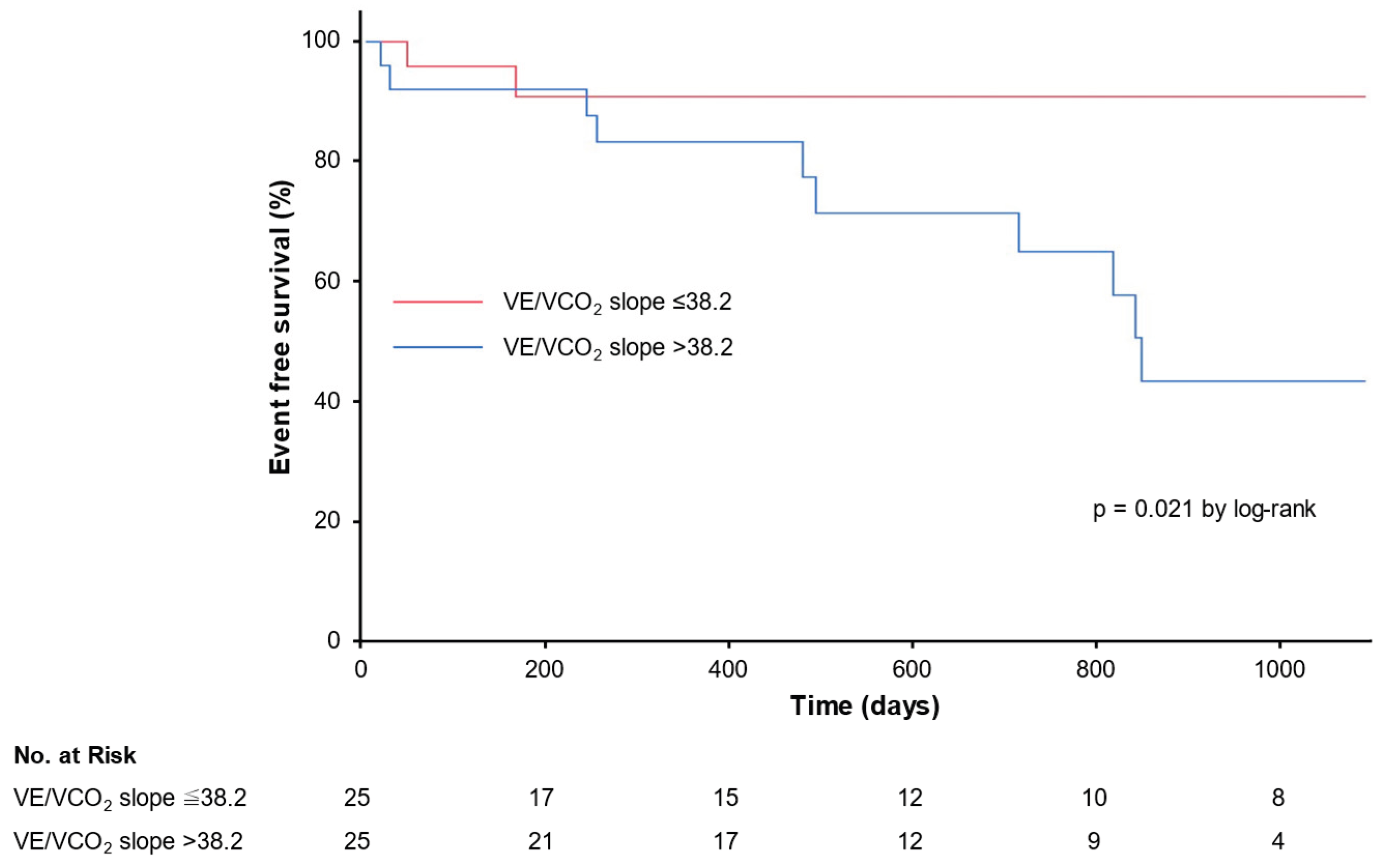
Background: Cardiopulmonary exercise testing (CPET) is widely used for prognostic assessment and risk stratification in patients with heart failure (HF). However, its feasibility and clinical utility in patients with advanced HF requiring intravenous inotropic support remain unclear. Methods: This observational study included hospitalized patients with HF who received intravenous inotropic therapy and underwent CPET during index hospitalization. The primary outcome was a composite of HF rehospitalization or cardiovascular death. Results: A total of 528 patients hospitalized for HF required intravenous inotropic therapy; 50 patients were included. CPET was safely completed in all patients without exercise-related adverse events. The median peak VO2 was 12.5 mL/kg/min, and the median VE/VCO2 slope was 38.3. During a median follow-up of 762 days, 12 primary events occurred. In univariable analysis, the VE/VCO2 slope, O2 pulse, and maximal workload were significantly associated with the primary outcome, whereas peak VO2 was not. A VE/VCO2 slope >38.2 was associated with a significantly higher 3-year event rate compared with a slope ≤38.2 (40% vs. 8%, p = 0.021). Conclusions: CPET is feasible and safe in selected patients with advanced HF requiring inotropic support, and the VE/VCO2 slope provides important prognostic information even in this high-risk population.

Keywords: 
;  ;  ;  

1. Introduction

Currently, cardiopulmonary exercise testing (CPET) is widely utilized not only to assess cardiopulmonary function in patients with heart failure (HF), but also for prognostic evaluation, cardiovascular risk stratification, assessment of therapeutic efficacy, and guidance of cardiac rehabilitation programs [1,2,3,4]. Historically, CPET was primarily used to identify candidates for heart transplantation (HTx) among patients with HF due to left ventricular dysfunction [5]. According to the 2016 International Society for Heart and Lung Transplantation (ISHLT) listing criteria, a peak oxygen uptake (VO2) ≤12 mL/kg/min is recommended to guide HTx listing in patients receiving β-blocker therapy, whereas a threshold of 14 mL/kg/min may be applied to those intolerant to β-blockers [6].
In recent years, survival among patients with HF with reduced ejection fraction (HFrEF) has improved owing to advances in pharmacological therapies and device-based and interventional therapies [7]. Consequently, patients currently considered for HTx often experience recurrent HF hospitalizations and are frequently unable to perform exercise testing due to severe symptoms at rest or with minimal exertion. Moreover, some candidates require intravenous inotropic support, further limiting the feasibility of CPET. Given that CPET is frequently not feasible in patients with extremely advanced HF, its utility for HTx candidate selection may be reduced in this population.
Conversely, CPET remains a cornerstone in the evaluation of advanced HF and continues to serve as a key complementary tool for risk stratification, including decision-making for HTx or durable ventricular assist device (VAD) in contemporary practice [8,9,10]. Although several studies have reported the utility of CPET in ambulatory patients with advanced HF, there are limited data on CPET performed in hospitalized patients with advanced HF, particularly those requiring intravenous inotropic support [11]. Therefore, the safety and clinical utility of CPET in this population remain unclear.
In our institution, CPET has been performed in patients with advanced HF requiring intravenous inotropic support, including those undergoing continuous inotropic infusion during testing, in accordance with our institutional criteria. In this study, we report our institutional experience with this approach. This investigation may provide clinically relevant insights into the feasibility, safety, and prognostic utility of CPET in this high-risk population, even during ongoing intravenous inotropic support.

2. Materials and Methods

2.1. Study Design

This study was a single-center, observational study designed to assess the feasibility, safety, and clinical utility of CPET in patients with HF receiving intravenous inotropes. Eligible patients were those hospitalized for worsening chronic HF or acute decompensated HF who were treated with intravenous inotropic agents and underwent at least one CPET during the index hospitalization at a tertiary academic center. Participants received guideline-directed medical therapy for HF, including renin-angiotensin system inhibitors (ACEI), angiotensin receptor blockers (ARB), or angiotensin receptor-neprilysin inhibitors (ARNI), beta-blockers, mineralocorticoid receptor antagonists, sodium-glucose cotransporter 2 inhibitors (SGLT2i), and diuretics, if applicable. The baseline was defined as the date of CPET performance. Baseline characteristics were retrieved on that day, including demographic, laboratory, and medication data. Standard echocardiographic findings were retrieved between admission and CPET.
Patients were excluded from enrollment if they met any of the following criteria: age <18 years, presence of durable VAD or prior HTx, pregnancy or breastfeeding, severe cognitive impairment or neuromuscular disease, and inability to undergo CPET.
In our institution, CPET was conducted only in participants who fulfilled all of the following criteria: (1) ambulatory within the ward without symptoms, (2) no worsening of subjective symptoms (e.g., dyspnea, fatigue) or physical findings (e.g., edema, pulmonary congestion) of HF, (3) no evidence of worsening HF (e.g., increase in B-type natriuretic peptide levels), (4) absence of uncontrolled arrhythmias, (5) absence of severe valvular disease, and (6) no unstable ischemic heart disease. In addition, CPET was not performed in patients deemed inappropriate by the attending physicians. This study evaluated the safety of CPET under these institutional criteria and examined the association between CPET parameters and subsequent clinical outcomes.
The study was conducted according to the principles outlined in the Declaration of Helsinki, and prior informed consent was diligently obtained from all individuals participating in this investigative endeavor. The local Institutional Ethics Board of Toyama University Hospital approved the study protocol (#R2015154).

2.2. Exercise Testing

CPET was conducted using a system consisting of a leg ergometer (Konami Sports Life, Tokyo, Japan), a breath-by-breath gas analyzer (Minato Medical Science, Osaka, Japan), and 12-lead electrocardiography for continuous monitoring (Fukuda Denshi, Tokyo, Japan). Our exercise protocol was based on the recommendations of the American Heart Association [12]. CPET began with 3 min of seated rest on the cycle ergometer, followed by a symptom-limited exercise test. Each test employed a ramp protocol with incremental workload, set at 7, 10, and 15 W/min to yield a fatigue-limited exercise duration of 8 to 12 minutes. All tests were supervised by a cardiologist.
During CPET, the following parameters were directly measured: oxygen uptake (VO2), carbon dioxide production (VCO2), minute ventilation (VE), and respiratory rate. Blood pressure and heart rate were measured at one-minute intervals during the test. Derived indices included the respiratory exchange ratio (RER), ventilatory efficiency (VE/VCO2 slope), anaerobic threshold (AT), and peak O2 pulse.
The VE/VCO2 slope was calculated from breath-by-breath data of VE and VCO2 plotted throughout exercise using linear regression analysis. AT was identified by a nonlinear increase in the VCO2–VO2 slope (V-slope method). Exercise testing was terminated when patients reached volitional exhaustion or at the discretion of the supervising physician based on safety concerns. A Borg scale questionnaire was administered after the test to assess symptom perception.

2.3. Outcomes

The primary outcome was a composite of unplanned hospitalization for HF or death from cardiovascular causes. Implantation of a VAD or HTx was considered equivalent to cardiovascular death. The secondary endpoint was all-cause mortality, including cardiovascular death, non-cardiovascular death, HTx, or VAD implantation.
Patients were censored at the time of last follow-up if they did not experience the primary outcome. Data for patients who did not have the endpoint event were censored on the last day they were known to have been free of the event. All patients were followed until the end of the observation period or for 36 months.

2.4. Statistical Analyses

All statistical computations were executed employing JMP® 18 (SAS Institute Inc., Cary, NC, USA). Significance was ascribed to outcomes featuring a two-sided p-value of <0.050. Continuous variables were conveyed as medians in conjunction with interquartile ranges (IQRs), and the appropriate statistical assessments were conducted employing Wilcoxon test. Categorical data were articulated in terms of counts and corresponding percentages, and the appropriate statistical assessments were conducted employing the chi-square test.
The Kaplan-Meier method was used to estimate the cumulative 3-year incidence of clinical events, and differences were assessed using log-rank test. Univariate Cox regression analysis was used to calculate the hazard ratio to assess the influence of various parameters on clinical outcomes. For univariate analysis, we selected values of patients’ characteristics, etiology of HF, major markers of HF, and CPET parameters. Receiver operator characteristic (ROC) analysis was carried out to determine the cut-off value of the parameters used in the Cox regression analysis.

3. Results

3.1. Follow-Up and Patient Characteristics

From January 2020 to October 2025, a total of 528 patients hospitalized for HF required intravenous inotropic therapy. Among these, 53 patients underwent CPET during the index hospitalization. One patient underwent CPET but was excluded from this study because, due to frailty, no workload could be applied using the ramp protocol. Two patients were excluded because they had previously undergone durable VAD implantation.
A total of 50 patients were included in this study. Twenty-one patients who underwent CPET while receiving intravenous inotropic therapy were defined as the inotrope group, whereas 29 patients, comprising three who underwent CPET before inotropic therapy and 26 who underwent CPET after discontinuation of inotropic therapy, were defined as the non-inotrope group (Figure 1).
Table 1 lists the baseline characteristics. The median age was 58 (49-67) years and 72% were male. The median left ventricular ejection fraction was 32 (22-43) %. Ischemic etiology was present in 14 patients (28%), and dilated cardiomyopathy in 17 patients (34%). Diabetes mellitus was present in 21 patients (42%). Medical therapy was optimized, with 100% of patients taking ACEI, ARB, or ARNI and 98% taking beta-blockers. Baseline plasma B-type natriuretic peptide level was 188 (120-339) pg/mL.
All patients in the inotrope group received dobutamine, and milrinone was co-administered in two cases. The median dose of dobutamine was 2.0 μg/kg/min. Patients in the inotrope group had lower systolic blood pressure and higher hemoglobin levels compared with those in the non-inotrope group. In addition, the prevalence of dilated cardiomyopathy was higher in the inotrope group.
All patients in the inotrope group were weaned off intravenous inotropes at a median of 30 (14-48) days after the CPET. Although 3 patients underwent implantation of a durable VAD during the index hospitalization, all patients were discharged home ambulatory following a median of 47 (24-68) days of hospitalization. The length of hospital stay was significantly longer in the inotrope group than in the non-inotrope group (64 [35-92] days versus 37 [21-53] days, p = 0.010). Following index discharge, patients were followed for a median period of 762 (346-1095) days. While the median follow-up period did not differ significantly between the two groups, follow-up duration varied among individuals.

3.2. Cardiopulmonary Exercise Test Parameters

CPET was completed safely without any exercise-related worsening of HF, ischemic symptoms, or new-onset arrhythmias requiring additional treatment. Results of the CPET variables are shown in Table 2, stratified by inotrope and non-inotrope groups. Overall, the median peak VO2 was 12.5 (10.6-15.4) mL/kg/min, and the median VE/VCO2 slope was 38.3 (34.1-46.8). Most CPET parameters were not significantly different between the inotrope and non-inotrope groups. The only parameter that differed significantly was the peak RER, which was higher in the inotrope group than in the non-inotrope group.

3.3. Clinical Outcomes

There were 12 primary composite events throughout the 3-year follow-up (9 HF hospitalizations and 3 durable VAD implantations). All three patients who underwent implantation of a durable VAD belonged to the inotrope group. Table 3 presents the results of the analysis of factors associated with the primary outcome. In univariable Cox proportional hazards analysis, the VE/VCO2 slope, peak O2 pulse, and maximal workload were identified as significant predictors of HF hospitalization or cardiovascular death, whereas peak VO2 was not significantly associated with the primary outcome. The use of intravenous inotropic agents at the time of CPET was not significantly associated with the primary outcome.
A cut-off of baseline VE/VCO2 slope to predict the primary endpoint was 38.2 (area under the curve 0.738, sensitivity 0.605, and specificity 0.833). The results from Kaplan-Meier analysis revealed a 3-year primary endpoint of 40% in patients with VE/VCO2 >38.2 and 8% in those with VE/VCO2 ≤38.2 (p = 0.021 by log-rank; Figure 2).
Secondary outcome events were observed in seven patients, including three cardiovascular deaths, two non-cardiovascular deaths, and three VAD implantations. Univariable Cox proportional hazards analysis showed that age and maximal workload achieved during CPET were associated with the secondary outcome (Table 4).

4. Discussion

In this study, we evaluated the feasibility, safety, and clinical utility of CPET in patients with advanced HF receiving intravenous inotropic therapy. CPET was performed only after hemodynamic stabilization using a cycle ergometer with an individualized ramp protocol. Under our institutional criteria, CPET was safely completed in all eligible patients, with no adverse events, and all patients were ultimately discharged.
Notably, CPET parameters were largely comparable between patients undergoing testing during inotropic therapy and those assessed before initiation or after discontinuation of inotropes, suggesting that ongoing inotropic support did not substantially influence CPET-derived parameters. Furthermore, CPET parameters, particularly indices of ventilatory efficiency, were significantly associated with the composite endpoint of HF hospitalization or cardiovascular death in this high-risk population, whereas peak VO2 was not associated with outcomes.

4.1. Feasibility of CPET

The present study demonstrates that CPET can be safely performed in carefully selected patients with advanced HF receiving intravenous inotropic support, provided that strict selection criteria and expert supervision are applied. To our knowledge, this is the first study to specifically evaluate CPET in this high-risk population. Safety is the foremost concern in this population, as these patients are often hemodynamically unstable and presumed to be at high risk during exercise testing. Nonetheless, in our cohort, no adverse events occurred, highlighting that CPET is feasible even under continuous inotropic therapy when conducted within a structured protocol and under the close supervision of experienced cardiologists. Furthermore, the peak RER observed in the present study was clearly higher than that reported in previous studies evaluating CPET in patients with advanced HF [11]. This finding suggests that the enrollment criteria in our study appropriately selected patients who were able to tolerate maximal exercise testing. Therefore, the CPET data obtained in this study are likely to be reliable for the assessment of exercise capacity and prognostic evaluation.
In the present study, one patient was excluded from CPET because severe lower-limb muscle weakness prevented the application of any exercise workload. One of the limitations of the present study is the difficulty in accurately assessing baseline exercise capacity prior to CPET, particularly in patients receiving intravenous inotropic support, in whom conventional functional assessment may be unreliable. Such patients may be identifiable prior to testing by assessing physical performance, such as with the Short Physical Performance Battery (SPPB) or objective measurements of lower-limb muscle strength [13]. Incorporating these assessments into our eligibility criteria may allow more appropriate patient selection and further enhance the safety of CPET in patients with advanced HF.
This finding provides important preliminary evidence that CPET may be more broadly applicable in the inpatient management of advanced HF than previously considered. The populations that have been studied by most of the previous trials regarding inotropic agents were composed of patients with worsening HF, for whom it is difficult to perform evaluations of functional capacity. However, in real-world clinical practice, we frequently encounter situations in which it is necessary to assess the functional capacity of patients who are receiving long-term intravenous inotropic therapy or who are dependent on inotropes. Indeed, in this setting, prognostic assessment and the decision regarding HTx listing are limited to the clinical outcome, which is influenced by several uncontrolled variables, and to a few measured parameters and mainly hemodynamic variables and BNP values. Given the limited data available in this high-risk group, our results help fill an important gap in the literature and suggest that CPET may offer reliable physiological information without compromising patient safety. In addition, in recent years, the potential benefits of cardiac rehabilitation have been advocated even for patients with advanced HF receiving intravenous inotropic therapy, and many institutions have begun to offer exercise training to these patients [14,15,16]. Our study suggests that CPET may provide a basis for prescribing appropriate exercise regimens tailored to such patients.

4.2. CPET Parameters with or Without Inotropic Therapy

CPET parameters were generally similar regardless of inotropic therapy status. Previous studies have shown that intravenous inotropic agents can acutely improve cardiac output and peripheral perfusion in patients with advanced HF, which may lead to transient improvements in exercise capacity [17,18]. Indeed, a previous study reported that the inodilator levosimendan increase the peak VO2 and decrease the VE/VCO2 slope in patients who admitted for HF [19]. Therefore, CPET parameters obtained during inotropic support may theoretically overestimate intrinsic exercise capacity.
Although most CPET parameters did not differ according to the use of inotropic therapy, peak RER was significantly higher in the inotrope group. Intravenous inotropic therapy may influence the RER through changes in cardiac output, metabolic rate, and exercise capacity, potentially modifying the ventilatory and metabolic responses during CPET. Importantly, because comparisons were made between different patients, the direct physiological effects of inotropic therapy could not be fully isolated. A within-subject design, with CPET performed both during and off inotropic support, would be required to more precisely define these effects.

4.3. Clinical Impact of CPET

This study demonstrated that CPET parameters were stronger predictors of clinical outcomes than etiology or major markers of HF. These findings are consistent with the recommendations of the ISHLT guidelines, which emphasize the importance of CPET parameters, particularly peak VO2 and the VE/VCO2 slope, for risk stratification and for determining candidacy for advanced therapies such as or HTx or durable VAD implantation. In the present study, the VE/VCO2 slope was identified as a significant predictor of adverse clinical outcomes, whereas peak VO2 was not. This finding may be explained by several factors specific to patients with advanced HF requiring intravenous inotropic therapy. Peak VO2 is highly dependent on patient effort and peripheral factors such as skeletal muscle function, frailty, and exercise motivation, all of which are frequently impaired in patients with advanced HF. Furthermore, intravenous inotropic agents may transiently augment cardiac output and peripheral perfusion, potentially improve exercise capacity and attenuate the relationship between peak VO2 and long-term prognosis. Consequently, peak VO2 may not accurately reflect the severity of underlying circulatory dysfunction in this population.
In contrast, the VE/VCO2 slope reflects ventilatory inefficiency associated with impaired pulmonary perfusion, ventilation-perfusion mismatch, and abnormal chemoreflex sensitivity, which are key pathophysiological features of advanced HF [20]. Because this parameter is less dependent on maximal effort and more closely reflects underlying cardiopulmonary pathophysiology, it may provide more robust prognostic information in patients receiving inotropic support. In clinical settings where peak VO2 may be influenced by inotropic therapy or reduced exercise tolerance, ventilatory efficiency parameters such as VE/VCO2 slope may therefore provide complementary and potentially more reliable prognostic information for identifying patients who require timely consideration of advanced therapeutic strategies. Indeed, several studies have demonstrated that a VE/VCO2 slope of >35 is of greater prognostic value than a peak VO2 of <14 ml/kg/min [21,22,23,24].
In the present study, the optimal cut-off value of the VE/VCO2 slope for predicting the primary outcome was 38.3, which is slightly higher than the threshold of approximately 35 commonly used for risk stratification in advanced HF and recommended by the ISHLT guidelines 6. One possible explanation is that our cohort included patients with more advanced disease requiring intravenous inotropic support, in whom ventilatory inefficiency tends to be more pronounced. Moreover, many of the studies that established a VE/VCO2 slope cut-off value of approximately 35 for prognostic stratification included patients who were not receiving contemporary HF therapies, such as β-blockers. In contrast, most participants in the present study were treated with current GDMT, including ARNI and SGLT2i. Therefore, the prognostic threshold of the VE/VCO2 slope may be higher in patients receiving contemporary GDMT than in those included in earlier studies.
From a clinical perspective, CPET performed under inotropic support may assist in tracking functional status, evaluating therapeutic response (including inotrope weaning), and guiding decisions regarding HTx or VAD candidacy. However, larger studies focusing specifically on patients undergoing CPET during inotropic therapy are needed to validate these findings and refine prognostic thresholds.

4.4. Limitations

This study has several limitations. First, it was a single-center observational study with a relatively small sample size, particularly in the subgroup receiving inotropic support. Second, the oxygen uptake efficiency slope (OUES), an established prognostic marker in advanced HF, could not be assessed due to technical limitations. Third, inotropic regimens were heterogeneous, reflecting real-world practice. Because CPET was performed under varying agents and infusion rates, the specific effects of individual drugs could not be distinguished from underlying disease severity. Finally, CPET findings may have influenced clinical decision-making, including listing for HTx or initiation of VAD therapy, introducing potential bias in outcome assessment.

5. Conclusions

In conclusion, CPET performed during continuous intravenous inotropic therapy is feasible and provides clinically actionable physiologic information. The results of this study suggest that an elevated VE/VCO2 slope is an independent predictor of adverse cardiac events in patients with advanced HF. Although CPET parameters should be interpreted with caution in this setting, submaximal indices and measures of ventilatory efficiency offer robust complementary information that may aid clinical decision-making in patients with advanced HF receiving inotropic support.

Author Contributions

M.N. (Masaki Nakagaito) wrote a draft. T.I. (Teruhiko Imamura) analyzed and interpreted the patient data. Y.H, T.I. (Toshihide Izumida), and R.U. collected data and reviewed the draft. M.N. (Makiko Nakamura) reviewed the draft. K.K. supervised the study. All authors have read and agreed to the published version of the manuscript.

Funding

This research received no external funding.

Institutional Review Board Statement

The study was conducted according to the guidelines of the Declaration of Helsinki and approved by the Institutional Ethics Board of Toyama University Hospital (#R2015154) on 11 April 2016.

Data Availability Statement

The data presented in this study are available on request from the corresponding author. The data are not publicly available due to privacy.

Acknowledgments

The authors would like to express their gratitude to the clinical laboratory technicians at University of Toyama Hospital for their valuable assistance with this study.

Conflicts of Interest

The authors declare no conflicts of interest.

References

  1. Alba, A. C.; Adamson, M. W.; MacIsaac, J.; Lalonde, S. D.; Chan, W. S.; Delgado, D. H.; Ross, H. J. The Added Value of Exercise Variables in Heart Failure Prognosis. J. Card. Fail From NLM. 2016, 22(7), 492–497. [Google Scholar] [CrossRef] [PubMed]
  2. Corrà, U.; Piepoli, M. F.; Adamopoulos, S.; Agostoni, P.; Coats, A. J.; Conraads, V.; Lambrinou, E.; Pieske, B.; Piotrowicz, E.; Schmid, J. P.; et al. Cardiopulmonary exercise testing in systolic heart failure in 2014: the evolving prognostic role: a position paper from the committee on exercise physiology and training of the heart failure association of the ESC. Eur. J. Heart Fail From NLM. 2014, 16(9), 929–941. [Google Scholar] [CrossRef] [PubMed]
  3. McDonagh, T. A.; Metra, M.; Adamo, M.; Gardner, R. S.; Baumbach, A.; Böhm, M.; Burri, H.; Butler, J.; Čelutkienė, J.; Chioncel, O. 2021 ESC Guidelines for the diagnosis and treatment of acute and chronic heart failure From NLM. Eur. Heart J. 2021, 42(36), 3599–3726. [Google Scholar] [CrossRef] [PubMed]
  4. Heidenreich, P. A.; Bozkurt, B.; Aguilar, D.; Allen, L. A.; Byun, J. J.; Colvin, M. M.; Deswal, A.; Drazner, M. H.; Dunlay, S. M.; Evers, L. R.; et al. 2022 AHA/ACC/HFSA Guideline for the Management of Heart Failure: A Report of the American College of Cardiology/American Heart Association Joint Committee on Clinical Practice Guidelines. Circulation From NLM. 2022, 145(18), e895–e1032. [Google Scholar] [CrossRef] [PubMed]
  5. Mancini, D. M.; Eisen, H.; Kussmaul, W.; Mull, R.; Edmunds, L. H., Jr.; Wilson, J. R. Value of peak exercise oxygen consumption for optimal timing of cardiac transplantation in ambulatory patients with heart failure. Circulation From NLM. 1991, 83(3), 778–786. [Google Scholar] [CrossRef] [PubMed]
  6. Mehra, M. R.; Canter, C. E.; Hannan, M. M.; Semigran, M. J.; Uber, P. A.; Baran, D. A.; Danziger-Isakov, L.; Kirklin, J. K.; Kirk, R.; Kushwaha, S. S.; et al. The 2016 International Society for Heart Lung Transplantation listing criteria for heart transplantation: A 10-year update. J. Heart Lung Transpl. From NLM. 2016, 35(1), 1–23. [Google Scholar] [CrossRef] [PubMed]
  7. Lietz, K.; Miller, L. W. Improved survival of patients with end-stage heart failure listed for heart transplantation: analysis of organ procurement and transplantation network/U.S. United Network of Organ Sharing data, 1990 to 2005. J. Am. Coll. Cardiol. From NLM. 2007, 50(13), 1282–1290. [Google Scholar] [CrossRef] [PubMed]
  8. Chua, T. P.; Ponikowski, P.; Harrington, D.; Anker, S. D.; Webb-Peploe, K.; Clark, A. L.; Poole-Wilson, P. A.; Coats, A. J. Clinical correlates and prognostic significance of the ventilatory response to exercise in chronic heart failure. J. Am. Coll. Cardiol. From NLM. 1997, 29(7), 1585–1590. [Google Scholar] [CrossRef] [PubMed]
  9. Corrà, U.; Agostoni, P. G.; Anker, S. D.; Coats, A. J. S.; Crespo Leiro, M. G.; de Boer, R. A.; Harjola, V. P.; Hill, L.; Lainscak, M.; Lund, L. H.; et al. Role of cardiopulmonary exercise testing in clinical stratification in heart failure. A position paper from the Committee on Exercise Physiology and Training of the Heart Failure Association of the European Society of Cardiology. Eur. J. Heart Fail From NLM. 2018, 20(1), 3–15. [Google Scholar] [CrossRef] [PubMed]
  10. Landsteiner, I.; Ikoma, T.; Lewis, G. D. Cardiopulmonary Exercise Testing in Advanced Heart Failure Management. Heart Fail Clin.;From NLM 2025, 21(1), 35–49. [Google Scholar] [CrossRef] [PubMed]
  11. Lala, A.; Shah, K. B.; Lanfear, D. E.; Thibodeau, J. T.; Palardy, M.; Ambardekar, A. V.; McNamara, D. M.; Taddei-Peters, W. C.; Baldwin, J. T.; Jeffries, N.; et al. Predictive Value of Cardiopulmonary Exercise Testing Parameters in Ambulatory Advanced Heart Failure. JACC Heart Fail From NLM. 2021, 9(3), 226–236. [Google Scholar] [CrossRef] [PubMed]
  12. Balady, G. J.; Arena, R.; Sietsema, K.; Myers, J.; Coke, L.; Fletcher, G. F.; Forman, D.; Franklin, B.; Guazzi, M.; Gulati, M. Clinician's Guide to cardiopulmonary exercise testing in adults: a scientific statement from the American Heart Association From NLM. Circulation 2010, 122(2), 191–225. [Google Scholar] [CrossRef] [PubMed]
  13. Kitai, T.; Shimogai, T.; Tang, W. H. W.; Iwata, K.; Xanthopoulos, A.; Otsuka, S.; Nakada, F.; Yokoyama, R.; Kamiya, K.; Saito, H.; et al. Short physical performance battery vs. 6-minute walking test in hospitalized elderly patients with heart failure. Eur. Heart J. Open 2021, 1(1), oeab006. [Google Scholar] [CrossRef]
  14. Kataoka, T.; Keteyian, S. J.; Marks, C. R.; Fedel, F. J.; Levine, A. B.; Levine, T. B. Exercise training in a patient with congestive heart failure on continuous dobutamine. Med. Sci. Sports Exerc From NLM. 1994, 26(6), 678–681. [Google Scholar] [CrossRef] [PubMed]
  15. Arena, R.; Humphrey, R.; Peberdy, M. A. Safety and efficacy of exercise training in a patient awaiting heart transplantation while on positive intravenous inotropic support. J. Cardiopulm. Rehabil. 2000, 20(4), 259–261. [Google Scholar] [CrossRef]
  16. Forestieri, P.; Guizilini, S.; Peres, M.; Bublitz, C.; Bolzan, D. W.; Rocco, I. S.; Santos, V. B.; Moreira, R. S.; Breda, J. R.; de Almeida, D. R. A Cycle Ergometer Exercise Program Improves Exercise Capacity and Inspiratory Muscle Function in Hospitalized Patients Awaiting Heart Transplantation: a Pilot Study From NLM. Braz. J. Cardiovasc Surg. 2016, 31(5), 389–395. [Google Scholar] [CrossRef] [PubMed]
  17. Maskin, C. S.; Forman, R.; Sonnenblick, E. H.; Frishman, W. H.; LeJemtel, T. H. Failure of dobutamine to increase exercise capacity despite hemodynamic improvement in severe chronic heart failure. Am. J. Cardiol. From NLM. 1983, 51(1), 177–182. [Google Scholar] [CrossRef] [PubMed]
  18. Wilson, J. R.; Martin, J. L.; Ferraro, N. Impaired skeletal muscle nutritive flow during exercise in patients with congestive heart failure: role of cardiac pump dysfunction as determined by the effect of dobutamine. Am. J. Cardiol. From NLM. 1984, 53(9), 1308–1315. [Google Scholar] [CrossRef] [PubMed]
  19. Mushtaq, S.; Andreini, D.; Farina, S.; Salvioni, E.; Pontone, G.; Sciomer, S.; Volpato, V.; Agostoni, P. Levosimendan improves exercise performance in patients with advanced chronic heart failure. ESC Heart Fail From NLM. 2015, 2(3), 133–141. [Google Scholar] [CrossRef] [PubMed]
  20. Arena, R.; Myers, J.; Abella, J.; Peberdy, M. A.; Bensimhon, D.; Chase, P.; Guazzi, M. Development of a ventilatory classification system in patients with heart failure From NLM. Circulation 2007, 115(18), 2410–2417. [Google Scholar] [CrossRef] [PubMed]
  21. Arena, R.; Myers, J.; Aslam, S. S.; Varughese, E. B.; Peberdy, M. A. Peak VO2 and VE/VCO2 slope in patients with heart failure: a prognostic comparison. Am. Heart J. From NLM. 2004, 147(2), 354–360. [Google Scholar] [CrossRef] [PubMed]
  22. Francis, D. P.; Shamim, W.; Davies, L. C.; Piepoli, M. F.; Ponikowski, P.; Anker, S. D.; Coats, A. J. Cardiopulmonary exercise testing for prognosis in chronic heart failure: continuous and independent prognostic value from VE/VCO(2)slope and peak VO(2). Eur. Heart J. From NLM. 2000, 21(2), 154–161. [Google Scholar] [CrossRef] [PubMed]
  23. Robbins, M.; Francis, G.; Pashkow, F. J.; Snader, C. E.; Hoercher, K.; Young, J. B.; Lauer, M. S. Ventilatory and heart rate responses to exercise: better predictors of heart failure mortality than peak oxygen consumption From NLM. Circulation 1999, 100(24), 2411–2417. [Google Scholar] [CrossRef] [PubMed]
  24. Bard, R. L.; Gillespie, B. W.; Clarke, N. S.; Egan, T. G.; Nicklas, J. M. Determining the best ventilatory efficiency measure to predict mortality in patients with heart failure. J. Heart Lung Transpl. From NLM.. 2006, 25(5), 589–595. [Google Scholar] [CrossRef] [PubMed]
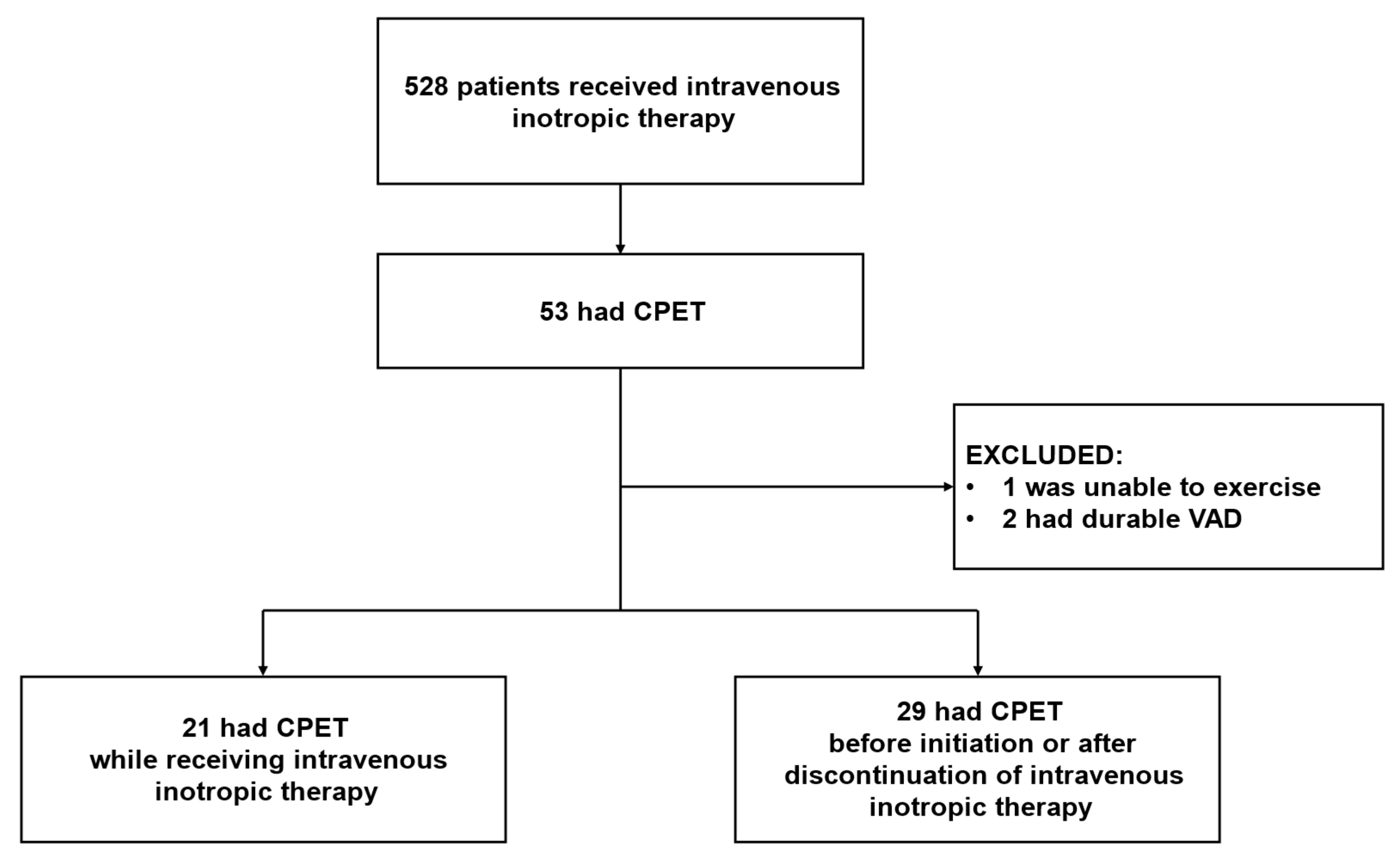
Figure 1. Patient diagram. CPET, cardiopulmonary exercise test; VAD, ventricular assist device.
Preprints 220351 g001
Figure 2. Kaplan-Meier curves for hospitalization for heart failure or cardiovascular death with VE/VCO2 slope threshold of 38.2.
Preprints 220351 g002
Table 1. Baseline characteristics.
Total
(n = 50)
Inotrope
(n = 21)
Non-inotrope
(n = 29)
P value
Age, years 58 (49-67) 58 (49-66) 57 (49-72) 0.753
Male, n (%) 36 (72) 16 (76) 20 (69) 0.574
Body mass index, kg/m2 20.5 (19.2-23.8) 21.5 (19.1-25.7) 20.4 (19.2-22.5) 0.467
Systolic blood pressure, mmHg 95 (83-107) 88 (81-98) 105 (84-113) 0.006*
Heart rate, beats per minutes 75 (68-84) 75 (63-80) 71 (64-83) 0.187
Diabetes mellitus, n (%) 21 (42) 11 (52) 10 (35) 0.206
Ischemic etiology, n (%) 14 (28) 4 (19) 10 (35) 0.230
Dilated cardiomyopathy, n (%) 17 (34) 13 (62) 4 (14) <0.001*
Atrial fibrillation, n (%) 12 (24) 5 (24) 7 (24) 0.979
Left ventricular ejection fraction, % 32 (22-43) 31 (21-40) 32 (26-47) 0.279
Hemoglobin, g/dL 12.7 (10.8-14.3) 13.2 (11.7-15.1) 11.7 (10.4-13.3) 0.043*
Serum albumin, g/dL 3.5 (3.1-3.8) 3.5 (3.1-4.0) 3.5 (3.2-3.8) 0.882
Serum sodium, mEq/L 139 (137-140) 139 (138-141) 139 (137-140) 0.197
Serum potassium, mEq/L 4.3 (4.0-4.8) 4.3 (4.1-4.7) 4.2 (4.0-4.9) 0.867
eGFR, mL/minute/1.73m2 54.8 (44.1-68.6) 48.6 (44.7-65.4) 57.0 (41.3-73.6) 0.569
Uric acid, mg/dL 4.5 (3.7-6.3) 3.9 (3.5-5.4) 4.6 (3.8-6.5) 0.109
Total bilirubin, mg/dL 0.6 (0.4-0.8) 0.6 (0.4-0.8) 0.6 (0.4-0.8) 0.819
Plasma BNP, pg/mL 188 (120-339) 229 (134-364) 167 (91-320) 0.238
Treatment
Dobutamine, μg/kg/min 0 (0-2.0) 2.0 (1.8-3.0) 0 (0-0) <0.001*
Beta-blockers, n (%) 49 (98) 21 (100) 28 (97) 0.390
ACEI/ARB/ARNI, n (%) 50 (100) 21 (100) 29 (100) NA
MRA, n (%) 46 (92) 20 (95) 26 (90) 0.473
SGLT2 inhibitors, n (%) 38 (76) 18 (86) 20 (69) 0.171
Loop diuretics, n (%) 24 (48) 11 (52) 13 (45) 0.598
Dose of loop diuretics, mg/day 0 (0-10) 10 (0-20) 0 (0-10) 0.472
eGFR, estimated glomerular filtration rate; BNP, b-type natriuretic peptide; ACEI, angiotensin converting enzyme inhibitors; ARB, angiotensin receptor blockers; ARNI, angiotensin receptor-neprilysin inhibitors; MRA, mineralocorticoid receptor antagonists; SGLT2 inhibitors, sodium-glucose cotransporter 2 inhibitors. *p <0.050.
Table 2. Baseline cardiopulmonary exercise variables.
Total
(n = 50)
Inotrope
(n = 21)
Non-inotrope
(n = 29)
P value
Peak VO2, mL/kg/min 12.5 (10.6-15.4) 13.1 (11.1-15.7) 12.2 (10.4-15.5) 0.549
VE/VCO2 slope 38.3 (34.1-46.8) 43.0 (35.2-47.9) 38.1 (33.2-44.3) 0.157
AT, mL/kg/min 8.5 (7.5-9.4) 8.4 (7.7-9.2) 8.5 (7.2-9.4) 0.714
Peak RER 1.19 (1.11-1.25) 1.23 (1.15-1.31) 1.14 (1.09-1.23) 0.018*
Peak O2 pulse, mL 6.5 (5.7-8.5) 6.7 (5.4-8.9) 6.5 (5.9-8.2) 0.829
HRR 13 (5-21) 13 (8-24) 13 (3-20) 0.271
Peak systolic blood pressure, mmHg 117 (102-128) 114 (102-123) 117 (107-131) 0.267
Peak heart rate, beats per minutes 116 (102-132) 120 (103-133) 110 (97-132) 0.461
Maximal workload, W 61 (45-82) 68 (46-88) 57 (43-80) 0.275
Borg score at peak exercise 14 (13-17) 14 (13-17) 14 (13-16) 0.520
VO2, oxygen uptake; VE, minute ventilation; VCO2, volume of exhaled carbon dioxide; AT, anaerobic threshold; RER, respiratory exchange ratio; HRR, heart rate recovery. *p <0.050.
Table 3. Variables associated with heart failure hospitalization or cardiovascular death.
Variables Outcome (-) (n = 38) Outcome (+) (n = 12) Hazzard Ratio (95% CI) p value
Inotrope group, n 16 (42) 5 (42) 1.21 (0.38, 3.84) 0.742
Age, years 56 (49-66) 65 (44-70) 1.02 (0.98, 1.06) 0.424
Body mass index, kg/m2 20.6 (19.4-26.1) 19.9 (17.5-23.1) 0.87 (0.72, 1.01) 0.078
Ischemic etiology, n 12 (32) 2 (17) 0.43 (0.09, 1.95) 0.271
LVEF, % 32 (22-45) 34 (21-42) 1.01 (0.97, 1.04) 0.671
Hemoglobin, g/dL 57.0 (44.7-71.6) 12.3 (11.4-14.3) 1.01 (0.80, 1.25) 0.912
eGFR, mL/minute/1.73m2 12.7 (10.7-14.3) 48.5 (33.9-67.8) 0.99 (0.97, 1.02) 0.551
ln BNP 5.15 (4.56-5.74) 5.62 (5.01-6.12) 1.42 (0.66, 2.89) 0.367
Peak VO2, mL/kg/min 13.4 (11.1-15.9) 11.3 (9.7-12.4) 0.83 (0.65, 1.01) 0.066
VE/VCO2 slope 37.5 (33.3-46.0) 45.5 (38.7-48.8) 1.09 (1.01, 1.20) 0.027*
AT, mL/kg/min 8.6 (7.8-9.4) 7.7 (6.5-8.8) 0.68 (0.36, 1.22) 0.197
Peak RER 1.19 (1.11-1.25) 1.20 (1.09-1.27) 2.28 (0.02, 166.69) 0.718
Peak O2 pulse, mL 6.8 (5.7-8.8) 6.0 (4.2-6.5) 0.60 (0.39, 0.88) 0.007*
HRR 14 (7-23) 9 (3-17) 0.96 (0.89, 1.03) 0.271
Peak SBP, mmHg 117 (102-132) 115 (102-121) 0.97 (0.94, 1.00) 0.115
Peak HR, beats per minutes 119 (103-132) 109 (92-123) 0.98 (0.95, 1.01) 0.178
Maximal workload, W 65 (48-86) 46 (38-65) 0.97 (0.93, 1.00) 0.022*
Borg score at peak exercise 14 (13-17) 14 (12-15) 0.90 (0.66, 1.20) 0.492
LVEF, left ventricular ejection fraction; eGFR, estimated glomerular filtration rate; BNP, b-type natriuretic peptide; VO2, oxygen uptake; VE, minute ventilation; VCO2, volume of exhaled carbon dioxide; AT, anaerobic threshold; RER, respiratory exchange ratio; HRR, heart rate recovery; SBP, systolic blood pressure; HR, heart rate. *p <0.050.
Table 4. Variables associated with all-cause death.
Variables Outcome (-) (n = 43) Outcome (+) (n = 7) Hazzard Ratio (95% CI) p value
Inotrope group, n 18 (42) 3 (43) 1.29 (0.29, 5.82) 0.737
Age, years 56 (48-66) 70 (65-75) 1.10 (1.03, 1.19) 0.004*
Body mass index, kg/m2 21.0 (19.3-25.8) 20.4 (18.8-20.6) 0.84 (0.62, 1.03) 0.104
Ischemic etiology, n 11 (26) 3 (43) 1.68 (0.38, 7.52) 0.498
LVEF, % 32 (21-42) 35 (27-47) 1.02 (0.98, 1.06) 0.311
Hemoglobin, g/dL 12.7 (10.8-14.7) 11.5 (10.4-13.0) 0.77 (0.51, 1.07) 0.133
eGFR, mL/minute/1.73m2 56.9 (45.1-68.2) 36.3 (33.1-73.2) 0.99 (0.96, 1.02) 0.492
ln BNP 5.28 (4.69-5.75) 5.09 (4.96-6.18) 1.08 (0.40, 2.90) 0.875
Peak VO2, mL/kg/min 13.3 (10.5-15.8) 11.8 (11.3-12.4) 0.87 (0.64, 1.11) 0.282
VE/VCO2 slope 38.2 (33.8-46.6) 42.6 (35.3-49.1) 1.03 (0.94, 1.15) 0.545
AT, mL/kg/min 8.5 (7.7-9.4) 7.1 (6.4-8.9) 0.74 (0.44, 1.21) 0.238
Peak RER 1.19 (1.10-1.25) 1.20 (1.11-1.27) 2.68 (0.01, 982.59) 0.748
Peak O2 pulse, mL/beat 6.7 (5.6-8.7) 5.9 (5.7-6.4) 0.70 (0.42, 1.08) 0.110
HRR 13 (6-21) 13 (3-25) 0.99 (0.91, 1.06) 0.851
Peak SBP, mmHg 116 (102-130) 117 (79-121) 0.97 (0.93, 1.01) 0.183
Peak HR, beats per minutes 117 (102-132) 103 (82-128) 0.97 (0.93, 1.01) 0.205
Maximal workload, W 66 (46-85) 45 (38-48) 0.95 (0.90, 0.99) 0.022*
Borg score at peak exercise 15 (13-17) 14 (13-14) 1.00 (0.69, 1.40) 0.993
LVEF, left ventricular ejection fraction; eGFR, estimated glomerular filtration rate; BNP, b-type natriuretic peptide; VO2, oxygen uptake; VE, minute ventilation; VCO2, volume of exhaled carbon dioxide; AT, anaerobic threshold; RER, respiratory exchange ratio; HRR, heart rate recovery; SBP, systolic blood pressure; HR, heart rate. *p <0.050.
Disclaimer/Publisher’s Note: The statements, opinions and data contained in all publications are solely those of the individual author(s) and contributor(s) and not of MDPI and/or the editor(s). MDPI and/or the editor(s) disclaim responsibility for any injury to people or property resulting from any ideas, methods, instructions or products referred to in the content.
Copyright: This open access article is published under a Creative Commons CC BY 4.0 license, which permit the free download, distribution, and reuse, provided that the author and preprint are cited in any reuse.
Prerpints.org logo

Preprints.org is a free preprint server supported by MDPI in Basel, Switzerland.

Subscribe

© 2026 MDPI (Basel, Switzerland) unless otherwise stated

Accessibility

Disclaimer

Terms of Use

Privacy Policy

Privacy Settings