

Submitted:
23 June 2026
Posted:
26 June 2026
You are already at the latest version
Abstract
Background and Clinical Significance: Iatrogenic atrophy following intralesional corticosteroid injections represents a rare but potentially disfiguring complication, particularly when administered in the facial region for the treatment of inflammatory acne. Unlike conventional post-acne atrophic scarring, corticosteroid-induced tissue loss involves both dermal and subcutaneous compartments, resulting in a clinically distinct presentation that poses significant therapeutic challenges, with no established consensus on optimal management. Case Presentation: We report the case of a 26-year-old Caucasian female (Fitzpatrick phototype III) presenting with severe iatrogenic facial atrophy of the left cheek, resulting from multiple intralesional corticosteroid injections performed by a previous physician for papulo-pustular acne. The condition had been clinically stable for approximately two years at first evaluation. The patient underwent a stepwise multimodal protocol over approximately 18 months, combining non-ablative fractional laser remodelling (LightScan – Eufoton), Autologous Regenerative Therapy (ART, Seffiller technique, Seffiline srl), hyperdiluted calcium hydroxylapatite (CaHA – Radiesse, Merz Aesthetics) biostimulation, and additional non-ablative fractional photothermolysis sessions, supported by a topical cosmeceutical protocol. Clinician-assessed scar severity improved from Goodman & Baron Grade 3 to Grade 2, and Vancouver Scar Scale score from 4–5/13 to 1–2/13. Global aesthetic improvement was rated as +2 ("Much improved") on the GAIS, with patient satisfaction of 5/5. Conclusions: This case demonstrates that a carefully sequenced multimodal approach combining energy-based devices, biological scaffolds, and biostimulatory agents can achieve substantial and durable correction of iatrogenic steroid-induced facial atrophy, providing a reproducible framework for managing this under-reported and therapeutically challenging condition.
Keywords:
iatrogenic atrophy
; steroid-induced lipoatrophy
; acne scar
; non-ablative fractional laser
; autologous regenerative therapy
; calcium hydroxylapatite hyperdilution
; multimodal aesthetic treatment
; case report
1. Introduction and Clinical Significance
Intralesional corticosteroid injection is a widely used therapeutic modality in dermatology, employed for the management of inflammatory acne lesions, hypertrophic scars, and keloids [1]. When administered at appropriate doses and in the correct tissue plane, this technique is generally safe and effective. However, inadvertent delivery into the subcutaneous compartment, or the use of excessive concentrations, can result in localised dermal and subdermal atrophy — a recognised but infrequently reported iatrogenic complication [2].
Steroid-induced facial atrophy is clinically distinct from post-inflammatory atrophic acne scarring. While the latter primarily involves dermal collagen loss with characteristic morphological subtypes (ice-pick, rolling, and boxcar scars) [3], iatrogenic atrophy typically involves a broader area of tissue depression, loss of subcutaneous volume, and disruption of the normal skin architecture across multiple structural layers. This compound pathology represents a significant therapeutic challenge: energy-based devices optimised for dermal remodelling may not adequately address the subcutaneous component, while volumising agents alone are insufficient to correct the textural and structural dermal deficits.
The absence of consensus guidelines for the management of this condition, and its relative rarity in the published literature, means that affected patients are frequently undertreated or managed sub-optimally. The present case report describes, to our knowledge, a novel structured multimodal protocol combining non-ablative fractional laser remodelling, Autologous Regenerative Therapy (ART), and hyperdiluted calcium hydroxylapatite biostimulation, achieving sustained clinical improvement in a young patient with severe, stabilised iatrogenic facial atrophy.
2. Case Presentation
2.1. Patient History
A 26-year-old Caucasian female (Fitzpatrick phototype III) presented to our clinic complaining of severe left facial depression and skin texture abnormality, with significant psychological and aesthetic impact. The patient reported a history of moderate-to-severe papulo-pustular acne of the left cheek, for which she had received multiple intralesional corticosteroid injections administered by a previous physician. Following these injections, she developed a progressive and eventually stable area of significant tissue atrophy involving the left cheek, characterised by both dermal and subcutaneous loss, which she described as “a scar and important loss of tissue.”
The atrophic defect had been clinically stable for approximately two years prior to her first evaluation at our centre. The patient denied any subsequent acne activity, current pharmacological therapy, or known drug allergies. She had previously received systemic isotretinoin therapy, with the last course completed in 2020. No hormonal therapy, prior surgical intervention to the area, or prior laser or energy-based device treatments to the left cheek were reported.
2.2. Clinical Examination
Examination of the left cheek revealed two anatomically distinct but coexisting pathological components:
- -
- Superior component (mid-cheek): Multiple atrophic scars with mixed ice-pick and rolling morphology distributed over an extended surface area, associated with chromatic heterogeneity and erythematous discolouration — consistent with residual post-acne dermal scarring.
- -
- Inferior-central component (lower cheek): A broad area of significant dermal and subcutaneous atrophy with tissue depression, irregular surface contour, and partial erythema — consistent with iatrogenic corticosteroid-induced lipoatrophy, anatomically and morphologically distinct from typical acne-related scarring.
Baseline severity of the atrophic acne scarring component (superior mid-cheek area) was graded as Grade 3 (moderate) on the Goodman & Baron Qualitative Grading System [3], indicating lesions clearly visible at conversational distance, not fully concealed by make-up, with mixed rolling and ice-pick morphology across an estimated surface area of 6–8 cm2. The iatrogenic atrophic defect (inferior cheek component) was independently assessed using the Vancouver Scar Scale (VSS), yielding an estimated baseline score of 4–5 out of 13, reflecting abnormal vascularity (score 1), mixed pigmentation with focal hypopigmentation (score 1), impaired elasticity with surface irregularity (score 2–3), and tissue depression below the cutaneous plane (height score: negative). Formal GAIS assessment — which quantifies improvement relative to a documented baseline rather than absolute severity — was performed at each subsequent follow-up visit and is reported in Section 2.4.
The superior component involved multiple atrophic scars of mixed rolling and ice-pick morphology, distributed over an estimated surface area of approximately 6–8 cm2 (approximately 3 cm in vertical height × 2–2.5 cm in width), with chromatic heterogeneity, areas of residual erythema, and focal hypopigmentation. Surface texture was irregular with partial loss of normal follicular architecture. The inferior component presented as a broad area of compound dermal and subcutaneous atrophy, estimated at 8–12 cm2 in surface extent (approximately 4 cm × 2.5–3 cm), with a characteristic crinkled-paper surface texture (crinkled paper sign), focal telangiectasia, and visible tissue depression consistent with iatrogenic corticosteroid-induced lipoatrophy involving both dermal and subdermal compartments. Precise dimensional measurements were not recorded at the initial 2022 assessment; clinical estimation is based on photographic documentation. Objective baseline parameters were established by VISIA® Complexion Analysis [4] from February 2024 onwards.
Photographic documentation of the baseline condition (T0) was obtained at the external centre in Bologna in October 2022 using a standard smartphone camera (Figure 1). No standardised complexion analysis instrumentation (VISIA® or equivalent) was available at that centre. This represents an inherent limitation in the pre-treatment baseline documentation, as discussed in Section 3. Objective instrumental assessment was initiated from the first session at our centre, with VISIA® Complexion Analysis System (Canfield Scientific) imaging performed from February 2024 onwards, providing standardised and quantitative baseline parameters for all subsequent comparative analyses.
2.3. Therapeutic Protocol
A stepwise multimodal protocol was designed with the dual objective of (i) stimulating neocollagenesis and structural dermal regeneration, and (ii) restoring the lost subcutaneous volume, while progressively improving the surface texture of the atrophic component. The sequence of treatments was planned to maximise synergy between the individual modalities: tissue preparation and remodelling preceding volume restoration, followed by surface refinement with non-ablative fractional photothermolysis. The complete treatment timeline is summarised in Table 1.
2.3.1. Non-Ablative Fractional Laser Treatment (LightScan – Eufoton) (Sessions 1–3)
In this protocol, a non-ablative fractional laser device (LightScan – Eufoton) was used in its dedicated fractional handpiece modality, operating at 1470 nm. This handpiece delivers controlled fractional energy to the dermis through a scanning mechanism, generating an array of microscopic thermal columns with dermal remodelling and neocollagenesis-stimulating effects, without ablation of the epidermal surface. This approach was selected for its suitability in treating the superficial and mid-dermal components of the atrophic defect, including the scar borders, where stimulation of the perilesional tissue was a specific therapeutic objective.
Three sessions were performed (November 2023, February 2024, April 2025), with progressive energy adjustment reflecting the evolving tissue response:
The fractional handpiece was applied using the “acne scarring” preset protocol at 20% density, as per the manufacturer’s established parameters for atrophic scar treatment. The technique involved rotating the handpiece over the treatment zone in a systematic scanning pattern, with approximately 4–5 passes per side across the full facial area treated. In the zone corresponding to the cicatricial defect, additional passes were performed relative to the contralateral side, with deliberate treatment of the scar border as well as the central atrophic area, in order to stimulate the perilesional tissue and promote progressive scar margin blending. The energy parameters delivered across the three sessions were as follows: Session 1 (November 2023): 5.5 W / 660 J; Session 2 (February 2024): 5.5 W / 1000 J; Session 3 (April 2025): 4.5 W / 450 J. The progressive increase in energy between Sessions 1 and 2 reflects deliberate escalation in response to satisfactory tissue tolerance, while the reduction in Session 3 reflects a maintenance strategy in a tissue environment already substantially remodelled by the intervening procedures.
All sessions were performed under topical anaesthesia with 20% lidocaine cream, applied to the treatment area prior to each session. Immediately following treatment, Betamethasone valerate 0.1% + Gentamicin sulphate 0.1% was applied to the treated surface as an anti-inflammatory and antimicrobial prophylactic measure. No additional post-procedural dressings or occlusive protocols were employed. No intra-procedural or post-procedural complications were recorded across any of the three sessions.
2.3.2. Autologous Regenerative Therapy (April 2024)
Autologous Regenerative Therapy (ART) is a technique for the preparation and injection of an autologous stromal vascular fraction (SVF)-enriched lipograft, obtained through mechanical emulsification of harvested adipose tissue without enzymatic processing (Seffiller technique, Seffiline srl). The resulting product is characterised by a high concentration of adipose-derived stromal cells (ADSCs), growth factors, and extracellular matrix components, with documented capacity to promote angiogenesis, tissue regeneration, and collagen synthesis in atrophic and scarred tissue beds [5].
A total volume of 18 mL of autologous adipose-derived cells was prepared according to the ART protocol and injected into the atrophic zone of the left cheek. Injection was performed using a 21-gauge cannula. A double-plane technique was employed, with product delivered into both the subcutaneous and the deeper subdermal plane, targeting the full volume deficit of the atrophic defect. Treatment was distributed across both the peripheral and the central areas of the lesion in order to achieve uniform volumetric restoration and tissue integration. No local anaesthesia was required, as the autologous fat preparation itself provides intrinsic analgesic effect.
2.3.3. CaHA Hyperdiluted 1:2 (September 2024)
Calcium hydroxylapatite (CaHA – Radiesse, Merz Aesthetics) administered in hyperdiluted formulation has established biostimulatory properties, promoting fibroblast activation and de novo collagen and elastin synthesis in the injected tissue plane, without the volumising effect of standard-concentration preparations [6,7]. The technique was employed, delivering the product in a highly diluted form to achieve a diffuse biostimulatory effect across the atrophic zone.
A 1:2 hyperdilution of CaHA was prepared as follows: 1.5 mL of CaHA (one full vial) was combined with 2.7 mL of normal saline (0.9% NaCl) and 0.3 mL of lidocaine, yielding a total volume of 4.5 mL of hyperdiluted product. The entire prepared volume was injected into the left cheek atrophic zone using a 25-gauge cannula, in a subdermal plane. Local anaesthesia was administered prior to injection. The technique involved retrograde linear threading with fan-pattern distribution to achieve homogeneous coverage of the treatment area.
2.3.4. Non-Ablative Fractional Photothermolysis 1470 nm (September 2024)
Non-ablative fractional laser treatment (LightScan – Eufoton, 1470 nm wavelength) was performed in combination with CaHA in the same session of September 2024. The 1470 nm wavelength targets both water and lipid chromophores within the dermis, generating microscopic treatment zones (MTZs) of controlled thermal injury while preserving surrounding tissue architecture, thereby stimulating a wound-healing cascade resulting in neocollagenesis, dermal remodelling, and surface texture refinement [8,9].
The non-ablative fractional laser treatment performed in September 2024 employed the same device described in Section 2.3.1, utilising the acne scarring preset at 20% density. Treatment was delivered in the same session as the CaHA injection, with the fractional laser applied first to prepare and stimulate the dermal compartment prior to product delivery. The technique and pass number were consistent with the approach described in Section 2.3.1. Energy parameters delivered in this session were 5.5 W / 600 J, consistent with the same preset and handpiece settings described in Section 2.3.1.
2.3.5. Cosmeceutical Support Protocol
Throughout the treatment course, the patient was maintained on a structured topical cosmeceutical protocol comprising a pharmaceutical-grade retinol preparation (ZO Skin Health®) in combination with a stabilised topical 10% of vitamin C serum. The retinol component was employed to prime the skin between sessions, upregulate fibroblast activity, and promote ongoing collagen synthesis, while topical vitamin C provided antioxidant support, enhanced collagen cross-linking, and contributed to progressive improvement in skin chromatic homogeneity. Broad-spectrum photoprotection (SPF 50+) was mandatory throughout the entire treatment period.
2.4. Outcome and Follow-Up
Clinical outcome was assessed at each follow-up visit through standardised clinical photography, VISIA® Complexion Analysis imaging (from February 2024 onwards), patient-reported outcome measures using a 5-point satisfaction scale, clinician-rated scar severity using the Goodman & Baron Qualitative Scale and the Vancouver Scar Scale (VSS), and global improvement relative to baseline using the GAIS.

Table 2.
Clinical and instrumental outcomes across the treatment timeline.
| Time Point | Data | Goodman & Baron | VSS (/13) | GAIS | Satisf. (/5) | VISIA® — Quantitative Parameters | ||
|---|---|---|---|---|---|---|---|---|
| Texture score | Eritema | Pigment. | ||||||
| T0 | Oct 2022 | Grade 3 | 4-5/13 | — | 0/5 | Smartphone photos only | n/d | n/d |
| T1 (1st Laser) | Nov 2023 | Grade 3 | 4-5/13 | +1 | 1/5 | n/d | n/d | n/d |
| T2 (2nd Laser) | Feb 2024 | Grade 3 | 5/13 | +1 | 1/5 | Baseline (reference) | Baseline (reference) | Baseline (reference) |
| T3 (ART) | Apr 2024 | Grade2-3 | 5/13 | +1 | 3/5 | Qualitative ↓ vs T2 | Qualitative ↓ vs T2 | Stable vs T2 |
| T4 (CaHA + Laser) | Sep 2024 | Grade2 | 4/13 | +2 | 4/5 | Qualitative ↓ vs T3 | Qualitative ↓ vs T3 | Qualitative ↓ vs T3 |
| Final T (3rd Laser) | Apr 2025 | Grade 2 | 1–2/13 | +2 (Much improved) | 5/5 | — | — | — |
Abbreviations: G&B = Goodman & Baron Qualitative Grading Scale; VSS = Vancouver Scar Scale (0–13); GAIS = Global Aesthetic Improvement Scale (+1 improved, +2 much improved, +3 very much improved); Satisf. = patient-reported satisfaction (1–5); n/d = not documented (no VISIA® available at centre); — = not assessed at this time point. VISIA® texture, erythema, and pigmentation columns (T2–T4) reflect qualitative visual comparison across standard-light, UV fluorescence, and haemoglobin/vascularity composite images (Figure 2, Figure 3 and Figure 4); formal numerical VISIA® scoring was not available for inclusion in this table. Cells in green: confirmed final outcome values.
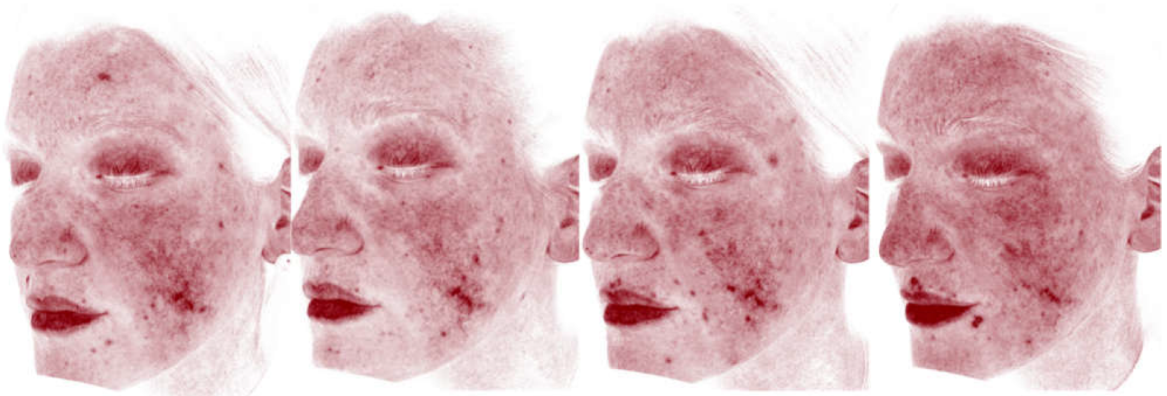
Serial VISIA® assessments, performed at the first instrumental assessment session (February 2024) and at subsequent visits (April 2024, September 2024, December 2024), demonstrated qualitative progressive improvement in surface texture and chromatic homogeneity on visual comparison of standard-light composite images. Representative VISIA® composite images are presented in Figure 2. In addition to standard-light complexion analysis, UV fluorescence and haemoglobin/vascularity (red areas) imaging were performed at each VISIA® session to assess porphyrin activity, subclinical pigmentary changes, and erythema/vascular components not fully apparent under standard lighting, providing complementary instrumental documentation of the treatment response across all time points (Figure 3 and Figure 4).
Figure 2.
VISIA® Complexion Analysis composite images of the left cheek, standardised lighting and positioning. Left panel: baseline assessment (February 2024), showing diffuse erythema overlying the atrophic scar complex, with marked chromatic contrast between the lesional and perilesional skin and an irregular surface texture. Right panel: follow-up assessment (September 2024), demonstrating reduced erythema, improved chromatic homogeneity between the scarred and surrounding skin, and a smoother overall surface texture, consistent with the progressive biostimulatory effect of the treatment protocol.
Figure 2.
VISIA® Complexion Analysis composite images of the left cheek, standardised lighting and positioning. Left panel: baseline assessment (February 2024), showing diffuse erythema overlying the atrophic scar complex, with marked chromatic contrast between the lesional and perilesional skin and an irregular surface texture. Right panel: follow-up assessment (September 2024), demonstrating reduced erythema, improved chromatic homogeneity between the scarred and surrounding skin, and a smoother overall surface texture, consistent with the progressive biostimulatory effect of the treatment protocol.
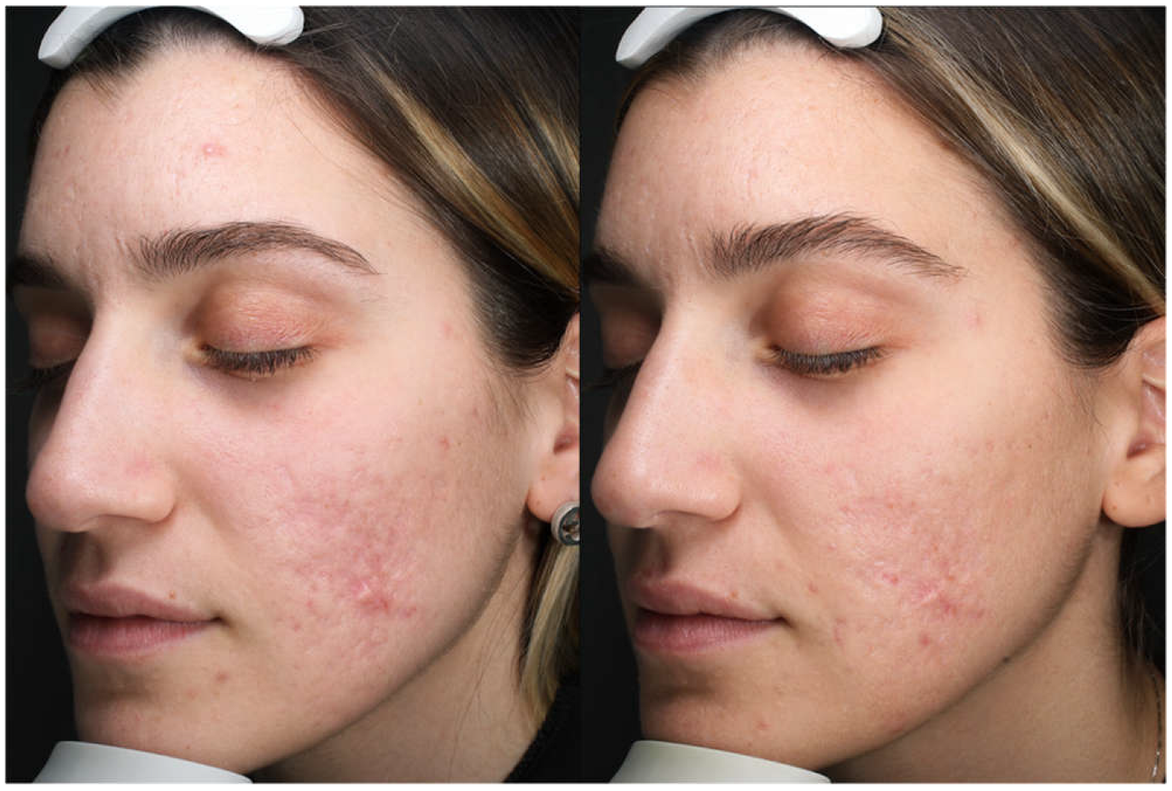
Figure 3.
Sequential VISIA® UV fluorescence imaging of the left cheek across four instrumental assessment time points, arranged left to right in chronological order: T1 (16 February 2024), T2 (22 April 2024), T3 (30 September 2024), and T4 (1 December 2024). UV fluorescence highlights porphyrin activity and subclinical changes not visible under standard lighting, providing complementary documentation of the progressive improvement in skin quality over the treatment course.
Figure 3.
Sequential VISIA® UV fluorescence imaging of the left cheek across four instrumental assessment time points, arranged left to right in chronological order: T1 (16 February 2024), T2 (22 April 2024), T3 (30 September 2024), and T4 (1 December 2024). UV fluorescence highlights porphyrin activity and subclinical changes not visible under standard lighting, providing complementary documentation of the progressive improvement in skin quality over the treatment course.
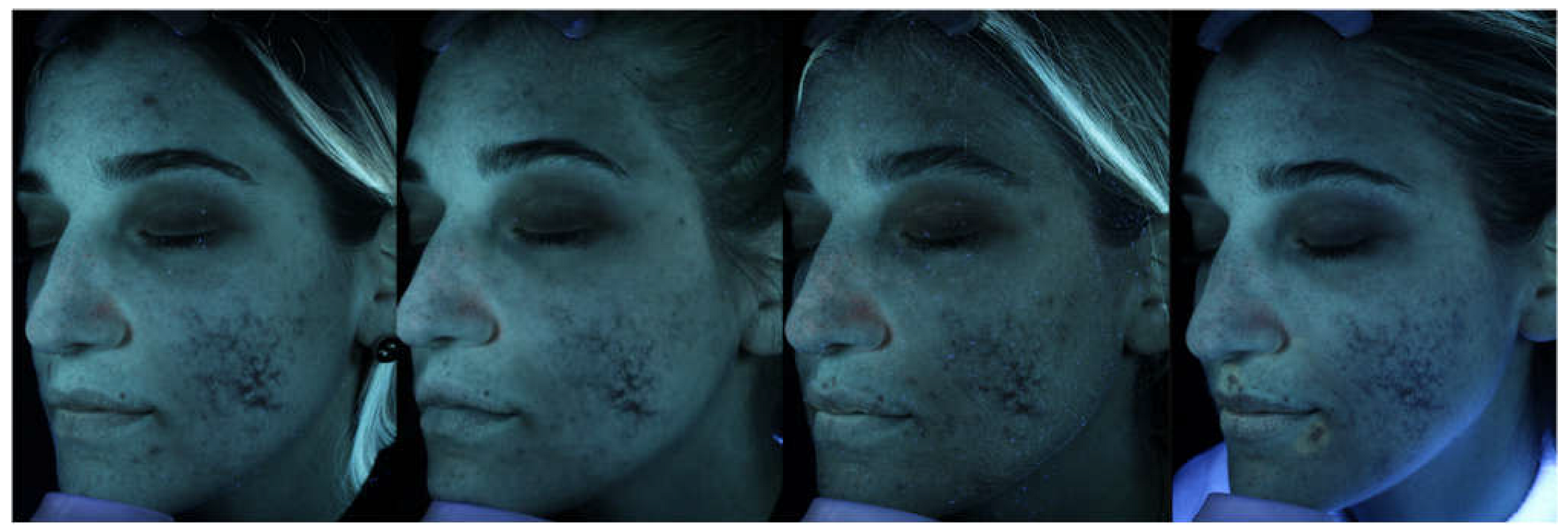
Figure 4.
Sequential VISIA® hemoglobin/vascularity (red areas) imaging of the left cheek across four instrumental assessment time points, arranged left to right in chronological order: T1 (16 February 2024), T2 (22 April 2024), T3 (30 September 2024), and T4 (1 December 2024). This modality isolates subsurface haemoglobin signal to visualise erythema and vascular components not fully apparent under standard lighting, complementing the texture and UV fluorescence assessments shown in Figure 2 and Figure 3.
Figure 4.
Sequential VISIA® hemoglobin/vascularity (red areas) imaging of the left cheek across four instrumental assessment time points, arranged left to right in chronological order: T1 (16 February 2024), T2 (22 April 2024), T3 (30 September 2024), and T4 (1 December 2024). This modality isolates subsurface haemoglobin signal to visualise erythema and vascular components not fully apparent under standard lighting, complementing the texture and UV fluorescence assessments shown in Figure 2 and Figure 3.
No post-procedural complications were recorded across any of the treatment sessions. The patient reported no adverse events between sessions and expressed high overall satisfaction with the aesthetic outcome. At final follow-up, clinician-assessed scar severity had improved to Goodman & Baron Grade 2, and VSS score had reduced to 1–2/13 (from a baseline of 4–5/13). Global aesthetic improvement relative to baseline was rated as +2 (“Much improved”) on the GAIS. Patient-reported satisfaction at final follow-up was 5/5 (Figure 5).
3. Discussion
The present case illustrates the clinical and therapeutic complexity of iatrogenic corticosteroid-induced facial atrophy — a condition that, despite being a recognised complication of intralesional steroid therapy, remains inadequately characterised in the aesthetic medicine literature and lacks established management guidelines.
The key pathophysiological distinction between steroid-induced lipoatrophy and conventional post-acne atrophic scarring has direct therapeutic implications. In common post-acne scarring, the primary target is dermal collagen remodelling, and fractional laser, microneedling, and subcision protocols address this effectively [10,11]. In contrast, corticosteroid-induced atrophy involves a compound deficit: not only dermal structural loss, but also subcutaneous tissue dissolution and fibrotic disruption of the dermo-hypodermal junction. This necessitates a multilayer therapeutic approach that addresses the subcutaneous compartment as a primary target, with surface refinement as a secondary objective.
The therapeutic sequence adopted in this case was designed with this rationale in mind. Non-ablative fractional laser treatment was employed as the foundational intervention: by delivering fractional 1470 nm energy transcutaneously via the dedicated scanning handpiece, it initiates a fibroblastic wound-healing response in the dermis and at the dermo-hypodermal interface, creating a progressively remodelled tissue environment favourable to the subsequent volumising and regenerative procedures. Of particular note was the deliberate treatment of the scar borders in addition to the central atrophic area, in order to stimulate perilesional tissue and promote gradual integration of the healthy surrounding dermis with the atrophic zone. The progressive adjustment of energy parameters across sessions (660 J, 1000 J, 450 J) reflects a deliberate clinical strategy of tissue conditioning followed by consolidation, adapted to the evolving tissue response.
TAR was selected for its unique biological composition: as a mechanically processed autologous lipograft enriched in ADSCs and growth factors, it provides both volumetric restoration and regenerative biological activity, without the risks associated with enzymatic SVF preparation or the limitations of cell-depleted fat grafting. Its application in a tissue bed pre-conditioned by non-ablative fractional laser thermal treatment may potentiate graft survival and biological efficacy through enhanced angiogenesis and tissue receptivity.
Hyperdiluted CaHA was subsequently introduced as a biostimulatory agent targeting the dermal–subdermal interface [12,13]. In its hyperdiluted form, CaHA acts primarily as a scaffold-and-stimulus for endogenous collagen and elastin synthesis, complementing the regenerative activity of the TAR and contributing to progressive structural improvement of the atrophic zone.
The integration of non-ablative fractional photothermolysis (1470 nm) in the same session as CaHA represents a synergistic combination: fractional laser-induced microcoagulation activates a controlled wound-healing cascade in the dermis, while the simultaneously administered CaHA provides a structural biostimulatory substrate for the ensuing neocollagenesis [14]. This “double-stimulation” approach to dermal remodelling has not, to our knowledge, been previously described in the context of steroid-induced atrophy.
A recent case report described the successful treatment of a single facial steroid-induced atrophic scar using a dual-session fractional CO2 laser protocol preceded by polydioxanone thread placement and a dimethylaminoethanol skin booster, with VSS improving from 9/13 at baseline to 2/13 at final follow-up [15]. This confirms the relevance of fractional laser-based dermal remodelling for this condition and supports the choice of fractional photothermolysis as a core component of the present protocol. However, that case addressed a recent (approximately one-month-old), single-component atrophic defect using a laser-only approach with adjunctive mechanical and biostimulatory priming, whereas the present case involved a long-stabilised (approximately two-year), compound dermal and subcutaneous defect requiring an additional volumetric and regenerative component (ART and hyperdiluted CaHA) beyond laser-induced dermal remodelling alone. This distinction underscores that the optimal therapeutic strategy for steroid-induced atrophy may depend substantially on lesion chronicity and the relative contribution of subcutaneous volume loss, supporting the rationale for a multimodal rather than single-modality approach in cases with established subcutaneous involvement.
The rationale for selecting ART and hyperdiluted CaHA as core components of this protocol is further grounded in the senior author’s prior published clinical experience with these techniques. A large multicentric retrospective series on guided SEFFI demonstrated the efficacy and favourable safety profile of autologous fat grafting leveraging SVF and ADSC-driven regenerative mechanisms in facial volume restoration [16]. Building on this foundation, a subsequent retrospective observational study of 158 patients established the efficacy of a combined guided SEFFI and diluted/hyperdiluted CaHA protocol, demonstrating consistent improvement in skin quality and facial volume across multiple facial regions, with substantial gains observed at 90 days and sustained through 150 days of follow-up, and a favourable safety profile limited to minor, self-resolving ecchymosis [17]. The biological compatibility of this combination was further substantiated by an in vitro characterisation study, which demonstrated that adipose-derived mesenchymal stem cells harvested with the guided SEFFI technique retained their proliferative capacity, immunophenotypic identity, and multilineage differentiation potential when co-cultured with CaHA, with no cytotoxic effect observed and an enhanced osteogenic marking suggestive of a synergistic biostimulatory interaction between the two components [18]. The present case extends this previously validated combined SEFFI/CaHA rationale to a markedly more complex clinical scenario — a compound, long-stabilised iatrogenic defect — and integrates it with non-ablative fractional laser remodelling, representing a deliberate, experience-driven escalation of a protocol already shown to be biologically sound, safe, and effective in a larger patient population.
The role of topical cosmeceutical support with retinol (ZO Skin Health®) throughout the treatment course should not be underestimated: retinol-mediated upregulation of keratinocyte and fibroblast activity serves both to prime the tissue between sessions and to consolidate procedural outcomes by maintaining an anabolic dermal environment.
This case has several limitations inherent to its nature as a single-patient report. The primary limitation concerns baseline documentation: the initial clinical assessment in October 2022 was performed at an external centre in Bologna where no standardised complexion analysis instrumentation was available. Photographic documentation at T0 was therefore limited to smartphone photography, without standardised lighting, fixed focal length, or instrumental analysis. Objective VISIA® baseline parameters were only established from February 2024 — approximately 15 months after the condition had already been present and photographically documented. This gap prevents precise quantitative comparison between the true baseline condition and the final outcome, and represents an acknowledged methodological limitation of this report. Additionally, the long-term durability of the achieved results beyond the current follow-up period of 18 months remains to be determined. A prospective series with standardised outcome measures and extended follow-up would be required to validate this protocol as a generalisable treatment framework.
4. Conclusions
To our knowledge, this represents the first reported case describing the combined sequential use of non-ablative fractional laser remodelling, autologous adipose-derived cell therapy, hyperdiluted calcium hydroxylapatite biostimulation, and additional fractional photothermolysis for the management of iatrogenic steroid-induced facial atrophy. We present a structured multimodal protocol combining non-ablative fractional laser remodelling, Autologous Regenerative Therapy (ART), hyperdiluted CaHA biostimulation, and additional non-ablative fractional photothermolysis sessions, supported by a cosmeceutical protocol of 1% topical retinol and 10% of vitamin C, in a patient with severe iatrogenic steroid-induced facial atrophy. This protocol builds directly on the senior author’s previously published clinical and biological experience with guided SEFFI and hyperdiluted CaHA in regenerative facial aesthetics [16,17,18], here adapted and extended to the specific challenge of compound iatrogenic atrophy.
The clinical outcome demonstrates that significant and sustained improvement of a complex multilayer iatrogenic atrophic defect is achievable through a carefully sequenced combination of energy-based, biological, and biostimulatory modalities. The rational sequence of tissue preparation, volumetric restoration, and surface refinement — applied over a structured 18-month timeline — may provide a reproducible clinical framework for managing this challenging and under-reported complication of corticosteroid therapy.
Further prospective studies with larger patient series, standardised scoring instruments, and extended follow-up are warranted to validate and refine this approach.
Author Contributions
Conceptualization, F.M. and G.C.; Methodology, F.M.; Investigation, F.M. and G.C.; Resources, F.M.; Data Curation, G.C.; Writing – Original Draft Preparation, G.C.; Writing – Review & Editing, F.M. and G.C.; Visualization, G.C.; Supervision, F.M. All authors have read and agreed to the published version of the manuscript.
Funding
This research received no external funding.
Institutional Review Board Statement
Ethical review and approval were waived for this study, as it describes routine clinical care delivered as part of standard aesthetic medical practice, with no experimental intervention, randomisation, or deviation from established standard-of-care procedures. The study was conducted in accordance with the Declaration of Helsinki.
Informed Consent Statement
Informed consent was obtained from all subjects involved in the study. Written informed consent has been obtained from the patient to publish this paper, including the accompanying clinical photographs. The patient’s identity has been anonymised in accordance with the journal’s editorial policies and applicable data protection legislation.
Data Availability Statement
The data presented in this study are available on request from the corresponding author due to patient privacy considerations.
Conflicts OF Interest
The authors declare no conflicts of interest. The technologies and techniques described (non-ablative fractional laser, Autologous Regenerative Therapy/Seffiller technique by Seffiline srl, calcium hydroxylapatite/Radiesse, ZO Skin Health, VISIA®) are referenced solely for scientific accuracy; no commercial relationships exist between the authors and the respective manufacturers.
References
- Richards, R.N. Update on intralesional steroid: focus on dermatoses. J. Cutan. Med. Surg. 2010, 14(1), 19–23. [Google Scholar] [CrossRef] [PubMed]
- Colwell, R.; Gullickson, M.; Cutlan, J.; Stratman, E. Cutaneous Atrophy Following Corticosteroid Injections for Tendonitis: Report of Two Cases. JMIR Dermatol. 2025, 8, e67921. [Google Scholar] [CrossRef] [PubMed]
- Goodman, G.J.; Baron, J.A. Postacne scarring: a qualitative global scarring grading system. Dermatol. Surg. 2006, 32(12), 1458–1466. [Google Scholar] [CrossRef] [PubMed]
- Henseler, H. Validation of the Visia® Camera System for skin analysis through assessment of the correlations among the three offered measurements - the percentile, feature count and absolute score - as well as the three capture perspectives, from the left, front and right. GMS Interdiscip. Plast. Reconstr. Surg. DGPW 2022, 11, Doc04. [Google Scholar] [CrossRef] [PubMed]
- Rossi, M.; Roda, B.; Zia, S.; et al. Characterization of the Tissue and Stromal Cell Components of Micro-Superficial Enhanced Fluid Fat Injection (Micro-SEFFI) for Facial Aging Treatment. Aesthet. Surg. J. 2020, 40(6), 679–690. [Google Scholar] [CrossRef] [PubMed]
- Muti, G.F.; Astolfi, G.; Renzi, M.; Rovatti, P.P. Calcium Hydroxylapatite for Augmentation of Face and Hands: A Retrospective Analysis in Italian Subjects. J. Drugs Dermatol. 2015, 14(9), 948–954. [Google Scholar] [PubMed]
- Rovatti, P.P.; Pellacani, G.; Guida, S. Hyperdiluted Calcium Hydroxylapatite 1: 2 for Mid and Lower Facial Skin Rejuvenation: Efficacy and Safety. Dermatol. Surg. 2020, 46(12), e112–e117. [Google Scholar] [CrossRef] [PubMed]
- Gozali, M.V.; Zhou, B. Effective treatments of atrophic acne scars. J. Clin. Aesthet. Dermatol. 2015, 8(5), 33–40. [Google Scholar] [CrossRef] [PubMed]
- Hedelund, L.; Moreau, K.E.; Beyer, D.M.; Nymann, P.; Haedersdal, M. Fractional nonablative 1,540-nm laser resurfacing of atrophic acne scars. A randomized controlled trial with blinded response evaluation. Lasers Med. Sci. 2010, 25(5), 749–754. [Google Scholar] [CrossRef] [PubMed]
- Hession, M.T.; Graber, E.M. Atrophic acne scarring: a review of treatment options. J. Clin. Aesthet. Dermatol. 2015, 8(1), 50–58. [Google Scholar] [PubMed]
- Alam, M.; Han, S.; Pongprutthipan, M.; et al. Efficacy of a needling device for the treatment of acne scars: a randomized clinical trial. JAMA Dermatol. 2014, 150(8), 844–849. [Google Scholar] [CrossRef] [PubMed]
- Jacovella, P.F. Use of calcium hydroxylapatite (Radiesse) for facial augmentation. Clin. Interv. Aging 2008, 3(1), 161–174. [Google Scholar] [CrossRef] [PubMed]
- Guida, S.; Longhitano, S.; Spadafora, M.; et al. Hyperdiluted calcium hydroxylapatite for the treatment of skin laxity of the neck. Dermatol. Ther. 2021, 34(5), e15090. [Google Scholar] [CrossRef] [PubMed]
- Emer, J.; Sundaram, H. Aesthetic applications of calcium hydroxylapatite volumizing filler: an evidence-based review and discussion of current concepts: (part 1 of 2). J. Drugs Dermatol. 2013, 12(12), 1345–1354. [Google Scholar] [PubMed]
- Braho, K.; Zevini, A.; Martinelli, D.; Barini, R. Advanced Fractional CO2 Laser Treatment for Steroid-Induced Atrophy Scars: Clinical Outcomes. Am. J. Case Rep. 2025, 26, e947900. [Google Scholar] [CrossRef] [PubMed]
- Gennai, A.; Baldessin, M.; Melfa, F.; Bovani, B.; Camporese, A.; Claysset, B.; Colli, M.; Diaspro, A.; Russo, R.; Strano, P.; Bollero, D.; Capparè, G.; Casadei, A.; Gallo, G.; Piccolo, D.; Salti, G.; Tesauro, P. Guided Superficial Enhanced Fluid Fat Injection (SEFFI) Procedures for Facial Rejuvenation: An Italian Multicenter Retrospective Case Report. Clin. Pract. 2023, 13(4), 924–943. [Google Scholar] [CrossRef] [PubMed]
- Melfa, F.; McCarthy, A.; Aguilera, S.B.; van Loghem, J.; Gennai, A. Guided SEFFI and CaHA: A Retrospective Observational Study of an Innovative Protocol for Regenerative Aesthetics. J. Clin. Med. 2024, 13(15), 4381. [Google Scholar] [CrossRef] [PubMed]
- Melfa, F.; Gennai, A.; Carfagna, G.; Bovani, B.; Piccolo, D.; Colli, M.; Baldessin, M.; Siragusa, D. Characterization of Adipose-Derived Mesenchymal Stem Cells from tissue harvested with the guided SEFFI technique and co-cultured with calcium hydroxyapatite. J. Appl. Cosmetol. 2023, 41(2), 4–19. [Google Scholar] [CrossRef]
Figure 1.
Clinical photographs at baseline (T0, October 2022), obtained with a standard smartphone camera at the referring centre in Bologna. Left lateral view demonstrating the extent of iatrogenic corticosteroid-induced atrophic defect of the left cheek, with compound dermal and subcutaneous tissue loss. No standardised imaging system was available at the time of initial evaluation.
Figure 1.
Clinical photographs at baseline (T0, October 2022), obtained with a standard smartphone camera at the referring centre in Bologna. Left lateral view demonstrating the extent of iatrogenic corticosteroid-induced atrophic defect of the left cheek, with compound dermal and subcutaneous tissue loss. No standardised imaging system was available at the time of initial evaluation.
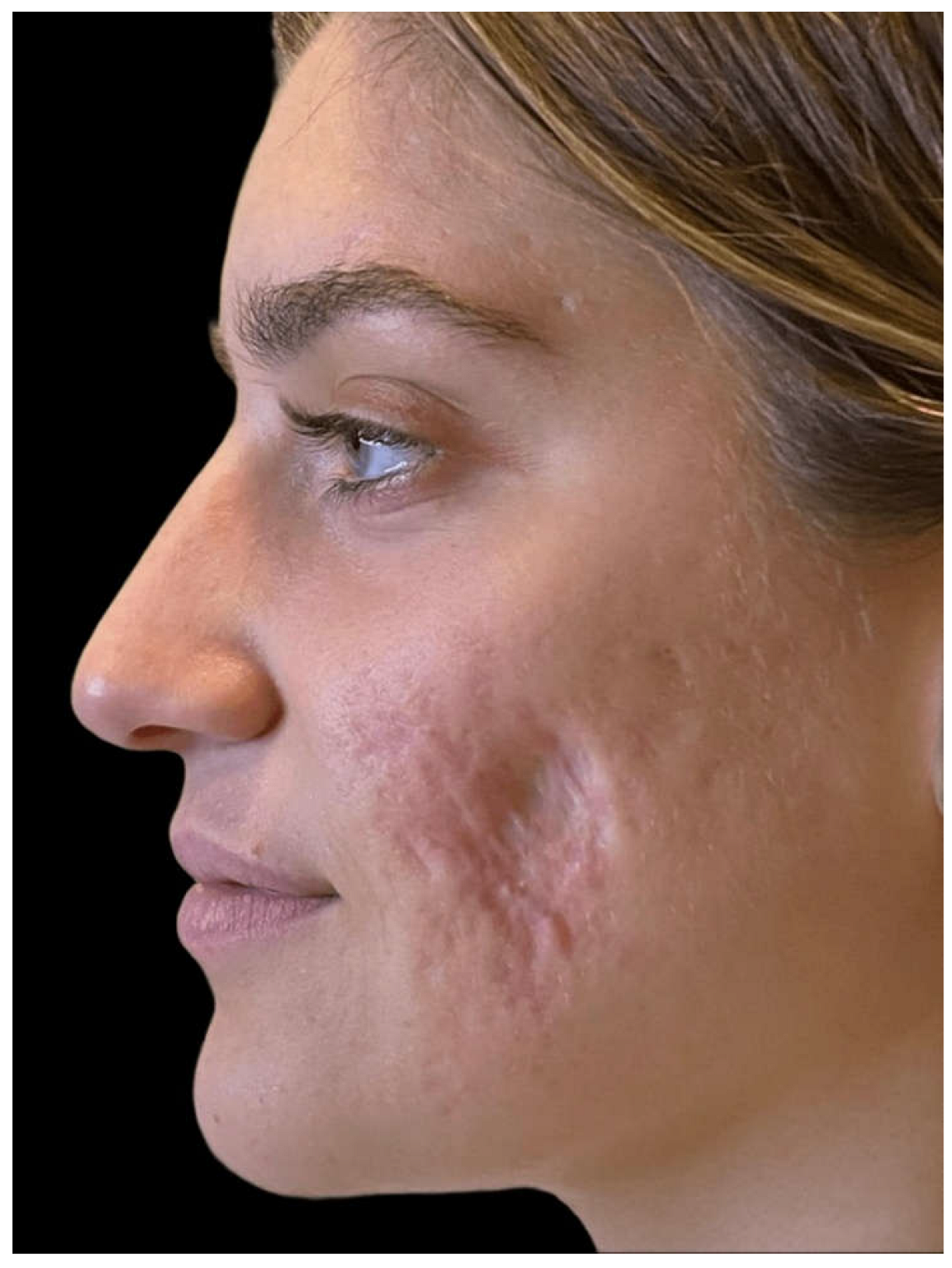
Figure 5.
Comparative clinical photographs of the left cheek. Left panel: baseline (T0, October 2022), obtained with a standard smartphone camera at the referring centre in Bologna, showing the full extent of the iatrogenic corticosteroid-induced atrophic defect with the characteristic linear hypertrophic/depressed scar component and overlying acneiform atrophic scarring. Right panel: 13-month follow-up (December 2024), VISIA® Complexion Analysis System acquisition, demonstrating substantial resolution of the hypertrophic scar component, marked improvement in surface texture and chromatic homogeneity, with only minimal residual scarring evident.
Figure 5.
Comparative clinical photographs of the left cheek. Left panel: baseline (T0, October 2022), obtained with a standard smartphone camera at the referring centre in Bologna, showing the full extent of the iatrogenic corticosteroid-induced atrophic defect with the characteristic linear hypertrophic/depressed scar component and overlying acneiform atrophic scarring. Right panel: 13-month follow-up (December 2024), VISIA® Complexion Analysis System acquisition, demonstrating substantial resolution of the hypertrophic scar component, marked improvement in surface texture and chromatic homogeneity, with only minimal residual scarring evident.
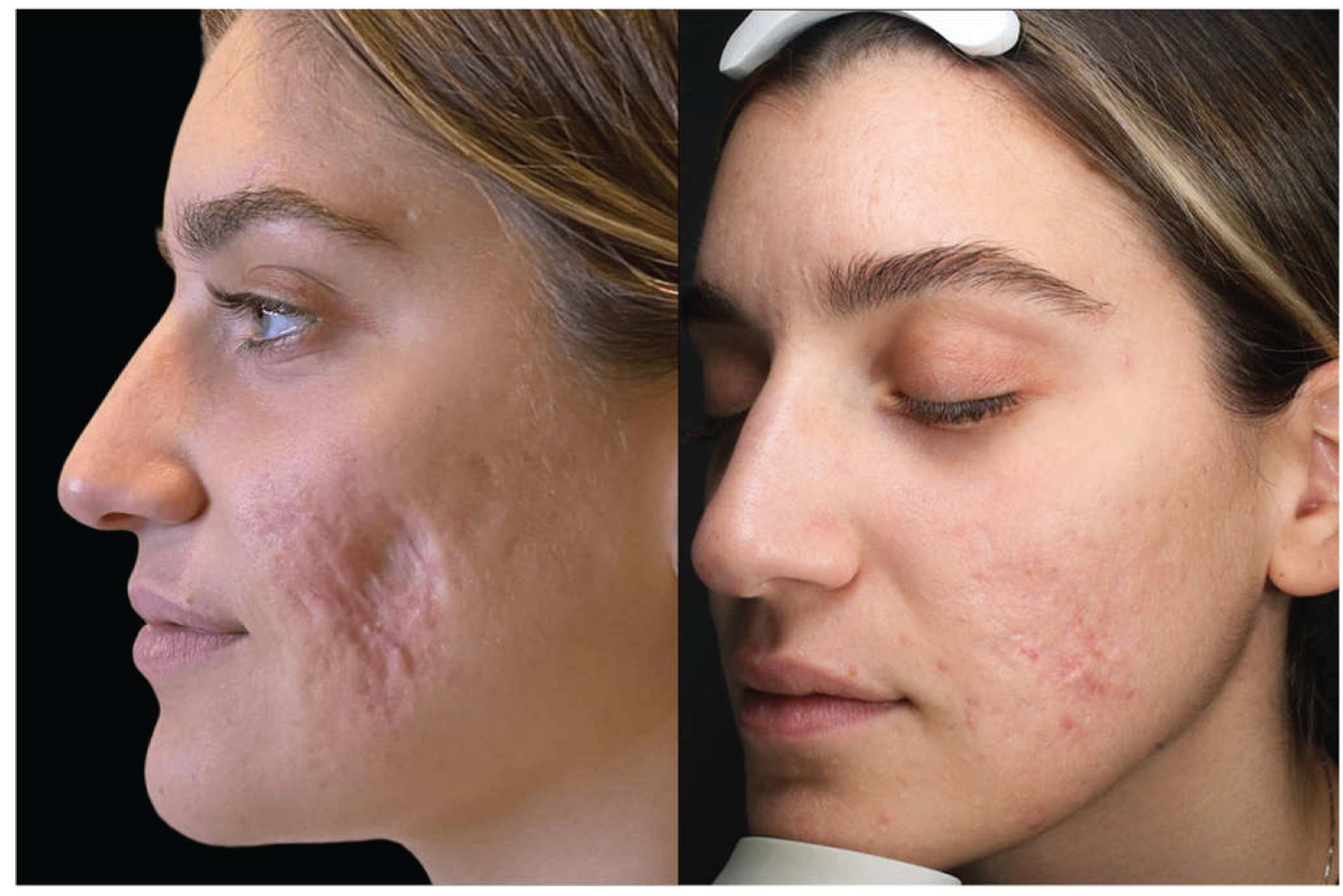
Table 1.
Chronological summary of all treatment sessions.
| Data | Location | Procedure | Parameters / Notes |
|---|---|---|---|
| 20/10/2022 | Bologna | Clinical evaluation + Videodermatoscopy | T0 photo documentation with smartphone. No Visia available. Unique pre-treatment photographic baseline. |
| Home-based | At-home facial treatment | Following the initial consultation, I began a treatment programme using ZO Skin Health products, comprising a facial cleanser, a scrub and 1% retinol (Wrinkle & Texture Repair) | |
| 08/11/2023 | Palermo | Non-Ablative Fractional Laser — Session 1 | Scarring Acne Preset 20% — non-ablative fractional handpiece. 5.5 W / 660 J total. Anesthesia: lidocaine cream 20%. Post-proc: Gentamicin and Betamethasone. |
| 16/02/2024 | Palermo | Non-Ablative Fractional Laser — Session 2 | Scarring Acne Preset 20% — non-ablative fractional handpiece. 5.5 W / 1000 J total. Anesthesia: lidocaine cream 20%. Post-proc: Gentamicin and Betamethasone. |
| 22/04/2024 | Palermo | Autologous Regenerative Therapy (ART) | 18 mL autologous fat cells. Cannula 21G. Double plane (subcutaneous + deep). Central and peripheral distribution. Integrated anesthesia of fat. |
| 30/09/2024 | Palermo | CaHA + Non-Ablative Fractional Laser | CaHA 1.5 mL + 2.7 mL NaCl + 0.3 mL lidocaine (1:2 dilution). 25G cannula, subdermal plane. Non-ablative fractional laser (LightScan – Eufoton): Scarring Acne Preset 20%, 5.5 W / 600 J total, same session. |
| 09/04/2025 | Palermo | Non-Ablative Fractional Laser — Session 3 | Scarring Acne Preset 20% — non-ablative fractional handpiece. 4.5 W / 450 J total (maintenance session). Anesthesia: lidocaine cream 20%. Post-proc: Gentamicin and Betamethasone. |
Disclaimer/Publisher’s Note: The statements, opinions and data contained in all publications are solely those of the individual author(s) and contributor(s) and not of MDPI and/or the editor(s). MDPI and/or the editor(s) disclaim responsibility for any injury to people or property resulting from any ideas, methods, instructions or products referred to in the content. |
© 2026 by the authors. Licensee MDPI, Basel, Switzerland. This article is an open access article distributed under the terms and conditions of the Creative Commons Attribution (CC BY) license (http://creativecommons.org/licenses/by/4.0/).
Copyright: This open access article is published under a Creative Commons CC BY 4.0 license, which permit the free download, distribution, and reuse, provided that the author and preprint are cited in any reuse.


