

Submitted:
25 June 2026
Posted:
26 June 2026
You are already at the latest version
Abstract
Liver fibrosis develops from chronic stress and death of hepatocytes, sustained by persistent injury, immune responses, metabolic stress, and vascular changes. Multiple cells and signaling pathways are involved, with inflammation amplifying fibrogenesis and hepatic stellate cell activation driving the process. Progressive fibrotic changes, potentially culminating in cirrhosis, contribute substantially to global morbidity and mortality, and their most advanced stages re-main often irreversible in clinical practice. Here, we present current insights into liver fibrosis, including epidemiological trends, clinical associations, and future research directions. Modulated by age, sex, lifestyle habits, comorbidities and drug use, liver fibrosis and cirrhosis occur in approximately 3.3% and 1.3% of the general population, respectively. We also discuss techniques for assessing liver fibrosis in clinical practice and research. Moreover, sex differences in liver fibrosis are analyzed together with the role of liver fibrosis in the development of liver-related and extrahepatic outcomes. Finally, we examine the principles of medical treatment of liver fibrosis in the context of its systemic nature and clinical implications.
Keywords:
cirrhosis
; epidemiology
; HCC
; molecular pathogenesis
; portal hypertension
; sex differences
1. Definitions, Burden and Aims
Liver fibrosis represents the dreadful outcome of advanced hepatic injury, irrespective of inciting etiologies, most often altered metabolic homeostasis in the context of metabolic dysfunction-associated steatotic liver disease (MASLD), excess alcohol consumption leading to alcohol-related liver disease (ALD), and chronic viral hepatitis [1]. Progressive fibrotic changes, eventually culminating in cirrhosis in a proportion of individuals, contribute substantially to global morbidity and mortality, and their most advanced stages often remain irreversible in clinical practice, with limited effectiveness of existing therapeutic approaches [2].
From a biological perspective, liver fibrosis results from a scarring reaction with which the liver tends to accumulate collagen fibers to repair damaged tissue while insulating the offending agent(s) [3]. The fibrogenic process involves activation of hepatic stellate cells (HSCs) and myofibroblasts, capillarization of sinusoidal endothelial cells—resulting in increased extracellular matrix deposition and portal pressure—and release of pro-inflammatory cytokines by macrophages and neutrophils, which further stimulate HSCs [4]. Assessment of liver fibrosis, commonly staged according to the METAVIR scoring system from F0 (none) to F4 (cirrhosis), has traditionally relied on histological assessment via liver biopsy; however, fibrosis assessment is increasingly performed non-invasively using either serum biomarkers or imaging-based techniques [5].
The clinical correlates of advanced liver fibrosis and cirrhosis comprise a spectrum of liver-related manifestations and complications, spanning from liver failure requiring transplantation, portal hypertension, and hepatocellular carcinoma (HCC) [6,7]. In addition to being a powerful indicator of advanced chronic liver disease, liver fibrosis is presently regarded as a sensitive barometer of systemic health, serving to assess the risk of extrahepatic disease [1]. Indeed, heterogeneous conditions such as all-cause mortality, cardiovascular disease, extrahepatic cancer, chronic kidney disease (CKD), and type 2 diabetes (T2D) are increasingly recognized in association with liver fibrosis, particularly in the context of MASLD [1,8,9].
A recent study aimed at assessing the prevalence of undiagnosed liver fibrosis in a large, prospective, European population-based, multinational cohort reported that chronic liver disease with fibrosis was present in 32% of participants, yielding an overall estimated prevalence of 1.6% among 30,199 screened individuals [10]. Cirrhosis leads to significant healthcare costs. In the United States, the first-year incremental cost for cirrhosis care is $35,029, dropping to $14,216–$17,629 in each of the following three years. Most expenses are due to complications, with hepatic encephalopathy or ascites costing more than varices or hepatocellular cancer, and subjects with acute kidney injury or multimorbidity incurring the highest first-year costs [11].
Given this complex background, our objective is to present current insights into liver fibrosis, including epidemiological trends, clinical associations, and future research directions.
2. Prevalence and Risk Factors of Liver Fibrosis
Recent meta-analytic evidence, summarized in Table 1 [12,13,14,15,16,17,18,19], has documented the notion that liver fibrosis and cirrhosis occur in 3.3% and 1.3% the general population [12].
Liver fibrosis exhibits a geographical gradient, with prevalence being higher in North America and Australia than in Europe and Asia [13,14]. Additional modifiers include the diagnostic technique used for assessing fibrosis, with Fibroscan being more accurate than ultrasonography [15]. Comorbidities such as psoriasis, people living with HIV (PLWH), and inflammatory bowel disease (IBD) are associated with a high risk of liver fibrosis [15,16,17]. Lifestyle habits and drugs also modulate the risk of fibrosis, with coffee, and antiplatelet drugs associated with a decreased risk [18,19] and methotrexate and alcohol consumption associated with an increased risk of fibrosis [16,20]. A multinational cohort study of 30,199 adults across nine European countries found undiagnosed liver fibrosis in 1.6% of participants. Screening using VCTE-determined liver stiffness showed a 6.9% positive rate, with 4.6% having LSM ≥8 kPa. Elevated LSM was linked to obesity, T2D, and harmful alcohol consumption [10].
Another study found that MASLD and related fibrosis are linked to a greater prevalence of advanced cardiovascular-kidney-metabolic (CKM) syndrome (stages 3 and 4), with CAP/LSM levels showing linear associations in both Chinese and US populations, suggesting that liver health assessment is important for CKM syndrome risk stratification [21].
Assessing insulin resistance (IR) could aid in identifying lean individuals at high risk of MASLD and liver fibrosis, regardless of diabetes status. Njei et al [22] found that MASLD and advanced liver fibrosis in lean individuals were linked to IR, irrespective of excess adiposity. A study conducted among 31,327 Japanese individuals who underwent health examinations [23] found that, in the general population, liver fibrosis progresses with age, especially among men, those with low BMI, and habitual drinkers.
Additional modifiers of liver fibrosis risk comprise tobacco smoking and the use of statins. Cigarette smoking enhances hepatic fibrosis through several interconnected mechanisms [reviewed in 24]. Constituents of cigarette smoke, including nicotine and reactive oxygen species (ROS), contribute to oxidative stress, elevate inflammatory cytokines such as TNF-α, IL-6, and TGF-β1, and directly stimulate hepatic stellate cells (HSCs). Furthermore, smoking induces sinusoidal endothelial dysfunction, reduces antioxidant defenses, and modifies immune cell activity, collectively facilitating increased extracellular matrix (ECM) deposition and furthering the progression of fibrosis. Several studies have supported the notion that statins are considered safe for patients with chronic liver disease and are not considered a risk factor for liver fibrosis; rather, they are generally associated with a reduced risk of liver fibrosis progression and liver-related complications [25,26,27].
Collectively, the studies discussed above support the notion that undiagnosed liver fibrosis is common in the general population and in selected cohorts. Age, sex, lifestyle habits, comorbidities, and drug use modulate the risk of fibrosis. These variables may guide precision medicine approaches in triaging suspected cases of liver fibrosis for referral to hepatological consultation for specialist assessment and prompt treatment.
3. Etiopathogenesis of Liver Fibrosis
Liver fibrosis represents a dynamic wound-healing response that develops when chronic hepatic injury triggers sustained inflammatory and fibrogenic signaling [28,29]. Rather than being a passive accumulation of extracellular matrix (ECM), fibrosis reflects a complex multicellular process integrating hepatocellular injury, immune activation, metabolic stress, and vascular remodeling. Over the past two decades, the conceptual framework of fibrogenesis has evolved from a simple stellate cell-centric model toward a systems-level view in which hepatocytes, non-parenchymal cells, immune populations, and systemic metabolic cues interact continuously [30]. Liver fibrogenesis results from a complex interplay between hepatocyte injury, inflammatory signaling, stellate cell activation, and systemic metabolic factors (Figure 1). Understanding this network is critical not only for clarifying disease mechanisms but also for identifying therapeutic targets.
3.1. Initiating Events: Hepatocyte Stress and Death
The fibrogenic cascade usually begins with persistent hepatocyte injury. Etiologies include MASLD, ALD, chronic viral hepatitis, autoimmune liver disease, cholestatic disorders, and exposure to hepatotoxic drugs or toxins [28,29]. Regardless of the underlying cause, injured hepatocytes release damage-associated molecular patterns (DAMPs), reactive oxygen species (ROS), apoptotic bodies, and lipid peroxidation products [31]. These signals activate Kupffer cells and recruit circulating immune cells.
Metabolic injury deserves particular emphasis in contemporary research. Lipotoxicity driven by saturated fatty acids, free cholesterol accumulation, and mitochondrial dysfunction produces oxidative stress and endoplasmic reticulum stress in hepatocytes [31]. This state amplifies inflammatory signaling pathways such as NF-κB, JNK, and inflammasome activation. Recent experimental work suggests that hepatocyte senescence and impaired autophagy further contribute to fibrogenic signaling by altering hepatocyte–immune crosstalk [31]. These insights help explain why fibrosis progression is particularly pronounced in metabolic liver disease, now the leading cause of chronic liver injury worldwide.
Cell death mechanisms also influence fibrogenesis. Apoptosis has long been recognized as a driver of fibrosis through engulfment of apoptotic bodies by Kupffer cells and stellate cells [32]. More recently, attention has shifted to necroptosis and pyroptosis, which release stronger inflammatory signals and may accelerate fibrosis progression. The relative importance of these pathways likely varies by disease etiology, representing a promising area for mechanistic investigation [33,34].
3.2. Inflammation and Immune Cell Orchestration
Inflammation acts as the central amplifier of fibrogenesis. Kupffer cells, the resident macrophages of the liver, are among the first responders to hepatocyte injury. Upon activation, they secrete cytokines such as TNF-α, IL-1β, and IL-6, as well as fibrogenic mediators including transforming growth factor-β (TGF-β) and platelet-derived growth factor (PDGF) [28]. These signals stimulate hepatic stellate cells (HSCs), the primary collagen-producing cells in the injured liver. However, the immune landscape of fibrogenesis is much more diverse than previously appreciated. Monocyte-derived macrophages infiltrate the liver and can adopt both pro-fibrotic and pro-resolution phenotypes depending on microenvironmental cues. T lymphocytes contribute through several mechanisms: Th17 cells promote inflammation and fibrosis via IL-17 signaling, while regulatory T cells may limit excessive immune activation [35]. Natural killer (NK) cells exert antifibrotic effects by inducing apoptosis of activated stellate cells, although their function can become impaired during chronic disease [36].
Recent research has emphasized the plasticity of macrophage populations in liver fibrosis. Single-cell transcriptomic studies have identified distinct macrophage subsets associated with scar formation and fibrosis resolution. These findings raise the possibility that therapeutic strategies could shift macrophage polarization toward reparative phenotypes rather than simply suppressing inflammation.
3.3. Hepatic Stellate Cell Activation: The Core Fibrogenic Event
Activation of hepatic stellate cells remains the hallmark of liver fibrogenesis [37]. In the healthy liver, HSCs reside in the space of Disse as quiescent vitamin A–storing cells. Following chronic injury, they undergo transdifferentiation into proliferative, contractile, myofibroblast-like cells that produce large quantities of ECM proteins, including type I and III collagen, fibronectin, and laminin. This activation process is driven by several key signaling pathways:
- TGF-β signaling. This is the central fibrogenic pathway, promoting transcription of collagen genes through SMAD-dependent mechanisms [38]
- PDGF signaling: This is a potent mitogenic stimulus that drives stellate cell proliferation and migration [39].
- Hedgehog and Wnt pathways: Both pathways are involved in stellate cell activation and liver regeneration [40].
- Integrin signaling and mechanotransduction: These pathways promote hepatic stellate cell activation in response to extracellular matrix stiffness and mechanical stress [41].
Importantly, the extracellular matrix itself becomes an active participant in disease progression. As fibrosis develops, increased matrix stiffness further stimulates stellate cell activation through mechanosensitive pathways such as YAP/TAZ signaling [42]. This creates a feed-forward loop in which fibrosis promotes additional fibrogenesis.
Another emerging concept is metabolic reprogramming within activated stellate cells [30]. Like cancer cells, activated HSCs shift toward glycolysis and increased glutamine metabolism to support proliferation and matrix synthesis. Targeting this metabolic adaptation has been proposed as a potential antifibrotic strategy [43].
3.4. Contributions of Other Hepatic Cell Populations
Although stellate cells are the principal source of scar tissue, other hepatic cell types contribute significantly to fibrogenesis.
- Liver sinusoidal endothelial cells (LSECs) undergo capillarization during chronic injury, losing fenestrations and producing vasoconstrictive mediators. This process disrupts normal hepatic microcirculation and enhances stellate cell activation through paracrine signaling [44].
- Hepatic progenitor cells become activated during severe or chronic injury. While they contribute to regeneration, they also produce signals that can amplify fibrogenesis, particularly in ductular reaction–associated fibrosis [47].
- Cells derived from epithelial–mesenchymal transition (EMT) may also contribute to the pool of fibrogenic cells. Under conditions of chronic injury and inflammatory signaling, epithelial cells such as hepatocytes or cholangiocytes can acquire mesenchymal characteristics, including enhanced motility and extracellular matrix production, thereby potentially contributing to myofibroblast populations. Conversely, mesenchymal-epithelial transition (MET) has been proposed as a mechanism involved in tissue repair and fibrosis regression, reflecting the dynamic plasticity of hepatic cell populations during chronic liver injury and remodeling [48,49].
3.5. Genetic and Epigenetic Determinants
Interindividual variability in fibrosis progression highlights the role of genetic susceptibility. Polymorphisms in genes such as PNPLA3, TM6SF2, and MBOAT7 strongly influence fibrosis risk in metabolic and alcohol-related liver disease [50,51]. These variants alter lipid metabolism, hepatocyte injury susceptibility, or inflammatory signaling.
Epigenetic regulation also plays a critical role. DNA methylation patterns, histone modifications, and non-coding RNAs, including microRNAs and long non-coding RNAs, modulate stellate cell activation and inflammatory signaling [52]. For example, microRNAs such as miR-21 and miR-34a promote fibrogenic pathways, whereas others exert antifibrotic effects [53,54]. Epigenetic mechanisms are particularly attractive therapeutic targets because they may be reversible.
3.6. Crosstalk with Systemic Metabolic and Inflammatory Pathways
Modern perspectives on liver fibrosis increasingly emphasize its integration with systemic disease processes. Insulin resistance, adipose tissue inflammation, and gut microbiota alterations all influence hepatic fibrogenesis [30,55].
The gut-liver axis is particularly relevant. Increased intestinal permeability allows bacterial products such as lipopolysaccharide (LPS) to reach the liver via the portal vein. Activation of Toll-like receptors on Kupffer cells and stellate cells amplifies inflammatory signaling and fibrosis progression [56]. Dysbiosis may also alter bile acid composition and short-chain fatty acid production, further influencing hepatic metabolism and immune responses.
Adipose tissue contributes through secretion of adipokines and inflammatory mediators. Leptin promotes stellate cell activation, whereas adiponectin generally exerts antifibrotic effects [57]. In obesity and metabolic syndrome, this balance shifts toward a pro-fibrotic state.
3.7. Fibrosis Regression and Remodeling
Importantly, fibrosis is not an irreversible process. Removal of the underlying injury can lead to partial or even substantial fibrosis regression [58]. This process involves apoptosis or inactivation of activated stellate cells, matrix degradation by matrix metalloproteinases (MMPs), and immune-mediated remodeling [59].
Recent work suggests that “inactivated” stellate cells may persist in a primed state, making the liver more susceptible to rapid fibrosis recurrence upon re-injury [60]. Understanding how to stabilize the regression phenotype represents an important therapeutic challenge.
3.8. Perspectives and Future Directions
Several research directions appear particularly promising. First, the integration of spatial transcriptomics and single-cell technologies is transforming our understanding of the fibrotic niche [61]. These tools allow precise mapping of cell–cell interactions within fibrotic tissue, revealing previously unrecognized cellular subsets and signaling networks.
Second, the convergence of metabolic dysfunction, immune activation, and mechanical signaling suggests that successful antifibrotic therapies will likely require multi-target approaches [62]. Single-pathway interventions have largely failed in clinical trials, underscoring the redundancy of fibrogenic signaling.
Third, fibrosis should increasingly be viewed as a systemic disease rather than a purely hepatic condition [63]. Cardiometabolic dysfunction, chronic inflammation, and aging biology all intersect with fibrogenesis [64]. Integrative research frameworks that bridge hepatology with metabolic and immunologic disciplines will likely yield the most impactful insights.
Finally, there is a growing need to understand sex-specific and genetic determinants of fibrogenesis, particularly as precision medicine approaches expand. These factors may influence not only disease susceptibility but also responses to emerging therapies [65,66]].
Taken together, the etiopathogenesis of liver fibrosis reflects a highly dynamic, multicellular process shaped by local injury signals and systemic metabolic context. Continued mechanistic research, especially using modern single-cell and systems-biology approaches, will be essential for translating these insights into effective antifibrotic therapies.
4. Assessment of Liver Fibrosis in Practice and Research
Liver biopsy, though the standard for assessing fibrosis, is limited by its invasiveness, risk of complications, and sampling variability [67,68,69]. A typical sample represents only about 1/50,000 of the liver, often failing to capture fibrosis distribution. This results in under-staging cirrhosis in 20–30% of cases and significant discordance (one or more stages) in 41%, particularly with MASLD/MASH [70].
4.1. Liver Fibrosis Assessment in Clinical Practice
Therefore, assessment of liver fibrosis in clinical settings is evolving from reliance on invasive biopsy procedures to the implementation of noninvasive tests (NITs), such as elastography (FibroScan) and blood-based scores (FIB-4), for the detection of advanced fibrosis (F3–F4). In practice, the focus is on rapidly identifying patients at high risk of cirrhosis (F4) or advanced fibrosis (F3) to initiate treatment. [71]. To accomplish this objective, the FIB-4 index, which incorporates age, AST, ALT, and platelet count, is frequently recommended as the initial risk assessment for primary care screening of patients with MASLD [72].
Although the FIB-4 index is widely recognized as an effective and economical initial screening tool for liver fibrosis, its diagnostic accuracy is influenced by patient age; optimal performance is observed in individuals between 40 and 60 years of age. In patients over 65, the test frequently produces false-positive results, whereas its sensitivity is notably reduced in those younger than 35, thus limiting its clinical utility in this demographic [73]. Age-dependent cut-off values may be used [72]. Alternative to FIB-4 comprises APRI, NFS, and AST/ALT ratio [74].
Whenever advanced fibrosis is suspected based on patient triaging in clinical practice (owing to FIB-4 ≥ 1.3 or in the range of 1.45-3.25), confirmatory Imaging Vibration-Controlled Transient Elastography (VCTE/FibroScan) is the most widely adopted imaging method to measure liver stiffness [75,76,77]. VCTE values generally ≥8 kPa (or often ≥7.1–7.9 kPa in literature) suggest advanced fibrosis (F3-F4). Patients with intermediate/high FIB-4 and high VCTE readings are referred to a hepatologist for Advanced Imaging. Magnetic Resonance Elastography (MRE) is considered the most accurate, especially in obese patients, but is expensive and has limited accessibility [5].
4.2. Liver Fibrosis Assessment in Research
Current research is directed toward validating serum biomarkers and imaging techniques for screening, diagnosis, and longitudinal monitoring, aiming to replace liver biopsy as the standard reference, given the previously discussed limitations of this invasive technique, including sampling error [5,76,78].
Serum biomarkers, particularly direct markers such as the Enhanced Liver Fibrosis (ELF) score and FibroTest [79], provide a reliable, repeatable alternative to liver biopsy by directly assessing ECM turnover. In contrast to indirect tests (for example, AST, ALT, and platelets), which indicate liver injury, direct markers measure collagen degradation products and synthesis byproducts—such as hyaluronic acid, TIMP-1, and PIIINP—that are released during fibrogenesis [80].
A primary objective is to establish consistent, reliable cut-off values across various etiologies (such as MASLD, Hepatitis, ALD, drug-induced, and autoimmune liver diseases) and to evaluate the effectiveness of serial imaging and biomarkers in measuring fibrosis regression following treatment.
4.3. Moving from Histology to Prognosis
Historically, cirrhosis has been diagnosed by identifying widespread nodular formations and bridging fibrosis on histological examination, typically confirmed via liver biopsy [4]. In the past decade, diagnosing compensated advanced chronic liver disease (cACLD)—which includes both advanced fibrosis and compensated cirrhosis—has shifted from invasive liver biopsy to non-invasive tests like VCTE. This change reflects a move toward a more practical, risk-based clinical approach rather than static histological assessment [81]. The advent of cACLD, established by the Baveno VI consensus in 2015, recognizes that severe fibrosis and early-stage cirrhosis form a continuum, frequently indistinguishable among asymptomatic patients [82]. Within this framework, the evaluation of clinically significant portal hypertension (CSPH) and clinical decompensation represents an important transition from merely identifying architectural alterations to assessing and stratifying the risk of clinically relevant outcomes [83,84].
4.4. Role of Portal Hypertension Assessment
Accurate measurement of portal hypertension (PH) is critical for the diagnosis and management of advanced liver fibrosis and cirrhosis, functioning as a direct marker of hepatic stiffness and complication risk. PH assessment provides valuable prognostic information regarding the progression of cirrhosis, likelihood of variceal bleeding, and mortality [85,86]. Measurement of PH plays a key role in staging the severity of liver disease, prognostication of liver disease, treatment monitoring, and risk assessment. While liver fibrosis leads to heightened vascular resistance, the assessment of PH facilitates precise staging of advanced fibrosis (F3-F4) and aids in the identification of individuals with Clinically Significant Portal Hypertension (CSPH) [83].
The degree of PH, especially as assessed by hepatic venous pressure gradient (HVPG), is closely associated with the likelihood of clinical decompensation—including ascites and esophageal varices—and with overall mortality risk [87]. An HVPG > 10 mmHg indicates CSPH and is a key threshold for predicting complications in compensated cirrhosis [88] and HVPG measurement is critical for evaluating the response to therapies aimed at reducing portal pressure [89].
Various methods exist for measuring/assessing PH. While the invasive HVPG remains the gold standard for evaluation, non-invasive modalities such as liver elastography play a key supplemental role. Liver Stiffness Measurement (LSM) by Elastography is deemed to be a highly effective, non-invasive method using VCTE to correlate liver stiffness with portal pressure, often used to avoid invasive tests [90].
Spleen Stiffness Measurement is an emerging non-invasive technique that can offer superior accuracy for assessing portal hypertension [91]
According to the expert consensus and practice guidance [92,93,94,95,96] measurement of the Hepatic Venous Pressure Gradient (HVPG) is indicated in the scenarios summarized in Table 2.
Sex differences significantly influence the development and progression of liver fibrosis. A seminal meta-analysis of 54 studies, Balakrishnan et al. [97] reported a sex-specific pattern in MASLD fibrosis progression: while women appear to have a lower overall prevalence of MASLD than men, once the disease is present, they have a higher risk of advanced fibrosis (pooled risk ratio [RR] 1.37, 95% CI, 1.12-1.68). Recent population-based data further support a more detailed, sex-dependent insights. In a cross-sectional analysis of 5,981 US adults from the National Health and Nutrition Examination Survey (NHANES) 2017–2020, Albhaisi et al. [98] demonstrated that clinically significant fibrosis (defined by liver stiffness ≥8 kPa on transient elastography) was more prevalent among men than women; however, the relative impact of cardiometabolic risk factors (CMRFs) on fibrosis severity was substantially greater in women. Specifically, central adiposity, glucose intolerance, and the presence of two or more CMRFs exhibited significantly stronger associations with fibrosis in women compared with men [98]. Collectively, these pattern supports the concept that cardiometabolic stress may attenuate the relative protection from fibrosis progression in women, potentially through interactions between CMRFs and sex-related biological factors, although causality and directionality remain uncertain.
Biological sex modulates susceptibility to fibrogenesis through complex interactions involving sex hormones, immune responses, adipose tissue distribution, metabolic regulation, and genetic factors [99]. Estrogen is generally considered to exert antifibrotic effects in the liver, whereas androgen signaling appears context-dependent [99]. In men, low testosterone levels have been associated with increased fibrosis risk [100]. In women, hyperandrogenic states such as polyendocrine metabolic ovarian syndrome (PMOS, formerly known as polycystic ovary syndrome) have been linked to more severe metabolic dysfunction and potentially higher fibrosis risk [100,101].
Sex-specific immune differences also contribute. Females generally mount stronger innate and adaptive immune responses, which may improve pathogen clearance in chronic viral hepatitis but also increase susceptibility to autoimmune liver diseases [65,102]. In contrast, males tend to show stronger activation of pro-inflammatory and profibrotic pathways, particularly in the context of metabolic dysfunction [103].
Sociobehavioral factors (i.e., gender related factors) further contribute to sex disparities in fibrosis risk. Men generally report higher rates of alcohol consumption and smoking, both of which are strongly associated with fibrosis progression [65]. However, women appear more susceptible to alcohol-induced liver injury at lower levels of alcohol exposure [104]. Similarly, obesity-related metabolic dysfunction may produce distinct fibrogenic phenotypes between sexes [98].
Emerging evidence also suggests that genetic and chromosomal determinants may contribute to sex-specific fibrogenesis. Variants such as PNPLA3 and HSD17B13 may exert differential effects according to sex and hormonal milieu [105,106]. In addition, X-chromosome gene dosage and epigenetic regulation have been proposed as contributors to sex-biased fibrogenesis but require further mechanistic validation [107,108].
Overall, therefore, recognition of sex differences in liver fibrosis has important implications for screening, risk stratification, and therapeutic development. Sex-specific thresholds for non-invasive tests, differential responses to pharmacologic therapies, and the interaction between hormonal status and fibrosis progression warrant further investigation in both clinical research and antifibrotic treatment strategies.
6. Hepatic Outcomes: Cirrhosis, Liver Failure, and Hepatocellular Carcinoma
Although liver fibrosis initially is an adaptive response, ongoing fibrosis will eventually disrupt hepatic histological architecture and impair liver function, constituting a principal mechanism in the advancement toward cirrhosis, liver failure, and HCC [31].
6.1. Progression to Cirrhosis
Cirrhosis represents the advanced, late stage of liver fibrosis exhibiting the triad of architectural distortion, vascular remodeling, and PH. In the context of cirrhotic nodule histogenesis, continuous scar tissue formation eventually "bridges" between different parts of the liver, creating fibrous septa that encircle clusters of regenerating hepatocytes [109]. Moreover, fibrosis causes the "capillarization" of liver sinusoids, where the normally porous blood vessels lose their fenestrae (pores) and develop a basement membrane [110]. Finally, these structural changes increase resistance to blood flow, leading to PH [111].
6.2. Role of Fibrosis in Liver Failure
Fibrosis leads to liver failure by progressively replacing functional liver tissue with non-functional scar tissue, which, in its turn, leads to nutrient and oxygen blockade, hepatocyte death and loss of function. For example, the capillarized sinusoids and thick ECM create a barrier that prevents the efficient exchange of nutrients and oxygen between the blood and hepatocytes [112]. Moreover, deprived of oxygen and nutrients, mature hepatocytes undergo dysfunction and eventually apoptosis or necrosis [113]. Finally, as the number of healthy hepatocytes declines, the liver can no longer perform its innumerable vital tasks, including protein synthesis, toxin clearance, and metabolic regulation, ultimately resulting in end-stage liver failure [114]. Acute decompensation (AD) of cirrhosis is characterized by the onset of ascites, hepatic encephalopathy, and/or variceal bleeding [115]. Ascites in patients with cirrhosis is typically attributed to splanchnic arterial vasodilation and left ventricular dysfunction [116]. Hepatic encephalopathy is believed to result from hyperammonemia [117], and variceal hemorrhage is associated with portal hypertension [118].
More recently, large-scale European observational studies have identified systemic inflammation as a defining feature of AD [119]. Arroyo et al have introduced the systemic inflammation hypothesis, proposing that systemic inflammation—through impairment of one or more major organ systems—may be a unifying factor that operates synergistically with established mechanisms of AD development [119].
Several pathways by which systemic inflammation may disrupt organ function are involved. The first involves nitric oxide-mediated enhancement of splanchnic vasodilation, which subsequently triggers overactivation of endogenous vasoconstrictor systems, resulting in pronounced vasoconstriction and reduced perfusion in specific vascular beds, notably the renal circulation [120]. Additionally, systemic inflammation may provoke immune-mediated tissue injury [121]. It can also induce substantial metabolic alterations, as inflammatory responses require significant energy expenditure, necessitating the redistribution of nutrients such as glucose, amino acids, and lipids to support immune activation [122]. Furthermore, systemic inflammation inhibits nutrient utilization in peripheral organs, potentially reallocating metabolic resources toward immune processes [119]. Finally, diminished nutrient consumption in these organs may lead to reduced mitochondrial ATP production and compromised organ function [123].
Patients with compensated liver cirrhosis (CLC) are mostly asymptomatic and maintain liver function [124]. With improved non-invasive detection tools, more cases of CLC are now being identified [125]. The shift from CLC to decompensated cirrhosis is driven by various factors, marking a sharp drop in median survival from 10–12 years to 1–2 years after the first decompensation [124].
Evidence demonstrates that the Hepatic Venous Pressure Gradient (HVPG) is a strong, independent predictor of cirrhosis decompensation, particularly in identifying the risk of developing ascites, variceal hemorrhage, and hepatic encephalopathy. Studies confirm that a baseline HVPG ≥ 10 mmHg (Clinically Significant Portal Hypertension - CSPH) indicates a high risk of future decompensation, while an HVPG of 16 mmHg is associated with an imminent risk of further decompensation, and an HVPG≥20 mmHg predicts high mortality and treatment failure [126,127].
6.3. Role of Fibrosis in Hepatocellular Carcinoma
Advanced fibrosis and cirrhosis are the strongest risk factors for HCC, with 80–90% of cases occurring in a cirrhotic liver [128]. This occurs through a variety of pathomechanisms comprising pro-tumor microenvironment, mechanical stiffness, immune escape, and angiogenesis support [129]. The fibrotic liver creates a "premalignant microenvironment" characterized by chronic inflammation, oxidative stress, and constant cell turnover, which increases the likelihood of genetic mutations [130]. Increased matrix stiffness from collagen cross-linking directly activates oncogenic signaling pathways (such as PI3K/Akt and YAP/TAZ) that promote cancer cell survival and proliferation [131]. Fibrosis may inhibit the immune system's capacity to eliminate pre-malignant cells. Activated hepatic stellate cells can promote the expansion of immunosuppressive cell populations such as regulatory T cells (Tregs) and myeloid-derived suppressor cells (MDSCs), thereby shielding tumors from eradication by endogenous immune responses [132]. Fibrotic tissue secretes factors like VEGF that promote the growth of new blood vessels, which are necessary to supply a growing tumor with nutrients [133].
In conclusion, the complex pathobiological processes underlying liver fibrosis are strongly associated with the full range of liver-related manifestations and complications. Advancing our knowledge of these interactions will facilitate improved risk assessment and enable more precise approaches in medicine.
7. Extrahepatic Outcomes: Cardiovascular Risk, Extrahepatic Cancers, Chronic Kidney Disease and Dementia
7.1. Cardiovascular Risk
Liver fibrosis is increasingly recognized not only as a marker of progressive liver disease but also as an important predictor of cardiovascular morbidity and mortality. Among individuals with MASLD, cardiovascular disease (CVD) remains the leading cause of death [134], and severity of liver fibrosis, rather than the presence of steatosis alone, seems to be the main determinant of CVD risk [135].
Fibrosis has also been linked to early manifestations of CVD, particularly subclinical atherosclerosis. In a meta-analysis of 12 studies including 4,725 patients with MASLD, liver fibrosis, assessed via biopsy or VCTE, was associated with more than a twofold higher risk of subclinical atherosclerosis (pooled OR 2.18, 95% CI 1.62–2.93) [136]. Moreover, this association strengthened progressively with increasing fibrosis severity, with odds ratios of 1.64 (95% CI 1.22–2.20) for fibrosis stage ≥F1, 2.22 (95% CI 1.37–3.62) for ≥F2, and 3.42 (95% CI 1.81–6.46) for ≥F3.
Importantly, the increased cardiovascular risk associated with fibrosis extends beyond subclinical disease and translates into a higher incidence of major adverse cardiovascular events (MACE). In a large population-based cohort study including 10,422 Swedish adults with biopsy-confirmed MASLD and 46,517 matched controls without CVD at baseline, cardiovascular risk increased progressively with fibrosis severity over a median follow-up of 13.6 years [137]. Compared to controls, the adjusted hazard ratios (aHR) for MACE were 1.58 (95% CI 1.50–1.67) in patients with simple steatosis, 1.67 (95% CI 1.47–1.89) in those with non-cirrhotic fibrosis, and 2.15 (95% CI 1.77–2.61) in those with cirrhosis [137].
Taken together, the available evidence supports liver fibrosis as a clinically meaningful marker of cardiovascular risk. Therefore, accurate assessment of fibrosis has implications that extend beyond liver-related outcomes and may help identify individuals who would benefit from more intensive cardiovascular risk assessment and aggressive management of modifiable risk factors.
7.2. Chronic Kidney Disease
A growing body of evidence supports a close relationship between liver fibrosis and chronic kidney disease (CKD), particularly in individuals with MASLD. The liver and kidney share several pathophysiological pathways, such that progression of disease in one organ may adversely affect the other [8].
Among patients with MASLD, fibrosis stage rather than steatosis alone appears to be the strongest determinant of renal outcomes. In a longitudinal cohort study involving 3,188 patients with type 2 diabetes mellitus and preserved renal function at baseline, Seo et al. [138] demonstrated that, among individuals with MASLD, advanced liver fibrosis (defined by a FIB-4 index ≥2.67) was independently associated with incident CKD during a mean follow-up of 8.3 years (aHR 1.75, 95% CI 1.15–2.66). In contrast, MASLD without advanced fibrosis was not a predictor of CKD risk [138]. Furthermore, liver fibrosis and CKD seem to have a bidirectional association [PMID: 40945850]. A recent longitudinal analysis [139] found that, over a median follow-up of 9 years, baseline CLD was independently associated with an increased risk of incident CKD (aHR 1.93, 95% CI 1.37–2.72), while baseline CKD was likewise associated with a higher risk of developing CLD (aHR 1.68, 95% CI 1.31–2.16).
Collectively, these findings suggest that liver and kidney diseases may mutually influence disease progression and have important clinical implications because both CKD and MASLD substantially increase cardiovascular risk, hospitalization, and mortality [140]. Consequently, assessment of renal function may be considered in patients with advanced liver fibrosis, and vice versa, the presence of CKD may warrant heightened vigilance for liver fibrosis progression. However, the overall clinical benefit, optimal screening strategy, and cost-effectiveness of integrated hepatorenal surveillance remain subjects of ongoing investigation.
7.3. Extrahepatic Cancers
Emerging evidence suggests that liver fibrosis may be associated not only with hepatocellular carcinoma but also with an increased risk of extrahepatic malignancies [141,142]. In a retrospective cohort study of 1,541 US veterans with chronic viral hepatitis who underwent transient elastography, Abutaleb et al. demonstrated that higher liver stiffness measurements were independently associated with a greater likelihood of extrahepatic cancers after adjustment for alcohol use, obesity, and viral hepatitis status [143]. The strongest associations were observed for prostate cancer, lung cancer, head and neck squamous cell carcinoma, and hematologic malignancies [143].
Additional evidence comes from a large retrospective longitudinal study of 43,080 individuals in Israel. Haimi et al. reported that individuals with a high probability of advanced fibrosis, defined by a FIB-4 score >2.67, had an increased risk of both prevalent and incident malignancies during a 10-year follow-up period [144]. Advanced fibrosis was associated with several cancer types, including pancreatic, breast, colorectal, and hepatocellular carcinoma, and was also linked to a higher risk of metastatic disease and all-cause mortality [144].
Collectively, these findings support the concept that advanced liver fibrosis may reflect a systemic pro-tumorigenic milieu, and beyond its established role as a predictor of liver-related outcomes, advanced fibrosis may therefore serve as a clinically relevant marker for identifying individuals at increased risk of extrahepatic malignancies. Nevertheless, further prospective studies are needed to clarify whether this association is causal and to determine the potential role of fibrosis-based risk stratification in cancer surveillance strategies.
7.4. Dementia
The liver–brain axis has received increasing attention in recent years, and liver fibrosis appears to be a potential contributor to neurocognitive decline and dementia. A growing body of evidence suggests that hepatic fibrosis may influence brain health through multiple mechanisms, which has been discussed in detail elsewhere [145]. In this context, a recent systematic review and meta-analysis by Jamalinia et al., which included 1,115,759 middle-aged individuals from eight longitudinal cohorts, demonstrated that liver fibrosis in the general population, regardless of its etiology, was associated with a 32% increased risk of incident all-cause dementia over a mean follow-up of 14 years [146]. Importantly, a dose–response relationship was observed, with dementia risk progressively increasing according to fibrosis severity: HR 1.06 (95% CI 0.67–1.68) for ≥F2, HR 1.32 (95% CI 1.06–1.64) for ≥F3, and HR 1.69 (95% CI 1.01–2.83) for F4 fibrosis [146].
Additional evidence suggests that, among individuals with MASLD, advanced fibrosis is associated with an increased risk of Alzheimer disease, the most common form of dementia. This association appears to be particularly pronounced in lean MASLD phenotypes, where the aHR was 1.21 (95% CI 0.99–1.48), compared to 1.08 (95% CI 1.01–1.14) in overweight or obese MASLD populations [147]. Although the effect sizes are modest, these findings are consistent with the hypothesis that metabolic dysregulation and ectopic fat distribution, rather than obesity alone, may play an important role in neurodegenerative risk.
Overall, these data suggest that patients with advanced liver fibrosis may benefit from closer cognitive monitoring, as well as aggressive optimization of vascular and metabolic risk factors. As the prevalence of MASLD continues to rise globally and populations age, liver fibrosis may emerge as an important and potentially modifiable contributor to the burden of dementia. However, further mechanistic studies and long-term prospective cohorts are needed to clarify causality and determine whether improvement or regression of fibrosis translates into meaningful protection against neurocognitive decline.
8. Principles of Medical Treatment of Liver Fibrosis
The medical management of liver fibrosis has undergone a profound conceptual shift. Historically, treatment focused primarily on addressing the underlying cause of liver injury, with the assumption that fibrosis would stabilize or regress once the injurious stimulus was removed [148]. While etiological therapy remains the cornerstone of management, contemporary research increasingly recognizes fibrosis as an active and potentially modifiable pathological process. As such, antifibrotic therapy is emerging as a major therapeutic frontier in hepatology [149]. Current and emerging therapeutic strategies for liver fibrosis target multiple complementary mechanisms, including removal of the underlying injury, modulation of inflammation, metabolic interventions, and direct antifibrotic approaches (Figure 2).
8.1. Etiology-Directed Therapy as the Foundation
The most effective antifibrotic intervention remains elimination or control of the primary cause of liver injury. Examples include antiviral therapy for chronic hepatitis B or C, alcohol cessation in alcohol-related liver disease, immunosuppressive treatment for autoimmune hepatitis, and metabolic risk factor control in MASLD [150,151,152].
The success of direct-acting antivirals for hepatitis C provides compelling proof that fibrosis can regress following removal of the injurious stimulus. Numerous studies have demonstrated reductions in liver stiffness, histological fibrosis stage, and portal hypertension after viral eradication [153]. However, fibrosis regression is often incomplete, particularly in advanced disease, emphasizing the need for complementary antifibrotic strategies.
In metabolic liver disease, lifestyle interventions remain central. Weight loss through diet, physical activity, and bariatric surgery can significantly reduce steatohepatitis and fibrosis progression [154]. However, sustained lifestyle modification is challenging, and the global prevalence of obesity suggests that pharmacological approaches will be increasingly necessary.
8.2. Targeting Hepatic Stellate Cell Activation
Because hepatic stellate cells represent the principal effector cells of fibrosis, many therapeutic strategies aim to prevent or reverse their activation. Approaches under investigation include inhibition of key signaling pathways such as TGF-β, PDGF, and integrin-mediated mechanotransduction [38,155].
However, directly targeting TGF-β has proven difficult because of its essential roles in immune regulation and tissue homeostasis. Broad inhibition may produce unacceptable adverse effects. Moreover, single-cell RNA sequencing has led to the identification of different subsets of hepatic stellate cells that might need special targeting strategies [156]. Consequently, current research focuses on more selective modulation of downstream signaling pathways or localized drug delivery systems.
Another promising approach involves inducing apoptosis or senescence of activated stellate cells, thereby reducing matrix production. Nanoparticle-based drug delivery systems that selectively target stellate cells are currently being explored in preclinical models [157].
8.3. Modulating Inflammation and Immune Responses
Given the central role of inflammation in fibrogenesis, immunomodulatory therapies represent another key strategy. Agents targeting chemokine signalling, macrophage recruitment, and inflammatory cytokines have been investigated in clinical trials.
For example, inhibition of the CCR2/CCR5 chemokine axis has shown potential in reducing inflammatory cell infiltration and fibrogenesis [158]. Similarly, modulation of macrophage polarization toward reparative phenotypes may promote fibrosis resolution. Nevertheless, purely anti-inflammatory approaches have often produced modest benefits, reflecting the redundancy and complexity of immune signaling pathways in chronic liver disease. Therefore, most promising approaches are to combine therapies with molecules that target multiple pathways, such as metabolism and inflammation [159].
8.4. Targeting Metabolic Pathways
The rapid rise of MASLD has shifted attention toward metabolic interventions with antifibrotic potential. Several classes of metabolic drugs are currently under investigation or in advanced clinical development:
- GLP-1 receptor agonists and dual incretin agonists improve weight loss, insulin sensitivity, and hepatic inflammation [160].
- FXR agonists regulate bile acid metabolism and may reduce steatosis and inflammation [161].
- PPAR agonists influence lipid metabolism, insulin sensitivity, and inflammatory signaling [162].
Although many of these agents were initially developed for metabolic disease, their pleiotropic effects may also influence fibrogenic pathways. Early clinical trials suggest that improvements in metabolic parameters often correlate with reductions in liver stiffness and fibrosis markers.
8.5. Promoting Matrix Degradation and Fibrosis Resolution
Another therapeutic concept involves accelerating degradation of the fibrotic extracellular matrix. MMPs naturally degrade collagen and other matrix components, while tissue inhibitors of metalloproteinases (TIMPs) suppress this activity [59]. In fibrotic liver disease, TIMP expression is typically increased, limiting matrix breakdown.
Strategies aimed at restoring the balance between MMPs and TIMPs could theoretically enhance fibrosis regression. However, systemic modulation of matrix remodeling carries potential risks, including unwanted tissue damage, tumor progression, and angiogenesis [163].
8.6. Combination Therapies and Systems Approaches
One of the key lessons from antifibrotic drug development is that liver fibrosis is driven by multiple overlapping pathways. As a result, single-target therapies may be insufficient [159]. Future treatment strategies will likely involve combination approaches addressing metabolic injury, inflammation, and stellate cell activation simultaneously.
This concept mirrors therapeutic strategies in other chronic diseases such as cancer and cardiovascular disease, where multi-drug regimens targeting different pathways have proven more effective than monotherapy.
8.7. Personalized and Precision Medicine
Genetic risk factors and metabolic heterogeneity strongly influence fibrosis progression and treatment response, which requires personalized therapies. Variants such as PNPLA3 may modify both disease susceptibility and therapeutic outcomes [50,51]. As genomic and biomarker technologies advance, personalized treatment strategies could become increasingly feasible.
Non-invasive biomarkers and imaging techniques will also play a crucial role in therapeutic development [164]. Reliable markers of fibrosis regression are essential for evaluating antifibrotic therapies in clinical trials and guiding individualized treatment decisions.
8.8. Future Perspectives
Despite substantial progress in understanding fibrogenesis, no universally approved antifibrotic drug currently exists for most chronic liver diseases [165]. Nevertheless, the therapeutic landscape is evolving rapidly. Advances in molecular biology, drug delivery technologies, and systems medicine are expanding the range of potential targets [52,166].
Several priorities should guide future research. First, antifibrotic trials should incorporate better patient stratification based on disease etiology, fibrosis stage, and metabolic profile [167]. Second, combination therapies targeting complementary pathways deserve greater attention [148]. Third, translational research bridging experimental models and human disease remains essential, as many promising preclinical therapies have failed to demonstrate efficacy in clinical settings. In this regard, it should be mentioned that over the last few decades, several hundred, or even a thousand, different drugs have been identified that consistently show beneficial effects on liver health in specific disease models. However, only a few of these compounds have been successfully translated from bench to bedside or incorporated into actual patient care [168]. Finally, it is important to recognize that fibrosis regression alone may not fully eliminate long-term complications such as HCC [169]. Therefore, antifibrotic therapy should be integrated into a broader strategy of risk reduction, surveillance, and management of systemic comorbidities.
In summary, the medical treatment of liver fibrosis is transitioning from a purely etiological approach toward targeted antifibrotic therapy. Continued integration of mechanistic insights, clinical trials, and precision medicine strategies will be essential to translate emerging discoveries into meaningful clinical benefit.
9. Research Agenda
The growing global burden of chronic liver disease underscores the urgent need to advance research on liver fibrosis from descriptive epidemiology to mechanism-driven prevention and therapy. Liver fibrosis represents a complex and dynamic process shaped by interactions among hepatocellular injury, immune activation, metabolic dysfunction, vascular changes, and extracellular matrix remodeling, making it unlikely that single-pathway interventions will adequately address disease progression. Consequently, future research should adopt integrative and systems-level approaches that capture the multifactorial nature of fibrogenesis and its links to systemic metabolic and inflammatory disorders.
One priority area involves improving early detection and population-level screening strategies. Non-invasive tests such as serum fibrosis scores and elastography have already begun to replace liver biopsy in routine practice, yet their diagnostic performance varies across age groups, disease etiologies, and stages of fibrosis. Future studies should focus on refining risk stratification algorithms that combine biochemical markers, imaging modalities, and clinical variables to enable accurate identification of individuals at risk for advanced fibrosis in both primary care and specialist settings. Large prospective cohorts and population-based screening initiatives will be particularly valuable in determining optimal thresholds and diagnostic pathways that balance sensitivity, specificity, accessibility, and cost-effectiveness.
A second major research direction concerns the molecular and cellular mechanisms underlying fibrogenesis and fibrosis regression. Recent advances in single-cell transcriptomics, spatial biology, and multi-omics technologies provide unprecedented opportunities to dissect the cellular heterogeneity of the fibrotic liver and the signaling networks that govern interactions among hepatocytes, immune cells, endothelial cells, and hepatic stellate cells. These approaches may identify previously unrecognized cellular subpopulations and regulatory pathways that drive scar formation or promote fibrosis resolution. Understanding the temporal dynamics of these interactions will be essential for identifying stage-specific therapeutic targets and biomarkers of treatment response.
Equally important is the growing recognition that liver fibrosis cannot be viewed solely as a hepatic condition but rather as a systemic manifestation of metabolic and inflammatory dysregulation. Increasing evidence links liver fibrosis with cardiovascular disease, T2D, CKD, extrahepatic malignancies, and dementia, particularly in the context of MASLD. Future research should therefore explore the bidirectional relationships between hepatic fibrogenesis and systemic cardiometabolic risk factors, integrating hepatology with disciplines such as endocrinology, immunology, and cardiovascular medicine. Such interdisciplinary frameworks may help clarify shared pathogenic mechanisms and identify interventions capable of improving both hepatic and extrahepatic outcomes.
Another key research priority lies in the development of effective antifibrotic therapies. Although removal of the underlying cause of liver injury remains the cornerstone of management, clinical experience demonstrates that fibrosis regression is often incomplete, particularly in advanced disease. The complexity of fibrogenic signaling pathways, including stellate cell activation, inflammatory cytokine networks, metabolic stress, and mechanical signaling, suggests that successful therapeutic strategies will likely require combination approaches targeting multiple complementary mechanisms. Advances in drug delivery systems, including nanoparticle-based targeting of hepatic stellate cells, may further enhance therapeutic specificity and minimize systemic adverse effects.
Precision medicine approaches also represent a promising avenue for future investigation. Genetic susceptibility variants, metabolic phenotypes, and environmental exposures all contribute to interindividual differences in fibrosis progression and therapeutic response. Integrating genomic data, biomarker profiles, and non-invasive imaging into clinical decision-making may enable more personalized risk prediction and treatment strategies. In parallel, improved surrogate endpoints and validated biomarkers of fibrosis regression will be essential for accelerating antifibrotic drug development and for evaluating treatment efficacy in clinical trials.
Finally, public health and implementation research should not be overlooked. The rising prevalence of MASLD, obesity, and diabetes highlights the need for preventive strategies that address lifestyle, metabolic health, and environmental determinants of liver injury. Health-system approaches integrating primary care screening, specialist referral pathways, and digital risk-assessment tools may substantially improve early detection and management of liver fibrosis on a population level.
10. Conclusions
In conclusion, liver fibrosis represents a major and evolving challenge in global health. Although significant progress has been made in understanding the biological mechanisms and clinical implications of fibrogenesis, important gaps remain in early detection, therapeutic development, and integration of liver disease within broader systemic health frameworks. Future advances will depend on multidisciplinary research efforts that combine epidemiology, mechanistic biology, clinical investigation, and precision medicine approaches. Through such integrative strategies, the field may move closer to achieving the ultimate goals of preventing fibrosis progression, promoting fibrosis regression, and reducing the substantial burden of liver-related and extrahepatic complications associated with chronic liver disease.
Author Contributions
Conceptualization, AL, MJ, and RW.; methodology, AL, MJ, and RW; writing—original draft preparation, AL, MJ, and RW; writing—review and editing, AL, MJ, and RW; visualization, AL, MJ, and RW; supervision, AL, MJ, and RW. All authors have read and agreed to the published version of the manuscript.
Funding
This research received no external funding.
Institutional Review Board Statement
Not applicable.
Informed Consent Statement
Not applicable.
Data Availability Statement
No new data were created.
Acknowledgments
The authors are grateful to Sabine Weiskirchen (IFMPEGKC) for preparing Figures 1 and 2 for this article.
Conflicts of Interest
RW is Associate Editor of Livers. However, he was not involved in the peer-review process, the selection of reviewers, or the editorial decision regarding the publication of this manuscript. All other authors declare no conflicts of interest.
Abbreviations
The following abbreviations are used in this manuscript:
| DAMP(s) | Damage-associated molecular pattern(s) |
| ECM | Epithelial-to-mesenchymal transition |
| HSC(s) | Hepatic stellate cell(s) |
| LPS | Lipopolysaccharides |
| LSEC(s) | Liver sinusoidal endothelial cell(s) |
| MASLD | Metabolic dysfunction–associated steatotic liver disease |
| MET | Mesenchymal-to-epithelial transition |
| MMP(s) | Metalloproteinase(s) |
| NK | Natural killer |
| PDGF | Platelet-derived growth factor |
| PNPLA3 | Patatin-like phospholipase domain-containing protein 3 |
| ROS | Reactive oxygen species |
| TGF-β | Transforming growth factor-β |
| TIMP(s) | Tissue inhibitors of metalloproteinases |
| TM6SF2 | Transmembrane 6 superfamily, member 2 |
References
- Lonardo A, Ballestri S, Baffy G, Weiskirchen R. Liver fibrosis as a barometer of systemic health by gauging the risk of extrahepatic disease. Metab Target Organ Damage. 2024;4:41. [CrossRef]
- Nair DG, B Nair D, Weiskirchen R. Reconstructing Liver Fibrosis: 3D Human Models, Microbiome Interfaces, and Therapeutic Innovation. Curr Issues Mol Biol. 2026;48(2):165. PMID: 41751427; PMCID: PMC12939559. [CrossRef]
- Bataller R, Brenner DA. Liver fibrosis. J Clin Invest. 2005 Feb;115(2):209-18. Erratum in: J Clin Invest. 2005;115(4):1100. PMID: 15690074. [CrossRef]
- Somnay K, Wadgaonkar P, Sridhar N, Roshni P, Rao N, Wadgaonkar R. Liver Fibrosis Leading to Cirrhosis: Basic Mechanisms and Clinical Perspectives. Biomedicines 2024;12(10):2229. [CrossRef]
- Lonardo A, Jamalinia M, Weiskirchen R. Recent advances in metabolic dysfunction-associated steatotic liver disease. Eurasian J Med Oncology 2026,10(2),026070085. [CrossRef]
- Zampino R, Patauner F, Durante-Mangoni E. Clinical trajectories in liver cirrhosis: An evidence-based reappraisal for the internist. Eur J Intern Med. 2025;136:31-36. [CrossRef]
- Juanola A, Pose E, Ginès P. Liver Cirrhosis: ancient disease, new challenge. Med Clin (Barc). 2025;164(5):238-246. English, Spanish. [CrossRef]
- Lonardo A. Association of NAFLD/NASH, and MAFLD/MASLD with chronic kidney disease: an updated narrative review. Metab Target Organ Damage. 2024;4:16. [CrossRef]
- Mantovani A, Petracca G, Beatrice G, Tilg H, Byrne CD, Targher G. Non-alcoholic fatty liver disease and risk of incident diabetes mellitus: an updated meta-analysis of 501 022 adult individuals. Gut 2021;70(5):962-969. [CrossRef]
- Graupera I, Thiele M, Castera L, Pera G, Piano S, Sòria A, Fabrellas N, Toran P, Chacon C, Bech KT, Schnefeld HL, Tonon M, Incicco S, Moussy J, Lévy V, Madir A, Kukic S, Jan Havaj D, Adamcova-Selcanova S, Pustjens J, van Kleef LA, Jiménez-Masip A, Pagès L, Zoncapè M, Weber SN, Galle PR, Harris R, Ibáñez-Samaniego L, Morillas RM, Diaz A, Detlefsen S, Serra-Burriel M, Arslanow A, Andersen P, Pich J, Bonfill E, Korenjak M, Fournier-Poizat C, Llorca A, Gourmelon MC, de Koning HJ, Pérez-Guasch M, Thu Ma A, Juanola A, Pose E, Arteaga I, Villesen I, Hansen JK, Calvino V, Gagliardi R, Boutouria B, Pastrovic F, Kujundzic PD, Zilincanova D, Sulejova KK, Rojo D, de Knegt RJ, Melo MD, Torrejón A, Hernández-Ibáñez R, Hoyo J, Muñoz L, López-Martos R, Griffin SJ, Manns M, Karlsen TH, Newsome PN, Kamath PS, Bañares R, Guha IN, Schattenberg JM, Lammert F, Tsochatzis E, Brouwer WP, Pericàs JM, Skladany L, Grgurevic I, Roulot D, Angeli P, Krag A, Caballeria L, Ginès P; LiverScreen Consortium Investigators. Prevalence of liver fibrosis in the general population (the LiverScreen project): a multinational European cohort study. Lancet 2026;407(10537):1448-1458. [CrossRef]
- Kanwal F, Nelson R, Liu Y, Kramer JR, Hernaez R, Cholankeril G, Rana A, Flores A, Smith D, Cao Y, Beech B, Asch SM. Cost of Care for Patients With Cirrhosis. Am J Gastroenterol. 2024;119(3):497-504. [CrossRef]
- Zamani M, Alizadeh-Tabari S, Ajmera V, Singh S, Murad MH, Loomba R. Global Prevalence of Advanced Liver Fibrosis and Cirrhosis in the General Population: A Systematic Review and Meta-analysis. Clin Gastroenterol Hepatol. 2025;23(7):1123-1134. [CrossRef]
- Owrangi S, Paik JM, Golabi P, de Avila L, Hashida R, Nader A, Paik A, Henry L, Younossi ZM. Meta-Analysis: Global Prevalence and Mortality of Cirrhosis in Metabolic Dysfunction-Associated Steatotic Liver Disease. Aliment Pharmacol Ther. 2025;61(3):433-443. [CrossRef]
- Kim HY, Yu JH, Chon YE, Kim SU, Kim MN, Han JW, Lee HA, Jin YJ, An J, Choi M, Jun DW. Prevalence of clinically significant liver fibrosis in the general population: A systematic review and meta-analysis. Clin Mol Hepatol. 2024;30(Suppl):S199-S213. [CrossRef]
- Navarro P, Gutiérrez-Ramírez L, Tejera-Muñoz A, Arias Á, Lucendo AJ. Systematic Review and Meta-Analysis: Prevalence of Non-Alcoholic Fatty Liver Disease and Liver Fibrosis in Patients with Inflammatory Bowel Disease. Nutrients 2023;15(21):4507. [CrossRef]
- Yongpisarn T, Namasondhi A, Iamsumang W, Rattanakaemakorn P, Suchonwanit P. Liver fibrosis prevalence and risk factors in patients with psoriasis: A systematic review and meta-analysis. Front Med (Lausanne) 2022;9:1068157. [CrossRef]
- Lyu H, Tang H, Liang Y, Huang S, Wang Y, Huang W, Zhou Y. Alcohol Consumption and Risk of Liver Fibrosis in People Living With HIV: A Systematic Review and Meta-Analysis. Front Immunol. 2022;13:841314. [CrossRef]
- Thongtan T, Deb A, Vutthikraivit W, Laoveeravat P, Mingbunjerdsuk T, Islam S, Islam E. Antiplatelet therapy associated with lower prevalence of advanced liver fibrosis in non-alcoholic fatty liver disease: A systematic review and meta-analysis. Indian J Gastroenterol. 2022;41(2):119-126. [CrossRef]
- Ebadi M, Ip S, Bhanji RA, Montano-Loza AJ. Effect of Coffee Consumption on Non-Alcoholic Fatty Liver Disease Incidence, Prevalence and Risk of Significant Liver Fibrosis: Systematic Review with Meta-Analysis of Observational Studies. Nutrients. 2021;13(9):3042. [CrossRef]
- Marti-Aguado D, Calleja JL, Vilar-Gomez E, Iruzubieta P, Rodríguez-Duque JC, Del Barrio M, Puchades L, Rivera-Esteban J, Perelló C, Puente A, Gomez-Medina C, Escudero-García D, Serra MA, Bataller R, Crespo J, Arias-Loste MT. Low-to-moderate alcohol consumption is associated with increased fibrosis in individuals with metabolic dysfunction-associated steatotic liver disease. J Hepatol. 2024;81(6):930-940. [CrossRef]
- Hu S, Wang D, Yu Q, Chen Z, Lu W, Meng Y, Peng X, Liu L, Wan H, Shen J. Metabolic Dysfunction-Associated Steatotic Liver Disease and Liver Fibrosis are Associated with Advanced Cardiovascular-Kidney-Metabolic Syndrome in Chinese and US Populations. Diabetes Metab Syndr Obes. 2025;18:4699-4713. [CrossRef]
- Njei B, Abdu M, Al-Ajlouni YA, Mohamed MF, Deng Y, Osta EG, Kanmounye US, Vilarinho S, Dranoff J, Lim JK, Md, Echouffo Tcheugui JB. Insulin resistance, metabolic dysfunction-associated steatotic liver disease, and advanced liver fibrosis in lean US adults: a population-based study. Proc (Bayl Univ Med Cent). 2025;38(5):637-645. [CrossRef]
- Kariyama K, Kawanaka M, Nouso K, Wakuta A, Shiota S, Kurisu A, Sugiyama A, Akita T, Kumada T, Tanaka J; Real-Life Practice Experts for HCC (RELPEC) Study Group. Identification of risk groups for advanced liver fibrosis in the general population using the Fibrosis-3 index. JGH Open. 2024;8(7):e70010. [CrossRef]
- El-Azab G, Elkhouly E, Abouyoussef R, Nagdy H. Smoking and liver diseases: an updated review of pathogenesis, progression, and therapeutic implications. Clin Exp Med. 2025;26(1):51. [CrossRef]
- Simon TG, King LY, Zheng H, Chung RT. Statin use is associated with a reduced risk of fibrosis progression in chronic hepatitis C. J Hepatol. 2015;62(1):18-23. [CrossRef]
- Kamal S, Khan MA, Seth A, Cholankeril G, Gupta D, Singh U, Kamal F, Howden CW, Stave C, Nair S, Satapathy SK, Ahmed A. Beneficial Effects of Statins on the Rates of Hepatic Fibrosis, Hepatic Decompensation, and Mortality in Chronic Liver Disease: A Systematic Review and Meta-Analysis. Am J Gastroenterol. 2017;112(10):1495-1505. [CrossRef]
- Schreiner AD, Zhang J, Petz CA, Moran WP, Koch DG, Marsden J, et al. Statin prescriptions and progression of advanced fibrosis risk in primary care patients with MASLD. BMJ Open Gastroenterology 2024;11:e001404. [CrossRef]
- Acharya P, Chouhan K, Weiskirchen S, Weiskirchen R. Cellular Mechanisms of Liver Fibrosis. Front Pharmacol. 2021;12:671640. [CrossRef]
- Wang Z, Cao Z, Dong Y, Hong Y, Zuo L, Wang H. Hepatocyte-hepatic stellate cell interactions in liver fibrosis: Mechanisms and therapeutic implications. Hepatol Commun. 2026;10(2):e0893. [CrossRef]
- Horn P, Tacke F. Metabolic reprogramming in liver fibrosis. Cell Metab. 2024;36(7):1439-1455. [CrossRef]
- Roehlen N, Crouchet E, Baumert TF. Liver Fibrosis: Mechanistic Concepts and Therapeutic Perspectives. Cells 2020;9(4):875. [CrossRef]
- Shojaie L, Iorga A, Dara L. Cell Death in Liver Diseases: A Review. Int J Mol Sci. 2020;21(24):9682. [CrossRef]
- Gan C, Cai Q, Tang C, Gao J. Inflammasomes and Pyroptosis of Liver Cells in Liver Fibrosis. Front Immunol. 2022;13:896473. [CrossRef]
- Ye Z, Zhang N, Lei H, Yao H, Fu J, Zhang N, Xu L, Zhou G, Liu Z, Lv Y. Immunogenic necroptosis in liver diseases: mechanisms and therapeutic potential. J Mol Med (Berl). 2023;101(11):1355-1363. [CrossRef]
- Lurje I, Gaisa NT, Weiskirchen R, Tacke F. Mechanisms of organ fibrosis: Emerging concepts and implications for novel treatment strategies. Mol Aspects Med. 2023;92:101191. [CrossRef]
- Yang M, Vanderwert E, Kimchi ET, Staveley-O'Carroll KF, Li G. The Important Roles of Natural Killer Cells in Liver Fibrosis. Biomedicines. 2023;11(5):1391. [CrossRef]
- Khomich O, Ivanov AV, Bartosch B. Metabolic Hallmarks of Hepatic Stellate Cells in Liver Fibrosis. Cells 2019;9(1):24. [CrossRef]
- Dewidar B, Meyer C, Dooley S, Meindl-Beinker AN. TGF-β in Hepatic Stellate Cell Activation and Liver Fibrogenesis-Updated 2019. Cells 2019;8(11):1419. [CrossRef]
- Mostafa S, Shetab Boushehri MA, Ezzat AA, Weiskirchen R, Lamprecht A, Mansour S, Tammam SN. Targeted Delivery of Anti-TGF-β1-siRNA Using PDGFR-β Peptide-Modified Chitosan Nanoparticles for the Treatment of Liver Fibrosis. Mol Pharm. 2025;22(11):6741-6758. [CrossRef]
- Nishikawa K, Osawa Y, Kimura K. Wnt/β-Catenin Signaling as a Potential Target for the Treatment of Liver Cirrhosis Using Antifibrotic Drugs. Int J Mol Sci. 2018;19(10):3103. [CrossRef]
- Sharip A, Kunz J. Mechanosignaling via Integrins: Pivotal Players in Liver Fibrosis Progression and Therapy. Cells 2025;14(4):266. [CrossRef]
- Du K, Maeso-Díaz R, Oh SH, Wang E, Chen T, Pan C, Xiang K, Dutta RK, Wang XF, Chi JT, Diehl AM. Targeting YAP-mediated HSC death susceptibility and senescence for treatment of liver fibrosis. Hepatology 2023;77(6):1998-2015. [CrossRef]
- Yin X, Peng J, Gu L, Liu Y, Li X, Wu J, Xu B, Zhuge Y, Zhang F. Targeting glutamine metabolism in hepatic stellate cells alleviates liver fibrosis. Cell Death Dis. 2022;13(11):955. [CrossRef]
- Poisson J, Lemoinne S, Boulanger C, Durand F, Moreau R, Valla D, Rautou PE. Liver sinusoidal endothelial cells: Physiology and role in liver diseases. J Hepatol. 2017;66(1):212-227. [CrossRef]
- Banales JM, Huebert RC, Karlsen T, Strazzabosco M, LaRusso NF, Gores GJ. Cholangiocyte pathobiology. Nat Rev Gastroenterol Hepatol. 2019;16(5):269-281. [CrossRef]
- Iwaisako K, Jiang C, Zhang M, Cong M, Moore-Morris TJ, Park TJ, Liu X, Xu J, Wang P, Paik YH, Meng F, Asagiri M, Murray LA, Hofmann AF, Iida T, Glass CK, Brenner DA, Kisseleva T. Origin of myofibroblasts in the fibrotic liver in mice. Proc Natl Acad Sci U S A. 2014;111(32):E3297-305. [CrossRef]
- So J, Kim A, Lee SH, Shin D. Liver progenitor cell-driven liver regeneration. Exp Mol Med. 2020;52(8):1230-1238. [CrossRef]
- Li Y, Wang J, Asahina K. Mesothelial cells give rise to hepatic stellate cells and myofibroblasts via mesothelial-mesenchymal transition in liver injury. Proc Natl Acad Sci U S A. 2013;110(6):2324-9. [CrossRef]
- Wang S, Link F, Han M, Chaudhary R, Asimakopoulos A, Liebe R, Yao Y, Hammad S, Dropmann A, Krizanac M, Rubie C, Feiner LK, Glanemann M, Ebert MPA, Weiskirchen R, Henis YI, Ehrlich M, Dooley S. The Interplay of TGF-β1 and Cholesterol Orchestrating Hepatocyte Cell Fate, EMT, and Signals for HSC Activation. Cell Mol Gastroenterol Hepatol. 2024;17(4):567-587. [CrossRef]
- Eslam M, Newsome PN, Sarin SK, Anstee QM, Targher G, Romero-Gomez M, Zelber-Sagi S, Wai-Sun Wong V, Dufour JF, Schattenberg JM, Kawaguchi T, Arrese M, Valenti L, Shiha G, Tiribelli C, Yki-Järvinen H, Fan JG, Grønbæk H, Yilmaz Y, Cortez-Pinto H, Oliveira CP, Bedossa P, Adams LA, Zheng MH, Fouad Y, Chan WK, Mendez-Sanchez N, Ahn SH, Castera L, Bugianesi E, Ratziu V, George J. A new definition for metabolic dysfunction-associated fatty liver disease: An international expert consensus statement. J Hepatol. 2020;73(1):202-209. [CrossRef]
- Weiskirchen R, Lonardo A. Obesity, Metabolic Dysfunction-Associated Steatotic Liver Disease and Hepatocellular Carcinoma: How Molecular Changes Impact Cellular Functions, Biocell 2026;50(5):1. [CrossRef]
- Liu R, Li Y, Zheng Q, Ding M, Zhou H, Li X. Epigenetic modification in liver fibrosis: Promising therapeutic direction with significant challenges ahead. Acta Pharm Sin B. 202;14(3):1009-1029. [CrossRef]
- Ichim C, Boicean A, Anderco P, Todor SB, Hașegan A, Bîrsan S, Bîrluțiu V. MicroRNAs in Liver Cirrhosis: Diagnostic and Therapeutic Perspectives-A Comprehensive Review. J Pers Med. 2025;15(8):376. [CrossRef]
- Zhao M, Qi Q, Liu S, Huang R, Shen J, Zhu Y, Chai J, Zheng H, Wu H, Liu H. MicroRNA-34a: A Novel Therapeutic Target in Fibrosis. Front Physiol. 2022;13:895242. [CrossRef]
- Lonardo A, Weiskirchen R. Insulin Resistance at the Crossroads of Metabolic Inflammation, Cardiovascular Disease, Organ Failure and Cancer. Biomolecules 2025;15(12):1745. [CrossRef]
- Albillos A, de Gottardi A, Rescigno M. The gut-liver axis in liver disease: Pathophysiological basis for therapy. J Hepatol. 2020;72(3):558-577. [CrossRef]
- Parola M, Pinzani M. Liver fibrosis in NAFLD/NASH: from pathophysiology towards diagnostic and therapeutic strategies. Mol Aspects Med. 2024;95:101231. [CrossRef]
- Kisseleva T, Brenner D. Molecular and cellular mechanisms of liver fibrosis and its regression. Nat Rev Gastroenterol Hepatol. 2021;18(3):151-166. [CrossRef]
- Roeb E. Matrix metalloproteinases and liver fibrosis (translational aspects). Matrix Biol. 2018;68-69:463-473. [CrossRef]
- Troeger JS, Mederacke I, Gwak GY, Dapito DH, Mu X, Hsu CC, Pradere JP, Friedman RA, Schwabe RF. Deactivation of hepatic stellate cells during liver fibrosis resolution in mice. Gastroenterology 2012;143(4):1073-83.e22. [CrossRef]
- Dobie R, Wilson-Kanamori JR, Henderson BEP, Smith JR, Matchett KP, Portman JR, Wallenborg K, Picelli S, Zagorska A, Pendem SV, Hudson TE, Wu MM, Budas GR, Breckenridge DG, Harrison EM, Mole DJ, Wigmore SJ, Ramachandran P, Ponting CP, Teichmann SA, Marioni JC, Henderson NC. Single-Cell Transcriptomics Uncovers Zonation of Function in the Mesenchyme during Liver Fibrosis. Cell Rep. 2019;29(7):1832-1847.e8. [CrossRef]
- Weiskirchen R, Weiskirchen S, Tacke F. Organ and tissue fibrosis: Molecular signals, cellular mechanisms and translational implications. Mol Aspects Med. 2019;65:2-15. [CrossRef]
- Yoneda M, Kobayashi T, Iwaki M, Nogami A, Saito S, Nakajima A. Nonalcoholic Fatty Liver Disease as a Systemic Disease and the Need for Multidisciplinary Care. Gut Liver. 2023;17(6):843-852. [CrossRef]
- Saidi AN, Theel WB, de Jong VD, van Mil SR, van der Lely AJ, Grobbee DE, Apers J, Beek EVZ, Castro Cabezas M. Liver Fibrosis as a Predictor of Cardiovascular Risk in Patients with Severe Obesity. J Clin Med. 2025;14(23):8532. [CrossRef]
- Jamalinia M, Lonardo A, Weiskirchen R. Sex and Gender Differences in Liver Fibrosis: Pathomechanisms and Clinical Outcomes. Fibrosis 29024;2(4):10006. [CrossRef]
- Zhang X, Chang KM, Yu J, Loomba R. Unraveling Mechanisms of Genetic Risks in Metabolic Dysfunction-Associated Steatotic Liver Diseases: A Pathway to Precision Medicine. Annu Rev Pathol. 2025;20(1):375-403. [CrossRef]
- Colapietro F, Viganò M, Cerini F, Plebani R, Savino A, Calabrese MP, Marra P, Martins de Mattos KD, Taboni S, Cosenza A, Loglio A, Selvaggio C, Lucà MG, Masellis C, Mori B, Pugliese N, Masetti C, Farina E, De Nicola S, Ceriani R, Lleo A, Muglia R, Sironi S, Fagiuoli S, Aghemo A. Incidence and Predictors of Complications Following Percutaneous Liver Biopsy: A Large Italian Multicentre Study. Liver Int. 2025;45(5):e70078. [CrossRef]
- Sumida Y, Nakajima A, Itoh Y. Limitations of liver biopsy and non-invasive diagnostic tests for the diagnosis of nonalcoholic fatty liver disease/nonalcoholic steatohepatitis. World J Gastroenterol. 2014;20(2):475-85. [CrossRef]
- Karlgren S, Hagström H, Nilsson H. High Heterogeneity of Several Liver Histology Parameters in Patients With Metabolic Dysfunction-Associated Steatotic Liver Disease Demonstrated by Biopsies From 7 Segments. Clin Gastroenterol Hepatol. 2025:S1542-3565(25)01034-1. [CrossRef]
- Ratziu V, Charlotte F, Heurtier A, Gombert S, Giral P, Bruckert E, Grimaldi A, Capron F, Poynard T; LIDO Study Group. Sampling variability of liver biopsy in nonalcoholic fatty liver disease. Gastroenterology 2005;128(7):1898-906. [CrossRef]
- Campos-Murguía A, Ruiz-Margáin A, González-Regueiro JA, Macías-Rodríguez RU. Clinical assessment and management of liver fibrosis in non-alcoholic fatty liver disease. World J Gastroenterol. 2020;26(39):5919-5943. [CrossRef]
- Sung S, Al-Karaghouli M, Tam M, Wong YJ, Jayakumar S, Davyduke T, Ma M, Abraldes JG. Age-dependent differences in FIB-4 predictions of fibrosis in patients with MASLD referred from primary care. Hepatol Commun. 2024;9(1):e0609. [CrossRef]
- Kaltenbach M. Why are non-invasive risk scores such as FIB-4 used in clinical practice? Available at: https://www.aasld.org/liver-fellow-network/core-series/why-series/why-are-non-invasive-risk-scores-such-fib-4-used (last accessed 25 June 2026).
- Amernia B, Moosavy SH, Banookh F, Zoghi G. FIB-4, APRI, and AST/ALT ratio compared to FibroScan for the assessment of hepatic fibrosis in patients with non-alcoholic fatty liver disease in Bandar Abbas, Iran. BMC Gastroenterol. 2021;21(1):453. [CrossRef]
- Chan WK, et al. Optimizing use of nonalcoholic fatty liver disease fibrosis score, fibrosis-4 score, and liver stiffness measurement to identify patients with advanced fibrosis. Clin. Gastroenterol. Hepatol. 2019;17:2570–2580.e37. [CrossRef]
- Kazi IN, Kuo L, Tsai E. Noninvasive Methods for Assessing Liver Fibrosis and Steatosis. Gastroenterol Hepatol (N Y). 2024;20(1):21-29.
- Castera L, Friedrich-Rust M, Loomba R. Noninvasive assessment of liver disease in patients with non-alcoholic fatty liver disease. Gastroenterology 2019;156:1264–1281.e4. [CrossRef]
- Castera L, Rinella ME, Tsochatzis EA. Noninvasive Assessment of Liver Fibrosis. N Engl J Med. 2025;393(17):1715-1729. [CrossRef]
- Karsdal MA, Daniels SJ, Holm Nielsen S, Bager C, Rasmussen DGK, Loomba R, Surabattula R, Villesen IF, Luo Y, Shevell D, Gudmann NS, Nielsen MJ, George J, Christian R, Leeming DJ, Schuppan D. Collagen biology and non-invasive biomarkers of liver fibrosis. Liver Int. 2020;40(4):736-750. [CrossRef]
- Rostami S, Parsian H. Hyaluronic Acid: from biochemical characteristics to its clinical translation in assessment of liver fibrosis. Hepat Mon. 2013;13(12):e13787. [CrossRef]
- Segna D, Mendoza YP, Lange NF, Rodrigues SG, Berzigotti A. Non-invasive tools for compensated advanced chronic liver disease and portal hypertension after Baveno VII - an update. Dig Liver Dis. 2023;55(3):326-335. [CrossRef]
- de Franchis R; Baveno VI Faculty. Expanding consensus in portal hypertension: Report of the Baveno VI Consensus Workshop: Stratifying risk and individualizing care for portal hypertension. J Hepatol. 2015;63(3):743-52. [CrossRef]
- Vanderschueren E, van der Merwe S, Laleman W. Understanding clinically significant portal hypertension: an in-depth look at pathogenesis, diagnosis and treatment. Ann Gastroenterol. 2025;38(4):380-391. [CrossRef]
- Practial Gastro. Understanding New Nomenclature in Advanced Chronic Liver Disease. Available at: https://practicalgastro.com/2025/09/15/understanding-new-nomenclature-in-advanced-chronic-liver-disease/ (last accessed 25 June 2026).
- Bolognesi M, Di Pascoli M, Sacerdoti D. Clinical role of non-invasive assessment of portal hypertension. World J Gastroenterol. 2017 7;23(1):1-10. [CrossRef]
- Mandorfer M, Abraldes JG, Berzigotti A. Non-invasive assessment of portal hypertension: Liver stiffness and beyond. JHEP Rep. 2024;7(3):101300. [CrossRef]
- Sathawane A, Khobragade H, Pal S. Correlation of Hepatic Venous Pressure Gradient Level With Clinical and Endoscopic Parameters in Decompensated Chronic Liver Disease. Cureus 2023;15(12):e51154. [CrossRef]
- Berzigotti A, Reig M, Abraldes JG, Bosch J, Bruix J. Portal Hypertension and the Outcome of Surgery for Hepatocellular Carcinoma in Compensated Cirrhosis: A Systematic Review and Meta-analysis. Hepatology. 2015;61:526–536. [CrossRef]
- Lu Q, Leong S, Lee KA, Patel A, Chua JME, Venkatanarasimha N, Lo RH, Irani FG, Zhuang KD, Gogna A, Chang PEJ, Tan HK, Too CW. Hepatic venous-portal gradient (HVPG) measurement: pearls and pitfalls. Br J Radiol. 2021;94(1124):20210061. [CrossRef]
- Hong WK, Kim MY, Baik SK, Shin SY, Kim JM, Kang YS, Lim YL, Kim YJ, Cho YZ, Hwang HW, Lee JH, Chae MH, Kim HA, Kang HW, Kwon SO. The usefulness of non-invasive liver stiffness measurements in predicting clinically significant portal hypertension in cirrhotic patients: Korean data. Clin Mol Hepatol. 2013;19(4):370-5. [CrossRef]
- Xu X, Liu J, Zhu Y, Rui F, Wu C, Li J. Spleen stiffness measurement as a non-invasive assessment in patients with portal hypertension. eGastroenterology 2024;2(1):e100031. [CrossRef]
- Qi X, Zhang X, Li Z, Hui J, Xiang Y, Chen J, Zhao J, Li J, Qi FZ, Xu Y. HVPG signature: A prognostic and predictive tool in hepatocellular carcinoma. Oncotarget 2016;7(38):62789-62796. [CrossRef]
- de Franchis R, Bosch J, Garcia-Tsao G, Reiberger T, Ripoll C; Baveno VII Faculty. Baveno VII - Renewing consensus in portal hypertension. J Hepatol. 2022 Apr;76(4):959-974. doi: 10.1016/j.jhep.2021.12.022. Epub 2021 Dec 30. Erratum in: J Hepatol. 2022;77(1):271. [CrossRef]
- Kaplan DE, Ripoll C, Thiele M, Fortune BE, Simonetto DA, Garcia-Tsao G, Bosch J. AASLD Practice Guidance on risk stratification and management of portal hypertension and varices in cirrhosis. Hepatology 2024;79(5):1180-1211. [CrossRef]
- Bettinger D, Berzigotti A, Mandorfer M, Ripoll C, Labenz C, Zizer E, Bruns T, De Gottardi A, Emrich J, Engelmann C, Maasoumy B, Ferlitsch A, Fuhrmann V, Hinrichs J, Jansen C, Lackner K, Matzberger R, Meyer C, Mozayani B, Praktiknjo M, Reuken PA, Schultheiss M, Zipprich A, Lange CM, Kloeckner R, Sarrazin C, Trebicka J, Reiberger T, Bosch J, Dollinger MM; German (D) – Austrian (A) – Swiss (CH) portal hypertension (DACH-PH) consortium. Transjugular diagnostic procedures in hepatology: Indications, techniques and interpretation. JHEP Rep. 2025;7(8):101437. [CrossRef]
- Bruni A, Colecchia L, Dajti E, Barbara G, Azzaroli F. New practice guidelines on risk stratification and management of portal hypertension: towards a personalized multidisciplinary approach. Hepatobiliary Surg Nutr. 2025;14(2):282-285. [CrossRef]
- Balakrishnan M, Patel P, Dunn-Valadez S, Dao C, Khan V, Ali H, El-Serag L, Hernaez R, Sisson A, Thrift AP, Liu Y, El-Serag HB, Kanwal F. Women Have a Lower Risk of Nonalcoholic Fatty Liver Disease but a Higher Risk of Progression vs Men: A Systematic Review and Meta-analysis. Clin Gastroenterol Hepatol. 2021;19(1):61-71.e15. [CrossRef]
- Albhaisi S, Kim S, Terrault N, Dodge JL. Sex-Specific Cardiometabolic Profiles and Severity of Liver Fibrosis. JAMA Netw Open. 2026;9(3):e260863. [CrossRef]
- Jamalinia M, Saeian S, Nikkhoo N, Nazerian A, Lankarani KB. Sex and gender differences in MASLD: pathophysiological mechanisms, clinical implications, and future directions. Metab Target Organ Damage. 2025;5:60. [CrossRef]
- Jaruvongvanich V, Sanguankeo A, Riangwiwat T, Upala S. Testosterone, Sex Hormone-Binding Globulin and Nonalcoholic Fatty Liver Disease: a Systematic Review and Meta-Analysis. Ann Hepatol. 2017;16(3):382-394.
- Kumarendran B, O'Reilly MW, Manolopoulos KN, Toulis KA, Gokhale KM, Sitch AJ, Wijeyaratne CN, Coomarasamy A, Arlt W, Nirantharakumar K. Polycystic ovary syndrome, androgen excess, and the risk of nonalcoholic fatty liver disease in women: A longitudinal study based on a United Kingdom primary care database. PLoS Med. 2018;15(3):e1002542. [CrossRef]
- Burra P, Zanetto A, Schnabl B, Reiberger T, Montano-Loza AJ, Asselta R, Karlsen TH, Tacke F. Hepatic immune regulation and sex disparities. Nat Rev Gastroenterol Hepatol. 2024;21(12):869-884. doi: 10.1038/s41575-024-00974-5. Erratum in: Nat Rev Gastroenterol Hepatol. 2024 Dec;21(12):887. [CrossRef]
- Booijink R, Ramachandran P, Bansal R. Implications of innate immune sexual dimorphism for MASLD pathogenesis and treatment. Trends Pharmacol Sci. 2024;45(7):614-627. [CrossRef]
- Cooper KM, Delk M, Devuni D, Sarkar M. Sex differences in chronic liver disease and benign liver lesions. JHEP Rep. 2023;5(11):100870. [CrossRef]
- Cherubini A, Rosso C, Della Torre S. Sex-specific effects of PNPLA3 I148M. Liver Int. 2025;45(3):e16088. [CrossRef]
- Vilar-Gomez E, Pirola CJ, Sookoian S, Wilson LA, Liang T, Chalasani N. The Protection Conferred by HSD17B13 rs72613567 Polymorphism on Risk of Steatohepatitis and Fibrosis May Be Limited to Selected Subgroups of Patients With NAFLD. Clin Transl Gastroenterol. 2021;12(9):e00400. [CrossRef]
- Jamalinia M, Targher G, Lonardo A. X-chromosome gene dosage shapes MASLD beyond sex hormones. Trends Endocrinol Metab. 2026:S1043-2760(26)00120-7. [CrossRef]
- Jamalinia M, Zare F, Mantovani A, Targher G, Lonardo A. Meta-Analysis: Liver Disease Burden and Associated Factors in Turner Syndrome. Aliment Pharmacol Ther. 2026;63(4):468-482. [CrossRef]
- Sharma B, John S. Hepatic Cirrhosis. [Updated 2022 Oct 31]. In: StatPearls [Internet]. Treasure Island (FL): StatPearls Publishing; 2026 Jan-. Available from: https://www.ncbi.nlm.nih.gov/books/NBK482419/ (last accessed 25 June 2026).
- DeLeve LD. Liver sinusoidal endothelial cells in hepatic fibrosis. Hepatology. 2015 May;61(5):1740-6. doi: 10.1002/hep.27376. Epub 2015 Mar 23. Erratum in: Hepatology 2015;62(1):326. [CrossRef]
- Tsochatzis EA, Bosch J, Burroughs AK. Liver cirrhosis. Lancet 2014;383(9930):1749-61. [CrossRef]
- Qing Z, Huang H, Yang S, Lin J, Zeng Z, Duan J, Yuan B, Ming T. Hypoxia maintains the fenestration of liver sinusoidal endothelial cells and promotes their proliferation through the SENP1/HIF-1α/VEGF signaling axis. Biochem Biophys Res Commun. 2021;540:42-50. [CrossRef]
- Guicciardi ME, Malhi H, Mott JL, Gores GJ. Apoptosis and necrosis in the liver. Compr Physiol. 2013;3(2):977-1010. [CrossRef]
- Haep N, Florentino RM, Squires JE, Bell A, Soto-Gutierrez A. The Inside-Out of End-Stage Liver Disease: Hepatocytes are the Keystone. Semin Liver Dis. 2021;41(2):213-224. [CrossRef]
- McCoy MS, Angeli P, Trebicka J. Acute and non-acute decompensation of liver cirrhosis. Liver Int. 2025;45(3):e15861. [CrossRef]
- Perri GA. Ascites in patients with cirrhosis. Can Fam Physician. 2013;59(12):1297-9; e538-40. PMID: 24336542.
- Jayakumar AR, Norenberg MD. Hyperammonemia in Hepatic Encephalopathy. J Clin Exp Hepatol. 2018;8(3):272-280. [CrossRef]
- Dib N, Oberti F, Calès P. Current management of the complications of portal hypertension: variceal bleeding and ascites. CMAJ. 2006;174(10):1433-43. [CrossRef]
- Arroyo V, Angeli P, Moreau R, Jalan R, Clària J, Trebicka J, Fernández J, Gustot T, Caraceni P, Bernardi M; investigators from the EASL-CLIF Consortium, Grifols Chair and European Foundation for the Study of Chronic Liver Failure (EF-Clif). The systemic inflammation hypothesis: Towards a new paradigm of acute decompensation and multiorgan failure in cirrhosis. J Hepatol. 2021;74(3):670-685. [CrossRef]
- Martell M, Coll M, Ezkurdia N, Raurell I, Genescà J. Physiopathology of splanchnic vasodilation in portal hypertension. World J Hepatol. 2010;2(6):208-20. [CrossRef]
- Kronsten VT, Shawcross DL. Clinical Implications of Inflammation in Patients With Cirrhosis. Am J Gastroenterol. 2025;120(1):65-74. [CrossRef]
- Liu H, Wang S, Wang J, Guo X, Song Y, Fu K, Gao Z, Liu D, He W, Yang LL. Energy metabolism in health and diseases. Signal Transduct Target Ther. 2025;10(1):69. [CrossRef]
- Do LH, Da Costa RT, Solesio ME. Effects of nutrients and diet on mitochondrial dysfunction: An opportunity for therapeutic approaches in human disease. Biomed Pharmacother. 2025;191:118493. [CrossRef]
- Kumar R, Kumar S, Prakash SS. Compensated liver cirrhosis: Natural course and disease-modifying strategies. World J Methodol. 2023;13(4):179-193. [CrossRef]
- Chaulagain RP, Dinislam K, Shrestha Y, Yadav DK, Ali A. Advancing Diagnosis of Liver Cirrhosis: Why Non-invasive Methods Are the Future? Cureus. 2025;17(12):e99071. [CrossRef]
- Paternostro R, Kwanten WJ, Hofer BS, Semmler G, Bagdadi A, Luzko I, Hernández-Gea V, Graupera I, García-Pagán JC, Saltini D, Indulti F, Schepis F, Moga L, Rautou PE, Llop E, Téllez L, Albillos A, Fortea JI, Puente A, Tosetti G, Primignani M, Zipprich A, Vuille-Lessard E, Berzigotti A, Taru MG, Taru V, Procopet B, Jansen C, Praktiknjo M, Gu W, Trebicka J, Ibanez-Samaniego L, Bañares R, Rivera-Esteban J, Pericas JM, Genesca J, Alvarado E, Villanueva C, Larrue H, Bureau C, Laleman W, Ardevol A, Masnou H, Vanwolleghem T, Trauner M, Mandorfer M, Francque S, Reiberger T; a study by the Baveno Cooperation: an EASL consortium. Hepatic venous pressure gradient predicts risk of hepatic decompensation and liver-related mortality in patients with MASLD. J Hepatol. 2024;81(5):827-836. [CrossRef]
- Vaishnav M, Biswas S, Anand A, Pathak P, Swaroop S, Aggarwal A, Arora U, Elhence A, Gamanagatti S, Goel A, Kumar R, Shalimar. Hepatic Venous Pressure Gradient Predicts Further Decompensation in Cirrhosis Patients with Acute Esophageal Variceal Bleeding. Diagnostics (Basel). 2023;13(14):2385. [CrossRef]
- Asafo-Agyei KO, Samant H. Hepatocellular Carcinoma. [Updated 2023 Jun 12]. In: StatPearls [Internet]. Treasure Island (FL): StatPearls Publishing; 2026 Jan-. Available from: https://www.ncbi.nlm.nih.gov/books/NBK559177/. (last accessed 25 June 2026).
- Ai J, Li H, Zhang M, Liu J, Liu L, Sun C. Mechanical Microenvironment in Tumor Immune Evasion: Bidirectional Regulation Between Matrix Stiffness and Immune Cells and Its Therapeutic Implications. Int J Biol Sci. 2026;22(1):280-307. [CrossRef]
- Refolo MG, Messa C, Guerra V, Carr BI, D'Alessandro R. Inflammatory Mechanisms of HCC Development. Cancers (Basel). 2020;12(3):641. [CrossRef]
- Zhang M, Zhang B. Extracellular matrix stiffness: mechanisms in tumor progression and therapeutic potential in cancer. Exp Hematol Oncol. 2025;14(1):54. [CrossRef]
- Yin Y, Feng W, Chen J, Chen X, Wang G, Wang S, Xu X, Nie Y, Fan D, Wu K, Xia L. Immunosuppressive tumor microenvironment in the progression, metastasis, and therapy of hepatocellular carcinoma: from bench to bedside. Exp Hematol Oncol. 2024;13(1):72. [CrossRef]
- Liu X, Zhang J, Yi T, Li H, Tang X, Liu D, Wu D, Li Y. Decoding tumor angiogenesis: pathways, mechanisms, and future directions in anti-cancer strategies. Biomark Res. 2025;13(1):62. [CrossRef]
- Sanyal AJ, Husain M, Diab C, Mangla KK, Shoeb A, Lingvay I, Tapper EB. Cardiovascular disease in patients with metabolic dysfunction-associated steatohepatitis compared with metabolic dysfunction-associated steatotic liver disease and other liver diseases: A systematic review. Am Heart J Plus. 2024;41:100386. [CrossRef]
- Ekstedt M, Hagström H, Nasr P, Fredrikson M, Stål P, Kechagias S, Hultcrantz R. Fibrosis stage is the strongest predictor for disease-specific mortality in NAFLD after up to 33 years of follow-up. Hepatology 2015;61(5):1547-54. [CrossRef]
- Jamalinia M, Zare F, Lankarani KB. Systematic review and meta-analysis: Association between liver fibrosis and subclinical atherosclerosis in nonalcoholic fatty liver disease. Aliment Pharmacol Ther. 2023;58(4):384-394. [CrossRef]
- Nabi O, Spaak J, Bergström G, Engström G, Johan Östgren C, Malinovschi A, Kullberg J, Blomberg A, Jernberg T, Andersson DP, Hagström H. Prevalence and risk factors for metabolic dysfunction-associated steatotic liver disease in Sweden: Insights from the SCAPIS cohort. J Intern Med. 2026;299(4):481-501. [CrossRef]
- Seo DH, Suh YJ, Cho Y, Ahn SH, Seo S, Hong S, Lee YH, Choi YJ, Lee E, Kim SH. Advanced Liver Fibrosis Is Associated with Chronic Kidney Disease in Patients with Type 2 Diabetes Mellitus and Nonalcoholic Fatty Liver Disease. Diabetes Metab J. 2022;46(4):630-639. [CrossRef]
- He Y, Zhang F, Zhang Z, Zhang X, Zhong Y. Bidirectional association between chronic liver disease and chronic kidney disease: a longitudinal study based on CHARLS 2011-2020 data. Ann Hepatol. 2026;31(1):102115. [CrossRef]
- Goh RSJ, Koh J, Utami Intaran MA, Chin Y, Kong G, Chong B, Chia J, Chan MY, Mehta A, Muthiah M, Khan MS, Chew NW. Population-Based Study on the Coexistence of Metabolic Dysfunction-Associated Steatotic Liver Disease and Chronic Kidney Disease. J Am Heart Assoc. 2025;14(19):e041834. [CrossRef]
- Tai J, Hsu CW, Chen WT, Yang SS, Chiu CH, Chien RN, Chang ML. Association of liver fibrosis with extrahepatic cancer in steatotic liver disease patients with PNPLA3 I148M GG genotype. Cancer Sci. 2024;115(2):564-574. [CrossRef]
- Zelber-Sagi S, Schonmann Y, Weinstein G, Yeshua H. Liver Fibrosis Marker FIB-4 Is Associated With Hepatic and Extrahepatic Malignancy Risk in a Population-Based Cohort Study. Liver Int. 2025;45(6):e70139. PMID: 40358032. [CrossRef]
- Abutaleb A, Almario JA, Alghsoon S, Yoon JA, Gheysens K, Kottilil S, Wilson E. Higher Levels of Fibrosis in a Cohort of Veterans with Chronic Viral Hepatitis are Associated with Extrahepatic Cancers. J Clin Exp Hepatol. 2021;11(2):195-200. [CrossRef]
- Haimi M, Baker FA, Vinker S, Safadi R, Israel A. Association Between High FIB-4 Score and the Risk of Malignancy Development and Mortality: A Retrospective Longitudinal Case-Control Study. Dig Dis Sci. 2025;70(10):3525-3537. [CrossRef]
- Jamalinia M, Weiskirchen R, Lonardo A. Liver Fibrosis and the Risks of Impaired Cognition and Dementia: Mechanisms, Evidence, and Clinical Implications. Med Sci (Basel) 2026;14(1):44. [CrossRef]
- Jamalinia M, Zare F, Lonardo A. Liver Fibrosis and Risk of Incident Dementia in the General Population: Systematic Review With Meta-Analysis. Health Sci Rep. 2025;8(11):e71530. [CrossRef]
- Lim TS, Chung SJ, Jeon J, Kim JK, Kim J. The Influence of Metabolic Dysfunction-Associated Steatotic Liver Disease and Body Mass Index on the Incidence of Alzheimer Disease: A Nationwide Cohort Study. Gut Liver. 2026;20(1):107-116. [CrossRef]
- Cerrito L, Galasso L, Iaccarino J, Pizzi A, Termite F, Esposto G, Borriello R, Ainora ME, Gasbarrini A, Zocco MA. Present and Future Perspectives in the Treatment of Liver Fibrosis. Pharmaceuticals (Basel) 2025;18(9):1321. [CrossRef]
- Zeng X, Huang D, Zhu Z, Cai Q, Yang Y, Lu H, Chen J. Mechanism-guided drug development and treatment for liver fibrosis: a clinical perspective. Front Pharmacol. 2025;16:1574385. [CrossRef]
- Terrault NA, Lok ASF, McMahon BJ, Chang KM, Hwang JP, Jonas MM, Brown RS Jr, Bzowej NH, Wong JB. Update on prevention, diagnosis, and treatment of chronic hepatitis B: AASLD 2018 hepatitis B guidance. Hepatology 2018;67(4):1560-1599. [CrossRef]
- Bhattacharya D, Aronsohn A, Price J, Lo Re V; AASLD-IDSA HCV Guidance Panel. Hepatitis C Guidance 2023 Update: AASLD-IDSA Recommendations for Testing, Managing, and Treating Hepatitis C Virus Infection. Clin Infect Dis. 2023:ciad319. [CrossRef]
- Weiskirchen R, Lonardo A. How 'miracle' weight-loss semaglutide promises to change medicine but can we afford the expense? Br J Pharmacol. 2025;182(8):1651-1670. [CrossRef]
- Martín-Carmona J, Corona-Mata D, Téllez F, Navarrete Lorite MN, Barroso I, Alados JC, Palacios Muñoz R, de Los Santos I, Vera-Méndez FJ, Imaz A, Raffo Márquez M, Morano Vázquez AC, Galindo MJ, Belinchón O, Serrano Fuentes M, López Zúñiga MÁ, Galera Peñaranda C, Reus-Bañuls SJ, Pineda JA, Macías J, Corma-Gómez A; GEHEP-011 study group. Long-term liver stiffness dynamics after sustained virological response in patients with HIV/HCV co-infection and advanced fibrosis. AIDS. 2026;40(2):151-159. [CrossRef]
- Schon HT, Weiskirchen R. Silent inflammation: a hidden cause of liver fibrosis. Front Pharmacol. 2026;17:1676534. [CrossRef]
- Ying HZ, Chen Q, Zhang WY, Zhang HH, Ma Y, Zhang SZ, Fang J, Yu CH. PDGF signaling pathway in hepatic fibrosis pathogenesis and therapeutics (Review). Mol Med Rep. 2017;16(6):7879-7889. [CrossRef]
- Du K, Jun JH, Dutta RK, Diehl AM. Plasticity, heterogeneity, and multifunctionality of hepatic stellate cells in liver pathophysiology. Hepatol Commun. 2024;8(5):e0411. [CrossRef]
- Zhang L, Liu T, Shen H, Liu B, Mei Y, Liu Y, Chen S, Ma X, He C, Hu T, Li B, Cao G, Yu S. Advanced strategies based on nanomedicine for Liver fibrosis treatment. Int J Pharm. 2026;688:126437. [CrossRef]
- Ratziu V, Sanyal A, Harrison SA, Wong VW, Francque S, Goodman Z, Aithal GP, Kowdley KV, Seyedkazemi S, Fischer L, Loomba R, Abdelmalek MF, Tacke F. Cenicriviroc Treatment for Adults With Nonalcoholic Steatohepatitis and Fibrosis: Final Analysis of the Phase 2b CENTAUR Study. Hepatology 2020;72(3):892-905. [CrossRef]
- Gilgenkrantz H, Sayegh RA, Lotersztajn S. Immunoregulation of Liver Fibrosis: New Opportunities for Antifibrotic Therapy. Annu Rev Pharmacol Toxicol. 2025;65(1):281-299. [CrossRef]
- Havranek B, Loh R, Torre B, Redfield R, Halegoua-DeMarzio D. Glucagon-like peptide-1 receptor agonists improve metabolic dysfunction-associated steatotic liver disease outcomes. Sci Rep. 2025;15(1):4947. [CrossRef]
- Wen YQ, Zou ZY, Zhao GG, Zhang MJ, Zhang YX, Wang GH, ShiJJ, Wang YY, Song YY, Wang HX, Chen RY, Zheng DX, Duan XQ, Liu YM, Gonzalez FJ, Fan JG, Xie C. FXR activation remodels hepatic and intestinal transcriptional landscapes in metabolic dysfunction-associated steatohepatitis. Acta Pharmacol Sin. 2024;45(11):2313-2327. [CrossRef]
- Li N, Wu S, Li X, Yan M, Ding Y, Zhang L, Brenner DA, Liu X, Kisseleva T. Peroxisome Proliferator-Activated Receptor Agonist IVA337 Alleviates Inflammation and Fibrosis in MASH by Restoring Lipid Homeostasis. Am J Pathol. 2025;195(10):1822-1838. [CrossRef]
- Mitropoulou G, Kompoura V, Saffioti F, Mavroeidis VK. The Role of Matrix Metalloproteinases in Liver Function and Disease. Front Biosci (Landmark Ed). 2025;30(5):27127. [CrossRef]
- Tagliaferro M, Marino M, Basile V, Pocino K, Rapaccini GL, Ciasca G, Basile U, Carnazzo V. New Biomarkers in Liver Fibrosis: A Pass through the Quicksand? J Pers Med. 2024;14(8):798. [CrossRef]
- Schwabe RF, Tacke F, Sugimoto A, Friedman SL. Antifibrotic therapies for metabolic dysfunction-associated steatotic liver disease. JHEP Rep. 2025;7(8):101421. [CrossRef]
- Salvati A, Poelstra K. Drug Targeting and Nanomedicine: Lessons Learned from Liver Targeting and Opportunities for Drug Innovation. Pharmaceutics. 2022 Jan 17;14(1):217. PMID: 35057111; PMCID: PMC8777931. [CrossRef]
- Thakral N, Desalegn H, Diaz LA, Cabrera D, Loomba R, Arrese M, Arab JP. A Precision Medicine Guided Approach to the Utilization of Biomarkers in MASLD. Semin Liver Dis. 2024;44(3):273-286. [CrossRef]
- Weiskirchen R. Hepatoprotective and Anti-fibrotic Agents: It's Time to Take the Next Step. Front Pharmacol. 2016;6:303. [CrossRef]
- Ioannou GN, Beste LA, Green PK, Singal AG, Tapper EB, Waljee AK, Sterling RK, Feld JJ, Kaplan DE, Taddei TH, Berry K. Increased Risk for Hepatocellular Carcinoma Persists Up to 10 Years After HCV Eradication in Patients With Baseline Cirrhosis or High FIB-4 Scores. Gastroenterology 2019;157(5):1264-1278.e4. [CrossRef]
Figure 1.
Cellular and molecular mechanisms underlying the development of liver fibrosis. Chronic liver injury arising from diverse etiologies, including metabolic dysfunction–associated steatotic liver disease, alcohol-related liver disease, viral hepatitis, autoimmune diseases, cholestatic disorders, and hepatotoxic exposures—induces persistent hepatocyte stress and death. Injured hepatocytes release damage-associated molecular patterns (DAMPs), reactive oxygen species, and apoptotic bodies that activate Kupffer cells and recruit inflammatory immune cells. These immune responses promote the production of fibrogenic mediators such as transforming growth factor-β (TGF-β), platelet-derived growth factor (PDGF), and inflammatory cytokines. These signals drive the activation and transdifferentiation of hepatic stellate cells from quiescent vitamin A–storing cells into proliferative, contractile myofibroblasts. Activated stellate cells represent the principal source of extracellular matrix components, including type I and III collagen, leading to progressive matrix deposition within the hepatic parenchyma. Concurrent changes in liver sinusoidal endothelial cells and extracellular matrix stiffness further amplify stellate cell activation through mechanotransduction pathways. Systemic factors, including gut-derived microbial products, adipose tissue inflammation, insulin resistance, and genetic susceptibility variants such as patatin-like phospholipase domain-containing protein 3 (PNPLA3) and transmembrane 6 superfamily, member 2 (TM6SF2), modulate fibrogenic signaling and influence disease progression. Persistent activation of these pathways ultimately leads to architectural remodeling of the liver and progression toward cirrhosis.
Figure 1.
Cellular and molecular mechanisms underlying the development of liver fibrosis. Chronic liver injury arising from diverse etiologies, including metabolic dysfunction–associated steatotic liver disease, alcohol-related liver disease, viral hepatitis, autoimmune diseases, cholestatic disorders, and hepatotoxic exposures—induces persistent hepatocyte stress and death. Injured hepatocytes release damage-associated molecular patterns (DAMPs), reactive oxygen species, and apoptotic bodies that activate Kupffer cells and recruit inflammatory immune cells. These immune responses promote the production of fibrogenic mediators such as transforming growth factor-β (TGF-β), platelet-derived growth factor (PDGF), and inflammatory cytokines. These signals drive the activation and transdifferentiation of hepatic stellate cells from quiescent vitamin A–storing cells into proliferative, contractile myofibroblasts. Activated stellate cells represent the principal source of extracellular matrix components, including type I and III collagen, leading to progressive matrix deposition within the hepatic parenchyma. Concurrent changes in liver sinusoidal endothelial cells and extracellular matrix stiffness further amplify stellate cell activation through mechanotransduction pathways. Systemic factors, including gut-derived microbial products, adipose tissue inflammation, insulin resistance, and genetic susceptibility variants such as patatin-like phospholipase domain-containing protein 3 (PNPLA3) and transmembrane 6 superfamily, member 2 (TM6SF2), modulate fibrogenic signaling and influence disease progression. Persistent activation of these pathways ultimately leads to architectural remodeling of the liver and progression toward cirrhosis.
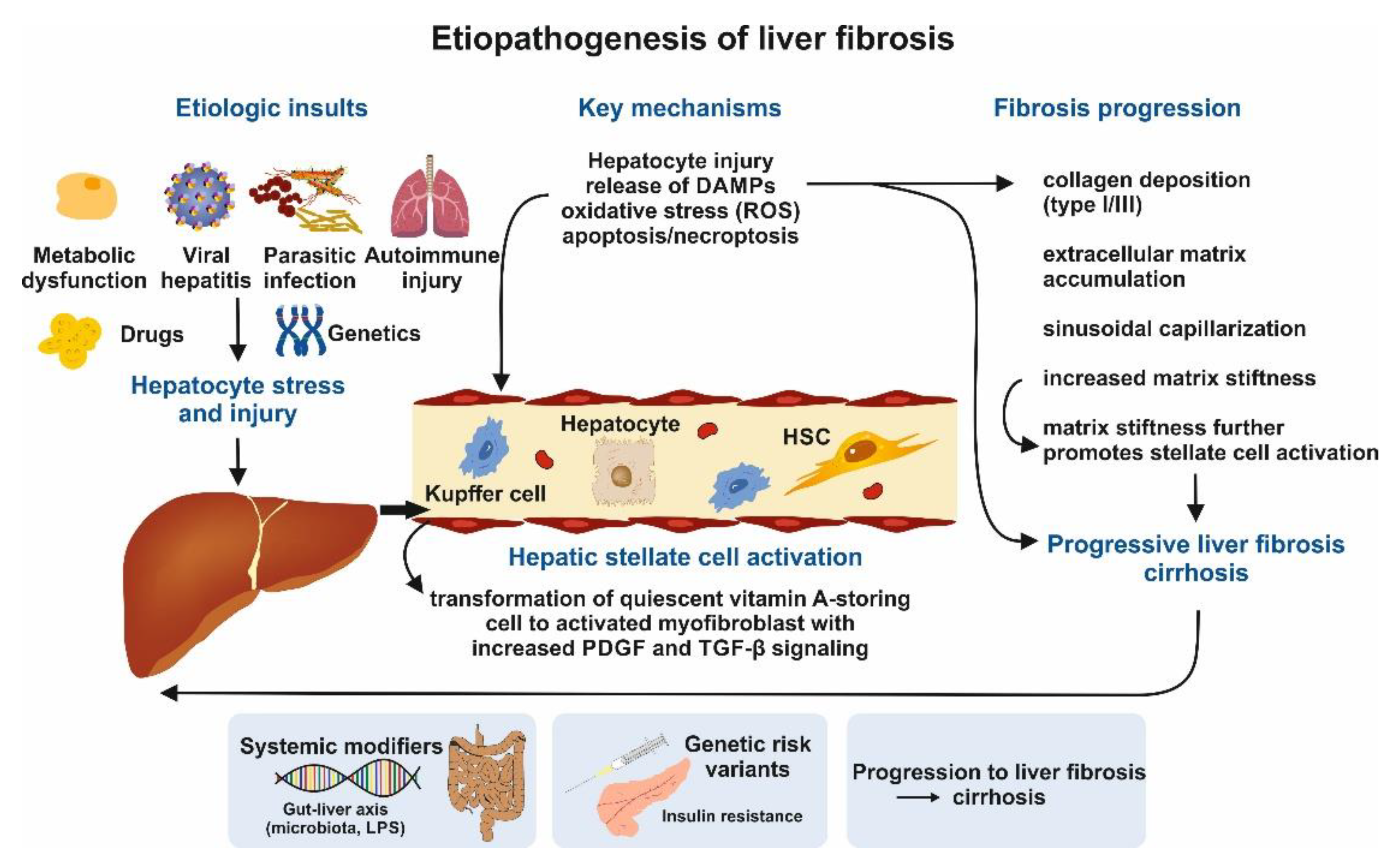
Figure 2.
Current and emerging therapeutic strategies targeting liver fibrosis. Medical treatment of liver fibrosis relies on both etiological therapy and emerging antifibrotic strategies targeting key pathogenic mechanisms. Removal or control of the primary cause of liver injury, including antiviral therapy for viral hepatitis, alcohol abstinence, immunosuppressive treatment for autoimmune liver disease, and metabolic risk reduction in metabolic dysfunction–associated steatotic liver disease, remains the foundation of fibrosis management. Additional therapeutic approaches aim to directly interfere with fibrogenic pathways. These include inhibition of hepatic stellate cell activation through modulation of signalling pathways such as TGF-β, PDGF, and integrin-mediated mechanotransduction. Anti-inflammatory and immunomodulatory therapies targeting macrophage recruitment and cytokine signalling seek to attenuate chronic hepatic inflammation. Metabolic therapies, including GLP-1 receptor agonists, farnesoid X receptor agonists, and peroxisome proliferator-activated receptor agonists, improve metabolic dysfunction and may indirectly reduce fibrogenesis. Strategies promoting extracellular matrix remodelling—through enhancement of matrix metalloproteinase activity or reduction of tissue inhibitors of metalloproteinases—aim to facilitate fibrosis regression. Future therapeutic paradigms will likely rely on combination approaches and precision medicine strategies integrating genetic susceptibility, metabolic phenotype, and non-invasive biomarkers to achieve effective and sustained antifibrotic responses.
Figure 2.
Current and emerging therapeutic strategies targeting liver fibrosis. Medical treatment of liver fibrosis relies on both etiological therapy and emerging antifibrotic strategies targeting key pathogenic mechanisms. Removal or control of the primary cause of liver injury, including antiviral therapy for viral hepatitis, alcohol abstinence, immunosuppressive treatment for autoimmune liver disease, and metabolic risk reduction in metabolic dysfunction–associated steatotic liver disease, remains the foundation of fibrosis management. Additional therapeutic approaches aim to directly interfere with fibrogenic pathways. These include inhibition of hepatic stellate cell activation through modulation of signalling pathways such as TGF-β, PDGF, and integrin-mediated mechanotransduction. Anti-inflammatory and immunomodulatory therapies targeting macrophage recruitment and cytokine signalling seek to attenuate chronic hepatic inflammation. Metabolic therapies, including GLP-1 receptor agonists, farnesoid X receptor agonists, and peroxisome proliferator-activated receptor agonists, improve metabolic dysfunction and may indirectly reduce fibrogenesis. Strategies promoting extracellular matrix remodelling—through enhancement of matrix metalloproteinase activity or reduction of tissue inhibitors of metalloproteinases—aim to facilitate fibrosis regression. Future therapeutic paradigms will likely rely on combination approaches and precision medicine strategies integrating genetic susceptibility, metabolic phenotype, and non-invasive biomarkers to achieve effective and sustained antifibrotic responses.
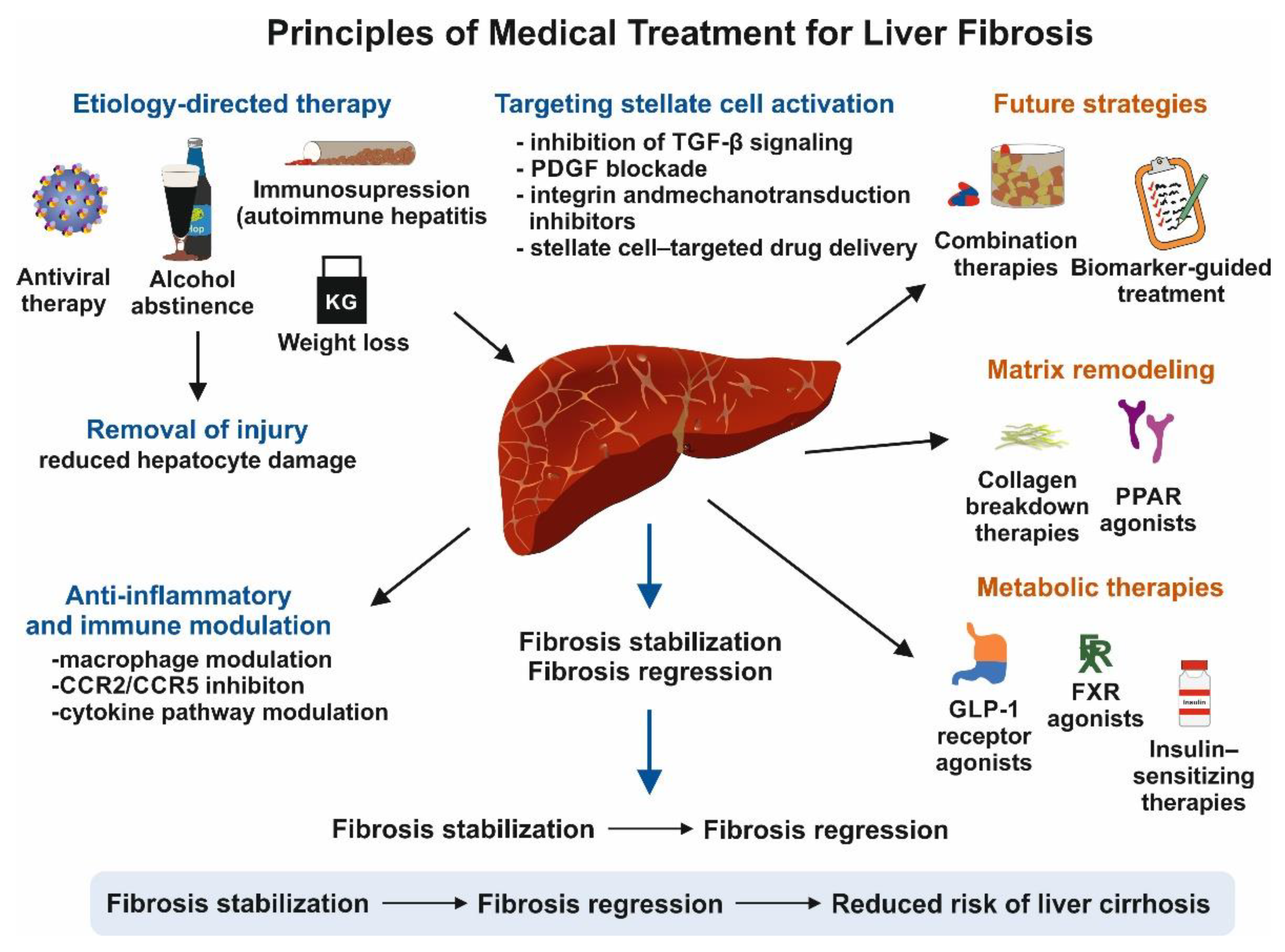

Table 1.
Meta-analytic evidence regarding the prevalence of liver fibrosis and cirrhosis.
| Author, year [Ref] | Number of studies/participants/setting | Prevalence of fibrosis and cirrhosis | Additional findings and conclusion |
|---|---|---|---|
| Zamani et al., [12] | 46/approximately 8,000,000 individuals /general population of 21 countries globally. | 3.3% (95% CI, 2.4%-4.2%) and 1.3% (95% CI, 0.9%-1.7%) worldwide. | After 2016, advanced fibrosis and cirrhosis became more common (P = .004 and P = .034, respectively). Significant geographic differences were found at both continental and national levels (P < .0001). Risk factors for cirrhosis included viral hepatitis, diabetes, heavy drinking, obesity, and being male. |
| Owrangi et al., 2025 [13] | 35 studies comprising 513,742 patients with MASLD worldwide | The pooled global prevalence of cirrhosis among MASLD patients was 3.26% (95% CI: 2.47%-4.31%) in general practice settings (4 studies) and 14.51% (95% CI: 11.22%-18.57%) among those in inpatient settings or referred for liver biopsy (31 studies). | Across regions, the prevalence rates in high-risk settings were highest in North America and Australia (18.38%; 95% CI: 9.06%-33.75%), compared to Europe (10.16%; 95% CI: 5.71%-17.44%) and Asia (9.12%; 95% CI: 6.11%-13.40%) (p = 0.007). Additionally, diagnoses using ICD criteria revealed a significantly greater prevalence of cirrhosis (27.43%) than those established through liver biopsy (13.24%; p < 0.001). |
| Kim et al., 2024 [14] | 45 eligible studies comprising 566,160 participants in the general population globally | Advanced liver fibrosis prevalence, based on the FIB-4 index, was 2.3% (95% CI: 1.2–3.7%). Using VCTE in the general population, significant fibrosis was 7.3% (95% CI: 5.9–8.8%), advanced fibrosis was 3.5% (95% CI: 2.7–4.5%), and cirrhosis was 1.2% (95% CI: 0.8–1.8%). | Region-based subgroup analysis indicated that the American region demonstrated the highest prevalence of advanced fibrosis according to the high probability cutoff for the FIB-4 index. Additionally, the American region showed the greatest prevalence of significant liver fibrosis, advanced liver fibrosis, and liver cirrhosis when evaluated using VCTE. |
| Navarro et al., [15] | 89 studies involving 1,387,184 people with IBD from 27 different countries worldwide | The overall prevalence of fibrosis was 16.7% (12.2-21.7) but varied greatly according to the measurement method. | The prevalence of fibrosis varied by diagnostic method: highest with CAP (38.8%), compared to ultrasonography (28.5%) or others. |
| Yongpisarn et al., 2022 [16] | 41 studies, enrolling a total of 3,868 patients with psoriasis between 1988 and 2022 | Patients with psoriasis at high risk for advanced liver fibrosis had a pooled prevalence of 9.66%, 95% C): 6.92-12.75%, I 2 = 76.34%, whereas patients at low risk for advanced liver fibrosis had a pooled prevalence of 77.79% (95% CI: 73.23-82.05%, I 2 = 85.72%). | MTX-naïve patients had a lower prevalence of advanced liver fibrosis (4.44%) compared to MTX users (12.25%). Meta-regression found no significant sources of heterogeneity among age, sex, BMI, PASI score, psoriasis duration, MTX dose, or related health conditions. Pooled odds ratios indicated higher risks associated with age over 50, BMI over 30, diabetes, hypertension, dyslipidemia, and metabolic syndrome. |
| Lyu et al., 2022 [17] | 15 studies totaling 22,676 PLWH were included in data analysis | Alcohol abuse significantly increases the risk of liver fibrosis in PLWH (pooled OR = 2.25). Consuming over 50 g of alcohol daily raised this risk further (pooled OR = 3.10), including for PLWH co-infected with HCV (OR = 2.48) and HIV mono-infected individuals (OR = 1.85). However, high-risk drinking based on AUDIT-C ≥ 4 showed little effect on developing liver fibrosis. | Alcohol consumption is associated with an increased risk of liver fibrosis in PLWH. HCV co-infection with alcohol abuse could possibly induce a higher risk of liver fibrosis than HIV mono-infected patients. |
| Thongtan et al., 2022 [18] | 4 studies involving 2,593 patients with NAFLD were included | The use of aspirin and/or P2Y12 receptor inhibitors was associated with a lower pooled OR of advanced liver fibrosis in patients with NAFLD (pooled OR = 0.66; 95% CI: 0.53-0.81, I2 = 0.0%; p < 0.001). | While initial findings from this meta-analysis indicate a potential protective relationship between antiplatelet therapy and the prevalence of advanced liver fibrosis in patients with NAFLD, the review is constrained by the limited number of studies included. |
| Ebadi et al., 2021 [19] | 11 articles were included in the meta-analysis totaling 4,303 subjects for the analysis of liver fibrosis | Coffee consumption was significantly associated with a 35% decreased odds of significant liver fibrosis (RR 0.65, 95% CI 0.54-0.78, p < 0.00001). There was no heterogeneity (I2 = 11%, p = 0.34) and no evidence of publication bias (p = 0.134). | This meta-analysis demonstrates an association between coffee consumption and protection against significant liver fibrosis in patients with NAFLD. However, it does not establish the threshold of coffee intake required to achieve hepatoprotective effects, which should be determined through prospective trials. |
List of abbreviation used: CAP, controlled attenuation parameter; CI, confidence intervals; FIB-4, fibrosis -4; HCV, hepatitis C virus; HIV, human immunodeficiency virus; IBD, inflammatory bowel disease; MTX, methotrexate; MASLD, metabolic dysfunction-associated steatotic liver disease; NAFLD, nonalcoholic fatty liver disease; OR, odds ratio; PLWH, people living with HIV; VCTE, vibration-controlled transient elastography.
Table 2.
Indications and cut-off values for HVPG measurement.
| Indication | Details | HVPG Threshold | Associated Outcome |
|---|---|---|---|
| Diagnosis | |||
| CSPH | Gold standard for viral/alcohol-related cirrhosis | ≥10 mmHg | Diagnosis of CSPH |
| Indeterminate NITs | LSM 15–25 kPa, platelet counts in gray zone | Unclear | Clarifies CSPH presence |
| Atypical presentation | Differentiates sinusoidal vs. non-sinusoidal PH | Not specified | Clarifies etiology |
| Pre-surgery | |||
| Liver resection (HCC) | Cirrhosis with HCC prior to resection | ≥10 mmHg | Higher risk of post-operative decompensation |
| Non-hepatic Surgery | Major abdominal surgery risk assessment | ≥16 mmHg | High short-term mortality risk |
| Therapy | |||
| Monitoring pharmacotherapy Response | Effectiveness of NSBBs | ≥20% reduction or < 12 mmHg | Defines response |
| Monitoring pre- and post-TIPS | Measured before/after TIPS placement | < 12 mmHg | Adequate pressure reduction |
| Clinical trials | Phase II trials for PH drugs | Not specified | Surrogate endpoint for clinical events |
| Prognostication | |||
| Acute variceal bleeding | Measured within 24 hours of acute bleed | ≥20 mmHg | Predicts treatment failure, need for early TIPS |
List of abbreviations used: CSPH, clinically significant portal hypertension; HCC, hepatocellular carcinoma; HVPG, hepatic venous pressure gradient; LSM, liver stiffness measurement; NITs, non-invasive tests; NSBBs, non-selective beta-blockers; PH, portal hypertension; TIPS, trans jugular intrahepatic porto-systemic shunting.5. Sex Differences in Liver Fibrosis.
Disclaimer/Publisher’s Note: The statements, opinions and data contained in all publications are solely those of the individual author(s) and contributor(s) and not of MDPI and/or the editor(s). MDPI and/or the editor(s) disclaim responsibility for any injury to people or property resulting from any ideas, methods, instructions or products referred to in the content. |
© 2026 by the authors. Licensee MDPI, Basel, Switzerland. This article is an open access article distributed under the terms and conditions of the Creative Commons Attribution (CC BY) license (http://creativecommons.org/licenses/by/4.0/).
Copyright: This open access article is published under a Creative Commons CC BY 4.0 license, which permit the free download, distribution, and reuse, provided that the author and preprint are cited in any reuse.


