

Submitted:
24 June 2026
Posted:
26 June 2026
You are already at the latest version
Abstract
Background and Objectives: Oral low-dose minoxidil has achieved widespread clinical adoption for androgenetic alopecia (AGA), yet its subclinical cardiovascular structural effects at dermatological doses remain uncharacterized. We aimed to evaluate the effects of dutasteride and three different doses of oral minoxidil (1, 2.5, and 5 mg/day) on carotid intima-media thickness (CIMT) and CIMT-derived vascular age in healthy young men. Methods: A prospective, randomized, four-group pharmacological study enrolling 44 healthy young male subjects assigned to dutasteride 0.5 mg/day (n = 12), oral minoxidil 1 mg/day (n = 8), 2.5 mg/day (n = 12), or 5 mg/day (n = 12). High-resolution carotid ultrasonography was performed at baseline and at the end of follow-up. Primary outcomes included bilateral CIMT, maximum CIMT, and CIMT-derived vascular age. Within-group changes were assessed with the Wilcoxon signed-rank test; between-group comparisons used the Kruskal-Wallis test with post hoc Mann-Whitney U testing. Results: Thirty-one participants completed the study. Groups were well balanced at baseline (all p > 0.05). The minoxidil 5 mg group exhibited statistically significant within-group increases in right CIMT (471 ± 78 vs. 555 ± 87 µm; p < 0.05), maximum CIMT (509 ± 79 vs. 575 ± 76 µm; p < 0.05), and CIMT-derived vascular age (35.9 ± 15.2 vs. 48.2 ± 14.9 years; p < 0.05). The right CIMT delta of the minoxidil 5 mg group (Δ = +79 ± 57 µm) was significantly greater than that of the dutasteride group (Δ = −21 ± 79 µm; p = 0.032). A clear dose-dependent gradient was observed across minoxidil groups for right CIMT and maximum CIMT deltas. Dutasteride showed a consistent, non-significant trend toward CIMT reduction across all parameters. Conclusions: Oral minoxidil at 5 mg/day is associated with adverse carotid arterial wall remodeling and accelerated vascular aging in healthy young men, with a dose-dependent pattern suggesting a pharmacodynamic threshold between 1 and 2.5 mg/day. Dutasteride showed a favorable vascular profile.
Keywords:
carotid intima-media thickness
; oral minoxidil
; dutasteride
; androgenetic alopecia
; vascular aging
; pharmacovigilance
; subclinical atherosclerosis
; vascular remodeling
; cardiovascular safety
1. Introduction
Androgenetic alopecia (AGA) is the most prevalent form of hair loss in men, affecting up to 50% of Caucasian males by the fifth decade and approaching 80% by the eighth decade [1]. Beyond its cosmetic dimension, AGA has been increasingly recognized as a clinically significant phenotypic marker: large epidemiological studies have demonstrated associations between early-onset male pattern baldness and elevated risk of coronary artery disease, hypertension, insulin resistance, and metabolic syndrome [2,3]. These associations have prompted interest not only in the dermatological management of AGA but also in the cardiovascular implications of the pharmacological agents employed in its treatment, particularly given the growing body of evidence linking the androgenic signaling axis to arterial wall biology.
The pharmacological management of AGA has historically relied on two approved systemic agents: finasteride (a selective type 2 5α-reductase inhibitor) and minoxidil (originally developed as an antihypertensive agent and subsequently repurposed for topical use in alopecia). Over the past decade, dutasteride, a dual type 1 and type 2 5α-reductase inhibitor with superior DHT suppression relative to finasteride [4] has been increasingly adopted off-label for AGA, particularly in Asia and Latin America where it holds regulatory approval for this indication. More recently, a paradigm shift has occurred with the emergence of oral low-dose minoxidil as a therapeutic option for both male and female pattern hair loss [5,6]. Randomized clinical trials have demonstrated the efficacy of oral minoxidil at doses ranging from 0.25 to 5 mg/day in promoting hair regrowth, with a seemingly favorable short-term symptomatic safety profile at these doses. As a result, oral minoxidil, particularly at 5 mg/day for men, has become widely prescribed across dermatological practice worldwide, often without systematic cardiovascular monitoring.
Despite this rapid clinical adoption, a critical pharmacovigilance gap exists: systematic data on the subclinical vascular structural effects of oral minoxidil at the doses used in dermatological practice are lacking in the literature. Clinical trials assessing oral minoxidil for AGA have uniformly relied on self-reported symptomatic outcomes and basic clinical monitoring (blood pressure, heart rate, and symptom questionnaires at best), without measurement of subclinical vascular endpoints such as carotid intima-media thickness (CIMT), arterial stiffness, or endothelial function [5,6,7]. This gap is particularly concerning given that minoxidil’s mechanism of action, the ATP-sensitive potassium (KATP) channel opening in vascular smooth muscle cells, invariably triggers compensatory hemodynamic responses, including reflex sympathetic activation and renin-angiotensin-aldosterone system (RAAS) stimulation [8,9]. These neurohormonal cascades are well-established drivers of arterial wall remodeling and intimal thickening, raising a biologically plausible hypothesis that chronic low-dose oral minoxidil may exert adverse structural vascular effects even at dermatological doses.
Carotid intima-media thickness is a validated, non-invasive ultrasonographic surrogate of subclinical arterial disease that reflects the cumulative structural consequence of hemodynamic, metabolic, and inflammatory stressors acting on the arterial wall [10]. The Mannheim CIMT and Plaque Consensus has provided internationally accepted measurement protocols that anchor the clinical and pharmacological interpretation of CIMT data [10]. Crucially, large prospective meta-analyses have demonstrated that each 0.1 mm increment in common carotid CIMT is independently associated with a 15–20% relative increase in myocardial infarction risk and a 13–18% relative increase in stroke risk [11]. The Framingham Heart Study has further shown that CIMT improves cardiovascular risk reclassification beyond traditional Framingham scoring [12]. Furthermore, the Bogalusa Heart Study established that subclinical arterial pathology originates early in life and correlates with modifiable risk factors measurable in childhood and young adulthood [13], highlighting the particular sensitivity of CIMT to pharmacological exposures in young populations, precisely the demographic most commonly receiving oral minoxidil for AGA.
On the other hand, the inclusion of dutasteride as a comparator arm in the present study is supported by emerging evidence that androgen signaling modulates vascular smooth muscle cell (VSMC) biology through androgen receptor-mediated transcriptional regulation of pro-fibrotic and pro-proliferative pathways [14]. Dihydrotestosterone (DHT), the most potent endogenous androgen, whose biosynthesis is effectively blocked by dutasteride, has been shown to stimulate VSMC proliferation, extracellular matrix deposition, and vascular inflammatory tone through androgen receptor signaling, as well as to upregulate VEGF in endothelial cells via paracrine mechanisms that further promote VSMC growth [14,15]. By achieving near-complete suppression of DHT through dual 5α-reductase inhibition [4], dutasteride may attenuate these androgen-driven pro-remodeling pathways, making it a pharmacologically informative comparator for assessing differential vascular effects within the AGA treatment landscape.
Against this background, the present study was designed to address the pharmacovigilance gap in oral minoxidil safety data by comparing, in a randomized, four-group pharmacological design, the effects of dutasteride and three different doses of oral minoxidil (1, 2.5, and 5 mg/day) on carotid intima-media thickness and CIMT-derived vascular age in a cohort of healthy young male subjects. To our knowledge, this represents one of the first randomized pharmacological trials to systematically evaluate subclinical vascular structural effects of oral minoxidil at the previously reported doses versus dihydrotestosterone suppression.
2. Materials and Methods
This was a prospective, randomized, double-blind, four-group pharmacological study designed to evaluate the subclinical vascular structural effects of oral dutasteride versus three different doses of oral minoxidil in healthy young male subjects. The study was conducted in accordance with the Declaration of Helsinki and its subsequent amendments. Ethical approval was obtained from the Comité de Ética en lnvestigación del Centro de Estudios de lnvestigación Básica y Clínica S.C. (CECEIBAC) review board (20.01.205), and all participants provided written informed consent before enrollment. 2.1. Study Population and Eligibility Criteria.
A total of 44 healthy young male subjects were enrolled. Eligible participants were adult males aged 18–45 years with a diagnosis of androgenetic alopecia (AGA) who were candidates for pharmacological treatment. Key inclusion criteria were: (1) absence of established cardiovascular disease (coronary artery disease, heart failure, cerebrovascular disease, or peripheral arterial disease); (2) no history of hypertension, diabetes mellitus, dyslipidemia, or other major cardiovascular risk factors; (3) no use of antihypertensive, lipid-lowering, antiplatelet, anticoagulant, or other cardiovascular medications; (4) no prior use of minoxidil or 5α-reductase inhibitors within six months of enrollment; and (5) no renal or hepatic dysfunction. Exclusion criteria included any contraindication to the assigned pharmacological intervention, tobacco use, and any systemic inflammatory or autoimmune condition.
Participants were randomly assigned in a 1:1:1:1 ratio to one of four pharmacological intervention groups: (1) dutasteride 0.5 mg/day (n = 12); (2) oral minoxidil 1 mg/day (n = 8); (3) oral minoxidil 2.5 mg/day (n = 12); or (4) oral minoxidil 5 mg/day (n = 12). Randomization was performed using a computer-generated allocation sequence. All study medications were administered orally once daily throughout the follow-up period. Participants were instructed to maintain their habitual lifestyle, dietary habits, and physical activity levels throughout the study. No dose adjustments or concomitant pharmacological interventions targeting the cardiovascular system were permitted during the study period.
Carotid intima-media thickness was measured by high-resolution B-mode (Esaote Mylab x1) ultrasound, performed by a trained and certified sonographer blinded to group allocation. Measurements were obtained bilaterally at the far wall of the common carotid artery, 1 cm proximal to the carotid bulb, following the standardized methodology defined by the Mannheim CIMT and Plaque Consensus [10]. For each side, three independent measurements were obtained and averaged. The following CIMT variables were recorded at baseline and at the end of the follow-up period: (1) right CIMT (µm); (2) left CIMT (µm); and (3) maximum CIMT (µm), defined as the highest value recorded across both carotid arteries. All measurements were performed using a dedicated vascular ultrasound unit with a high-frequency linear transducer (7.5–10 MHz). Intraobserver and interobserver reproducibility were verified prior to study initiation, with intraclass correlation coefficients exceeding 0.90 for all CIMT measures.
CIMT-derived vascular age was calculated from the maximum CIMT measurement using a validated algorithm that translates carotid CIMT values into an estimate of the biological equivalent chronological vascular age, based on sex- and age-specific reference values from large normative cohort studies [16]. This metric provides a clinically interpretable framework for quantifying the pharmacological impact on arterial aging trajectory and has been employed in prior pharmacological intervention studies to contextualize CIMT-based vascular changes.
At baseline, standardized anthropometric measurements were obtained, including body weight (kg), height (cm), and body mass index (BMI, kg/m2). Blood pressure and heart rate were measured in triplicate after 5 minutes of seated rest using a calibrated automated sphygmomanometer (OMRON BP7320). Baseline clinical and demographic characteristics across all groups are summarized in Table 1.
2.1. Statistical Analysis
Continuous variables are expressed as mean ± standard deviation (SD). Given the small sample sizes and the non-parametric distribution of CIMT delta values, non-parametric statistical tests were employed throughout. Baseline between-group comparisons were performed using the Kruskal-Wallis test. Within-group longitudinal changes from baseline to final assessment were evaluated using the Wilcoxon signed-rank test. Between-group comparisons of absolute delta values (Δ = final − baseline) were performed using the Kruskal-Wallis test; in cases that reached nominal significance (p < 0.05), post hoc pairwise comparisons were conducted using the Mann-Whitney U test with a Bonferroni correction for multiple comparisons. Statistical significance was defined as p < 0.05 (two-tailed). All statistical analyses were performed using SPSS Statistics version 28.0 (IBM Corp., Armonk, NY, USA).
3. Results
A total of 44 healthy young subjects were enrolled and randomly assigned to one of four pharmacological intervention groups: dutasteride (n=12), minoxidil 1 mg (n=8), minoxidil 2.5 mg (n=12), and minoxidil 5 mg (n=12). Baseline anthropometric and vascular characteristics across all groups are presented in Table 1. No statistically significant differences were observed between groups at baseline for age, weight, height, BMI, right CIMT, left CIMT, maximum CIMT, or CIMT-derived vascular age (all p > 0.05, Kruskal-Wallis test), confirming adequate baseline comparability across the four study arms.
At the end of the follow-up period, 31 participants completed the study: 9 in the dutasteride group, 4 in the minoxidil 1 mg group, 9 in the minoxidil 2.5 mg group, and 9 in the minoxidil 5 mg group. CIMT and vascular age values obtained at the final assessment are summarized in Table 2. Cross-sectional comparisons at the end of the intervention revealed no statistically significant differences among groups for any of the outcome variables: right CIMT (p=0.443), left CIMT (p=0.763), maximum CIMT (p=0.937), or vascular age (p=0.933).
Within-group changes between baseline and final measurements are summarized in Table 3. In the dutasteride group, right CIMT, left CIMT, maximum CIMT, and vascular age did not change significantly over time (546 ± 77 vs. 539 ± 53 µm; 525 ± 119 vs. 547 ± 98 µm; 577 ± 100 vs. 576 ± 78 µm; 43.8 ± 14.6 vs. 44.7 ± 10.6 years, respectively). Similarly, neither the minoxidil 1 mg nor the minoxidil 2.5 mg group showed statistically significant changes in any of the assessed variables.
In contrast, the minoxidil 5 mg group exhibited statistically significant increases in right CIMT (471 ± 78 vs. 555 ± 87 µm; p<0.05), maximum CIMT (509 ± 79 vs. 575 ± 76 µm; p<0.05), and vascular age (35.9 ± 15.2 vs. 48.2 ± 14.9 years; p<0.05) by the end of the intervention period, suggesting an adverse effect on arterial vascular structure in this group.

Table 3.
Within-group comparison of vascular parameters before and after pharmacological intervention.
Table 3.
Within-group comparison of vascular parameters before and after pharmacological intervention.
| Variable | Dutas 0.5 mg Baseline |
Dutas 0.5 mg Final | Min 1 mg Baseline | Min 1 mg Final |
Min 2.5 mg Baseline | Min 2.5 mg Final | Min 5 mg Baseline | Min 5 mg Final |
|---|---|---|---|---|---|---|---|---|
| Right CIMT, (µm) | 546±77 | 539±53 | 509±76 | 450±108 | 472±46 | 517±74 | 471±78 | 555±87* |
| Left CIMT, (µm) | 525±119 | 547±98 | 537±89 | 540±69 | 503±61 | 561±125 | 491±82 | 498±82 |
| Maximum CIMT, (µm) | 577±100 | 576±78 | 554±87 | 547±72 | 513±49 | 582±107 | 509±79 | 575±76* |
| Vascular age, (years) | 43.8±14.6 | 44.7±10.6 | 44.5±16.7 | 43.0±13.9 | 36.7±9.5 | 46.6±20.4 | 35.9±15.2 | 48.2±14.9* |
CIMT: carotid intima-media thickness. Data expressed as mean ± standard deviation. * p= < 0.05 versus baseline within the same group (Wilcoxon signed-rank test).
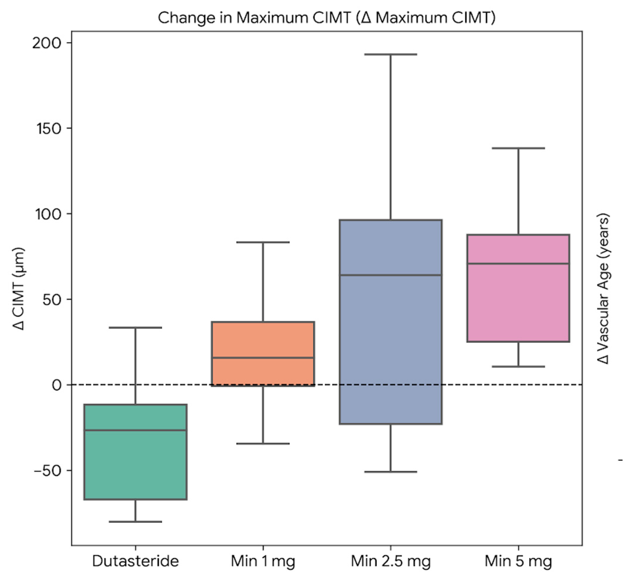
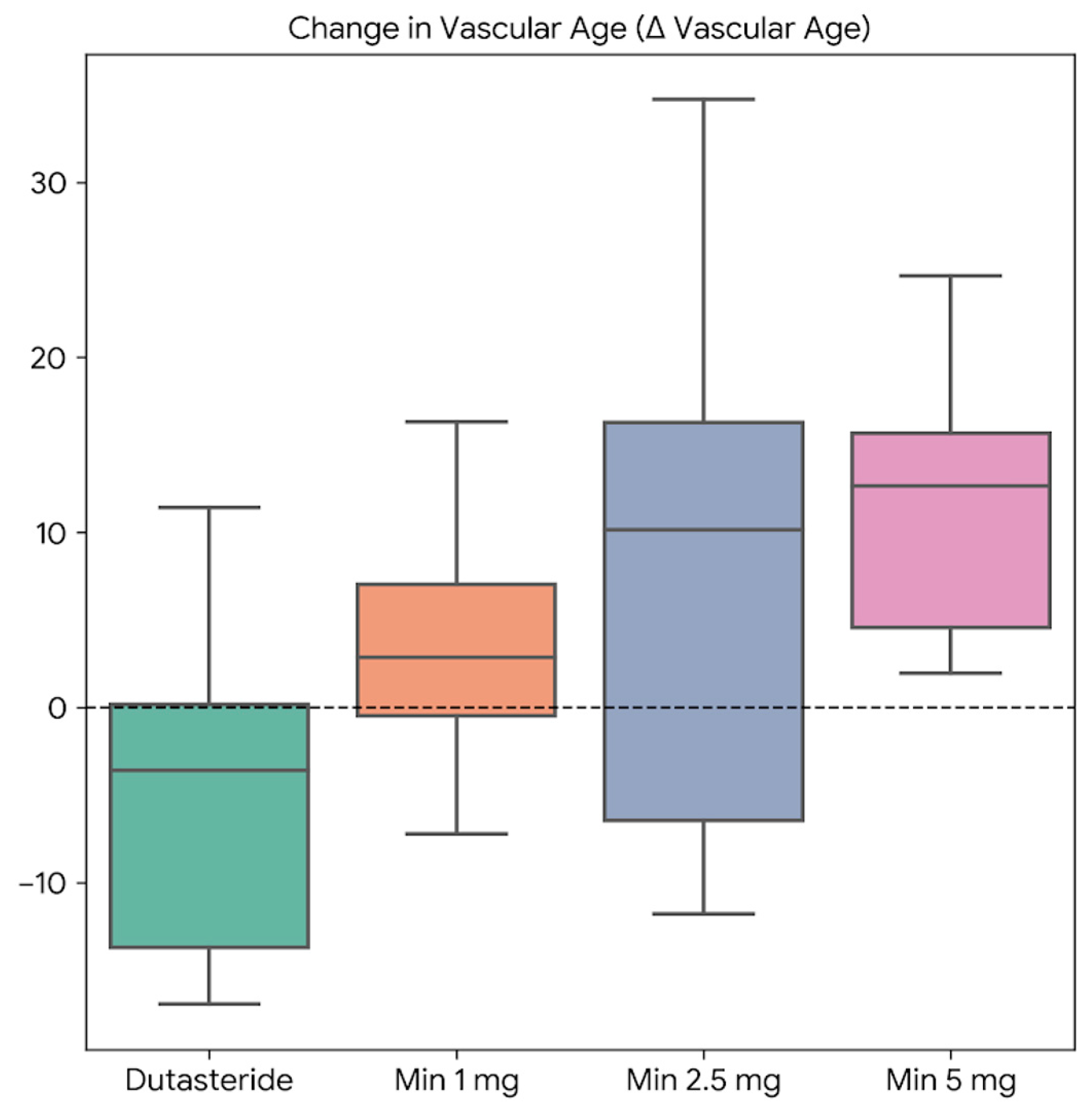
To compare changes across groups, absolute deltas (Δ = final value − baseline value) were calculated for each outcome and are shown in Table 4 and Figure 1, Figure 2, Figure 3 and Figure 4. Right CIMT was the only outcome with a statistically significant between-group difference (p = 0.032). Post hoc analysis showed that the minoxidil 5 mg group had a significantly greater increase in right CIMT than the dutasteride group (Δ = 79 ± 57 µm vs. −21 ± 79 µm; p < 0.05), indicating that dutasteride tended to reduce right CIMT, whereas minoxidil 5 mg increased it.
The deltas for maximum CIMT (p=0.077) and vascular age (p=0.188) showed a similar pattern, with the most negative values observed in the dutasteride group (Δ = −30 ± 84 µm and −4.4 ± 17.5 years, respectively) and the highest values in the minoxidil 5 mg group (Δ = 67 ± 45 µm and 12 ± 8 years, respectively), although these differences did not reach conventional statistical significance. The delta for left CIMT did not differ significantly among groups (p=0.796).
Table 4.
Comparison of absolute changes (deltas) across the four study groups.
| Variable | Dutaste 0.5 mg (n=9) |
Minoxidil 1 mg (n=4) | Minoxidil 2.5 mg (n=9) |
Minoxidil 5 mg (n=9) |
p |
|---|---|---|---|---|---|
| ΔRight CIMT, (µm) | −21 ± 79 | −28 ± 90 | 39 ± 55 | 79 ± 57* | 0.032 |
| ΔLeft CIMT, (µm) | −15 ± 113 | 23 ± 61 | 49 ± 88 | 7 ± 102 | 0.796 |
| ΔMaximum CIMT (µm) | −30 ± 84 | 20 ± 42 | 57 ± 86 | 67 ± 45* | 0.077 |
| ΔVascular age (years) | −4.4 ± 17.5 | 3.7 ± 8.4 | 8.8 ± 16.4 | 12 ± 8* | 0.188 |
Δ: absolute change (final value − baseline value). CIMT: carotid intima-media thickness. Data expressed as mean ± standard deviation. Between-group comparisons were performed using the Kruskal-Wallis test and Mann-Whitney U test. * p < 0.05 between dutasteride and minoxidil 5 mg groups.
Figure 1 displays the mean absolute changes in right CIMT for each treatment group. The dutasteride and minoxidil 1 mg groups showed mean reductions from baseline (Δ = −21 µm and −28 µm, respectively), suggesting a trend toward improvement in right intimal thickness. In contrast, the minoxidil 2.5 mg and minoxidil 5 mg groups demonstrated mean increases (Δ = +39 µm and +79 µm, respectively), with the delta of the minoxidil 5 mg group being significantly greater than that of the dutasteride group (p<0.05). These findings indicate a dose-dependent pattern in the minoxidil-treated groups, with greater right CIMT increases observed at higher doses.
Figure 1.
Mean absolute changes in right carotid intima-media thickness (CIMT) for each treatment group.
Figure 1.
Mean absolute changes in right carotid intima-media thickness (CIMT) for each treatment group.
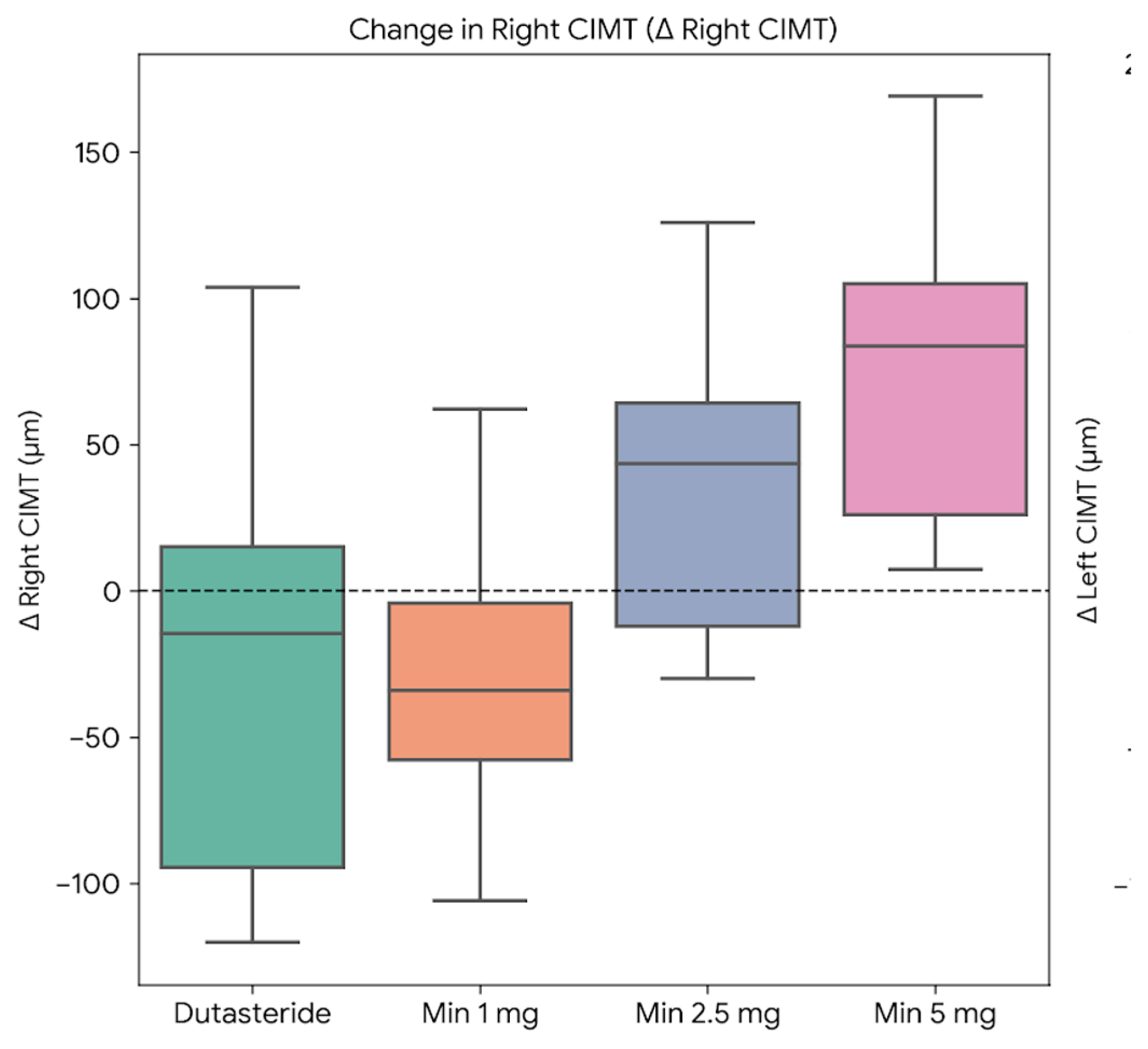
Figure 2 illustrates the mean absolute changes in left CIMT across groups. Unlike right CIMT, the distribution of deltas did not follow a consistent dose-response pattern. The dutasteride group was the only one to show a negative delta (Δ = −15 µm), while the minoxidil 1 mg, 2.5 mg, and 5 mg groups exhibited mean increases of +23, +49, and +7 µm, respectively. The minoxidil 2.5 mg group displayed the highest delta among all four groups; however, between-group differences did not reach statistical significance (p=0.796). The substantial interindividual variability in this measurement, reflected in the wide standard deviations, may have reduced the statistical power to detect significant differences.
Figure 2.
Mean absolute changes in left carotid intima-media thickness (CIMT) for each treatment group.
Figure 2.
Mean absolute changes in left carotid intima-media thickness (CIMT) for each treatment group.
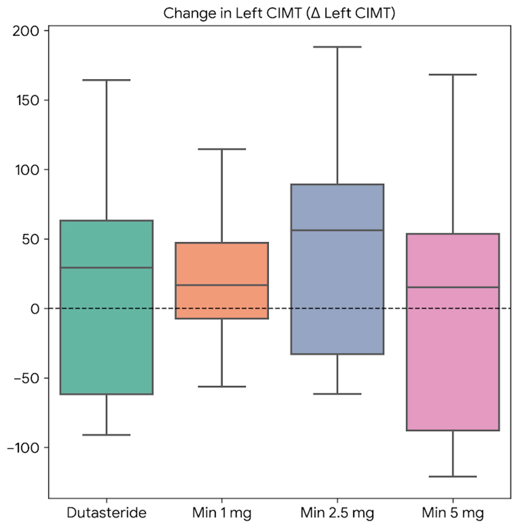
Figure 3 presents the mean absolute changes in maximum CIMT, defined as the highest value recorded across both carotid arteries. The dutasteride group was the only one with a negative delta (Δ = −30 µm), contrasting with the increases observed in the minoxidil 1 mg (Δ = +20 µm), minoxidil 2.5 mg (Δ = +57 µm), and minoxidil 5 mg (Δ = +67 µm) groups. Although the overall between-group comparison did not reach formal statistical significance (p=0.077), the trend is clinically meaningful: post hoc analysis confirmed that the delta of the minoxidil 5 mg group was significantly greater than that of dutasteride (p<0.05), and a progressive dose-related gradient was observed across the minoxidil treatment groups.
Figure 3.
Mean absolute changes in maximum carotid intima-media thickness (CIMT) for each treatment group.
Figure 3.
Mean absolute changes in maximum carotid intima-media thickness (CIMT) for each treatment group.
Finally, Figure 4 shows the mean absolute changes in CIMT-derived vascular age. The dutasteride group was the only one to exhibit a negative delta (Δ = −4.4 years), which may be interpreted as a modest improvement in vascular profile among participants in that group. In the minoxidil-treated groups, progressive increases in vascular age were observed with escalating doses: +3.7 years in the 1 mg group, +8.8 years in the 2.5 mg group, and +12 years in the 5 mg group. Although the overall between-group difference did not reach statistical significance (p=0.188), post hoc analysis demonstrated that the delta of the minoxidil 5 mg group was significantly greater than that of the dutasteride group (p<0.05). Taken together, these findings suggest that oral minoxidil at a dose of 5 mg may be associated with accelerated vascular aging in healthy young subjects, an effect not observed with dutasteride during the study follow-up period.
Figure 4.
Mean absolute changes in CIMT-derived vascular age for each treatment group.
4. Discussion
The present study evaluated the effects of oral dutasteride and three different doses of oral minoxidil (1, 2.5, and 5 mg/day) on carotid intima-media thickness (CIMT) and CIMT-derived vascular age in a randomized cohort of healthy young subjects. The principal and most clinically significant finding is that oral minoxidil at 5 mg/day was associated with statistically significant increases in right CIMT, maximum CIMT, and CIMT-derived vascular age within the study follow-up period, a pattern not observed in subjects receiving dutasteride, who instead exhibited a consistent, although non-significant, trend toward CIMT reduction across all measured vascular parameters. To our knowledge, this represents one of the first randomized pharmacological studies to systematically assess subclinical vascular structural effects of low-dose oral minoxidil at doses currently in widespread clinical use for androgenetic alopecia, constituting a timely and clinically relevant pharmacovigilance contribution.
The choice of CIMT as the primary vascular outcome measure in this study is based on a robust body of epidemiological and clinical evidence. As a non-invasive ultrasonographic parameter, CIMT reflects the cumulative structural consequence of hemodynamic, metabolic, and inflammatory stressors acting on the arterial wall, providing an integrative, reproducible, and validated readout of subclinical vascular injury [10]. The Mannheim Carotid Intima-Media Thickness and Plaque Consensus, now in its fourth iteration, has provided internationally accepted measurement protocols, quality criteria, and reference values that anchor the clinical interpretation of CIMT data in both population and pharmacological studies [10]. The well-known meta-analysis by Lorenz et al., which pooled data from prospective cohort studies enrolling over 37,000 subjects, demonstrated that each 0.1 mm increment in common carotid CIMT is independently associated with a 15-20% relative increase in the risk of myocardial infarction and a 13-18% relative increase in stroke risk [11]. These associations were further corroborated by Polak and colleagues in the Framingham Heart Study offspring cohort, who confirmed that CIMT-based vascular assessment improves cardiovascular risk reclassification beyond traditional Framingham risk scoring [12]. Notably, the value of CIMT as an outcome measure extends to young populations: the landmark Bogalusa Heart Study established that subclinical arterial pathology, reflected as fatty streak burden in coronary and aortic vessels at autopsy, correlated significantly with classic cardiovascular risk factors measurable during life in children and young adults [13], highlighting the capacity of early vascular measurements to capture pharmacologically relevant changes long before clinical events manifest.
The four intervention groups were well balanced at baseline across all anthropometric and vascular variables assessed, confirming the validity and adequacy of the randomization procedure. No statistically significant between-group differences were observed in body weight, height, body mass index, right CIMT, left CIMT, maximum CIMT, or vascular age at baseline, with p-values ranging from 0.077 to 0.749. It is noteworthy, however, that baseline CIMT values across groups ranging from 471 to 546 µm for right CIMT and from 491 to 537 µm for left CIMT reflect the substantial physiological interindividual variability in carotid wall thickness that characterizes even apparently healthy adult populations, as documented in large cohort studies [4,16]. The Cardiovascular Risk in Young Finns Study, for instance, demonstrated that carotid wall thickness in young adults is significantly modified by the cumulative exposure to cardiovascular risk factors during childhood and adolescence, even in the absence of clinically overt disease [4]. The wide standard deviations observed at baseline in the present study are therefore biologically expected and were addressed methodologically through within-group longitudinal analysis and calculation of absolute deltas (Δ = final − baseline) as the primary strategy, which maximizes sensitivity to individual-level pharmacological effects.
While dutasteride produced a consistent, directionally uniform trend toward CIMT reduction across all four vascular outcome variables (Δ = −21 ± 79 µm for right CIMT, Δ = −15 ± 113 µm for left CIMT, Δ = −30 ± 84 µm for maximum CIMT, and Δ = −4.4 ± 17.5 years for vascular age), none of these deltas achieved individual statistical significance, attributable in part to the small sample size and the intrinsically wide interindividual variability in CIMT measurements; nonetheless, the biological coherence and directional consistency of the findings warrant careful mechanistic consideration. Dutasteride is a dual type 1 and type 2 5α-reductase inhibitor that, at the standard therapeutic dose of 0.5 mg/day, achieves suppression of serum DHT exceeding 90% from baseline [18], a degree of androgen suppression notably greater than that produced by finasteride, which selectively inhibits only the type 2 isoform and reduces DHT by approximately 65-70% [14]. DHT, the most potent endogenous androgen, acts through androgen receptors expressed on vascular smooth muscle cells (VSMCs), endothelial cells, and macrophages within the arterial wall. Through these receptors, DHT modulates gene transcription programs leading to VSMC proliferation, extracellular matrix deposition, and vascular inflammatory tone [15]. Androgen receptor signaling in VSMCs has been shown to regulate the expression of key pro-fibrotic mediators including transforming growth factor-β (TGF-β) and platelet-derived growth factor (PDGF), both of which are central to the cellular cascades underlying intimal thickening and early arteriosclerotic remodeling [14]. Furthermore, androgen-mediated upregulation of VEGF in endothelial cells has been shown to promote VSMC proliferation through a paracrine mechanism [8]. By achieving near-complete suppression of DHT across both 5α-reductase isoforms, dutasteride may attenuate these androgen-driven pro-remodeling pathways in the arterial wall, providing a plausible biological mechanism for the observed trend toward CIMT stability or modest reduction. These observations are consistent with emerging evidence that androgen modulation influences vascular phenotype in men, though the directionality and clinical magnitude of such effects in non-hypogonadal subjects participating in androgenetic alopecia pharmacotherapy, as opposed to castration or androgen deprivation therapy for prostate cancer, remain incompletely defined and warrant further investigation.
In remarkable contrast to dutasteride, oral minoxidil at 5 mg/day produced statistically significant within-group increases in right CIMT (471 ± 78 vs. 555 ± 87 µm; p < 0.05), maximum CIMT (509 ± 79 vs. 575 ± 76 µm; p < 0.05), and CIMT-derived vascular age (35.9 ± 15.2 vs. 48.2 ± 14.9 years; p < 0.05), as assessed by the Wilcoxon signed-rank test. The delta analysis corroborated and extended these within-group findings at the between-group level: the right CIMT delta of the minoxidil 5 mg group (Δ = +79 ± 57 µm) was significantly greater than that of the dutasteride group (Δ = −21 ± 79 µm; p < 0.05, Mann-Whitney U test), and this difference survived the overall Kruskal-Wallis between-group comparison for right CIMT (p = 0.032). Post hoc pairwise analysis further confirmed that the maximum CIMT delta (Δ = +67 ± 45 µm vs. −30 ± 84 µm; p < 0.05) and vascular age delta (Δ = +12 ± 8 years vs. −4.4 ± 17.5 years; p < 0.05) of the minoxidil 5 mg group were significantly greater than those of the dutasteride group, despite the overall between-group comparisons for these two variables not reaching conventional significance thresholds (p = 0.077 and p = 0.188, respectively), a pattern most plausibly explained by the limited statistical power available after accounting for the degrees of freedom across four groups when modest sample sizes are included, as in the present study.
Interpreting the vascular structural effects of oral minoxidil at this dose requires a detailed understanding of its molecular pharmacology. Minoxidil is a piperidino-pyrimidine derivative that, after hepatic sulfation to its active form, minoxidil sulfate, opens ATP-sensitive potassium (KATP) channels in the plasma membrane of vascular smooth muscle cells [19]. The consequent potassium efflux hyperpolarizes the VSMC membrane, inhibits voltage-gated L-type calcium channels, reduces cytosolic calcium concentration, promotes myosin light-chain dephosphorylation, and produces marked arteriolar vasodilation [9].
At the doses used in the treatment of refractory hypertension, classically 5 to 40 mg/day, this vasodilation is powerful and well-characterized, but it invariably triggers a cascade of hemodynamic counterregulatory responses that act directly on vascular structure. Baroreceptor unloading secondary to the drop in peripheral vascular resistance activates the sympathetic nervous system, increasing heart rate, cardiac output, and circulating catecholamines [9]. Simultaneously, decreased renal perfusion pressure and direct sympathoadrenal stimulation of the juxtaglomerular apparatus activate the renin-angiotensin-aldosterone system (RAAS), with consequent increases in circulating angiotensin II and aldosterone [9]. Even at the 5 mg/day dose used in dermatological practice, these homeostatic responses, though attenuated relative to antihypertensive doses, may be sufficient to induce vascular structural changes in susceptible individuals over the course of a pharmacological follow-up period.
A complementary mechanism through which minoxidil may promote adverse vascular remodeling involves its capacity to upregulate vascular endothelial growth factor (VEGF) expression. Minoxidil has been shown to dose-dependently stimulate VEGF mRNA and protein expression in human dermal papilla cells, likely through downstream signaling cascades activated by KATP channel opening and associated alterations in intracellular calcium dynamics [20]. VEGF is canonically recognized as the primary driver of physiological and pathological angiogenesis, but its role in VSMC biology extends beyond the regulation of new vessel formation: at supraphysiological concentrations or in the context of concurrent pro-inflammatory signaling, VEGF potently stimulates VSMC proliferation and migration through receptors expressed on the smooth muscle cell surface [20]. Furthermore, the active metabolite of minoxidil, minoxidil sulfate, produced by cytosolic sulfotransferases within VSMCs, may exert direct intracellular proliferative effects through mechanisms partly independent of KATP channel activation, as suggested by studies demonstrating that cells deficient in sulfotransferase activity show attenuated, but not absent, mitogenic responses to minoxidil [5]. Whether this direct proliferative pathway contributes meaningfully to CIMT changes at the doses assessed in the present study remains to be established, but it cannot be excluded as an additional mechanism amplifying the effects of the sympathoadrenal and RAAS-mediated remodeling pathways.
One of the most compelling features of the present dataset is the clear dose-response relationship observed across the three oral minoxidil treatment groups for right CIMT and maximum CIMT deltas. For right CIMT, the mean deltas were −28 µm (1 mg group), +39 µm (2.5 mg group), and +79 µm (5 mg group), representing a progressive, near-linear gradient with escalating dose. A closely parallel pattern was observed for maximum CIMT: Δ = +20 µm (1 mg), Δ = +57 µm (2.5 mg), Δ = +67 µm (5 mg). A dose-response gradient of this nature substantially strengthens the causal inference linking minoxidil to the adverse vascular structural changes, as it satisfies one of the classical Bradford Hill criteria for establishing pharmacological causality in observational and quasi-experimental designs. The apparent threshold between a neutral or potentially beneficial CIMT trajectory, as suggested by the non-significant negative delta at 1 mg, and a deleterious trajectory appears to lie somewhere between 1 and 2.5 mg/day, though the limited sample size at the 1 mg dose level (only four completers) prevents a precise pharmacodynamic threshold determination. This observation is of immediate practical relevance to the dermatological community, where doses between 0.25 and 5 mg/day are actively debated as the optimal range for oral minoxidil treatment of androgenetic alopecia in men [6,7].
The CIMT-derived vascular age analysis merits particular emphasis within the broader context of cardiovascular aging biology. The concept of vascular age, operationalized through validated algorithms that translate carotid CIMT measurements into an estimate of biologically equivalent chronological vascular age, provides a clinically interpretable metric for communicating the cardiovascular significance of structural arterial changes to clinicians and patients alike [4,16]. Lakatta and Levy, in their review of arterial and cardiac aging, described how cumulative exposure to hemodynamic, inflammatory, and metabolic stressors accelerates the structural remodeling of the arterial wall, including intimal thickening, medial calcification, and loss of elastic fiber integrity, producing a vascular phenotype that is biologically older than the individual’s chronological age [16]. The observation in the present study that subjects in the minoxidil 5 mg group experienced a mean increase in CIMT-derived vascular age equivalent to approximately 12 chronological years over the follow-up period is particularly notable when considered against the young and ostensibly healthy profile of the study population. An accelerated vascular aging signal of this magnitude in young individuals receiving a drug primarily for a cosmetic indication such as androgenetic alopecia raises important questions about the risk-benefit balance of high-dose oral minoxidil treatment and underscores the need for prospective long-term cardiovascular surveillance in this patient population. Arterial stiffness, a related but distinct vascular aging biomarker, has been shown to independently predict cardiovascular mortality in multiple large prospective studies [23], and it is plausible that the structural CIMT changes documented here may foreshadow or develop in parallel with functional vascular aging changes that carry direct prognostic significance.
The clinical implications of the present findings must be interpreted in the context of the rapidly growing use of oral low-dose minoxidil for androgenetic alopecia worldwide. Following early reports and subsequent randomized evidence demonstrating superior hair regrowth efficacy of oral minoxidil compared to topical formulations in both male and female pattern hair loss [5,6], oral minoxidil has achieved broad adoption in dermatological practice with a favorable perceived safety profile at low doses. The cardiovascular safety profile reported in published clinical trials to date has been predominantly reassuring for symptomatic adverse events, with fluid retention, peripheral edema, and reflex tachycardia documented at low frequencies and typically manageable with dose reduction [7]. However, these trials have uniformly relied on self-reported symptomatic outcomes and basic clinical monitoring, without systematic assessment of subclinical vascular structural endpoints such as CIMT or arterial stiffness [5,6,7]. The present study addresses this critical gap in the evidence base. The demonstration that oral minoxidil at 5 mg/day, the dose most commonly prescribed for male androgenetic alopecia in clinical practice, produces measurable adverse changes in carotid arterial wall structure over the study follow-up period in young, healthy subjects should prompt a reassessment of cardiovascular monitoring recommendations for patients receiving this treatment. At minimum, although not always accessible as routine practice, a baseline CIMT measurement and periodic cardiovascular assessment appear warranted in men prescribed 5 mg/day oral minoxidil for alopecia, particularly those with additional cardiovascular risk factors or elevated baseline CIMT values.
Direct comparative pharmacological data on the vascular structural effects of minoxidil at doses used for alopecia are largely absent from the existing literature, making the present study genuinely novel in its contribution. Prior cardiovascular safety data for minoxidil predominantly derive from its antihypertensive use at doses of 5 to 40 mg/day, where pericardial effusion, significant fluid retention requiring diuretic co-administration, and marked reflex tachycardia necessitating concurrent beta-adrenergic blockade are well-recognized class effects [7,8]. At these higher doses, the structural cardiovascular sequelae have been attributed to the intense degree of sympathoadrenal and RAAS activation described above; our data suggest that even at the 5 mg dose used in dermatological practice, the magnitude of these neurohormonal responses may be sufficient to initiate measurable arterial wall remodeling in the short term. Meanwhile, in the cardiovascular pharmacological literature, data from prostate cancer prevention and benign prostatic hyperplasia treatment trials in older populations, often with established cardiovascular comorbidities and frequently under concomitant cardioprotective pharmacotherapy, make direct extrapolation to young, healthy men receiving low-dose dutasteride for alopecia inappropriate. The present study provides the first pharmacologically controlled vascular data for dutasteride in this specific clinical population and demonstrates that, at least over the study follow-up period, dutasteride does not adversely affect and may modestly benefit carotid arterial wall structure in young healthy men.
4.1. Limitations
Several limitations of the present study must be acknowledged in the interpretation of its findings. First, the modest sample sizes across groups, particularly the minoxidil 1 mg group, which was reduced to four completers at the final assessment, substantially constrain the statistical power available for between-group comparisons and limit the precision of effect size estimates, as reflected in the wide standard deviations of the delta values. Second, the absence of a placebo control arm precludes the formal exclusion of regression to the mean or non-pharmacological temporal trends in CIMT as contributors to the observed changes; however, the internal consistency of the dose-response gradient and the directional divergence between dutasteride and minoxidil groups argue against a non-causal interpretation. Third, the study was conducted in healthy young male subjects specifically selected to be free of cardiovascular risk factors, which, while necessary to isolate the pharmacological effects of the study drugs, limits the generalizability of findings to populations with androgenetic alopecia who may differ in cardiovascular risk profile, hormonal milieu, renal function, or concomitant medication use. Fourth, the absence of concurrent measurement of blood pressure, heart rate, serum testosterone and DHT levels, RAAS biomarkers (plasma renin activity, aldosterone), inflammatory markers, and endothelial function (flow-mediated dilation) prevents the mechanistic attribution of observed CIMT changes to specific pharmacodynamic pathways and represents a significant limitation for understanding the causal chain linking drug exposure to vascular structural change. Finally, the duration of follow-up, while sufficient to detect statistically significant within-group changes at the 5 mg minoxidil dose, may not capture the full trajectory of vascular remodeling that would be expected to develop during the months to years of continuous treatment that characterize real-world alopecia pharmacotherapy, raising the possibility that the observed changes represent only an early signal of a more substantial long-term adverse vascular effect.
5. Conclusions
The present study provides robust, internally consistent, and biologically plausible evidence that oral minoxidil at 5 mg/day exerts adverse effects on carotid arterial wall structure in healthy young subjects, manifesting as significant increases in right CIMT, maximum CIMT, and CIMT-derived vascular age, with a dose-dependent gradient extending across the 2.5 mg dose level. Dutasteride, in contrast, showed no adverse vascular signal and a consistent, directionally favorable, although non-significant, trend toward CIMT reduction. Given the rapidly growing clinical use of oral minoxidil for androgenetic alopecia at doses at which our data now reveal subclinical vascular risk, these findings support the incorporation of systematic vascular monitoring, including baseline and follow-up CIMT measurement, blood pressure tracking, and, where feasible, assessment of RAAS biomarkers, into clinical care protocols for patients receiving 5 mg/day oral minoxidil.
Finally, future prospective studies in larger, adequately powered cohorts, with longer follow-up periods, hormonal profiling, and multi-modal vascular phenotyping where achievable, are needed to confirm and extend these findings, characterize the dose threshold below which vascular safety is preserved, and evaluate whether concomitant pharmacological interventions attenuate the adverse vascular remodeling signal observed at this dose level.
Supplementary Materials
Not applicable.
Author Contributions
FGP, DCM: Conceptualization, LESD. and EGCM.; methodology, MGRZ.; formal analysis, FGP, DCM, MGRZ.; investigation, LJMB, MLC, DGQH, JAMA, writing—original draft, MGRZ, FGP.; writing—review and editing, DCM, DGQH, supervision: JAMA, MLQL. All authors have read and agreed to the published version of the manuscript.
Funding
This research received no external funding.
Institutional Review Board Statement
The study was conducted in accordance with the Declaration of Helsinki and its subsequent amendments, and was approved by the Comité de Ética en lnvestigación del Centro de Estudios de lnvestigación Básica y Clínica S.C. (CECEIBAC) review board (20.01.205), January 2023.
Informed Consent Statement
Informed consent was obtained from all subjects involved in the study. All participants provided written informed consent before any study-related procedures were performed.
Data Availability Statement
The data presented in this study are available on request from the corresponding author.
Acknowledgments
No other author contributions are needed to declare.
Conflicts of Interest
The authors declare no conflicts of interest.
Abbreviations
The following abbreviations are used in this manuscript:
| CIMT | Carotid intima-media thickness |
| AGA | Androgenetic alopecia |
| DHT | Dihydrotestosterone |
| RAAS | Renin-angiotensin-aldosterone system |
| KATP | ATP-sensitive potassium (channel) |
| VSMC | Vascular smooth muscle cell |
| BMI | Body mass index |
| SD | Standard deviation |
| VEGF | Vascular endothelial growth factor |
| TGF-β | Transforming growth factor beta |
| PDGF | Platelet-derived growth factor |
| IRB | Institutional Review Board |
References
- Norwood, O.T. Male pattern baldness: classification and incidence. South Med. J. 1975, 68, 1359–1365. [CrossRef]
- Lotufo, P.A.; Chae, C.U.; Ajani, U.A.; Hennekens, C.H.; Manson, J.E. Male pattern baldness and coronary heart disease: the Physicians’ Health Study. Arch. Intern. Med. 2000, 160, 165–171. [CrossRef]
- Su, L.H.; Chen, T.H. Association of androgenetic alopecia with metabolic syndrome in men: a community-based survey. Br. J. Dermatol. 2010, 163, 371–377. [CrossRef]
- Clark, R.V.; Hermann, D.J.; Cunningham, G.R.; Wilson, T.H.; Morrill, B.B.; Hobbs, S. Marked suppression of dihydrotestosterone in men with benign prostatic hyperplasia by dutasteride, a dual 5α-reductase inhibitor. J. Clin. Endocrinol. Metab. 2004, 89, 2179–2184. [CrossRef]
- Randolph, M.; Tosti, A. Oral minoxidil treatment for hair loss: a review of efficacy and safety. J. Am. Acad. Dermatol. 2021, 84, 737–746. [CrossRef]
- Ramos, P.M.; Sinclair, R.D.; Kasprzak, M.; Miot, H.A. Minoxidil 1 mg oral versus minoxidil 5% topical solution for the treatment of female-pattern hair loss: a randomized clinical trial. J. Am. Acad. Dermatol. 2020, 82, 252–253. [CrossRef]
- Rossi, A.; Cantisani, C.; Melis, L.; Iorio, A.; Scali, E.; Calvieri, S. Minoxidil use in dermatology, side effects and recent patents. Recent Pat. Inflamm. Allergy Drug Discov. 2012, 6, 130–136. [CrossRef]
- Hamilton, T.C.; Weston, A.H. Cromakalim, nicorandil and pinacidil: novel drugs which open potassium channels in smooth muscle. Gen. Pharmacol. 1989, 20, 1–9. [CrossRef]
- Pettinger, W.A.; Mitchell, H.C. Minoxidil—an alternative to nephrectomy for refractory hypertension. N. Engl. J. Med. 1973, 289, 167–171. [CrossRef]
- Touboul, P.J.; Hennerici, M.G.; Meairs, S.; Adams, H.; Amarenco, P.; Bornstein, N.; et al. Mannheim carotid intima-media thickness and plaque consensus (2004–2006–2011). Cerebrovasc. Dis. 2012, 34, 290–296. [CrossRef]
- Lorenz, M.W.; Markus, H.S.; Bots, M.L.; Rosvall, M.; Sitzer, M. Prediction of clinical cardiovascular events with carotid intima-media thickness: a systematic review and meta-analysis. Circulation 2007, 115, 459–467. [CrossRef]
- Polak, J.F.; Pencina, M.J.; Pencina, K.M.; O’Donnell, C.J.; Wolf, P.A.; D’Agostino, R.B., Sr. Carotid-wall intima-media thickness and cardiovascular events. N. Engl. J. Med. 2011, 365, 213–221. [CrossRef]
- Berenson, G.S.; Srinivasan, S.R.; Bao, W.; Newman, W.P., 3rd; Tracy, R.E.; Wattigney, W.A. Association between multiple cardiovascular risk factors and atherosclerosis in children and young adults. The Bogalusa Heart Study. N. Engl. J. Med. 1998, 338, 1650–1656. [CrossRef]
- Kelly, D.M.; Jones, T.H. Testosterone: a metabolic hormone in health and disease. J. Endocrinol. 2013, 217, R25–R45. [CrossRef]
- Cai, J.; Hong, Y.; Weng, C.; Tan, C.; Imperato-McGinley, J.; Zhu, Y.S. Androgen stimulates endothelial cell proliferation via an androgen receptor/VEGF/cyclin A-mediated mechanism. Am. J. Physiol. Heart Circ. Physiol. 2011, 301, H1210–H1223. [CrossRef]
- Lakatta, E.G.; Levy, D. Arterial and cardiac aging: major shareholders in cardiovascular disease enterprises: Part I: aging arteries: a ‘set up’ for vascular disease. Circulation 2003, 107, 139–146. [CrossRef]
- Juonala, M.; Järvisalo, M.J.; Mäki-Torkko, N.; Kähönen, M.; Viikari, J.S.; Raitakari, O.T. Risk factors identified in childhood and decreased carotid artery elasticity in adulthood: the Cardiovascular Risk in Young Finns Study. Circulation 2005, 112, 1486–1493. [CrossRef]
- Gormley, G.J.; Stoner, E.; Bruskewitz, R.C.; et al. The effect of finasteride in men with benign prostatic hyperplasia. The Finasteride Study Group. N. Engl. J. Med. 1992, 327, 1185–1191. [CrossRef]
- Quayle, J.M.; Nelson, M.T.; Standen, N.B. ATP-sensitive and inwardly rectifying potassium channels in smooth muscle. Physiol. Rev. 1997, 77, 1165–1232. [CrossRef]
- Intengan, H.D.; Schiffrin, E.L. Vascular remodeling in hypertension: roles of apoptosis, inflammation, and fibrosis. Hypertension 2001, 38, 581–587. [CrossRef]
- Lachgar, S.; Charveron, M.; Gall, Y.; Bonafe, J.L. Minoxidil upregulates the expression of vascular endothelial growth factor in human hair dermal papilla cells. Br. J. Dermatol. 1998, 138, 407–411. [CrossRef]
- Shorter, K.; Farjo, N.P.; Picksley, S.M.; Randall, V.A. Human hair follicles contain two forms of ATP-sensitive potassium channels, only one of which is sensitive to minoxidil. FASEB J. 2008, 22, 2064–2076. [CrossRef]
- Vlachopoulos, C.; Aznaouridis, K.; Stefanadis, C. Prediction of cardiovascular events and all-cause mortality with arterial stiffness: a systematic review and meta-analysis. J. Am. Coll. Cardiol. 2010, 55, 1318–1327. [CrossRef]
Table 1.
Baseline characteristics of the four study groups.
| Variable | Dutasteride 0.5 mg (n=12) |
Minoxidil 1 mg (n=8) | Minoxidil 2.5 mg (n=12) | Minoxidil 5 mg (n=12) | p |
|---|---|---|---|---|---|
| Age, years | 30.3 ± 4.6 | 32.4 ± 6.2 | 29.0 ± 4.9 | 27.4 ± 3.5 | 0.148 |
| Weight (kg) | 85.0 ± 20.6 | 78.1 ± 15.7 | 79.9 ± 10.2 | 87.2 ± 15.6 | 0.267 |
| Height (cm) | 179 ± 8.5 | 175 ± 8 | 178 ± 5 | 175 ± 8 | 0.507 |
| BMI (kg/m2) | 26.2 ± 4.5 | 25.1 ± 4.2 | 25.0 ± 2.9 | 28.1 ± 4.5 | 0.078 |
| Right CIMT (µm) | 546 ± 77 | 509 ± 76 | 472 ± 46 | 471 ± 78 | 0.077 |
| Left CIMT (µm) | 525 ± 119 | 537 ± 89 | 503 ± 61 | 491 ± 82 | 0.749 |
| Maximum CIMT (µm) | 577 ± 100 | 554 ± 87 | 513 ± 49 | 509 ± 79 | 0.309 |
| Vascular age (years) | 43.8 ± 14.6 | 44.5 ± 16.7 | 36.7 ± 9.5 | 35.9 ± 15.2 | 0.463 |
BMI: body mass index; CIMT: carotid intima-media thickness. Data expressed as mean ± standard deviation. Between-group comparisons were performed using the Kruskal-Wallis test.
Table 2.
Final assessment of vascular parameters across the four study groups.
| Variable | Dutaste 0.50 mg (n=9) | Minoxidil 1 mg (n=4) | Minoxidil 2.5 mg (n=9) | Minoxidil 5 mg (n=9) | p |
|---|---|---|---|---|---|
| Right CIMT (µm) | 539 ± 53 | 450 ± 108 | 517 ± 74 | 555 ± 87 | 0.443 |
| Left CIMT (µm) | 547 ± 98 | 540 ± 69 | 561 ± 125 | 498 ± 82 | 0.763 |
| Maximum CIMT (µm) | 576 ± 78 | 547 ± 72 | 582 ± 107 | 575 ± 76 | 0.937 |
| Vascular age (years) | 44.7 ± 10.6 | 43.0 ± 13.9 | 46.6 ± 20.4 | 48.2 ± 14.9 | 0.933 |
CIMT: carotid intima-media thickness. Data expressed as mean ± standard deviation. Between-group comparisons were performed using the Kruskal-Wallis test.
Disclaimer/Publisher’s Note: The statements, opinions and data contained in all publications are solely those of the individual author(s) and contributor(s) and not of MDPI and/or the editor(s). MDPI and/or the editor(s) disclaim responsibility for any injury to people or property resulting from any ideas, methods, instructions or products referred to in the content. |
© 2026 by the authors. Licensee MDPI, Basel, Switzerland. This article is an open access article distributed under the terms and conditions of the Creative Commons Attribution (CC BY) license (http://creativecommons.org/licenses/by/4.0/).
Copyright: This open access article is published under a Creative Commons CC BY 4.0 license, which permit the free download, distribution, and reuse, provided that the author and preprint are cited in any reuse.


