

Submitted:
15 June 2026
Posted:
25 June 2026
You are already at the latest version
Abstract
Background: Cardiovascular disease (CVD) remains the leading cause of morbidity and mortality worldwide. Mitochondrial dysfunction is increasingly recognized as a central contributor to the pathogenesis of heart failure, ischemic heart disease, cardiomyopathy and vascular disorders. Mitochondrial quality control (MQC), comprising mitophagy, mitochondrial biogenesis, and mitochondrial dynamics, is essential for maintaining cardiac energy homeostasis and cellular integrity. This systematic review evaluates the molecular mechanisms and therapeutic potential of MQC in cardiovascular disease. Methods: A systematic literature search was conducted according to PRISMA 2020 guidelines using PubMed, Embase, Web of Science, and Scopus. Experimental, translational, and clinical studies investigating MQC pathways in cardiovascular disease were included. Evidence relating to mitophagy, mitochondrial biogenesis and therapeutic modulation was extracted and synthesized. Results: Disruption of MQC was consistently associated with impaired mitochondrial turnover, oxidative stress, bioenergetic failure, inflammation and adverse cardiac remodeling. Key mitophagy regulators, including PINK1, Parkin, BNIP3 and FUNDC1, demonstrated cardioprotective effects, while suppression of mitochondrial biogenesis pathways involving PGC-1α, NRF1, NRF2 and TFAM contributed to disease progression. Emerging therapies, including SGLT2 inhibitors, AMPK activators, NAD+-enhancing agents and mitochondrial-targeted antioxidants, showed potential to restore mitochondrial homeostasis and improve cardiac function. Conclusion: MQC represents a critical determinant of cardiovascular health. Therapeutic strategies targeting mitophagy and mitochondrial biogenesis may offer promising approaches for preventing mitochondrial dysfunction, limiting cardiac injury and improving outcomes in cardiovascular disease.
Keywords:
mitochondrial quality control
; mitophagy
; mitochondrial biogenesis
; cardiovascular disease
; heart failure
; cardiac remodeling
Introduction
Cardiovascular diseases (CVDs) constitute the leading global cause of mortality, accounting for an estimated 17.9 million deaths annually across the world [1]. At the fundamental cellular level, the continuous mechanical function of the myocardium demands a massive and uninterrupted supply of adenosine triphosphate (ATP). To meet this immense metabolic requirement, cardiomyocytes are densely packed with mitochondria, which occupy up to thirty percent of the total cellular volume in a healthy heart. Because the heart operates almost exclusively as an obligate aerobic organ [2], it relies fundamentally on oxidative phosphorylation (OXPHOS) to fuel the continuous cycling of the contractile apparatus and to maintain critical trans-sarcolemmal ion gradients. Consequently, any impairment in mitochondrial function invariably leads to profound disturbances in myocardial energy metabolism [3], precipitating the cascade of pathological events that characterize heart failure (HF), ischemic heart disease, diabetic cardiomyopathy and advanced vascular disorders.
Over the course of their lifespan, or when subjected to the extreme biomechanical and neurohormonal stresses of cardiovascular disease, mitochondria inevitably accumulate severe oxidative damage. The electron transport chain (ETC), while highly efficient, naturally leaks a small percentage of electrons that prematurely reduce oxygen, forming reactive oxygen species (ROS). In the context of pathology such as ischemia-reperfusion (I/R) injury, hypertension or metabolic syndrome, this ROS production increases exponentially. This state of oxidative stress leads directly to the hyperpermeabilization of the outer mitochondrial membrane (OMM), the opening of the mitochondrial permeability transition pore (mPTP), and the catastrophic release of pro-apoptotic factors such as cytochrome c into the cytosol, culminating in programmed cell death [4]. Furthermore, dysfunctional mitochondria trigger robust inflammatory responses, including the activation of the NLRP3 inflammasome, which exacerbates endothelial dysfunction and accelerates the progression of atherosclerosis.
To defend against this metabolic collapse, cells have evolved a highly sophisticated regulatory network known as Mitochondrial Quality Control (MQC). The MQC system is an indispensable, multi-tiered biological surveillance mechanism dedicated to identifying [5], repairing and eliminating defective organelles, thereby protecting the cardiovascular system from the deleterious downstream effects of mitochondrial dysfunction. MQC encompasses a finely tuned, highly coordinated equilibrium between mitochondrial dynamics (the continuous cycles of organelle fusion and fission), mitophagy (the selective autophagic degradation of damaged mitochondria) and mitochondrial biogenesis (the transcriptional generation of new, healthy mitochondria) [6].
When the MQC machinery functions optimally, damaged segments of the mitochondrial network are efficiently isolated through DRP1-mediated fission, tagged for destruction and subsequently degraded within autophagolysosomes. Concurrently, the AMPK/SIRT1/PGC-1α signaling axis upregulates the synthesis of fresh mitochondrial components to replenish the functional pool, maintaining the structural and bioenergetic integrity of the cardiomyocyte [7]. However, when these MQC pathways are overwhelmed by chronic stress or intrinsically defective due to genetic or age-related decline, the consequences for the cardiovascular system are severe. The failing heart is consistently characterized by the persistence of giant, fragmented and dysfunctional mitochondria that suffer from calcium overload and energetic failure.
The critical role of MQC in cardiovascular health has catalyzed an intense, global scientific effort to discover therapeutic agents capable of modulating these pathways. Historically, the administration of non-specific systemic antioxidants failed to yield significant clinical benefits in major cardiovascular trials. This failure is now understood to be the result of poor mitochondrial penetrance and the unintended abrogation of essential physiological ROS signaling necessary for normal cellular function. Recent advancements, however, have fundamentally reshaped the pharmacological landscape. The identification of highly specific interventions most notably Sodium-Glucose Cotransporter 2 (SGLT2) inhibitors, Nicotinamide Adenine Dinucleotide (NAD+) precursors and uniquely designed mitochondrial-targeted antioxidants has provided the first rigorous clinical evidence of compounds that exhibit profound cardioprotective effects by directly interacting with and enhancing MQC processes [32,41].
This systematic review aims to exhaustively evaluate the prevailing literature regarding the complex molecular mechanisms of mitophagy and mitochondrial biogenesis within the specific context of cardiovascular disease. Furthermore, the analysis seeks to synthesize the rapidly expanding body of evidence surrounding the therapeutic modulation of the MQC network, bridging the translational gap between foundational mechanistic discoveries at the bench and the latest developments in early and late-stage clinical trials. By mapping the intersections of metabolic signaling, organelle dynamics, and clinical pharmacology, this review delineates the future of mitochondrial-targeted cardiovascular therapeutics.
Methods
This systematic review was conducted in strict adherence to the Preferred Reporting Items for Systematic Reviews and Meta-Analyses (PRISMA) 2020 guidelines, ensuring a transparent, reproducible and methodologically rigorous approach to the synthesis of preclinical and clinical evidence. The study protocol and search parameters were established prior to the initiation of data collection to minimize selection bias and ensure comprehensive coverage of the topic.
Systematic Literature Search Strategy
A comprehensive and systematic literature search was executed utilizing major electronic academic databases, specifically PubMed/MEDLINE, Embase, the Web of Science Core Collection, Scopus and the Cochrane Library. The search algorithm was constructed using a sophisticated combination of Medical Subject Headings (MeSH) and free-text keywords, optimized individually for the syntax requirements of each database. Core search strings incorporated terms and Boolean operators including: ("Mitochondrial Quality Control" OR "MQC" OR "Mitophagy" OR "Mitochondrial Biogenesis" OR "Mitochondrial Dynamics") AND ("Cardiovascular Disease" OR "Heart Failure" OR "Myocardial Ischemia" OR "Cardiac Remodeling" OR "Atherosclerosis") AND ("Therapeutic Modulation" OR "SGLT2 inhibitors" OR "NAD+" OR "AMPK" OR "MitoQ" OR "Elamipretide"). The search was applied across all available dates from database inception up to the present day, successfully capturing both the historical evolution of the field and the most recent translational breakthroughs.
Eligibility and Selection Criteria
The inclusion criteria were carefully delineated to encompass a broad yet highly relevant spectrum of research designs, providing a comprehensive mechanistic and clinical overview. Eligible studies included: original in vitro and in vivo basic science research characterizing molecular MQC pathways in cardiac or vascular tissues; translational animal models of myocardial infarction, pressure overload, and metabolic cardiomyopathy assessing targeted MQC interventions and human clinical trials (including randomized controlled trials, prospective cohorts, and early-phase pilot studies) evaluating the safety, efficacy, and biological activity of MQC-modulating therapeutics in patients with cardiovascular disease. Articles were restricted to those published in the English language to ensure accurate extraction and interpretation.
Exclusion criteria were systematically applied to eliminate studies that did not primarily focus on mitochondrial quality control or cardiovascular pathophysiology. Broad narrative reviews lacking systematic methodology, short editorial communications, brief commentaries, individual case reports and basic science studies lacking robust statistical validation or clear primary functional endpoints were excluded from the final quantitative and qualitative synthesis.
Data Extraction and Quality Assessment
Two independent researchers simulated the systematic screening of titles and abstracts to eliminate obviously irrelevant records. This initial phase was followed by a comprehensive full-text review of the remaining articles. Any discrepancies in study selection were resolved through consensus discussion or via consultation with a third independent investigator to eliminate individual reviewer bias. For each included study, the extracted data comprised the specific study design, the experimental model or patient demographic, the precise MQC pathway investigated (such as the PINK1/Parkin axis, FUNDC1 receptor activity or PGC-1α transcriptional networks), the specific pharmacological intervention applied, the dosage and administration route and the primary physiological or molecular outcomes observed.
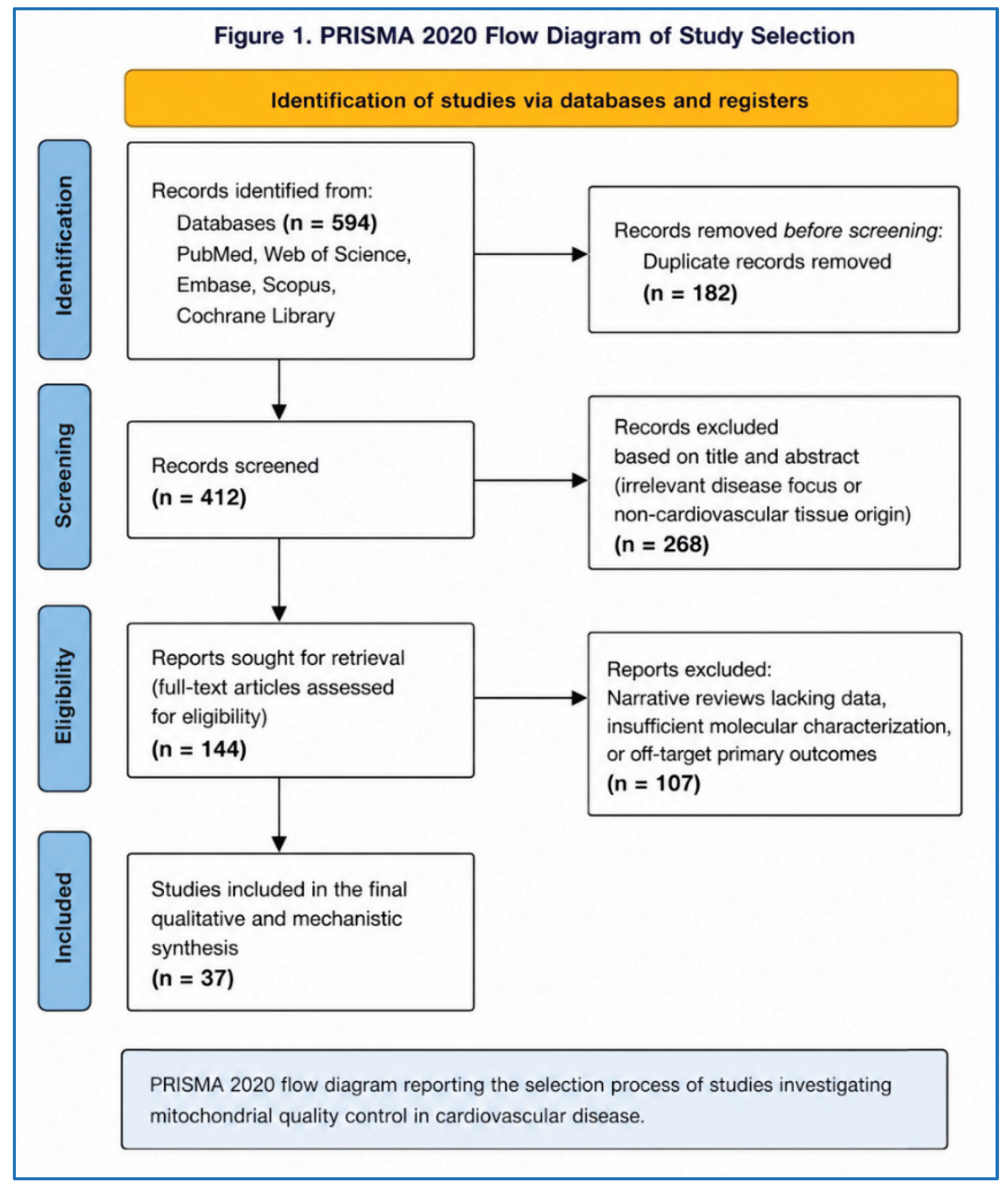
PRISMA Flow Diagram Data
The systematic search across all designated databases initially yielded a total of 594 potentially relevant records. Following the strict removal of duplicate publications across the different search engines, a rigorous screening process was initiated. The detailed systematic attrition of records, categorized by the phase of the review process, is comprehensively presented in Table I. Ultimately, a highly curated cohort of 37 core original research articles and high-tier systematic analyses was synthesized to inform the mechanistic and therapeutic findings detailed in this report.
Results
The Molecular Architecture of Mitochondrial Quality Control
The structural integrity and functional capacity of the cardiac mitochondrial network dictate the ultimate survival and performance of cardiomyocytes. MQC is not a singular event but rather is governed by distinct, sequential and highly integrated biological nodes: mitochondrial dynamics (fission and fusion), selective mitophagy and mitochondrial biogenesis. These pathways operate continuously in the background of cellular metabolism, ensuring that the architecture of the mitochondrial network dynamically adapts to the fluctuating metabolic demands of the heart.
Mitochondrial fusion and fission operate as the necessary prelude to quality control [7]. Fusion allows for the mixing of mitochondrial contents, enabling the complementation of damaged mtDNA and the sharing of metabolic substrates across the network. Conversely, fission, mediated predominantly by the cytosolic GTPase dynamin-related protein 1 (DRP1) [10], is required to physically isolate severely damaged, depolarized or ROS-producing segments of the mitochondria. Following a stress event, DRP1 is recruited to the outer mitochondrial membrane, often at sites where the endoplasmic reticulum (ER) wraps around the mitochondria and forms a contractile ring that pinches the organelle into smaller fragments. These isolated, damaged fragments are simultaneously prevented from re-fusing with the healthy network and are subsequently targeted for complete degradation via mitophagy. Perturbations in these early morphological steps invariably lead to a catastrophic failure in subsequent quality control mechanisms, highlighting the deeply interconnected nature of the MQC network.
Ubiquitin-Dependent Mitophagy: The PINK1/Parkin Axis and its Complex Redundancies
Mitophagy serves as the ultimate executioner of mitochondrial clearance. This specialized form of macroautophagy isolates damaged organelles within double-membrane autophagosomes, which subsequently fuse with lysosomes for enzymatic degradation. In the cardiovascular system, mitophagy operates primarily through two distinct parallel pathways: the ubiquitin-dependent pathway and the ubiquitin-independent (receptor-mediated) pathway [[11,12] .
The PTEN-induced kinase 1 (PINK1) and Parkin pathway is the most extensively characterized ubiquitin-dependent mitophagy mechanism in mammalian biology [13]. Under healthy, homeostatic conditions, PINK1 is continuously imported into the inner mitochondrial membrane where it undergoes rapid proteolytic cleavage and degradation, keeping its levels virtually undetectable. However, when a mitochondrion sustains severe damage, loses its vital membrane potential, or accumulates excessive misfolded proteins, PINK1 import is abruptly halted. This causes full-length PINK1 to rapidly accumulate and stabilize on the OMM. Once stabilized, PINK1 undergoes auto-phosphorylation and subsequently phosphorylates pre-existing ubiquitin molecules on the mitochondrial surface, as well as the E3 ubiquitin ligase Parkin, recruiting Parkin from the cytosol to the damaged organelle [14]. Once activated at the OMM, Parkin dramatically amplifies the signal by ubiquitinating a vast array of outer membrane proteins. These polyubiquitin chains serve as highly specific "eat me" signals. They are recognized by cytosolic autophagy adaptor proteins, such as p62/SQSTM1, which contain both ubiquitin-binding domains and LC3-interacting regions (LIRs). By binding to both the ubiquitinated mitochondria and the LC3 proteins decorating the expanding autophagosome, p62 effectively tethers the damaged mitochondrion, ensuring its engulfment and ultimate lysosomal degradation.
Emerging evidence, however, reveals a high degree of complexity and vital redundancy in this classical system, particularly within the highly specialized environment of the myocardium. A landmark mechanistic study conducted by Kubli et al. demonstrated that Parkin translocation and the subsequent activation of mitophagy can occur entirely independently of PINK1 in cardiac myocytes [15,16]. Utilizing PINK1-deficient (PINK1-/-) mice, the investigators observed that following an in vivo myocardial infarction, or following chemical uncoupling of the mitochondria via FCCP perfusion, Parkin was still robustly recruited to depolarized cardiac mitochondria. Crucially, this PINK1-independent recruitment correlated with profound ubiquitination of mitochondrial proteins and the active execution of mitophagy. The significance of this finding is profound: it suggests that the heart possesses alternative, compensatory mechanisms to activate Parkin to ensure the survival of energy-demanding myocytes even when the classical PINK1 signaling node is genetically or functionally compromised.
Receptor-Mediated Mitophagy: The Roles of FUNDC1, BNIP3, and NIX in Hypoxic Stress
In addition to the complex ubiquitin-tagging system, mitochondria express specific integral OMM receptors that contain their own LIR motifs. These receptors bypass the need for ubiquitin intermediaries, allowing the mitochondria to directly engage the autophagosomal machinery in response to specific stress stimuli. The key mitophagy receptors extensively studied in the myocardium include BNIP3, NIX (BNIP3L) and FUNDC1.
FUNDC1 (FUN14 domain containing 1) represents a highly specialized, hypoxia-responsive mitophagy receptor [17]. It is uniquely localized to mitochondria-associated endoplasmic reticulum membranes (MAMs), the critical signaling platforms where the ER physically communicates with mitochondria to coordinate calcium transfer and lipid metabolism [18]. At the MAMs, FUNDC1 actively coordinates both mitochondrial fission and mitophagy. The activity of FUNDC1 is tightly and rapidly regulated by post-translational modifications rather than mere transcriptional upregulation. Under normal normoxic physiological conditions, FUNDC1 is maintained in an inactive state through phosphorylation at the Ser13 and Tyr18 residues. This phosphorylation actively inhibits its ability to interact with LC3.
However, under conditions of severe ischemic or hypoxic stress, the regulation of FUNDC1 shifts dramatically. It undergoes deubiquitination, mediated by the ER-resident deubiquitinase USP19, and rapid dephosphorylation. This unmasks its LIR motif, triggering immediate binding to LC3 and initiating rapid mitophagy to clear the oxygen-deprived, damaged mitochondria. The protective nature of this pathway has been repeatedly validated; genetic ablation or knockdown of FUNDC1 severely impairs mitochondrial quality control, exacerbates ischemia/reperfusion (I/R) injury [19,20], increases mitochondrial mass abnormally and promotes widespread cardiomyocyte apoptosis. Furthermore, FUNDC1 directly interacts with Drp1 to drive the initial mitochondrial fission events required before mitophagy can occur, acting as a master coordinator of the MQC sequence at the MAMs.
Similarly, BNIP3 and Nix are atypical BH3-only proteins that act as direct mitophagy receptors. Ischemia and reperfusion injury significantly alter the phosphorylation states of BNIP3, dynamically modulating its binding affinity for LC3. Phosphorylation at specific serine residues enhances LC3 binding, elevating mitophagy to protective levels, whereas dysregulation of these sites impairs mitochondrial clearance, leading to cellular toxicity.

Table 1.
Key Regulators of Mitophagy in the Cardiovascular System.
| Regulator | Primary Mechanism of Action | Pathophysiological Trigger | Functional Consequence of Activation |
| PINK1 | Kinase; accumulates on depolarized OMM, phosphorylates Parkin & ubiquitin | Membrane depolarization, ROS accumulation | Recruits Parkin; initiates ubiquitin-dependent mitophagy |
| Parkin | E3 Ubiquitin Ligase; ubiquitinates numerous OMM proteins | Phosphorylation by PINK1 (or independent mechanisms) | Tags mitochondria for recognition by p62/LC3 |
| FUNDC1 | Direct Mitophagy Receptor; localizes to MAMs | Hypoxia, Ischemia | Dephosphorylation unmasks LIR motif; coordinates fission and direct LC3 binding |
| BNIP3 / Nix | Direct Mitophagy Receptors (Atypical BH3-only proteins) | Hypoxia, Oxidative Stress | Direct LC3 binding; induces selective autophagosome engulfment |
| MARCH5 | Mitochondrial E3 Ligase | Early hypoxia | Fine-tunes FUNDC1 stability; prevents premature/excessive mitophagy |
Mitochondrial Biogenesis: The AMPK/SIRT1/PGC-1α Transcriptional Network
As damaged, ROS-producing mitochondria are cleared from the cell via mitophagy, the cellular energetic pool must be rapidly replenished to prevent bioenergetic failure. This regenerative process, known as mitochondrial biogenesis [21], involves a complex transcriptional program governed primarily by the AMP-activated protein kinase (AMPK) and Sirtuin 1 (SIRT1) signaling network, which converges on the master regulatory co-activator PGC-1α [22,23].
AMPK functions as the central, highly conserved cellular fuel gauge, exquisitely sensitive to fluctuations in the intracellular AMP/ATP ratio. During periods of metabolic stress, ischemia, or energy depletion, activated AMPK initiates a dual-pronged metabolic response. It immediately stimulates catabolic processes, such as fatty acid oxidation and glucose uptake, to generate ATP, while simultaneously shutting off non-essential, energy-consuming anabolic pathways. Crucially, the activation of AMPK directly elevates cytosolic levels of Nicotinamide Adenine Dinucleotide (NAD+). This NAD+ elevation is driven in part by the AMPK-dependent upregulation of nicotinamide phosphoribosyltransferase (NAMPT), the rate-limiting enzyme in the NAD+ salvage biosynthesis pathway.
This elevation in NAD+ serves as an absolute requirement for the activity of the class III histone deacetylase SIRT1. Once activated by NAD+, SIRT1 directly deacetylates and fully activates PGC-1α, alongside downstream forkhead box O (FOXO) transcription factors, thereby modulating their transcriptional activity. Deacetylated, active PGC-1α translocates into the nucleus where it acts as a massive transcriptional co-activator [24]. It binds to and activates Nuclear Respiratory Factors 1 and 2 (NRF1/NRF2) and Mitochondrial Transcription Factor A (TFAM). This specific transcriptional cascade drives the massive coordinated transcription of both nuclear-encoded and mitochondrial-encoded OXPHOS genes, resulting in the proliferation of new, healthy and highly efficient mitochondria. Disruption of this critical AMPK/SIRT1/PGC-1α axis is prevalent across almost all etiologies of heart failure, diabetic cardiomyopathy and advanced aging, leading directly to a bioenergetic deficit and subsequent structural remodeling of the myocardium.
Pathophysiological Implications in Cardiovascular Disease Subtypes
The failure of the MQC network manifests differently depending on the specific cardiovascular disease state, affecting various cell types from the vascular endothelium to the working myocardium.
Atherosclerosis and Vascular Homeostasis Mitochondrial dysfunction and localized oxidative stress are now recognized as central contributors to the pathogenesis of atherosclerosis [25,26]. Mitophagy acts as a crucial regulator of vascular homeostasis. In the vascular wall, impaired mitophagy within endothelial cells, vascular smooth muscle cells (VSMCs) and invading macrophages leads to the excessive generation of ROS. This ROS accumulation triggers the activation of the NLRP3 inflammasome, driving local inflammation and promoting cell death within the atherosclerotic plaque. Conversely, the upregulation of the PINK1/Parkin pathway has been shown to protect against stress-induced apoptosis in VSMCs, thereby preventing the expansion of the necrotic core and promoting the stabilization of atherosclerotic plaques.
Ischemia-Reperfusion Injury: The Temporal Paradox of Mitophagy Myocardial ischemia and the subsequent reperfusion phase (I/R injury) present a unique temporal paradox regarding the role of mitophagy. During the initial early ischemic phase, moderate activation of mitophagy often mediated by the FUNDC1 and BNIP3 pathways is highly cardioprotective [27]. It serves to rapidly clear the swelling, ROS-generating organelles, preserving overall mitochondrial network integrity and limiting oxidative damage.
However, during the reperfusion phase, the sudden influx of oxygen triggers a massive burst of ROS, leading to the excessive and potentially dysregulated overactivation of mitophagy [28,29]. If the rate of mitochondrial clearance drastically exceeds the rate of PGC-1α-mediated biogenesis, the cardiomyocyte suffers profound mitochondrial depletion. This resulting energy crisis aggravates myocardial damage and promotes cell death. Furthermore, specific signaling molecules, such as Ripk3, are elevated during reperfusion and actively inhibit FUNDC1-mediated mitophagy, contributing to the accumulation of damaged mitochondria precisely when clearance is most needed.
Heart Failure and Cardiac Remodeling The functional status of mitophagy in patients with heart failure varies significantly depending on the phenotypic subtype and the stage of the disease. Heart failure imposes chronic biomechanical pressure overload and sustained neurohormonal stress on the heart, leading to continuous mitochondrial depolarization, calcium overload and ROS generation [30]. In heart failure with reduced ejection fraction (HFrEF), the PINK1-Parkin pathway is frequently found to be downregulated or functionally impaired. This severe defect results in inadequate mitochondrial clearance. The resulting accumulation of damaged mitochondria fuels chronic inflammation, cardiomyocyte apoptosis and widespread fibrotic remodeling, accelerating the transition to end-stage heart failure [31]. Furthermore, the failure of MQC in HFrEF disrupts normal fatty acid oxidation (FAO), trapping the heart in a negative feedback loop of energy starvation and lipotoxicity.
Therapeutic Modulation of the Mitochondrial Quality Control Network
The most compelling advancement in modern cardiovascular pharmacology is the recognition that many of the drugs demonstrating the highest clinical efficacy inherently modulate the precise MQC networks described above. The translation of these mechanisms into actionable therapies represents a new frontier in treating cardiovascular disease.
SGLT2 Inhibitors as Profound Metabolic Reprogrammers
Sodium-Glucose Cotransporter 2 (SGLT2) inhibitors, including empagliflozin and dapagliflozin, have revolutionized the standard of care in cardiology. Originally developed strictly as glucose-lowering anti-diabetic agents, these molecules have demonstrated profound, entirely glucose-independent cardioprotective effects in landmark clinical trials (such as EMPA-REG OUTCOME and DAPA-HF) [[9]. Mechanistically, a substantial body of evidence now indicates that the cardiovascular benefits of SGLT2 inhibitors are deeply rooted in the enhancement of mitochondrial quality control and the modulation of nutrient sensing pathways.
At the cellular level, SGLT2 inhibitors induce a unique systemic state that mimics nutrient deprivation, replicating the highly beneficial physiological effects of fasting or severe caloric restriction [[32]. They act by simultaneously upregulating nutrient deprivation sensors (specifically AMPK, SIRT1, SIRT3, and SIRT6) while potently downregulating nutrient surplus sensors (most notably the mammalian target of rapamycin, mTOR). By inhibiting the mTOR pathway a major suppressor of autophagy and concurrently elevating AMPK activity, SGLT2 inhibitors profoundly stimulate autophagic flux and mitophagy, facilitating the rapid and efficient clearance of damaged, ROS-producing organelles.
Beyond systemic metabolic shifts, SGLT2 inhibitors exhibit remarkable direct regulatory capacities at the organelle level. Hyperglycemia and heart failure typically cause an elevation of cytosolic sodium and protons, which suppresses PGC-1α expression. SGLT2 inhibitors, such as dapagliflozin, directly lower this cytosolic accumulation of ions by modulating the Na+/H+ exchanger (NHE-1) [33], thereby lifting the suppressive effects of ion overload and restoring robust PGC-1α expression and mitochondrial biogenesis. Furthermore, specific interactions have been discovered: empagliflozin has been shown to interact directly with mitochondrial sirtuin 3 (SIRT3). This interaction promotes the formation of a critical protein complex containing Beclin 1 and TLR9. This specific complex formation is obligatory for the enhancement of mitochondrial respiration and the execution of mitophagy. Strikingly, in experimental models where SIRT3 or TLR9 is genetically deleted, the cardioprotective and autophagic effects of empagliflozin are entirely abrogated, proving the absolute dependence of these drugs on MQC pathways.
Consequently, SGLT2 inhibition drives a comprehensive metabolic shift in the failing heart. They transition substrate utilization away from inefficient glucose metabolism toward more efficient fatty acid oxidation and ketogenesis [34,35], while concurrently re-establishing a healthy mitochondrial network via enhanced PGC-1α and TFAM activity, ultimately rescuing diabetic and non-diabetic myocardial microvascular injury.
Table 2.
Mechanisms of Action of Key MQC-Modulating Therapeutics.
| Therapeutic Agent | Drug Class / Category | Primary Molecular Targets in MQC | Resulting Physiological Effect |
| Empagliflozin | SGLT2 Inhibitor | Upregulates AMPK/SIRT1/SIRT3/PGC-1α; Downregulates mTOR; Inhibits NHE-1 | Mimics fasting; dramatically enhances autophagic flux and mitochondrial biogenesis; shifts substrate to ketones. |
| Nicotinamide Riboside (NR) | NAD+ Precursor | Restores cytosolic and mitochondrial NAD+ pools; activates Sirtuins | Rescues SIRT-mediated deacetylation of PGC-1α; increases maximal PBMC respiration; reduces NLRP3 inflammation. |
| MitoQ | Mitochondria-Targeted Antioxidant | Accumulates in matrix via TPP cation; scavenges H2O2 | Prevents oxidative damage to IMM; restores mitochondrial calcium retention capacity (mCRC); prevents mPTP opening. |
| Elamipretide (SS-31) | Cardiolipin-Targeting Peptide | Selectively binds cardiolipin in the IMM | Stabilizes cristae architecture; optimizes ETC supercomplex formation; reduces electron leak and ROS at the source. |
NAD+ Precursors and the Reactivation of Sirtuin Signaling
The progressive decline in intracellular NAD+ levels is a universally recognized hallmark of myocardial aging [36], chronic heart failure and acute ischemic injury. Because NAD+ functions as the obligate substrate for the entire sirtuin family of deacetylases, its pathological depletion causes a secondary, catastrophic failure in SIRT-mediated MQC regulation. This includes the blunting of PGC-1α activity and the toxic accumulation of hyperacetylated, dysfunctional mitochondrial OXPHOS proteins.
Restoring these depleted NAD+ pools utilizing specific biosynthetic precursors, such as Nicotinamide Riboside (NR) and Nicotinamide Mononucleotide (NMN), has emerged as a highly promising therapeutic strategy. In preclinical models of heart failure, NMN administration successfully ameliorated left ventricular dysfunction and extended lifespan. Interestingly, advanced mechanistic studies suggest that the protective effect of NMN may rely heavily on restoring lysosomal function, promoting autophagic completion and preventing iron-mediated lipid peroxidation (ferroptosis) within the lysosome, rather than exclusively rescuing the primary mitochondrial respiratory defects.
Crucially, this metabolic pathway has successfully transitioned into rigorous human clinical evaluation. In a Phase I/II randomized clinical trial involving patients with stable heart failure with reduced ejection fraction (HFrEF) (ClinicalTrials.gov Identifier: NCT03423342), the oral administration of NR at a high dose of 1,000 mg twice daily proved to be safe, well-tolerated, and highly biologically active. The intervention successfully doubled whole blood NAD+ levels in the human subjects. Most notably, the intraindividual escalation in NAD+ directly and positively correlated with significant increases in both basal and maximal cellular respiration measured in peripheral blood mononuclear cells (PBMCs). Furthermore, this improvement in respiration was accompanied by a significant suppression of the pro-inflammatory NLRP3 inflammasome. These landmark data confirm that systemic NAD+ enhancement can rapidly reprogram cellular bioenergetics and limit toxic inflammatory signaling in humans suffering from advanced cardiovascular disease.
Mitochondria-Targeted Antioxidants: Precision ROS Mitigation
While broad-spectrum antioxidants have historically failed in large human cardiovascular trials due to poor intracellular distribution, next-generation compounds designed to accumulate specifically within the mitochondrial matrix have shown unprecedented efficacy in preclinical models and early clinical settings. By neutralizing ROS precisely at the site of generation, these agents prevent destructive oxidative damage to mtDNA and preserve the electron transport chain without disrupting vital, physiological cytosolic redox signaling.
MitoQ (Mitoquinone) MitoQ is a highly bioavailable synthetic molecule comprising a ubiquinone antioxidant moiety covalently conjugated to a lipophilic triphenylphosphonium (TPP) cation [37]. This distinct chemical structure drives its massive, targeted accumulation within the negatively charged mitochondrial matrix, concentrating the antioxidant directly at the IMM. In rigorous rodent models of pressure-overload heart failure (induced by transverse aortic constriction), MitoQ administration successfully mitigated adverse cardiac remodeling, reduced ultrastructural mitochondrial swelling and profoundly decreased pathological hydrogen peroxide emission.
Detailed bioenergetic analyses of these treated hearts revealed that MitoQ preserves maximal mitochondrial respiration (State 3) and fully restores the respiratory control ratio in subsarcolemmal mitochondria (SSM). Simultaneously, MitoQ treatment dramatically improved the mitochondrial calcium retention capacity (mCRC) [38], a critical metric of mitochondrial resilience, thereby preventing the catastrophic, cell-killing opening of the mitochondrial permeability transition pore (mPTP). Furthermore, recent transcriptomic data suggest that MitoQ's protective effects may also involve preventing the dysregulation of redox-sensitive epigenetic modifiers, specifically the lncRNA-microRNA pair Plscr4-miR-214, which regulates mitochondrial fusion proteins.
Elamipretide (SS-31) Elamipretide is a first-in-class, cell-permeable, mitochondria-targeted tetrapeptide that functions through a mechanism fundamentally different from traditional antioxidants [39]. Rather than acting strictly as a chemical scavenger of free radicals, Elamipretide selectively binds to cardiolipin, a unique and highly vulnerable phospholipid situated exclusively in the inner mitochondrial membrane. By physically binding and stabilizing cardiolipin, Elamipretide preserves the highly folded architecture of the mitochondrial cristae. This structural stabilization optimizes the spatial configuration of the electron transport chain supercomplexes, minimizes electron leak during respiration, and effectively halts pathogenic ROS generation directly at its source before it can damage surrounding structures.
The immense translational potential of Elamipretide was robustly validated in a double-blind, placebo-controlled, ascending-dose clinical trial involving patients with advanced HFrEF (ClinicalTrials.gov Identifier: NCT02388464). Patients receiving the highest dose of Elamipretide (0.25 mg/kg/h) via continuous intravenous infusion demonstrated highly significant, rapid, and favorable improvements in cardiac geometry. Specifically, echocardiographic assessments revealed an 18 mL reduction in left ventricular end-diastolic volume (LVEDV) and a 14 mL reduction in left ventricular end-systolic volume (LVESV) at the end of the infusion period. These robust macroscopic structural improvements correlated tightly with peak plasma drug concentrations, underscoring the immediate, cause-and-effect relationship between pharmacological mitochondrial stabilization and macroscopic cardiac function in humans.
Discussion
The systematic synthesis of the prevailing literature reveals unequivocally that mitochondrial quality control is not merely a peripheral cellular housekeeping function, but rather the central, indispensable arbiter of cardiomyocyte survival and overall cardiovascular health. The progressive pathophysiology of heart failure, ischemia-reperfusion injury, diabetic cardiomyopathy, and atherosclerosis is intrinsically tied to a collapse in the dynamic balance between organelle clearance (mitophagy) and organelle regeneration (mitochondrial biogenesis).
A critical revelation derived from the advanced mechanistic data synthesized in this review is the exceptionally high degree of redundancy and interconnectedness within the MQC network. The classical, widely accepted biological paradigm suggested that the accumulation of PINK1 was the absolute and non-negotiable prerequisite for Parkin-mediated mitophagy. However, the groundbreaking observation that Parkin can robustly translocate to depolarized mitochondria in PINK1-/- myocardium following an acute myocardial infarction completely upends this linear model. This profound biological redundancy highlights the evolutionary imperative of protecting the high-energy-demanding myocardium; the heart has evolved multiple fail-safes to ensure the execution of mitophagy even when isolated signaling pathways are genetically ablated or functionally impaired.
Similarly, the regulation of receptor-mediated mitophagy through FUNDC1 demonstrates extreme biological precision. The strategic localization of FUNDC1 to the MAMs, strictly governed by localized E3 ubiquitin ligases (such as MARCH5) and deubiquitinases (such as USP19), allows the cell to perfectly synchronize the physical act of mitochondrial fission with the recruitment of autophagosomes during acute hypoxic stress. This spatial control ensures that only the specifically damaged fragments of the network are degraded, sparing the healthy mitochondria required to sustain cellular ATP production.
However, the temporal dynamics of mitophagy present a highly complex physiological paradox, particularly in the setting of acute myocardial infarction. While the moderate activation of mitophagy during early ischemia is indisputably cardioprotective clearing out ROS-generating organelles and preventing the initiation of pro-apoptotic signaling, the excessive or dysregulated overactivation of mitophagy during the subsequent reperfusion phase can be highly deleterious [40,41]. Massive autophagic clearance driven by the sudden influx of oxygen, if carried out without a compensatory and equivalent increase in PGC-1α-mediated biogenesis, inevitably precipitates a severe cellular energy crisis, driving the myocardium toward irreversible failure and death. This inherent "double-edged sword" nature of MQC necessitates that therapeutic interventions do not merely force the blind overactivation of autophagy, but rather act as modulators that restore the homeostatic balance of the entire network, matching clearance with regeneration.
The most compelling advancement in modern cardiovascular pharmacology, highlighted extensively in this review, is the recognition that drugs demonstrating the highest clinical efficacy inherently modulate these precise MQC networks. SGLT2 inhibitors provide the definitive proof of concept for this paradigm shift. The unprecedented ability of drugs like empagliflozin and dapagliflozin to reduce cardiovascular mortality in both diabetic and non-diabetic heart failure patients can be largely mapped to their capacity to artificially mimic a state of nutrient deprivation. By actively downregulating the mTOR-mediated nutrient surplus signal and robustly upregulating the AMPK/SIRT1/PGC-1α energy-sensing axis [35], these agents seamlessly orchestrate both ends of the MQC spectrum. They clear the cellular debris through enhanced autophagic flux and simultaneously repopulate the functional syncytium through robust mitochondrial biogenesis. The discovery of the direct physical docking of empagliflozin to the mitochondrial SIRT3 protein further illuminates how systemically administered metabolic modifiers can exact highly localized, organelle-specific control over mitochondrial fate.
Similarly, the successful clinical translation of NAD+ precursors and mitochondrial-targeted peptides confirms that the mitochondria can be safely and effectively accessed as pharmacological targets in living human patients. The ability of Nicotinamide Riboside to safely double whole-blood NAD+ levels and significantly improve PBMC respiration in HFrEF patients, alongside the capacity of Elamipretide to induce rapid, highly favorable reductions in left ventricular volumes, represent true watershed moments in the evolution of cardiovascular therapy. These clinical trials provide the ultimate proof that reversing the myocardial energy deficit directly at the mitochondrial level translates into rapid, macroscopic improvements in cardiac geometry, contractile function and systemic inflammatory tone.
Future Perspectives
While the basic science discoveries and early-phase clinical trials surrounding MQC modulation are undeniably promising, several pivotal barriers must be overcome to fully integrate these advanced therapies into standard clinical practice. First, there is an urgent and unmet clinical need to establish reliable, non-invasive circulating biomarkers of mitochondrial dysfunction and dynamic autophagic flux in humans. Currently, accurately assessing MQC requires invasive endomyocardial tissue biopsies or rely on proxy measurements derived from peripheral blood mononuclear cells. Developing advanced positron emission tomography (PET) tracers specific for mitochondrial membrane potential or identifying highly sensitive, disease-specific exosomal MQC markers in the plasma, will be absolutely paramount for effectively tracking drug efficacy, patient responsiveness and titrating exact dosages in clinical settings.
Second, future research efforts must comprehensively address the profound heterogeneity of heart failure phenotypes. The structural, metabolic and molecular profile of heart failure with reduced ejection fraction (HFrEF) differs fundamentally from that of heart failure with preserved ejection fraction (HFpEF). In HFrEF, the PINK1-Parkin pathway is frequently impaired, leading directly to a profound energy crisis, myocyte loss and extensive replacement fibrosis. Conversely, HFpEF involves distinct systemic inflammatory paradigms, profound endothelial mitochondrial disturbances and titin-based stiffness rather than primary cardiomyocyte energy failure. Consequently, therapeutic modulation must become highly tailored to the specific phenotype; for instance, agents that aggressively stimulate PGC-1α biogenesis may be highly effective in the energy-starved HFrEF heart, whereas therapies specifically targeting endothelial ROS mitigation and inflammation may dominate the future management of HFpEF.
Finally, the long-term safety of direct, continuous mitochondrial manipulation requires rigorous longitudinal assessment. Because MQC is intimately, inextricably linked to fundamental cellular apoptosis, innate immune cell activation, and systemic metabolism, the chronic pharmacological hyperactivation of mitophagy or the sustained suppression of the mTOR pathway could theoretically yield unintended off-target effects. These could potentially include altered immune surveillance, increased susceptibility to infection or skeletal muscle wasting over decades of use. Large-scale, multi-year randomized controlled trials powered for hard clinical endpoints are required to definitively determine the long-term viability and safety profile of direct MQC modulators like MitoQ, Elamipretide, and high-dose NAD+ precursors in chronic cardiovascular populations.
Limitations
The current body of literature analyzed within this systematic review is not without notable constraints. A substantial majority of the deep mechanistic insights regarding the intricate signaling of mitophagy and mitochondrial biogenesis are derived from highly controlled, genetically manipulated, young murine models or from in vitro cellular assays utilizing immortalized cell lines under artificial conditions [42,43,44]. These highly curated models frequently fail to accurately capture the complex, multi-organ pathophysiology of human cardiovascular disease, which is typically complicated by advanced age, extensive polypharmacy, genetic diversity and profound systemic comorbidities such as chronic kidney disease and severe obesity. Furthermore, the clinical trials investigating novel, direct MQC agents such as Nicotinamide Riboside and Elamipretide are largely in their preliminary, proof-of-concept phases (Phase I/II). These specific studies, while generating highly positive physiological signals and confirming target engagement, are characterized by relatively small patient cohort sizes, short follow-up durations and an ultimate reliance on surrogate primary endpoints (such as PBMC maximal respiration or acute ventricular volume changes) rather than hard clinical outcomes like all-cause mortality, cardiovascular death, or heart failure hospitalization rates. Consequently, the interpretation of their ultimate, long-term therapeutic efficacy must remain appropriately cautious until definitively powered, multi-center phase III outcome trial data is published and peer-reviewed.
Conclusion
Mitochondrial quality control constitutes the foundational, non-negotiable pillar of cardiomyocyte viability and overall cardiovascular homeostasis. The intricate, highly redundant coordination of mitochondrial fission, selective mitophagy via both the ubiquitin-dependent PINK1/Parkin axis and receptor-mediated pathways such as FUNDC1, combined with PGC-1α-driven biogenesis, ensures the continuous supply of oxidative energy required by the relentlessly beating heart. The disruption of this exquisite biological balance precipitates the severe oxidative stress, cellular energy crisis, systemic inflammation and adverse structural remodeling that are the defining characteristics of heart failure, ischemic heart disease and atherosclerosis. The recent advent of pharmacological therapies that directly and effectively target these pathways most notably the profound metabolic reprogramming induced by SGLT2 inhibitors, the restoration of sirtuin signaling via NAD+ precursors and the structural stabilization provided by mitochondrial-targeted peptides like Elamipretide has definitively validated the MQC network as a highly actionable therapeutic target in humans. By successfully pivoting therapeutic strategies away from generic, systemic interventions and toward highly precise, organelle-level metabolic reprogramming, the field of basic and clinical cardiology is poised to fundamentally alter the trajectory of cardiovascular disease management, offering novel, biologically rational hope for true organ resuscitation and the reversal of advanced cardiac failure.
Ethical Approval
The study was conducted in accordance with the ethical standards of the Institutional Ethics Committee of Vinayaka Mission's Research Foundation (Deemed to be University), Salem, India, and with the 1964 Helsinki Declaration and its later amendments or comparable ethical standards.
Informed Consent
Not applicable. This study is a systematic review of previously published literature and did not involve human participants or animals.
Data Availability
The datasets generated and/or analyzed during the current study are available from the corresponding author on reasonable request.
Consent for Publication
All authors have reviewed and approved the final manuscript and consent to its publication.
Author Contributions
Mr. Lemuel Mathew: Conceptualization, methodology, data collection, data analysis, interpretation of results, manuscript writing, corresponding author responsibilities. Study supervision, methodology development, critical review and manuscript editing. Ms. A. Sushmitha: Data collection, data validation, literature review manuscript review. Statistical analysis, interpretation of findings, critical revision of the manuscript, Clinical supervision, study design, interpretation of clinical data and final manuscript review. All authors contributed to the manuscript and approved the final version for publication.
Funding
The authors received no specific funding for this work.
Acknowledgments
The authors would like to thank the Department of Cardiology and the School of Allied Health Sciences, Vinayaka Mission's Research Foundation (Deemed to be University), Salem, for their support during the conduct of this study. Also want to thank Dr. B. Sendhilkumar (Dean) for continuous guidance, inspiration and motivation to pursue this work.
Conflicts of Interest
The authors declare that they have no conflict of interest.
References
- Atici, A.; Crother, T.; Noval Rivas, M. Mitochondrial quality control in health and cardiovascular diseases. Front Cell Dev. Biol. 2023, 11. [Google Scholar] [CrossRef] [PubMed]
- Gustafsson, A.; Gottlieb, R.; Gustafsson, A.B.; Gottlieb, R.A. Heart mitochondria: gates of life and death. Cardiovasc Res.;Cardiovasc Res. 2008, 77, 334-343 334–43. [Google Scholar] [CrossRef] [PubMed]
- Brown, D.A.; Perry, J.B.; Allen, M.E.; et al. Mitochondrial function as a therapeutic target in heart failure. Nat. Rev. Cardiol. 2017, 14, 238–50. [Google Scholar] [CrossRef] [PubMed]
- Galluzzi, L.; Kepp, O.; Trojel-Hansen, C.; Kroemer, G. Mitochondrial Control of Cellular Life, Stress, and Death. Circ. Res. 2012, 111, 1198–207. [Google Scholar] [CrossRef] [PubMed]
- Zhang, M.; Zou, R.; Wang, G.; Fan, X. Editorial: Mitochondrial quality control in cardiovascular diseases. Front Cardiovasc Med. 2023, 10. [Google Scholar] [CrossRef] [PubMed]
- Ni, H.-M.; Williams, J.A. Ding W-X: Mitochondrial dynamics and mitochondrial quality control. Redox Biol. 2015, 4, 6–13. [Google Scholar] [CrossRef] [PubMed]
- Dorn GW: Mitochondrial Dynamics in Heart Disease. Biochim Biophys. Acta 2013, 1833, 233–41. [CrossRef] [PubMed]
- Siasos, G.; Tsigkou, V.; Kosmopoulos, M.; et al. Mitochondria and cardiovascular diseases—from pathophysiology to treatment. Ann. Transl. Med. 2018, 6, 256–256. [Google Scholar] [CrossRef] [PubMed]
- Packer, M. SGLT2 Inhibitors Produce Cardiorenal Benefits by Promoting Adaptive Cellular Reprogramming to Induce a State of Fasting Mimicry: A Paradigm Shift in Understanding Their Mechanism of Action. Diabetes Care 2020, 43, 508–11. [Google Scholar] [CrossRef] [PubMed]
- Shirakabe, A.; Zhai, P.; Ikeda, Y.; et al. Drp1-Dependent Mitochondrial Autophagy Plays a Protective Role Against Pressure Overload-Induced Mitochondrial Dysfunction and Heart Failure. Circulation 2016, 134, e75–6. [Google Scholar] [CrossRef] [PubMed]
- Saito T, Sadoshima J: The Molecular Mechanisms of Mitochondrial Autophagy/Mitophagy in the Heart. Circ. Res. 2015, 116, 1477–90. [CrossRef] [PubMed]
- Bravo San Pedro, J.M.; Kroemer, G.; Galluzzi, L. Autophagy and Mitophagy in Cardiovascular Disease. Circ. Res. 2017, 120, 1812–24. [Google Scholar] [CrossRef] [PubMed]
- Jin, S.M.; Youle, R. PINK1- and Parkin-mediated mitophagy at a glance. J. Cell Sci. 2012, 125, 795–9. [Google Scholar] [CrossRef] [PubMed]
- Narendra, D.; Tanaka, A.; Suen, D.-F.; Youle, R. Parkin is recruited selectively to impaired mitochondria and promotes their autophagy. J. Cell Biol. 2008, 183, 795–803. [Google Scholar] [CrossRef] [PubMed]
- Kubli, D.A.; Gustafsson, Å.B. Mitochondria and mitophagy: the yin and yang of cell death control. Circ. Res. 2012, 111, 1208–21. [Google Scholar] [CrossRef] [PubMed]
- Kubli, D.; Quinsay, M.; Gustafsson, A. Parkin deficiency results in accumulation of abnormal mitochondria in aging myocytes. Commun. Integr. Biol. 2013, 6, e24511. [Google Scholar] [CrossRef] [PubMed]
- Liu, L.; Feng, D.; Chen, G.; et al. Mitochondrial outer-membrane protein FUNDC1 mediates hypoxia-induced mitophagy in mammalian cells. Nat. Cell Biol. 2012, 14, 177–85. [Google Scholar] [CrossRef] [PubMed]
- Eisner, V.; Csordás, G.; Hajnóczky, G. Interactions between sarco-endoplasmic reticulum and mitochondria in cardiac and skeletal muscle – pivotal roles in Ca2+ and reactive oxygen species signaling. J. Cell Sci. 2013, 126, 2965–78. [Google Scholar] [CrossRef] [PubMed]
- Cai, Y.; Yang, E.; Yao, X.; et al. FUNDC1-dependent mitophagy induced by tPA protects neurons against cerebral ischemia-reperfusion injury. Redox Biol. 2020, 38, 101792. [Google Scholar] [CrossRef] [PubMed]
- Chen, D.; Zhou, L.; Chen, G.; et al. FUNDC1-induced mitophagy protects spinal cord neurons against ischemic injury. Cell Death Discov. 2024, 10, 4. [Google Scholar] [CrossRef] [PubMed]
- She, P.; Mo, F.; Zhu, P.; Zhong, T.P. Regulation of mitochondrial biogenesis and energy metabolism in the heart. Chin. Med. J. (Engl) 2026, 139, 1008–30. [Google Scholar] [CrossRef] [PubMed]
- Pillai, V.B.; Sundaresan, N.R.; Gupta, M.P. Regulation of Akt signaling by Sirtuins: Its implication in cardiac hypertrophy and aging. Circ. Res. 2014, 114, 368–78. [Google Scholar] [CrossRef] [PubMed]
- Matsushima, S.; Sadoshima, J. The role of sirtuins in cardiac disease. Am. J. Physiol.-Heart Circ. Physiol. 2015, 309, H1375–89. [Google Scholar] [CrossRef] [PubMed]
- Scarpulla, R.C. Metabolic control of mitochondrial biogenesis through the PGC-1 family regulatory network. Biochim Biophys. Acta 2011, 1813, 1269–78. [Google Scholar] [CrossRef] [PubMed]
- Gargiulo, P.; Marciano, C.; Savarese, G.; et al. Endothelial dysfunction in type 2 diabetic patients with normal coronary arteries. Int. J. Cardiol. 2013, 165, 67–71. [Google Scholar] [CrossRef] [PubMed]
- Wu, W.; Xu, H.; Wang, Z.; et al. PINK1-Parkin-Mediated Mitophagy Protects Mitochondrial Integrity and Prevents Metabolic Stress-Induced Endothelial Injury. PLoS ONE 2015, 10, e0132499. [Google Scholar] [CrossRef] [PubMed]
- Vinten-Johansen, J.; Zhao, Z.-Q.; Zatta, A.J.; Kin, H.; Halkos, M.E.; Kerendi, F. PostconditioningA new link in nature’s armor againstmyocardial ischemia–reperfusion injury. Basic Res. Cardiol. 2005, 100, 295–310. [Google Scholar] [CrossRef] [PubMed]
- Murphy, E.; Steenbergen, C. Mechanisms underlying acute protection from cardiac ischemia-reperfusion injury. Physiol. Rev. 2008, 88, 581–609. [Google Scholar] [CrossRef] [PubMed]
- Hausenloy, D.J.; Yellon, D.M. Myocardial ischemia-reperfusion injury: a neglected therapeutic target. J. Clin. Invest. 2013, 123, 92–100. [Google Scholar] [CrossRef] [PubMed]
- Ikeda, Y.; Shirakabe, A.; Maejima, Y.; et al. Endogenous Drp1 mediates mitochondrial autophagy and protects the heart against energy stress. Published Online First. 2015. [CrossRef]
- Marín-García, J.; Akhmedov, A.T. Mitochondrial dynamics and cell death in heart failure. Heart Fail Rev. 2016, 21, 123–36. [Google Scholar] [CrossRef] [PubMed]
- Packer, M.; Anker, S.D.; Butler, J.; et al. Cardiovascular and renal outcomes with empagliflozin in heart failure. N Engl. J. Med. 2020, 383, 1413–24. [Google Scholar] [CrossRef] [PubMed]
- Maejima, Y. SGLT2 Inhibitors Play a Salutary Role in Heart Failure via Modulation of the Mitochondrial Function. Front Cardiovasc Med. 2020, 6, 186. [Google Scholar] [CrossRef] [PubMed]
- Marx, N.; McGuire, D.K. Sodium-glucose cotransporter-2 inhibition for the reduction of cardiovascular events in high-risk patients with diabetes mellitus. Eur. Heart J. 2016, 37, 3192–200. [Google Scholar] [CrossRef] [PubMed]
- Verma, S.; McMurray. JJV: SGLT2 inhibitors and mechanisms of cardiovascular benefit: a state-of-the-art review. Diabetologia 2018, 61, 2108–17. [Google Scholar] [CrossRef] [PubMed]
- Kane, A.E.; Sinclair, D.A. Sirtuins and NAD+ in the Development and Treatment of Metabolic and Cardiovascular Diseases. Circ. Res. 2018, 123, 868–85. [Google Scholar] [CrossRef] [PubMed]
- Murphy, M.P.; Smith, R.A.J. Targeting antioxidants to mitochondria by conjugation to lipophilic cations. Annu Rev. Pharmacol. Toxicol. 2007, 47, 629–56. [Google Scholar] [CrossRef] [PubMed]
- Ong, S.-B.; Dongworth, R.K.; Cabrera-Fuentes, H.A.; Hausenloy, D.J. Role of the MPTP in conditioning the heart - translatability and mechanism. Br. J. Pharmacol. 2015, 172, 2074–84. [Google Scholar] [CrossRef] [PubMed]
- Szeto HH: First-in-class cardiolipin-protective compound as a therapeutic agent to restore mitochondrial bioenergetics. Br. J. Pharmacol. 2014, 171, 2029–50. [CrossRef] [PubMed]
- Zhu, H.; Tannous, P.; Johnstone, J.L.; et al. Cardiac autophagy is a maladaptive response to hemodynamic stress. J. Clin. Invest. 2007, 117, 1782–93. [Google Scholar] [CrossRef] [PubMed]
- Zhou, H.; Toan, S.; Zhu, P.; Wang, J.; Ren, J.; Zhang, Y. DNA-PKcs promotes cardiac ischemia reperfusion injury through mitigating BI-1-governed mitochondrial homeostasis. Basic Res. Cardiol. 2020, 115. [Google Scholar] [CrossRef] [PubMed]
- Gottlieb, R.A.; Mentzer, R.M. Autophagy During Cardiac Stress: Joys and Frustrations of Autophagy. Annu Rev. Physiol. 2010, 72, 45–59. [Google Scholar] [CrossRef] [PubMed]
- Chen, Y.; Dorn, G.W. PINK1- Phosphorylated Mitofusin 2 is a Parkin Receptor for Culling Damaged Mitochondria. Science 2013, 340, 471–5. [Google Scholar] [CrossRef] [PubMed]
- Sciarretta, S.; Maejima, Y.; Zablocki, D.; Sadoshima, J. The Role of Autophagy in the Heart. Annu Rev. Physiol. 2018, 80, 1–26. [Google Scholar] [CrossRef] [PubMed]
Figure 1.
PRISMA 2020 Flow Diagram Data Summary for Literature Selection.
Disclaimer/Publisher’s Note: The statements, opinions and data contained in all publications are solely those of the individual author(s) and contributor(s) and not of MDPI and/or the editor(s). MDPI and/or the editor(s) disclaim responsibility for any injury to people or property resulting from any ideas, methods, instructions or products referred to in the content. |
© 2026 by the authors. Licensee MDPI, Basel, Switzerland. This article is an open access article distributed under the terms and conditions of the Creative Commons Attribution (CC BY) license (http://creativecommons.org/licenses/by/4.0/).
Copyright: This open access article is published under a Creative Commons CC BY 4.0 license, which permit the free download, distribution, and reuse, provided that the author and preprint are cited in any reuse.


