
Submitted:
24 June 2026
Posted:
26 June 2026
You are already at the latest version
Abstract
Desminopathies are rare myofibrillar myopathies caused by variants in the desmin (DES) gene on chromosome 2q35, encoding the protein desmin. These variants, typically autosomal dominant missense, result in diverse clinical phenotypes, including progressive skeletal myopathy and cardiac involvement. Over 180 pathogenic variants and 700 variants of uncertain significance have been reported.
We describe a 46-year-old male with progressive distal and proximal muscle weakness since age 30, functional impairment, and intermittent dysphagia. There was no family history or respiratory involvement. Electromyography demonstrated a myopathic pattern, and muscle biopsy revealed desmin- positive aggregates. The patient had a prior syncopal episode with minor chronic electrocardiographic abnormalities, suggesting possible cardiac involvement.
Whole exome sequencing identified a novel heterozygous DES variant, c.1327_1328delinsTT, p.(Lys443Leu). The combined clinical, electrophysiological, and histopathological findings strongly support the likely pathogenicity of the identified variant. This case expands the spectrum of DES variants and highlights the importance of multidisciplinary evaluation.
Keywords:
desminopathy
; novel de novo heterozygous DES variant
; desmin tail-domain variants
; variant c.1327_1328delinsTT
; p.(Lys443Leu)
; likely pathogenic desmin gene variant
1. Introduction
Desminopathies are a subgroup of myofibrillar myopathies caused by variants in the desmin (DES) gene on chromosome 2q35, which encodes desmin, a muscle-specific intermediate filament protein essential for maintaining structural and functional integrity of muscle cells [1].
While most reported variants are autosomal dominant missense variants, recessive inheritance patterns and sporadic cases have also been documented. Currently, over 180 pathogenic DES variants and over 700 variants of uncertain clinical significance have been reported [2].
Desmin is an essential intermediate filament protein in cardiac and skeletal muscle that maintains cell integrity and mediates mechanochemical signaling. The desmin molecule is composed of 470 amino acids and is organized into three domains: a highly conserved α-helical core of 308 amino acid residues flanked by globular N- and C- terminal (“head” and “tail”) structures [3]. It forms a network that aligns myofibrils, positions organelles, and transduces signals. Desmin facilitates protein interactions to maintain the connection between the contractile apparatus and other muscle fiber components, crucial for cell integrity, force transmission, and signaling. Desmin gene variants can result in diverse clinical phenotypes [4,5,6].
The pathogenesis of DES- related disease is frequently attributed to the toxic gain-of-function of many pathogenic DES variants, leading to desmin misfolding, aggregation, and accumulation within muscle fibers [2].
The age of disease onset, clinical/myopathological features and the rate of progression vary significantly depending on the type of inheritance and the location of the causative variant, particularly within functional domains of the desmin protein [7].
Desminopathies are phenotypically characterized by progressive, bilateral skeletal muscle weakness typically initiating in distal leg muscles and subsequently affecting proximal limb muscles, limb girdle, scapuloperoneal musculature, and manifesting as generalized myopathy. Proximal, truncal, bulbar, or facial muscle involvement may also occur. Cardiac issues like conduction defects and cardiomyopathy are common. Additional symptoms may include respiratory dysfunction, cataracts, dysphagia, and gastrointestinal dysfunction [5,6,7].
2. Case Report
A 46-year-old Caucasian male was admitted to the neurology clinic exhibiting a progressive onset of distal and proximal muscle weakness since the age of 30. Initial presentation involved the lower extremities, subsequently progressing to the upper extremities, resulting in an inability to rise from a squat, navigate stairs, and maintain a grasp. The patient has required a walking aid for ambulation for the past year and reports intermittent dysphagia.
Two months prior to admission, he was hospitalized following a syncopal episode. Cardiac evaluation included electrocardiography (ECG) and 24-hour Holter monitoring. Echocardiography demonstrated preserved left ventricular systolic function without valvular abnormalities. ECG revealed ST-segment depressions consistent with prior recordings. Holter monitoring showed sinus rhythm (55–75 bpm) with infrequent isolated ventricular ectopic beats (Lown class I).
Neurological examination upon admission to the neurology clinic revealed a myopathic syndrome characterized by severe hypotrophy of distal and proximal muscles in both upper and lower extremities, with more significant involvement of the left lower extremity. Examination revealed no abnormalities in facial, bulbar, or truncal musculature, and no associated muscular atrophies were identified beyond those noted. The patient reports a negative family history for similar complaints and denies any respiratory symptoms.
Laboratory findings indicated an elevated creatine kinase level of 834 U/L (1- 190). Electromyography demonstrated myopathic changes in both upper and lower extremities, characterized by heterogeneous involvement of the examined muscles. More pronounced abnormalities were observed in the proximal and distal muscles of the lower limbs, as well as in the finger extensor muscles bilaterally. Motor unit potentials were reduced in amplitude, with markedly decreased or absent compound motor action potentials in the lower extremities, most likely reflecting advanced muscle atrophy. Although these findings are consistent with a primary myopathic process, the presence of concomitant peripheral nerve involvement cannot be entirely excluded.
Brain MRI was unremarkable, aside from incidental bilateral maxillary sinusitis. Cervical spine MRI demonstrated moderate spondyloarthrosis with mild foraminal stenosis at C3–C5 and a small median disc protrusion at C4–C5. Muscle MRI was not performed.
Whole exome sequencing (WES) did not identify a clearly pathogenic variant but revealed a heterozygous DES variant: c.1327_1328delinsTT, p.(Lys443Leu), initially classified as a variant of uncertain significance (VUS). Subsequent analysis using the Franklin by Genoox platform [8] classified the variant as likely pathogenic, according to the ACMG/AMP criteria [9].
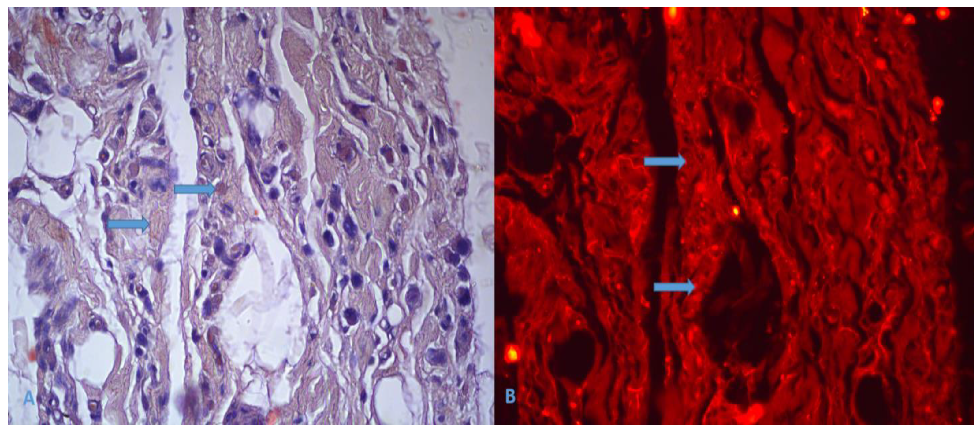
Muscle biopsy of the left triceps surae revealed sarcoplasmic eosinophilic inclusions consistent with protein aggregates. There was variation in muscle fiber diameter with the presence of atrophic fibers. Focal fatty infiltration and mild fibrosis were identified. Immunohistochemically, the inclusions showed positivity for desmin with an abnormal granular cytoplasmic desmin accumulation, consistent with aggregation of intermediate filaments. Congo red staining demonstrated congophilic material, with fluorescence microscopy showing bright red granular inclusions. The morphological and immunohistochemical findings were consistent with a myofibrillar myopathy of the desminopathy type. (Figure 1 and Figure 2).
3. Discussion
We report a novel DES variant, c.1327_1328delinsTT, p.(Lys443Leu), in a patient with clinical, electrophysiological, and histopathological features consistent with desminopathy. The variant is with a very high variant priority score (1.00), high confidence, a genotype– phenotype correlation score of 0.52. and is consistent with the known autosomal dominant inheritance pattern of DES-related disease. Nonetheless, this interpretation should be considered with caution. Multi-nucleotide variants (MNVs; i.e., closely spaced substitutions or indels affecting adjacent nucleotides) [10] are often imperfectly captured in population databases, which may lead to underestimation or uncertainty in their true allele frequency. Such discrepancy could leave the variant as VUS, although one leaning pathogenic. Moreover, no clinical diagnostic laboratories have submitted a clinical-significance assertion for this specific variant to ClinVar to date, and additional segregation or functional data would further strengthen its classification.
Although initially classified as a variant of uncertain significance, the cumulative clinical, histopathological, and computational evidence supports reclassification toward likely pathogenic according to ACMG/AMP variant interpretation guidelines: PM1 (pathogenic moderate)( non-truncating non-synonymous variant is located in a mutational hot spot and/or critical and well-established functional domain); PP2 (pathogenic supporting) (missense variant in a gene with low rate of benign missense mutations and for which missense mutation is a common mechanism of a disease); PM2 (extremely low frequency in gnomAD population databases) and PP4 (patient's phenotype or family history is highly specific for a disease with a single genetic etiology) [9]. The absence of the variant from large population databases is consistent with moderate evidence for pathogenicity (PM2). The patient’s phenotype is highly specific for a disease with a single gene etiology, namely DES- related myofibrillar myopathy, supporting evidence (PP4). Multiple computational tools, as integrated within the Franklin platform, predict a deleterious effect on protein function (PP3). Furthermore, the identified variant is located within the functionally critical C-terminal tail domain of desmin, a region enriched for pathogenic variants and associated with both skeletal and cardiac phenotypes (PM1).
The heterozygous state of the variant is consistent with the established autosomal dominant inheritance pattern of DES-related disease. Although a de novo occurrence is suspected based on negative family history, lack of parental genetic testing precludes application of strong de novo criteria (PS2). Taken together, the available evidence supports classification of the variant as likely pathogenic.
The patient presented with a myopathic syndrome characterized by asymmetric involvement of limb muscles, with possible cardiac involvement suggested by a prior syncopal episode, although objective evidence of cardiomyopathy or conduction abnormalities remains limited. Possible dysphagia was reported based on patient history. At this stage, there was no evidence of facial or axial muscle involvement, nor of respiratory impairment.
This clinical phenotype is consistent with the known effects of DES tail-domain variants, which are associated with both skeletal muscle disease and cardiac manifestations. Although such variants may not significantly impair filament assembly in vitro, they can disrupt higher-order filament network organization, intracellular signaling, and mechanotransduction. These alterations are thought to exert dominant-negative effects, contributing to progressive cellular dysfunction [11]. However, clinical expression remains variable, even among patients with variants affecting the same domain.
Although the findings are predominantly myopathic, electromyography raises the possibility of concomitant peripheral nerve involvement, as described in a subset of patients with desminopathy [12].
Disease onset of desminopathies, clinical and myopathological presentation, and disease progression are variable, influenced by inheritance pattern and variant location. Onset typically occurs around 30 years of age, with earlier presentation observed in recessive cases and those involving isolated cardiac manifestations. The precise incidence and prevalence of desmin myopathy and/or cardiomyopathy remain undefined. Desminopathies have been documented across various ethnic populations and affect both sexes [5,6,7].
The age of onset of the disease in our patient falls within the typical range for desminopathies. Since we do not have objective information about heredity, we can assume that in our case it is a sporadic variant.
Variants within the DES gene result in a heterogeneous spectrum of phenotypes encompassing skeletal myopathy, cardiomyopathy, arrhythmia, and cardiac conduction disease, variably expressed in combination. Distinct clinical phenotypes may arise from variants affecting different functional domains of the DES gene. Symptoms may manifest at disease onset or during progression, occurring in isolation or concurrently. The clinical course is progressive, and a specific treatment for this severely debilitating condition remains unavailable [2,5,6,12].
The patient's cardiomyopathic symptoms may have emerged later in the disease progression, ultimately constituting the classic phenotypic presentation of desminopathy.
The C-terminal non-helical tail domain, spanning residues 416-470, primarily mediates interactions with other cytoskeletal proteins to facilitate the establishment of a cytoplasmic intermediate filament network. Variants within this domain exhibit pleiotropic functional consequences, impacting mechanochemical signaling, intracellular transport, and protein-protein interactions, ultimately leading to diverse cellular dysfunctions and variable clinical phenotypes [7,11].
Variants clustered in the C-terminal tail domain, are strongly associated with skeletal muscle dysfunction and cardiac pathology. The C-terminus non-α-helical tail domain, harboring p.Lys443Leu, adjacent to previously reported variants such as p.Lys449Thr, is essential for maintaining desmin structural integrity. Although some tail variants do not markedly disrupt filament assembly in vitro, they induce severe disease in patients, suggesting interference with muscle cell structural stability, often manifesting as a dominant-negative effect [11].
The DES variant p.Lys443Leu could compromise desmin filament network structure, leading to a progressive, inherited disease characterized by skeletal muscle weakness and cardiac manifestations, including cardiomyopathy and/or conduction blocks.
In conclusion, we report a novel de novo heterozygous DES variant, c.1327_1328delinsTT, p.(Lys443Leu), initially classified as a variant of uncertain significance. However, the characteristic clinical phenotype, supportive electromyographic findings, and histopathological features consistent with desminopathy, together with possible cardiac involvement, collectively suggest that this variant is likely pathogenic. This case expands the mutational spectrum of DES, particularly within the functionally critical tail domain, and highlights the importance of a multidisciplinary approach integrating clinical, electrophysiological, pathological, and genetic data in the interpretation of rare variants.
Funding
The research did not receive funding.
Institutional Review Board Statement
This study was conducted in accordance with the Declaration of Helsinki.
Informed Consent Statement
Written informed consent has been obtained from the patient.
References
- Sun N, Wang C, Yang S. Severe conduction block and cardiomyopathy associated with desminopathy. Cardiology in the Young. 2025;35(4):850-852. [CrossRef]
- Yeow D, Katz M, Henderson R, Prasad S, Denman R, Blum S, Davis M, Robertson T and McCombe P (2023) Phenotypic variability within the desminopathies: A case series of three patients. Front. Neurol. 13:1110934. [CrossRef]
- Bär H, Strelkov SV, Sjöberg G, Aebi U, Herrmann H. The biology of desmin filaments: how do mutations affect their structure, assembly, and organisation? J Struct Biol. 2004 Nov;148(2):137-52. PMID: 15477095 . [CrossRef]
- Xiao H, Song L, Tao L. A case report of adolescent myofibrillar myopathy due to a de novo R406W pathogenic variant in desmin with symptoms of "hypertrophic cardiomyopathy". Heliyon. 2024 Jan 21;10(3):e25009. PMID: 38314304; PMCID: PMC10837553 . [CrossRef]
- Clemen CS, Herrmann H, Strelkov SV, Schröder R. Desminopathies: pathology and mechanisms. Acta Neuropathol. 2013 Jan;125(1):47-75. Epub 2012 Nov 11. PMID: 23143191; PMCID: PMC3535371 . [CrossRef]
- Alonso-Pérez J, Barrachina-Esteve O, González-Quereda L, Viguera-Martinez ML, Luján-Torné M, Guitart-Feliubadaló M, Miguel Martínez J, Carbayo Á, Gallano P, Díaz-Manera J, Olivé M, Rojas-Garcia R. Novel DES variant presenting with isolated restrictive respiratory failure. Expanding the clinical spectrum. Neurologia (Engl Ed). 2025 Jun;40(5):433-441. PMID: 40523712 . [CrossRef]
- Maddison P, Damian MS, Sewry C, McGorrian C, Winer JB, Odgerel Z, Shatunov A, Lee HS, Goldfarb LG. Clinical and myopathological characteristics of desminopathy caused by a variant in desmin tail domain. Eur Neurol. 2012;68(5):279-86. Epub 2012 Oct 5. PMID: 23051780; PMCID: PMC3551980 . [CrossRef]
- Genoox. Franklin by Genoox. Available at: https://franklin.genoox.com, Accessed January, 2026.
- Richards S, Aziz N, Bale S, Bick D, Das S, Gastier-Foster J, Grody WW, Hegde M, Lyon E, Spector E, Voelkerding K, Rehm HL; ACMG Laboratory Quality Assurance Committee. Standards and guidelines for the interpretation of sequence variants: a joint consensus recommendation of the American College of Medical Genetics and Genomics and the Association for Molecular Pathology. Genet Med. 2015 May;17(5):405-24. Epub 2015 Mar 5. PMID: 25741868; PMCID: PMC4544753 . [CrossRef]
- Wang Q, Pierce-Hoffman E, Cummings BB, Alföldi J, Francioli LC, Gauthier LD, Hill AJ, O'Donnell-Luria AH; Genome Aggregation Database Production Team; Genome Aggregation Database Consortium; Karczewski KJ, MacArthur DG. Landscape of multi-nucleotide variants in 125,748 human exomes and 15,708 genomes. Nat Commun. 2020 May 27;11(1):2539. Erratum in: Nat Commun. 2021 Feb 2;12(1):827. doi: 10.1038/s41467-021-21077-8. PMID: 32461613; PMCID: PMC7253413 . [CrossRef]
- Bär H, Goudeau B, Wälde S, Casteras-Simon M, Mücke N, Shatunov A, Goldberg YP, Clarke C, Holton JL, Eymard B, Katus HA. Conspicuous involvement of desmin tail mutations in diverse cardiac and skeletal myopathies. Human mutation. 2007 Apr;28(4):374-86. PMID: 17221859 . [CrossRef]
- Geng L, Wang M, Wang K, Xu L, Li J, Liu F, Lu J. Desmin-related myopathy manifested by various types of arrhythmias: a case report and literature review. J Int Med Res. 2024 Nov;52(11):3000605241291741. PMID: 39501717; PMCID: PMC11539263 . [CrossRef]
Figure 1.
Hematoxylin-eosin staining and immunohistochemical analysis. (A) –Skeletal muscle fibers with atrophic changes and finely granular eosinophilic cytoplasmic inclusions, lipomatosis and fibrosis; x400, H&E. (B) – Finely granular, heterogeneous cytoplasmic expression of desmin; x400.
Figure 1.
Hematoxylin-eosin staining and immunohistochemical analysis. (A) –Skeletal muscle fibers with atrophic changes and finely granular eosinophilic cytoplasmic inclusions, lipomatosis and fibrosis; x400, H&E. (B) – Finely granular, heterogeneous cytoplasmic expression of desmin; x400.
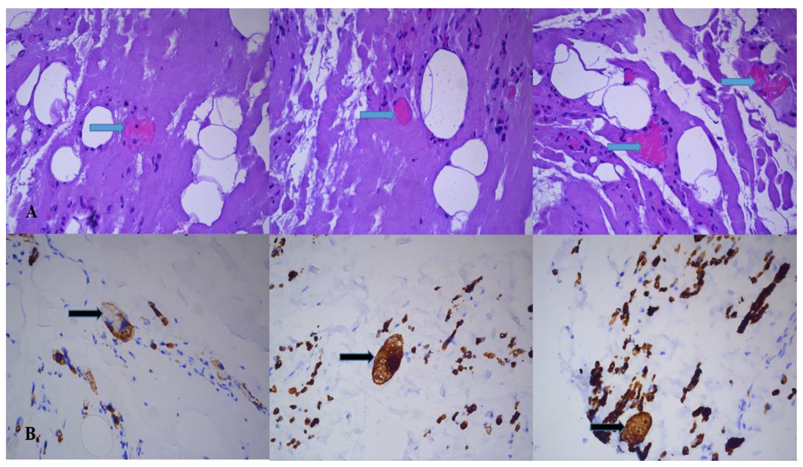
Figure 2.
Congo red staining. (A) – Finely granular cytoplasmic inclusions showing weak congophilia; x400. (В) – Congo red staining viewed with fluorescence microscope using a 545-580 nm excitation filter, showing bright red, finely granular cytoplasmic inclusions; x400.
Figure 2.
Congo red staining. (A) – Finely granular cytoplasmic inclusions showing weak congophilia; x400. (В) – Congo red staining viewed with fluorescence microscope using a 545-580 nm excitation filter, showing bright red, finely granular cytoplasmic inclusions; x400.
Disclaimer/Publisher’s Note: The statements, opinions and data contained in all publications are solely those of the individual author(s) and contributor(s) and not of MDPI and/or the editor(s). MDPI and/or the editor(s) disclaim responsibility for any injury to people or property resulting from any ideas, methods, instructions or products referred to in the content. |
© 2026 by the authors. Licensee MDPI, Basel, Switzerland. This article is an open access article distributed under the terms and conditions of the Creative Commons Attribution (CC BY) license (http://creativecommons.org/licenses/by/4.0/).
Copyright: This open access article is published under a Creative Commons CC BY 4.0 license, which permit the free download, distribution, and reuse, provided that the author and preprint are cited in any reuse.


