Preprint
Article

This version is not peer-reviewed.

Investigation of the Effect of Local Melatonin Application on Biomechanic Bone-İmplant Connection in Titanium İmplants with Different Surface Placed in Rat Tibias

Submitted:

23 June 2026

Posted:

24 June 2026

You are already at the latest version

Abstract

Background and Objectives: The aim of this study was to investigate the effects of local melatonin application on osseointegration of machined (MAC), resorbable blast material (RBM), and sandblasted and acid-etched (SLA) surface implants placed in rat tibiae, utilizing the reverse torque analysis. Materials and Methods: A total of 72 female Sprague-Dawley rats, weighing between 270 and 300 g, were included in the study. The rats were divided into six groups, and samples in which the implants were not properly placed were excluded from the study: control groups consisting of MAC-CNT (n = 12), RBM-CNT (n = 11), and SLA-CNT (n = 12), and local melatonin (MLT)-treated groups consisting of MAC-MLT (n = 12), RBM-MLT (n = 10), and SLA-MLT (n = 10). The implants were surgically placed into the tibiae of the rats under general anesthesia. Following a four-week experimental period, the biomechanical bone–implant connection level was evaluated using reverse torque analysis. Results: The lowest mean biomechanical bone–implant connection value was observed in the MAC-CNT group, whereas the highest value was recorded in the SLA-MLT group. Compared with the MAC-CNT group, all other groups demonstrated statistically significantly higher biomechanical connection values (p < 0.05). The SLA-MLT group showed significantly higher osseointegration levels than both the MAC-MLT and RBM-MLT groups (p < 0.05). In addition, the RBM-MLT group demonstrated significantly higher values compared with the MAC-MLT group (p < 0.05). Both MAC-MLT and SLA-MLT groups exhibited statistically significantly higher biomechanical bone-implant connection values compared to control groups (p < 0.05). Conclusions: Local melatonin application positively affected osseointegration in SLA and MAC surfaced implants. In contrast, local melatonin application did not provide any additional contribution to osseointegration in RBM surfaced implants.

Keywords: 
;  ;  ;  ;  

1. Introduction

In contemporary dentistry, dental implants represent a highly predictable and widely utilized therapeutic approach for restoring missing dentition. The clinical predictability of these fixtures heavily relies on osseointegration, which is characterized as the direct structural and functional anchorage achieved between viable bone tissue and the implant’s biomaterial surface [1]. Successful osseointegration is achieved through the biological harmony between bone resorption mediated by osteoclastic activity and bone formation mediated by osteoblastic activity, a process referred to as bone remodeling [2]. Any factor affecting this dynamic process of bone healing and remodeling may lead to complications in the bone surrounding dental implants and even result in implant loss [3].
Parameters such as the surface topography of dental implants (micro- and nanoscale roughness), chemical composition, surface energy, and hydrophilic properties play a decisive role in successful osseointegration and the long-term stability of implants. Therefore, various surface modification methods have been developed to improve the biological response of implant surfaces. These methods include TiUnite anodization, resorbable blast media (RBM) blasting, acid etching, sandblasting and acid etching (SLA), NanoTite anodization, plasma spraying/titanium plasma spraying, laser ablation, and biological coatings such as hydroxyapatite coatings [4]. In addition, to enhance the osseointegration success of dental implants, growth factors and hormones such as estradiol, parathyroid hormone, and melatonin—which can directly influence bone healing—and anti-osteoclastic drugs are applied locally or systemically to the implant surface [5,6]. Recent studies have focused on the effects of these different biological agents on the osseointegration success of implants with varying surface characteristics [7,8,9].
Melatonin is a sleep hormone primarily produced by the pineal gland and plays a key role in regulating the circadian rhythm. In addition to its anti-inflammatory and antioxidant properties, melatonin exerts significant effects on bone metabolism. It suppresses osteoclastic activity by promoting the formation of type I collagen fibers [10]. Furthermore, it contributes to bone formation by stimulating the differentiation and proliferation of mesenchymal stem cells into osteoblasts [2,10]. Melatonin has also been shown to accelerate angiogenesis, the process of new blood vessel formation. This effect suggests that melatonin may provide additional support to the bone healing process, which is closely associated with vascularization [11]. Considering its effects on inflammatory responses and bone metabolism, melatonin has emerged as a promising molecule for implant applications. Numerous studies have demonstrated that local melatonin application promotes bone healing and positively affects the osseointegration of implants [12,13,14].
According to the literature, there is no study investigating the effect of melatonin, which is known to have beneficial effects on bone tissue, on the osseointegration of implants with different surface characteristics. Therefore, this experimental study aims to biomechanically evaluate the effect of local melatonin administration on bone–implant connection in titanium implants with different surface properties placed in rat tibiae, using a reverse torque analysis.

2. Materials and Methods

Ethical approval for the all experimental procedures and and surgical protocols was officially granted by the Animal Experiments Local Ethics Committee of Fırat University (Approval No. 13–16, August 6, 2025). Every experimental phase of this study was conducted at the Experimental Research Center of Fırat University (Elazığ, Türkiye). Throughout the study period, all procedures involving animals were performed in strict accordance with the ARRIVE guidelines and the relevant regulations governing the use of experimental animals in research. All rats utilized in the investigation were sourced from the identical breeding institution, namely the Experimental Research Center of Fırat University (Elazığ, Türkiye).

2.1. Implant Surface Characterization

A total of 60 sterile endosseous titanium implants manufactured from Ti-6Al-4V alloy (Implance Dental Implant Systems, AGS Medical, Istanbul, Türkiye), with a diameter of 2.5 mm and a length of 4 mm, were used in the study. Three different implant surface topographies were evaluated: MAC, SLA, and RBM surfaces. The average roughness (Ra) values of the implants were the same (Ra: 1–2 μm).

2.2. Animals and Study Design

A total of 72 female Sprague–Dawley rats, with an age range of 6 to 12 months, were utilized to perform this experimental investigation. At baseline, the animals had body weights ranging from 270 to 300 g. The rats were housed in specially designed cages under controlled environmental conditions, including a temperature of 22 ± 2°C and relative humidity of 55%, with a 12-h light/12-h dark cycle. Standard laboratory chow and water were provided ad libitum throughout the experimental period. To determine the appropriate animal cohort size before initiating the study, a sample size estimation was conducted through G*Power software (version 3.1.9.4; Franz Faul, Kiel University, Kiel, Germany). The power analysis indicated that to achieve a statistical power of 90% with an effect size of 0.5 and a significance threshold (α) set at 0.05 across the six experimental cohorts, a minimum sample size of eight rats per group was essential [15]. Although power analysis indicated that at least eight rats per group were required, twelve rats were included in each group to compensate for the risk of rat mortality and to increase the reliability of the findings. To compensate for potential mortality risks during the surgical interventions and the subsequent experimental follow-up, each group initially comprised 12 rats. To ensure hormonal standardization, vaginal smear examinations were performed, and all animals were confirmed to be in the same stage of the estrous cycle. Seventy-two female rats were randomly assigned to six equal groups using a computer-generated randomization protocol. Animals in which proper implant placement could not be achieved were excluded from the study.
  • Machined Surface Control Group (MAC-CNT) (n = 12): Machined-surface titanium implants were inserted into the right tibiae of the animals. Following a 4-week healing interval, the animals were euthanized for subsequent biomechanical evaluation.
  • RBM Surface Control Group (RBM-CNT) (n = 12): RBM-surface titanium implants were inserted into the right tibiae of the animals. Following a 4-week healing interval, the animals were euthanized for subsequent biomechanical evaluation.
  • SLA Surface Control Group (SLA-CNT) (n = 12): SLA-surface titanium implants were inserted into the right tibiae of the animals. Following a 4-week healing interval, the animals were euthanized for subsequent biomechanical evaluation.
  • Machined Surface Local Melatonin Group (MAC-MLT) (n = 12): Following local administration of 3 mg melatonin into the implant osteotomy site to the maximum capacity of the prepared implant bed, machined-surface titanium implants were inserted into the right tibiae of the animals. Following a 4-week healing interval, the animals were euthanized for subsequent biomechanical evaluation [16].
  • RBM Surface Local Melatonin Group (RBM-MLT) (n = 12): Following local administration of 3 mg melatonin into the implant osteotomy site to the maximum capacity of the prepared implant bed, RBM-surface titanium implants were inserted into the right tibiae of the animals. Following a 4-week healing interval, the animals were euthanized for subsequent biomechanical evaluation [16].
  • SLA Surface Local Melatonin Group (SLA-MLT) (n = 12): Following local administration of 3 mg melatonin into the implant osteotomy site to the maximum capacity of the prepared implant bed, SLA-surface titanium implants were inserted into the right tibiae of the animals. Following a 4-week healing interval, the animals were euthanized for subsequent biomechanical evaluation [16].

2.3. Surgical Procedures

All surgical procedures were performed under general anesthesia under sterile conditions. General anesthesia was induced by intraperitoneal administration of xylazine (5 mg/kg) and ketamine (50 mg/kg) using an appropriate syringe. The surgical site was shaved and disinfected with povidone-iodine solution. To access the metaphyseal region of the right tibia for implant placement, a 2-cm incision was created above the tibial crest using a No. 15 scalpel. During the preparation of the implant osteotomy sites, sterile saline solution was continuously used for irrigation to prevent thermal bone injury. Titanium implants (Ti-6Al-4V; Implant Dental Implant Systems, AGS Medical, Istanbul, Türkiye), measuring 4 mm in length and 2.5 mm in diameter, were inserted into the cortico-cancellous bone of the right tibiae of the rats. The soft tissues were closed using 4-0 resorbable sutures (polyglactin) (Figure 1). Following surgery, antibiotic (penicillin, 50 mg/kg) and analgesic (tramadol hydrochloride, 0.1 mg/kg) treatments were administered intramuscularly for 3 consecutive days to prevent postoperative infection and to manage pain.

2.4. Biomechanical Analysis

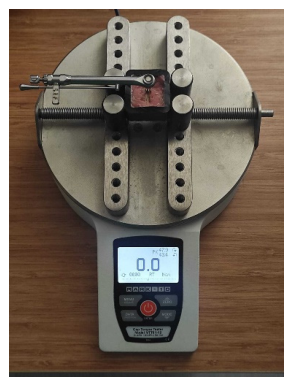
Upon completion of the 4-week post-operative healing phase, euthanasia was performed on the animals. Subsequently, the titanium fixtures, along with the adjacent osseous structures, were meticulously harvested and immersed in a formaldehyde solution for preservation. The degree of osseointegration was evaluated using a reverse torque analysis performed with a biomechanical testing device (Mark-10, NY, USA) by a blinded examiner who was unaware of the group allocations (M.T.). The force required to initiate the first rotation of the implant within the socket was recorded as the biomechanical osseointegration value. A hexagonal driver was engaged into the internal hex connection of the implant, and force was applied using a ratchet system. Care was taken to maintain a 90° angle between the ratchet and the hex driver during torque application. All measurements were recorded in Newton/centimeters (N/cm) (Figure 2).

2.5. Statistical Analysis

The IBM SPSS Statistics Statistics software (v. 22.0; IBM Corp., Armonk, NY, USA) was employed to process all data. While data normality was verified through the Shapiro–Wilk normality test, variance homogeneity was confirmed using Levene’s methodology. One-way analysis of variance (ANOVA) was implemented to execute statistical comparisons among multiple independent groups, provided that the data adhered to a normal distribution. When the assumption of homogeneity of variances was met, post hoc multiple comparisons were performed using Tukey’s honestly significant difference (HSD) test. In cases where variance homogeneity was violated, the Games–Howell post hoc test was applied. Pairwise comparisons between groups with similar surface characteristics were performed using the Student’s t-test. A p-value of less than 0.05 was considered statistically significant.

3. Results

Throughout the experimental period, no mortality, postoperative infection, or wound dehiscence was observed among the animals. Due to improper implant placement, one specimen from the RBM-CNT group and two specimens each from the RBM-MLT and SLA-MLT groups were excluded from the final analysis.
The biomechanical bone–implant interface strength values (N/cm) of the study groups are presented in Table 1. The lowest mean value was observed in the MAC-CNT group (3.31 ± 0.69), whereas the highest value was recorded in the SLA-MLT group (7.50 ± 2.31). A statistically significant difference was detected among the groups (p < 0.05). Compared with the MAC-CNT group, all other groups exhibited significantly higher biomechanical bone–implant interface strength values (p < 0.05). Furthermore, the SLA-MLT group demonstrated significantly higher biomechanical bone–implant interface strength values not only compared with the MAC-CNT group but also with the MAC-MLT group (p < 0.05).
The within-group comparisons of biomechanical bone–implant interface strength values according to implant surface characteristics are presented in Table 2. Among the control groups, a statistically significant difference was observed between the surface characteristics (p < 0.001). Both the RBM-CNT (5.69 ± 1.12) and SLA-CNT (5.55 ± 1.07) groups exhibited significantly higher biomechanical bone–implant interface strength values than the MAC-CNT group (3.31 ± 0.69) (p < 0.05). Similarly, a statistically significant difference was found among the melatonin-treated groups according to surface characteristics (p < 0.001). The SLA-MLT group (7.50 ± 2.31) demonstrated the highest biomechanical bone–implant interface strength values and exhibited significantly higher values than both the MAC-MLT (4.58 ± 0.71) and RBM-MLT (5.12 ± 0.52) groups (p < 0.05). In addition, the RBM-MLT group showed significantly higher biomechanical bone–implant interface strength values compared with the MAC-MLT group (p < 0.05).
Pairwise comparisons of groups according to implant surface characteristics are presented in Table 3. For machined surfaces, the melatonin-treated group (4.58 ± 0.71) exhibited significantly higher biomechanical bone–implant interface strength values than the control group (3.31 ± 0.69) (p < 0.001). For RBM surfaces, although the melatonin-treated group (5.12 ± 0.52) demonstrated slightly lower mean values than the control group (5.69 ± 1.12), this difference did not reach statistical significance (p = 0.150). In contrast, for SLA surfaces, the melatonin-treated group (7.50 ± 2.31) exhibited significantly higher biomechanical bone–implant interface strength values compared with the control group (5.55 ± 1.07) (p = 0.030).

4. Discussion

Although numerous studies have investigated the effects of melatonin on the osseointegration of dental implants [13,17,18], no study has, to the best of our knowledge, evaluated this effect in the context of implants with different surface characteristics. Therefore, the present study aimed to investigate the effect of locally applied melatonin on bone–implant integration around titanium implants with three distinct surface characteristics (MAC, RBM, and SLA) placed in rat tibiae. The findings demonstrated that differences in implant surface modifications influenced the degree of osseointegration achieved. Furthermore, local melatonin application was found to enhance osseointegration in implants with both MAC and SLA surface characteristics.
In a study investigating the influence of implant surface roughness on osseointegration, Lagdive et al. reported that moderately rough surfaces (Ra = 1.5 μm) provide the most favorable conditions for osteoblast activity, cell proliferation, and bone mineralization (19). Similarly, another study demonstrated that excessively rough surfaces may promote bacterial contamination and, consequently, do not outperform moderately rough surfaces in terms of bone–implant integration [20]. Therefore, to enhance the reliability of the findings, implants with a moderate surface roughness (Ra = 1–2 μm), which has been associated with superior osseointegration outcomes, were selected for use in the present study.
Dündar et al. investigated the effects of different locally administered melatonin doses on osseointegration during implant surgery and reported that the application of 3 mg melatonin resulted in greater bone–implant contact than the 1.2 mg dose [16]. Consistent with these findings, another study using 3 mg of locally applied melatonin demonstrated a significant increase in new bone formation around implants in the melatonin-treated group [21]. In contrast, a study evaluating the effects of 1.2 mg local melatonin reported comparable bone–implant contact and bone density values between the melatonin and control groups at 5 and 8 weeks following osseointegration [22]. Based on these findings, a 3 mg dose of locally administered melatonin, which has been associated with superior osseointegration outcomes, was selected in the present study to maximize the potential biological effects of melatonin on bone healing [16]. The results demonstrated that local melatonin application enhanced osseointegration, particularly in implants with SLA and MAC surface characteristics, which is in agreement with previous experimental studies reporting the osteogenic and osseointegration-promoting effects of melatonin.
Bingül et al. evaluated the osseointegration of implants with different surface characteristics placed in 60 rat tibiae using the reverse torque analysis and reported that the lowest osseointegration levels were observed in MAC surfaces, followed by SLA and RBM surfaces, respectively [15]. Similarly, recent studies comparing SLA and RBM surfaces with MAC surfaces have reported that these surface characteristics exhibit superior outcomes in terms of osteoblastic activity and bone–implant contact. In addition, RBM surfaces have been reported to provide higher levels of bone–implant contact compared with SLA surfaces [23,24]. However, Özcan et al., in a study evaluating osseointegration in the presence of concomitant allogeneic bone grafting, reported that SLA surfaces demonstrated higher biomechanical osseointegration levels compared with RBM and MAC surfaces [25]. These discrepancies suggest that the influence of implant surface characteristics on osseointegration remains controversial, highlighting the need for further well-designed studies. In the present study, osseointegration of implants with different surface characteristics was also evaluated using the reverse torque analysis. The findings of the present study demonstrated that, among the control groups, the lowest level of osseointegration was observed in MAC surfaces, followed by SLA and RBM surfaces, respectively. In addition, the significantly higher bone–implant interface strength values observed in both the RBM-CNT and SLA-CNT groups compared with the MAC-CNT group are consistent with previous findings reported in the literature.
Brignardello-Petersen et al. evaluated the effects of adding melatonin to autogenous bone grafts in a study involving 26 patients and 52 immediately placed implants, and reported that the melatonin-treated group exhibited lower marginal bone resorption, probing depth, and gingival index values [26]. In another clinical study, Chaves Cavalcante Kischinhevsky et al. investigated the effects of a melatonin gel (1.2 mg) incorporated into a xenogeneic collagen sponge applied to extraction sockets in 20 patients following premolar or molar tooth extraction. They reported that melatonin enhanced osteoblastic activity accompanied by a higher number of osteocytes, accelerated bone healing, and increased bone density [27]. Wu et al. investigated the effects of melatonin on peri-implantitis and reported that melatonin delays disease progression and reduces its incidence by decreasing pro-inflammatory cytokine levels, promoting angiogenesis, inhibiting osteoclast formation, and stimulating osteogenesis [28]. Tanrısever et al., in a study evaluating the effects of different doses of melatonin (1.2 and 3 mg) on the healing of bone defects created in rat tibiae, demonstrated that melatonin accelerated bone healing in a dose-independent manner [29]. In the present study, significantly higher biomechanical bone–implant interface strength values were observed in all melatonin-treated groups compared with their respective controls, except for the RBM surface group, which is consistent with the current literature. The absence of a statistically significant difference between the melatonin and control groups in RBM surface implants suggests that the effect of locally applied melatonin may vary depending on implant surface characteristics and may be more limited in RBM-treated surfaces.
El-Gammal et al. conducted a 12-month clinical and radiological follow-up study on immediately loaded SLA-surfaced dental implants in 14 patients receiving local melatonin application and reported that melatonin was associated with good implant stability and minimal marginal bone resorption [30]. Similarly, Salomó-Coll et al. investigated the effects of locally applied melatonin and vitamin D on 36 SLA-surfaced immediately placed implants and reported that melatonin enhanced peri-implant bone formation following a 12-week osseointegration period [31]. In the present study, the SLA-MLT group demonstrated the highest biomechanical bone–implant interface strength values, which were significantly higher than those observed in both the RBM-MLT and MAC-MLT groups, in agreement with the findings reported in the literature.
Bingül et al. investigated the effects of locally applied zoledronic acid, a pharmacological agent known to influence bone metabolism, on the osseointegration of implants with different surface characteristics, and reported that in the zoledronic acid–treated group, SLA and RBM surfaced implants exhibited higher biomechanical bone–implant interface strength values compared with MAC surfaced implants [15]. In the present study, the effects of locally applied melatonin, which is also known to exert beneficial effects on bone metabolism, on osseointegration across different implant surface characteristics were evaluated. The findings demonstrated that the SLA-MLT group exhibited significantly higher biomechanical bone–implant interface strength values compared with both the RBM-MLT and MAC-MLT groups, while the RBM-MLT group also showed significantly higher values than the MAC-MLT group. These results are consistent with the findings reported by Bingül et al., suggesting a similar pattern of surface-dependent enhancement of osseointegration in response to locally applied bioactive agents.
Park et al. evaluated the effects of laser ablation applied to RBM-surfaced implants on bone–implant contact and reported no statistically significant differences in total or cortical bone–implant contact between the laser-ablated and control groups [32]. Similarly, Conserva et al. investigated FasL expression that promotes osseointegration and the preservation of bone mass in mesenchymal stromal cell–cultured environments using different implant surfaces and reported a significant increase in FasL expression only on MAC surfaces compared with RBM and Ca²⁺ nanostructured surfaces. The authors attributed this finding to the fact that the microporous topography of RBM and Ca²⁺ nanostructured surfaces already optimizes early cell adhesion, fibrin retention, and osteoblastic differentiation [33]. In the present study, the effects of locally applied melatonin on bone–implant integration across different implant surface characteristics were evaluated. The findings suggested that melatonin did not provide an additional beneficial effect on RBM-surfaced implants, which may be explained by the fact that RBM surfaces already offer a highly optimized microtopography for osteoblast adhesion and new bone formation, resulting in a high baseline level of osseointegration potential. This condition may have masked the potential incremental biological effects of melatonin through a so-called “ceiling effect” mechanism.
Lee et al. evaluated the effects of implants coated with a nano/micro-combined hydroxyapatite (HA) structure using a laser-induced one-step coating technique and reported that the surface roughness ranking was HA > RBM > SLA > MAC, whereas the surface wettability ranking was RBM > HA > MAC > SLA (24). In a study by Sun et al., it was demonstrated that as implant material hydrophilicity increases, both cell adhesion and bone-binding capacity also increase [34]. In the present study, although the highest numerical reverse torque values were observed in the RBM surface group, no additional beneficial effect of local melatonin application was detected. This finding may be explained, in agreement with the literature, by the inherently high osseointegration capacity of RBM surfaces, which is attributed to their favorable surface roughness and superior wettability. Accordingly, these advantageous properties that optimize osseointegration in RBM surfaces may have masked the incremental biological effects of melatonin on osseointegration.
In the present study, the effects of locally applied melatonin on osseointegration of titanium implants with different surface characteristics were evaluated using a rat tibia model. The lack of clinical-level assessments and the absence of systemic melatonin application represent the main limitations of this study. In addition, osseointegration was assessed using reverse torque analysis. Another limitation of the present study is the absence of histomorphometric, micro-CT, and immunohistochemical analyses.

5. Conclusions

Within the limitations of the present study, local melatonin application during implant surgery appears to positively influence osseointegration in SLA- and MAC-surfaced implants. This effect may be associated with the anti-inflammatory and antioxidant properties of melatonin, as well as its ability to enhance angiogenesis, suppress osteoclastic activity, and promote osteoblastic differentiation, thereby increasing bone density and consequently improving implant stability. In contrast, local melatonin application did not provide an additional benefit for osseointegration in RBM-surfaced implants. Nevertheless, further well-designed long-term experimental and clinical studies with larger sample sizes, different dosages, and alternative administration routes (local or systemic) are required to more clearly elucidate the effects of melatonin on osseointegration across different implant surface characteristics.

Author Contributions

Conceptualization, methodology, project administration, K.S. and E.C.O.; Supervision and funding acquisition, U.K.C.; Literature review, investigation, data analysis, and writing—original draft preparation, K.S.; Surgical procedures, animal care, S.D.; Data acquisition, M.T.; Data analysis and study finalization, K.S. and O.I.. All authors have read and agreed to the published version of the manuscript.

Funding

This research received no external funding.

Institutional Review Board Statement

This study was approved by the Animal Experiments Local Ethics Committee of Fırat University (Approval No. 13–16, August 6, 2025). It was carried out at the Experimental Research Center of Fırat University, and the Helsinki Declaration rules were strictly followed during the experiments. The female Sprague–Dawley rats used in the experiments were obtained from the Experimental Research Center of Fırat University. In these studies, experiments were carried out without causing suffering to animals, and all ethical rules were respected.

Data Availability Statement

The raw data supporting the conclusions of this article will be made available by the authors on request.

Acknowledgments

The authors wish to thank Implance Dental Implant System, AGS Medical Corporation, Istanbul, Turkey, for providing the titanium implants.

Conflicts of Interest

The authors declare no conflicts of interest.

Abbreviations

MAC Machined
RBM Resorbable blast material
SLA Sandblasted and acid-etched
CNT
MLT
HA
Control
Melatonin
Hydroxyapatite

References

  1. Alameeri Z.A.; Jawad H.A. Comparative analysis of osteointegration in photostimulated dental implants: 650-976 nm diode lasers versus growth factors. J. Med. Life. 2024, 17, 769-775.
  2. Abu Alfaraj T.; Al-Madani S.; Alqahtani N.S.; Almohammadi A.A.; Alqahtani A.M.; AlQabbani H.S.; Bajunaid M.K.; Alharthy B.A.; Aljalfan N. Optimizing osseointegration in dental implantology: a cross-disciplinary review of current and emerging strategies. Cureus. 2023, 15, 47943.
  3. Liu Y.; Shen Z.; Xu Y.; Zhu Y.W.; Chen W.; Qiu J. Layer-by-layer self-assembly of PLL/CPP-ACP multilayer on SLA titanium surface: Enhancing osseointegration and antibacterial activity in vitro and in vivo. Colloids Surf. Bç Biointerfaces. 2024, 240, 113966.
  4. Sarvaiya B.B.; Kumar S.; Pathan M.S.H.; Patel S.; Gupta V.; Haque M. The impact of implant surface modifications on the osseointegration process: an overview. Cureus. 2025, 17, 81576.
  5. Jafariandehkordi A.; Jafariandehkordi Z. A biomechanical assessment of dental implant stability under the effect of changes in bone remodeling factors and implant elastic modulus. Comput. Biol. Med. 2025, 192, 110318.
  6. Talo Yildirim T.; Dündar S.; Bozoğlan A.; Karaman T.; Tekin S.; Kahraman O.E. Evaluation of the effects of ß-adrenergic receptor-propranolol on osseointegration of the titanium implants. J. Craniofac. Surg. 2021, 32, 783-786.
  7. Sghaireen M.G.; Alam M.K.; Mohamedeissa A.A.S.; Kazma J.M. In-vitro study of osseointegration: evaluating the influence of surface modifications on dental implant stability. J. Pharm Bioallied. Sci. 2024, 16, 2518-2520.
  8. Wang Y.Y.; Gong P.; Zhang J. Effects of different implant surface properties on the biological behavior of Schwann cells. Hua Xi Kou Qiang Yi Xue Za Zhi. 2021, 39, 279-285.
  9. Gadzo N.; Ioannidis A.; Naenni N.; Hüsler J.; Jung R.E.; Thoma D.S. Survival and complication rates of two dental implant systems supporting fixed restorations: 10-year data of a randomized controlled clinical study. Clin. Oral Investig. 2023, 27, 7327-7336.
  10. Ahmadpour E.; Azizi B.; Behradfar M.; Javadi S.; Jabbari Hagh E.; Ahmadpour F. Local delivery of melatonin along with bone grafts to improve osteogenesis. J. Craniofac. Surg. 2025, 36, 1430-1436.
  11. Tanrisever M.; Tekin B.; Can U.K.; Istek O.; Ozcan E.C.; Ozercan I.H.; Gelic T.; Dundar S. The effect of local melatonin application on bone fracture healing in rat tibias. Medicina (Kaunas). 2025, 61, 146.
  12. Cutando A.; Montero J.; Gómez-de Diego R.; Ferrera M.J.; Lopez-Valverde A. Effect of topical application of melatonin on serum levels of C-reactive protein (CRP), interleukin-6 (IL-6) and tumor necrosis factor-alpha (TNF-α) in patients with type 1 or type 2 diabetes and periodontal disease. J. Clin. Exp. Dent. 2015, 7, 628-633.
  13. Ravi Kiran S., Bammidi N., Kumar A.K.; Kumar P.S.; Karnam Y. Evaluation of the effect of topical melatonin application on immediately placed dental implants using cone beam computed tomography (CBCT). Cureus. 2022, 14, 25233.
  14. Montarele L.F.; Pitol D.L.; Pereira B.F.; Feldman S.; Fazan V.P.S.; Issa J.P.M. Histological and immunohistochemical analysis of the effects of topical melatonin treatment associated with collagen sponge and rhBMP-2 protein on bone remodeling. Biomolecules. 2022, 12, 1738.
  15. Bingul M.B.; Gul M.; Dundar S.; Bozoglan A.; Kirtay M.; Ozupek M.F.; Ozcan E.C.; Habek O.; Tasdemir I. Effects of the application local zoledronic acid on different dental implants in rats on osseointegration. Drug. Des. Devel Ther. 2024, 18, 2249-2256.
  16. Dundar S.; Yaman F.; Saybak A.; Ozupek M.F.; Toy V.E.; Gul M.; Ozercan I.H. Evaluation of effects of topical melatonin application on osseointegration of dental implant: an experimental study. J. Oral Implantol. 2016, 42, 386-389.
  17. Petraki G.; Boutiou S.S.; Athanasiou A.; Velivasakis A.S.; Kafas P.; Al Khawalde M.; Lillis T.; Dabarakis N. Melatonin as a modulator of osseointegration: mechanisms, evidence and clinical potential in implant dentistry. Pak. J. Biol. Sci. 2026, 29, 102-109.
  18. Wang Z.; Chen T.; Wu Z.; Jiang X.; Hou Q.; Miao S.; Xia R.; Wang L. The dual-effects of PLGA@MT electrospun nanofiber coatings on promoting osteogenesis at the titanium-bone interface under diabetic conditions. J. Mater. Chem. B. 2022, 10, 4020-4030.
  19. Lagdive S.S,; Vasani A.; Madhad V.; Tulsi S.P.S.; Panda A.; Gupta S.P.; Patadiya H.H.; Lunkad H. In vitro analysis of bone response to different implant surface roughness. J. Pharm Bioallied. Sci. 2025, 17, 1104-1106.
  20. Wennerberg A.; Albrektsson T. Effects of titanium surface topography on bone integration: a systematic review. Clin. Oral Implants Res. 2009, 20, 172-184.
  21. Tresguerres I.F.; Clemente C.; Blanco L.; Khraisat A.; Tamimi F.; Tresguerres J.A. Effects of local melatonin application on implant osseointegration. Clin. Implant Dent. Relat. Res. 2012, 14, 395-399.
  22. Muñoz F.; López-Peña M.; Miño N.; Gómez-Moreno G.; Guardia J.; Cutando A. Topical application of melatonin and growth hormone accelerates bone healing around dental implants in dogs. Clin. Implant Dent. Relat. Res. 2012, 14, 226-235.
  23. KS. K.; Sreevidya B.; TK. R.; Bm. D., Dedeepya N.R.; Kamal Badiyani B.; Kumar A. Comparative analysis of surface modification techniques for assessing oral implant osseointegration: an animal study. Cureus. 2024, 16, 54014.
  24. Lee J.T.; Kim S.; Lee S.H.; Han D.W.; Lee K.; Lee S.; Kwon D.; Lee K.; Jeon H.; Jung T.G.; Kim B. Evaluation of dental implant with hydroxyapatite coating by laser-induced hydrothermal synthesis: in vitro and in vivo experimental study. Discov. Nano. 2025, 20, 134.
  25. Özcan E.C.; Aydin M.A.; Dundar S., Tanrisever M.; Bal A.; Karasu N.; Kirtay M. Biomechanical investigation of the osseointegration of titanium implants with different surfaces placed with allogeneic bone transfer. J. Craniofac. Surg. 2024, 35, 2184-2188.
  26. Brignardello-Petersen R. Melatonin as an adjunct to autogenous bone grafts may result in small benefits in probing depth, marginal bone loss, and gingival index of sites with immediate implants. J. Am. Den.t Assoc. 2018, 149, 85.
  27. Chaves Cavalcante Kischinhevsky I.; Alves A.T.N.N.; Santos C.M.S.F.F.D.; Bonato R.; Sartoretto S.C.; Mourão C.F.; Granjeiro J.M.; Diuana Calasans-Maia M. Does melatonin gel improve alveolar preservation? A triple-blind randomized controlled clinical trial. J. Appl. Biomater. Funct. Mater. 2026, 24, 22808000261422958.
  28. Wu X.; Qiao S.; Wang W.; Zhang Y.; Shi J.; Zhang X.; Gu W.; Zhang X.; Li Y.; Ding X.; Wei J.; Gu Y.; Lai H. Melatonin prevents peri implantitis via suppression of TLR4/NF-κB. Acta Biomater. 2021, 134, 325-336.
  29. Tanrisever M.; Bereket–Altintas M.; Can U.K.; Istek O.; Ozcan E.C.; Ozercan I.H.; Gelic T.; Dundar, S. Evaluation of the effectiveness of local Melatonin applied at different doses on healing of bone defect in rat tibias. Revista Cienifica-Facultad De Ciencias Veterinarias. 2025, 35, 3.
  30. El-Gammal M.Y.; Salem A.S.; Anees M.M.; Tawfik M.A. Clinical and radiographic evaluation of immediate loaded dental implants with local application of melatonin: a preliminary randomized controlled clinical trial. J. Oral Implantol. 2016, 42, 119-125.
  31. Salomó-Coll O.; Maté-Sánchez de Val J.E.; Ramírez-Fernández M.P.; Satorres-Nieto M.; Gargallo-Albiol J.; Calvo-Guirado J.L. Osseoinductive elements for promoting osseointegration around immediate implants: a pilot study in the foxhound dog. Clin. Oral Implants Res. 2016, 27, 167-175.
  32. Park E.Y.; Sohn H.O.; Kim E.K. Comparison of the removal torque and a histomorphometric evaluation of the RBM treated implants with the RBM followed by laser treated implants: an experimental study in rabbits. Yeungnam Univ. J. Med. 2019, 36, 43-49.
  33. Conserva E.; Pisciotta A.; Borghi F.; Nasi M.; Pecorini S.; Bertoni L.; de Pol A.; Consolo U.; Carnevale G. Titanium surface properties influence the biological activity and FasL expression of craniofacial stromal cells. Stem Cells Int. 2019, 2019, 4670560.
  34. Sun Y.; Sun J.; Wu X.; Li Y.; Li X.; Li R.; Wang T.; Bi W.; Cui W.; Yu Y. Mechanism of zirconia microgroove surface structure for osseointegration. Materials Today Advances. 2021, 12, 100159.
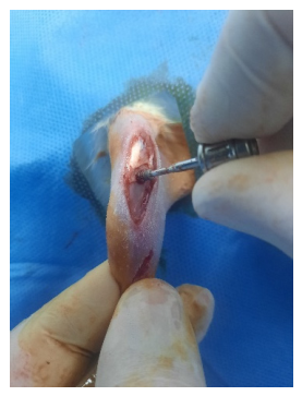
Figure 1. Surgical placement of titanium implants measuring 4 mm in length and 2.5 mm in diameter into the cortico-cancellous bone of the right tibiae of the rats.
Preprints 219943 g001
Figure 2. Performing the reverse torque analysis with a biomechanical testing machine (Mark-10, NY, USA).
Preprints 219943 g002
Table 1. Biomechanic bone implant connection (N/cm) levels of the groups.
Groups Mean (N/cm) Std. Deviation p* Value
MAC-CNT (n=12) 3.31 0.69
RBM-CNT (n=11)a 5.69 1.12
SLA-CNT (n=12)a 5.55 1.07 0.000
MAC-MLT (n=12)a 4.58 0.71
RBM-MLT (n=10)a 5.12 0.52
SLA-MLT (n=10)a,b 7.50 2.31
*One Way Anova (p<0.05). a: Different compared with the MAC-CNT group. b: Different compared with the MAC-MLT group.
Table 2. Intragroup comparison of biomechanical bone-implant connection values according to implant surface characteristics.
Mean (N/cm) Std. Dev p*
CNT MAC 3.31 0.69 p1=0.000
RBMa 5.69 1.12
SLAa 5.55 1.07
MAC 4.58 0.71 p2=0.000
MLT RBMb 5.12 0.52
SLAb,c 7.50 2.31
*One Way Anova (P<0.05). a: Statistically significantly different compared with machined (Tukey HSD). b: Statistically significantly different compared with MAC-MLT group (Games-Howell test). c: Statistically significantly different compared with RBM-MLT group (Games-Howell test).
Table 3. Pairwise comparisons of groups based on implant surface characteristics.
Groups Mean (N/cm) Std. Dev p*
MAC MLTa 4.58 0.71 p1=0.000
CNT 3.31 0.69
RBM MLT 5.12 0.52 p2=0.150
CNT 5.69 1.12
SLA MLTb 7.50 2.31 p2=0.030
CNT 5.55 1.07
*Student T Test a: Statistically significant difference between with MAC-CNT and MAC-MLT group (p<0,05). b: Statistically significant difference between with SLA-CNT and SLA-MLT group (p<0,05). No statistically significant difference between with RBM groups (p>0,05).
Disclaimer/Publisher’s Note: The statements, opinions and data contained in all publications are solely those of the individual author(s) and contributor(s) and not of MDPI and/or the editor(s). MDPI and/or the editor(s) disclaim responsibility for any injury to people or property resulting from any ideas, methods, instructions or products referred to in the content.
Copyright: This open access article is published under a Creative Commons CC BY 4.0 license, which permit the free download, distribution, and reuse, provided that the author and preprint are cited in any reuse.
Prerpints.org logo

Preprints.org is a free preprint server supported by MDPI in Basel, Switzerland.

Subscribe

© 2026 MDPI (Basel, Switzerland) unless otherwise stated

Accessibility

Disclaimer

Terms of Use

Privacy Policy

Privacy Settings