

Submitted:
23 June 2026
Posted:
24 June 2026
You are already at the latest version
Abstract
Coronary artery disease is a leading cause of morbidity and mortality in modern medicine. In contrast, surgical myocardial revascularization via coronary artery bypass grafting (CABG) remains the gold standard of treatment for complex multivessel disease. The great saphenous vein remains the most frequently used conduit due to its availability and technical simplicity, but its long-term patency is significantly inferior to that of arterial grafts. The primary pathological process responsible for vein graft failure is intimal hyperplasia, which represents a complex response of the vascular wall to surgical trauma, vein arterialization, inflammation, and hemodynamic stress. This process is characterized by endothelial dysfunction, inflammatory cell activation, proliferation and migration of vascular smooth muscle cells, and extracellular matrix remodeling. Underpinning these alterations are numerous molecular pathways, including NF-κB, MAPK, PI3K/Akt, TGF-β, and mTOR signaling, as well as substantial contributions from oxidative stress, cytokines, growth factors, and microRNAs. Contemporary research indicates that the phenotypic transformation of vascular smooth muscle cells constitutes the central event in the development of intimal hyperplasia. Understanding the cellular and molecular mechanisms underlying this disease's onset enables the development of novel therapeutic strategies to preserve long-term graft patency. This review paper aims to provide a systematic overview of current knowledge regarding the molecular and cellular mechanisms of intimal hyperplasia development in vein grafts following CABG.
Keywords:
CABG
; vein graft
; intimal hyperplasia
; vascular smooth muscle cells
; inflammation
; endothelial dysfunction
1. Introduction
Coronary artery disease represents one of the most critical healthcare challenges in the modern world. Despite significant advancements in percutaneous coronary intervention, coronary surgery continues to serve as the cornerstone therapeutic modality for patients with complex multivessel disease, diabetes, and left main coronary artery stenosis [1]. The great saphenous vein is used in most CABG procedures owing to its accessibility, length, and technical convenience. However, the long-term patency of vein grafts remains limited. It is established that approximately 10–15% of grafts thrombose within the first year, whereas up to 40–50% of vein grafts exhibit significant stenosis after 10 years [2].
In preparing this review paper, a systematic search of the literature on the pathophysiology of vein graft intimal hyperplasia following coronary artery bypass grafting (CABG) was conducted. The literature search was conducted using electronic databases including PubMed, Scopus, Web of Science, and Google Scholar. Articles published between 1990 and 2026 were analyzed, with particular focus on contemporary research from the last ten years. The keywords and combinations utilized included: 'vein graft intimal hyperplasia', 'saphenous vein graft failure', 'vascular smooth muscle cells', 'endothelial dysfunction', 'vascular inflammation', 'molecular pathways', 'oxidative stress', 'microRNA', 'CABG', 'vein graft remodeling', and 'phenotypic switching'. The search was restricted to articles published in the English language and available in full text.
Inclusion criteria comprised original experimental and clinical studies, review articles, meta-analyses, and translational research evaluating the molecular, cellular, and inflammatory mechanisms of vein graft intimal hyperplasia development after CABG. Particular attention was dedicated to studies investigating the role of vascular smooth muscle cells, endothelial dysfunction, oxidative stress, cytokines, signaling pathways, and epigenetic mechanisms in disease development. Papers with insufficient methodological data, small sample sizes lacking clear statistical analysis, publications not directly related to vein graft pathophysiology, and non-peer-reviewed publications were excluded from the analysis. Following the initial screening of titles and abstracts, articles holding the highest scientific and clinical relevance to the scope of this literature review were thoroughly analyzed.
The pathophysiology of vein graft failure unfolds through three distinct phases: early thrombosis, intimal hyperplasia, and late graft atherosclerosis (Figure 1). Intimal hyperplasia serves as a critical transitional event linking early thrombosis to late atherosclerosis. The process arises as a response of the vein to its exposure to arterial pressure and altered shear stress forces following implantation into the arterial system [3]. This adaptive response involves endothelial activation, inflammatory infiltration, migration, and proliferation of vascular smooth muscle cells (VSMCs), as well as accelerated extracellular matrix synthesis [4,5].
At first, the graft can fail soon after surgery because of blood clots. This usually happens within a few hours or days. Over time, the graft narrows as cells in the graft wall proliferate and deposit additional material. This process, called intimal hyperplasia, can take weeks or months. Later on, the graft can become diseased, just like the original arteries. This is called accelerated atherosclerosis. It's characterized by the buildup of fat, plaque, and scar tissue, which can cause the graft to narrow or even block completely. This can take months or even years to happen. The progression of saphenous vein graft failure is a complex process that involves multiple stages. Understanding these stages is crucial for developing effective strategies to prevent or delay graft failure. By recognizing the different phases of graft failure, doctors can take steps to reduce the risk of complications and improve patient outcomes.
Modern molecular biology has provided a deeper understanding of the signaling pathways involved in the development of intimal hyperplasia. Particular interest has centered on the NF-κB, MAPK, and TGF-β signaling pathways, which regulate inflammation, cellular proliferation, and vascular remodeling [6,7].
2. Histological and Functional Characteristics of the Vein Graft
The great saphenous vein is physiologically adapted to low-pressure, low-shear-stress conditions. Upon implantation into the arterial system, an abrupt shift in biomechanical conditions occurs. Veins are immediately subjected to high arterial pressure, pulsatile flow, and increased tangential stress. This sudden change activates mechanosensitive receptors and triggers a complex vascular remodeling process. The normal structure of the vein wall comprises a thinner media with fewer smooth muscle cells and a less developed internal elastic lamina compared to arteries. The venous endothelium exhibits an attenuated capacity for nitric oxide production and heightened susceptibility to oxidative stress. These precise anatomical and functional features contribute to its predisposition toward developing intimal hyperplasia [8,9]. The lining of our blood vessels, called the endothelium, plays a crucial role in keeping our vascular system healthy. But when we harvest and prepare veins for grafting, the endothelium is damaged by handling, stretching, and reduced blood flow. This damage reduces the production of important substances such as nitric oxide and prostacyclin, which help our blood vessels relax and prevent blood clots. At the same time, the damaged endothelium begins producing more adhesion molecules, such as VCAM-1, ICAM-1, and selectins, which attract white blood cells to the area. These white blood cells then stick to the blood vessel wall and move into the surrounding tissue, causing inflammation. The endothelium also releases proinflammatory cytokines such as TNF-α, IL-1β, and IL-6, which worsen inflammation. All these changes can trigger a response that leads to thickening of the blood vessel wall, known as intimal hyperplasia. A key player in this process is the NF-κB signaling pathway, which controls the expression of genes involved in inflammation and the production of cytokines and adhesion molecules. When this pathway is activated, it triggers a chain reaction that drives the inflammatory response and subsequent changes in the blood vessel wall [10,11].
Figure 2.
Temporal evolution of intimal hyperplasia in saphenous vein grafts after coronary artery bypass grafting (CABG): cellular, molecular, and immunological mechanisms.
Figure 2.
Temporal evolution of intimal hyperplasia in saphenous vein grafts after coronary artery bypass grafting (CABG): cellular, molecular, and immunological mechanisms.
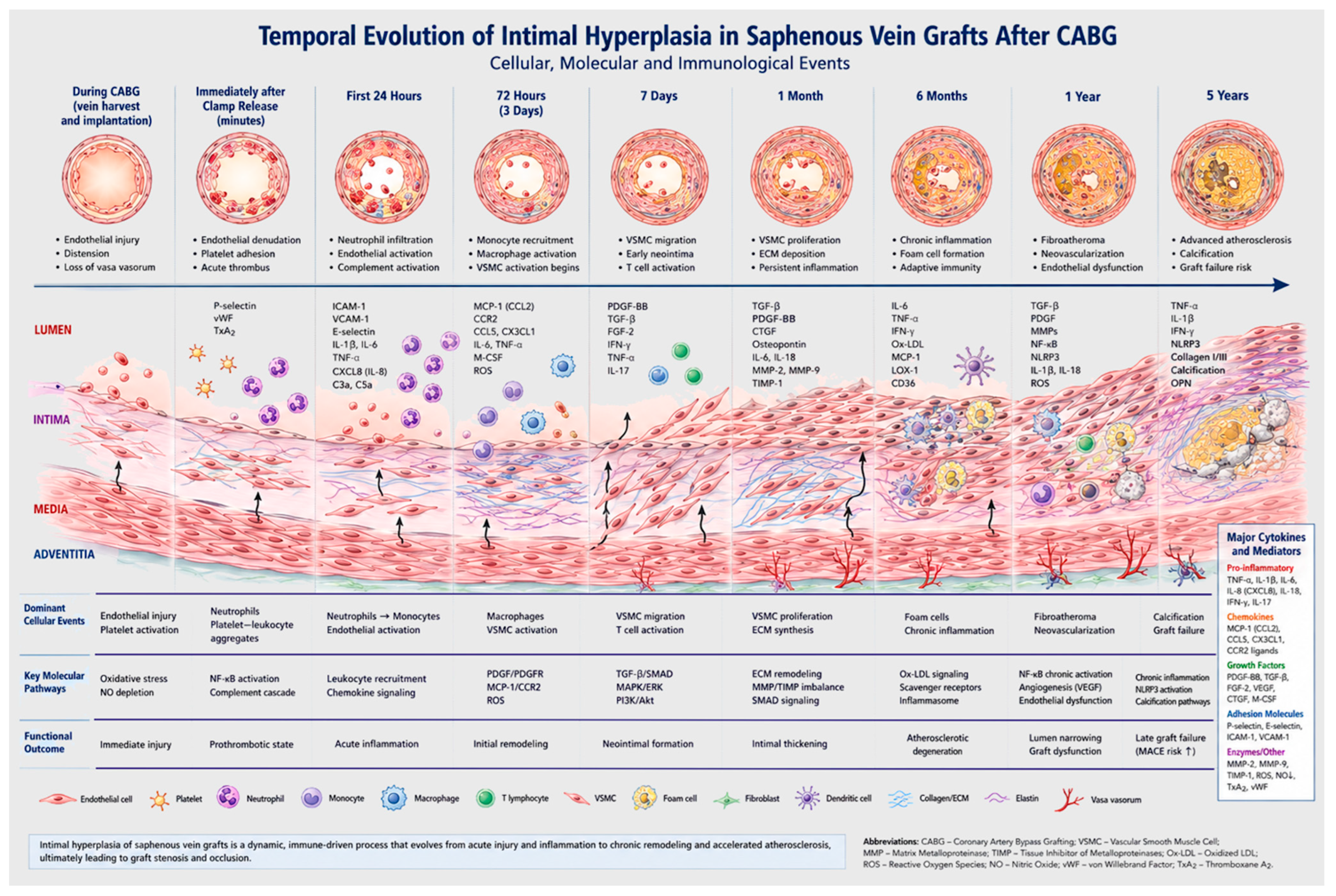
The figure illustrates the progression of intimal hyperplasia and accelerated vein graft disease following coronary artery bypass grafting (CABG), emphasizing the sequential cellular, molecular, and immunological events from the moment of graft implantation through late graft failure. The pathological process begins during graft harvesting and implantation, when the saphenous vein is exposed to mechanical trauma, ischemia-reperfusion injury, overdistension, and sudden arterial hemodynamic stress. These factors result in endothelial injury, loss of nitric oxide bioavailability, oxidative stress, and disruption of the vasa vasorum, thereby initiating an acute inflammatory and prothrombotic response.
Immediately after clamp release, endothelial denudation promotes platelet adhesion and activation, leading to the subsequent release of thromboxane A2, P-selectin, and von Willebrand factor. Activated platelets interact with circulating leukocytes and stimulate recruitment of neutrophils through chemokine-mediated signaling pathways. Concurrently, complement activation and reactive oxygen species (ROS) generation amplify endothelial dysfunction and vascular inflammation.
Within the first 24 hours, the inflammatory response intensifies with marked neutrophil infiltration and monocyte adhesion to the activated endothelium. Upregulation of adhesion molecules, including ICAM-1, VCAM-1, and E-selectin, facilitates leukocyte transmigration into the vessel wall. Activation of the NF-κB signaling pathway induces transcription of multiple proinflammatory cytokines, including TNF-α, IL-1β, IL-6, and IL-8, thereby propagating vascular inflammation and endothelial apoptosis.
By 72 hours, macrophages become the predominant inflammatory cells within the graft wall. Simultaneously, vascular smooth muscle cells (VSMCs) transition from a contractile to a synthetic phenotype under the influence of platelet-derived growth factor (PDGF), transforming growth factor-β (TGF-β), fibroblast growth factor-2 (FGF-2), and vascular endothelial growth factor (VEGF). Matrix metalloproteinases (MMP-2 and MMP-9) degrade extracellular matrix components, facilitating migration of VSMCs from the media into the intimal layer. Adventitial fibroblast activation further contributes to vascular remodeling.
At approximately 7 days after implantation, early neointimal formation becomes evident. Proliferation and migration of VSMCs are accompanied by accumulation of macrophages and activation of T lymphocytes. Intracellular signaling pathways including MAPK/ERK and PI3K/Akt regulate cellular proliferation, survival, and extracellular matrix synthesis. Increased deposition of collagen, fibronectin, and proteoglycans progressively thickens the intimal layer.
One month after CABG, intimal hyperplasia becomes well established. Synthetic phenotype VSMCs dominate the neointima, while persistent macrophage-mediated inflammation maintains chronic cytokine production. Elevated levels of osteopontin, MCP-1, IL-18, and macrophage colony-stimulating factor (M-CSF) perpetuate leukocyte recruitment and extracellular matrix remodeling. An imbalance between matrix metalloproteinases and their tissue inhibitors of metalloproteinases (TIMPs) further promotes pathological vascular remodeling and luminal narrowing.
At six months, the pathological process gradually transitions from isolated intimal hyperplasia toward accelerated graft atherosclerosis. Chronic inflammatory infiltrates persist within the vessel wall, and foam cells accumulate as macrophages take up oxidized low-density lipoprotein (ox-LDL) via scavenger receptor-mediated pathways. Ongoing activation of inflammatory mediators such as IL-6 and TNF-α contributes to endothelial senescence and progressive vascular degeneration.
By one year, fibroatheromatous plaque formation becomes increasingly apparent. Chronic NF-κB activation, oxidative stress, and reduced endothelial nitric oxide synthase (eNOS) activity sustain endothelial dysfunction and inflammatory signaling. Neovascularization of the neointima may occur through VEGF-mediated angiogenic pathways, while persistent macrophage and T-cell activity promotes plaque progression and instability.
At five years, advanced vein graft disease is characterized by diffuse fibroatherosclerosis, calcification, chronic inflammation, and plaque instability. Activation of the NLRP3 inflammasome and sustained production of TNF-α, IL-1β, and IFN-γ maintain chronic immune dysregulation and oxidative injury. Extensive extracellular matrix remodeling, fibrosis, and calcification ultimately lead to severe graft stenosis, thrombosis, myocardial ischemia, and late graft failure.
Overall, the figure demonstrates that intimal hyperplasia in saphenous vein grafts is not a single, isolated event but rather a complex, temporally coordinated process involving endothelial dysfunction, innate and adaptive immune activation, vascular smooth muscle cell phenotypic transformation, extracellular matrix remodeling, and progressive atherosclerotic degeneration.
3. Inflammation and Immune Response
The inflammatory response represents the principal driver of intimal hyperplasia development. Following endothelial injury, neutrophils, monocytes, and T-lymphocytes are recruited into the graft wall. Monocytes differentiate into macrophages, which secrete numerous proinflammatory mediators and growth factors. Macrophages release PDGF, TGF-β, FGF, and VEGF, which directly stimulate the proliferation and migration of vascular smooth muscle cells. Concurrently, the generation of reactive oxygen species (ROS) further amplifies inflammation and endothelial injury [12,13]. T-lymphocytes exert important regulatory functions through the production of interferon-γ and other cytokines that modulate macrophage and VSMC activity. Chronic inflammation sustains this proliferative response and drives lesion progression [14].
The central cellular event in the development of intimal hyperplasia is the phenotypic transformation of vascular smooth muscle cells from a contractile to a synthetic phenotype (Table 1). Under physiological conditions, VSMCs maintain vascular tone and express contractile proteins such as α-SMA, SM22α, and calponin. Under the influence of inflammatory cytokines, oxidative stress, and hemodynamic alterations, VSMCs shed their contractile phenotype and acquire proliferative and migratory features. The synthetic phenotype is characterized by accelerated proliferation, migration into the intima, and extensive extracellular matrix synthesis [15]. A crucial regulator of this process is PDGF-BB, which activates the MAPK and PI3K/Akt signaling pathways. TGF-β further stimulates the synthesis of collagen and fibronectin, while the mTOR signaling pathway regulates cellular growth and proliferation.
This table summarizes the sequential biological processes occurring in saphenous vein grafts following implantation into the arterial circulation. Immediately after graft reperfusion, endothelial injury, platelet activation, oxidative stress, and inflammatory cell recruitment initiate a complex cascade of vascular remodeling. During the first 24 hours, neutrophil infiltration and activation of pro-inflammatory signaling pathways amplify endothelial dysfunction and promote leukocyte adhesion and transmigration.
A critical transition occurs approximately 72 hours after graft implantation, when vascular smooth muscle cells (VSMCs) begin to undergo phenotypic switching from a contractile to a synthetic phenotype, representing one of the earliest and most important events in the development of intimal hyperplasia. Under the influence of growth factors such as platelet-derived growth factor (PDGF), transforming growth factor-β (TGF-β), fibroblast growth factor-2 (FGF-2), and vascular endothelial growth factor (VEGF), VSMCs lose their differentiated contractile characteristics and acquire enhanced proliferative, migratory, and extracellular matrix–producing capabilities. Concurrent activation of matrix metalloproteinases (particularly MMP-2 and MMP-9) facilitates degradation of the extracellular matrix and internal elastic structures, enabling VSMC migration from the medial layer toward the intima.
By the end of the first week, synthetic VSMCs become the dominant cellular component of the developing neointima. These cells actively proliferate and secrete extracellular matrix proteins, including collagen, elastin, fibronectin, and proteoglycans, leading to progressive neointimal formation. The process is further sustained by macrophage-derived cytokines and activation of signaling pathways such as MAPK/ERK and PI3K/Akt. At one month and beyond, persistent synthetic VSMC activity and chronic low-grade inflammation contribute to progressive luminal narrowing and structural graft remodeling.
Overall, the table highlights that although endothelial injury initiates the pathological process, the early phenotypic transformation of VSMCs, occurring as early as 72 hours after CABG, is the pivotal biological event driving subsequent intimal hyperplasia and long-term vein graft failure. Understanding the molecular mechanisms that regulate this phenotypic switch may identify important therapeutic targets to improve long-term graft patency.
Phenotypic transformation of vascular smooth muscle cells (VSMCs) is currently recognized as the central cellular event in the initiation and progression of vein graft intimal hyperplasia after CABG [16,17]. Under physiological conditions, the VSMCs within the media of the venous wall reside in a so-called contractile phenotype, characterized by minimal proliferative activity and dominant expression of contractile proteins, including α-smooth muscle actin (α-SMA), smooth muscle myosin heavy chain (SM-MHC), calponin, and SM22α [17]. Their fundamental functions are to regulate vascular tone and maintain the structural integrity of the vessel wall.
Following implantation of the saphenous vein into the arterial system, an abrupt transformation in biomechanical and hemodynamic conditions occurs. Veins are exposed to high arterial pressure, elevated circumferential stress, and altered shear stress forces. Simultaneously, surgical manipulation, graft distension, and ischemia-reperfusion injury lead to endothelial breakdown and the release of inflammatory mediators [4,6]. These stimuli activate intracellular signaling cascades that induce a phenotypic switch of VSMCs from a contractile to a synthetic state.
The synthetic phenotype is characterized by increased proliferation, migration from the media to the intima, heightened extracellular matrix synthesis, and downregulated expression of contractile proteins [16]. These cells become metabolically hyperactive and acquire the capacity to produce type I and III collagens, fibronectin, proteoglycans, and matrix metalloproteinases. This results in progressive intimal thickening and vascular wall remodeling. VSMC migration represents a critical step in neointima development. For cells to migrate through the internal elastic lamina, matrix metalloproteinases—specifically MMP-2 and MMP-9—are activated to degrade the extracellular matrix, thereby permitting cellular translocation [18,19]. Following intimal migration, VSMCs undergo intense proliferation driven by growth factors and cytokines.
Platelet-derived growth factor (PDGF), particularly the PDGF-BB isoform, plays a uniquely pivotal role because it is released from activated platelets, macrophages, and endothelial cells [14]. Binding of PDGF to the PDGFR-β receptor triggers a series of signaling cascades that induce VSMC proliferation and migration. Beyond PDGF, substantial contributions are made by transforming growth factor-beta (TGF-β), fibroblast growth factor (FGF), epidermal growth factor (EGF), and insulin-like growth factor-1 (IGF-1). Contemporary studies demonstrate that VSMCs possess exceptional phenotypic plasticity [20]. Aside from the synthetic phenotype, certain subpopulations can exhibit macrophage-like, osteochondrogenic, or fibroblastic phenotypes, thereby further driving the progression of vascular remodeling and graft atherosclerosis [15].
4. Molecular Signaling Pathways in Intimal Hyperplasia
NF-κB Signaling Pathway
Nuclear factor kappa B (NF-κB) stands as one of the most critical regulators of vascular wall inflammation [21]. Under physiological conditions, NF-κB resides in the cytoplasm bound to the inhibitory protein IκB. Upon stimulation by inflammatory factors such as TNF-α, IL-1β, oxidative stress, and mechanical injury, IκB kinase (IKK) is activated, leading to the phosphorylation and subsequent degradation of IκB proteins. Following its release, NF-κB translocates into the nucleus, where it orchestrates the transcription of numerous proinflammatory genes, including TNF-α, IL-6, MCP-1, VCAM-1, ICAM-1, selectins, and MMPs. Activation of the NF-κB pathway permits monocyte and T-lymphocyte recruitment, perpetuates inflammation, and stimulates VSMC proliferation [11,17]. Sustained activation of this pathway is linked to progressive intimal thickening and graft failure.
MAPK Signaling Pathway
The mitogen-activated protein kinase (MAPK) signaling pathway plays a key role in cellular proliferation, migration, and differentiation [22]. This system comprises three primary branches: ERK1/2 (extracellular signal-regulated kinase), JNK (c-Jun N-terminal kinase), and p38 MAPK. PDGF, FGF, and oxidative stress activate the Ras/Raf/MEK/ERK cascade, driving VSMC proliferation and the expression of genes involved in cell cycle progression. ERK1/2 predominantly regulates cellular proliferation, whereas JNK and p38 MAPK play prominent roles in the inflammatory response and apoptosis. The activation of p38 MAPK is explicitly linked to the production of inflammatory cytokines and extracellular matrix synthesis. MAPK signaling also contributes to the phenotypic transformation of VSMCs by reducing contractile protein expression and upregulating synthetic markers [22].
PI3K/Akt/mTOR Signaling Pathway
The PI3K/Akt/mTOR signaling pathway is a central regulator of cell growth, metabolism, and survival [19]. Activation begins with growth factor binding to receptor tyrosine kinases, followed by activation of phosphoinositide 3-kinase (PI3K). PI3K generates PIP3, which activates Akt kinase. Akt subsequently regulates multiple processes: inhibition of apoptosis, cellular proliferation, protein synthesis, VSMC migration, and cellular metabolism. One of the primary effectors of Akt signaling is the mammalian target of rapamycin (mTOR). Activation of the mTOR complex stimulates protein synthesis and VSMC proliferation, thereby contributing to neointimal development. Inhibitors of mTOR, such as sirolimus and everolimus, have demonstrated a significant reduction of intimal hyperplasia in both experimental and clinical settings [23].
TGF-β/Smad Signaling Pathway
Transforming growth factor beta (TGF-β) serves a dual role in vascular biology [24]. In early stages, it can exert anti-inflammatory effects, whereas chronic activation contributes to fibrosis and pathological vascular remodeling. Upon binding its receptor, TGF-β activates Smad2 and Smad3 proteins, which form a complex with Smad4 and enter the nucleus to regulate the expression of genes involved in collagen synthesis, fibronectin production, inhibition of matrix degradation, and vascular wall fibrosis. This signaling pathway contributes substantially to extracellular matrix accumulation and neointimal stabilization.
Oxidative Stress Signaling Pathways
Oxidative stress represents a major driver of vascular remodeling [25]. The primary source of reactive oxygen species (ROS) within the vascular wall is NADPH oxidase [26]. ROS activate multiple signaling cascades: NF-κB, MAPK, PI3K/Akt, and inflammatory cytokine pathways [3]. Simultaneously, nitric oxide (NO) bioavailability decreases, exacerbating endothelial dysfunction [7]. The superoxide anion reacts with NO to form peroxynitrite—a potent oxidant that damages cell membranes, DNA, and proteins. ROS also induce VSMC proliferation and migration via redox-dependent activation of transcription factors.
MicroRNA and Epigenetic Regulation
MicroRNAs play vital regulatory roles in the phenotypic transformation of VSMCs [27,28]. Specifically, miR-145 and miR-143 preserve the contractile phenotype of smooth muscle cells by stimulating contractile protein expression. Attenuated expression of these microRNAs triggers a phenotypic switch toward the synthetic state. Conversely, miR-21 promotes proliferation and inhibits VSMC apoptosis. Epigenetic mechanisms include histone acetylation, DNA methylation, and chromatin remodeling. These alterations enable long-term shifts in the expression of genes involved in inflammation and vascular cell proliferation [29].
5. Therapeutic Strategies
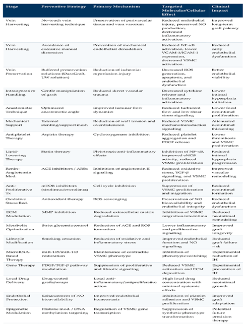
Given that intimal hyperplasia arises from the combined impact of surgical trauma, endothelial dysfunction, inflammation, hemodynamic stress, and vascular smooth muscle cell proliferation, prevention must target all phases of the CABG procedure—from vein harvesting and intraoperative graft handling to postoperative pharmacological and metabolic control (Table 2).
The table summarizes the major preventive strategies aimed at preventing intimal hyperplasia in saphenous vein grafts following CABG, organized by the perioperative and postoperative phases of graft management. The presented approaches include surgical, pharmacological, molecular, hemodynamic, and experimental strategies aimed at preserving endothelial integrity, reducing inflammatory activation, inhibiting vascular smooth muscle cell (VSMC) proliferation and migration, and minimizing extracellular matrix remodeling.
During vein harvesting and graft preparation, techniques such as no-touch harvesting, controlled low-pressure distension, and optimized graft preservation solutions primarily aim to prevent endothelial injury and reduce oxidative stress. Preservation of endothelial nitric oxide bioavailability and vasa vasorum integrity is crucial for limiting early inflammatory activation and phenotypic switching of VSMCs.
Intraoperative preventive measures emphasize optimizing graft geometry and hemodynamics, including appropriate anastomotic configuration, avoidance of graft kinking or competitive flow, and use of external support devices to reduce circumferential wall stress and mechanotransduction-mediated cellular activation. These interventions are directed toward maintaining laminar flow patterns and preventing low-shear-stress–induced endothelial dysfunction.
Postoperative pharmacological strategies primarily target thromboinflammatory and proliferative pathways. Antiplatelet agents reduce platelet-derived growth factor (PDGF) release and thrombus formation. At the same time, statins exert pleiotropic anti-inflammatory and endothelial-protective effects through inhibition of NF-κB signaling and improvement of endothelial nitric oxide synthase (eNOS) activity. Additional therapies, including ACE inhibitors, angiotensin receptor blockers, antioxidants, and mTOR inhibitors, modulate oxidative stress, transforming growth factor beta (TGF-β) signaling, and VSMC proliferation.
The table also includes emerging therapeutic approaches such as microRNA modulation, gene therapy, epigenetic regulation, and local drug-delivery systems, which represent promising future strategies for targeted inhibition of neointimal formation and long-term preservation of graft patency. Overall, the summarized mechanisms demonstrate that preventing vein graft intimal hyperplasia requires a multifactorial, biologically integrated approach that preserves endothelial homeostasis, suppresses inflammatory signaling, and modulates vascular remodeling.
The 'no-touch' vein-harvesting technique preserves perivascular tissue and minimizes endothelial breakdown, thereby significantly improving long-term graft patency. Avoiding excessive manual distension and executing gentle tissue manipulation attenuates the acute inflammatory response. Aspirin and statins form the baseline of pharmacological prevention against graft failure. Statins exhibit powerful pleiotropic benefits, including suppression of vascular inflammation and inhibition of VSMC proliferation [20]. mTOR inhibitors such as sirolimus have demonstrated profound efficacy in reducing intimal hyperplasia. Antioxidants and NF-κB inhibitors likewise represent viable therapeutic targets. Current cutting-edge research is heavily focused on gene therapies directed against proliferative signaling axes, while microRNA-based therapeutics stand out as highly promising avenues for future clinical implementation.
6. Conclusion
Vein graft intimal hyperplasia after CABG is a highly intricate, multifactorial biological cascade encompassing endothelial dysfunction, robust inflammation, vascular smooth muscle cell proliferation, and extracellular matrix remodeling. The pivotal biological event is the phenotypic switching of VSMCs driven by a synergistic combination of inflammatory cytokines, local oxidative stress, and dramatic alterations in arterial hemodynamics. Contemporary molecular research has identified a myriad of regulatory signaling networks involved in this disease, including the NF-κB, MAPK, TGF-β, and PI3K/Akt/mTOR pathways. Fully elucidating these pathways opens critical avenues for targeted molecular therapies to prevent vein graft disease. Although refinement of surgical techniques and modern medical therapies have optimized outcomes after CABG, vein graft failure persists as a serious clinical obstacle. Future investigations must prioritize personalized therapeutic approaches and the rapid translation of molecular discoveries into bedside clinical practice.
Author Contributions
Conceptualization, D.M.L.,S.J. and M.K.K.; methodology, D.C. and S.N. ; software, D.I. and V.A.; validation, D.M.L. and S.J.; formal analysis, M.J.K. and D.G.; investigation, D.C. V.J.; resources, D.M.L.; data curation, M.K.K and S.N.; writing—original draft preparation, D.M.L.; writing—review and editing, D.M.L. and S.J. and J.K.; visualization, J.K. and V.J.; supervision, D.C. and S.J. and D.I.; project administration, M.J.K. and S.N.; funding acquisition, V.A. and M.J.K. All authors have read and agreed to the published version of the manuscript.
Funding
This research received no external funding.
Conflicts of Interest
The authors declare no conflict of interest.
References
- Mitra AK, Gangahar DM, Agrawal DK. Cellular, molecular and immunological mechanisms in the pathophysiology of vein graft intimal hyperplasia. Immunol Cell Biol. 2006;84(2):115-124. [CrossRef] [PubMed]
- de Vries MR, Simons KH, Jukema JW, Braun J, Quax PH. Vein graft failure: from pathophysiology to clinical outcomes. Nat Rev Cardiol. 2016;13(8):451-470. [CrossRef] [PubMed]
- Lazović DM, Karadžić Kočica M, Ivanišević D, Aleksić V et al. Molecular and Cellular Mechanisms of Cardioplegic Protection in Surgical Myocardial Revascularization. Cells. 2026;15(2):173. [CrossRef] [PubMed]
- Sur S, Sugimoto JT, Agrawal DK. Coronary artery bypass graft: why is the saphenous vein prone to intimal hyperplasia? Can J Physiol Pharmacol. 2014;92(7):531-545. [CrossRef] [PubMed]
- Goldman S, Zadina K, Moritz T, et al. Long-term patency of saphenous vein and left internal mammary artery grafts after CABG. J Am Coll Cardiol. 2004;44(11):2149-2156. [CrossRef]
- Newby AC, Zaltsman AB. Molecular mechanisms in intimal hyperplasia. J Pathol. 2000;190(3):300-309. [CrossRef] [PubMed]
- Juricic S, Klac J, Stojkovic S, Beleslin B, et al. Molecular and Cellular Mechanisms of Myocardial Ischemia and Reperfusion Injury: A Narrative Review. Cells. 2026;15(3):265. [CrossRef] [PubMed]
- Owens CD. Adaptive changes in autogenous vein grafts for arterial reconstruction: clinical implications. J Vasc Surg. 2010;51(3):736-746. [CrossRef] [PubMed]
- Dashwood MR, Tsui JC. ‘No-touch’ saphenous vein harvesting improves graft performance. Vascul Pharmacol. 2013;58(3):240-250. [CrossRef]
- Angelini GD, Bryan AJ, Williams HM, Morgan R, Newby AC. Distention promotes platelet and leukocyte adhesion in human saphenous vein grafts. Circulation. 1990;82(5 Suppl):IV61-IV70.
- Jeremy JY, Dashwood MR, Mehta D, et al. Nitric oxide, prostacyclin and cyclic nucleotide formation in saphenous vein grafts. Br J Surg. 1997;84(4):540-545. [CrossRef]
- Libby P. Inflammation in atherosclerosis. Nature. 2002;420(6917):868-874. [CrossRef] [PubMed]
- Hansson GK. Inflammation, atherosclerosis, and coronary artery disease. N Engl J Med. 2005;352(16):1685-1695. [CrossRef] [PubMed]
- Ross R. Atherosclerosis—an inflammatory disease. N Engl J Med. 1999;340(2):115-126. [CrossRef] [PubMed]
- Raines EW. PDGF and cardiovascular disease. Cytokine Growth Factor Rev. 2004;15(4):237-254. [CrossRef] [PubMed]
- Wang L, Deng Y. Phenotypic switching of vascular smooth muscle cells: a central mechanism in vein graft intimal hyperplasia. Front Cardiovasc Med. 2025;12:1713297. [CrossRef] [PubMed]
- Owens GK, Kumar MS, Wamhoff BR. Molecular regulation of vascular smooth muscle cell differentiation. Physiol Rev. 2004;84(3):767-801. [CrossRef] [PubMed]
- Newby AC. Matrix metalloproteinase inhibition therapy for vascular diseases. Vasc Pharmacol. 2012;56(5-6):232-244. [CrossRef] [PubMed]
- Galis ZS, Khatri JJ. Matrix metalloproteinases in vascular remodeling. Circ Res. 2002;90(3):251-262. [CrossRef]
- Juricic S, Klac J, Stojkovic S, Tesic M, et al. Molecular and Pathophysiological Mechanisms Leading to Ischemic Heart Disease in Patients with Diabetes Mellitus. Int J Mol Sci. 2025 Apr 22;26(9):3924. [CrossRef] [PubMed]
- Monaco C, Andreakos E, Kiriakidis S, et al. Canonical pathway of nuclear factor kappaB activation selectively regulates proinflammatory and prothrombotic responses in human atherosclerosis. Proc Natl Acad Sci USA. 2004;101(15):5634-5639. [CrossRef] [PubMed]
- Pearson G, Robinson F, Beers, Gibson T, et al. Mitogen-activated protein kinase pathways. Endocr Rev. 2001;22(2):153-183. [CrossRef] [PubMed]
- Schachner T, Zou Y, Oberhuber A, et al. Local application of rapamycin inhibits neointimal hyperplasia. Ann Thorac Surg. 2004;77(5):1580-1585. [CrossRef] [PubMed]
- Border WA, Noble NA. Transforming growth factor beta in tissue fibrosis. N Engl J Med. 1994;331(19):1286-1292. [CrossRef] [PubMed]
- Madamanchi NR, Vendrov A, Runge MS. Oxidative stress and vascular disease. Arterioscler Thromb Vasc Biol. 2005;25(1):29-38. [CrossRef] [PubMed]
- Guzik TJ, West NE, Black E, et al. Vascular superoxide production by NAD(P)H oxidase. Circ Res. 2000;86(9):E85-E90. [CrossRef] [PubMed]
- Ji R, Cheng Y, Yue J, et al. MicroRNA expression signature and antisense-mediated depletion reveal an essential role of miR-21 in vascular neointimal lesion formation. Circ Res. 2007;100(11):1579-1588. [CrossRef] [PubMed]
- Cordes KR, Sheehy NT, White MP, et al. miR-145 and miR-143 regulate smooth muscle cell fate. Nature. 2009;460(7256):705-710. [CrossRef] [PubMed]
- Greißel A, Culmes M, Burgkart R, et al. Histone acetylation and methylation in vascular smooth muscle cells. Mol Med. 2016;22:458-467. [CrossRef]
Figure 1.
Progression of Saphenous Vein Graft Failure After CABG.
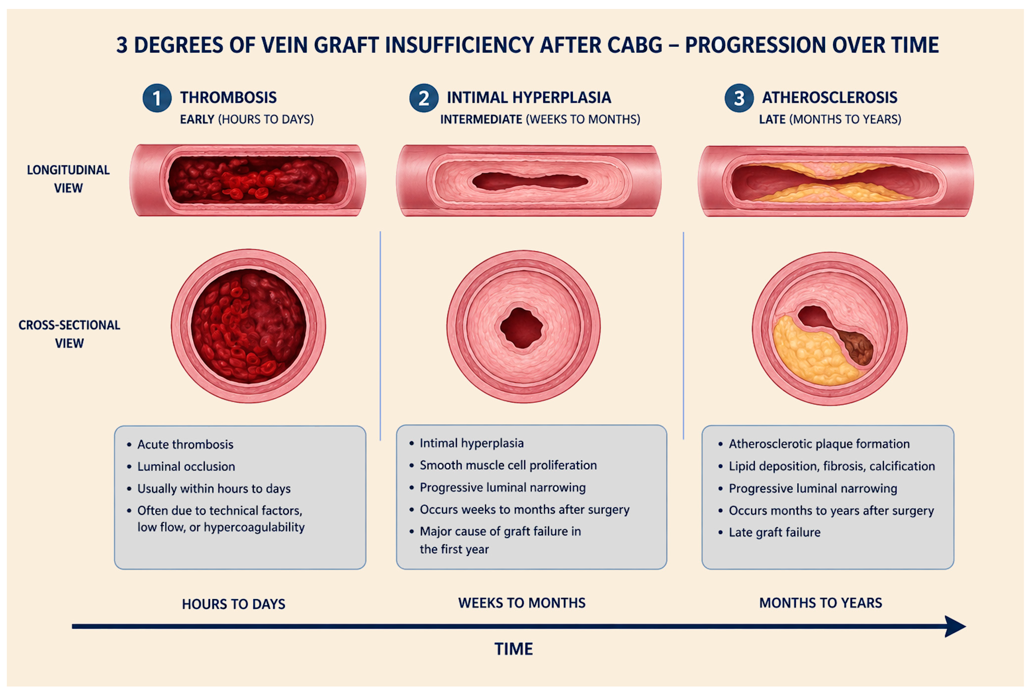

Table 1.
Temporal evolution of cellular and molecular events leading to saphenous vein graft intimal hyperplasia after coronary artery bypass grafting (CABG).
Table 1.
Temporal evolution of cellular and molecular events leading to saphenous vein graft intimal hyperplasia after coronary artery bypass grafting (CABG).
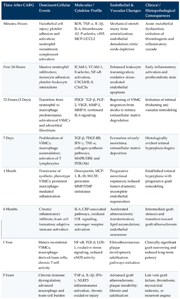 |
Table 2.
Principles and mechanisms of prevention of saphenous vein graft intimal hyperplasia after coronary artery bypass grafting (CABG).
Table 2.
Principles and mechanisms of prevention of saphenous vein graft intimal hyperplasia after coronary artery bypass grafting (CABG).
 |
Disclaimer/Publisher’s Note: The statements, opinions and data contained in all publications are solely those of the individual author(s) and contributor(s) and not of MDPI and/or the editor(s). MDPI and/or the editor(s) disclaim responsibility for any injury to people or property resulting from any ideas, methods, instructions or products referred to in the content. |
© 2026 by the authors. Licensee MDPI, Basel, Switzerland. This article is an open access article distributed under the terms and conditions of the Creative Commons Attribution (CC BY) license (http://creativecommons.org/licenses/by/4.0/).
Copyright: This open access article is published under a Creative Commons CC BY 4.0 license, which permit the free download, distribution, and reuse, provided that the author and preprint are cited in any reuse.


