

Submitted:
23 June 2026
Posted:
24 June 2026
You are already at the latest version
Abstract
Background/Objectives: Beta-thalassemia (β-thal) is an inherited hemoglobin disorder caused by mutations in the HBB gene. Hb Malay (HBB:c.59A>G, CD19), a missense substitution (AAC→AGC; Asn→Ser) in exon 1 of the β-globin gene causing β⁺-thalassemia, has been mainly described in Southeast Asian populations but remains rarely documented in Indonesia. This study aimed to identify the Hb Malay mutation and investigate its familial segregation in an Indonesian extended family. Methods: Cascade molecular screening was initiated following detection of the mutation in a proband referred for genetic testing. Sixteen family members consented to participate. Genomic DNA was extracted from peripheral blood and a 1.9-kb β-globin gene fragment was amplified by PCR. Mutation screening was performed using the GenoArray Thalassemia Detection Kit (Hybribio) and confirmed by Sanger sequencing. A three-generation pedigree was constructed from molecular results and family interviews.Results: The Hb Malay mutation (HBB:c.59A>G) was identified in 13 of 16 individuals: 12 heterozygous carriers (A/G) and one homozygous individual (G/G, subject KA, aged 15 years). Three individuals showed the normal genotype (A/A). Pedigree analysis demonstrated autosomal recessive inheritance across three generations. No α-globin deletions were detected. Conclusions: This is the first report of a large Indonesian family carrying the Hb Malay variant, confirmed by dual molecular methods. These findings highlight the importance of cascade molecular screening for early carrier identification, particularly where hemoglobin fractionation testing is unavailable.
Keywords:
β-thalassemia
; Hb Malay
; HBB:c.59A>G
; familial segregation
; cascade screening
; molecular diagnosis
; Indonesia
1. Introduction
β-Thalassemia (β-thal) is one of the most common inherited hemoglobin disorders worldwide, caused by mutations in the HBB gene that lead to reduced or absent production of β-globin chains. The clinical manifestations range from mild anemia in carriers to severe transfusion-dependent conditions in individuals with β-thal major [1,2,3]. Indonesia has one of the highest numbers of thalassemia cases in Southeast Asia, with an estimated 3–10% of the population being carriers and over 10,000 registered cases of thalassemia major, a number that continues to rise due to insufficient public awareness and prevention programs [4,5,6].
β-Thal arises from over 300 different mutations in the HBB gene, broadly categorized into β⁰-thal (complete absence of β-globin synthesis) and β⁺-thal (reduced but not absent synthesis). One such variant occurs at codon 19 of the β-globin gene, known as hemoglobin Malay (Hb Malay; HBB:c.59A>G), also referred to as β-thal CD19. This missense substitution replaces asparagine (AAC) with serine (AGC), resulting in β⁺-thalassemia. Hb Malay has been frequently reported in Malaysia [7,8,9,10,11,12,13,14] and Thailand [15], but remains rarely documented in Indonesia compared with more common mutations such as IVS-1-5 (G>C), CD26 (HbE), and CD41/42 (−TTCT) [16,17].
When β-thal is discovered in a large family, it raises important concerns about the inheritance pattern and health risks for family members, requiring cascade screening and genetic counselling [18,19,20]. This study aimed to identify the β-thal CD19 mutation in an extended family using molecular techniques, to construct a three-generation pedigree, and to highlight the importance of molecular screening for early carrier identification.
2. Detailed Case Description
The index case (proband, NZY) is a 15-year-old female who was initially identified during a school-based anemia screening program. Laboratory findings revealed a hemoglobin (Hb) level of 9.3 g/dL and a mean corpuscular volume (MCV) of 80.5 fL, indicating mild-to-moderate anemia with a near-normal MCV. This hematological profile prompted referral to our molecular diagnostic laboratory for further evaluation.
A relevant family history was noted: the proband’s mother (LRN, F2, 46 years old) had previously been diagnosed with thalassemia-associated anemia at another laboratory, but the specific mutation type and subtype had not been disclosed to her at the time of diagnosis. This reflects a broader challenge in Indonesia, where thalassemia remains underrecognized and underdiagnosed in clinical practice, largely overshadowed by the more commonly suspected iron deficiency anemia. Consequently, many carriers and affected individuals remain unaware of their precise genetic status and the hereditary implications for their families.
Given the proband’s anemia and her mother’s history of thalassemia-associated anemia, molecular analysis was initiated at our laboratory. The proband did not report any clinical symptoms at the time of examination. Capillary hemoglobin electrophoresis was not available at the study site; therefore, HbA₂ quantification could not be performed. Nevertheless, molecular screening using the GenoArray Thalassemia Detection Kit (Hybribio) identified the Hb Malay mutation (HBB: c.59A>G, CD19) in the proband in the heterozygous state (A/G genotype), subsequently confirmed by Sanger sequencing.
Following detection of the Hb Malay mutation in the proband, the extended family expressed interest in determining their carrier status. A total of 16 family members across three generations voluntarily submitted peripheral blood samples for molecular analysis. All participants provided written informed consent (or parental consent for minors) specifically for molecular testing. No additional clinical examination or complete hematological profiling was performed, as participants consented exclusively to molecular genetic screening. The family was originally from South Sumatra and had relocated to Jakarta, a region where the Hb Malay variant is known to be prevalent among the Malay ethnic population [21]. Following disclosure of results, all family members were provided with information on the hereditary nature of β-thalassemia and the importance of genetic counseling, particularly for those of reproductive age and for the homozygous individual (KA, F3, aged 15 years).
3. Materials and Methods
3.1. Subjects and Ethical Approval
One patient (proband, NZY) with clinical suspicion of thalassemia initiated this study. After detection of the Hb Malay mutation, cascade screening was extended to 16 consenting family members. The study was approved by the Ethics Committee of the Faculty of Medicine, UIN Syarif Hidayatullah Jakarta (No. B-019/F12/KEPK/TL.00/08/2025) and conducted in accordance with the Declaration of Helsinki. Written informed consent was obtained from all participants or their legal guardians.
3.2. DNA Extraction and PCR Amplification
Genomic DNA was extracted from peripheral blood using the gSYNC™ DNA Extraction Kit (Geneaid, Taiwan) per manufacturer’s instructions. A 1.9-kb segment of the β-globin gene was amplified by PCR using forward primer 5′-AGTAGCAATTTGTACTGATGGTATGG-3′ and reverse primer 5′-TTTCCCAAGGTTTGAACTAGCTCTT-3′. PCR conditions: initial denaturation 94°C/3 min; 35 cycles of 94°C/40 s, 62°C/30 s, 72°C/1 min; final extension 72°C/2 min. Products were verified by agarose gel electrophoresis [22,23].
3.3. Mutation Screening Using GenoArray
The GenoArray Thalassemia Detection Kit (Hybribio, China) was used to screen for common α- and β-globin gene variants per manufacturer’s instructions, enabling simultaneous detection of multiple thalassemia mutations and α-globin deletions.
3.4. DNA Sequencing and Data Analysis
All GenoArray-positive samples for β-thal CD19 were confirmed by Sanger sequencing (Apical Scientific Sdn. Bhd., Malaysia). Chromatograms were analyzed using ApE (A Plasmid Editor) software to identify nucleotide alterations at codon 19 of the β-globin gene (HBB).
3.5. Pedigree Analysis
A three-generation pedigree was constructed based on molecular results and structured interviews with the proband and available family members to illustrate the distribution and segregation of the mutation within the family.
4. Results
4.1. Molecular Analysis Overview
As described in the Case Presentation, the proband (NZY) presented with a hemoglobin level of 9.3 g/dL and MCV of 80.5 fL. The remaining 15 family members underwent molecular analysis only, as they consented exclusively to genetic screening without additional hematological profiling. Capillary hemoglobin electrophoresis was unavailable at the study site; therefore, HbA₂ levels could not be determined. GenoArray screening followed by Sanger sequencing allowed accurate identification of the HBB:c.59A>G (CD19) mutation and demonstrated its segregation within this extended family.
4.2. Pedigree and Genotypic Distribution
The segregation pattern of the Hb Malay mutation is shown in Figure 1. A total of 16 individuals from three generations (F1–F3) were analyzed. The genotypic distribution is summarized in Table 1, demonstrating normal (A/A), heterozygous carrier (A/G), and homozygous mutant (G/G) genotypes within the family.
4.3. Sanger Sequencing Confirmation
All GenoArray-positive samples for β-thal CD19 were confirmed by Sanger sequencing. Sequencing demonstrated a single base substitution (A→G) at codon 19, changing AAC to AGC (Figure 2). The homozygous individual (KA, F3, aged 15 years) showed a complete A→G substitution with no residual normal allele (Figure 3). None of the samples showed α-globin (HBA) deletions.
5. Discussion
This study presents the first report of a large Indonesian extended family carrying the Hb Malay variant (HBB:c.59A>G), confirmed through dual molecular methods: GenoArray screening and Sanger sequencing. The investigation was initiated following detection of the mutation in the proband (NZY), prompting cascade screening of 16 family members.
Hb Malay is a β⁺-thalassemia-causing missense mutation in exon 1 of the β-globin gene at codon 19, specifically AAC→AGC (Asn→Ser) [7,9,10,11,13,15]. The molecular basis of this substitution has been well characterized: it reduces but does not abolish β-globin production, consistent with the β⁺ phenotype [24]. Heterozygous carriers typically present with mild microcytic anemia with low MCV and MCH, and slightly elevated HbA₂ levels [8,12,25]. In the homozygous state or compound heterozygosity with other hemoglobinopathies, clinical severity increases, potentially resembling thalassemia intermedia or major, depending on the degree of β-globin chain imbalance [2,14,21,24,26,30]. Subject KA, the sole homozygous individual (G/G) identified in this family, was 15 years of age at the time of screening. Although complete hematological data were unavailable due to limited consent, prior reports indicate that homozygous Hb Malay typically presents as a mild disease with severe hypochromic microcytic anemia but without transfusion dependence, distinguishing it from β-thalassemia major [5,14,28].
Pedigree analysis across three generations demonstrated autosomal recessive inheritance, with 12 heterozygous carriers and one homozygous individual (KA, aged 15, F3). The high carrier rate (75%) is consistent with familial clustering when both parental lineages carry the mutation. Notably, the family originated from South Sumatra, where Hb Malay has been reported to be prevalent in the Malay ethnic population [21]. This highlights the importance of considering ethnic background when selecting mutation panels for molecular β-thal testing in Indonesia, where commonly screened mutations (IVS-1-5, HbE, CD41/42) may miss less prevalent variants such as CD19 [16,17,27,29].
The absence of complete hematological data and HbA₂ measurements for most family members is a recognized limitation. However, GenoArray and Sanger sequencing demonstrated full concordance, supporting the reliability of molecular-based cascade screening in resource-limited settings [31,32,33]. While Hb Malay is well-documented in Malaysia [7,8,9,10,11,12,13] and Thailand [14,15,34], its documentation in Indonesia remains scarce despite a carrier rate exceeding 5% [6,21]. This report contributes important molecular epidemiological data and underscores the need to expand mutation screening panels to include rare variants such as CD19, particularly in populations of South Sumatran Malay ancestry. The clinical context of this case also highlights an important public health issue in Indonesia. The proband’s mother (LRN) had previously been informed of a thalassemia-associated anemia diagnosis at another facility, yet the specific mutation type was never disclosed to her. This lack of precise genetic information reflects the current state of thalassemia care in Indonesia, where hemoglobinopathy screening is not yet systematically integrated into routine healthcare, and awareness remains low compared to more commonly recognized conditions such as iron deficiency anemia [4,6]. As a result, carriers and affected individuals may remain unaware of the hereditary implications for their offspring, delaying appropriate genetic counseling and cascade family screening. This case underscores the critical importance of not only detecting thalassemia but also ensuring that patients and families receive complete, accurate, and actionable genetic information following diagnosis.
6. Conclusions
The integration of GenoArray and Sanger sequencing provides a reliable approach for detecting rare β-globin gene variants. This report documents the Hb Malay (HBB:c.59A>G) mutation in a three-generation Indonesian family, representing the first report of this variant in a large Indonesian family. The identification of multiple heterozygous carriers and one homozygous individual underscores the value of cascade molecular screening where hemoglobin fractionation testing is limited. These findings support the integration of molecular diagnosis and family-based cascade screening into national thalassemia prevention programs in Indonesia.
Author Contributions
Conceptualization: C.A. and A.Z.; Methodology: C.A., G.P.S., M.F., Y.M., L.A.H., S. and A.L.; Formal analysis: C.A., R.P., S.P., P.S. and S.I.; Investigation: A.Z., G.P.S., M.F. and Y.M.; Data curation: C.A. and R.P.; Writing – original draft: C.A. and A.Z.; Writing – review & editing: C.A., S.P., P.S. and S.I.; Visualization: G.P.S. and M.F.; Supervision: C.A.; Project administration: C.A. All authors have read and agreed to the published version of the manuscript.
Funding
This research received no external funding.
Institutional Review Board Statement
The study was conducted in accordance with the Declaration of Helsinki, and approved by the Ethics Committee of the Faculty of Medicine, Universitas Islam Negeri Syarif Hidayatullah Jakarta (protocol code B-019/F12/KEPK/TL.00/08/2025).
Informed Consent Statement
Written informed consent was obtained from all participants (or their legal guardians) prior to inclusion in the study. Written informed consent for publication has been obtained from the participants.
Data Availability Statement
The data supporting the findings of this study are available from the corresponding author upon reasonable request, subject to institutional ethics approval.
Acknowledgments
The authors express sincere gratitude to the proband and all family members for their participation, and to the Faculty of Medicine and the Molecular Diagnostic and Research Center (MDRC) at UIN Syarif Hidayatullah Jakarta for institutional support. During the preparation of this manuscript, the authors used Claude (Anthropic) and Gemini (Google) for language editing and writing clarity. The authors have reviewed and edited the output and take full responsibility for the content of this publication.
Conflicts of Interest
The authors declare no conflicts of interest.
Abbreviations
The following abbreviations are used in this manuscript:
β-thal: β-thalassemia; CD19: codon 19 mutation; HBB: hemoglobin subunit beta gene; Hb Malay: hemoglobin Malay (HBB:c.59A>G); MCV: mean corpuscular volume; MCH: mean corpuscular hemoglobin; PCR: polymerase chain reaction.
References
- Langer, A.L. Beta-thalassemia. In GeneReviews®; Adam, M.P., Ed.; University of Washington: Seattle, WA, USA, 2000; Updated 2024.
- Bajwa, H.; Basit, H. Thalassemia. In StatPearls; StatPearls Publishing: Treasure Island, FL, USA, 2023.
- Sadiq, I.Z.; Abubakar, F.S.; Usman, H.S.; Abdullahi, A.D.; Ibrahim, B.; Kastayal, B.S.; Ibrahim, M.; Hassan, H.A. Thalassemia: pathophysiology, diagnosis, and advances in treatment. Thalass. Rep. 2024, 14, 81–102. [CrossRef]
- Susanah, S.; Sari, N.M.; Prihatni, D.; Sinaga, P.O.; Trisaputra, J.O.; Rakhmilla, L.E.; Sribudiani, Y. Extended family thalassemia screening as a feasible alternative method to be implemented in identifying carriers in West Java, Indonesia. J. Community Genet. 2021, 13, 103. [CrossRef]
- Petrou, M. Genetic counselling. In Prevention of Thalassaemias and Other Haemoglobin Disorders, 2nd ed.; Old, J., Ed.; Thalassaemia International Federation: Nicosia, Cyprus, 2013.
- Wahidiyat, P.A.; Sari, T.T.; Rahmartani, L.D.; Iskandar, S.D.; Pratanata, A.M.; Yapiy, I.; Setianingsih, I.; Atmakusuma, T.D.; Lubis, A.M. Thalassemia in Indonesia. Hemoglobin 2022, 46, 39–44. [CrossRef]
- Yang, K.G.; Kutlar, F.; George, E.; Wilson, J.B.; Kutlar, A.; Stoming, T.A.; Gonzales-Redondo, J.M.; Huisman, T.H. Molecular characterization of beta-globin gene mutations in Malay patients with Hb E-beta-thalassaemia and thalassaemia major. Br. J. Haematol. 1989, 72, 73–80. [CrossRef]
- George, E.; Huisman, T.H.; Yang, K.G.; Kutlar, F.; Wilson, J.B.; Kutlar, A.; Stoming, T.A.; Gonzales-Redondo, J.M.; Faridah, K.; Khalid, A.K. First observation of haemoglobin Malay alpha 2B2 26 (B1) Asn→Ser: A case report. Med. J. Malaysia 1989, 44, 259–262.
- Rozitah, R.; Nizam, M.Z.; Nur Shafawati, A.R.; Nor Atifah, M.A.; Dewi, M.; Kannan, T.P.; Ariffin, N.; Norsarwany, M.; Setianingsih, I.; Harahap, A.; et al. Detection of beta-globin gene mutations among Kelantan Malay thalassaemia patients by PCR-RFLP. Singapore Med. J. 2008, 49, 1046–1049.
- Hassan, S.; Ahmad, R.; Zakaria, Z.; Zulkafli, Z.; Abdullah, W.Z. Detection of β-globin gene mutations among β-thalassemia carriers and patients in Malaysia: application of multiplex ARMS-PCR. Malays. J. Med. Sci. 2013, 20, 13–20.
- Koh, D.X.R.; Raja Sabudin, R.Z.A.; Mohd Yusoff, M.; Hussin, N.H.; Ahmad, R.; Othman, A.; Ismail, E. Molecular characterisation of α- and β-thalassaemia among indigenous Senoi Orang Asli communities in Peninsular Malaysia. Ann. Hum. Genet. 2017, 81, 205–212. [CrossRef]
- Hassan, S.; Zulkafli, Z.; Saleh, R.M.; Ahmad, R.; Abdullah, W.Z. β-globin gene mutations among β-thalassemia and β-variant patients in a teaching hospital in Universiti Sains Malaysia, Kubang Kerian, Kelantan. GMJ 2022, 33, 215–219.
- Asri, A.S.; Samsuddin, M.H.; Jalil, N.; Mohamad Tahir, N.; Hashim, H.; Azma, R.Z.; Esa, E.; Albert, R.A.; Alauddin, H. Characterization of hemoglobin Malay phenotypes in tertiary hospitals. Hemoglobin 2024, 48, 153–160. [CrossRef]
- Fucharoen, S.; Sanchaisuriya, K.; Fucharoen, G.; Surapot, S. Molecular characterization of thalassemia intermedia with homozygous Hb Malay and Hb Malay/HbE in Thai patients. Haematologica 2001, 86, 657–658.
- Theingi, M.; Ruengdit, C.; Punyamung, M.; Pornprasert, S. Case Report: Two cases of Hb Malay (HBB: c.59A>G) found in Northern Thailand. J. Assoc. Med. Sci. 2025, 59, 80–86. [CrossRef]
- Rujito, L.; Maritska, Z.; Sofro, A.S. β-thalassemia mutation flow in Indonesia: a migration perspective. Thalass. Rep. 2023, 13, 253–261. [CrossRef]
- Hernaningsih, Y.; Syafitri, Y.; Indrasari, Y.N.; Rahmawan, P.A.; Andarsini, M.R.; Lesmana, I.; Moses, E.J.; Abdul Rahim, N.A.; Yusoff, N.M. Analysis of common beta-thalassemia mutations in East Java, Indonesia. Front. Pediatr. 2022, 10, 925599.
- Iqbal, R.; Sidrah; Akhtar, A. Family screening of thalassemic patients and effectiveness of education in providing knowledge about prevention of thalassemia. Pak. J. Med. Health Sci. 2023, 17, 253. [CrossRef]
- Kaur, G.; Chatterjee, T.; Ahuja, A.; Sen, A. Challenges in diagnosis of thalassemia syndromes. Med. J. Armed Forces India 2024, 88, 632–637. [CrossRef]
- Cao, A.; Galanello, R. Beta-thalassemia. Genet. Med. 2010, 12, 61–76.
- Sari, D.P.; Wahidiyat, P.A.; Setianingsih, I.; Timan, I.S.; Gatot, D.; Kekalih, A. Hematological parameters in individuals with beta thalassemia trait in South Sumatra, Indonesia. Anemia 2022, 2022, 3572986. [CrossRef]
- Adhiyanto, C.; Hendarmin, L.; Puspitaningrum, R. Pengenalan Dasar Teknik Bio-Molekuler [Basic introduction to biomolecular techniques]; Deepublish: Yogyakarta, Indonesia, 2020.
- Adhiyanto, C.; Yamashiro, Y.; Hattori, Y.; Nitta, T.; Hino, M.; Matar, M.; Takagi, F.; Kimoto, M. A new β⁰-thalassemia mutation (codon 102, AAC>ATCAC) in coexistence with a heterozygous P4.2 Nippon gene. Hemoglobin 2013, 37, 227–240. [CrossRef]
- Thein, S.L. The molecular basis of β-thalassemia. Cold Spring Harb. Perspect. Med. 2013, 3, a011700.
- Hariharan, P.; Gorivale, M.; Sawant, P.; Dalvi, R.; Mehta, P.; Nadkarni, A. Significance of genetic modifiers of hemoglobinopathies leading towards precision medicine. Sci. Rep. 2021, 11, 20906. [CrossRef]
- Iyevhobu, K.O.; Lucky, E.O.; Okobi, T.J.; Usoro, E.R.; Ivie, A.A.; Ken-Iyevhobu, B.; Omokpo, V.O. Overview of beta-thalassemia. IntechOpen 2023. [CrossRef]
- Adhiyanto, C.; Susianti, Y.; Rahmawati, N.M.; Badriah, F.; Harriati, Z. Screening of β-globin gene mutations in adolescent schoolgirls in rural Malang and Sukabumi City, Java Province, Indonesia. In Proceedings of the 1st International Integrative Conference on Health, Life and Social Sciences (ICHLaS 2017), Malang, Indonesia, December 2017; pp. 9–12.
- UCSF Thalassemia Center. Beta Thalassemia. Available online: https://thalassemia.ucsf.edu/thalassemia-information/what-is-thalassemia/beta-thalassemia (accessed on 20 July 2025).
- Batubara, N.R.; Adhiyanto, C.; Fahmida, U.; Freisleben, H.J. Hemoglobin E allele screening in adolescent girls at Kepanjen and Gondanglegi Districts, Malang Regency, East Java-Indonesia. Adv. Health Sci. Res. 2017, 10, 5–8.
- Sharifi A, Mahdieh N. HBB mutations and HbA2 level: Escaping the carrier screening programs. Clin Case Rep. 2021;9(2):973-977. [CrossRef]
- Huang R, Liu Y, Xu J, et al. Back-to-back comparison of third-generation sequencing and next-generation sequencing in carrier screening of thalassemia. Arch Pathol Lab Med. 2024;148(7):797-804. [CrossRef]
- Zhao J, Li J, Lai Q, Yu Y. Combined use of gap-PCR and next-generation sequencing improves thalassaemia carrier screening among premarital adults in China. J Clin Pathol. 2020;73(8):488-492. [CrossRef]
- Russo R, Marra R, Rosato BE, Iolascon A, Andolfo I. Genetics and genomics approaches for diagnosis and research into hereditary anemias. Front Physiol. 2020;11:613559. [CrossRef]
- Boonyawat, B.; Monsereenusorn, C.; Traivaree, C. Molecular analysis of beta-globin gene mutations among Thai beta-thalassemia children: results from a single center study. Appl. Clin. Genet. 2014, 7, 253–258. [CrossRef]
Figure 1.
Pedigree of the extended family showing segregation of the Hb Malay (HBB:c.59A>G) mutation. Squares represent males; circles represent females. Open symbols: normal genotype (A/A); half-filled symbols: heterozygous carriers (A/G); filled symbols: homozygous mutant (G/G). Numbers denote individuals whose samples were analyzed; alphabetical codes represent family members without samples.
Figure 1.
Pedigree of the extended family showing segregation of the Hb Malay (HBB:c.59A>G) mutation. Squares represent males; circles represent females. Open symbols: normal genotype (A/A); half-filled symbols: heterozygous carriers (A/G); filled symbols: homozygous mutant (G/G). Numbers denote individuals whose samples were analyzed; alphabetical codes represent family members without samples.
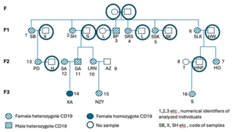
Figure 2.
Sanger sequencing chromatograms. (A) Normal β-globin showing the unaltered AAC codon at position 19. (B) Heterozygous β-thal CD19 showing both AAC and AGC at codon 19 (arrow indicates the mutation site).
Figure 2.
Sanger sequencing chromatograms. (A) Normal β-globin showing the unaltered AAC codon at position 19. (B) Heterozygous β-thal CD19 showing both AAC and AGC at codon 19 (arrow indicates the mutation site).
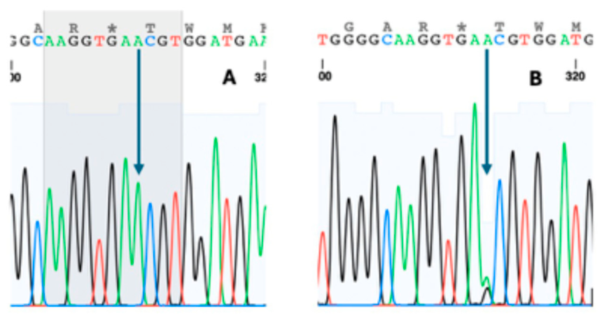
Figure 3.
Sanger sequencing result for subject KA (homozygous β-thal CD19). Complete AAC→AGC substitution at codon 19 with no residual normal allele (arrow), confirming homozygosity.
Figure 3.
Sanger sequencing result for subject KA (homozygous β-thal CD19). Complete AAC→AGC substitution at codon 19 with no residual normal allele (arrow), confirming homozygosity.
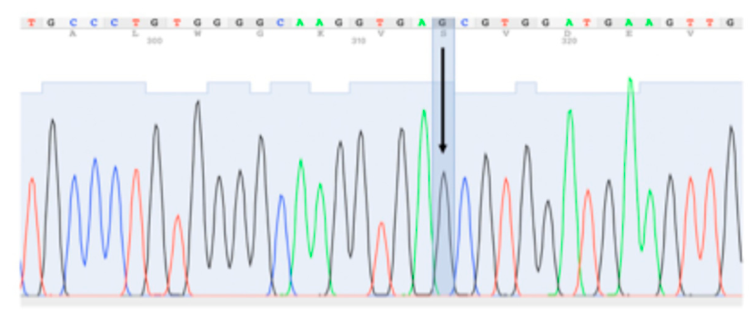

Table 1.
Genotypic distribution of the Hb Malay (HBB:c.59A>G) mutation among analyzed family members.
Table 1.
Genotypic distribution of the Hb Malay (HBB:c.59A>G) mutation among analyzed family members.
| No | Sample Code | Generation | Sex | Age (years) | Genotype | Interpretation |
|---|---|---|---|---|---|---|
| 1 | SB | F1 | Female | 77 | A/G | Heterozygous carrier |
| 2 | SH | F1 | Female | 73 | A/G | Heterozygous carrier |
| 3 | BP | F1 | Male | 76 | A/G | Heterozygous carrier |
| 4 | SRS | F1 | Female | 70 | A/G | Heterozygous carrier |
| 5 | SSK | F1 | Female | 68 | A/G | Heterozygous carrier |
| 6 | SLK | F1 | Female | 62 | A/G | Heterozygous carrier |
| 7 | HO | F2 | Female | 40 | A/G | Heterozygous carrier |
| 8 | T | F2 | Female | 33 | A/A | Normal |
| 9 | AZ | F2 | Male | 46 | A/A | Normal |
| 10 | LRN | F2 | Female | 46 | A/G | Heterozygous carrier |
| 11 | GA | F2 | Male | 48 | A/G | Heterozygous carrier |
| 12 | SA | F2 | Female | 46 | A/G | Heterozygous carrier |
| 13 | PD | F2 | Female | 53 | A/G | Heterozygous carrier |
| 14 | KA | F3 | Female | 15 | G/G | Homozygous mutant |
| 15 | NZY | F3 | Female | 15 | A/G | Heterozygous carrier |
| 16 | S | F3 | Female | 7 | A/G | Heterozygous carrier |
1Genotypes were determined by GenoArray screening and confirmed by Sanger sequencing. Hematological data are unavailable as family members consented only to molecular testing.
Disclaimer/Publisher’s Note: The statements, opinions and data contained in all publications are solely those of the individual author(s) and contributor(s) and not of MDPI and/or the editor(s). MDPI and/or the editor(s) disclaim responsibility for any injury to people or property resulting from any ideas, methods, instructions or products referred to in the content. |
© 2026 by the authors. Licensee MDPI, Basel, Switzerland. This article is an open access article distributed under the terms and conditions of the Creative Commons Attribution (CC BY) license (http://creativecommons.org/licenses/by/4.0/).
Copyright: This open access article is published under a Creative Commons CC BY 4.0 license, which permit the free download, distribution, and reuse, provided that the author and preprint are cited in any reuse.


