

Submitted:
22 June 2026
Posted:
23 June 2026
You are already at the latest version
Abstract
Atopic dermatitis (AD) is a chronic, relapsing inflammatory skin disease affecting more than 171 million individuals worldwide. Increasing evidence indicates that its pathogenesis extends beyond cutaneous immune dysregulation and involves systemic interactions mediated by the gut microbiome. This narrative review summarizes current knowledge regarding the gut–skin axis in AD and highlights three emerging areas of translational relevance: (i) the relationship between gut microbial dysbiosis, microbial metabolite alterations, and validated disease severity indices; (ii) the role of short-chain fatty acids (SCFAs), tryptophan-derived aryl hydrocarbon receptor (AhR) ligands, and secondary bile acids as key mediators of gut–skin immune communication; and (iii) the integration of artificial intelligence (AI) and multi-omics technologies in precision microbiome medicine. Gut dysbiosis in AD is characterized by reduced abundance of beneficial taxa, including Bifidobacterium, Faecalibacterium prausnitzii, Blautia, and Akkermansia muciniphila, resulting in impaired regulatory T-cell function, enhanced Th2 polarization, increased IgE production, and disruption of epithelial barrier homeostasis. Altered concentrations of microbial metabolites, particularly SCFAs and tryptophan-derived compounds, are associated with disease activity and correlate with clinical severity scores, including the Eczema Area and Severity Index (EASI) and SCORing Atopic Dermatitis (SCORAD). Intestinal barrier dysfunction, reflected by elevated zonulin, lipopolysaccharide-binding protein, and Reg3A, further amplifies systemic inflammation and cutaneous sensitization. Microbiome-targeted interventions—including probiotics, synbiotics, dietary modulation, and fecal microbiota transplantation (FMT)—have demonstrated promising effects on disease severity, immune regulation, and the restoration of microbial diversity. Emerging AI-driven multi-omics approaches are enabling the identification of disease endotypes, microbial signatures, and metabolite profiles with potential diagnostic, prognostic, and therapeutic value. Additionally, evidence from critical illness research suggests that ICU-associated dysbiosis may represent an extreme model of gut–skin axis disruption, providing novel insights into microbiome-driven immune dysfunction. Collectively, current evidence supports the gut microbiome as a driver, biomarker, and therapeutic target in AD. Future research should prioritize standardized multi-omics studies, metabolite-based biomarkers, and precision microbiome interventions to facilitate the translation of discoveries from the gut–skin axis into clinical practice.
Keywords:
atopic dermatitis
; gut microbiome
; gut–skin axis
; dysbiosis
; short-chain fatty acids
; aryl hydrocarbon receptor
; microbial metabolites
; intestinal permeability
; probiotics
; fecal microbiota transplantation
; multi-omics
; artificial intelligence
; precision medicine
; critical illness
; ICU dysbiosis
1. Introduction
Atopic dermatitis (AD) is a chronic, relapsing inflammatory skin disease characterized by intense pruritus, erythematous lesions, and a profoundly compromised quality of life. Once regarded primarily as a pediatric condition, AD is now recognized as a persistent and increasingly prevalent disorder across all age groups. According to data from the Global Burden of Disease (GBD) consortium, at least 171 million individuals were affected worldwide in 2019, corresponding to approximately 2.23% of the global population [1]. More recent estimates place global prevalence at 11.1% among children and adolescents and 6.3% among adults, with the highest burden observed in Asia and in urban environments in low- to middle-income countries [2,3]. Beyond its epidemiological scope, AD imposes a substantial multidimensional burden: patients experience sleep disturbances, anxiety, depression, and social stigmatization, while healthcare systems face escalating direct and indirect costs estimated to exceed $5 billion annually in the United States alone [4,5].
The pathogenesis of AD is multifactorial and involves the interplay of genetic predisposition, epidermal barrier dysfunction, immune dysregulation, and environmental exposures. At its core, AD is driven by a Th2-polarized immune response characterized by elevated levels of interleukin (IL)-4, IL-5, IL-13, and IL-31, along with alarmin release (thymic stromal lymphopoietin [TSLP], IL-25, and IL-33) from damaged keratinocytes [6]. Loss-of-function mutations in the filaggrin gene (FLG), present in 10-30% of AD patients, exemplify the genetic basis for impaired skin barrier integrity, thereby facilitating allergen penetration, microbial colonization — particularly by Staphylococcus aureus — and perpetuating the inflammatory cycle [7]. In the chronic phase, Th22 and Th17 responses further contribute to epidermal thickening and barrier deterioration through IL-22, which suppresses filaggrin expression and antimicrobial peptide production [8].
Despite significant advances in targeted therapies — including dupilumab (anti-IL-4Ra), tralokinumab (anti-IL-13), and JAK inhibitors — a substantial proportion of patients continue to experience inadequately controlled disease, recurrence upon treatment discontinuation, and comorbidities extending beyond the skin into the atopic march (asthma, allergic rhinitis, food allergy) [9]. These limitations underscore the need for a broader mechanistic understanding of AD and for identifying novel therapeutic targets that operate upstream of or in parallel with the type 2 inflammatory cascade.
In recent years, increasing attention has been directed toward the gut microbiome as a critical yet underexplored modulator of AD pathogenesis. The "gut-skin axis" — a bidirectional biological conduit linking the intestinal and cutaneous microenvironments through immune, neuroendocrine, and metabolic pathways — has emerged as a conceptual framework of growing clinical relevance [10,11]. Notably, the skin and gut share fundamental structural and functional characteristics: both are primary interfaces between the host and the external environment, both are central components of mucosal immunity, and both are directly colonized by complex microbial communities whose composition profoundly influences systemic immune tone [12]. The shared embryonic origin of the gut, brain, and skin further illuminates their functional and immunological congruence [13].
Epidemiological observations have long suggested a gut-skin relationship in AD: infants colonized by Clostridium difficile or Escherichia coli in early life, those born by cesarean section, and those exposed to antibiotics in the perinatal period demonstrate higher rates of subsequent AD development — all scenarios associated with gut microbiome perturbation [14]. Conversely, a growing number of studies have documented consistent patterns of gut dysbiosis in AD patients, including reduced microbial diversity, depletion of butyrate-producing taxa (Faecalibacterium prausnitzii, Bifidobacterium, Blautia, Eubacterium), and alterations in short-chain fatty acid (SCFA) and tryptophan metabolite profiles that correlate with disease severity [15,16].
Two dimensions of this relationship merit particular emphasis in current research. First, microbial metabolites — principally SCFAs (butyrate, propionate, acetate) and tryptophan-derived compounds (indoles, kynurenines) — function as key immunological mediators bridging the gut lumen with systemic and cutaneous immune responses, modulating regulatory T cell (Treg) differentiation, IL-10 and TGF-β production, and aryl hydrocarbon receptor (AhR) activation [17]. Second, the integration of multi-omics platforms (metagenomics, metabolomics, transcriptomics) with artificial intelligence and machine learning tools is beginning to reveal individualized microbial signatures with diagnostic and prognostic value, offering the prospect of precision medicine approaches to AD management [18].
Against this background, the present narrative review aims to synthesize the current evidence on the gut microbiome's role in AD, with a dual focus on (i) the mechanistic underpinnings of the gut-skin axis, with particular attention to microbial metabolite pathways, and (ii) microbiota-targeted therapeutic strategies, from probiotic and prebiotic interventions to fecal microbiota transplantation (FMT). Additionally, we discuss the emerging contribution of AI-assisted multi-omics analysis in decoding the complexity of gut-skin interactions and its implications for future personalized therapeutic strategies. By integrating mechanistic insights with translational and clinical perspectives, this review aims to serve both dermatologists and researchers in microbiology and immunology who seek a comprehensive, up-to-date synthesis of this rapidly evolving field.
2. The Gut-Skin Axis: Bidirectional Crosstalk in Atopic Dermatitis
2.1. Conceptual Framework and Structural Parallels
The gut-skin axis (GSA) describes a complex, bidirectional communication network linking the gastrointestinal tract with the skin through immune modulation, systemic inflammation, neuroendocrine signaling, and microbiota-derived metabolites [11,19]. Far from being a purely theoretical construct, this axis reflects genuine biological convergence: both the gut and the skin are large epithelial interfaces of approximately 400 m² and 2 m², respectively, that serve as primary barriers between the host and the external environment. Both tissues are continuously exposed to antigens, allergens, and commensal microorganisms, and both play essential and overlapping roles in mucosal immunity [12].
At a structural level, the gut and skin share several key features. Both are regulated by tight junction (TJ) proteins — including occludin, claudins, and zonula occludens-1 (ZO-1) — that govern paracellular permeability and determine barrier selectivity [20]. In healthy conditions, these junctions prevent the translocation of microbial products and antigens into the systemic circulation. Both barriers are also rich in innate immune sentinels (dendritic cells, macrophages, mast cells) and adaptive immune effectors (Th1, Th2, Th17, and regulatory T cells [Tregs]), creating tissue-specific but interconnected immunological niches [15]. The shared embryonic origin of the skin and gut, as well as their functional parallels in neural innervation and endocrine signaling, further underscore their intrinsic biological relationship [10].
2.2. Immune Crosstalk: Th1/Th2 Imbalance, IgE Dysregulation, and Treg Insufficiency
A pivotal mechanism through which the gut microbiome influences AD is via modulation of systemic immune balance. Under homeostatic conditions, a diverse and stable gut microbiota promotes the development of Tregs — both thymus-derived (tTregs) and peripherally induced (pTregs) — which suppress mucosal Th2-mediated inflammation, inhibit IgE class switching, and maintain immunological tolerance [16]. Gut-resident genera such as Bifidobacterium, Clostridium, Bacteroides, Streptococcus, and Lactobacillus produce short-chain fatty acids (SCFAs) — principally butyrate and propionate — that act as potent Treg inducers via histone deacetylase (HDAC) inhibition and GPR41/GPR43 receptor signaling [16,17].
In AD patients, gut dysbiosis disrupts this regulatory balance. Reduced microbial diversity and depletion of SCFA-producing taxa result in a shift toward unchecked Th2 polarization, with elevated production of IL-4, IL-5, and IL-13, and increased IgE synthesis by B cells [14]. Intestinal immune cells — particularly dendritic cells and Th17 cells — activated in the dysbiotic gut can migrate via systemic circulation to the skin and amplify local inflammation, a phenomenon documented in both AD and psoriasis [21,22]. This conceptual link between gut microbial poverty and allergic sensitization is central to the "hygiene hypothesis" and its more recent reformulation as the "old friends" hypothesis [23,24].
2.3. The Intestinal Barrier: Leaky Gut as an Amplifier of Cutaneous Inflammation
A critical component of gut-skin communication in AD is the integrity of the intestinal epithelial barrier. When the TJ function is compromised, a condition commonly referred to as "leaky gut" occurs, in which luminal contents (including lipopolysaccharides (LPS), bacterial toxins, dietary antigens, and pathogen-associated molecular patterns (PAMPs)) gain access to the lamina propria and the systemic circulation [25]. Zonulin, a physiological modulator of intestinal TJ permeability, has emerged as a measurable biomarker of gut barrier dysfunction. Elevated serum zonulin levels have been demonstrated in AD patients, particularly in children, and correlate independently with disease presence and severity, irrespective of total IgE and eosinophil counts [26]. Additional biomarkers include LBP, Reg3A, IL-10, and IL-22 [27]. This parallel impairment of intestinal and cutaneous TJs, both driven by Th2 cytokines, represents a mechanistic convergence reinforcing the notion of a systemic barrier disease [28].
2.4. Neuroendocrine Pathways: The HPA Axis, Cortisol, and the Itch-Stress Loop
Beyond immunological mechanisms, the gut-skin axis is connected through neuroendocrine signaling, most notably the hypothalamic-pituitary-adrenal (HPA) axis. HPA axis dysfunction documented in AD patients elevates systemic cortisol, paradoxically promoting Th2 skewing and suppressing antimicrobial immune responses [29]. Psychological stress activates the HPA axis via CRH and ACTH, simultaneously impairing gut barrier integrity and propagating downstream cutaneous inflammation [30]. Tryptophan, as the primary serotonin precursor, influences central serotonin concentrations and itch perception in AD independently of immune mechanisms, while SCFAs stimulate the sympathetic nervous system to modulate cutaneous lesion phenotype through neuronal pathways [31,32].
2.5. A Systems View: The Bidirectionality of the Gut-Skin Axis in AD
The gut-skin axis in AD is a genuinely bidirectional feedback system, not a unidirectional chain. Skin barrier dysfunction — characterized by increased TEWL, S. aureus colonization, and alarmin release — induces systemic immune activation, altering gut permeability and microbiota composition, thereby creating a self-perpetuating inflammatory loop [21]. Mechanical skin injury has been shown to promote food anaphylaxis by driving intestinal mast cell expansion, while UV-B skin exposure modulates the intestinal microbiome — illustrating the skin's capacity to influence gut ecology [33]. This bidirectional, multi-pathway architecture positions the gut-skin axis as a systems-level concept of major translational relevance.
Emerging evidence indicates that AD is not solely a skin-restricted inflammatory disorder but rather a systemic disease strongly influenced by gut microbial ecology and intestinal immune homeostasis. Gut dysbiosis in AD is characterized by depletion of SCFA-producing and immunoregulatory taxa, disruption of intestinal barrier integrity, systemic immune activation, and neuroendocrine dysregulation. These interconnected pathways converge to amplify Th2/Th17-driven inflammation, epidermal barrier dysfunction, and chronic pruritic skin lesions (Figure 1).
3. Gut Microbiome Composition in Atopic Dermatitis
3.1. Overview: Compositional Dysbiosis as a Defining Feature of AD
One of the most consistently replicated findings in gut microbiome research of AD is compositional dysbiosis — reduced alpha diversity, depletion of beneficial anaerobic taxa, and enrichment of potentially pathogenic microorganisms [14,15]. AD patients exhibit recurring dysbiotic patterns across diverse cohorts and age groups converging on a common theme: insufficiency of SCFA-producing, immunoregulatory bacteria and compensatory overgrowth of taxa associated with barrier disruption and Th2 skewing [34,35].
3.2. Depletion of Beneficial Taxa: The Immunoregulatory Deficit
The most clinically significant finding in AD gut dysbiosis is the consistent reduction of immunoprotective genera:
- Blautia, Coprococcus eutactus, and Eubacterium spp. — SCFA-producing Lachnospiraceae members whose reduction correlates with diminished butyrate output, more pronounced in severe AD [37].
- Lactobacillus spp. — broadly reduced in AD; associated with increased flare frequency and disease severity [34].
3.3. Enrichment of Potentially Pathogenic Taxa
The gut microbiome of AD patients is enriched for pro-inflammatory taxa: early colonization with Clostridium difficile and Escherichia coli predicts eczema development by dysregulating Treg induction; Klebsiella spp. and E. coli show significant overabundance (p < 0.05) in AD infants [40]; shifts in the Firmicutes/Bacteroidetes ratio are observed in both human and murine AD models [41]; and Ruminococcus gnavus enrichment shows context-dependent dysbiotic roles in AD subgroups [39]. In adult AD, Bacteroidales, Enterobacteriaceae, and Clostridium perfringens are more characteristic of the fecal microbiota compared to healthy adults [34].
3.4. Pediatric vs. Adult Gut Microbiome Profiles
Infant and Early Childhood AD
The first three years of life are a critical window of gut microbiome establishment [42]. In infants with AD, dysbiosis includes reduced Bifidobacterium richness from 4-26 weeks postpartum, increased Parabacteroides and Enterobacteriaceae, and disordered temporal colonization by SCFA-producing taxa — leading to butyrate deficiency and microaerophilic destabilization of the anaerobic microbiome [43]. Dietary patterns significantly modulate severity: processed-food-dominant diets are associated with higher abundances of Dorea and Anaerostipes and worse EASI/SCORAD scores [44,45].
Adult AD
3.5. Potential Biomarker Taxa
Bacteroidaceae and Porphyromonadaceae have been identified as potential fecal biomarker families for AD diagnosis [17]. Baseline gut microbiome composition predicted treatment response to E3 probiotics via ML analysis, with responders showing enrichment of Clostridium, Lactobacillus, and Streptococcus [46]. The gut microbiota serves three clinical roles in AD: an early predictive biomarker, a treatment response indicator, and a therapeutic target [15].
3.6. Methodological Considerations
16S rRNA amplicon sequencing remains the predominant platform but is limited in species-level resolution and functional gene content. Whole-metagenome sequencing (WMS) provides species-level resolution and direct functional gene annotation — essential for capturing SCFA biosynthesis, vitamin production, and immune signaling dysbiosis not visible in compositional profiles alone [39,44]. Methodological standardization — uniform sequencing depth, bioinformatic pipelines, fecal sample handling — remains a significant unmet need.
4. Microbial Metabolites as Key Mediators in the Gut-Skin Axis
The gut microbiome exerts much of its immunological influence through bioactive metabolites that enter systemic circulation and reach distant organs, including the skin. Among the most extensively studied are SCFAs, tryptophan-derived metabolites, and secondary bile acids — whose production is critically dependent on microbial diversity and whose depletion in AD patients correlates closely with disease activity [15,17] (Figure 2).
4.1. Short-Chain Fatty Acids (SCFAs): Immunoregulatory Keystones
4.1.1. Treg Induction and Th2 Suppression
SCFAs — butyrate, propionate, and acetate — are produced by anaerobic fermentation of dietary fiber by gut commensals including Faecalibacterium prausnitzii, Bifidobacterium, Blautia, Roseburia, and Coprococcus spp. [16]. They act through two major molecular mechanisms: (i) activation of GPCRs — GPR41, GPR43, and GPR109A; and (ii) inhibition of histone deacetylases (HDACs), driving epigenetic remodeling that promotes immune tolerance [47,48]. Butyrate enhances histone H3 acetylation at the Foxp3 locus in CD4+ T cells, amplifying Treg generation [48,49]. Propionate exerts analogous effects, while acetate acts through GPR43 to suppress neutrophil-mediated inflammation [47]. SCFA depletion in AD results in reduced FOXP3+ Treg activity, unopposed Th2 polarization, and elevated IgE [50].
4.1.2. Clinical Correlations: SCFAs and AD Severity
A case-control study of 50 adult AD patients and 25 controls found that serum concentrations of caproic acid and isocaproic acid showed a strong negative correlation with EASI and SCORAD (r = -0.33 to -0.37, p < 0.05) [51]. Additional SCFAs, propionic acid, butyric acid, and valeric acid, are also inversely correlated with disease extent during remission [27]. A longitudinal infant cohort confirmed significant negative associations between plasma concentrations of caproic, acetic, and succinic acids and the subsequent development of atopic sensitization and eczema [52].
4.2. Tryptophan Metabolites and the Aryl Hydrocarbon Receptor (AhR) Pathway
4.2.1. AhR Activation: Barrier Reinforcement and Anti-Inflammatory Signaling
Tryptophan catabolism by intestinal microbiota generates indole derivatives — indole-3-aldehyde (IAld), indole-3-acetic acid (IAA), indole-3-propionic acid (IPA), indole-3-lactic acid (I3LA), and tryptamine — which function as AhR ligands [17,53,54,55]. AhR activation in keratinocytes upregulates filaggrin and barrier proteins, reduces TSLP expression, and promotes antimicrobial peptide production. IAld levels are significantly lower in both lesional and non-lesional skin of AD patients; topical IAld application in a murine AD model significantly attenuated skin inflammation, inhibited Th2 cytokines and TSLP — effects abolished in AhR-null mice and by AhR antagonist treatment [56]. I3LA treatment of IL-4/IL-13-stimulated human skin equivalents restored barrier protein expression and reduced AD-associated gene expression [53].
4.2.2. AhR Dysregulation in AD
In AD, multiple converging factors deplete AhR signaling: gut dysbiosis reduces tryptophan catabolism; Th2 inflammation suppresses AhR target gene expression (CYP1A1 is significantly reduced in lesional skin by scRNA-seq); and accumulation of toxic indoxyl sulfate disrupts skin vascular homeostasis and worsens neuroinflammation and pruritus [15,57].
4.3. Secondary Bile Acids: Emerging Modulators of Barrier and Immunity
Secondary bile acids, lithocholic acid (LCA) and omega-muricholic acid (omega-MCA), modulate immunity via the farnesoid X receptor (FXR) and TGR5. Patients with AD exhibit reduced omega-MCA levels, thereby weakening FXR signaling and impairing Treg function [15]. LCA and omega-MCA enhance AMP expression in the skin and improve intestinal barrier function. In murine AD models, bile acid supplementation reduced epidermal thickening and cutaneous inflammatory cytokines — though this has not yet been validated in human trials [15,58].
4.4. Other Bioactive Metabolites
TMAO is reduced in AD patients and may modulate systemic inflammatory tone [27]. Linoleic acid and its microbially modified derivatives alleviate AD-like symptoms in mouse models through lipid-microbiome crosstalk. Polyamines produced by gut bacteria support intestinal epithelial renewal with emerging immunomodulatory relevance in AD [58].
4.5. Metabolomics as a Translational Bridge
The metabolite-centric perspective provides mechanistically interpretable, clinically measurable endpoints correlatable with EASI/SCORAD and potentially leverageable for patient stratification. Integration of metabolomics data with multi-omics platforms is beginning to reveal individualized gut-skin metabolite "fingerprints" in AD — the foundation for AI-assisted precision medicine strategies discussed in Section 5.
The growing body of metabolomic evidence suggests that several gut-derived metabolites and intestinal barrier-associated biomarkers are not only mechanistically involved in AD pathogenesis but also exhibit measurable associations with disease severity. Collectively, these molecules represent promising candidates for biomarker development and patient stratification. The principal gut-derived biomarkers currently associated with AD severity are summarized in Table 1.
While individual metabolites may serve as candidate biomarkers of disease activity, their biological relevance extends far beyond clinical stratification. These gut-derived molecules act as central mediators of host–microbiome communication, influencing epithelial integrity, immune tolerance, and inflammatory signaling across the gut–skin axis. A mechanistic overview of these interconnected pathways is presented in Figure 2.
5. Artificial Intelligence and Multi-Omics Integration in Decoding the Gut-Skin Axis
5.1. The Imperative for Integrative Approaches
The gut-skin axis in AD is a multidimensional biological system not amenable to characterization by a single analytical layer. Classical single-omics approaches provide valuable but inherently incomplete snapshots [59,60,61,62,63]. The proliferation of multi-omics platforms simultaneously interrogating metagenomics, metabolomics, transcriptomics, proteomics, epigenomics, and lipidomics — combined with AI/ML capable of identifying complex, non-linear patterns in high-dimensional datasets — is beginning to reveal previously unrecognized links between microbial metabolic pathways and host immune signaling networks [17,64,65].
5.2. Multi-Omics Platforms: Layering the Biological Architecture of AD
5.2.1. From Single to Integrated Omics
A landmark multi-omics study employing microbiota analysis, metabolomics, and intestinal epithelial transcriptomics across 2,247 children from the COCOA birth cohort linked early-life gut microbial composition to metabolite-driven epigenetic modifications associated with subsequent AD development [17]. Multi-omics overlap analyses confirm that filaggrin (FLG) is the only candidate consistently implicated across genomic, epigenomic, transcriptomic, and proteomic layers, underscoring the value of integrated analysis [59].
5.2.2. Endotype Discovery Through Multi-Omics Integration
Integrated metagenomics, metabolomics, and single-cell transcriptomics identified two endotype-specific gut-skin axis signatures: a Bacteroides-enriched enterotype associated with LPS-driven systemic inflammation and impaired barrier function, and a Prevotella-dominant cluster linked to enhanced AhR activation and epithelial-protective metabolite profiles [66]. Single-cell RNA sequencing and spatial transcriptomics identified unique inflammatory fibroblast subpopulations (COL18A1+) and complex intercellular signaling networks not visible with bulk transcriptomics [67]. scRNA-seq of Langerhans cells from AD patients revealed altered transcriptional profiles that bridge S. aureus recognition and T cell activation [68].
5.3. Machine Learning in Gut Microbiome Analysis
5.3.1. Interpretable ML Models for AD-Associated Gut Features
Ma et al. constructed an interpretable ML framework applying five algorithms — random forest (RF), LightGBM, XGBoost, support vector machine, and logistic regression — to 16S rRNA datasets after centered log-ratio (CLR) transformation. RF achieved the highest discriminatory performance. SHAP values quantified each taxon's marginal contribution to AD risk, identifying Bifidobacterium and Collinsella as among the most predictively significant features, with partial dependence plots characterizing their non-linear influence on AD risk [64].
5.3.2. Graph Neural Networks and Deep Learning
ATOMIC, a graph attention network for AD prediction using gut microbiome data, demonstrated that modeling the relational structure of microbial co-occurrence networks substantially improved predictive accuracy over feature-based classifiers, capturing ecological dependencies within the gut community invisible to standard ML models [69].
5.3.3. Mendelian Randomization: Establishing Causality
A two-sample MR study using GWAS data from 207 gut microbial taxa (Dutch Microbiome Project, n = 7,738) and AD outcome data (FinnGen biobank, 5,321 AD patients, 213,146 controls) identified 17 distinct bacterial taxa — spanning 2 orders, 4 families, 5 genera, and 6 species — with statistically supported causal associations with AD risk [70]. These findings provide genetic-level evidence for the biological plausibility of the gut-skin axis.
5.4. AI-Assisted Multi-Omics for Precision Medicine in AD
Metabolomic profiling has identified alterations in SCFA and tryptophan metabolites that correlate with disease severity, while transcriptomic analyses link these metabolite changes to downstream gene expression networks in keratinocytes and immune cells [66]. AI-assisted multi-omics models are being developed to predict individual responses to microbiota-targeted interventions based on baseline gut microbiome and metabolite profiles — the most rapidly growing frontier in AD-microbiome research [17]. Key limitations include small sample sizes, absence of standardized protocols, limited racial diversity, and interpretability challenges in deep learning models [59,60,64].
5.5. Future Directions: Toward AI-Driven Precision Microbiome Medicine
The convergence of AI and multi-omics outlines a trajectory toward "precision microbiome medicine" — individualized microbiota-targeted interventions matched to specific gut-skin metabolite fingerprints and endotype classifications. Research priorities include multicenter longitudinal cohort studies with harmonized multi-omics protocols, validation of endotype signatures in prospective clinical trials, integration of AI decision-support tools into clinical workflows, and causal inference frameworks to move from association to mechanism [71].
The integration of AI with multi-omics technologies is transforming microbiome research in AD, shifting from associative observation to causal inference and precision medicine. By combining metagenomics, metabolomics, transcriptomics, proteomics, and single-cell analyses, advanced computational models can identify molecular endotypes, predict therapeutic responsiveness, and facilitate individualized microbiome-based interventions (Figure 3).
6. Therapeutic Strategies Targeting the Gut Microbiome in Atopic Dermatitis
6.1. Rationale for Microbiome-Targeted Therapies
Microbiome-targeted interventions aim to restore the ecological and metabolic conditions that underpin immune homeostasis — addressing upstream drivers of AD rather than its symptomatic manifestations [72,73]. This section reviews the principal therapeutic approaches: probiotics, prebiotics, postbiotics, synbiotics, and fecal microbiota transplantation (FMT).
6.2. Probiotics: Mechanisms, Strains, and Clinical Evidence
6.2.1. Immunological Mechanisms of Action
Probiotics modulate AD through multiple mechanisms: increasing secretory IgA and suppressing IgE-mediated sensitization; promoting Treg differentiation and IL-10/TGF-b production; enhancing tight junction expression; producing SCFAs that reduce intestinal permeability; and competing with S. aureus colonization through antimicrobial peptide secretion [72,74,75].
6.2.2. Clinical Efficacy: Strain-Specific Evidence
A systematic review of 32 RCTs (2005) identified L. paracasei K71, L. plantarum IS-10506, and L. acidophilus L-92 as strains with the most consistent evidence for moderately reducing SCORAD and EASI. L. fermentum showed significant SCORAD reductions (MD = -11.42, 95% CI: -13.81 to -9.04); L. salivarius showed similarly significant effects (MD = -7.21, 95% CI: -9.63 to -4.78); while L. rhamnosus GG showed no significant effect (MD = 3.29, p = 0.07) [76,77]. A meta-analysis of 25 pediatric RCTs (Hedges' g ~= 0.65, p < 0.05) indicated moderate-to-large improvement in AD severity [78].
6.2.3. Timing: Prenatal vs. Postnatal
Combined prenatal and postnatal probiotic strategies confer the greatest preventive benefit, particularly in high-risk infants. The EAACI 2024 task force meta-analysis confirmed significant reductions in SCORAD with probiotics alone or in combination with prebiotics, along with additional improvements in patient-reported itch and sleep outcomes [73].
6.3. Prebiotics, Postbiotics, and Synbiotics
Prebiotics (FOS, GOS, HMOs) show more consistent benefit in AD prevention than treatment, with the 2025 umbrella meta-analysis confirming no significant SCORAD reduction from prebiotic-only interventions [79]. Postbiotics — preparations of inanimate microorganisms and/or their components — offer a heat-stable, bacteremia-free alternative that activates pattern recognition receptors and promotes Treg responses, currently limited to pilot studies [80]. Synbiotics demonstrated the most consistent SCORAD reductions across the subgroups analyzed in the 2025 umbrella meta-analysis, as the prebiotic component enhances probiotic colonization and the durability of effect [79].
6.4. Fecal Microbiota Transplantation (FMT)
6.4.1. Rationale and Approaches
FMT enables comprehensive restoration of gut microbial ecology, aiming to reset the dysbiotic gut ecosystem toward an SCFA-producing, immune-tolerant composition [81,82]. Washed microbiota transplantation (WMT) employs additional filtration to reduce bacterial load while preserving bioactive fractions, thereby improving safety compared with conventional FMT [83].
6.4.2. Clinical Trial Evidence
A pivotal randomized, double-blind, placebo-controlled trial evaluated FMT in 63 adult patients with moderate-to-severe AD: EASI scores and EASI-50 response rates were significantly higher in the FMT group. FMT decreased Th2/Th17 cell proportions, reduced serum TNF-a and total IgE, and produced significant shifts in gut microbial composition. No serious adverse reactions occurred [82]. WMT in 23 moderate-to-severe AD patients demonstrated significant decreases in SCORAD, EASI, DLQI, and NRS itch scores (all p < 0.05), with reduction in peripheral basophils, increased gut microbial diversity, enrichment of Bifidobacterium and Faecalibacterium, and metabolomic shifts in SCFAs and bile acid derivatives [17,83].
6.5. Dietary Modulation
High dietary fiber intake and Mediterranean dietary patterns are associated with greater gut microbial diversity and lower levels of systemic inflammatory markers relevant to AD [84]. Processed-food-dominant diets in pediatric AD are associated with worse EASI/SCORAD scores [85]. Breastfeeding — the most potent dietary modulator of early microbiome composition — consistently demonstrates protective effects against AD development through HMO-mediated Bifidobacterium enrichment in the neonatal gut [84].
6.6. Comparative Summary of Microbiome-Targeted Interventions
As interest in microbiome-based therapeutics continues to expand, a growing body of evidence has evaluated diverse interventions to restore intestinal microbial homeostasis in patients with atopic dermatitis. These strategies range from conventional probiotics and synbiotics to emerging approaches such as postbiotics, FMT, washed microbiota transplantation (WMT), and dietary modulation. The following table (Table 2) summarizes the current evidence landscape, highlighting the level of supporting evidence, principal clinical outcomes, and major limitations associated with each intervention.
Overall, the available evidence suggests that microbiome-targeted interventions exert heterogeneous but generally favorable effects on disease severity in atopic dermatitis. Multi-strain probiotic formulations, particularly those enriched with Lactobacillus species, currently have the strongest evidence base, as supported by umbrella meta-analyses demonstrating significant reductions in SCORAD scores. Synbiotics appear similarly beneficial, although direct comparisons with probiotic monotherapy remain limited. In contrast, single-strain Bifidobacterium supplementation has yielded inconsistent clinical results, suggesting that broader modulation of the microbial ecosystem may be necessary to achieve meaningful therapeutic effects.
Prebiotics alone have shown limited efficacy in reducing disease severity, indicating that their primary value may reside in prevention or long-term microbiome support rather than active treatment. Emerging postbiotic approaches are particularly attractive because they target microbial metabolites and immune signaling pathways directly; however, current evidence remains restricted to pilot studies and requires validation in larger randomized trials.
Among the most innovative interventions, FMT and WMT have demonstrated encouraging clinical outcomes, including significant improvements in EASI, SCORAD, and patient-reported symptom scores. Notably, the recent randomized controlled trial of conventional FMT provides some of the strongest mechanistic and clinical evidence for microbiome restoration as a therapeutic strategy in AD. Nevertheless, important challenges remain, including protocol standardization, donor selection, safety monitoring, and long-term efficacy assessment.
Dietary interventions represent a complementary strategy capable of enhancing microbial diversity while reducing disease severity. However, interpretation of dietary studies is complicated by substantial confounding factors, differences in dietary patterns, and difficulties maintaining long-term adherence. Collectively, these findings support the concept that microbiome modulation is a promising therapeutic avenue in AD, while emphasizing the need for standardized, multicenter, randomized trials and mechanistically informed precision-medicine approaches.
Therapeutic modulation of the gut microbiome represents one of the most promising translational strategies in AD. Interventions including probiotics, synbiotics, postbiotics, dietary modulation, and FMT aim to restore microbial diversity, normalize metabolite production, strengthen epithelial barriers, and rebalance systemic immune responses. Increasing clinical evidence demonstrates that microbiome-targeted therapies can improve both immunologic and clinical outcomes in AD (Figure 4).
7. Challenges, Research Gaps, Novel Contributions, and Discussion
7.1. Positioning This Review Within the Existing Literature
Most published reviews on the gut microbiome in AD focus either on compositional dysbiosis or on microbiota-targeted interventions, while few integrate mechanistic microbial metabolite pathways with emerging computational approaches. Furthermore, the rapidly expanding fields of AI, machine learning, and multi-omics analysis have rarely been discussed within a unified gut–skin axis framework. The present review addresses these gaps by integrating microbiome composition, metabolite-mediated immune regulation, microbiota-targeted therapeutics, and AI-assisted precision medicine into a single translational model of AD pathogenesis and management.
7.2. From Dysbiosis to Clinical Severity: A Quantitative Framework
A major conceptual contribution of this review is the integration of three interconnected biological layers: gut microbial composition, metabolite production, and clinical disease severity. Depletion of beneficial taxa such as Bifidobacterium, Faecalibacterium prausnitzii, Blautia, and Akkermansia muciniphila is consistently associated with reduced production of SCFA and tryptophan-derived metabolites, while altered concentrations of caproic acid, isocaproic acid, butyrate, indoxyl, and biomarkers of intestinal permeability correlate with EASI and SCORAD scores [27]. This integrated dysbiosis–metabolite–severity axis provides a biologically plausible framework for developing composite biomarker panels that combine microbial metabolites with markers of intestinal barrier dysfunction and systemic inflammation.
7.3. Microbial Metabolites as Therapeutic Targets
The evidence reviewed supports a transition from viewing microbial metabolites as passive biomarkers to considering them as active therapeutic targets. SCFAs regulate immune tolerance through HDAC inhibition, FOXP3 induction, and GPR43/GPR109A signaling, whereas tryptophan-derived metabolites modulate keratinocyte differentiation and immune homeostasis through AhR activation [15,48,50,56]. Similarly, secondary bile acids influence epithelial integrity and Treg function through FXR and TGR5 signaling pathways [50]. These findings provide a mechanistic rationale for targeted microbiome interventions designed to restore metabolite production rather than simply modify microbial composition.
7.4. AI and Multi-Omics as the Foundation of Precision Microbiome Medicine
Recent advances in AI and multi-omics technologies are transforming gut microbiome research from descriptive association studies toward clinically actionable precision medicine. Interpretable machine learning approaches, including SHAP-based models and graph neural networks, enable identification of key microbial taxa and metabolite signatures associated with AD risk and treatment response [17,64,66]. In parallel, Mendelian randomization studies provide increasing support for causal microbiome–AD relationships [70]. The integration of metagenomics, metabolomics, transcriptomics, and single-cell analyses is beginning to reveal biologically distinct AD endotypes that may ultimately guide personalized therapeutic selection and disease monitoring.
7.5. Current Challenges and Research Gaps
Despite significant progress, several important limitations remain. Microbiome studies continue to be affected by methodological heterogeneity, including differences in sequencing platforms, bioinformatic pipelines, and sample-processing procedures [79]. Geographic and ethnic representation remains limited, with most available datasets originating from East Asian and European populations [82]. Furthermore, although growing evidence supports causal links between gut dysbiosis and AD, many observations remain associative and require validation in longitudinal and interventional studies [1,86]. Additional uncertainties include interactions between microbiome-targeted therapies and established treatments, such as biologics and JAK inhibitors, as well as regulatory and safety considerations for FMT and live biotherapeutic products.
7.6. Future Research Priorities
Several research priorities emerge from the current evidence. First, multi-analyte biomarker panels that combine SCFAs, tryptophan metabolites, and markers of intestinal permeability should be prospectively validated as indicators of disease activity and treatment response. Second, clinical trials investigating targeted metabolite supplementation, including butyrate- and AhR-based interventions, are warranted. Third, harmonized international multi-omics cohorts are needed to improve reproducibility and facilitate cross-population comparisons. Finally, AI-assisted endotype classification systems integrating microbiome, metabolomic, and clinical data may represent a critical step toward precision microbiome medicine in AD.
7.7. Integrating the Evidence Across the Gut–Skin Axis
Collectively, the evidence reviewed supports a systems-level model in which gut dysbiosis, microbial metabolite deficiency, epithelial barrier dysfunction, and immune dysregulation act as interconnected drivers of AD. The depletion of SCFA-producing and AhR-ligand-generating microbial communities contributes not only to altered immune tolerance but also to measurable clinical outcomes, as reflected in EASI and SCORAD scores [15,27]. Microbiome-targeted interventions, particularly probiotics, synbiotics, and FMT, demonstrate the potential to restore these disrupted pathways and improve disease severity [79,82].
Importantly, AI-driven multi-omics analyses provide the analytical framework necessary to identify disease endotypes, predict therapeutic responsiveness, and translate microbiome research into individualized clinical applications [17,64,66,70]. Together, the integration of dysbiosis profiles, metabolite-centered mechanisms, precision analytics, and microbiota-targeted therapeutics represents the principal conceptual advance of this review and outlines a roadmap for the future development of precision microbiome medicine in atopic dermatitis.
8. Critically Ill Patients, ICU-Associated Dysbiosis, and the Gut–Skin Axis in Atopic Dermatitis
Critically ill patients admitted to intensive care units (ICUs) represent an extreme biological model of gut microbiome disruption and systemic immune dysregulation. Increasing evidence suggests that critical illness induces profound intestinal dysbiosis characterized by rapid loss of microbial diversity, depletion of beneficial anaerobic commensals, overgrowth of opportunistic pathogens, intestinal barrier dysfunction, and excessive systemic inflammation. These alterations overlap mechanistically with pathways implicated in AD pathogenesis, including reduced SCFA production, impaired Treg signaling, exaggerated Th2/Th17 responses, epithelial barrier disruption, and altered neuroimmune communication. Although direct clinical evidence specifically linking ICU-associated dysbiosis to AD exacerbation remains limited, critically ill patients may constitute a translational model for studying severe gut–skin axis dysfunction and its immunologic consequences [87,88,89].
8.1. ICU-Associated Gut Dysbiosis as a Model of Extreme Gut–Skin Axis Disruption
Critical illness profoundly alters the intestinal microbiome within hours after ICU admission. Sepsis, trauma, mechanical ventilation, broad-spectrum antibiotics, proton-pump inhibitors, vasopressor use, opioid exposure, parenteral nutrition, and prolonged hospitalization collectively contribute to rapid microbial collapse and conversion of the commensal microbiota into a pathogenic “pathobiome”. ICU-associated dysbiosis is characterized by marked reductions in microbial diversity, depletion of obligate anaerobes, and expansion of opportunistic pathogens, particularly members of the Proteobacteria and Enterobacteriaceae phyla [87,90,91].
Several microbial taxa depleted in critically ill patients overlap with those consistently reduced in AD, including Bifidobacterium, Faecalibacterium prausnitzii, and other SCFA-producing commensals. Loss of these organisms contributes to decreased production of butyrate and other SCFAs that normally maintain epithelial integrity, suppress inflammation, and support immune tolerance [34,89,92].
Importantly, ICU-associated dysbiosis is accompanied by profound intestinal barrier dysfunction. Critical illness induces disruption of tight junction proteins, increased intestinal permeability, epithelial apoptosis, and systemic translocation of microbial-associated molecular patterns (MAMPs), including lipopolysaccharide (LPS), flagellin, and bacterial DNA. Elevated zonulin, lipopolysaccharide-binding protein (LBP), and inflammatory cytokines further reflect the development of a “leaky gut” phenotype [87,88].
This pathophysiologic cascade strongly parallels mechanisms implicated in AD pathogenesis. In AD, gut dysbiosis and impaired barrier integrity promote systemic immune activation characterized by reduced FOXP3+ Treg activity, exaggerated Th2 polarization, elevated IgE synthesis, eosinophilic inflammation, and impaired keratinocyte homeostasis. Reduced microbial metabolite production—particularly SCFAs and tryptophan-derived aryl hydrocarbon receptor (AhR) ligands—further amplifies epithelial dysfunction and inflammatory signaling [34,93,94].
From a mechanistic perspective, the critically ill patient may therefore represent an amplified model of gut–skin axis disruption in which severe dysbiosis, systemic inflammation, metabolic collapse, and epithelial barrier failure converge. ICU-associated reductions in SCFA-producing bacteria may impair GPR43/GPR109A signaling and histone deacetylase (HDAC) inhibition, resulting in diminished Treg induction and enhanced inflammatory responses. Simultaneously, altered tryptophan metabolism and reduced AhR activation may compromise keratinocyte differentiation and epidermal barrier integrity [92,94].
Neuroimmune mechanisms may provide an additional point of convergence between critical illness and AD. Severe stress responses in ICU patients activate the HPA axis and alter gut–brain communication pathways, including vagal signaling and serotonin metabolism. Since gut microbial metabolites participate in neuroimmune regulation and pruritus-related pathways, ICU-associated dysbiosis may theoretically exacerbate itch amplification and inflammatory skin responses through interactions along the gut–brain–skin axis [88,95,96].
Although prospective clinical studies directly examining AD outcomes in ICU populations remain lacking, the overlap between ICU-associated dysbiosis and AD-associated gut–skin axis dysfunction suggests several translational implications. Critically ill patients may serve as valuable human models for studying severe microbiome collapse, epithelial barrier failure, immunometabolic dysregulation, and microbiome-targeted interventions. Moreover, microbiota-restoring strategies currently explored in critical care, including probiotics, synbiotics, postbiotics, enteral nutrition optimization, and FMT, may provide mechanistic insights relevant to future AD therapeutics [87,97].
Beyond its relevance to dermatology, the gut–skin axis provides a broader conceptual framework for understanding how microbial dysbiosis, barrier dysfunction, and systemic immune alterations contribute to chronic inflammatory disease. The critically ill patient represents a unique human model in which these processes converge, offering important insights into the pathophysiology of AD and future microbiome-based therapeutic strategies. The integrated translational model proposed in this review is summarized in Figure 5.
9. Conclusions
Atopic dermatitis is increasingly recognized as a systemic disorder involving complex interactions among the gut microbiome, microbial metabolites, epithelial barriers, immune networks, and neuroendocrine pathways. Accumulating evidence demonstrates that gut dysbiosis contributes to AD pathogenesis through depletion of beneficial microbial taxa, impaired production of immunoregulatory metabolites, disruption of intestinal barrier integrity, and amplification of Th2/Th17-driven inflammatory responses. Beyond their role as disease-associated signatures, gut-derived metabolites—including short-chain fatty acids, tryptophan-derived aryl hydrocarbon receptor ligands, and secondary bile acids—emerge as central mechanistic mediators linking microbial composition to immune homeostasis and clinical disease severity. A major conceptual advance highlighted in this review is the integration of microbiome composition, metabolite-centered mechanisms, and validated clinical severity indices, supporting the development of composite biomarker panels for patient stratification and disease monitoring. In parallel, microbiome-targeted interventions—including probiotics, synbiotics, postbiotics, dietary modulation, and fecal microbiota transplantation—demonstrate growing potential to restore microbial homeostasis and attenuate inflammatory pathways relevant to AD. Recent advances in artificial intelligence, machine learning, and multi-omics technologies are transforming microbiome research from descriptive association studies toward causal inference and precision medicine. The integration of metagenomics, metabolomics, transcriptomics, and single-cell approaches is beginning to reveal biologically distinct AD endotypes that may ultimately guide individualized therapeutic strategies and improve prediction of treatment response. Furthermore, evidence from critical care medicine suggests that critically ill patients may represent an extreme biological model of gut–skin axis disruption, characterized by profound dysbiosis, intestinal barrier failure, altered microbial metabolism, systemic immune dysregulation, and neuroendocrine stress activation. Although direct clinical evidence linking ICU-associated dysbiosis to AD outcomes remains limited, this translational framework provides unique opportunities to investigate microbiome-driven inflammatory mechanisms and identify novel therapeutic targets. Collectively, the current evidence supports the gut microbiome as a driver, biomarker, and therapeutic target in atopic dermatitis. Future research should prioritize harmonized multicenter multi-omics cohorts, metabolite-based biomarker validation, AI-assisted endotype classification, and mechanistically informed clinical trials to facilitate the translation of gut–skin axis discoveries into precision microbiome medicine for AD.
Author Contributions
L.B. and A.E.G.; methodology, L.B. and A.E.G.; validation, V.G. and V.P.; resources, G.G.M.; data curation, V.G. and V.P.; writing—original draft preparation, R.P. and M.-Z.A.; writing—review and editing, R.P. and M.-Z.A.; supervision, V.P. and M.B.N.; project administration, M.V.B. All authors have read and agreed to the published version of the manuscript.
Funding
The Article Processing Charges were funded by the University of Medicine and Pharmacy of Craiova, Romania.
Institutional Review Board Statement
Not applicable.
Informed Consent Statement
Not applicable.
Data Availability Statement
The data used to support the findings of this study are available from the corresponding author upon reasonable request.
Acknowledgments
Alice Elena Ghenea and Lidia Boldeanu share equal contributions and status as main/first authors. The authors have reviewed and edited the output and take full responsibility for the content of this publication.
Conflicts of Interest
The authors declare no conflicts of interest.
References
- Faye, O.; Flohr, C.; Kabashima, K.; Ma, L.; Paller, A. S.; Rapelanoro, F. R.; Steinhoff, M.; Su, J. C.; Takaoka, R.; Wollenberg, A.; Yew, Y. W.; Postigo, J. A. R.; Schmid-Grendelmeier, P.; Taïeb, A. Atopic dermatitis: A global health perspective. J. Eur. Acad. Dermatol. Venereol. 2024, 38(5), 801–811. [Google Scholar] [CrossRef] [PubMed]
- Migliavaca, C. B.; Lazzarini, R.; Stein, C.; Escher, G. N.; de Gaspari, C. N.; Dos Santos, H. W. G.; Myers, D. E.; Lemeshow, A. R.; Pinheiro, M. R. T.; Falavigna, M.; Pachito, D. V. Prevalence of Atopic Dermatitis: A Systematic Review and Meta-Analysis. Dermatitis 2025, 36(6), 575–582. [Google Scholar] [CrossRef] [PubMed]
- Puerta Durango, K.; Chiesa Fuxench, Z. C. Global Burden of Atopic Dermatitis: Examining Disease Prevalence Across Pediatric and Adult Populations World-Wide. Dermatol. Clin. 2024, 42(4), 519–525. [Google Scholar] [CrossRef] [PubMed]
- Adamson, A.S. The Economic Impact of Atopic Dermatitis. In Management of Atopic Dermatitis. Advances in Experimental Medicine and Biology; Feldman, S.R., Strowd, L.C., Lovell, K.K., Eds.; Springer: Cham, 2024; vol 1447. [Google Scholar] [CrossRef] [PubMed]
- Gkalpakiotis, S.; Kannenberg, S.; Kingo, K.; Nada, H. R.; Rakhmatulina, M. R.; Lesiak, A.; Nicolescu, A. C.; Darlenski, R.; Masri, A.; Zhou, L.; Albuquerque, T.; Hammad, S.; Almasry, I. Real-World Clinical, Psychosocial, and Economic Burden of Atopic Dermatitis: Results From the ESSENTIAL AD Multicountry Study. Dermatol. Ther. (Heidelb) 2024, 14(5), 1173–1187. [Google Scholar] [CrossRef] [PubMed]
- Yamamura, Y.; Nakashima, C.; Otsuka, A. Interplay of cytokines in the pathophysiology of atopic dermatitis: insights from Murin models and human. Front Med. 2024, 11, 1342176. [Google Scholar] [CrossRef] [PubMed]
- Yue, C.; Zhou, H.; Wang, X.; Yu, J.; Hu, Y.; Zhou, P.; Zhao, F.; Zeng, F.; Li, G.; Li, Y.; Feng, Y.; Sun, X.; Huang, S.; He, M.; Wu, W.; Huang, N.; Li, J. Atopic dermatitis: pathogenesis and therapeutic intervention. MedComm (2020) 2024, 5(12), e70029. [Google Scholar] [CrossRef] [PubMed]
- Xie, M.; Kong, L.; Li, G.; Chen, Y.; Hou, L.; Hou, J. Atopic Dermatitis: Updated Insights Into Pathophysiology and Emerging Therapies. Drug Dev. Res. 2025, 86(7), e70161. [Google Scholar] [CrossRef] [PubMed]
- Criton, V.J.; Sanjana, J. Targeting the gut microbiome for atopic dermatitis. Med. Res. Arch. 2024, 12(5). [Google Scholar] [CrossRef]
- Qu, B.; Zhang, X. E.; Feng, H.; Yan, B.; Bai, Y.; Liu, S.; He, Y. Microbial perspective on the skin-gut axis and atopic dermatitis. Open Life Sci. 2024, 19(1), 20220782. [Google Scholar] [CrossRef] [PubMed]
- Jimenez-Sanchez, M.; Celiberto, L. S.; Yang, H.; Sham, H. P.; Vallance, B. A. The gut-skin axis: a bi-directional, microbiota-driven relationship with therapeutic potential. Gut Microbes 2025, 17(1), 2473524. [Google Scholar] [CrossRef] [PubMed]
- Chen, M.; Wang, R.; Wang, T. Gut microbiota and skin pathologies: Mechanism of the gut-skin axis in atopic dermatitis and psoriasis. Int. Immunopharmacol. 2024, 141, 112658. [Google Scholar] [CrossRef] [PubMed]
- Rios-Carlos, M.; Cervantes-García, D.; Córdova-Dávalos, L. E.; Bermúdez-Humarán, L. G.; Salinas, E. Unraveling the gut-skin axis in atopic dermatitis: exploiting insights for therapeutic strategies. Gut Microbes 2024, 16(1), 2430420. [Google Scholar] [CrossRef] [PubMed]
- Tang, H.; Li, W.; Xu, Y.; Zhou, Y.; Hamblin, M. R.; Wen, X. Gut microbiota modulation: a key determinant of atopic dermatitis susceptibility in children. Front Microbiol. 2025, 16, 1549895. [Google Scholar] [CrossRef] [PubMed]
- Hou, B.; Shao, H.; Yuan, D.; Tham, E. H. Skin and gut microbiome in atopic dermatitis: Mechanisms and therapeutic opportunities. Pediatr. Allergy Immunol. 2025, 36(12), e70265. [Google Scholar] [CrossRef] [PubMed]
- Alia, K.; Khan, H.; Muzaffar, H.; Perveen, N.; Alrashedi, S.; Al Dhaheri, Y.; Waheed, Y.; Alam, M. T.; Naseem, M.; Muhammad, K. Gut dysbiosis and microbial metabolites in atopic dermatitis: implications for immune regulation along gut-skin axis. Front Microbiol. 2026, 17, 1829876. [Google Scholar] [CrossRef] [PubMed]
- Zhang, Z.; Wang, R.; Li, M.; Lu, M. Current insights and trends in atopic dermatitis and microbiota interactions: a systematic review and bibliometric analysis. Front Microbiol. 2025, 16, 1613315. [Google Scholar] [CrossRef] [PubMed]
- Wang, Y.; Wang, B.; Sun, S.; Wang, Z. Mapping the relationship between atopic dermatitis and gut microbiota: a bibliometric analysis, 2014-2023. Front Microbiol. 2024, 15, 1400657. [Google Scholar] [CrossRef] [PubMed]
- Singla, N.; Singla, K.; Attauabi, M.; Aggarwal, D. Gut-skin axis: Emerging insights for gastroenterologists-a narrative review. World J. Gastrointest. Pathophysiol. 2025, 16(3), 108952. [Google Scholar] [CrossRef] [PubMed]
- Katsarou, S.; Makris, M.; Vakirlis, E.; Gregoriou, S. The Role of Tight Junctions in Atopic Dermatitis: A Systematic Review. J. Clin. Med. 2023, 12, 1538. [Google Scholar] [CrossRef] [PubMed]
- Calvo-Barreiro, L.; Zhang, L.; Abdel-Rahman, S.A.; Naik, S.P.; Gabr, M. Gut Microbial-Derived Metabolites as Immune Modulators of T Helper 17 and Regulatory T Cells. Int. J. Mol. Sci. 2023, 24, 1806. [Google Scholar] [CrossRef] [PubMed]
- Ohara, D.; Takeuchi, Y.; Hirota, K. Type 17 immunity: novel insights into intestinal homeostasis and autoimmune pathogenesis driven by gut-primed T cells. Cell Mol. Immunol. 2024, 21(11), 1183–1200. [Google Scholar] [CrossRef] [PubMed]
- Camilleri, M. Leaky gut: mechanisms, measurement and clinical implications in humans. Gut 2019, 68(8), 1516–1526. [Google Scholar] [CrossRef] [PubMed]
- Di Vincenzo, F.; Del Gaudio, A.; Petito, V.; Lopetuso, L. R.; Scaldaferri, F. Gut microbiota, intestinal permeability, and systemic inflammation: a narrative review. Intern Emerg. Med. 2024, 19(2), 275–293. [Google Scholar] [CrossRef] [PubMed]
- Ghosh, S. S.; Wang, J.; Yannie, P. J.; Ghosh, S. Intestinal Barrier Dysfunction, LPS Translocation, and Disease Development. J. Endocr. Soc. 2020, 4(2), bvz039. [Google Scholar] [CrossRef] [PubMed]
- Sheen, Y. H.; Jee, H. M.; Kim, D. H.; Ha, E. K.; Jeong, I. J.; Lee, S. J.; Baek, H. S.; Lee, S. W.; Lee, K. J.; Lee, K. S.; Jung, Y. H.; Sung, M.; Kim, M. A.; Han, M. Y. Serum zonulin is associated with presence and severity of atopic dermatitis in children, independent of total IgE and eosinophil. Clin. Exp. Allergy 2018, 48(8), 1059–1062. [Google Scholar] [CrossRef] [PubMed]
- Blicharz, L.; Samborowska, E.; Zagożdżon, R.; Bukowska-Ośko, I.; Czuwara, J.; Zych, M.; Roszczyk, A.; Perlejewski, K.; Makowska, K.; Nowaczyk, J.; Zaremba, M.; Kazimierska, K.; Waśkiel-Burnat, A.; Dadlez, M.; Samochocki, Z.; Olszewska, M.; Radkowski, M.; Rudnicka, L. Severity of atopic dermatitis is associated with gut-derived metabolites and leaky gut-related biomarkers. Sci. Rep. 2025, 15(1), 26146. [Google Scholar] [CrossRef] [PubMed]
- Akdis, C. A. Does the epithelial barrier hypothesis explain the increase in allergy, autoimmunity and other chronic conditions? Nat. Rev. Immunol. 2021, 21(11), 739–751. [Google Scholar] [CrossRef] [PubMed]
- Lin, T.-K.; Zhong, L.; Santiago, J.L. Association between Stress and the HPA Axis in the Atopic Dermatitis. Int. J. Mol. Sci. 2017, 18, 2131. [Google Scholar] [CrossRef] [PubMed]
- Guo, Z.; Yang, J.; Zang, R.; Yang, Y.; Wang, Q.; Xu, C. The brain-gut-skin axis in inflammatory and disfiguring skin diseases: mechanistic insights, clinical correlations, and therapeutic strategies. Front Immunol. 2026, 17, 1737303. [Google Scholar] [CrossRef] [PubMed]
- Huang, Y.; Chen, L.; Liu, F.; Xiong, X.; Ouyang, Y.; Deng, Y. Tryptophan, an important link in regulating the complex network of skin immunology response in atopic dermatitis. Front Immunol. 2024, 14, 1300378. [Google Scholar] [CrossRef]
- Fan, J.; Mishra, S. K. The emerging role of neuroimmune interactions in atopic dermatitis and itch. FEBS J. 2022, 289(10), 2723–2735. [Google Scholar] [CrossRef] [PubMed]
- Leyva-Castillo, J. M.; Galand, C.; Kam, C.; Burton, O.; Gurish, M.; Musser, M. A.; Goldsmith, J. D.; Hait, E.; Nurko, S.; Brombacher, F.; Dong, C.; Finkelman, F. D.; Lee, R. T.; Ziegler, S.; Chiu, I.; Austen, K. F.; Geha, R. S. Mechanical Skin Injury Promotes Food Anaphylaxis by Driving Intestinal Mast Cell Expansion. Immunity 2019, 50(5), 1262–1275.e4. [Google Scholar] [CrossRef] [PubMed]
- Díez-Madueño, K.; de la Cueva Dobao, P.; Torres-Rojas, I.; Fernández-Gosende, M.; Hidalgo-Cantabrana, C.; Coto-Segura, P. Gut Dysbiosis and Adult Atopic Dermatitis: A Systematic Review. J. Clin. Med. 2025, 14, 19. [Google Scholar] [CrossRef] [PubMed]
- Wang, Y.; Hou, J.; Tsui, J.C.-C.; Wang, L.; Zhou, J.; Chan, U.K.; Lo, C.J.Y.; Siu, P.L.K.; Loo, S.K.F.; Tsui, S.K.W. Unique Gut Microbiome Signatures among Adult Patients with Moderate to Severe Atopic Dermatitis in Southern Chinese. Int. J. Mol. Sci. 2023, 24, 12856. [Google Scholar] [CrossRef] [PubMed]
- Shen, Y.; Fan, N.; Ma, S. X.; Cheng, X.; Yang, X.; Wang, G. Gut Microbiota Dysbiosis: Pathogenesis, Diseases, Prevention, and Therapy. MedComm (2020) 2025, 6(5), e70168. [Google Scholar] [CrossRef] [PubMed]
- Reddel, S.; Del Chierico, F.; Quagliariello, A.; Giancristoforo, S.; Vernocchi, P.; Russo, A.; Fiocchi, A.; Rossi, P.; Putignani, L.; El Hachem, M. Gut microbiota profile in children affected by atopic dermatitis and evaluation of intestinal persistence of a probiotic mixture. Sci. Rep. 2019, 9(1), 4996. [Google Scholar] [CrossRef] [PubMed]
- Sung, M.; Choi, Y.; Park, H.; Huh, C. S. Gut Microbiome Characteristics in Mothers and Infants According to the Presence of Atopic Dermatitis. BioMed Res. Int. 2022, 2022, 8145462. [Google Scholar] [CrossRef] [PubMed]
- Lee, M. J.; Kang, M. J.; Lee, S. Y.; Lee, E.; Kim, K.; Won, S.; Suh, D. I.; Kim, K. W.; Sheen, Y. H.; Ahn, K.; Kim, B. S.; Hong, S. J. Perturbations of gut microbiome genes in infants with atopic dermatitis according to feeding type. J. Allergy Clin. Immunol. 2018, 141(4), 1310–1319. [Google Scholar] [CrossRef] [PubMed]
- Pantazi, A. C.; Nori, W.; Kassim, M. A. K.; Balasa, A. L.; Mihai, C. M.; Chisnoiu, T.; Mihai, L.; Ungureanu, A.; Frecus, C. E.; Chirila, S. I.; Cambrea, S. C. Gut microbiota profile and atopic dermatitis in the first year of life. J. Med. Life 2024, 17(10), 948–952. [Google Scholar] [CrossRef] [PubMed]
- Ye, S.; Wang, J.; Luo, F.; Jia, J.; Mo, X.; Chen, D. Gut microbiota metabolite acetate mediates free fatty acid receptor 2 expression to alleviate atopic dermatitis. Front Microbiol. 2025, 16, 1595532. [Google Scholar] [CrossRef] [PubMed]
- Nunez, H.; Nieto, P. A.; Mars, R. A.; Ghavami, M.; Sew Hoy, C.; Sukhum, K. Early life gut microbiome and its impact on childhood health and chronic conditions. Gut Microbes 2025, 17(1), 2463567. [Google Scholar] [CrossRef] [PubMed]
- Lee, M. J.; Park, Y. M.; Kim, B.; Tae, I. H.; Kim, N. E.; Pranata, M.; Kim, T.; Won, S.; Kang, N. J.; Lee, Y. K.; Lee, D. W.; Nam, M. H.; Hong, S. J.; Kim, B. S. Disordered development of gut microbiome interferes with the establishment of the gut ecosystem during early childhood with atopic dermatitis. Gut Microbes 2022, 14(1), 2068366. [Google Scholar] [CrossRef] [PubMed]
- Patumcharoenpol, P.; Kingkaw, A.; Nakphaichit, M.; Chatchatee, P.; Suratannon, N.; Panagiotou, G.; Vongsangnak, W. Exploring Longitudinal Gut Microbiome towards Metabolic Functional Changes Associated in Atopic Dermatitis in Early Childhood. Biology 2023, 12, 1262. [Google Scholar] [CrossRef] [PubMed]
- Park, J.; Lee, S. J.; Yan, J.; Kim, S.; Kim, J.; Lim, C.; Kim, H.; Jung, M. Associations between dietary patterns, gut microbiome diversity, and itch severity in preschool aged children with atopic dermatitis: A cross-sectional study. Pediatr. Allergy Immunol. 2025, 36(9), e70202. [Google Scholar] [CrossRef] [PubMed]
- Wang, Y.; Choy, C.T.; Lin, Y.; Wang, L.; Hou, J.; Tsui, J.C.C.; Zhou, J.; Wong, C.H.; Yim, T.K.; Tsui, W.K.; et al. Effect of a Novel E3 Probiotics Formula on the Gut Microbiome in Atopic Dermatitis Patients: A Pilot Study. Biomedicines 2022, 10, 2904. [Google Scholar] [CrossRef] [PubMed]
- Du, Y.; He, C.; An, Y.; Huang, Y.; Zhang, H.; Fu, W.; Wang, M.; Shan, Z.; Xie, J.; Yang, Y.; et al. The Role of Short Chain Fatty Acids in Inflammation and Body Health. Int. J. Mol. Sci. 2024, 25, 7379. [Google Scholar] [CrossRef] [PubMed]
- Yu, W.; Sun, S.; Fu, Q. The role of short-chain fatty acid in metabolic syndrome and its complications: focusing on immunity and inflammation. Front Immunol. 2025, 16, 1519925. [Google Scholar] [CrossRef] [PubMed]
- Furusawa, Y.; Obata, Y.; Fukuda, S.; Endo, T. A.; Nakato, G.; Takahashi, D.; Nakanishi, Y.; Uetake, C.; Kato, K.; Kato, T.; Takahashi, M.; Fukuda, N. N.; Murakami, S.; Miyauchi, E.; Hino, S.; Atarashi, K.; Onawa, S.; Fujimura, Y.; Lockett, T.; Clarke, J. M.; Ohno, H. Commensal microbe-derived butyrate induces the differentiation of colonic regulatory T cells. Nature 2013, 504(7480), 446–450. [Google Scholar] [CrossRef] [PubMed]
- Schwarz, A.; Bruhs, A.; Schwarz, T. The Short-Chain Fatty Acid Sodium Butyrate Functions as a Regulator of the Skin Immune System. J. Invest Dermatol. 2017, 137(4), 855–864. [Google Scholar] [CrossRef] [PubMed]
- Zeng, L.; Qian, Y.; Cui, X.; Zhao, J.; Ning, Z.; Cha, J.; Wang, K.; Ge, C.; Jia, J.; Dou, T.; Chen, H.; Liu, L.; Bao, Z.; Jian, Z. Immunomodulatory role of gut microbial metabolites: mechanistic insights and therapeutic frontiers. Front Microbiol. 2025, 16, 1675065. [Google Scholar] [CrossRef] [PubMed]
- Barman, M.; Gio-Batta, M.; Andrieux, L.; Stråvik, M.; Saalman, R.; Fristedt, R.; Rabe, H.; Sandin, A.; Wold, A. E.; Sandberg, A. S. Short-chain fatty acids (SCFA) in infants' plasma and corresponding mother's milk and plasma in relation to subsequent sensitisation and atopic disease. EBioMedicine 2024, 101, 104999. [Google Scholar] [CrossRef] [PubMed]
- Kim, K.; Kim, H.; Sung, G.Y. Effects of Indole-3-Lactic Acid, a Metabolite of Tryptophan, on IL-4 and IL-13-Induced Human Skin-Equivalent Atopic Dermatitis Models. Int. J. Mol. Sci. 2022, 23, 13520. [Google Scholar] [CrossRef] [PubMed]
- Ye, S.; Wang, J.; Luo, F.; Jia, J.; Mo, X.; Chen, D. Gut microbiota metabolite acetate mediates free fatty acid receptor 2 expression to alleviate atopic dermatitis. Front Microbiol. 2025, 16, 1595532. [Google Scholar] [CrossRef] [PubMed]
- Losol, P.; Wolska, M.; Wypych, T. P.; Yao, L.; O'Mahony, L.; Sokolowska, M. A cross talk between microbial metabolites and host immunity: Its relevance for allergic diseases. Clin. Transl. Allergy 2024, 14(2), e12339. [Google Scholar] [CrossRef] [PubMed]
- Yu, J.; Luo, Y.; Zhu, Z.; Zhou, Y.; Sun, L.; Gao, J.; Sun, J.; Wang, G.; Yao, X.; Li, W. A tryptophan metabolite of the skin microbiota attenuates inflammation in patients with atopic dermatitis through the aryl hydrocarbon receptor. J. Allergy Clin. Immunol. 2019, 143(6), 2108–2119.e12. [Google Scholar] [CrossRef] [PubMed]
- Kyoreva, M.; Li, Y.; Hoosenally, M.; Hardman-Smart, J.; Morrison, K.; Tosi, I.; Tolaini, M.; Barinaga, G.; Stockinger, B.; Mrowietz, U.; Nestle, F. O.; Smith, C. H.; Barker, J. N.; Di Meglio, P. CYP1A1 Enzymatic Activity Influences Skin Inflammation Via Regulation of the AHR Pathway. J. Invest Dermatol. 2021, 141(6), 1553–1563.e3. [Google Scholar] [CrossRef] [PubMed]
- Mohammed, A. D.; Ball, R. A. W.; Kubinak, J. L. The interplay between bile acids and mucosal adaptive immunity. PLoS Pathog. 2023, 19(6), e1011356. [Google Scholar] [CrossRef] [PubMed]
- Ghosh, D.; Bernstein, J. A.; Khurana Hershey, G. K.; Rothenberg, M. E.; Mersha, T. B. Leveraging Multilayered "Omics" Data for Atopic Dermatitis: A Road Map to Precision Medicine. Front Immunol. 2018, 9, 2727. [Google Scholar] [CrossRef] [PubMed]
- Nakajima, S.; Nakamizo, S.; Nomura, T.; Ishida, Y.; Sawada, Y.; Kabashima, K. Integrating multi-omics approaches in deciphering atopic dermatitis pathogenesis and future therapeutic directions. Allergy 2024, 79(9), 2366–2379. [Google Scholar] [CrossRef] [PubMed]
- Wang, Z.; Deng, S.; Wang, X.; Chang, X.; Guo, N.; Chen, P.; Yang, H. Progress of metabolomics-centric multi-omics research in medicine. iMetaOmics 2026, e70096. [Google Scholar] [CrossRef]
- Mohr, A.E.; Ortega-Santos, C.P.; Whisner, C.M.; Klein-Seetharaman, J.; Jasbi, P. Navigating Challenges and Opportunities in Multi-Omics Integration for Personalized Healthcare. Biomedicines 2024, 12, 1496. [Google Scholar] [CrossRef] [PubMed]
- Sanches, P.H.G.; de Melo, N.C.; Porcari, A.M.; de Carvalho, L.M. Integrating Molecular Perspectives: Strategies for Comprehensive Multi-Omics Integrative Data Analysis and Machine Learning Applications in Transcriptomics, Proteomics, and Metabolomics. Biology 2024, 13, 848. [Google Scholar] [CrossRef] [PubMed]
- Ma, J.; Fang, Y.; Li, S.; Zeng, L.; Chen, S.; Li, Z.; Ji, G.; Yang, X.; Wu, W. Interpretable machine learning algorithms reveal gut microbiome features associated with atopic dermatitis. Front Immunol. 2025, 16, 1528046. [Google Scholar] [CrossRef] [PubMed]
- Guo, Y.; Luo, L.; Zhu, J.; Li, C. Multi-Omics Research Strategies for Psoriasis and Atopic Dermatitis. Int. J. Mol. Sci. 2023, 24, 8018. [Google Scholar] [CrossRef] [PubMed]
- Xie, M.; Kong, L.; Hou, L.; Chen, Y.; Hou, J. Atopic dermatitis: Multi-omics insights into microbiota-driven modulation of the gut-skin axis. Microb. Pathog. 2026, 216, 108504. [Google Scholar] [CrossRef] [PubMed]
- Mitamura, Y.; Reiger, M.; Kim, J.; Xiao, Y.; Zhakparov, D.; Tan, G.; Rückert, B.; Rinaldi, A. O.; Baerenfaller, K.; Akdis, M.; Brüggen, M. C.; Nadeau, K. C.; Brunner, P. M.; Roqueiro, D.; Traidl-Hoffmann, C.; Akdis, C. A. Spatial transcriptomics combined with single-cell RNA-sequencing unravels the complex inflammatory cell network in atopic dermatitis. Allergy 2023, 78(8), 2215–2231. [Google Scholar] [CrossRef] [PubMed]
- Tamminga, S. M.; Van Der Wal, M. M.; Saager, E. S.; Van Der Gang, L. F.; Boesjes, C. M.; Hendriks, A.; Pannekoek, Y.; De Bruin, M. S.; Van Wijk, F.; Van Sorge, N. M. Single-cell sequencing of human Langerhans cells identifies altered gene expression profiles in patients with atopic dermatitis. Immunohorizons 2025, 9(2), vlae009. [Google Scholar] [CrossRef] [PubMed]
- Bong, H.; Min, J.; Kim, S.; Lim, W.; Lim, D.; Eom, H.; Her, Y.; Jeon, M. ATOMIC: a graph attention network for atopic dermatitis prediction using human gut microbiome. Front Immunol. 2026, 16, 1670993. [Google Scholar] [CrossRef] [PubMed]
- Li, W.; Li, A. Exploring the causal relationship between gut microbiota and atopic dermatitis: A Mendelian randomization study. Medicine 2024, 103(52), e40193. [Google Scholar] [CrossRef] [PubMed]
- Fang, X.; Wang, Y.; Wei, H.; Huang, Y. Precision Microbiome: A New Era of Targeted Therapy with Core Probiotics. Research (Wash D C) 2025, 8, 0658. [Google Scholar] [CrossRef] [PubMed]
- Micu, A.E.; Popescu, I.A.; Halip, I.A.; Mocanu, M.; Vâță, D.; Hulubencu, A.L.; Gheucă-Solovăstru, D.F.; Gheucă-Solovăstru, L. From Gut Dysbiosis to Skin Inflammation in Atopic Dermatitis: Probiotics and the Gut–Skin Axis—Clinical Outcomes and Microbiome Implications. Int. J. Mol. Sci. 2026, 27, 365. [Google Scholar] [CrossRef] [PubMed]
- 73; Vassilopoulou, E.; Comotti, A.; Douladiris, N.; Konstantinou, G. Ν.; Zuberbier, T.; Alberti, I.; Agostoni, C.; Berni Canani, R.; Bocsan, I. C.; Corsello, A.; De Cosmi, V.; Feketea, G.; Laitinen, K.; Mazzocchi, A.; Monzani, N. A.; Papadopoulos, N. G.; Peroni, D. G.; Pitsios, C.; Roth-Walter, F.; Skypala, I.; Milani, G. P. A systematic review and meta-analysis of nutritional and dietary interventions in randomized controlled trials for skin symptoms in children with atopic dermatitis and without food allergy: An EAACI task force report. Allergy 2024, 79(7), 1708–1724. [Google Scholar] [CrossRef] [PubMed]
- Kim, J.E.; Kim, H.S. Microbiome of the Skin and Gut in Atopic Dermatitis (AD): Understanding the Pathophysiology and Finding Novel Management Strategies. J. Clin. Med. 2019, 8, 444. [Google Scholar] [CrossRef] [PubMed]
- 75; Rios-Carlos, M.; Cervantes-García, D.; Córdova-Dávalos, L. E.; Bermúdez-Humarán, L. G.; Salinas, E. Unraveling the gut-skin axis in atopic dermatitis: exploiting insights for therapeutic strategies. Gut Microbes 2024, 16(1), 2430420. [Google Scholar] [CrossRef] [PubMed]
- Sędziak, O.; Majchrzak, I. Probiotics in atopic dermatitis: A systematic literature review. Forum Dermatol. 2026, 12, e05026025. [Google Scholar] [CrossRef]
- Fijan, S.; Kolč, N.; Hrašovec, M.; Jamtvedt, G.; Pogačar, M.Š.; Mičetić Turk, D.; Maver, U. Single-Strain Probiotic Lactobacilli for the Treatment of Atopic Dermatitis in Children: A Systematic Review and Meta-Analysis. Pharmaceutics 2023, 15, 1256. [Google Scholar] [CrossRef] [PubMed]
- Gaikwad, R.; Kondle, S.; Chang, S.; Barnes, C.; Kubba, R.; Lane, C.; Uppu, S.; Frezza, E. Impact of Probiotics on Atopic Dermatitis in Pediatric Patients: A Systematic Review and Meta-Analysis. Medicina 2025, 61, 2090. [Google Scholar] [CrossRef] [PubMed]
- Wang, L.; Xu, L. The impact of prebiotics, probiotics and synbiotics on the prevention and treatment of atopic dermatitis in children: an umbrella meta-analysis. Front Pediatr. 2025, 13, 1498965. [Google Scholar] [CrossRef] [PubMed]
- Aguwa, C.; Enwereji, N.; Santiago, S.; Hine, A.; Kels, G. G.; McGee, J.; Lu, J. Targeting dysbiosis in psoriasis, atopic dermatitis, and hidradenitis suppurativa: the gut-skin axis and microbiome-directed therapy. Clin. Dermatol. 2023, 41(5), 640–649. [Google Scholar] [CrossRef] [PubMed]
- Ma, J.; Fang, Y.; Hu, J.; Li, S.; Zeng, L.; Chen, S.; Li, Z.; Meng, R.; Yang, X.; Zhang, F.; Ji, G.; Liao, P.; Chen, L.; Wu, W. Innovative microbial strategies in atopic dermatitis. Front Immunol. 2025, 16, 1605434. [Google Scholar] [CrossRef] [PubMed]
- Liu, X.; Luo, Y.; Chen, X.; Wu, M.; Xu, X.; Tian, J.; Gao, Y.; Zhu, J.; Wang, Z.; Zhou, Y.; Zhang, Y.; Wang, X.; Li, W.; Lu, Q.; Yao, X. Fecal microbiota transplantation against moderate-to-severe atopic dermatitis: A randomized, double-blind controlled explorer trial. Allergy 2025, 80(5), 1377–1388. [Google Scholar] [CrossRef] [PubMed]
- Deng, W.; Chen, D.; Wei, Y.; Chen, W.; Chen, K.; Zhong, H.; He, X. Washed microbiota transplantation relieves atopic dermatitis via gut-skin microbiome rebalancing. BMC Microbiol. 2026, 26(1), 149. [Google Scholar] [CrossRef] [PubMed]
- Pan, Jiayao; Liu, Lunfei. Exploring the Impact of Nutrition on Atopic Dermatitis: A Review. Dermatol. Ther. 2026, 5683408, 1–24. [Google Scholar] [CrossRef]
- 85; Park, J.; Lee, S. J.; Yan, J.; Kim, S.; Kim, J.; Lim, C.; Kim, H.; Jung, M. Associations between dietary patterns, gut microbiome diversity, and itch severity in preschool aged children with atopic dermatitis: A cross-sectional study. Pediatr. Allergy Immunol. 2025, 36(9), e70202. [Google Scholar] [CrossRef] [PubMed]
- Fyhrquist, N.; Yang, Y.; Karisola, P.; Alenius, H. Endotypes of atopic dermatitis. J. Allergy Clin. Immunol. 2025, 156(1), 24–40.e4. [Google Scholar] [CrossRef] [PubMed]
- Shimizu, K.; Ogura, H.; Oda, J. Gut dysbiosis and its treatment in patients with critical illness. Acute Med. Surg. 2025, 12(1), e70068. [Google Scholar] [CrossRef] [PubMed]
- Oami, T.; Shimazui, T.; Yumoto, T.; Otani, S.; Hayashi, Y.; Coopersmith, C. M. Gut integrity in intensive care: alterations in host permeability and the microbiome as potential therapeutic targets. J. Intensive Care 2025, 13(1), 16. [Google Scholar] [CrossRef] [PubMed]
- Tang, H.; Li, W.; Xu, Y.; Zhou, Y.; Hamblin, M. R.; Wen, X. Gut microbiota modulation: a key determinant of atopic dermatitis susceptibility in children. Front Microbiol. 2025, 16, 1549895. [Google Scholar] [CrossRef] [PubMed]
- Park, G.; Munley, J. A.; Kelly, L. S.; Kannan, K. B.; Mankowski, R. T.; Sharma, A.; Upchurch, G.; Casadesus, G.; Chakrabarty, P.; Wallet, S. M.; Maile, R.; Bible, L. E.; Wang, B.; Moldawer, L. L.; Mohr, A. M.; Efron, P. A.; Nagpal, R. mycobiome dysbiosis after sepsis and trauma. Crit. Care 2024, 28(1), 18. [Google Scholar] [CrossRef] [PubMed]
- Cho, N. A.; Strayer, K.; Dobson, B.; McDonald, B. Pathogenesis and therapeutic opportunities of gut microbiome dysbiosis in critical illness. Gut Microbes 2024, 16(1), 2351478. [Google Scholar] [CrossRef] [PubMed]
- Alam, M.J.; Xie, L.; Yap, Y.-A.; Marques, F.Z.; Robert, R. Manipulating Microbiota to Treat Atopic Dermatitis: Functions and Therapies. Pathogens 2022, 11, 642. [Google Scholar] [CrossRef] [PubMed]
- Mao, Q.; Wang, X.; Cai, H.; Yang, J.; Zhang, Y.; Min, W.; Qian, Q.; Zeng, Y. Research Progress on the Correlation of Atopic Dermatitis with Gut Microbiota. Clin. Cosmet. Investig. Dermatol. 2024, 17, 1613–1619. [Google Scholar] [CrossRef] [PubMed]
- Susanto, P. M.; Hardianty, A.; Waspodo, N. N.; Widita, W.; Muhlis, M.; Djawad, K.; et al. Mechanism of Gut-Skin Axis in Atopic Dermatitis: a Narrative Review. Dermatol. Rev. Dermatol. 2025, 112(6), 361–367. [Google Scholar] [CrossRef]
- Paz, M.; Lio, P. Postbiotics and Atopic Dermatitis: Aiming to Modulate the Gut-Skin Axis: Postbiotics, nonviable microorganisms and their byproducts, hold potential in managing atopic dermatitis through modulation of the gut-skin axis and the neuro-immuno-cutaneous-endocrine (NICE) system. JOID 2024, 1(1), 1–6. [Google Scholar] [CrossRef]
- Ortlek, B. E.; Akan, O. B. Modeling and Analysis of SCFA-Driven Vagus Nerve Signaling in the Gut-Brain Axis via Molecular Communication. 2025. [Google Scholar] [CrossRef]
- Neag, M.A.; Mitre, A.O.; Pomana, I.G.; Velescu, M.A.; Militaru, C.; Nagy, G.; Melincovici, C.S. Host–Microbiome Interaction in the Intensive Care Unit. Diseases 2025, 13, 250. [Google Scholar] [CrossRef] [PubMed]
Figure 1.
The Gut–Skin Axis in Atopic Dermatitis: From Intestinal Dysbiosis to Cutaneous Inflammation. (Figure created in Canva). Gut dysbiosis in AD is characterized by reduced abundance of beneficial microbial taxa, including Bifidobacterium, Faecalibacterium prausnitzii, Akkermansia muciniphila, and Blautia, alongside expansion of pathobionts. Loss of SCFA- and indole-producing bacteria impairs intestinal epithelial integrity, increasing permeability and the systemic translocation of microbial products, including lipopolysaccharide (LPS), flagellin, and allergens. Barrier dysfunction is associated with elevated zonulin, lipopolysaccharide-binding protein (LBP), and Reg3A. Systemic immune activation leads to reduced FOXP3+ regulatory T cells (Tregs), exaggerated Th2/Th17 polarization, elevated IgE production, and increased levels of inflammatory cytokines. Cutaneous consequences include keratinocyte activation, epidermal barrier disruption, pruritus, and chronic eczema lesions. Neuroendocrine pathways involving hypothalamic-pituitary-adrenal (HPA) axis dysregulation and altered tryptophan-serotonin metabolism further amplify the gut–brain–skin inflammatory cycle.
Figure 1.
The Gut–Skin Axis in Atopic Dermatitis: From Intestinal Dysbiosis to Cutaneous Inflammation. (Figure created in Canva). Gut dysbiosis in AD is characterized by reduced abundance of beneficial microbial taxa, including Bifidobacterium, Faecalibacterium prausnitzii, Akkermansia muciniphila, and Blautia, alongside expansion of pathobionts. Loss of SCFA- and indole-producing bacteria impairs intestinal epithelial integrity, increasing permeability and the systemic translocation of microbial products, including lipopolysaccharide (LPS), flagellin, and allergens. Barrier dysfunction is associated with elevated zonulin, lipopolysaccharide-binding protein (LBP), and Reg3A. Systemic immune activation leads to reduced FOXP3+ regulatory T cells (Tregs), exaggerated Th2/Th17 polarization, elevated IgE production, and increased levels of inflammatory cytokines. Cutaneous consequences include keratinocyte activation, epidermal barrier disruption, pruritus, and chronic eczema lesions. Neuroendocrine pathways involving hypothalamic-pituitary-adrenal (HPA) axis dysregulation and altered tryptophan-serotonin metabolism further amplify the gut–brain–skin inflammatory cycle.
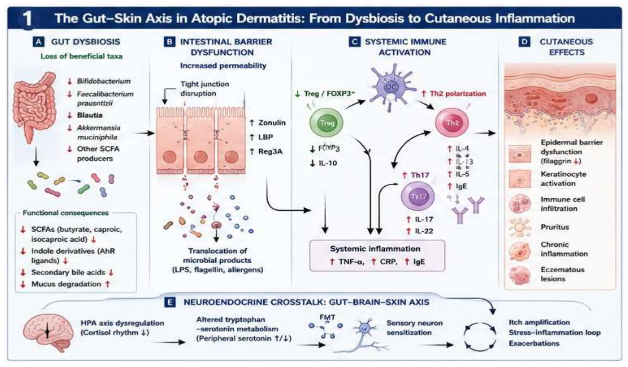
Figure 2.
Microbial metabolites as key immunologic mediators in atopic dermatitis (Figure created in Canva). SCFAs including butyrate, caproic acid, and isocaproic acid activate GPR43 (FFAR2) and GPR109A signaling pathways, inhibit histone deacetylases (HDACs), and promote FOXP3+ Treg differentiation, IL-10 production, epithelial barrier integrity, and suppression of inflammation. Tryptophan-derived indole metabolites activate the aryl hydrocarbon receptor (AhR) in immune cells and keratinocytes, enhancing epidermal differentiation, antimicrobial peptide production, and immune tolerance while reducing pro-inflammatory cytokine signaling. Secondary bile acids regulate epithelial and immune homeostasis through activation of the FXR and TGR5 receptors. Reduced circulating levels of microbial metabolites correlate inversely with EASI and SCORAD scores and positively with inflammatory biomarkers, supporting their role as mechanistic drivers and candidate biomarkers of AD severity.
Figure 2.
Microbial metabolites as key immunologic mediators in atopic dermatitis (Figure created in Canva). SCFAs including butyrate, caproic acid, and isocaproic acid activate GPR43 (FFAR2) and GPR109A signaling pathways, inhibit histone deacetylases (HDACs), and promote FOXP3+ Treg differentiation, IL-10 production, epithelial barrier integrity, and suppression of inflammation. Tryptophan-derived indole metabolites activate the aryl hydrocarbon receptor (AhR) in immune cells and keratinocytes, enhancing epidermal differentiation, antimicrobial peptide production, and immune tolerance while reducing pro-inflammatory cytokine signaling. Secondary bile acids regulate epithelial and immune homeostasis through activation of the FXR and TGR5 receptors. Reduced circulating levels of microbial metabolites correlate inversely with EASI and SCORAD scores and positively with inflammatory biomarkers, supporting their role as mechanistic drivers and candidate biomarkers of AD severity.
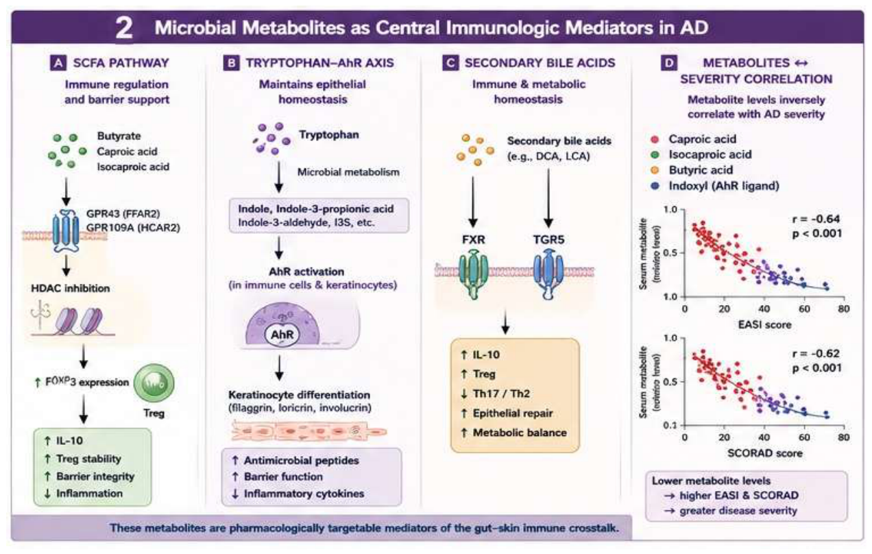
Figure 3.
AI-driven multi-omics integration and precision medicine in atopic dermatitis (Figure created in Canva). Integrated multi-omics platforms combine metagenomic, metabolomic, transcriptomic, proteomic, and single-cell datasets to characterize host–microbiome interactions in AD. Machine learning approaches, including random forest models, graph attention networks, SHAP explainability analysis, and causal inference frameworks, enable identification of key microbial drivers, metabolite signatures, inflammatory pathways, and predictive biomarkers. Multi-omics analyses support stratification of biologically distinct AD endotypes, including SCFA-deficient, barrier-disrupted, neuroimmune, and Th17-high inflammatory subtypes. These approaches facilitate precision therapeutics through personalized probiotics, metabolite-guided therapies, biomarker panels, predictive response models, and individualized microbiome-targeted interventions. Translational integration of AI and systems biology is expected to accelerate biomarker validation, clinical trial optimization, and the implementation of precision dermatology.
Figure 3.
AI-driven multi-omics integration and precision medicine in atopic dermatitis (Figure created in Canva). Integrated multi-omics platforms combine metagenomic, metabolomic, transcriptomic, proteomic, and single-cell datasets to characterize host–microbiome interactions in AD. Machine learning approaches, including random forest models, graph attention networks, SHAP explainability analysis, and causal inference frameworks, enable identification of key microbial drivers, metabolite signatures, inflammatory pathways, and predictive biomarkers. Multi-omics analyses support stratification of biologically distinct AD endotypes, including SCFA-deficient, barrier-disrupted, neuroimmune, and Th17-high inflammatory subtypes. These approaches facilitate precision therapeutics through personalized probiotics, metabolite-guided therapies, biomarker panels, predictive response models, and individualized microbiome-targeted interventions. Translational integration of AI and systems biology is expected to accelerate biomarker validation, clinical trial optimization, and the implementation of precision dermatology.
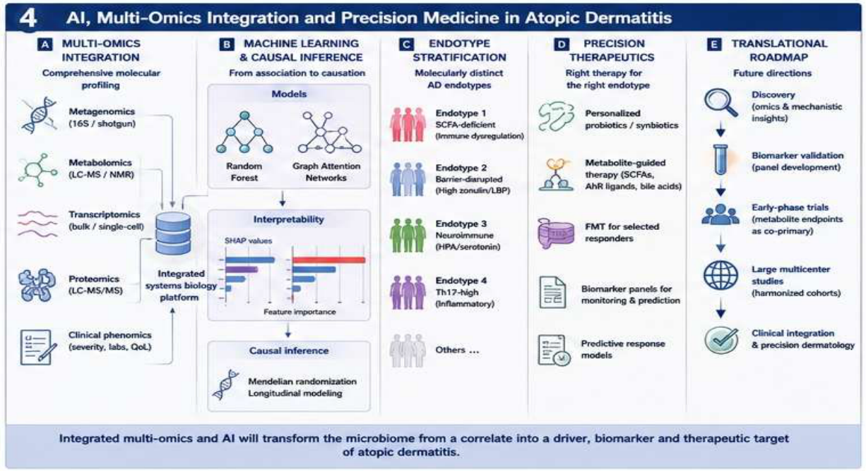
Figure 4.
Therapeutic modulation of the gut microbiome in atopic dermatitis (Figure created in Canva). Probiotics and synbiotics containing Lactobacillus and Bifidobacterium species promote SCFA production, Treg induction, epithelial barrier integrity, and suppression of Th2 cytokine responses. Fecal microbiota transplantation (FMT) restores microbial diversity and metabolite production while reducing systemic inflammation, Th2/Th17 activation, TNF-α, and total IgE levels. Postbiotic and metabolite-based therapies include direct supplementation with SCFAs, AhR agonists, indole derivatives, and secondary bile acids to restore immunometabolic signaling. Clinical studies and meta-analyses demonstrate reductions in EASI and SCORAD scores, decreased inflammatory biomarkers, improved skin barrier function, and enhanced quality of life following microbiome-targeted interventions.
Figure 4.
Therapeutic modulation of the gut microbiome in atopic dermatitis (Figure created in Canva). Probiotics and synbiotics containing Lactobacillus and Bifidobacterium species promote SCFA production, Treg induction, epithelial barrier integrity, and suppression of Th2 cytokine responses. Fecal microbiota transplantation (FMT) restores microbial diversity and metabolite production while reducing systemic inflammation, Th2/Th17 activation, TNF-α, and total IgE levels. Postbiotic and metabolite-based therapies include direct supplementation with SCFAs, AhR agonists, indole derivatives, and secondary bile acids to restore immunometabolic signaling. Clinical studies and meta-analyses demonstrate reductions in EASI and SCORAD scores, decreased inflammatory biomarkers, improved skin barrier function, and enhanced quality of life following microbiome-targeted interventions.
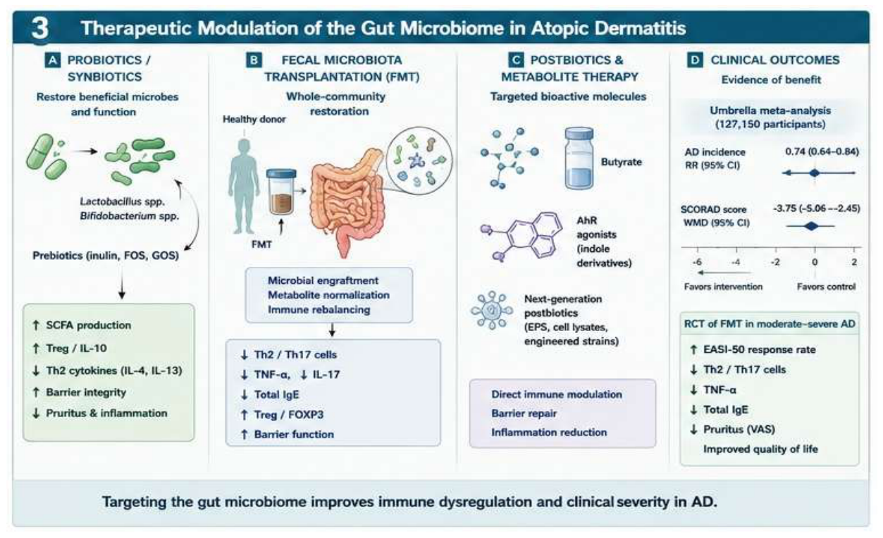
Figure 5.
A critically ill patient is an extreme model of gut–skin axis disruption in atopic dermatitis (Figure created in Canva). Critical illness-associated factors, including sepsis, broad-spectrum antibiotic exposure, mechanical ventilation, altered nutrition, and physiological stress, promote profound gut dysbiosis characterized by depletion of beneficial commensal bacteria and expansion of opportunistic pathogens. Microbial alterations contribute to intestinal barrier dysfunction (“leaky gut”), increased epithelial permeability, and systemic translocation of microbial-associated molecular patterns (MAMPs), including lipopolysaccharide (LPS), flagellin, peptidoglycan, and bacterial DNA. These processes trigger systemic immune dysregulation, characterized by altered innate and adaptive immune responses, cytokine imbalance, and impaired immune tolerance. Concurrent reductions in short-chain fatty acids (SCFAs), tryptophan-derived aryl hydrocarbon receptor (AhR) ligands, and secondary bile acid signaling further disrupt epithelial and immunologic homeostasis. Neuroendocrine alterations involving the hypothalamic–pituitary–adrenal (HPA) axis and gut–brain–skin communication amplify inflammatory signaling. Collectively, these mechanisms contribute to epidermal barrier dysfunction, keratinocyte activation, chronic inflammation, and amplification of AD-related pathogenic pathways. The figure also highlights potential microbiome-targeted therapeutic strategies to restore gut microbial homeostasis and immune regulation in critically ill patients. Abbreviations: AD, atopic dermatitis; ICU, intensive care unit; SCFA, short-chain fatty acid; AhR, aryl hydrocarbon receptor; LPS, lipopolysaccharide; LBP, lipopolysaccharide-binding protein; Reg3A, regenerating islet-derived protein 3A; HPA, hypothalamic–pituitary–adrenal.
Figure 5.
A critically ill patient is an extreme model of gut–skin axis disruption in atopic dermatitis (Figure created in Canva). Critical illness-associated factors, including sepsis, broad-spectrum antibiotic exposure, mechanical ventilation, altered nutrition, and physiological stress, promote profound gut dysbiosis characterized by depletion of beneficial commensal bacteria and expansion of opportunistic pathogens. Microbial alterations contribute to intestinal barrier dysfunction (“leaky gut”), increased epithelial permeability, and systemic translocation of microbial-associated molecular patterns (MAMPs), including lipopolysaccharide (LPS), flagellin, peptidoglycan, and bacterial DNA. These processes trigger systemic immune dysregulation, characterized by altered innate and adaptive immune responses, cytokine imbalance, and impaired immune tolerance. Concurrent reductions in short-chain fatty acids (SCFAs), tryptophan-derived aryl hydrocarbon receptor (AhR) ligands, and secondary bile acid signaling further disrupt epithelial and immunologic homeostasis. Neuroendocrine alterations involving the hypothalamic–pituitary–adrenal (HPA) axis and gut–brain–skin communication amplify inflammatory signaling. Collectively, these mechanisms contribute to epidermal barrier dysfunction, keratinocyte activation, chronic inflammation, and amplification of AD-related pathogenic pathways. The figure also highlights potential microbiome-targeted therapeutic strategies to restore gut microbial homeostasis and immune regulation in critically ill patients. Abbreviations: AD, atopic dermatitis; ICU, intensive care unit; SCFA, short-chain fatty acid; AhR, aryl hydrocarbon receptor; LPS, lipopolysaccharide; LBP, lipopolysaccharide-binding protein; Reg3A, regenerating islet-derived protein 3A; HPA, hypothalamic–pituitary–adrenal.
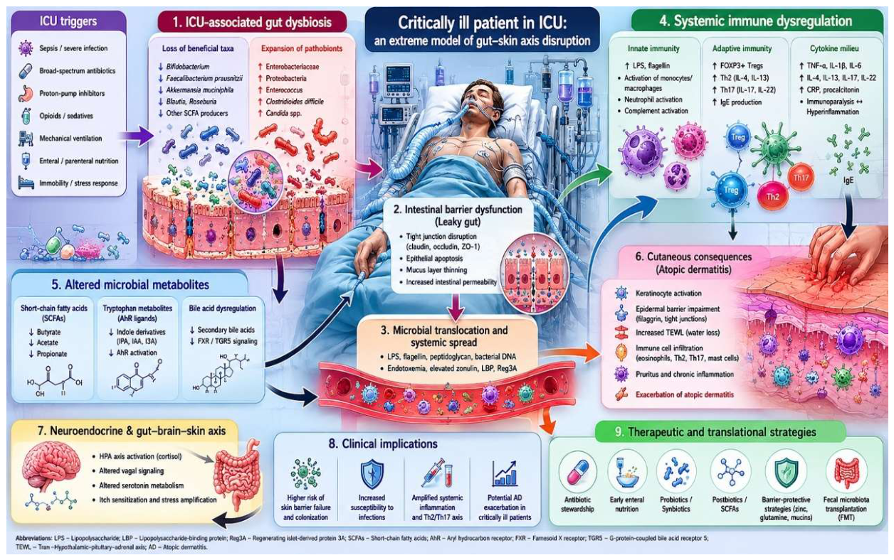

Table 1.
Gut-Derived Metabolites and Intestinal Barrier Biomarkers Associated with Clinical Severity in Atopic Dermatitis.
Table 1.
Gut-Derived Metabolites and Intestinal Barrier Biomarkers Associated with Clinical Severity in Atopic Dermatitis.
| Biomarker | Direction | Clinical correlation | Evidence |
|---|---|---|---|
| Butyrate | ↓ | EASI/SCORAD | High |
| Caproic acid | ↓ | EASI | Moderate |
| Indoxyl | ↑ | Severity | Moderate |
| Zonulin | ↑ | Barrier dysfunction | High |
| LBP | ↑ | Inflammation | High |
| Reg3A | ↑ | Severity | Emerging |
Legend: AD, atopic dermatitis; EASI, Eczema Area and Severity Index; SCORAD, SCORing Atopic Dermatitis; LBP, lipopolysaccharide-binding protein. ↑ denotes increased circulating levels or abundance, whereas ↓ denotes decreased circulating levels or abundance. Associations are reported according to their relationship with AD severity and disease activity.
Table 2.
Comparative Overview of Microbiome-Targeted Interventions in Atopic Dermatitis: Evidence Strength, Clinical Outcomes, and Current Limitations.
Table 2.
Comparative Overview of Microbiome-Targeted Interventions in Atopic Dermatitis: Evidence Strength, Clinical Outcomes, and Current Limitations.
| Intervention | Best Evidence Level | Key Outcome | Key Limitation |
|---|---|---|---|
| Multi-strain probiotics (Lactobacillus) | Umbrella meta-analysis (2025) | SCORAD WMD -3.75 | Strain heterogeneity; moderate disease only |
| Synbiotics | Umbrella meta-analysis (2025) | SCORAD significantly decreased | Limited head-to-head vs. probiotics alone |
| Bifidobacterium monostrain | Multiple meta-analyses | Inconsistent SCORAD effect | Insufficient as monotherapy |
| Prebiotics alone | Multiple meta-analyses | No significant SCORAD effect | Preventive > therapeutic role |
| Postbiotics | Pilot studies | IgE decreased; barrier improved | Early stage; no large RCTs |
| FMT (conventional) | RCT (2025, Allergy) | EASI-50 significantly increased | Standardization; long-term safety unknown |
| WMT | Prospective cohort (2025) | SCORAD/EASI/NRS significantly decreased | No RCT design; small samples |
| Dietary intervention | Observational/RCT | Microbiome diversity increased; severity decreased | Confounders; long-term adherence |
Legend: SCORAD, Scoring Atopic Dermatitis; EASI, Eczema Area and Severity Index; NRS, Numeric Rating Scale; IgE, immunoglobulin E; FMT, fecal microbiota transplantation; WMT, washed microbiota transplantation; RCT, randomized controlled trial; WMD, weighted mean difference.
Disclaimer/Publisher’s Note: The statements, opinions and data contained in all publications are solely those of the individual author(s) and contributor(s) and not of MDPI and/or the editor(s). MDPI and/or the editor(s) disclaim responsibility for any injury to people or property resulting from any ideas, methods, instructions or products referred to in the content. |
© 2026 by the authors. Licensee MDPI, Basel, Switzerland. This article is an open access article distributed under the terms and conditions of the Creative Commons Attribution (CC BY) license (http://creativecommons.org/licenses/by/4.0/).
Copyright: This open access article is published under a Creative Commons CC BY 4.0 license, which permit the free download, distribution, and reuse, provided that the author and preprint are cited in any reuse.


