

Submitted:
22 June 2026
Posted:
23 June 2026
You are already at the latest version
Abstract
The goal of study was to identify bacterial species isolated from canine non-healing ulcers and evaluate temporal changes in resistance patterns. Corneal microbiology samples were collected from 228 non-healing ulcers of 221 dogs between January 2019 and February 2023. Samples were cultured aerobically and isolates were identified using MALDI-ToF mass spectrometry. Antibiotic susceptibility testing was performed using Kirby-Bauer disc diffusion according to current CLSI guidelines. Acquired resistance patterns were compared between 2019–2020 and 2021–2023, and multidrug-resistant (MDR) and possible extensively drug-resistant (XDR) isolates were characterized. Bacterial growth was observed in 71% (163/228) of samples, yielding 170 isolates. The most frequently isolated species were Staphylococcus epidermidis (21%), Staphylococcus pseudintermedius (18%), Staphylococcus capitis (15%), and other coagulase-negative staphylococci (14%). Overall susceptibility was highest to combinations involving neomycin-polymyxin-bacitracin (neopolybac). A statistically significant increase in overall bacterial resistance (P = 0.000008) and acquired resistance (P = 0.000504) was observed from 2019–2020 to 2021–2023. Methicillin-resistant Staphylococcus spp. comprised 61% of staphylococcal isolates, with an increase from 47% (2019–2020) to 70% (2021–2023). Over half the isolates (53%) were multidrug-resistant. Topical antibiotic regimens containing neomycin-polymyxin-bacitracin provided the broadest coverage. Increasing resistance, particularly methicillin resistance in staphylococci, highlights the concern about the development of highly resistant bacteria which can potentially increase crossover infections between humans and dogs.
Keywords:
cornea
; ulcer
; non-healing
; antibiogram
1. Introduction
Spontaneous chronic corneal epithelial defects (SCCEDs), also known as indolent or non-healing corneal ulcers, represent one of the most common chronic corneal diseases encountered in canine ophthalmology. The condition is characterized by failure of normal epithelial adhesion, abnormalities of the epithelial basement membrane, and formation of an acellular hyaline stromal zone that interferes with normal re-epithelialization, with frequent topical antibiotic treatment for weeks to months until healing is achieved. Histopathologic studies have demonstrated disruption of epithelial-stromal attachment mechanisms, resulting in persistent epithelial defects despite the absence of significant stromal inflammation or neurogenic keratitis component [1,2]. Although SCCEDs are well recognized in veterinary medicine, they share important clinical and biological similarities with human persistent epithelial defects (PEDs). In both species, chronic disruption of the corneal epithelial barrier predisposes the ocular surface to microbial colonization, inflammation, stromal degradation, and delayed wound healing. Human persistent epithelial defects may develop secondary to neurotrophic disease, herpetic keratitis, ocular surface inflammation, limbal stem cell deficiency, graft-versus-host disease, autoimmune disease, or ocular surgery, and remain a significant cause of visual morbidity worldwide [3,4]. Despite differences in underlying etiology, SCCEDs and human PEDs share prolonged epithelial instability, recurrent healing failure, and dependence on supportive therapies designed to restore epithelial integrity [3,5]. A growing body of evidence suggests that chronic epithelial defects should not be viewed solely as disorders of epithelial adhesion but also as disturbances of the ocular surface microbial ecosystem. The normal ocular surface harbors a diverse microbial community that contributes to immune homeostasis and barrier defense [6,7,8]. Loss of epithelial integrity alters this relationship and may permit colonization by opportunistic organisms, particularly coagulase-negative staphylococci and other commensal bacteria. In human ophthalmology, bacterial colonization and secondary infection are recognized complications of persistent epithelial defects and neurotrophic keratopathy, often necessitating prophylactic antimicrobial therapy to prevent stromal ulceration and corneal perforation [4]. Simultaneously, antimicrobial resistance has emerged as one of the most significant challenges facing both human and veterinary medicine. Ocular surveillance programs have documented increasing prevalence of methicillin-resistant staphylococci, multidrug-resistant organisms, and declining susceptibility to commonly used ophthalmic antibiotics [9,10].Companion animals and humans occupy increasingly shared environments, creating opportunities for exchange of antimicrobial-resistant bacteria and resistance determinants [11,12]. Consequently, antimicrobial resistance in ocular pathogens has become an important One Health concern with implications extending beyond individual species [13]. Current treatment recommendations for canine SCCEDs typically include epithelial debridement procedures such as diamond burr keratectomy, grid keratotomy, superficial keratectomy, or thermal cautery, combined with topical antimicrobial therapy [14,15,16,17,18,19,20]. However, despite the routine use of antibiotics, surprisingly little is known regarding the microbiological characteristics of SCCEDs. To date, only a single dedicated study has evaluated bacterial isolates from canine indolent ulcers and reported positive bacterial cultures in approximately 20% of cases [21]. The prevalence of bacterial colonization, distribution of microbial species, antimicrobial susceptibility patterns, and temporal trends in resistance remain largely undefined. The objectives of the present study were therefore to characterize bacterial species isolated from canine SCCED/non-healing corneal ulcers, determine their antimicrobial susceptibility profiles, evaluate temporal changes in resistance patterns, and identify optimal empirical antimicrobial strategies. Additionally, we sought to explore the broader translational relevance of chronic epithelial defects as potential reservoirs of antimicrobial resistance and to compare microbial patterns observed in canine SCCEDs with those reported in human chronic corneal epithelial disease.
2. Materials and Methods
2.1. Sample Collection and Processing
Samples for microbiology analyses were collected from 221 dogs and a total of 228 eyes with non-healing corneal ulcers in the period January 2019 – February 2023. All patients had a complete eye examination. The antibiotic treatment was initialized with triple antibiotic ointment cream (neomycin-polymyxin-bacitracin) q8h followed by the change of the topical antibiotic based on the antibiogram results of antibiogram. The inclusion criteria for the study were presence of corneal epithelial defect for the duration longer than 10 days despite appropriate medical therapy, absence of corneal stromal involvement, and presence of the loose epithelial margins at the ulcer edges at the time of initial diagnosis. Data from additional 62 patients with non-healing ulcers treated where standard ofloxacin protocol (q6h) without pursuing ocular microbiology were included in the analysis (patients were seen in the period 2012–2013). Additional 3 patients (two patients with a non-healing ulcer, and one patient with corneal stromal ulcer seen in 2025), had corneal confocal microscopy performed (VivaScope 3000, Caliber ID, Rochester, NY, USA) under the topical anesthesia in combination with bacterial culture testing. Clinical canine microbiology samples were collected from corneal non-healing ulcers using a flocked swab and placed in the provided transport media (BD ESwabTM Collection Kit, COPAN ITALIA SpA, Brescia, Italy). After applying topical anesthetic on the ocular surface (Propracaine 0.05%, Akorn Pharmaceuticals, Lake Forest, IL, USA) samples were harvested and then placed on ice packs until sent to the laboratory or were immediately cultured. Approximately half of each sample in ESwab transport media (roughly 0.5ml, concentrated via centrifugation to increase recovery) was cultured onto MacConkey agar (Hardy Diagnostics, Santa Maria, CA, USA) whereas the other half of the sample was cultured onto Chocolate agar (Hardy Diagnostics Hardy Diagnostics, Santa Maria, CA, USA). The plates were then placed in the incubator at 370C in 5% CO2, and inspected at 24, 48 and 96 hours. Presence or absence of bacterial colonies was recorded.
2.2. Standard Microbial Culture and Antimicrobial Susceptibility Testing
Standard microbial culture (aerobic) and antimicrobial susceptibility testing were performed as previously described [22]. Briefly, samples were inoculated onto Chocolate and MacConkey agar, incubated aerobically and anaerobically at 37 °C for 24–96 hours. Growth was assessed morphologically, and isolates were identified using Gram staining, and automated MALDI-ToF identification (Bruker BioTyper; MBT Compass database, updated biannually). Antibiotic susceptibility was determined via Kirby-Bauer disk diffusion on Mueller-Hinton agar, interpreting zones as susceptible (S), intermediate (counted as S for analysis), or resistant (R). WHONET database software (World Health Organization) containing 2021 (or later) CLSI breakpoints for dogs was used to manage and analyze microbiology laboratory data and antibiotic susceptibility test results. Other CLSI animal breakpoints were used if breakpoints were not available for dogs. If no other animal breakpoints were available, human CLSI breakpoints were applied. The breakpoints were based upon CLSI VET08 Performance Standards for Antimicrobial Disk and Dilution Susceptibility Tests for Bacteria Isolated From Animals (https://clsi.org/media/2321/vet08ed4_sample.pdf), CLSI M100 Performance Standards for Antimicrobial Susceptibility Testing (updated yearly, https://em100.edaptivedocs.net/Login.aspx) and Hardy Diagnostics Disk Diffusion Zone Diameter Chart (https://www.keyscientific.com/files/Other%20Manufacturers/Hardy%20Diagnostics/AST%20Discs/Hardy%20AST%20Disc%20Insert.pdf). This chart was also used for polymyxin B breakpoints in Pseudomonas aeruginosa (R ≤ 11; S≥12). To detect penicillin-binding protein 2A (PBP2A) in staphylococcal isolates, the AlereTM PBP2A SA Culture Colony Test was performed following manufacturer’s instructions (Alere Scarborough, Inc., 10 Southgate Road, Scarborough, ME 04074). All bacterial isolates were assigned into resistant and susceptible categories relative to their in vitro response to antibiotics. Susceptible and intermediate interpretations were grouped together to the susceptible class considering the high concentration of antibiotics that can be delivered directly to eyes. Tested antibiotics were grouped into the antibiotic categories of aminoglycosides (amikacin, gentamicin, neomycin, and tobramycin), polypeptides/polymyxins (bacitracin, polymixin B), anti-staphylococcal β-lactams (cephamycins; oxacillin, cefoxitin), tetracyclines (tetracycline), non-extended spectrum cephalosporins (1st and 2nd generation cephalosporins; cefazolin), penicillins with and without β-lactamase inhibitors (amoxicillin/clavulanic acid, penicillin G), macrolides (erythromycin), and fluoroquinolones (ofloxacin, moxifloxacin, and ciprofloxacin). Additionally, each clinical isolate was allocated to a not multidrug-resistant (Not MDR), multidrug-resistant (MDR), or possible extensively multidrug-resistant (possible XDR) group based upon its susceptibility data. The MDR group was defined as resistance to at least one antibiotic in three or more antibiotic categories. XDR was defined as resistance to at least one antibiotic in all but two or fewer antibiotic categories (i.e., bacterial isolates remain susceptible to only one or two categories) as previously published [23,24]. Further, if an isolate had intrinsic resistance to a particular antibiotic, then the corresponding resistance was excluded from the analysis [25]. Total of 176 eyes in which ocular microbiology analysis was performed and 62 eyes without microbiology analysis treated with ofloxacin only were treated with diamond burr and placement of the soft contact lens (PureVision, Bauch and Lomb, St Louis, Missouri, USA) as previously described [15]. The healing time was calculated from the time of treatment to the time of the corneal epithelium defect closure, which was determined via slit lamp examination and negative fluorescein staining of the defect.
2.3. Statistical Analysis
Commercial software was used for statistical analyses as described in the manuscript and as previously reported (Prism, version 5.0; GraphPad, San Diego, CA) [22].
3. Results
3.1. Growth of Bacteria from Patient Samples
Samples were collected from 221 dogs and a total of 228 non-healing corneal ulcer samples were plated. In 71% (163/228) of plated samples a bacterial growth was observed, while 29% (65/228) of plated samples yielded no growth in the culture media. A total of 170 isolates were obtained and analyzed in this study (some corneal ulcer samples had multiple isolates accounting for the larger number of isolates compared to the number of corneal ulcers with positive growth).
3.2. Distribution of Bacterial Species Isolated from Patients With Corneal Non-Healing Ulcers
The most frequent microbial species identified in the corneal non-healing samples were Staphylococcus epidermidis 21% (36/170), Staphylococcus pseudintermedius 18% (31/170), and Staphylococcus capitis 15% (25/170) followed by coagulase negative staphylococci present in 14% (24/170) of samples (Table 1).
3.3. Antibiotic Susceptibility Profile of Bacteria Isolated From Corneal Non-Healing Ulcers
To determine a suitable antibiotic treatment for canine corneal non-healing ulcers, we examined overall bacterial resistance regarding the use of a single or combination antibiotic between two time points 2019–2020 and 2021–2023. Based upon the general antibiotic susceptibility data, neopolybac alone (94%) or a combination of neopolybac with ofloxacin (98%) or amikacin (96%) appeared to be the optimal antibiotic treatment for canine corneal non-healing ulcers (Figure 1). Importantly, statistically significant increase in bacterial resistance (p = 0.000008, Paired t-test) was observed for the period 2021–2023 in comparison to the period 2019–2020 regarding the use of a single or combination antibiotic (Figure 1).
In samples tested with topical antibiotics, bacterial species were mainly resistant topolymyxin B 69% (117/170) and oxacillin 62% (21/34) followed by cefazolin 55% (94/170), and cefoxitin 51% (85/166). The least resistant species were against bacitracin 16% (27/170), amikacin 18% (31/170), ofloxacin 23% (39/170), and gentamicin 23% (39/169; Table 2). If intrinsic resistance was excluded from the analysis therefore considering only acquired resistance, a similar trend of resistance was observed. Bacterial species were predominantly resistant to polymyxin B 68% (114/167), oxacillin 62% (21/34), cefazolin 52% (81/157), and cefoxitin 48% (76/157). The least resistant species were against amikacin 15% (24/163), bacitracin 15% (25/168), gentamicin 20% (32/162), and neomycin 20% (32/158; Table 2).
Moreover, lower numbers of acquired bacterial resistance are detected comparing to overall resistant numbers. The exception from this pattern was no difference between acquired and total bacterial resistance in the case of oxacillin (Table 2). In samples tested with systemic antibiotics (usually samples from patients with the extensive corneal neovascularization reaching the corneal ulcer region), bacterial species were predominantly resistant to ampicillin 92% (12/13), penicillin G 87% (14/16), cephalexin 62% (10/16), clindamycin 62% (10/16), oxacillin 62% (21/34), cefoxitin 51% (85/166), amoxicillin/clavulanic acid 40% (36/89) followed by sulfamethoxazole/trimethoprim 31% (5/16), ciprofloxacin 31% (25/81), enrofloxacin 25% (4/16), marbofloxacin 25% (4/16), amikacin 18% (31/170), and doxycycline 13% (2/15).
3.4. Temporal Increase of Acquired Resistance In Isolates From Corneal Non-Healing Ulcers
To analyze dynamics of acquired resistance over time, we examine antibiotic susceptibility profile of isolates between two time points 2019–2020 and 2021-2023. In the period 2019-2020, isolates were predominantly resistant to polymyxin B (65%), oxacillin (45%), and cefazolin (41%). A similar trend was observed for the period 2021-2023 (polymyxin B (70%), oxacillin (70%), and cefazolin (59%; Figure 2). The smallest percentage of isolates was resistant to bacitracin (7%), amikacin (12%), gentamicin (17%), and neomycin (17%) in the period 2019-2020. In the period 2021–2023, the lowest percentage of isolates was resistant to amikacin (16%), bacitracin (20%), gentamicin (22%), and neomycin (23%). Overall, apparently higher percentages of resistance were noticed in the period 2021-2023 than in the period 2019–2020. Accordingly, statistically significant changes (p = 0.000504, Paired t-test) in acquired resistance were observed between these two time points (Figure 2).
3.5. Distribution of Bacterial Species In Patients Diagnosed with Corneal Non-Healing Ulcers Relative to Previous Patient’s Antibiotic Treatment
The most frequent bacteria identified in the corneal non-healing samples relative to the patient’s previous antibiotic treatments were Staphylococcus pseudintermedius, coagulase negative staphylococci, Staphylococcus capitis, Staphylococcus epidermidis, and Pseudomonas aeruginosa (Table 3). In comparison to the overall data in Table 1, most of isolates were present in relatively similar percentages. Consequently, no statistically significant difference (p = 0.313141, paired t-test) was observed in the distribution of bacterial species relative to the patient’s previous antibiotic treatments.
3.6. Susceptibility Profile of Isolates From Corneal Non-Healing Ulcers Relative To Patient’s Previous Antibiotic Treatments
To address a possible temporal change of acquired resistance relative to previous the patient’s antibiotic treatments, we analyzed isolates within an approximately four-year period from 2019 to 2023. In the group of patients without previous antibiotic treatments, the highest percentage of isolates was resistant to oxacillin, polymyxin B, cefazolin, and cefoxitin with a similar trend in the group previously exposed to antibiotics (Figure 3). The lowest percentage of isolates was resistant to amikacin/ofloxacin and bacitracin/amikacin in the group of patients without previous antibiotic treatments and the group previously exposed to antibiotic, respectively. Moreover, no statistically significant changes (p = 0.439938, paired t-test) in acquired resistance were observed relative to the patient’s previous antibiotic treatments within the examined four-year period (Figure 3).
3.7. Resistance Profile Based on The Number of Resistant Antibiotics in Canine Corneal Non-Healing Ulcers
The highest percentage of bacteria was resistant to 1 antibiotic 17% (29/170) and 2 antibiotics 15% (25/170). The lowest percentage of bacteria was resistant to 12 antibiotics 1% (1/170), 10 antibiotics 2% (4/170), 9 antibiotics 3% (5/170), and 11 antibiotics 4% (6/170; Figure 4).
3.8. Distribution of Multidrug-Resistant Bacteria in Clinical Corneal Non-Healing Ulcers
Overall, in the corneal non-healing ulcer samples, 47% (80/170) were not MDR, 20% (34/170) belong the MDR group while 33% (56/170) were possible XDR. Thus, over a half of isolates belong to a general multi-drug resistant category. In the period 2019–2020, 57% (40/70) were not MDR isolates, 20% (14/70) were MDR isolates while 23% (16/70) were possible XDR isolates. In the period 2021–2023, 40% (40/100) were not MDR isolates, 20% (20/100) were MDR isolates while 40% (40/100) were possible XDR isolates. Moreover, no statistically significant difference (p = 0.411720, Paired t-test) was observed in multidrug resistance although a decrease of not MDR and increase of XDR percentages were noticed between 2019–2020 and 2021–2023.
3.9. Methicillin-Resistant Staphylococcus Spp. in Clinical Corneal Non-Healing Ulcers
To address methicillin resistance in Staphylococcus species, the isolates were probed with cefoxitin disks 15. Per CLSI VET08 guidelines (https://clsi.org/media/2321/vet08ed4_sample.pdf) for Staphylococcus pseudintermedius and Staphylococcus schleiferi, the results were confirmed with oxacillin disks. Additionally, the penicillin-binding protein 2A (PBP2A) antibody test was used to determine a status of methicillin resistance in a few species. Overall, in the 2019-2023 period, 61% (68/112) isolates were methicillin-resistant. Regarding patient’s previous antibiotic treatments, a conspicuous increase in the percentage of methicillin resistant isolates was detected in the group with no previous antibiotic treatment 74% (14/19). In the group with previous antibiotic treatment 58% (54/93) of isolates were methicillin resistant. Over time, a noticeable increase in the percentage of methicillin-resistant Staphylococcus species was observed from 2019-2020 (47%; 20/43) to 2021-2023 (70%; 48/69).
4.0. Difference in Healing Time Between Non-Healing Ulcers With And Without Bacterial Growth
There was no statistically significant difference in healing times between ulcers with bacterial growth vs. no growth observed in this study (Welch’s t-test, p = 0.948): no growth group (51 eyes) = 17.25+/-1.16 days (mean+/-SEM) and bacterial growth group (125 eyes) 17.34+/=0.74 days. Some patients did not elect to pursue treatment or present for the recheck evaluations resulting in the discrepancy in numbers between total dogs with corneal microbiology isolates, and number of dogs for which healing time data was available. We have further evaluated whether there is a statistical difference in healing time between non-healing ulcers in dogs with bacterial growth, and non-healing ulcers (57 eyes) treated with generic ofloxacin protocol (q6h) without pursuing corneal microbiology testing. Ofloxacin treated dogs had significantly longer healing time at 20.39 +/- 1.58 (Welch’s t-test, p = 0.02) when compared to non-healing ulcers with bacterial growth group. Conversion to corneal stromal ulcer occurred in 0/125 (0%) eyes in non-healing ulcer group with bacterial growth and initially treated with neopolybac, and 5/62 (8.1%) eyes in ofloxacin group, representing a significantly increased risk in ofloxacin group (Fisher’s exact test, p = 0.003; OR 24.0, 95% CI 1.3–438.6). Confocal corneal imaging in limited number of patients (Figure 5) revealed numerous presence of inflammatory cells in ulcerated corneas with positive bacterial growth.
4. Discussion
The present study provides the largest microbiological characterization of canine spontaneous chronic corneal epithelial defects (SCCEDs) reported to date and offers several observations that may have significance beyond veterinary ophthalmology. Although SCCEDs have traditionally been regarded as sterile or minimally contaminated lesions, bacterial growth was identified in 71% of cases, a prevalence substantially higher than previously reported in dogs and remarkably similar to frequencies reported in human persistent epithelial defects (PEDs) [4,21]. These findings suggest that chronic epithelial defects, regardless of species, may create a shared biological niche that promotes bacterial colonization of the ocular surface. The predominance of coagulase-negative staphylococci, including Staphylococcus epidermidis and Staphylococcus capitis, is particularly noteworthy. Historically regarded as relatively benign commensals, these organisms are now increasingly recognized in human medicine as important opportunistic pathogens and major reservoirs of antimicrobial resistance genes [26,27]. Coagulase-negative staphylococci frequently harbor mecA-containing SCCmec elements and other mobile resistance determinants capable of horizontal transfer to more pathogenic species [28]. The observation that approximately half of all isolates in this study belonged to this bacterial group suggests that chronic corneal epithelial defects may serve as localized reservoirs of resistance-associated microorganisms within the ocular microbiome. Despite the high prevalence of bacterial isolation, culture-positive ulcers did not heal more slowly than culture-negative ulcers following diamond burr debridement and bandage contact lens placement. This finding supports the hypothesis that many recovered organisms may function primarily as colonizers rather than invasive pathogens. Similar observations have been reported in human persistent epithelial defects, neurotrophic keratopathy, and ocular graft-versus-host disease, where microbial colonization frequently accompanies epithelial breakdown without necessarily serving as the primary driver of delayed epithelial healing. Instead, the underlying epithelial dysfunction appears to be the dominant pathological mechanism, while bacterial colonization develops as a secondary consequence of prolonged barrier disruption [3,4,5]. From a translational perspective, the most concerning finding was the progressive increase in antimicrobial resistance observed during the study period. Overall resistance and acquired resistance both increased significantly between 2019–2020 and 2021–2023. Furthermore, methicillin-resistant staphylococci increased from 47% to 70% during the same interval, while more than half of all isolates met criteria for multidrug-resistant (MDR) or possible extensively drug-resistant (XDR) phenotypes. These trends parallel resistance patterns increasingly reported in human ophthalmology, where methicillin-resistant Staphylococcus aureus (MRSA), methicillin-resistant coagulase-negative staphylococci, and fluoroquinolone-resistant ocular pathogens have emerged as major therapeutic challenges [10]. The similarities between resistance trends observed in veterinary and human ocular isolates suggest that common ecological and antimicrobial selection pressures may be operating across species. The current findings also support the growing importance of One Health approaches to antimicrobial resistance surveillance. Companion animals share close environmental and physical contact with humans, creating opportunities for bidirectional exchange of bacterial organisms and resistance determinants. Previous studies have demonstrated genetic similarities between antimicrobial-resistant staphylococcal isolates recovered from dogs and their owners, suggesting that companion animals may participate in shared resistance ecosystems [11,29]. Although transmission dynamics were not evaluated in the present study, the rapid increase in methicillin resistance and multidrug resistance observed within a relatively short time frame raises important questions regarding environmental, healthcare-associated, and household factors contributing to resistance emergence. An additional translational implication involves the utility of canine SCCEDs as a naturally occurring model of chronic epithelial disease. Human persistent epithelial defects remain a significant therapeutic challenge and are associated with neurotrophic keratopathy, herpetic disease, autoimmune ocular surface disorders, limbal stem cell deficiency, ocular graft-versus-host disease, and post-surgical epithelial dysfunction. While SCCEDs differ in etiology, both conditions share prolonged epithelial barrier disruption, susceptibility to bacterial colonization, recurrent healing failure, and dependence on supportive therapies to restore epithelial integrity. The comparable prevalence of bacterial recovery observed between canine SCCEDs and human PEDs suggests that naturally occurring canine disease may provide a valuable translational platform for studying host-microbe interactions, ocular surface microbiome dynamics, antimicrobial stewardship strategies, and novel approaches to epithelial regeneration. The antibiotic susceptibility findings further reinforce the importance of evidence-based antimicrobial stewardship. Although neomycin-polymyxin B-bacitracin provided the broadest empirical coverage in this population, the rapid increase in resistance observed during the study period emphasizes that empirical antibiotic selection should not replace microbiological evaluation in refractory or atypical cases. The goal of antimicrobial therapy in chronic epithelial defects may be less about eradication of invasive infection and more about preventing secondary bacterial overgrowth while preserving the effectiveness of available antibiotics. This concept mirrors contemporary recommendations in human ophthalmology, where increasing resistance rates have prompted greater emphasis on culture-guided therapy and antimicrobial stewardship. Several limitations should be acknowledged. The study was retrospective and performed at a single referral center, potentially limiting geographic generalizability. Anaerobic organisms and molecular microbial characterization were not evaluated. In addition, whole-genome sequencing and resistance gene analysis were not performed, preventing assessment of specific resistance mechanisms or comparison with human ocular isolates. Future studies integrating metagenomics, microbiome profiling, and genomic epidemiology may help clarify whether resistant organisms identified in canine ocular disease share common evolutionary pathways with those observed in human ophthalmology.
5. Conclusions
Canine SCCEDs appear to represent a chronic ocular surface disorder characterized by frequent bacterial colonization, increasing antimicrobial resistance, and substantial methicillin-resistant staphylococcal involvement. The similarities between canine SCCEDs and human persistent epithelial defects suggest that naturally occurring canine disease may serve as a useful translational model for studying chronic epithelial failure, ocular surface microbiology, and antimicrobial resistance. Continued microbiological surveillance and antimicrobial stewardship efforts are likely essential for preserving therapeutic efficacy in both veterinary and human ophthalmology.
Author Contributions
Conceptualization, S.G., T.L., M.J. and B.F.; methodology, S.G., B.F., J.C., M.J., T.L.; software, D.R.; validation S.G., BF.; formal analysis M.J., J.C., D.R., S.G.; investigation, SG, M.J., J.C.; resources, S.G., T.L.; data curation, S.G., T.L., B.F.; writing—original draft preparation, S.G., M.J., T.L., B.F.; writing—review and editing, S.G., B.F.; visualization, S.G., D.R.; supervision S.G., T.L.; project administration, S.G., T.L.; funding acquisition, S.G., T.L. All authors have read and agreed to the published version of the manuscript.
Funding
Animal Eye Consultants of Iowa Internal Research Funding.
Institutional Review Board Statement
This study complies with the Guidelines for Ethical Research in Veterinary Ophthalmology (GERVO) and is exempt from approval by an ethics committee due to the retrospective nature of the study.
Informed Consent Statement
Animal owners or owners’ representatives provided written consent for the treatment provided.
Data Availability Statement
Limited data availability will be provided without client and animal identifiers upon the request to the corresponding author due to privacy laws.
Acknowledgments
None.
AI Use Statement
No AI tools were used for data analysis presented in this manuscript. Artificial intelligence tool (ChatGPT, OpenAI) was used to assist with manuscript organization and language editing.
Conflicts of Interest
The authors declare no conflicts of interest.
Abbreviations
The following abbreviations are used in this manuscript:
| CLSI | Clinical and Laboratory Standards Institute |
| ITS | Internal Transcribed Spacer |
References
- Bentley E. Spontaneous chronic corneal epithelial defects in dogs: a review. J Am Anim Hosp Assoc. 2005;41(3):158–65.
- Bentley E, Abrams GA, Covitz D, Cook CS, Fischer CA, Hacker D, et al. Morphology and immunohistochemistry of spontaneous chronic corneal epithelial defects (SCCED) in dogs. Investigative ophthalmology & visual science. 2001;42(10):2262–9.
- Ong ES, Jeng BH. Current and future therapies for persistent corneal epithelial defects and neurotrophic keratopathy. Curr Opin Ophthalmol. 2021;32(3):262–7.
- Sepulveda-Beltran PA, Carletti P, Banda V, Mulpuri L, Levine H, Amescua G, et al. Infectious and Noninfectious Corneal Ulcers in Ocular Graft-Versus-Host Disease: Epidemiology, Clinical Characteristics, and Outcomes. Am J Ophthalmol. 2024;257:236–46.
- Cheung AY, Holland EJ. Neurotrophic Keratopathy: Current Concepts, Management, and Emerging Treatments. Cornea. 2026;45(6):673–85.
- Kugadas A, Gadjeva M. Impact of Microbiome on Ocular Health. Ocul Surf. 2016;14(3):342–9.
- Li JJ, Yi S, Wei L. Ocular Microbiota and Intraocular Inflammation. Front Immunol. 2020;11:609765.
- Delbeke H, Younas S, Casteels I, Joossens M. Current knowledge on the human eye microbiome: a systematic review of available amplicon and metagenomic sequencing data. Acta Ophthalmol. 2021;99(1):16–25.
- Asbell PA, Sanfilippo CM, Sahm DF, DeCory HH. Trends in Antibiotic Resistance Among Ocular Microorganisms in the United States From 2009 to 2018. JAMA Ophthalmol. 2020;138(5):439–50.
- Asbell PA, Sahm DF, Shaw M, Draghi DC, Brown NP. Increasing prevalence of methicillin resistance in serious ocular infections caused by Staphylococcus aureus in the United States: 2000 to 2005. J Cataract Refract Surg. 2008;34(5):814–8.
- Boost MV, O’Donoghue MM, James A. Prevalence of Staphylococcus aureus carriage among dogs and their owners. Epidemiol Infect. 2008;136(7):953–64.
- Nebbia P, Robino P, Iamone G, Schena R, Arslan S, Cavalli S, et al. Nasal carriage of Staphylococcus aureus in dogs and owners: Spa typing and antimicrobial resistance in two Italian veterinary hospitals. Res Vet Sci. 2026;205:106155.
- Pomba C, Rantala M, Greko C, Baptiste KE, Catry B, van Duijkeren E, et al. Public health risk of antimicrobial resistance transfer from companion animals. J Antimicrob Chemother. 2017;72(4):957–68.
- Boutin MP, Coutellier M, Ollivier FJ. Cotton-tip debridement, scalpel blade debridement, and superficial grid keratotomy for treatment of spontaneous chronic corneal epithelial defects (SCCED): A retrospective evaluation of 308 cases. Vet Ophthalmol. 2020;23(6):979–86.
- Dees DD, Fritz KJ, Wagner L, Paglia D, Knollinger AM, Madsen R. Effect of bandage contact lens wear and postoperative medical therapies on corneal healing rate after diamond burr debridement in dogs. Vet Ophthalmol. 2017;20(5):382–9.
- Dees DD, Keys DA. Use of autologous serum or Vizoovet to improve healing rates of spontaneous chronic corneal epithelial defects after diamond burr debridement in dogs. Vet Ophthalmol. 2022;25(1):6–11.
- Edelmann ML, Mohammed HO, Wakshlag JJ, Ledbetter EC. Clinical trial of adjunctive autologous platelet-rich plasma treatment following diamond-burr debridement for spontaneous chronic corneal epithelial defects in dogs. J Am Vet Med Assoc. 2018;253(8):1012–21.
- Gimenez E, Crasta M. Comparision Between the Use of Topical Antibiotic and Septostil in Spontaneous Chronic Corneal Epithelial Defects in Dogs. Vet Ophthalmol. 2025.
- Hung JH, Leidreiter K, White JS, Bernays ME. Clinical characteristics and treatment of spontaneous chronic corneal epithelial defects (SCCEDs) with diamond burr debridement. Vet Ophthalmol. 2020;23(4):764–9.
- Landrevie C, Marchal J, Robin MC, Douet JY. Treatment of canine spontaneous chronic corneal epithelial defects (SCCEDs) with corneal thermal cautery: A retrospective study of 89 cases. Vet Ophthalmol. 2023;26(4):280–7.
- Levitt S, Osinchuk SC, Bauer B, Sandmeyer LS. Bacterial isolates of indolent ulcers in 43 dogs. Vet Ophthalmol. 2020;23(6):1009–13.
- Joksimovic M, Ford BA, Lazic T, Soldatovic I, Luzetsky S, Grozdanic S. Antibiotic Recommendations for Treatment of Canine Stromal Corneal Ulcers. Vet Sci. 2023;10(2).
- Magiorakos AP, Srinivasan A, Carey RB, Carmeli Y, Falagas ME, Giske CG, et al. Multidrug-resistant, extensively drug-resistant and pandrug-resistant bacteria: an international expert proposal for interim standard definitions for acquired resistance. Clin Microbiol Infect. 2012;18(3):268–81.
- Schwarz S, Silley P, Simjee S, Woodford N, van Duijkeren E, Johnson AP, et al. Editorial: assessing the antimicrobial susceptibility of bacteria obtained from animals. J Antimicrob Chemother. 2010;65(4):601–4.
- Sweeney MT, Lubbers BV, Schwarz S, Watts JL. Applying definitions for multidrug resistance, extensive drug resistance and pandrug resistance to clinically significant livestock and companion animal bacterial pathogens. J Antimicrob Chemother. 2018;73(6):1460–3.
- Widerström M. Significance of Staphylococcus epidermidis in Health Care-Associated Infections, from Contaminant to Clinically Relevant Pathogen: This Is a Wake-Up Call! J Clin Microbiol. 2016;54(7):1679–81.
- Otto M. Staphylococcus epidermidis--the ‘accidental’ pathogen. Nat Rev Microbiol. 2009;7(8):555–67.
- Tolo I, Thomas JC, Fischer RSB, Brown EL, Gray BM, Robinson DA. Do Staphylococcus epidermidis Genetic Clusters Predict Isolation Sources? J Clin Microbiol. 2016;54(7):1711–9.
- LoPinto AJ, Mohammed HO, Ledbetter EC. Prevalence and risk factors for isolation of methicillin-resistant Staphylococcus in dogs with keratitis. Vet Ophthalmol. 2015;18(4):297–303.
Figure 1.
Increase in percentage of resistant isolates to a single or combination antibiotics. Percentage (%) of resistant bacterial species isolated from patients with corneal non-healing ulcers. For the period 2019–2020, a number of isolates is 70 for each antibiotic/antibiotic combination except 69 for gentamicin, 66 for cefoxitin and 11 for oxacillin. For the period 2021–2023, a number of isolates is 100 for each antibiotic/antibiotic combination except 23 for oxacillin and 19 for amoxicillin/clavulanic acid. *-although amoxicillin/clavulanic acid is not used topically, data for this antibiotic are presented here since it is often used postoperatively as a systemic antibiotic. Abbreviations: AMC, amoxicillin/clavulanic acid; AMK, amikacin; BAC, bacitracin; CZO, cefazolin; FOX, cefoxitin; GEN, gentamicin; NEO, neomycin; NP, neomycin and polymixin B; NPB, neomycin, polymixin B, and bacitracin; OFX, ofloxacin; OXA, oxacillin; POL, polymyxin B; TCY, tetracycline; TOB, tobramycin.
Figure 1.
Increase in percentage of resistant isolates to a single or combination antibiotics. Percentage (%) of resistant bacterial species isolated from patients with corneal non-healing ulcers. For the period 2019–2020, a number of isolates is 70 for each antibiotic/antibiotic combination except 69 for gentamicin, 66 for cefoxitin and 11 for oxacillin. For the period 2021–2023, a number of isolates is 100 for each antibiotic/antibiotic combination except 23 for oxacillin and 19 for amoxicillin/clavulanic acid. *-although amoxicillin/clavulanic acid is not used topically, data for this antibiotic are presented here since it is often used postoperatively as a systemic antibiotic. Abbreviations: AMC, amoxicillin/clavulanic acid; AMK, amikacin; BAC, bacitracin; CZO, cefazolin; FOX, cefoxitin; GEN, gentamicin; NEO, neomycin; NP, neomycin and polymixin B; NPB, neomycin, polymixin B, and bacitracin; OFX, ofloxacin; OXA, oxacillin; POL, polymyxin B; TCY, tetracycline; TOB, tobramycin.
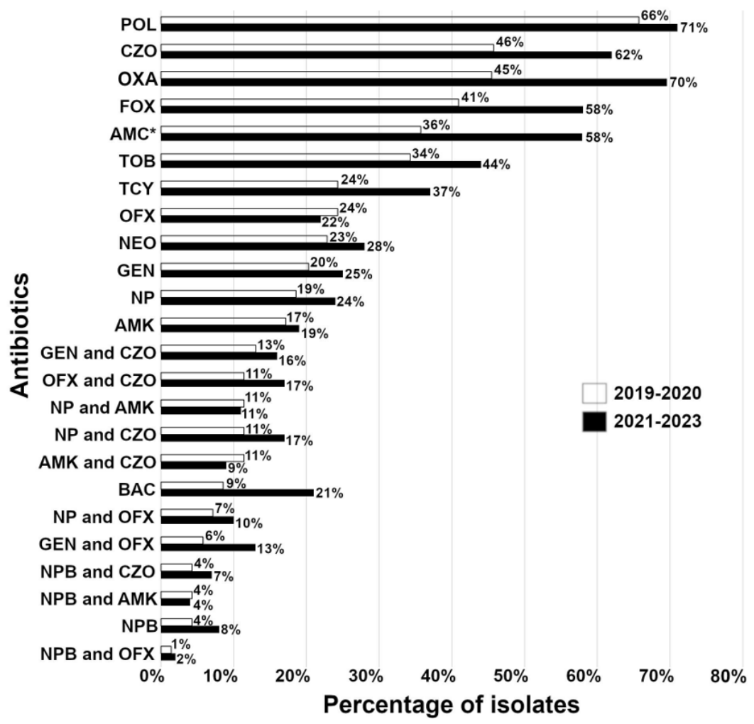
Figure 2.
Temporal distribution of acquired resistance in isolates from corneal non-healing ulcers. Percentage (%) of resistant bacterial species from patient samples excluding intrinsic resistance of isolates. A number of isolates range from 11 to 70 for the period 2019–2020 and from 19 to 99 for the period 2021–2023. * - although amoxicillin/clavulanic acid is not used topically, data for this antibiotic are presented here since it is often used postoperatively as a systemic antibiotic. Abbreviations: AMC, amoxicillin/clavulanic acid; AMK, amikacin; BAC, bacitracin; CZO, cefazolin; FOX, cefoxitin; GEN, gentamicin; NEO, neomycin; OFX, ofloxacin; OXA, oxacillin; POL, polymyxin B; TCY, tetracycline; TOB, tobramycin.
Figure 2.
Temporal distribution of acquired resistance in isolates from corneal non-healing ulcers. Percentage (%) of resistant bacterial species from patient samples excluding intrinsic resistance of isolates. A number of isolates range from 11 to 70 for the period 2019–2020 and from 19 to 99 for the period 2021–2023. * - although amoxicillin/clavulanic acid is not used topically, data for this antibiotic are presented here since it is often used postoperatively as a systemic antibiotic. Abbreviations: AMC, amoxicillin/clavulanic acid; AMK, amikacin; BAC, bacitracin; CZO, cefazolin; FOX, cefoxitin; GEN, gentamicin; NEO, neomycin; OFX, ofloxacin; OXA, oxacillin; POL, polymyxin B; TCY, tetracycline; TOB, tobramycin.
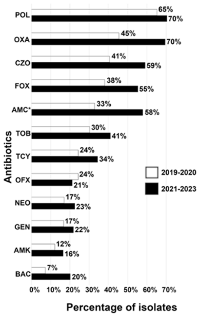
Figure 3.
Acquired resistance in isolates from corneal non-healing ulcers regarding the patient’s previous antibiotic treatments. Percentage (%) of resistant bacteria against antibiotics is presented excluding intrinsic resistance of isolates. The number of isolates range from 8 to 27 for the patients without previous antibiotic treatments and from 27 to 142 for the patients with previous antibiotic treatments. * = although amoxicillin/clavulanic acid is not used topically, data for this antibiotic are presented here since it is often used postoperatively as a systemic antibiotic. Abbreviations: AMC, amoxicillin/clavulanic acid; AMK, amikacin; BAC, bacitracin; CIP, ciprofloxacin; CZO, cefazolin; FOX, cefoxitin; GEN, gentamicin; MFX, moxifloxacin; NEO, neomycin; OFX, ofloxacin; OXA, oxacillin; POL, polymyxin B; TCY, tetracycline; TOB, tobramycin.
Figure 3.
Acquired resistance in isolates from corneal non-healing ulcers regarding the patient’s previous antibiotic treatments. Percentage (%) of resistant bacteria against antibiotics is presented excluding intrinsic resistance of isolates. The number of isolates range from 8 to 27 for the patients without previous antibiotic treatments and from 27 to 142 for the patients with previous antibiotic treatments. * = although amoxicillin/clavulanic acid is not used topically, data for this antibiotic are presented here since it is often used postoperatively as a systemic antibiotic. Abbreviations: AMC, amoxicillin/clavulanic acid; AMK, amikacin; BAC, bacitracin; CIP, ciprofloxacin; CZO, cefazolin; FOX, cefoxitin; GEN, gentamicin; MFX, moxifloxacin; NEO, neomycin; OFX, ofloxacin; OXA, oxacillin; POL, polymyxin B; TCY, tetracycline; TOB, tobramycin.
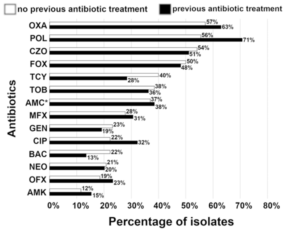
Figure 4.
Distribution of resistant isolates against multiple antibiotics. Percentage of the isolates that are resistant to the corresponding number of antibiotics. Bacterial samples were isolated from canine corneal non-healing ulcers (number of isolates = 170).
Figure 4.
Distribution of resistant isolates against multiple antibiotics. Percentage of the isolates that are resistant to the corresponding number of antibiotics. Bacterial samples were isolated from canine corneal non-healing ulcers (number of isolates = 170).
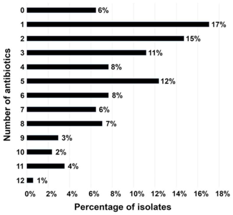
Figure 5.
Confocal microscopy evaluation of corneal ulcers. (A) A non-healing corneal ulcer from a dog with negative bacterial growth. The open arrow points to the region of the corneal ulcer; (A1) Confocal microscopy image from the same patient. Star marks loose epithelium at the ulcer region. (B) A non-healing corneal ulcer from a dog with positive bacterial growth. The open arrow points to the region of the corneal ulcer; (B1 Confocal microscopy image from the same patient, small arrows point to inflammatory cells; (C) An anterior stromal corneal ulcer from a dog with positive bacterial growth. The open arrow points to the region of the corneal ulcer; (C1) Confocal microscopy image from the same patient, small arrows point to numerous inflammatory cells. Corneal stromal disorganization is evident in the region of heavy inflammatory cell infiltrates. This patient had a conjunctival island graft one year prior due to the infected corneal ulcer.
Figure 5.
Confocal microscopy evaluation of corneal ulcers. (A) A non-healing corneal ulcer from a dog with negative bacterial growth. The open arrow points to the region of the corneal ulcer; (A1) Confocal microscopy image from the same patient. Star marks loose epithelium at the ulcer region. (B) A non-healing corneal ulcer from a dog with positive bacterial growth. The open arrow points to the region of the corneal ulcer; (B1 Confocal microscopy image from the same patient, small arrows point to inflammatory cells; (C) An anterior stromal corneal ulcer from a dog with positive bacterial growth. The open arrow points to the region of the corneal ulcer; (C1) Confocal microscopy image from the same patient, small arrows point to numerous inflammatory cells. Corneal stromal disorganization is evident in the region of heavy inflammatory cell infiltrates. This patient had a conjunctival island graft one year prior due to the infected corneal ulcer.
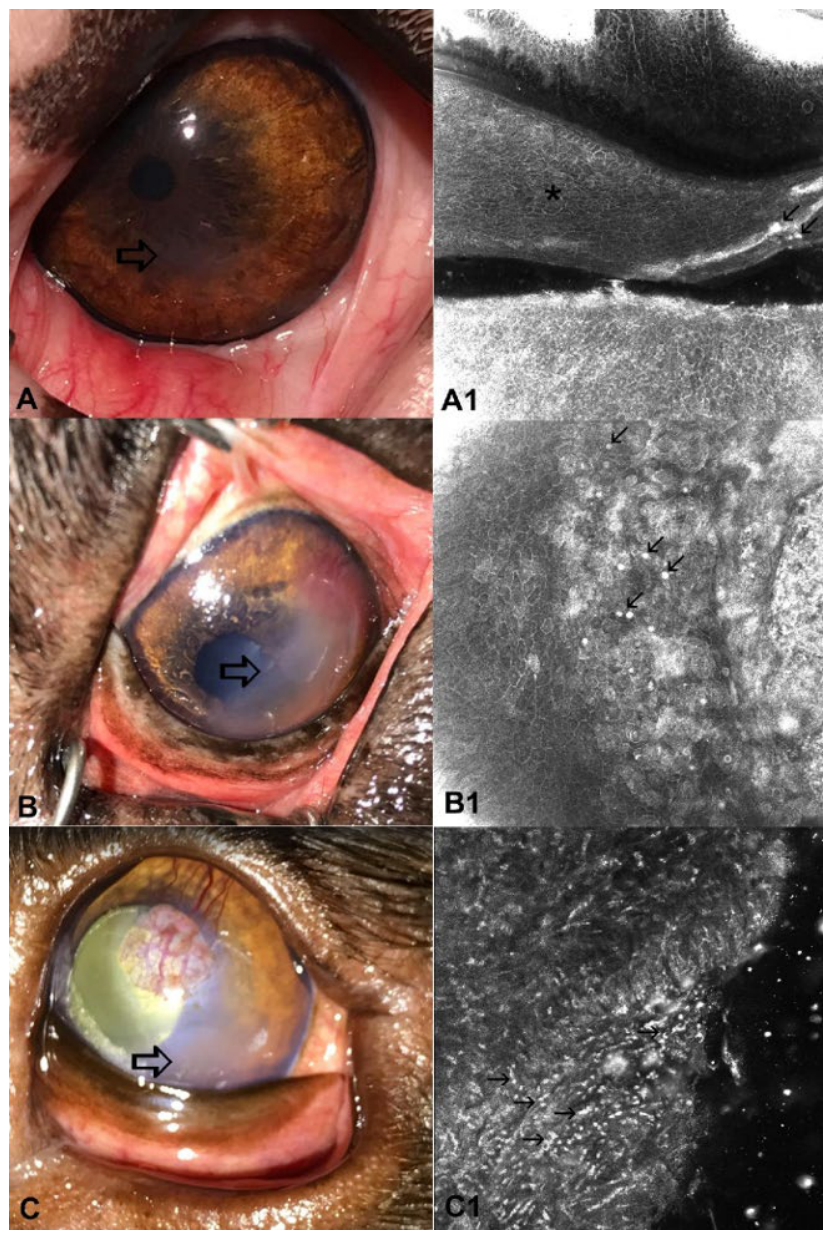

Table 1.
Bacterial species distribution from corneal non-healing ulcer samples. Distribution of incidence of the specific microorganism detection in canine patients. Data are presented as a percentage of total isolates (n = 170).
Table 1.
Bacterial species distribution from corneal non-healing ulcer samples. Distribution of incidence of the specific microorganism detection in canine patients. Data are presented as a percentage of total isolates (n = 170).
| Organism | (n = 170) |
| Staphylococcus epidermidis | 21% |
| Staphylococcus pseudintermedius | 18% |
| Staphylococcus capitis | 15% |
| coagulase negative staphylococci a | 14% |
| Rothia sp. | 4% |
| Gram positive bacteria | 3% |
| Pseudomonas aeruginosa | 3% |
| Other b | 9% |
| Other c | 13% |
aStaphylococcus spp. (S. aureus, S. auricularis, S. caprae, S. haemolyticus, S. hominis, S. intermedius, S. pasteuri, S. schleiferi, S. sciuri, and S. warneri) excluding S. capitis, S. epidermidis, and S. pseudintermedius; bpresence of each isolate of 2% represented by Bacillus pumilus, Corynebacterium sp., Enterococcus faecalis, Ochrobactrum sp., and Streptococcus canis; cpresence of each isolate of 1% represented by Acinetobacter sp., Actinomyces sp., Bacillus cereus, Bacillus megaterium, Bacillus thuringiensis, Corynebacterium amycolatum, Escherichia coli, Gram negative bacteria, Kocuria sp., Klebsiella pneumoniae, Macrococcus sp., Micrococcus luteus, Moraxella canis, Neisseria subflava, Serratia marcescens, Stenotrophomonas maltophilia, Streptococcus gordonii, Streptococcus mitis, and Streptococcus sp.
Table 2.
Resistance profile of isolates from corneal non-healing ulcer samples from 2019–2023.
| ANTIBIOTIC | TOTAL | INTRINSIC | ACQUIRED |
| polymyxin B (n = 170) | 69% (n = 117) | 1% (n = 2) | 68% (n = 115) |
| Oxacillin (n = 34) | 62% (n = 21) | 0% (n = 0) | 62% (n = 21) |
| Cefazolin (n = 170) | 55% (n = 93) | 3% (n = 5) | 52% (n = 88) |
| Cefoxitin (n = 166) | 51% (n = 84) | 3% (n = 5) | 48% (n = 79) |
| amoxicillin/clavulanic acid * (n = 89) | 40% (n = 35) | 2% (n = 2) | 38% (n = 33) |
| Tobramycin (n = 170) | 40% (n = 68) | 3% (n = 5) | 37% (n = 63) |
| Tetracycline (n = 170) | 32% (n = 54) | 2% (n = 3) | 30% (n = 51) |
| Ciprofloxacin (n = 81) | 31% (n = 25) | 1% (n = 1) | 30% (n = 24) |
| Moxifloxacin (n = 81) | 31% (n = 25) | 1% (n = 1) | 30% (n = 24) |
| Ofloxacin (n = 170) | 23% (n = 39) | 1% (n = 2) | 22% (n = 37) |
| Neomycin (n = 170) | 26% (n = 44) | 6% (n = 10) | 20% (n = 34) |
| Gentamicin (n = 169) | 23% (n = 39) | 3% (n = 5) | 20% (n = 34) |
| Amikacin (n = 170) | 18% (n = 30) | 3% (n = 5) | 15% (n = 25) |
| Bacitracin (170) | 16% (n = 27) | 1% (n = 2) | 15% (n = 25) |
Percentage (%) of resistant bacterial species from patient samples presented as combined resistance (TOTAL; intrinsic and acquired resistance together), intrinsic resistance only (intrinsic), or acquired resistance only (acquired). * - although amoxicillin/clavulanic acid is not used topically, data for this antibiotic are presented here since it is often used postoperatively as a systemic antibiotic.
Table 3.
Bacterial species distribution from corneal non-healing ulcer samples relative to previous patient’s antibiotic treatments.
Table 3.
Bacterial species distribution from corneal non-healing ulcer samples relative to previous patient’s antibiotic treatments.
| ORGANISM. | PREVIOUS ANTIBIOTIC TREATMENT | |
| NO (n = 27) | YES (n = 143) | |
| Staphylococcus pseudintermedius | 26% | 17% |
| coagulase negative staphylococci a | 22% | 13% |
| Staphylococcus capitis | 11% | 15% |
| Staphylococcus epidermidis | 11% | 23% |
| Pseudomonas aeruginosa | 11% | 2% |
| Other b | 19% | 30% |
Distribution of isolates in canine patients. Data are presented as a percentage of total isolates (n = 27 and n = 143). aStaphylococcus spp. (S. aureus, S. auricularis, S. caprae, S. haemolyticus, S. hominis, S. intermedius, S. pasteuri, S. schleiferi, S. sciuri, and S. warneri) excluding S. capitis, S. epidermidis, and S. pseudintermedius; bpresence of each isolate of 4% or less represented by Acinetobacter sp., Actinomyces sp., Bacillus cereus, Bacillus megaterium, Bacillus pumilus, Bacillus thuringiensis, Corynebacterium amycolatum, Corynebacterium sp., Enterococcus faecalis, Escherichia coli, Gram negative bacteria, Gram positive bacteria, Klebsiella pneumoniae, Kocuria sp., Macrococcus sp., Micrococcus luteus, Moraxella canis, Neisseria subflava, Ochrobactrum sp., Rothia sp., Serratia marcescens, Stenotrophomonas maltophilia, Streptococcus canis, Streptococcus gordonii, Streptococcus mitis, and Streptococcus sp., excluding Pseudomonas aeruginosa in the group with previous patient’s antibiotic treatments.
Disclaimer/Publisher’s Note: The statements, opinions and data contained in all publications are solely those of the individual author(s) and contributor(s) and not of MDPI and/or the editor(s). MDPI and/or the editor(s) disclaim responsibility for any injury to people or property resulting from any ideas, methods, instructions or products referred to in the content. |
© 2026 by the authors. Licensee MDPI, Basel, Switzerland. This article is an open access article distributed under the terms and conditions of the Creative Commons Attribution (CC BY) license (http://creativecommons.org/licenses/by/4.0/).
Copyright: This open access article is published under a Creative Commons CC BY 4.0 license, which permit the free download, distribution, and reuse, provided that the author and preprint are cited in any reuse.


