

Submitted:
22 June 2026
Posted:
23 June 2026
You are already at the latest version
Abstract
Background/Objectives: Quiescent leukemia stem cells (LSCs) are self-renewing, pluripotent cells that represent a major obstacle to successful curative treatment of chronic myeloid leukemia (CML). LSCs are independent of BCR-ABL1 signaling and persist following tyrosine-kinase inhibitor treatment. The mechanisms that enable LSCs' survival are currently the focus of CML research. The review examines the complex relationship between signaling pathways and discusses recent advancements in research on energy metabolism in the pathogenesis of CML.
Methods: This comprehensive narrative review used a systematic approach to selecting studies published in peer-reviewed journals, without year or publication-type limits.
Results: Energy metabolism assumes a critical role in the biology of CML LSCs. These cells show dependency on oxidative phosphorylation (OXPHOS) and mitochondrial homeostasis, with fatty acid oxidation as the primary source of ATP. Studies highlight significant alterations in signaling networks with a dynamic interplay among dominant pathways in the CML clone. TGF-β-FOXO signaling is proposed to be crucial for maintaining the self-renewal capacity of quiescent LSCs, unlike the majority of proliferating mature CML cells that exhibit strong activation of the PI3K/AKT pathway and glycolysis. Additionally, unique aspects of LSCs' metabolism underline the contribution of leukemic cell interactions, including the induction of a more permissive microenvironment.
Conclusion: Fatty acid oxidation appears critical to the survival and self-renewal of CML LSCs. Adaptation of mitochondrial function is concordant with signaling changes and presumes adjustment of mitochondrial respiration with stimulated OXPHOS. Research findings unveil many potential targets to consider to overcoming survival mechanisms, with disrupting the mitochondrial support as a promising strategy to selectively eradicate CML LSCs.
Keywords:
leukemia stem cell
; oxidative phosphorylation
; free fatty acid
; transforming growth factor beta
; lysophospholipids
; autophagy
1. Introduction
Quiescent leukemia stem cells (LSCs) are the foundation of residual disease in chronic myeloid leukemia (CML) due to inherent insensitivity to tyrosine kinase inhibitors (TKIs). These self-renewing pluripotent myeloid stem cells may re-initiate leukemogenesis following discontinuation of TKI treatment and currently represent a major obstacle to successful CML treatment [1,2,3]. Inhibition of BCR-ABL1 tyrosine kinase signaling effectively targets mature CML cells but fails to eradicate the disease-initiating persistent LSCs. Experiments with transgenic mouse models and in vitro CD34(+) CML cells derived from patients confirm that CML stem cell survival is BCR-ABL1 kinase-independent [2,4,5].
The mechanisms that enable LSCs' survival under TKI treatment are currently the focus of research, together with attempts to achieve treatment-free survival in these patients. In order to develop novel anticancer strategies targeting LSCs, it is crucial to understand CML pathogenesis and elucidate the mechanisms underlying LSC survival. These potentially curative approaches focus on BCR-ABL-independent mechanisms of resistance [1,2,6].
Research evidence proposes several BCR-ABL-independent mechanisms of CML stem cell survival, including actions of cytokines and growth factors, alternative activation of signaling pathways, enhanced lipid mediator signaling, dysregulation of immune response, alterations in the microenvironment, epigenetic mechanisms (DNA methylation, histone acetylation, and microRNA expression), alternative splicing, etc [1,2,4,7,8,9,10,11].
Along with persistent LSCs, a group of progenitor stem-like CML cells may arise with acquired drug resistance during therapy (treatment-resistant cells). Most of their resistance comes from genetic instability and additional mutations in the BCR-ABL1 oncogene or other genes important for cell-cycle and maintenance [3]. Part of the TKI resistance is due to alterations occurring in the kinase domain of the BCR-ABL protein. This can sometimes be overcome by different TKI types (such as nilotinib, dasatinib, and bosutinib) or other drugs that inhibit tyrosine kinase. The resistance can also arise from acquired mutations or alternative activation of the pathways downstream of the BCR-ABL kinase [4,7]. Another important issue to consider is the induction of antiproliferative effects in CML stem/progenitor cells by TKI. By blocking BCR-ABL activity, other independent signaling pathways remain unopposed, leading to cell cycle arrest, a state known as induced quiescence [5,12].
Recent advances in understanding unique aspects of LSCs' metabolism point to the contribution of leukemic cell interactions, including induced modifications that would shape a more permissive microenvironment for survival. A growing field of science, known as immunometabolism, explores the interconnection between cell activation signaling and metabolic changes that further govern the cell phenotype, function, and fate. Several cellular metabolites are found to be cofactors, enhancers, or inhibitors of enzymes, transcription factors, and epigenetic modifiers involved in cell cycle control and survival. Their activation network demonstrates that epigenetic and metabolic reprogramming are profoundly intertwined. Mitochondria are central to this process, given their involvement in energy production, redox reactions, regulation of cell death, immune response, and other processes [1,2,3,4,8,9].
This comprehensive narrative review describes and discusses the complex relationship between signaling pathways and energy metabolism in CML and their profound effects on cellular behavior and survival. The review presents the current understanding of CML pathogenesis, with recent and major findings emphasizing metabolic alterations and related intercellular interactions with the microenvironment. The article also summarizes proposed novel biomarkers of treatment response and strategies to address treatment resistance.
2. Materials and Methods
The review was conducted through a comprehensive analysis of relevant scientific literature. A detailed literature search strategy presumed investigation of scientific sources focused on CML pathogenesis, CML metabolism, and related processes, including the PubMed/MEDLINE, ScienceDirect, and Google Scholar databases. The review method involved a systematic approach to selecting studies published in peer-reviewed journals. We did not limit our search by the year the studies were published. The review encompassed 173 studies at the end, spanning the period from 2002 to 2026.
3. Signaling Networks in the Pathogenesis of CML
The BCR-ABL1 fusion gene, generated by the chromosomal translocation 9:22, is considered the cause of CML development. Detecting the BCR-ABL1 sequence is critical for CML diagnosis, while measuring gene transcript levels is used to monitor a patient's response to treatment, treatment-free remission, and detect resistance or recurrence of the disease [6,7,8]. The BCR-ABL1 fusion protein exhibits enhanced dimerization and sustained tyrosine kinase activity, initiating a complex signaling cascade that dysregulates cellular homeostasis, including accelerating and prolonging the cell cycle, inhibiting apoptosis, and impairing DNA repair, with limited dependence on growth factors [4,7,13].
The initial phase of CML is characterized by aberrant proliferation and survival of granulocytic cells, with neutrophils comprising a major portion of the CML clone. Later, the disease may progress into the blastic phase, with accumulation of differentiation-arrested blast cells, mostly due to additional mutations in the clone [4,7,13].
A multitude of intracellular signaling pathways are initiated by the BCR-ABL1 oncoprotein, directly or indirectly, including the RAS pathway, mitogen-activated protein kinases (MAPK), the MYC protooncogene, signal transducer and activator of transcription (STAT) transcription factors, phosphoinositide 3-kinase (PI3K)/Akt/ mammalian target of rapamycin complexes (mTORC), etc. [4,7,14]. The more recently described Wnt signaling (β-catenin/LEF-1) and Hedgehog pathways are also found to be important for the development of myeloid leukemias and for LSC maintenance [15]. The survival of the malignant clone is facilitated by modulation of tumor-suppressor phosphatases and FOXO transcription factors signaling, among other mechanisms, as well as by increased expression of anti-apoptotic proteins (e.g., BCL-X(L)). Consequently, expression of epigenetic modulators is often altered [4,7,16].
BCR-ABL1 activity causes constitutive activation of RAS downstream signaling cascades, leading to MEK and MAPK activation, thereby supporting abnormal cell proliferation [14]. The PI3K/Akt/mTORC1 signaling pathway is required for malignant transformation in CML and governs proliferation in the vast majority of mature CML cells. The pathway is initiated by the BCR-ABL kinase as well as by other signaling pathways independently, including STAT5, as demonstrated in imatinib-treated patients. Central to its effects is the support of proliferation and cell survival by activating mTORC1 and suppressing the FOXO transcription factor [10,14].
PI3K, a lipid kinase, produces phosphatidylinositol (3,4,5)-trisphosphate (PIP3), which mediates activation of phosphoinositide-dependent protein kinase 1 (PDK1). PDK1, together with mTORC2, activates serine/threonine kinase Akt (protein kinase B), a central kinase in the pathway that controls signaling networks, including mTORC1, FOXO, MYC, HIF-1α, and glycogen synthase kinase 3 (GSK3) [17]. Thus, the PI3K pathway exhibits multiple roles in controlling proliferation, survival, metabolism, angiogenesis, autophagy, and in suppressing apoptosis (e.g., inactivation of the proapoptotic molecule BAD) [14,17]. The PI3K/Akt signaling significantly affects glucose metabolism and mitochondrial oxidative phosphorylation (OXPHOS), as discussed in the following sections (Figure 1) [17,18].
Transcription factors STAT1, STAT3, and STAT5 activate the key downstream pathways in CML. The BCR-ABL oncoprotein directly activates STAT5, which signals to the nucleus, eliciting changes in gene transcription (e.g., cyclin D, c-MYC, Bcl-2, MMP-2). Given the sustained function of the oncoprotein, its downstream STAT5 follows the pace, and its constantly high levels are maintained, resulting in increased cell proliferation and survival [7,19]. The STAT5B isoform is the major initiator of leukemogenesis, while suppression of IFN-α/γ responses represents one of its mechanisms that enables malignant transformation [20]. High STAT5 expression may even lead to imatinib resistance and disease progression [4,21].
STAT3 and STAT5 are activated by different mechanisms and bind to distinct genomic loci, but they can exhibit significant functional crosstalk. Besides direct binding to gene promoter regions, their actions include combined interaction with other transcription factors, epigenetic regulation, and shaping the chromatin landscape [19,20,21,22,23,24]. Additionally, STAT proteins can elicit a number of processes even when in their non-tyrosine-phosphorylated form, such as transcriptional modifications (some of which differ from those of phospho-STATs), changes in mitochondrial function, and alterations in chromatin structure [19,25]. Moreover, activation of STAT3 is observed as one of the mechanisms led by external stimuli, that is, BCR-ABL1-independent, and which might add to TKI resistance [4,26].
The signaling network, which combines the crosstalk among the PI3K/Akt, MAPK, TGFβ, and Wnt pathways, regulates the switch between self-renewal and differentiation in pluripotent stem cells. The network works in conjunction with Smad2/3 proteins, the main signal transducers of TGF-β superfamily receptors. When PI3K/Akt is inhibited and the Wnt pathway is activated, higher Smad2/3 activity collaborates with β-catenin to target genes required for early differentiation [27,28].
3.1. Cytokines in CML Pathogenesis
Increased levels of circulating proinflammatory cytokines have been reported in patients with CML. Cytokines and chemokines are produced by leukemic cells and by non-malignant immune and stromal cells within bone marrow (BM) niches [29,30,31,32,33]. Research evidence shows that locally produced cytokines may favor malignant hematopoiesis by activating distinct signaling routes, e.g., the PIK3/Akt/mTOR pathway or STAT3 activation, thereby supporting disease progression and TKI resistance. Therefore, cytokine-directed intervention addresses BCR-ABL1-independent mechanisms and represents one perspective in the future management of CML [4,29,30,31,34,35].
Tumor necrosis factor alpha (TNF-α) has been repeatedly investigated and found to be significant in the pathogenesis of CML [29,30,31,32]. For instance, Giustacchini et al. demonstrate that compared with normal hematopoietic cells, LSCs express higher levels of inflammation-related genes, such as TNF-α and TGF-β. According to single-cell transcriptomics, these two cytokines were associated with increased stem cell quiescence and TKI resistance [32].
CML stem/progenitor cells produce TNF-α at higher levels than their normal counterparts, which provides them with survival signals through the NF-κB/TNF-α feedback loop, IL-3 expression, and GM-CSF β-chain receptor signaling. Importantly, TNF-α production is not BCR-ABL kinase-dependent [4,29,30,31]. Additionally, TNF-α levels at CML diagnosis were significantly higher in patients who did not achieve remission after 6 months of imatinib therapy than in those who did [36].
There are several TNF-α-mediated metabolic implications. TNF-α can stimulate lipolysis, regulate droplet dynamics, and promote triglyceride accumulation in differentiated human adipocytes, which make up a substantial part of adult BM. Lipolysis is enhanced by activation of MEK/ERK and cAMP-dependent protein kinase A. Through the same kinases, TNF-α decreases peroxisome proliferator-activated receptor gamma (PPAR-γ) transcriptional activity and reduces the expression of CIDEC (cell death-inducing DFF45-like effector C), an important regulator of lipolysis [37,38,39].
A recent work identified JAK1-STAT3-activating signaling and IL-6 as responsible for TKI-resistant CML cases, proposing novel, potentially curative therapeutic targets in CML. Indeed, the combined use of BCR-ABL and JAK1/2 selective inhibitors reduced CML cell proliferation and induced apoptosis, even in quiescent LSCs [35]. Another study identified MCP-1 and IL-6 as novel, strong, and predictive plasma biomarkers for treatment-free remission in CML, among 20 cytokines tested. MCP-1 and IL-6 levels were markedly increased in CML patients in treatment-free remission, whereas low MCP-1/IL-6 levels were associated with poorer relapse-free survival [40].
Transforming growth factor beta (TGF-β) is a pleiotropic cytokine that can regulate a myriad of cellular functions. Accordingly, aberrant TGF-β signaling is found in many disease states. Plasma TGF-β levels are reported to be elevated in patients with CML, while TKI dasatinib treatment significantly reduced these levels after 3 months of therapy. However, higher baseline levels were associated with a lower likelihood of achieving a good therapeutic response. One explanation implicates a relationship between TGF-β1 and regulatory T cells (Tregs). That is, demonstrating dasatinib-related inhibition of Tregs' ability to suppress the immune system is reflected in a decrease in both the percentage of Treg cells and serum TGF-β1 levels. Therefore, TGF-β values are suggested to be used as a biomarker for the disease [27,41,42].
3.2. The PI3K/AKT and TGF-β/FOXO Signaling in CML
In fast-proliferating CML cells, BCR-ABL-mediated activation of the PI3K/AKT/mTORC1 pathway drives cellular metabolism and survival. In these cells, BCR-ABL activity was associated with inactivation of FOXO transcription factors (FOXO1, 3a, and 4) at the post-transcriptional level. On the contrary, in quiescent CML LSCs, the level of AKT phosphorylation is found to decrease, while the TGF-β signaling and its downstream target FOXO were active. Also, the FOXOs phosphorylation levels were higher in CML than in normal CD34+ cells. Therefore, research underlines that the TGF-β-FOXO signaling is crucial for maintaining the self-renewal capacity of LSCs [5,12,17,43,44].
The PI3K and TGF-β signaling networks seem to diverge in governing the fate of LSCs. Nevertheless, the complex interaction among TGF-β, AKT, and FOXO activity in normal hematopoietic stem cells (HSCs) and LSC cells also depends on the cellular context. Through cell-intrinsic and extrinsic inputs, TGF-β1 can influence the tumor microenvironment and the immune system, especially Tregs. This way, it may exert pro- and anti-tumor effects, in general [5,11,12,42,43,44].
In experiments with leukemia-initiating cells (LIC), TGF-β was associated with decreased Akt phosphorylation and increased nuclear localization of Foxo3a, the predominant FOXO in these cells. On the other hand, Foxo3a is significantly affected by strong PI3K/AKT signaling in a way that activated Akt phosphorylates nuclear Foxo3a, causing it to move out of the nucleus and thereby hindering its transcriptional activity. Foxo3a is subsequently degraded in the cytoplasm. In quiescent CML LSCs, Akt activity is low, and Foxo3a factor remains within the nucleus and may lead to G1 cell cycle arrest or apoptosis [12,43]. The experiment using a mouse model showed that Foxo3a deficiency impedes the ability of CML cells to propagate. The number of Foxo3a -/- LICs decreased after subsequent BM transplantation. The same results of colony-forming ability and importance of TGF–β–FOXO signaling were observed when using primary human CML LICs [43]. FOXO3a may undergo multiple modifications that fine-tune its function, stability, DNA binding, and intracellular localization [45,46,47,48].
An interconnection between the PI3K pathway and the cellular localization of the FOXO1 transcription factor was also described in CML. The TKI-resistant cells with activated PI3K/AKT signaling exhibit increased AKT-mediated phosphorylation of FOXO1 and cytoplasmic sequestration of FOXO1 (and its levels). Upon inhibiting the PI3K/AKT pathway, FOXO1 translocated to the nucleus [4,49].
Research also finds that FOXOs underpin the antiproliferative activity of TKIs. The introduction of a TKI inhibits the PI3K/AKT pathway, leading to unopposed FOXO activity and cell cycle arrest. This creates a protective state in CML against genetic instability and apoptosis [5]. Pellicano et al. showed that dasatinib significantly reduces FOXO phosphorylation and causes relocalization of total FOXO1 and 3a from the cytoplasm to the nucleus, thereby increasing their transcriptional activity. In another experiment, shRNA-mediated knockdown of FOXO3a enabled TKI-induced apoptosis in CML cells [5].
Some of the key FOXO target genes include Cyclin D1, ATM, CDKN1C (p57), and BCL6. As a critical downstream effector of FOXO signaling, BCL6 is considered essential for CML stem cell survival. Its levels were upregulated in response to active FOXO in 24 h TKI-treated CD34+ CML cells. BCL6 represses transcription of Cyclin D2, Bcl-XL, Arf, and p53, thereby maintaining LSC survival under TKI treatment. On the contrary, in untreated CML, BCL6 is repressed in a BCR-ABL-dependent manner [5,50,51].
Notably, TGF-β had different effects on Foxo3a activation and localization in LICs and non-LICs (progenitors), thereby affecting the survival of these two leukemia cell populations. In non-LICs, Foxo3a activation upon TKI treatment leads to cell cycle arrest and apoptosis via TNF-α- or p53-induced pathways [5,43,52].
In a genome-wide transcriptome comparison, the authors observed greater similarity in expression patterns between CML LSCs and HSCs than between CML LSCs and CML progenitors. Nevertheless, the study reports downregulation of pro-differentiation and TGF-β/BMP signaling pathways in CML LSCs (CD34+CD38−ALDHhigh [aldehyde dehydrogenase 1 activity]) compared with normal HSCs. There was strong inhibition of the TGF-β/BMP transcriptional output, along with activation of pathway antagonists [11].
Moreover, co-culture systems with stromal cells modulate the effects of TGF-β and other soluble factors, underscoring the importance of cellular interactions in tuning malignant cell behavior. Actually, the colony-forming ability of LSCs was not disturbed when cells were grown in a stroma-free system and with TGF-β inhibitors, suggesting an additional extrinsic (perhaps stromal) source or mechanism of TGF-β is involved in CML pathogenesis [11,43,44].
4. Energy Metabolism in CML Cells
Recent studies report that cancer stem cells have metabolic flexibility that enables them to switch between glycolysis and OXPHOS in response to environmental cues to secure energy and evade apoptosis and immune attack [3,12,17,53]. A phenomenon of the metabolic shift/switch is intrinsic to leukocytes and occurs physiologically during leukocyte activation, when they undergo substantial metabolic changes to adjust to their tasks in the inflammatory response [54,55].
BCR-ABL-positive proliferating cells show a high glycolytic activity. When imatinib is applied, a strong suppression of cytosolic glycolysis occurs, accompanied by an increase in mitochondrial fatty-acid oxidation (FAO) and tricarboxylic acid cycle (TCA) intermediates. Mitochondrial energy supply appears necessary to maintain the survival of CML BCR-ABL-active cells under TKI exposure (Figure 2) [56,57,58].
4.1. The PI3K/Akt/mTOR Metabolic Implications
As already mentioned, the majority of proliferating mature CML cells exhibit strong BCR-ABL1 signaling accompanied by activation of the PI3K/Akt/mTORC1 pathway. This pathway exhibits a profound impact on metabolic events in malignant cells. In general, the PI3K/Akt signaling supports glycolytic processes by upregulating glucose transporters (e.g., high-affinity glucose transporter GLUT1) and glycolytic enzymes, while inhibiting glycogen synthase (GSK3). It promotes de novo lipid synthesis and directly stimulates cholesterol and fatty acid (FA) biosynthesis, while inhibiting gluconeogenesis and FAO (e.g., by directly phosphorylating peroxisome proliferator-activated receptor gamma coactivator 1-alpha (PGC-1α)). It inevitably affects the pentose phosphate pathway (PPP), OXPHOS in mitochondria (complex I activity), and redox homeostasis by replenishing the NADPH cytosolic pool, a reducing agent. Overall, the PI3K/Akt pathway tends to store energy and avoid oxidative stress [10,17,18,59,60,61].
Protein kinase B (Akt) is a central component of the PI3K pathway, playing several roles in regulating cellular metabolism by influencing enzymes, nutrient transporters, and various signaling networks. It has a substantial effect on enzymes that regulate glycolysis. The glycolytic enzyme hexokinase (HK) isoform 2 (HK2) is a direct target of Akt, producing bidirectional effects on glycolytic intensity and promoting pro-survival cues [17,62,63]. For example, HK2 binds to the voltage-dependent anion channel at the outer mitochondrial membrane (OMM), thereby interfering with apoptosis by limiting the opening of the mitochondrial permeability transition pore (mPTP) [17,63,64]. HK2 also binds to the mitochondria-associated membranes (MAMs) (sites of communication between the OMM and ER), where it participates in the regulation of Ca2+ turnover. Displacement of HK2 from MAMs promotes Ca2+ release, Ca2+-dependent calpain activation, and finally cell death [17,65]. Another example of the Akt/HK2 axis is the formation of the HK2 complex with fructose-2,6-bisphosphatase (TP53-induced glycolysis and apoptosis regulator (TIGAR), a p53-inducible protein) under hypoxia, which amplifies the PPP. This raises NADPH production and limits mitochondrial ROS production [17,66]. Although it protects mitochondria from oxidative stress, HK2 facilitates autophagy in response to starvation (glucose deprivation) [62,67].
In CML, detachment of HK2 from mitochondria was found to promote collateral sensitivity in multidrug-resistant CML cells that primarily resort to glycolysis to generate ATP. The detachment also destabilized ROS production and glutathione levels, leading to increased drug accumulation and cell death. Manipulation of HK2 is proposed as a promising strategy to enhance sensitivity in chemotherapy-refractory CML patients [68].
Pyruvate kinase (PK) is another key regulatory enzyme in glycolysis that is modulated by the PI3K pathway, specifically via the mTOR/c-Myc/hnRNP/PKM signaling cascade. Also, under IGF-1-induced PI3K signaling, Akt can directly interact with and phosphorylate PKM2. There are four isomeric, tissue-specific forms, with PKM2 (muscle type 2) present in all proliferating cells, including tumors [17,56,69]. As a dimer or „low activity“ form, PKM2 acts as a protein kinase, while as a tetramer, it is an effective pyruvate kinase [70]. This way, the dimeric form of PKM2 diverts glycolysis toward the production of several glycolytic intermediates that can be used for the biosynthesis of amino acids, lipids, and nucleotides [71,72]. Additionally, as a dimer, PKM2 can translocate to the nucleus, mitochondria, or outside the cell. Following nuclear translocation, PKM2 can bind to STAT5A and induce cyclin D1 expression [17,58,72,73].
Studies report multiple binding partners of PKM2 and related influences on gene transcription, epigenetic and post-translational modulation, and exosome-related communication. Therefore, PKM2 is designated as a glycolytic enzyme implicated in intercellular communication with potential for microenvironment reprogramming [71,72].
High expression of PKM2 in cancer cells plays a specific role in their metabolic reprogramming, namely, aerobic glycolysis [17,58,72,73]. PKM2 is elevated in CML and plays a significant role in promoting glycolysis and biosynthesis, redox homeostasis, cancer cell survival, and drug resistance. Higher PKM2 levels were observed in TKI-resistant CML primary cells, whereas PKM2 knockdown reduced glycolysis and lactate production, inhibited cell growth, and induced apoptosis [74,75].
As a phosphotyrosine-binding protein, PKM2 can interact, among others, with the BCR-ABL kinase, with proposed reciprocal modulation of both. By interacting with Bcl-2 under oxidative stress, or with p53 in the presence of the MDM2 oncogene, it hinders apoptotic pathways. Knockdown of PKM2, or induced switching of PKM2 to PKM1, caused autophagic cell death via the Bcl-2 mechanism and increased ROS levels [56,76]. Additionally, it phosphorylates STAT3, enhancing its activity and, consequently, the transcription of MEK5 and other STAT3-regulated genes [70,71,72,77]. Moreover, secreted dimeric PKM2 has been shown to mediate the migration of colon cancer cells via extracellular activation of the PI3K/Akt and Wnt/β-catenin signaling pathways [78].
4.2. CML Stem Cell Metabolism
Unlike normal HSCs and rapidly proliferating CML cells, CML LSC metabolism relies on increased OXPHOS and FA metabolism. A genome-wide comparison study showed altered cellular functions in CML LSCs compared to normal HSCs, with upregulated gene sets, including mitochondrial inner membrane, electron transport chain (ETC), mitochondrial matrix, NADH-to-ubiquinone, and Krebs-TCA cycle (in the oxidative metabolism group set) [11]. Nevertheless, LSCs are metabolically less active compared to the bulk of their clonal progeny. They are capable of using versatile fuel sources compared to healthy HSCs [3]. Evidence also shows that different CML LSC subpopulations can be metabolically distinct [79].
Chronic phase CML LSCs highly express mitochondrial respiratory chain genes and exhibit upregulated mitochondrial respiration and increased TCA cycle flux. Additionally, several studies show high ROS levels in these cells, which may be related to increased mitochondrial respiration [3,80,81,82]. Moreover, new evidence points to significant roles for lipid mediators, such as lysophospholipid (LysoPL) and branched-chain amino acid (BCAA) metabolism, in LSC survival, stemness, and TKI resistance [10,53].
Fatty acid and amino acid metabolisms significantly contribute to OXPHOS maintenance in LSCs, and FAO appears crucial to the survival and self-renewal of these cells. It produces several products that help cells generate energy while coordinating intracellular signaling, transcription, and protein function. Primarily, increased FAO supports OXPHOS as an energetic fuel by supplying acetyl-CoA to the TCA cycle and enhancing ATP production [53,79,83,84].
Furthermore, acetyl-CoA can exit mitochondria through the citrate-pyruvate cycle. Under glucose-deprived conditions, FAO-derived acetyl-CoA is utilized to produce cytosolic NADPH, among others, thereby compensating for the reduced PPP-derived NADPH. Acetyl-CoA is used to form citrate and malate, which are then converted by isocitrate and malate dehydrogenases, respectively, to produce NADPH. This way, NADPH significantly supports antioxidant regeneration, ameliorates redox imbalance, and helps in the repression of ROS-induced apoptosis [53,84,85,86,87,88].
Increased production of acetyl-CoA can enhance protein and histone acetylation by providing the acetyl group required by histone acetyltransferase, thereby allowing greater access to DNA and changing gene expression [84].
Increased FAO in CD34+ CML cells is associated with elevated acylcarnitine levels and with metabolites that enrich the TCA cycle and amino acid pool. Also, the TCA flux is supported by enhanced activity of anaplerotic pyruvate carboxylase, which catalyzes the conversion of pyruvate to oxaloacetate [89]. Metabolic profiling of CD34+ and CD34- CML cells revealed high levels of glycerol-3-phosphate and carnitine, while the levels of FFAs, such as oleic and stearic acids, were decreased in patient-derived stem cell-enriched CD34+ CML cells compared to differentiated CD34- CML cells [80].
Research shows that FAO is significant for tumor cell chemoresistance by promoting tumor cell autophagy, altering apoptosis, and affecting immune evasion. By increasing NADPH and inducing autophagy, FAO lowers oxidative stress species and interferes with apoptosis [60,90].
Using chemoresistant breast cancer cells, enhanced FAO was shown to activate STAT3 via acetylation, leading to increased phospholipid biosynthesis through STAT3-mediated upregulation of long-chain acyl-CoA synthetase 4. Phospholipids are incorporated into mitochondrial membranes, thereby increasing their lipid content and integrity, as well as membrane potential. These cells show greater resistance to apoptosis [91].
In the study by Ye et al., a higher rate of FAO was determined in leukemia cells compared with non-leukemia cells, as well as in LSCs (Lin-) compared to more differentiated leukemia cells (Lin+). Using a murine BC-CML co-culture with adipocytes, a subpopulation of LSCs utilized gonadal adipose tissue (GAT) as a niche to support their metabolism, suggesting that FAO might be regulated differently across LSC subpopulations. Additionally, inhibition of carnitine palmitoyltransferase-1 (CPT1) (with etomoxir) on the OMM reduced FAO by about 60%, suggesting that peroxisomal FAO makes a substantial contribution to the total FAO in CML LSCs [79].
Several studies report that FAO is activated and supported by BM adipocytes. An in vitro study of acute monocytic leukemia (AMoL) cells under nutrient-starvation conditions shows that co-culture with BM adipocytes increased the viability of malignant cells. Specifically, this was achieved, at least in part, by the upregulation of Bcl2 mRNA and the induction of a heat shock protein (HSP) chaperone response. The cells were protected to a greater extent than those co-cultured with mesenchymal stem cells (MSCs) [92]. These findings indicate that FAO in malignant cells was activated by co-culture with BM adipocytes. Ketone bodies produced by FAO were higher in the co-culture media than in the control media. Results demonstrate a significant increase in HADHA enzyme, a crucial component of the mitochondrial trifunctional protein (MTP) that catalyzes the final three steps of the mitochondrial FAO, thereby confirming that BM adipocytes stimulate FAO in AMoL cells. Moreover, changes in lipid metabolism seem required for the anti-apoptotic actions in this cellular system. That is, inhibition of FAO by a selective CPT1 inhibitor abolished the anti-apoptotic effects of BM adipocytes [84,92].
The co-culture with BM adipocytes upregulated stress-response kinases, p-AMPK (AMP-activated protein kinase) and p-p38 MAPK, along with autophagy, and downregulated Akt/mTOR signaling with depletion of p-Akt. At the same time, higher levels of the dominant aerobic glycolysis products were determined, implying activation of anaplerotic pathways and their role in supporting mitochondrial FAO [92,93].
Gene expression analysis showed increased expression of proteins involved in lipid metabolism in AMoL compared to control cells, including PPARγ, FABP4, CD36, and the CPT-1 gene. Once activated, PPARγ augmented its downstream target genes, including FABP4, CD36, and BCL2 [92,94]. PPARγ is a nuclear receptor that regulates glucose and lipid metabolism and is considered the key adipogenesis regulator [95]. In addition, the gene for adiponectin major receptor (ADIPOR1) was upregulated, thereby adding to the activation of the AMPK pathway. On the other hand, BM adipocytes expressed high levels of adiponectin and secreted it at significantly higher levels than parental MSCs [92].
The findings demonstrate downregulation of genes encoding HK2, apelin receptor (AGTRL1), ghrelin, CCL2 (MCP-1), the Calcineurin B-encoding gene PPP3R1, and the p53 transcription regulator, among others. AGTRL1 participates in the regulation of glucose and lipid metabolism by enhancing glucose uptake and inhibiting adipogenesis and lipolysis [92,96,97,98]. Overall, the study demonstrates a change in metabolic balance that enhances FA uptake, FAO, and the AMPK pathway, while downregulating processes that support glucose uptake and glycolysis, such as the PI3K/Akt pathway and its targets.
4.3. Lysophospholipids in CML
Beyond serving as an energy source, lipids are now considered to extend beyond their immediate interpretation in cancer pathogenesis, as demonstrated by their much wider role. They represent signaling molecules designated as bioactive lipids, such as sphingosine-1-phosphate (S1P), PGE2, lipoxin, and ceramide, and are involved in epigenetic modifications through fatty acylation [99,100,101].
Adequate lysophospholipid (LysoPL) metabolism is required for the maintenance of CML LSCs in vivo. The LysoPL pathway produces lipid mediators that act as lipid second messengers, mediating regulatory signaling in an oncogene-independent manner [10,95].
A group revealed higher expression of the Gdpd3 gene encoding a lysophospholipase D enzyme in murine CML LCSs compared with normal stem cells. Lysophospholipase D is an ER membrane-associated enzyme that converts LysoPL to lysophosphatidic acids (LPAs) in the glycerol phosphate pathway. Disruption of lysophospholipase D changed the levels of certain LPAs and lipid mediators in the analyzed CML stem cells. Also, Gdpd3 gene deficiency impaired the self-renewal capacity of CML LSCs and attenuated their ability to cause disease relapses [10,95].
Specifically, the Gdpd3 gene appeared to be a key suppressor of the AKT/mTORC1 pathway in CML stem cells, whereas in Gdpd3 gene-deficient cells, this pathway became highly active and disrupted LSC quiescence. Apparently, through its inhibitory action on Akt, Gdpd3 supports Foxo3a nuclear localization and transcriptional activity, as well as Foxo3a - β-catenin nuclear interaction [10,12]. Previous studies showed that β-catenin in Wnt signaling is required for the self-renewal of both normal and neoplastic stem cells [15,102]. In addition, the levels of prostaglandins, eicosanoids, and docosanoids were decreased in Gdpd3-deficient CML cells. Overall, the research emphasizes that GDPD3 is a major repressor of proliferation and a contributor to LSCs' self-renewal [10].
Mice overexpressing human GDPD3 in the liver showed significantly increased hepatic LPA production and increased FA uptake compared to control animals, but without effects on de novo FA synthesis, triglyceride content, or normal circulating lipid concentration [95].
Besides LysoPL and the PPARγ-mediated signaling pathway, the importance of other lipid-related mediators in the survival of CML LSCs is documented, including arachidonate 5-lipoxygenase (Alox5) and arachidonate 15-lipoxygenase (Alox15). These mediators are also proposed to be potential targets for combination therapy to overcome resistance to TKIs [10,103].
4.4. The AMPK Signaling and Autophagy in CML
The AMPK signaling acts as a central cellular energy sensor. It is activated by metabolic stress, low intracellular energy levels (low ATP), ER stress, hypoxia, ROS, and stimulation of the adiponectin receptor, among others. During energy stress (glucose-deficient conditions), the Liver Kinase B1 (LKB1)-AMPK cascade promotes mitochondrial respiration (OXPHOS) and lipid utilization (FAO). The AMPK influences metabolic pathways by inhibiting mTOR (a negative feedback loop), suppressing ATP-consuming processes, and recruiting AMPK-dependent cytoprotective autophagy and mitochondrial biogenesis [82,87,88,92]. Fatty acid oxidation increases, while FA, sterol, and glycogen synthesis decrease. There is an increase in TCA cycle capacity, including anaplerosis, while maintaining NADPH levels in tumor cells. Moreover, AMPK regulates FAO by hindering phosphorylation of acetyl-CoA carboxylase (ACC), which produces malonyl-CoA, an allosteric inhibitor of CPT1A and CPT1B [84,104]. Additionally, the LKB1-AMPK pathway counteracts oxidative stress caused by disturbed glycolysis or high OXPHOS, thereby preventing cancer cells from undergoing apoptosis during energy deprivation [53,86,88]. However, AMPK will also initiate pathways leading to cell death when ATP levels are low enough [57,82,105].
According to some studies, AMPK activators may have anti-leukemic effects against Ph+ leukemic cells, especially when combined with TKI and autophagy inhibitors [57,105,106]. Autophagy is an important cellular adaptation that results from impaired energy metabolism, which supports mitochondrial activity to overcome energy imbalance. Together with heightened mitochondrial function, autophagy is designated as an essential process for LSCs' quiescence and survival. Autophagy is often represented as a protective mechanism in tumor cells, including imatinib-treated BCR/ABL-expressing cells and the stem/progenitor (CD34+38−) cell population, with CML LSCs CD34+ stem cells displaying a high level of autophagic flux or higher autophagy levels compared to normal cells [57,82,107,108,109].
The BCR-ABL/PI3K/Akt/mTOR signaling conveys an inhibition of autophagy through several mechanisms, including ATF5-mediated regulation of mTOR transcription, induction of miRNA-30a expression, and downregulation of key autophagy genes. However, evidence also demonstrates that autophagy is essential for BCR-ABL-dependent leukaemogenesis, while TKI treatment can restore autophagy and enable cell survival, thereby antagonizing its own proapoptotic effects [4,13,14,56,82,93,108,110].
In a study using K562 cells with active BCR-ABL signaling (therefore not LSCs), continuous imatinib treatment inhibited glycolytic activity, leading to autophagy, as evidenced by activation of the AMPK pathway. These cells relied on activated mitochondrial functions to maintain viability. Surprisingly, these events during prolonged exposure increased the sensitivity of CML cell lines to TKI-induced death. Mitochondrial activity, mitochondrial membrane potential (MMP), and cell death (by ATP-synthase inhibitor) were all higher in imatinib-treated cells [111]. Perhaps, despite FAO association with cancer progression and chemoresistance, a high FAO activity level is expected to exhaust FAs and subsequently decrease de novo phospholipid biogenesis [91], unless additional salvage sources are provided, perhaps from the microenvironment.
The upregulation of mitochondrial biosynthesis-related genes is observed in many OXPHOS-dependent cancers. CML LSCs exhibit increased mitochondrial biogenesis, characterized by a greater number and mass of mitochondria, as well as increased MMP, features that are found critical for their survival. Increased MMP is associated with altered electron transport and NAD+ regeneration, resulting in a reduction in the NAD+/NADH ratio. AMPK adds to the replenishment of NADPH supplies by stimulating the PPP [53,82,112,113].
To counteract the BCR-ABL-independent mechanism, several studies propose targeting autophagy in combination with TKIs or mTOR inhibition as a novel approach in CML treatment [13,114,115]. Autophagy protects CML stem cells following TKIs administration and might contribute to disease persistence. Imatinib was shown to induce autophagy in various CML cell types through ER stress and depletion of intracellular calcium. Autophagy did not depend on BCR-ABL activity and was not related to imatinib-induced apoptosis. On the other hand, suppression of autophagy (including knockdown of autophagy-related genes (ATG) 5 and ATG7) enhanced the effects of TKIs and could substantially eliminate CML stem cells [108,110].
Activation of the serine/threonine kinase ULK1 complex represents an initiation step of autophagy under stress. It connects the energy-sensing receptors (mTOR and AMPK) to downstream components of the autophagosomes. Inhibition of ULK1 in combination with TKIs selectively targeted CML LSCs and led to loss of quiescence, with differentiation by increasing oxidative stress and reinduction of sensitivity to TKIs [109,116,117].
Moreover, the significance of autophagy is highlighted in cells that have gained TKI resistance. Imatinib-induced autophagy was associated with reduced ROS levels, seemingly acting cytoprotectively, and with increasing intracellular glutamate levels. Findings show that acquired TKI resistance strongly depends on glutamine and other amino acid metabolic pathways. High glutamine consumption and glutamate production were associated with the generation of self-protective mechanisms that increased proline synthesis, supporting proliferation, and glutathione metabolism for ROS scavenging. Such and similar autophagy-related mechanisms of oxidative stress modulation (e.g., KEAP1-NRF2 system, PGC-1α-dependent antioxidant response) seem to protect mitochondria from degradation in LSCs [57,82,118]. It is assumed that the interplay between high ROS levels, AMPK activation, and autophagy may lead to maintaining ROS below a threshold that would evoke LSC differentiation [82].
Furthermore, FA-induced autophagy is an important pro-survival mechanism in CML. An interesting study on autophagic cell death in the K562 cell line reports the importance of the fatty-acid (3-decenoic acid) derivative AIC-47 and the modulation of the PKM isoform expression profile. The authors observed that the switch from PKM2 to PKM1 expression forces cells to use OXPHOS, and that, in turn, causes autophagic death by increasing ROS levels. The AIC-47 repression of BCR-ABL signaling went through the PPARγ/β-catenin pathway and downregulation of c-Myc. This delineates one mechanism by which FAs induce autophagy flux in BCR-ABL-positive cells [56].
Another important facet is the connection of autophagy to TGF-β/FOXO signaling. It is found that TGF-β signaling, by inactivating AKT, allows FOXO3a to relocalize into the nucleus, where it supports the transcription of autophagy genes [82]. At the same time, in metabolically stressed cells, AMPK phosphorylates FOXO3a at specific sites, thereby dictating its transcriptional profile of mitochondrial genes to support mitochondrial metabolism. The AMPK–FOXO3a axis has been revealed to regulate autophagy-related (e.g., Beclin-1 and LC3B) and antioxidant genes (e.g., thioredoxin) [45,47,48,119,120]. When signaling shifts in terms of the FOXOs activity loss, a marked increase in ROS is observed [5].
4.5. The PPAR-γ Coactivator (PGC)-1α and Sirtuins
The transcription co-activator PGC-1α is another major player in maintaining metabolism, influencing mitochondrial function, biogenesis, and oxygen consumption rate. In CML, PGC-1α is upregulated in stem/progenitor cells and has an integrative role in metabolic fitness, thereby supporting the viability of leukemia cells. It enhances mitochondrial activity and increases OXPHOS. It has been linked to TKI resistance in CML stem/progenitor cells, while its inhibition raised susceptibility to apoptosis when combined with nilotinib. However, PGC-1α may present a dual role in cancers, often depending on tissue specificity [53,121,122,123,124,125,126]. Additionally, PGC-1α might modulate the inflammatory response by directly activating the promoter of the anti-inflammatory cytokine IL-10 gene. Interleukin-10 was shown to increase FAO in macrophages via PPARγ-FABP5-CPT1A signaling, thereby enhancing macrophage-mediated resistance in myeloma cells [60,127,128].
Important regulation of PGC-1α is achieved by sirtuin molecule 1 (SIRT1), which deacetylates PGC-1α in dependence on cellular NAD+ levels, that is, the cellular energy status. Silent information regulators (sirtuins) are NAD+-dependent deacetylases and signaling proteins that participate in critical biological processes. They act as stress sensors that regulate cell cycle, redox balance, and may enhance mitochondrial function. Decreased glycolytic pathway and related alterations in the NAD/NADH ratio activate sirtuins. In addition to PGC-1α, their target proteins include p53, NF-κB, FOXOs, HMGCS2, superoxide dismutase 2, structural proteins, the DNA repair enzyme Ku70, and others [45,122,126,129].
FOXO3a deacetylation is a key post-translational modification that improves its activity. SIRT1-mediated deacetylation tunes FOXO3a activity and leads to upregulation of several target genes. In oxidative stress, SIRT1 was found to decrease the expression of the pro-apoptotic Bim protein via FOXO3a, thereby preventing cell death, while upregulating antioxidative enzymes (e.g., MnSOD) and cell-cycle arrest genes (e.g., cyclin-dependent kinase inhibitor 1B). In combination with SIRT2, SIRT1 allows cells to repair DNA damage prior to division [45,46,47,122]. Nevertheless, intensified SIRT1 activity in CML cells has been reported to interfere with DNA repair mechanisms, thereby promoting the acquisition of genetic mutations for drug resistance [130].
SIRT1 overexpression in CML LSCs is regarded as a survival pathway activated by BCR-ABL/STAT5 signals. It seems that STAT5 directly upregulates the SIRT1 promoter. However, STAT5 knockdown only partially reduces its expression, arguing that SIRT1 activation is not fully dependent on BCR-ABL kinase activity. Accordingly, combined action against both SIRT1 and BCR-ABL kinase led to better survival in an animal model of CML [131].
SIRT1 overexpression is involved in mitochondrial metabolic rewiring associated with LSC maintenance and TKI resistance [122,132]. Studies show that the SIRT1/PGC1α axis participates in metabolic alterations, a shift from glycolysis toward OXPHOS, in a colon cancer model [133]. Moreover, suppression of the Sirt1/Pgc-1α/Nrf2 pathway increased susceptibility of wild-type p53 cancer cells to oxidative stress and apoptosis [134]. Among others, SIRT1 overexpression correlated with enhanced autophagy-associated conversion of the LC3 protein, a key ubiquitin-like protein essential for autophagy, in the K562 cell line [129].
Sirtuins participate in gene expression regulation that is responsible for FAO, thereby supporting FAO and OXPHOS. For example, SIRT1 was associated with OXPHOS gene expression signatures [122]. Also, CPT1A and LCAD (long-chain acyl-CoA dehydrogenase) are activated through sirtuin deacetylation [84,135]. Knockdown and transgenic mouse experiments demonstrate the SIRT1/PGC-1α axis is an important contributor to enhancing OXPHOS and TKI resistance in CML LSCs [121,122,131].
By genetically deleting SIRT1 in transgenic CML mice, Abracham and colleagues observed that SIRT1-deleted cells showed significantly reduced basal and maximal mitochondrial respiration and reduced mitochondrial reserve capacity compared with control CML c-Kit+ cells. Conversely, SIRT1-deleted CML cells did not show alteration in maximal glycolysis nor glycolytic reserve. The effects of SIRT1 deletion on mitochondrial function were not observed in normal stem/progenitor cells [122].
In a transcriptomic analysis of the three-node miRNA feed-forward loop in CML, novel SIRT1 connections were detected. A SIRT1-dependent mechanism of mitochondrial metabolic rewiring regulation in CML involved a gene regulatory network of miRNA-transcription factors, precisely miR-34a/miR-205/ETS1 and/or GFM1 mRNA. The mitochondrial translation pathway appeared as a molecular alternative to the BCR-ABL pathway, which can be exploited for LSC eradication or sensitization to TKI [132].
Interestingly, leucine supplementation increased SIRT1 expression and stabilized mitochondrial function in high-fat diet-induced obese mice. This was concordant with the deacetylation of PGC-1α and FoxO1 and upregulation of genes that control FAO. Ultimately, leucine attenuated mitochondrial dysfunction in obese mice [136].
It is worth noting that SIRT3 is also critically involved in mitochondrial homeostasis and affects major signaling pathways regulating mitochondrial function and biogenesis, including FOXO3. In AML, SIRT3 was found to confer chemoresistance in leukemia cells by modulating OXPHOS [91,137,138].
Of note, dasatinib was observed as a potent inducer of PGC-1α expression in various adipocyte types in lean and obese mice. Increased hepatic PGC-1α levels upregulated gluconeogenic gene expression and caused glucose intolerance in obese mice, but not lean mice. The mechanism of PGC-1α is supposed to rely on dasatinib inhibition of multiple kinases, not only BCR-ABL1. Accordingly, glucose homeostasis should be monitored in obese and diabetic CML patients taking this TKI [139].
Overall, altered mitochondrial respiration with stimulated OXPHOS in CML LSCs was concordant with SIRT1/PGC-1α axis-related adaptation of mitochondrial function and energy production. Research findings argue that sirtuins should be considered potential targets for overcoming survival mechanisms and drug resistance in CML LSCs.
5. Involvement of BM Microenvironment in CML
Another important aspect to consider in CML is the reciprocal influence between leukemia cells and cells in the BM microenvironment. The HSC niche comprises several cell types, including mesenchymal stem/stromal cells (MSCs), adipocytes, endothelial cells, fibroblasts, osteoblasts, and megakaryocytes. In adults, most of the BM volume is occupied by adipocytes (up to 70%). Establishment of a CML clone induces changes in the HSC niche, altering its cellular composition and molecular milieu, and thereby creating a tumor-supportive environment that maintains LSCs. Evidence suggests that TKI resistance is at least partly mediated by a dysregulated BM microenvironment in CML [8,31,140,141].
A number of soluble factors have already been investigated and reported to participate in this interaction, sending signals that finally favor malignant over normal HSCs, including cytokines, chemokines, and growth factors (IL-1, IL-6, TNF-α, MIP-1α/β, MCP-1, G-CSF, etc) [8,31,32,40]. For example, TNF-α-induced changes in chemokine ligand and receptor expression support LSCs' proliferation and self-renewal in a murine CML model. The changes involved higher expression of CXCL1 in stromal progenitors and its receptor (CXCR2) in LSCs, while their interaction ultimately supported LSCs [8,29,30,31]. Increased IL-8 expression exhibits both autocrine and paracrine effects, thereby facilitating CML LSCs' adhesion to MSCs and sustaining their survival [142].
Single-cell transcriptomics analyses revealed a cell-extrinsic disruption of hematopoiesis in CML, with heterogeneity in the CML stem cell population, including a subgroup that persisted during prolonged therapy [32]. Mesenchymal stromal cells have been shown to exert immunoregulatory functions and to protect CD34+ CML progenitors from imatinib. This is achieved through direct contact and the secretion of soluble factors [99,143,144,145,146]. For instance, the CXCL12(SDF-1)/CXCR4 interaction was observed between MSCs and CML cells, acting protectively on CML cells and against caspase-3-dependent cell death [144].
It was reported that MSCs support quiescence in CML cell lines and could inhibit CML cell proliferation in vitro by producing high levels of IFN-α. MSCs prevented apoptosis in CML cells by decreasing Cyclin D2, caspase-3, and Bax; regulating the expression of apoptosis-related proteins (e.g., Bcl-2); secreting IL-7; and activating JAK1-STAT5 signaling in a BCR-ABL-independent manner [143,147,148]. In blastic phase CML, MSCs increased CML cells' anti-apoptotic ability by modulating the expression of apoptosis proteins and by activating the Wnt/β-catenin signaling pathway [146,147].
N-Cadherin and CD34-enriched CML cells were protected from imatinib-induced cell death when interacting with stromal cells. The cells remained in a non-proliferative, non-cycling quiescent state and exhibited increased phosphorylation of ERK1/2 MAPK (via the actin cytoskeleton) and SMAD1/8 signaling molecules. Interestingly, prolonged interaction led to the acquisition of TKI chemoresistance without stroma adherence [15,149]. However, Jalilivand et al. demonstrated that BM MSC-exosomes isolated from a healthy donor exert an inhibitory effect on signaling pathways in AML cells. These exosomes caused a substantial reduction in JAK2, STAT3, and STAT5 expression, and by blocking JAK/STAT signaling, hampered leukemia growth. This resulted from increased ROS, p53, p21, BAX, and FOXO4, and decreased BCL2 and c-Myc levels [151,152].
Tumor-associated adipocytes (TAAs) have been shown to support the proliferation and progression of cancer cells. The complex bidirectional dialog between the two is governed by cancer cells and appears dominated by lipid metabolism. Adipocytes undergo significant structural and functional alterations when co-cultured with cancer cells. Additionally, transformed adipocytes can secrete a variety of adipokines and exosomes, thereby affecting the metabolism of cancer cells [141,152,153,154].
Stromal cells surrounding tumors actively exchange metabolites with cancer cells [53]. In this regard, LSCs establish a firm interconnection with adipocytes from the surrounding microenvironment [79]. Bone marrow adipocytes are a distinct type of adipocyte with a significant role in hematologic malignancies. Their contribution to malignancies appears to depend on the secretion of factors such as adipokines and FFA [155].
Research evidence indicates that leukemic blasts grow in an adipocyte-rich environment in the BM and that AML and BP-CML cells can exploit nearby adipocytes to meet their bioenergetic and biosynthetic demands [3,141]. Adipocytes may serve as an energy source by releasing FAs for LSCs' use, thereby assisting in their viability. These LSCs show enhanced TCA cycle and β-oxidation activity, with wide TCA substrate utilization [3,79,93,141].
The AML blasts induced metabolic changes in TAA by activating hormone-sensitive lipase and upregulating the transcription of the lipid chaperone fatty acid-binding protein 4 (FABP4), thereby substantially facilitating intercellular FA transport. Namely, adipocytes incorporate FABP4 in their membranes but can also release it from the surface. Then, cancer cells acquire FABP4 and accelerate FA transport [141,154,156]. Moreover, FABP4 triggers FA transport-unrelated processes, such as stimulation of the expression of additional FA transporters, e.g., CD36 and FABP4 [154,156]. Fatty acid translocase, or CD36, promotes FA uptake and represents one of the downstream targets of the IL-6/STAT3 pathway [154,157].
Activated lipolysis in adipocytes releases FAs that further promote activation of STAT3 (phosphorylation) and thereby induce its nongenomic regulatory effects on lipid metabolism. STAT3 inhibits enzymes in the glycerol lipid-synthesis pathway utilized for TG synthesis. This way, STAT3 affects the fate of FAs within adipocytes but not the rate of lipolysis [157,158,159].
Ye et al. demonstrated a distinct LSC CD36+ subpopulation in a mouse model of blast crisis CML. The LSCs resided in gonadal adipose tissue, where they formed a niche with supportive characteristics, including drug resistance. These LSCs induced lipolysis in adipocytes and used FA to fuel their FAO. This was particularly evident in cells expressing CD36, which consequently had a higher rate of FAO than CD36(-) LSCs. As expected, the CD36 inhibitor caused a selective reduction in the FAO rate. The gonadal adipose tissue released significantly more free FA and FABP4 than control tissue. The authors also demonstrated that LSCs exhibited a pro-inflammatory phenotype with the most strongly up-regulated cytokines IL-1, IL-6, TNF-α, CXCL1-3, and GM-CSF. Interestingly, the CD36+ LSCs had lower ATP content than CD36(-) LSCs, which is dependent on glycolysis, and were more resistant to the Complex I inhibitor. They seemed relatively quiescent compared with the CD36(-) subpopulation, with higher expression of cell cycle inhibitors (including Foxo1) and lower expression of cell cycle promoters (such as Myc) [79].
Adipocytes are a source of adipocytokines, including pro-inflammatory and angiogenic factors, such as IL-6, TNF-α, IL-1, MCP-1, MMP-11, chemokines (e.g., CCL-2, CCL-5), VEGF, and many others [153,154,160,161]. Pro-inflammatory cytokines from adipocytes may partially contribute to FAs release [154,162].
Exosomes in CML
Exosomes present a means of crosstalk between leukemia and stromal cells. In vitro and in vivo studies report that CML-derived exosomes promote the proliferation and survival of the malignant clone through several mechanisms. An exosome autocrine loop was demonstrated for CML-derived exosome-associated TGF-β1, which was followed by activation of ERK/Akt/NF-κB signaling and anti-apoptotic pathways. Specifically, exosome-targeted CML cells (LAMA84 cell line) demonstrated increased levels of anti-apoptotic proteins (BCL-w, BCL-xl, and survivin) and reduced levels of pro-apoptotic proteins (BAD, BAX, and PUMA). Inhibition of the TGF-β1 receptor significantly reversed exosome-mediated changes. The autocrine loop is also supported by evidence that the BCR-ABL oncogene upregulates TGF-β1 expression [163,164,165]. Importantly, exposure of the K562 leukemia cell line to a TKI significantly reduced total exosome release [166].
By examining various cell types, Wei and colleagues report that tumor cell exosome secretion is influenced by PKM2. Exosome release correlated positively with aerobic glycolysis in tumor cells. By phosphorylating synaptosome-associated protein 23 (SNAP-23), PKM2 enables the formation of the SNARE complex (proteins that mediate membrane fusion) and exosome exocytosis from tumor cells. In fact, high levels of aerobic glycolysis were required for this process. The amount of exosomes released correlated positively with the PKM2 level. Also, TNFα-activated aerobic glycolysis enhanced the exosome release by tumor cells [167]. Fatty acid oxidation, which is important for LSCs' survival, was also found to contribute to extracellular vesicle biogenesis in normal HSCs through the production of NADPH [168].
Corrado et al. demonstrated that CML-exosomes (LAMA84 cell line) can modulate the BM microenvironment through the activation of the EGF receptor on stromal cells (HS5 and primary BM stromal cells). This is achieved by the interaction of amphiregulin (AREG, a growth factor in the EGF family) contained in CML-exosomes, and the EGF receptor on stromal cells. The interaction initiates signaling by upregulating the SNAIL transcription factor and its targets, MMP-9 and IL-8, thereby stimulating CML cell proliferation. Additionally, CML-exosomes induced annexin A2 expression, leading to increased adhesion of CML cells to the stromal monolayer [142,169,170].
Importantly, AREG is produced by genotoxically damaged stromal cells that have entered senescence. By paracrine action, AREG induces programmed cell death 1 ligand (PD-L1) expression in cancer cells, thereby impairing the capacity of immune attack. AREG also sustains the suppressive function of T-regulatory cells via the EGFR/GSK-3β/Foxp3 axis. And all of this finally creates acquired resistance and enhances cancer progression [170,171].
6. Circulating Metabolic Biomarkers and Implications for Patient Care
Metabolomic profiling and bioanalysis show pronounced metabolic changes in CML patients, particularly in energy and nucleotide metabolism [172]. Several studies have tested levels of different metabolites in the blood plasma and leukocytes in CML patients in relation to TKI response and prognosis. For instance, myristate and glycerol emerged as potential biomarkers for TKI responsiveness. Glutamate, hypoxanthine, and D-galactonic acid were key differentiating metabolites between CML patients achieving deep molecular response and those with poorer responses. The global metabolic profiles clearly distinguished newly diagnosed CML patients from TKI-treated patients, with more evident changes in metabolites in the leukocytes than in the patient plasma. Newly diagnosed patients show increased levels of phosphorylated hexoses intracellularly (in leukocytes), accompanied by decreased levels of their plasma hexoses, being attributed to enhanced PIK3 pathway. TKI treatment reversed these changes; it reduced the leukocyte levels of phosphorylated hexoses, while their plasma levels corresponded to those of controls. Additionally, there is a decline in glycerate-3-phosphate and most of the TCA intermediates compared to the TKI group. Suppression with TKI normalized values of citric acid cycle parameters. The aspartate level was markedly reduced, unlike the majority of amino acids that were increased compared to controls. Also, there were differences in amino acid and acylcarnitine levels between responders and nonresponders to TKI therapy. A markedly different metabolic pattern also existed in dasatinib treatment in comparison with imatinib or nilotinib. In general, newly diagnosed patients show distinct metabolic disruptions in the blood that tend to normalize following TKI treatment [58,61,89,172].
A panel of metabolic biomarkers in CML identified several differentiated metabolites compared to healthy controls. Newly diagnosed CML patients had higher levels of lactic acid, carbohydrates, glycine, and isoleucine compared to healthy individuals, while the levels of FAs were decreased. Among the analyzed biomarkers, glycerol and myristic acid (a long-chain saturated FFA) were most significantly associated with TKI responses. TKI treatment effectively altered the metabolic profile, with a tendency towards healthy levels, in patients sensitive to TKI (“with optimal curative effect”), unlike in those resistant to TKI [58].
Isoleucine, a branched-chain amino acid, levels were significantly higher in the plasma of CML patients compared to healthy controls. Besides protein synthesis, this amino acid participates in signaling pathways and glucose metabolism [173].
7. Conclusions
A growing body of evidence reveals many novel, BCR-ABL kinase-independent survival mechanisms in CML stem/progenitor cells that could serve as therapeutic targets [4,29]. Current findings show substantial metabolic changes in LSCs that profoundly affect their survival. Alterations in energy metabolism, increased FAO, and enhanced OXPHOS, inseparable from mitochondrial function, constitute a metabolic distinction of CML LSCs, concordant with their adaptation to the microenvironment and with substantial differences in signaling pathways. Energy metabolism shows tight associations with signaling cues from both intracellular and extracellular sources, as well as oncogenic and physiological control mechanisms [2,3,4,58,80,86,108].
Most studies indicate that combination therapy targeting metabolic vulnerabilities with TKIs is a necessary treatment strategy that will provide favorable outcomes for CML patients.
Author Contributions
All authors have read and agreed to the published version of the manuscript.
Funding
This research was funded by the Ministry of Science, Technological Development, and Innovation, Republic of Serbia, grant number 451-03-34/2026-03/200113. The APC was funded by the first author, Jelena Milenkovic.
Institutional Review Board Statement
Not applicable.
Informed Consent Statement
Not applicable.
Data Availability Statement
Data sharing is not applicable.
Acknowledgments
The authors have reviewed and edited the output and take full responsibility for the content of this publication.
Conflicts of Interest
The authors declare no conflicts of interest. The funders had no role in the design of the study; in the collection, analyses, or interpretation of data; in the writing of the manuscript; or in the decision to publish the results.
Abbreviations
The following abbreviations are used in this manuscript:
| ACC | acetyl-CoA carboxylase |
| ADIPOR | adiponectin major receptor |
| AGTRL | apelin receptor |
| Akt | protein kinase B |
| ALDH | aldehyde dehydrogenase |
| Alox | arachidonate lipoxygenase |
| AML | acute myeloid leukemia |
| AMoL | acute monocytic leukemia |
| AMPK | AMP-activated protein kinase |
| AREG | amphiregulin |
| ATG | autophagy-related gene |
| ATM | ataxia-telangiectasia mutated |
| BAD | Bcl-2-associated death promoter |
| BCAA | branched-chain amino acid |
| Bcl-2 | B-cell lymphoma 2 |
| Bcl-6 | B-cell lymphoma 6 |
| BCL-X(L) | B-cell lymphoma-extra large |
| BCR-ABL | breakpoint cluster region (BCR) and Abelson murine leukemia viral oncogene homolog 1 (ABL1) |
| BM | bone marrow |
| BMP | bone morphogenic protein |
| cAMP | cyclic adenosine monophosphate |
| CDKN1C | cyclin dependent kinase inhibitor 1C |
| CIDEC | cell death-inducing DFF45-like effector CCD – cluster of differentiation |
| CML | chronic myeloid leukemia |
| CPT1 | carnitine palmitoyltransferase 1 |
| CXCL | chemokine C-X-C motif ligand |
| EGF | epidermal growth factor |
| ERK | extracellular signal-regulated kinase |
| ETC | electron transport chain |
| FA | fatty acid |
| FABP4 | fatty acid-binding protein 4 |
| FAO | fatty-acid oxidation |
| FOXO | forkhead box O |
| Gdpd3 | Glycerophosphodiester Phosphodiesterase Domain Containing 3 |
| GLUT | glucose transporter |
| GM-CSF | granulocyte-macrophage colony-stimulating factor |
| GSK3 | glycogen synthase kinase 3 |
| HIF-1α | hypoxia-inducible factor 1 |
| HK | hexokinase |
| HSC | hematopoietic stem cells |
| HSP | heat shock protein |
| IFN | interferon |
| IL | interleukin |
| JAK | Janus kinase |
| KEAP1 | Kelch-like ECH-associated protein 1 |
| LCAD | long-chain acyl-CoA dehydrogenase |
| LEF-1 | lymphoid enhancer-binding factor 1 |
| LIC | leukemia-initiating cells |
| LKB1 | liver kinase B1 |
| LSC | leukemia stem cells |
| LysoPL | lysophospholipid |
| MAM | mitochondria-associated membranes |
| MAPK | mitogen-activated protein kinases |
| MCP-1 | monocyte chemoattractant protein-1 |
| MDM2 | mouse double minute 2, an oncogene |
| MMP | mitochondrial membrane potential |
| MMPs | matrix metalloproteinases |
| mPTP | mitochondrial permeability transition pore |
| MSC | mesenchymal stem cells |
| mTORC | mammalian target of rapamycin complexes |
| MTP | mitochondrial trifunctional protein |
| NADPH | nicotinamide adenine dinucleotide phosphate (reduced) |
| NF-κB | nuclear factor kappa B |
| NRF2 | Nuclear factor erythroid 2-related factor 2 |
| OMM | outer mitochondrial membrane |
| OXPHOS | oxidative phosphorylation |
| PDK1 | phosphoinositide-dependent protein kinase 1 |
| PD-L1 | programmed cell death 1 ligand |
| PGC-1α | peroxisome proliferator-activated receptor gamma coactivator 1-alpha |
| PI3K | phosphoinositide 3-kinase |
| PIP3 | phosphatidylinositol (3,4,5)-trisphosphate |
| PK | pyruvate kinase |
| PKM | pyruvate kinase muscle (M) type |
| PPAR-γ | peroxisome proliferator-activated receptor gamma |
| PPP | pentose phosphate pathway |
| PUMA | p53 upregulated modulator of apoptosis |
| RAS pathway | rat sarcoma pathway |
| SDF | stromal cell-derived factor |
| SIRT1 | sirtuin molecule 1 |
| SMAD | suppressor of mothers against decapentaplegic, transcription factors |
| SNAP | synaptosome-associated protein |
| SNARE | soluble N-ethylmaleimide-sensitive factor attachment protein receptor |
| STAT | signal transducer and activator of transcription |
| TAA | tumor-associated adipocytes |
| TCA | tricarboxylic acid cycle |
| TGFβ | transforming growth factor beta |
| TIGAR | TP53-induced glycolysis and apoptosis regulator |
| TKI | tyrosine kinase inhibitors |
| TNF-α | tumor necrosis factor alpha |
References
- Tortorella, S.M.; Hung, A.; Karagiannis, T.C. The implication of cancer progenitor cells and the role of epigenetics in the development of novel therapeutic strategies for chronic myeloid leukemia. Antioxid. Redox Signal 2015, 22, 1425–1462. [Google Scholar] [CrossRef] [PubMed]
- Hamilton, A.; Helgason, G.V.; Schemionek, M.; Zhang, B.; Myssina, S.; Allan, E.K.; Nicolini, F.E.; Müller-Tidow, C.; Bhatia, R.; Brunton, V.G.; Koschmieder, S.; Holyoake, T.L. Chronic myeloid leukemia stem cells are not dependent on Bcr-Abl kinase activity for their survival. Blood 2012, 119, 1501–1510. [Google Scholar] [CrossRef] [PubMed]
- de Beauchamp, L.; Himonas, E.; Helgason, G.V. Mitochondrial metabolism as a potential therapeutic target in myeloid leukaemia. Leukemia 2022, 36, 1–12. [Google Scholar] [CrossRef] [PubMed]
- Poudel, G.; Tolland, M.G.; Hughes, T.P.; Pagani, I.S. Mechanisms of Resistance and Implications for Treatment Strategies in Chronic Myeloid Leukaemia. Cancers 2022, 14, 3300. [Google Scholar] [CrossRef] [PubMed]
- Pellicano, F.; Scott, M.T.; Helgason, G.V.; Hopcroft, L.E.; Allan, E.K.; Aspinall-O'Dea, M.; Copland, M.; Pierce, A.; Huntly, B.J.; Whetton, A.D.; Holyoake, T.L. The antiproliferative activity of kinase inhibitors in chronic myeloid leukemia cells is mediated by FOXO transcription factors. Stem Cells 2014, 32, 2324–2337. [Google Scholar] [CrossRef] [PubMed]
- Yhim, H.Y.; Lee, N.R.; Song, E.K.; Yim, C.Y.; Jeon, S.Y.; Lee, B.; Kim, J.A.; Kim, H.S.; Cho, E.H.; Kwak, J.Y. Long-Term Outcomes after Imatinib Mesylate Discontinuation in Chronic Myeloid Leukemia Patients with Undetectable Minimal Residual Disease. Acta Haematol. 2016, 135, 133–139. [Google Scholar] [CrossRef] [PubMed]
- El-Tanani, M.; Nsairat, H.; Matalka, I.I.; Lee, Y.F.; Rizzo, M.; Aljabali, A.A.; Mishra, V.; Mishra, Y.; Hromić-Jahjefendić, A.; Tambuwala, M.M. The impact of the BCR-ABL oncogene in the pathology and treatment of chronic myeloid leukemia. Pathol. Res. Pract. 2024, 254, 155161. [Google Scholar] [CrossRef] [PubMed]
- Agarwal, P.; Li, H.; Choi, K.; Hueneman, K.; He, J.; Welner, R.S.; Starczynowski, D.T.; Bhatia, R. TNF-α-induced alterations in stromal progenitors enhance leukemic stem cell growth via CXCR2 signaling. Cell Rep. 2021, 36, 109386. [Google Scholar] [CrossRef] [PubMed]
- Camacho, V.; Kuznetsova, V.; Welner, R.S. Inflammatory Cytokines Shape an Altered Immune Response During Myeloid Malignancies. Front Immunol. 2021, 12, 772408. [Google Scholar] [CrossRef] [PubMed]
- Naka, K.; Ochiai, R.; Matsubara, E.; Kondo, C.; Yang, K.M.; Hoshii, T.; Araki, M.; Araki, K.; Sotomaru, Y.; Sasaki, K.; Mitani, K.; Kim, D.W.; Ooshima, A.; Kim, S.J. The lysophospholipase D enzyme Gdpd3 is required to maintain chronic myelogenous leukaemia stem cells. Nat. Commun. 2020, 11, 4681. [Google Scholar] [CrossRef] [PubMed]
- Gerber, J.M.; Gucwa, J.L.; Esopi, D.; Gurel, M.; Haffner, M.C.; Vala, M.; Nelson, W.G.; Jones, R.J.; Yegnasubramanian, S. Genome-wide comparison of the transcriptomes of highly enriched normal and chronic myeloid leukemia stem and progenitor cell populations. Oncotarget 2013, 4, 715–728. [Google Scholar] [CrossRef] [PubMed]
- Naka, K. Role of Lysophospholipid Metabolism in Chronic Myelogenous Leukemia Stem Cells. Cancers 2021, 13, 3434. [Google Scholar] [CrossRef] [PubMed]
- Calabretta, B.; Salomoni, P. Inhibition of autophagy: a new strategy to enhance sensitivity of chronic myeloid leukemia stem cells to tyrosine kinase inhibitors. Leuk. Lymphoma 2011, 52, 54–59. [Google Scholar] [CrossRef] [PubMed]
- Cilloni, D.; Saglio, G. Molecular pathways: BCR-ABL. Clin. Cancer Res. 2012, 18, 930–937. [Google Scholar] [CrossRef] [PubMed]
- Zhang, B.; Li, M.; McDonald, T.; Holyoake, T.L.; Moon, R.T.; Campana, D.; Shultz, L.; Bhatia, R. Microenvironmental protection of CML stem and progenitor cells from tyrosine kinase inhibitors through N-cadherin and Wnt-β-catenin signaling. Blood 2013, 121, 1824–1838. [Google Scholar] [CrossRef] [PubMed]
- Gutiérrez-Castellanos, S.; Cruz, M.; Rabelo, L.; Godínez, R.; Reyes-Maldonado, E.; Riebeling-Navarro, C. Differences in BCL-X(L) expression and STAT5 phosphorylation in chronic myeloid leukaemia patients. Eur. J. Haematol. 2004, 72, 231–238. [Google Scholar] [CrossRef] [PubMed]
- Fontana, F.; Giannitti, G.; Marchesi, S.; Limonta, P. The PI3K/Akt Pathway and Glucose Metabolism: A Dangerous Liaison in Cancer. Int. J. Biol. Sci. 2024, 20, 3113–3125. [Google Scholar] [CrossRef] [PubMed]
- Fang, J.; Zhou, S.-H.; Fan, J.; Yan, S.-X. Roles of glucose transporter-1 and the phosphatidylinositol 3-kinase/protein kinase B pathway in cancer radioresistance. Mol. Med. Rep. 2015, 11, 1573–1581. [Google Scholar] [CrossRef] [PubMed]
- Wingelhofer, B.; Neubauer, H.A.; Valent, P.; Han, X.; Constantinescu, S.N.; Gunning, P.T.; Müller, M.; Moriggl, R. Implications of STAT3 and STAT5 signaling on gene regulation and chromatin remodeling in hematopoietic cancer. Leukemia 2018, 32, 1713–1726. [Google Scholar] [CrossRef] [PubMed]
- Kollmann, S.; Grundschober, E.; Maurer, B.; Warsch, W.; Grausenburger, R.; Edlinger, L.; Huuhtanen, J.; Lagger, S.; Hennighausen, L.; Valent, P.; Decker, T.; Strobl, B.; Mueller, M.; Mustjoki, S.; Hoelbl-Kovacic, A.; Sexl, V. Twins with different personalities: STAT5B-but not STAT5A-has a key role in BCR/ABL-induced leukemia. Leukemia 2019, 33, 1583–1597. [Google Scholar] [CrossRef] [PubMed]
- Warsch, W.; Kollmann, K.; Eckelhart, E.; Fajmann, S.; Cerny-Reiterer, S.; Hölbl, A.; Gleixner, K. V.; Dworzak, M.; Mayerhofer, M.; Hoermann, G.; Herrmann, H.; Sillaber, C.; Egger, G.; Valent, P.; Moriggl, R.; Sexl, V. High STAT5 levels mediate imatinib resistance and indicate disease progression in chronic myeloid leukemia. Blood 2011, 117, 3409–3420. [Google Scholar] [CrossRef] [PubMed]
- Walker, S.R.; Nelson, E.A.; Yeh, J.E.; Pinello, L.; Yuan, G.C.; Frank, D.A. STAT5 outcompetes STAT3 to regulate the expression of the oncogenic transcriptional modulator BCL6. Mol. Cell Biol. 2013, 33, 2879–2890. [Google Scholar] [CrossRef] [PubMed]
- Halim, C.E.; Deng, S.; Ong, M.S.; Yap, C.T. Involvement of STAT5 in Oncogenesis. Biomedicines 2020, 8, 316. [Google Scholar] [CrossRef] [PubMed]
- Takeuchi, A.; Nishioka, C.; Ikezoe, T.; Yang, J.; Yokoyama, A. STAT5A regulates DNMT3A in CD34(+)/CD38(-) AML cells. Leuk. Res. 2015, 39, 897–905. [Google Scholar] [CrossRef] [PubMed]
- Cheon, H.; Yang, J.; Stark, G. R. The functions of signal transducers and activators of transcriptions 1 and 3 as cytokine-inducible proteins. J. Interferon Cytokine Res. 2011, 31, 33–40. [Google Scholar] [CrossRef] [PubMed]
- Eiring, A. M.; Kraft, I. L.; Page, B. D.; O'Hare, T.; Gunning, P. T.; Deininger, M. W. STAT3 as a mediator of BCR-ABL1-independent resistance in chronic myeloid leukemia. Leuk. Suppl. 2014, 3, S5–S6. [Google Scholar] [CrossRef] [PubMed]
- Singh, A. M.; Reynolds, D.; Cliff, T.; Ohtsuka, S.; Mattheyses, A. L.; Sun, Y.; Menendez, L.; Kulik, M.; Dalton, S. Signaling network crosstalk in human pluripotent cells: a Smad2/3-regulated switch that controls the balance between self-renewal and differentiation. Cell Stem Cell 2012, 10, 312–326. [Google Scholar] [CrossRef] [PubMed]
- Dalton, S. Signaling networks in human pluripotent stem cells. Curr. Opin. Cell Biol. 2013, 25, 241–246. [Google Scholar] [CrossRef] [PubMed]
- Gallipoli, P.; Pellicano, F.; Morrison, H.; Laidlaw, K.; Allan, E. K.; Bhatia, R.; Copland, M.; Jørgensen, H. G.; Holyoake, T. L. Autocrine TNF-α production supports CML stem and progenitor cell survival and enhances their proliferation. Blood 2013, 122, 3335–3339. [Google Scholar] [CrossRef] [PubMed]
- Shen, N.; Liu, S.; Cui, J.; Li, Q.; You, Y.; Zhong, Z.; Cheng, F.; Guo, A.Y.; Zou, P.; Yuan, G.; Zhu, X. Tumor necrosis factor α knockout impaired tumorigenesis in chronic myeloid leukemia cells partly by metabolism modification and miRNA regulation. Onco Targets Ther. 2019, 12, 2355–2364. [Google Scholar] [CrossRef] [PubMed]
- Herrmann, O.; Kuepper, M. K.; Bütow, M.; Costa, I. G.; Appelmann, I.; Beier, F.; Luedde, T.; Braunschweig, T.; Koschmieder, S.; Brümmendorf, T. H.; Schemionek, M. Infliximab therapy together with tyrosine kinase inhibition targets leukemic stem cells in chronic myeloid leukemia. BMC Cancer 2019, 19, 658. [Google Scholar] [CrossRef] [PubMed]
- Giustacchini, A.; Thongjuea, S.; Barkas, N.; Woll, P. S.; Povinelli, B. J.; Booth, C. A. G.; Sopp, P.; Norfo, R.; Rodriguez-Meira, A.; Ashley, N.; Jamieson, L.; Vyas, P.; Anderson, K.; Segerstolpe, Å.; Qian, H.; Olsson-Strömberg, U.; Mustjoki, S.; Sandberg, R.; Jacobsen, S. E. W.; Mead, A. J. Single-cell transcriptomics uncovers distinct molecular signatures of stem cells in chronic myeloid leukemia. Nat. Med. 2017, 23, 692–702. [Google Scholar] [CrossRef] [PubMed]
- Patterson, S.D.; Copland, M. The Bone Marrow Immune Microenvironment in CML: Treatment Responses, Treatment-Free Remission, and Therapeutic Vulnerabilities. Curr. Hematol. Malig. Rep. 2023, 18, 19–32. [Google Scholar] [CrossRef] [PubMed]
- Welner, R.S.; Amabile, G.; Bararia, D.; Czibere, A.; Yang, H.; Zhang, H.; Pontes, L.L.; Ye, M.; Levantini, E.; Di Ruscio, A.; Martinelli, G.; Tenen, D.G. Treatment of chronic myelogenous leukemia by blocking cytokine alterations found in normal stem and progenitor cells. Cancer Cell 2015, 27, 671–681. [Google Scholar] [CrossRef] [PubMed]
- Kuepper, M.K.; Bütow, M.; Herrmann, O.; Ziemons, J.; Chatain, N.; Maurer, A.; Kirschner, M.; Maié, T.; Costa, I.G.; Eschweiler, J.; Koschmieder, S.; Brümmendorf, T.H.; Müller-Newen, G.; Schemionek, M. Stem cell persistence in CML is mediated by extrinsically activated JAK1-STAT3 signaling. Leukemia 2019, 33, 1964–1977. [Google Scholar] [CrossRef] [PubMed]
- Dokwal, S.; Ghalaut, V.S.; Kulshrestha, M.R.; Bansal, P.; Ghalaut, P.S.; Dokwal, S.K. Prognostic Relevance of Tumor Necrosis Factor Alpha (TNF-α) and Beta 2 Microglobulin (B2M) in Chronic Myeloid Leukemia (CML). Sch. Acad. J. Biosci. 2015, 3, 271–277. [Google Scholar] [CrossRef]
- Zhang, H.H.; Halbleib, M.; Ahmad, F.; Manganiello, V.C.; Greenberg, A.S. Tumor necrosis factor-alpha stimulates lipolysis in differentiated human adipocytes through activation of extracellular signal-related kinase and elevation of intracellular cAMP. Diabetes 2002, 51, 2929–2935. [Google Scholar] [CrossRef] [PubMed]
- Sharma, V.M.; Puri, V. Mechanism of TNF-α-induced lipolysis in human adipocytes uncovered. Obesity 2016, 24, 990–990. [Google Scholar] [CrossRef] [PubMed]
- Tan, X.; Cao, Z.; Li, M.; Xu, E.; Wang, J.; Xiao, Y. TNF-α downregulates CIDEC via MEK/ERK pathway in human adipocytes. Obesity 2016, 24, 1070–1080. [Google Scholar] [CrossRef] [PubMed]
- Pavlovsky, C.; Cordoba, B.V.; Sanchez, M.B.; Moiraghi, B.; Varela, A.; Custidiano, R.; Fernandez, I.; Freitas, M.J.; Ventriglia, M.V.; Bendek, G.; Mariano, R.; Mela Osorio, M.J.; Pavlovsky, M.A.; de Labanca, A.G.; Foncuberta, C.; Giere, I.; Vera, M.; Juni, M.; Mordoh, J.; Sanchez Avalos, J.C.; Cueto, G.; Miranda, S.; Levy, E.M.; Bianchini, M. Elevated plasma levels of IL-6 and MCP-1 selectively identify CML patients who better sustain molecular remission after TKI withdrawal. J. Hematol. Oncol. 2023, 16, 43. [Google Scholar] [CrossRef] [PubMed]
- Abdel Hammed, M.R.A.; Ahmed, Y.A.; Adam, E.N.; Bakry, R.; Elnaggar, M.G. sVCAM-1, and TGFβ1 in chronic phase, chronic myeloid leukemia patients treated with tyrosine kinase inhibitors. Egypt J. Immunol. 2022, 29, 163–173. [Google Scholar] [CrossRef] [PubMed]
- Wang, C.Z.; Zhang, Z.Q.; Zhang, Y.; Zheng, L.F.; Liu, Y.; Yan, A.T.; Zhang, Y.C.; Chang, Q.H.; Sha, S.; Xu, Z.J. Comprehensive characterization of TGFB1 across hematological malignancies. Sci. Rep. 2023, 13, 19107. [Google Scholar] [CrossRef] [PubMed]
- Naka, K.; Hoshii, T.; Muraguchi, T.; Tadokoro, Y.; Ooshio, T.; Kondo, Y.; Nakao, S.; Motoyama, N.; Hirao, A. TGF-β-FOXO signalling maintains leukaemia-initiating cells in chronic myeloid leukaemia. Nature 2010, 463, 676–680. [Google Scholar] [CrossRef] [PubMed]
- Toofan, P.; Busch, C.; Morrison, H.; O'Brien, S.; Jørgensen, H.; Copland, M.; Wheadon, H. Chronic myeloid leukaemia cells require the bone morphogenic protein pathway for cell cycle progression and self-renewal. Cell Death Dis. 2018, 9, 927. [Google Scholar] [CrossRef] [PubMed]
- Wang, X.; Hu, S.; Liu, L. Phosphorylation and acetylation modifications of FOXO3a: Independently or synergistically? Oncol. Lett. 2017, 13, 2867–2872. [Google Scholar] [CrossRef] [PubMed]
- Brunet, A.; Sweeney, L.B.; Sturgill, J.F.; Chua, K.F.; Greer, P.L.; Lin, Y.; Tran, H.; Ross, S.E.; Mostoslavsky, R.; Cohen, H.Y.; Hu, L.S.; Cheng, H.L.; Jedrychowski, M.P.; Gygi, S.P.; Sinclair, D.A.; Alt, F.W.; Greenberg, M.E. Stress-dependent regulation of FOXO transcription factors by the SIRT1 deacetylase. Science 2004, 303, 2011–2015. [Google Scholar] [CrossRef] [PubMed]
- Boccitto, M.; Kalb, R.G. Regulation of Foxo-dependent transcription by post-translational modifications. Curr. Drug Targets 2011, 12, 1303–1310. [Google Scholar] [CrossRef] [PubMed]
- Jin, H.; Zhang, L.; He, J.; Wu, M.; Jia, L.; Guo, J. Role of FOXO3a Transcription Factor in the Regulation of Liver Oxidative Injury. Antioxidants 2022, 11, 2478. [Google Scholar] [CrossRef] [PubMed]
- Wagle, M.; Eiring, A.M.; Wongchenko, M.; Lu, S.; Guan, Y.; Wang, Y.; Lackner, M.; Amler, L.; Hampton, G.; Deininger, M.W.; O'Hare, T.; Yan, Y. A role for FOXO1 in BCR-ABL1-independent tyrosine kinase inhibitor resistance in chronic myeloid leukemia. Leukemia 2016, 30, 1493–1501. [Google Scholar] [CrossRef] [PubMed]
- Hurtz, C.; Hatzi, K.; Cerchietti, L.; Braig, M.; Park, E.; Kim, Y.M.; Herzog, S.; Ramezani-Rad, P.; Jumaa, H.; Müller, M.C.; Hofmann, W.K.; Hochhaus, A.; Ye, B.H.; Agarwal, A.; Druker, B.J.; Shah, N.P.; Melnick, A.M.; Müschen, M. BCL6-mediated repression of p53 is critical for leukemia stem cell survival in chronic myeloid leukemia. J. Exp. Med. 2011, 208, 2163–2174. [Google Scholar] [CrossRef] [PubMed]
- Fernández de Mattos, S.; Essafi, A.; Soeiro, I.; Pietersen, A.M.; Birkenkamp, K.U.; Edwards, C.S.; Martino, A.; Nelson, B.H.; Francis, J.M.; Jones, M.C.; Brosens, J.J.; Coffer, P.J.; Lam, E.W. FoxO3a and BCR-ABL regulate cyclin D2 transcription through a STAT5/BCL6-dependent mechanism. Mol. Cell Biol. 2004, 24, 10058–10071. [Google Scholar] [CrossRef] [PubMed]
- Ghaffari, S.; Jagani, Z.; Kitidis, C.; Lodish, H.F.; Khosravi-Far, R. Cytokines and BCR-ABL mediate suppression of TRAIL-induced apoptosis through inhibition of forkhead FOXO3a transcription factor. Proc. Natl. Acad. Sci. USA 2003, 100, 6523–6528. [Google Scholar] [CrossRef] [PubMed]
- Tanabe, A.; Sahara, H. The metabolic heterogeneity and flexibility of cancer stem cells. Cancers 2020, 12, 2780. [Google Scholar] [CrossRef] [PubMed]
- Johnson, A.R.; Qin, Y.; Cozzo, A.J.; Freemerman, A.J.; Huang, M.J.; Zhao, L.; Sampey, B.P.; Milner, J.J.; Beck, M.A.; Damania, B.; Rashid, N.; Galanko, J.A.; Lee, D.P.; Edin, M.L.; Zeldin, D.C.; Fueger, P.T.; Dietz, B.; Stahl, A.; Wu, Y.; Mohlke, K.L.; Makowski, L. Metabolic reprogramming through fatty acid transport protein 1 (FATP1) regulates macrophage inflammatory potential and adipose inflammation. Mol. Metab. 2016, 5, 506–526. [Google Scholar] [CrossRef] [PubMed]
- Cheng, S.C.; Quintin, J.; Cramer, R.A.; Shepardson, K.M.; Saeed, S.; Kumar, V.; Giamarellos-Bourboulis, E.J.; Martens, J.H.; Rao, N.A.; Aghajanirefah, A.; Manjeri, G.R.; Li, Y.; Ifrim, D.C.; Arts, R.J.; van der Veer, B.M.; Deen, P.M.; Logie, C.; O'Neill, L.A.; Willems, P.; van de Veerdonk, F.L.; van de Veerdonk, F.L.; van der Meer, J.W.; Ng, A.; Joosten, L.A.; Wijmenga, C.; Stunnenberg, H.G.; Xavier, R.J.; Netea, M.G. mTOR- and HIF-1α-mediated aerobic glycolysis as metabolic basis for trained immunity. Science 2014, 345, 1250684. [Google Scholar] [CrossRef] [PubMed]
- Shinohara, H.; Taniguchi, K.; Kumazaki, M.; Yamada, N.; Ito, Y.; Otsuki, Y.; Uno, B.; Hayakawa, F.; Minami, Y.; Naoe, T.; Akao, Y. Anti-cancer fatty-acid derivative induces autophagic cell death through modulation of PKM isoform expression profile mediated by bcr-abl in chronic myeloid leukemia. Cancer Lett. 2015, 360, 28–38. [Google Scholar] [CrossRef] [PubMed]
- Hirao, T.; Yamaguchi, M.; Kikuya, M.; Chibana, H.; Ito, K.; Aoki, S. Altered intracellular signaling by imatinib increases the anti-cancer effects of tyrosine kinase inhibitors in chronic myelogenous leukemia cells. Cancer Sci. 2018, 109, 121–131. [Google Scholar] [CrossRef] [PubMed]
- Yang, B.; Wang, C.; Xie, Y.; Xu, L.; Wu, X.; Wu, D. Monitoring tyrosine kinase inhibitor therapeutic responses with a panel of metabolic biomarkers in chronic myeloid leukemia patients. Cancer Sci. 2018, 109, 777–784. [Google Scholar] [CrossRef] [PubMed]
- Liang, J.; Yu, M.; Li, Y.; Zhao, L.; Wei, Q. Glycogen synthase kinase-3: A potential immunotherapeutic target in tumor microenvironment. BioMed Pharmacother. 2024, 173, 116377. [Google Scholar] [CrossRef] [PubMed]
- Lei, Y.; Cai, S.; Zhang, J.K.; Ding, S.Q.; Zhang, Z.H.; Zhang, C.D.; Dai, D.Q.; Li, Y.S. The role and mechanism of fatty acid oxidation in cancer drug resistance. Cell Death Discov. 2025, 11, 277. [Google Scholar] [CrossRef] [PubMed]
- Karlíková, R.; Široká, J.; Friedecký, D.; Faber, E.; Hrdá, M.; Mičová, K.; Fikarová, I.; Gardlo, A.; Janečková, H.; Vrobel, I.; Adam, T. Metabolite Profiling of the Plasma and Leukocytes of Chronic Myeloid Leukemia Patients. J. Proteome Res. 2016, 15, 3158–3166. [Google Scholar] [CrossRef] [PubMed]
- Ciscato, F.; Filadi, R.; Masgras, I.; Pizzi, M.; Marin, O.; Damiano, N.; Pizzo, P.; Gori, A.; Frezzato, F.; Chiara, F.; Trentin, L.; Bernardi, P.; Rasola, A. Hexokinase 2 displacement from mitochondria-associated membranes prompts Ca2+ -dependent death of cancer cells. EMBO Rep. 2020, 21, e49117. [Google Scholar] [CrossRef] [PubMed]
- Majewski, N.; Nogueira, V.; Robey, R.B.; Hay, N. Akt inhibits apoptosis downstream of BID cleavage via a glucose-dependent mechanism involving mitochondrial hexokinases. Mol. Cell Biol. 2004, 24, 730–740. [Google Scholar] [CrossRef] [PubMed]
- Pastorino, J.G.; Shulga, N.; Hoek, J.B. Mitochondrial binding of hexokinase II inhibits Bax-induced cytochrome c release and apoptosis. J. Biol. Chem. 2002, 277, 7610–7618. [Google Scholar] [CrossRef] [PubMed]
- Cieri, D.; Vicario, M.; Giacomello, M.; Vallese, F.; Filadi, R.; Wagner, T.; Pozzan, T.; Pizzo, P.; Scorrano, L.; Brini, M.; Calì, T. SPLICS: a split green fluorescent protein-based contact site sensor for narrow and wide heterotypic organelle juxtaposition. Cell Death Differ. 2018, 25, 1131–1145. [Google Scholar] [CrossRef] [PubMed]
- Cheung, E.C.; Ludwig, R.L.; Vousden, K.H. Mitochondrial localization of TIGAR under hypoxia stimulates HK2 and lowers ROS and cell death. Proc. Natl. Acad. Sci. U S A 2012, 109, 20491–20496. [Google Scholar] [CrossRef] [PubMed]
- Roberts, D.J.; Miyamoto, S. Hexokinase II integrates energy metabolism and cellular protection: Akting on mitochondria and TORCing to autophagy. Cell Death Differ. 2015, 22, 248–257. [Google Scholar] [CrossRef] [PubMed]
- Oliveira, T.; Lemos, D.; Jean, L.; Kawashima, J.M.; de Azevedo, V.R.; Salustiano, E.J.; Rumjanek, V.M.; Monteiro, R.Q. Detachment of Hexokinase II From Mitochondria Promotes Collateral Sensitivity in Multidrug Resistant Chronic Myeloid Leukemia Cells. Front Oncol. 2022, 12, 852985. [Google Scholar] [CrossRef] [PubMed]
- Zahra, K.; Dey, T.; Ashish; Mishra, S.P.; Pandey, U. Pyruvate Kinase M2 and Cancer: The Role of PKM2 in Promoting Tumorigenesis. Front Oncol. 2020, 10, 159. [Google Scholar] [CrossRef] [PubMed]
- Gao, X.; Wang, H.; Yang, J.J.; Liu, X.; Liu, Z.R. Pyruvate kinase M2 regulates gene transcription by acting as a protein kinase. Mol. Cell 2012, 45, 598–609. [Google Scholar] [CrossRef] [PubMed]
- Hsu, M.C.; Hung, W.C. Pyruvate kinase M2 fuels multiple aspects of cancer cells: from cellular metabolism, transcriptional regulation to extracellular signaling. Mol. Cancer 2018, 17, 35. [Google Scholar] [CrossRef] [PubMed]
- Xu, D.; Liang, J.; Lin, J.; Yu, C. PKM2: A Potential Regulator of Rheumatoid Arthritis via Glycolytic and Non-Glycolytic Pathways. Front Immunol. 2019, 10, 2919. [Google Scholar] [CrossRef] [PubMed]
- Park, Z.Y.; Park, K.C.; Yeom, Y.I. AKT-induced PKM2 phosphorylation signals for IGF-1-stimulated cancer cell growth. Oncotarget 2016, 7, 48155–48167. [Google Scholar] [CrossRef] [PubMed]
- Tong, L.; Xu, N.; Zhou, X.; Huang, J.; wan-Er, W.; Chen, C.; Liang, L.; Liu, Q.; Xiaoli, L. PKM2 Mediates Chronic Myeloid Leukemia Imatinib Resistance By Regulating Glycolysis Energy Metabolism. Blood 2018, 132, 1724. [Google Scholar] [CrossRef]
- Yang, G.J.; Wu, J.; Leung, C.H.; Ma, D.L.; Chen, J. A review on the emerging roles of pyruvate kinase M2 in anti-leukemia therapy. Int. J. Biol. Macromol. 2021, 19, 1499–1506. [Google Scholar] [CrossRef] [PubMed]
- Liang, J.; Cao, R.; Wang, X.; Zhang, Y.; Wang, P.; Gao, H.; Li, C.; Yang, F.; Zeng, R.; Wei, P.; Li, D.; Li, W.; Yang, W. Mitochondrial PKM2 regulates oxidative stress-induced apoptosis by stabilizing Bcl2. Cell Res. 2017, 27, 329–351. [Google Scholar] [CrossRef] [PubMed]
- Hitosugi, T.; Kang, S.; Vander Heiden, M.G.; Chung, T.W.; Elf, S.; Lythgoe, K.; Dong, S.; Lonial, S.; Wang, X.; Chen, G.Z.; Xie, J.; Gu, T.L.; Polakiewicz, R.D.; Roesel, J.L.; Boggon, T.J.; Khuri, F.R.; Gilliland, D.G.; Cantley, L.C.; Kaufman, J.; Chen, J. Tyrosine phosphorylation inhibits PKM2 to promote the Warburg effect and tumor growth. Sci. Signal 2009, 2, ra73. [Google Scholar] [CrossRef] [PubMed]
- Yang, P.; Li, Z.; Wang, Y.; Zhang, L.; Wu, H.; Li, Z. Secreted pyruvate kinase M2 facilitates cell migration via PI3K/Akt and Wnt/β-catenin pathway in colon cancer cells. Biochem Biophys. Res. Commun. 2015, 459, 327–332. [Google Scholar] [CrossRef] [PubMed]
- Ye, H.; Adane, B.; Khan, N.; Sullivan, T.; Minhajuddin, M.; Gasparetto, M.; Stevens, B.; Pei, S.; Balys, M.; Ashton, J.M.; et al. Leukemic Stem Cells Evade Chemotherapy by Metabolic Adaptation to an Adipose Tissue Niche. Cell Stem Cell 2016, 19, 23–37. [Google Scholar] [CrossRef] [PubMed]
- Kuntz, E.M.; Baquero, P.; Michie, A.M.; Dunn, K.; Tardito, S.; Holyoake, T.L.; Helgason, G.V.; Gottlieb, E. Targeting mitochondrial oxidative phosphorylation eradicates therapy-resistant chronic myeloid leukemia stem cells. Nat. Med. 2017, 23, 1234–1240. [Google Scholar] [CrossRef] [PubMed]
- Flis, K.; Irvine, D.; Copland, M.; Bhatia, R.; Skorski, T. Chronic myeloid leukemia stem cells display alterations in expression of genes involved in oxidative phosphorylation. Leuk. Lymphoma 2012, 53, 2474–2478. [Google Scholar] [CrossRef] [PubMed]
- Baquero, P.; Dawson, A.; Mukhopadhyay, A.; Kuntz, E.M.; Mitchell, R.; Olivares, O.; Ianniciello, A.; Scott, M.T.; Dunn, K.; Nicastri, M.C.; Winkler, J.D.; Michie, A.M.; Ryan, K.M.; Halsey, C.; Gottlieb, E.; Keaney, E.P.; Murphy, L.O.; Amaravadi, R.K.; Holyoake, T.L.; Helgason, G.V. Targeting quiescent leukemic stem cells using second generation autophagy inhibitors. Leukemia 2019, 33, 981–994. [Google Scholar] [CrossRef] [PubMed]
- Panuzzo, C.; Jovanovski, A.; Pergolizzi, B.; Pironi, L.; Stanga, S.; Fava, C.; Cilloni, D. Mitochondria: A Galaxy in the Hematopoietic and Leukemic Stem Cell Universe. Int. J. Mol. Sci. 2020, 21, 3928. [Google Scholar] [CrossRef] [PubMed]
- Ma, J.; Wang, S.; Zhang, P.; Zheng, S.; Li, X.; Li, J.; Pei, H. Emerging roles for fatty acid oxidation in cancer. Genes Dis. 2024, 12, 101491. [Google Scholar] [CrossRef] [PubMed]
- Carracedo, A.; Cantley, L.C.; Pandolfi, P.P. Cancer metabolism: fatty acid oxidation in the limelight. Nat. Rev. Cancer 2013, 13, 227–232. [Google Scholar] [CrossRef] [PubMed]
- Yadav, U. P.; Singh, T.; Kumar, P.; Sharma, P.; Kaur, H.; Sharma, S.; Singh, S.; Kumar, S.; Mehta, K. Metabolic Adaptations in Cancer Stem Cells. Front Oncol. 2020, 10, 1010. [Google Scholar] [CrossRef] [PubMed]
- Jeon, S.-M.; Chandel, N.S.; Hay, N. AMPK regulates NADPH homeostasis to promote tumour cell survival during energy stress. Nature 2012, 485, 661–665. [Google Scholar] [CrossRef] [PubMed]
- Ren, Y.; Shen, H.M. Critical role of AMPK in redox regulation under glucose starvation. Redox Biol. 2019, 25, 101154. [Google Scholar] [CrossRef] [PubMed]
- Nemkov, T.; D'Alessandro, A.; Reisz, J.A. Metabolic underpinnings of leukemia pathology and treatment. Cancer Rep. 2019, 2, e1139. [Google Scholar] [CrossRef] [PubMed]
- Liu, Z.; Liu, W.; Wang, W.; Ma, Y.; Wang, Y.; Drum, D.L.; Cai, J.; Blevins, H.; Lee, E.; Shah, S.; Fisher, P.B.; Wang, X.; Fang, X.; Guo, C.; Wang, X.Y. Cpt1a-mediated fatty acid oxidation confers cancer cell resistance to immune-mediated cytolytic killing. Proc. Natl. Acad. Sci. USA 2023, 120, e2302878120. [Google Scholar] [CrossRef] [PubMed]
- Li, Y.J.; Fahrmann, J.F.; Aftabizadeh, M.; Zhao, Q.; Tripathi, S.C.; Zhang, C.; Yuan, Y.; Ann, D.; Hanash, S.; Yu, H. Fatty acid oxidation protects cancer cells from apoptosis by increasing mitochondrial membrane lipids. Cell Rep. 2022, 39, 110870. [Google Scholar] [CrossRef] [PubMed]
- Tabe, Y.; Yamamoto, S.; Saitoh, K.; Sekihara, K.; Monma, N.; Ikeo, K.; Mogushi, K.; Shikami, M.; Ruvolo, V.; Ishizawa, J.; Hail, N., Jr.; Kazuno, S.; Igarashi, M.; Matsushita, H.; Yamanaka, Y.; Arai, H.; Nagaoka, I.; Miida, T.; Hayashizaki, Y.; Konopleva, M.; Andreeff, M. Bone Marrow Adipocytes Facilitate Fatty Acid Oxidation Activating AMPK and a Transcriptional Network Supporting Survival of Acute Monocytic Leukemia Cells. Cancer Res. 2017, 77, 1453–1464. [Google Scholar] [CrossRef] [PubMed]
- Samudio, I.; Harmancey, R.; Fiegl, M.; Kantarjian, H.; Konopleva, M.; Korchin, B.; Kaluarachchi, K.; Bornmann, W.; Duvvuri, S.; Taegtmeyer, H.; Andreeff, M. Pharmacologic inhibition of fatty acid oxidation sensitizes human leukemia cells to apoptosis induction. J. Clin. Invest 2010, 120, 142–156. [Google Scholar] [CrossRef] [PubMed]
- Herroon, M.K.; Rajagurubandara, E.; Hardaway, A.L.; Powell, K.; Turchick, A.; Feldmann, D.; Podgorski, I. Bone marrow adipocytes promote tumor growth in bone via FABP4-dependent mechanisms. Oncotarget 2013, 4, 2108–2123. [Google Scholar] [CrossRef] [PubMed]
- Key, C.C.; Bishop, A.C.; Wang, X.; Zhao, Q.; Chen, G.Y.; Quinn, M.A.; Zhu, X.; Zhang, Q.; Parks, J.S. Human GDPD3 overexpression promotes liver steatosis by increasing lysophosphatidic acid production and fatty acid uptake. J. Lipid Res. 2020, 61, 1075–1086. [Google Scholar] [CrossRef] [PubMed]
- Than, A.; Cheng, Y.; Foh, L.C.; Leow, M.K.; Lim, S.C.; Chuah, Y. J.; Kang, Y.; Chen, P. Apelin inhibits adipogenesis and lipolysis through distinct molecular pathways. Mol. Cell Endocrinol. 2012, 362, 227–241. [Google Scholar] [CrossRef] [PubMed]
- Bertrand, C.; Valet, P.; Castan-Laurell, I. Apelin and energy metabolism. Front Physiol. 2015, 6, 115. [Google Scholar] [CrossRef] [PubMed]
- He, H.; Liu, X.; Lv, L.; Liang, H.; Leng, B.; Zhao, D.; Zhang, Y.; Du, Z.; Chen, X.; Li, S.; Lu, Y.; Shan, H. Calcineurin suppresses AMPK-dependent cytoprotective autophagy in cardiomyocytes under oxidative stress. Cell Death Dis. 2014, 5, e997. [Google Scholar] [CrossRef] [PubMed]
- Jin, H.R.; Wang, J.; Wang, Z.J.; Xi, M.J.; Xia, B.H.; Deng, K.; Yang, J.L. Lipid metabolic reprogramming in tumor microenvironment: from mechanisms to therapeutics. J. Hematol. Oncol. 2023, 16, 103. [Google Scholar] [CrossRef] [PubMed]
- Rohrig, F.; Schulze, A. The multifaceted roles of fatty acid synthesis in cancer. Nat. Rev. Cancer 2016, 16, 732–749. [Google Scholar] [CrossRef] [PubMed]
- Shang, S.; Liu, J.; Hua, F. Protein acylation: mechanisms, biological functions and therapeutic targets. Signal Transduct. Target Ther. 2022, 7, 396. [Google Scholar] [CrossRef] [PubMed]
- Zhao, C.; Blum, J.; Chen, A.; Kwon, H.Y.; Jung, S.H.; Cook, J.M.; Lagoo, A.; Reya, T. Loss of β-catenin impairs the renewal of normal and CML stem cells in vivo. Cancer Cell 2007, 12, 528–541. [Google Scholar] [CrossRef]
- Jung, H.E.; Shim, Y.R.; Oh, J.E.; Oh, D.S.; Lee, H.K. The autophagy protein Atg5 plays a crucial role in the maintenance and reconstitution ability of hematopoietic stem cells. Immune Netw. 2019, 19, e12. [Google Scholar] [CrossRef] [PubMed]
- Trefts, E.; Shaw, R.J. AMPK: restoring metabolic homeostasis over space and time. Mol. Cell 2021, 81, 3677–3690. [Google Scholar] [CrossRef] [PubMed]
- Vallianou, N.G.; Evangelopoulos, A.; Kazazis, C. Metformin and cancer. Rev. Diabet. Stud. 2013, 10, 228–235. [Google Scholar] [CrossRef] [PubMed]
- Vakana, E.; Altman, J.K.; Glaser, H.; Donato, N.J.; Platanias, L.C. Antileukemic effects of AMPK activators on BCR-ABL-expressing cells. Blood 2011, 118, 6399–6402. [Google Scholar] [CrossRef] [PubMed]
- Kawaguchi, M.; Aoki, S.; Hirao, T.; Morita, M.; Ito, K. Autophagy is an important metabolic pathway to determine leukemia cell survival following suppression of the glycolytic pathway. Biochem Biophys. Res. Commun. 2016, 474, 188–192. [Google Scholar] [CrossRef] [PubMed]
- Bellodi, C.; Lidonnici, M.R.; Hamilton, A.; Helgason, G.V.; Soliera, A.R.; Ronchetti, M.; Galavotti, S.; Young, K.W.; Selmi, T.; Yacobi, R.c.J. Clin. Investig. 2013, 123, 3634. [CrossRef]
- Kausar, M.A.; Anwar, S.; Khan, Y.S.; Saleh, A.A.; Ahmed, M.A.A.; Kaur, S.; Iqbal, N.; Siddiqui, W.A.; Najm, M.Z. Autophagy and Cancer: Insights into Molecular Mechanisms and Therapeutic Approaches for Chronic Myeloid Leukemia. Biomolecules 2025, 15, 215. [Google Scholar] [CrossRef] [PubMed]
- Helgason, G.V.; Mukhopadhyay, A.; Karvela, M.; Salomoni, P.; Calabretta, B.; Holyoake, T.L. Autophagy in chronic myeloid leukaemia: stem cell survival and implication in therapy. Curr. Cancer Drug Targets 2013, 13, 724–34. [Google Scholar] [CrossRef] [PubMed]
- Hirao, T.; Yamaguchi, M.; Kikuya, M.; Chibana, H.; Ito, K.; Aoki, S. Altered intracellular signaling by imatinib increases the anti-cancer effects of tyrosine kinase inhibitors in chronic myelogenous leukemia cells. Cancer Sci. 2018, 109, 121–131. [Google Scholar] [CrossRef] [PubMed]
- Luengo, A.; Li, Z.; Gui, D.Y.; Sullivan, L.B.; Zagorulya, M.; Do, B.T.; Ferreira, R.; Naamati, A.; Ali, A.; Lewis, C.A.; Thomas, C.J.; Spranger, S.; Matheson, N.J.; Vander Heiden, M.G. Increased demand for NAD+ relative to ATP drives aerobic glycolysis. Mol. Cell 2021, 81, 691–707.e6. [Google Scholar] [CrossRef] [PubMed]
- Saito, Y.; Chapple Richard, H.; Lin, A.; Kitano, A.; Nakada, D. AMPK protects leukemia-initiating cells in myeloid leukemias from metabolic stress in the bone marrow. Cell Stem Cell 2015, 17, 585–596. [Google Scholar] [CrossRef] [PubMed]
- Maggi, F.; Morelli, M.B.; Aguzzi, C.; Zeppa, L.; Nabissi, M.; Polidori, C.; Santoni, G.; Amantini, C. Calcium influx, oxidative stress, and apoptosis induced by TRPV1 in chronic myeloid leukemia cells: Synergistic effects with imatinib. Front Mol. Biosci. 2023, 10, 1129202. [Google Scholar] [CrossRef] [PubMed]
- Mitchell, R.; Hopcroft, L.E. M.; Baquero, P.; Allan, E.K.; Hewit, K.; James, D.; Hamilton, G.; Mukhopadhyay, A.; O'Prey, J.; Hair, A.; Melo, J.V.; Chan, E.; Ryan, K.M.; Maguer-Satta, V.; Druker, B.J.; Clark, R.E.; Mitra, S.; Herzyk, P.; Nicolini, F.E.; Salomoni, P.; Shanks, E.; Calabretta, B.; Holyoake, T.L.; Helgason, G.V. Targeting BCR-ABL-Independent TKI Resistance in Chronic Myeloid Leukemia by mTOR and Autophagy Inhibition. J. Natl. Cancer Inst. 2018, 110, 467–478. [Google Scholar] [CrossRef] [PubMed]
- Li, M.; Li, J.; Zhang, S.; Zhou, L.; Zhu, Y.; Li, S.; Li, Q.; Wang, J.; Song, R. Progress in the study of autophagy-related proteins affecting resistance to chemotherapeutic drugs in leukemia. Front Cell Dev. Biol. 2024, 12, 1394140. [Google Scholar] [CrossRef] [PubMed]
- Ianniciello, A.; Helgason, G.V. Targeting ULK1 in cancer stem cells: insight from chronic myeloid leukemia. Autophagy 2022, 18, 1734–1736. [Google Scholar] [CrossRef] [PubMed]
- Mostazo, M.G.C.; Kurrle, N.; Casado, M.; Fuhrmann, D.; Alshamleh, I.; Häupl, B.; Martín-Sanz, P.; Brüne, B.; Serve, H.; Schwalbe, H.; Schnütgen, F.; Marin, S.; Cascante, M. Metabolic Plasticity Is an Essential Requirement of Acquired Tyrosine Kinase Inhibitor Resistance in Chronic Myeloid Leukemia. Cancers 2020, 12, 3443. [Google Scholar] [CrossRef] [PubMed]
- Greer, E.L.; Oskoui, P.R.; Banko, M.R.; Maniar, J.M.; Gygi, M.P.; Gygi, S.P.; Brunet, A. The energy sensor AMP-activated protein kinase directly regulates the mammalian FOXO3 transcription factor. J. Biol. Chem. 2007, 282, 30107–19. [Google Scholar] [CrossRef] [PubMed]
- Li, X.N.; Song, J.; Zhang, L.; LeMaire, S.A.; Hou, X.; Zhang, C.; Coselli, J.S.; Chen, L.; Wang, X.L.; Zhang, Y.; Shen, Y.H. Activation of the AMPK-FOXO3 pathway reduces fatty acid-induced increase in intracellular reactive oxygen species by upregulating thioredoxin. Diabetes 2009, 58, 2246–2257. [Google Scholar] [CrossRef] [PubMed]
- Wang, Y.; Peng, J.; Yang, D.; Xing, Z.; Jiang, B.; Ding, X.; Jiang, C.; Ouyang, B.; Su, L. From metabolism to malignancy: the multifaceted role of PGC1α in cancer. Front Oncol. 2024, 14, 1383809. [Google Scholar] [CrossRef] [PubMed]
- Abraham, A.; Qiu, S.; Chacko, B.K.; Li, H.; Paterson, A.; He, J.; Agarwal, P.; Shah, M.; Welner, R.; Darley-Usmar, V.M.; Bhatia, R. SIRT1 regulates metabolism and leukemogenic potential in CML stem cells. J. Clin. Invest 2019, 129, 2685–2701. [Google Scholar] [CrossRef] [PubMed]
- Lee, K.M.; Giltnane, J.M.; Balko, J.M.; Schwarz, L.J.; Guerrero-Zotano, A.L.; Hutchinson, K.E.; Nixon, M.J.; Estrada, M.V.; Sánchez, V.; Sanders, M.E.; Lee, T.; Gómez, H.; Lluch, A.; Pérez-Fidalgo, J.A.; Wolf, M.M.; Andrejeva, G.; Rathmell, J.C.; Fesik, S.W.; Arteaga, C.L. MYC and MCL1 Cooperatively Promote Chemotherapy-Resistant Breast Cancer Stem Cells via Regulation of Mitochondrial Oxidative Phosphorylation. Cell Metab. 2017, 26, 633–647.e7. [Google Scholar] [CrossRef] [PubMed]
- LeBleu, V.S.; O'Connell, J.T.; Gonzalez Herrera, K.N.; Wikman, H.; Pantel, K.; Haigis, M.C.; de Carvalho, F.M.; Damascena, A.; Domingos Chinen, L.T.; Rocha, R.M.; Asara, J.M.; Kalluri, R. PGC-1α mediates mitochondrial biogenesis and oxidative phosphorylation in cancer cells to promote metastasis. Nat. Cell Biol. 2014, 16, 1–15. [Google Scholar] [CrossRef] [PubMed]
- Tan, Z.; Luo, X.; Xiao, L.; Tang, M.; Bode, A.M.; Dong, Z.; Cao, Y. The Role of PGC1α in Cancer Metabolism and its Therapeutic Implications. Mol. Cancer Ther. 2016, 15, 774–782. [Google Scholar] [CrossRef] [PubMed]
- Duszka, K.; Gregor, A.; Guillou, H.; König, J.; Wahli, W. Peroxisome Proliferator-Activated Receptors and Caloric Restriction-Common Pathways Affecting Metabolism, Health, and Longevity. Cells 2020, 9, 1708. [Google Scholar] [CrossRef] [PubMed]
- Zhang, M.; Chen, J.; Zhang, H.; Dong, H.; Yue, Y.; Wang, S. Interleukin-10 increases macrophage-mediated chemotherapy resistance via FABP5 signaling in multiple myeloma. Int. Immunopharmacol. 2023, 124, 110859. [Google Scholar] [CrossRef] [PubMed]
- Wan, X.; Zhu, X.; Wang, H.; Feng, Y.; Zhou, W.; Liu, P.; Shen, W.; Zhang, L.; Liu, L.; Li, T.; Diao, D.; Yang, F.; Zhao, Q.; Chen, L.; Ren, J.; Yan, S.; Li, J.; Yu, C.; Ju, Z. PGC1α protects against hepatic steatosis and insulin resistance via enhancing IL10-mediated anti-inflammatory response. FASEB J. 2020, 34, 10751–10761. [Google Scholar] [CrossRef] [PubMed]
- Duncan, M.; DeLuca, T.A.; Kuo, H.Y.; Yi, M.; Mrksich, M.; Miller, W.M. SIRT1 is a critical regulator of K562 cell growth, survival, and differentiation. Exp. Cell Res. 2016, 344, 40–52. [Google Scholar] [CrossRef] [PubMed]
- Wang, Z.; Yuan, H.; Roth, M.; Stark, J.M.; Bhatia, R.; Chen, W.Y. SIRT1 deacetylase promotes acquisition of genetic mutations for drug resistance in CML cells. Oncogene 2013, 32, 589–598. [Google Scholar] [CrossRef] [PubMed]
- Yuan, H.; Wang, Z.; Li, L.; Zhang, H.; Modi, H.; Horne, D.; Bhatia, R.; Chen, W. Activation of stress response gene SIRT1 by BCR-ABL promotes leukemogenesis. Blood 2012, 119, 1904–1914. [Google Scholar] [CrossRef] [PubMed]
- Tasneem, A.; Sharma, A.; Syed, M.A.; Dohare, R. Transcriptomic analysis delineates potential regulatory network as therapeutic alternatives in chronic myeloid leukemia. Egypt J. Med. Hum. Genet 2024, 25, 145. [Google Scholar] [CrossRef]
- Yamakuchi, M. MicroRNA Regulation of SIRT1. Front Physiol. 2012, 3, 68. [Google Scholar] [CrossRef] [PubMed]
- Do, M.T.; Kim, H.G.; Choi, J.H.; Jeong, H.G. Metformin induces microRNA-34a to downregulate the Sirt1/Pgc-1α/Nrf2 pathway, leading to increased susceptibility of wild-type p53 cancer cells to oxidative stress and therapeutic agents. Free Radic. Biol. Med. 2014, 74, 21–34. [Google Scholar] [CrossRef] [PubMed]
- O'Brien, C.; Ling, T.; Berman, J.M.; Culp-Hill, R.; Reisz, J.A.; Rondeau, V.; Jahangiri, S.; St-Germain, J.; Macwan, V.; Astori, A.; Zeng, A.; Hong, J.Y.; Li, M.; Yang, M.; Jana, S.; Gamboni, F.; Tsao, E.; Liu, W.; Dick, J.E.; Lin, H.; Melnick, A.; Tikhonova, A.; Arruda, A.; Minden, M.D.; Raught, B.; D'Alessandro, A.; Jones, C.L. Simultaneous inhibition of Sirtuin 3 and cholesterol homeostasis targets acute myeloid leukemia stem cells by perturbing fatty acid beta-oxidation and inducing lipotoxicity. Haematologica 2023, 108, 2343–2357. [Google Scholar] [CrossRef] [PubMed]
- Li, H.; Xu, M.; Lee, J.; He, C.; Xie, Z. Leucine supplementation increases SIRT1 expression and prevents mitochondrial dysfunction and metabolic disorders in high-fat diet-induced obese mice. Am. J. Physiol. Endocrinol. Metab. 2012, 303, E1234–E1244. [Google Scholar] [CrossRef] [PubMed]
- Ma, J.; Liu, B.; Yu, D.; Zuo, Y.; Cai, R.; Yang, J.; Cheng, J. SIRT3 deacetylase activity confers chemoresistance in AML via regulation of mitochondrial oxidative phosphorylation. Br. J. Haematol. 2019, 187, 49–64. [Google Scholar] [CrossRef] [PubMed]
- Tseng, A.H.; Shieh, S.S.; Wang, D.L. SIRT3 deacetylates FOXO3 to protect mitochondria against oxidative damage. Free Radic. Biol. Med. 2013, 63, 222–234. [Google Scholar] [CrossRef] [PubMed]
- Sylow, L.; Long, J.Z.; Lokurkar, I.A.; Zeng, X.; Richter, E.A.; Spiegelman, B.M. The Cancer Drug Dasatinib Increases PGC-1α in Adipose Tissue but Has Adverse Effects on Glucose Tolerance in Obese Mice. Endocrinology 2016, 157, 4184–4191. [Google Scholar] [CrossRef] [PubMed]
- Zhao, Y.; Shen, M.; Wu, L.; Yang, H.; Yao, Y.; Yang, Q.; Du, J.; Liu, L.; Li, Y.; Bai, Y. Stromal cells in the tumor microenvironment: accomplices of tumor progression? Cell Death Dis. 2023, 14, 587. [Google Scholar] [CrossRef] [PubMed]
- Shafat, M.S.; Oellerich, T.; Mohr, S.; Robinson, S.D.; Edwards, D.R.; Marlein, C.R.; Piddock, R.E.; Fenech, M.; Zaitseva, L.; Abdul-Aziz, A.; Turner, J.; Watkins, J.A.; Lawes, M.; Bowles, K.M.; Rushworth, S.A. Leukemic blasts program bone marrow adipocytes to generate a protumoral microenvironment. Blood 2017, 129, 1320–1332. [Google Scholar] [CrossRef] [PubMed]
- Corrado, C.; Saieva, L.; Raimondo, S.; Santoro, A.; De Leo, G.; Alessandro, R. Chronic myelogenous leukaemia exosomes modulate bone marrow microenvironment through activation of epidermal growth factor receptor. J. Cell Mol. Med. 2016, 20, 1829–1839. [Google Scholar] [CrossRef] [PubMed]
- Vukotić, M.; Kapor, S.; Simon, F.; Cokic, V.; Santibanez, J.F. Mesenchymal stromal cells in myeloid malignancies: Immunotherapeutic opportunities. Heliyon 2024, 10, e25081. [Google Scholar] [CrossRef] [PubMed]
- Vianello, F.; Villanova, F.; Tisato, V.; Lymperi, S.; Ho, K.K.; Gomes, A.R.; Marin, D.; Bonnet, D.; Apperley, J.; Lam, E.W.; Dazzi, F. Bone marrow mesenchymal stromal cells non-selectively protect chronic myeloid leukemia cells from imatinib-induced apoptosis via the CXCR4/CXCL12 axis. Haematologica 2010, 95, 1081–1089. [Google Scholar] [CrossRef] [PubMed]
- Müller, L.; Tunger, A.; Wobus, M.; von Bonin, M.; Towers, R.; Bornhäuser, M.; Dazzi, F.; Wehner, R.; Schmitz, M. Immunomodulatory Properties of Mesenchymal Stromal Cells: An Update. Front Cell Dev. Biol. 2021, 9, 637725. [Google Scholar] [CrossRef] [PubMed]
- Zhang, B.; Li, M.; McDonald, T.; Holyoake, T.L.; Moon, R.T.; Campana, D.; Shultz, L.; Bhatia, R. Microenvironmental protection of CML stem and progenitor cells from tyrosine kinase inhibitors through N-cadherin and Wnt-β-catenin signaling. Blood 2013, 121, 1824–1838. [Google Scholar] [CrossRef] [PubMed]
- Han, Y.; Wang, Y.; Xu, Z.; Li, J.; Yang, J.; Li, Y.; Shang, Y.; Luo, J. Effect of bone marrow mesenchymal stem cells from blastic phase chronic myelogenous leukemia on the growth and apoptosis of leukemia cells. Oncol. Rep. 2013, 30, 1007–1013. [Google Scholar] [CrossRef] [PubMed]
- Zhang, X.; Tu, H.; Yang, Y.; Jiang, X.; Hu, X.; Luo, Q.; Li, J. Bone marrow-derived mesenchymal stromal cells promote resistance to tyrosine kinase inhibitors in chronic myeloid leukemia via the IL-7/JAK1/STAT5 pathway. J. Biol. Chem. 2019, 294, 12167–12179. [Google Scholar] [CrossRef] [PubMed]
- Kumar, A.; Bhattacharyya, J.; Jaganathan, B.G. Adhesion to stromal cells mediates imatinib resistance in chronic myeloid leukemia through ERK and BMP signaling pathways. Sci. Rep. 2017, 7, 9535. [Google Scholar] [CrossRef] [PubMed]
- Jalilivand, S.; Nabigol, M.; Bakhtiyaridovvombaygi, M.; Gharehbaghian, A. Bone marrow mesenchymal stem cell exosomes suppress JAK/STAT signaling pathway in acute myeloid leukemia in vitro. Blood Res. 2024, 5, 43. [Google Scholar] [CrossRef] [PubMed]
- Jalilivand, S.; Izadirad, M.; Vazifeh Shiran, N.; Gharehbaghian, A.; Naserian, S. The effect of bone marrow mesenchymal stromal cell exosomes on acute myeloid leukemia's biological functions: a focus on the potential role of LncRNAs. Clin. Exp. Med. 2024, 24, 108. [Google Scholar] [CrossRef] [PubMed]
- Gazi, E.; Gardner, P.; Lockyer, N.P.; Hart, C.A.; Brown, M.D.; Clarke, N.W. Direct evidence of lipid translocation between adipocytes and prostate cancer cells with imaging FTIR microspectroscopy. J. Lipid Res. 2007, 48, 1846–1856. [Google Scholar] [CrossRef] [PubMed]
- Yao, H.; He, S. Multi-faceted role of cancer-associated adipocytes in the tumor microenvironment (Review). Mol. Med. Rep. 2021, 24, 866. [Google Scholar] [CrossRef] [PubMed]
- Corn, K.C.; Windham, M.A.; Rafat, M. Lipids in the tumor microenvironment: From cancer progression to treatment. Prog. Lipid Res. 2020, 80, 101055. [Google Scholar] [CrossRef] [PubMed]
- Starling, S. Characterizing bone marrow adipocytes. Nat. Rev. Endocrinol. 2020, 16, 196. [Google Scholar] [CrossRef] [PubMed]
- Guaita-Esteruelas, S.; Bosquet, A.; Saavedra, P.; Gumà, J.; Girona, J.; Lam, E.W.; Amillano, K.; Borràs, J.; Masana, L. Exogenous FABP4 increases breast cancer cell proliferation and activates the expression of fatty acid transport proteins. Mol. Carcinog. 2017, 56, 208–217. [Google Scholar] [CrossRef] [PubMed]
- Rozovski, U.; Harris, D.M.; Li, P.; Liu, Z.; Jain, P.; Ferrajoli, A.; Burger, J.; Thompson, P.; Jain, N.; Wierda, W.; Keating, M.J.; Estrov, Z. STAT3-activated CD36 facilitates fatty acid uptake in chronic lymphocytic leukemia cells. Oncotarget 2018, 9, 21268–21280. [Google Scholar] [CrossRef] [PubMed]
- Ahmadian, M.; Aksu, A.M.; Dhillon, P.; Zerbel, Z.J.; Kelemen, Y.; Gbayisomore, O.; Gómez-Banoy, N.; Chen, S.J.; Reilly, S.M. Fatty acids promote uncoupled respiration via ATP/ADP carriers in white adipocytes. Nat. Metab. 2026, 8, 572–586. [Google Scholar] [CrossRef] [PubMed]
- Yehuda-Shnaidman, E.; Buehrer, B.; Pi, J.; Kumar, N.; Collins, S. Acute stimulation of white adipocyte respiration by PKA-induced lipolysis. Diabetes 2010, 59, 2474–2483. [Google Scholar] [CrossRef] [PubMed]
- Wu, H.L.; Yang, P.; Hu, W.L.; Wang, Y.Y.; Lu, Y.X.; Zhang, L.C.; Fan, Y.; Xiao, H.; Li, Z. Overexpression of PKM2 promotes mitochondrial fusion through attenuated p53 stability. Oncotarget 2016, 7, 78069–78082. [Google Scholar] [CrossRef] [PubMed]
- Maximus, P.S.; Al Achkar, Z.; Hamid, P.F.; Hasnain, S.S.; Peralta, C.A. Adipocytokines: Are they the Theory of Everything? Cytokine 2020, 133, 155144. [Google Scholar] [CrossRef] [PubMed]
- Wang, Y.Y.; Attané, C.; Milhas, D.; Dirat, B.; Dauvillier, S.; Guerard, A.; Gilhodes, J.; Lazar, I.; Alet, N.; Laurent, V.; Le Gonidec, S.; Biard, D.; Hervé, C.; Bost, F.; Ren, G.S.; Bono, F.; Escourrou, G.; Prentki, M.; Nieto, L.; Valet, P.; Muller, C. Mammary adipocytes stimulate breast cancer invasion through metabolic remodeling of tumor cells. JCI Insight 2017, 2, e87489. [Google Scholar] [CrossRef] [PubMed]
- Deng, W.; Wang, L.; Pan, M.; Zheng, J. The regulatory role of exosomes in leukemia and their clinical significance. J. Int. Med. Res. 2020, 48, 300060520950135. [Google Scholar] [CrossRef] [PubMed]
- Raimondo, S.; Saieva, L.; Corrado, C.; Fontana, S.; Flugy, A.; Rizzo, A.; De Leo, G.; Alessandro, R. Chronic myeloid leukemia-derived exosomes promote tumor growth through an autocrine mechanism. Cell Commun. Signal 2015, 13, 8. [Google Scholar] [CrossRef] [PubMed]
- Zhu, X.; Wang, L.; Zhang, B.; Li, J.; Dou, X.; Zhao, R.C. TGF-beta1-induced PI3K/Akt/NF-kappaB/MMP9 signalling pathway is activated in Philadelphia chromosome-positive chronic myeloid leukaemia hemangioblasts. J. Biochem 2011, 149, 405–414. [Google Scholar] [CrossRef] [PubMed]
- Mineo, M.; Garfield, S.H.; Taverna, S.; Flugy, A.; De Leo, G.; Alessandro, R.; Kohn, E.C. Exosomes released by K562 chronic myeloid leukemia cells promote angiogenesis in a src-dependent fashion. Angiogenesis 2011, 15, 33–45. [Google Scholar] [CrossRef] [PubMed]
- Wei, Y.; Wang, D.; Jin, F.; Bian, Z.; Li, L.; Liang, H.; Li, M.; Shi, L.; Pan, C.; Zhu, D.; Chen, X.; Hu, G.; Liu, Y.; Zhang, C.Y.; Zen, K. Pyruvate kinase type M2 promotes tumour cell exosome release via phosphorylating synaptosome-associated protein 23. Nat. Commun. 2017, 8, 14041. [Google Scholar] [CrossRef] [PubMed]
- Bonora, M.; Morganti, C.; van Gastel, N.; Ito, K.; Calura, E.; Zanolla, I.; Ferroni, L.; Zhang, Y.; Jung, Y.; Sales, G.; Martini, P.; Nakamura, T.; Lasorsa, F.M.; Finkel, T.; Lin, C.P.; Zavan, B.; Pinton, P.; Georgakoudi, I.; Romualdi, C.; Scadden, D.T.; Ito, K. A mitochondrial NADPH-cholesterol axis regulates extracellular vesicle biogenesis to support hematopoietic stem cell fate. Cell Stem Cell 2024, 31, 359–377.e10. [Google Scholar] [CrossRef] [PubMed]
- Wilson, K.J.; Mill, C.; Lambert, S.; Buchman, J.; Wilson, T.R.; Hernandez-Gordillo, V.; Gallo, R.M.; Ades, L.M.; Settleman, J.; Riese, D.J., 2nd. EGFR ligands exhibit functional differences in models of paracrine and autocrine signaling. Growth Factors 2012, 30, 107–116. [Google Scholar] [CrossRef] [PubMed]
- Xu, Q.; Long, Q.; Zhu, D.; Fu, D.; Zhang, B.; Han, L.; Qian, M.; Guo, J.; Xu, J.; Cao, L.; Chin, Y.E.; Coppé, J.P.; Lam, E.W.; Campisi, J.; Sun, Y. Targeting amphiregulin (AREG) derived from senescent stromal cells diminishes cancer resistance and averts programmed cell death 1 ligand (PD-L1)-mediated immunosuppression. Aging Cell 2019, 18, e13027. [Google Scholar] [CrossRef] [PubMed]
- Wang, Y.Y.; Attané, C.; Milhas, D.; Dirat, B.; Dauvillier, S.; Guerard, A.; Gilhodes, J.; Lazar, I.; Alet, N.; Laurent, V.; Le Gonidec, S.; Biard, D.; Hervé, C.; Bost, F.; Ren, G.S.; Bono, F.; Escourrou, G.; Prentki, M.; Nieto, L.; Valet, P.; Muller, C. Mammary adipocytes stimulate breast cancer invasion through metabolic remodeling of tumor cells. JCI Insight 2017, 2, e87489. [Google Scholar] [CrossRef] [PubMed]
- Sayın, S.; Yıldırım, M.; Erdoğdu, B.; Kaplan, O.; Koç, E.; Bulduk, T.; Cömert, M.; Güney, M.; Çelebier, M.; Aylı, M. Metabolomic Profiling and Bioanalysis of Chronic Myeloid Leukemia: Identifying Biomarkers for Treatment Response and Disease Monitoring. Metabolites 2025, 15, 376. [Google Scholar] [CrossRef] [PubMed]
- Yang, B.; Wang, C.; Xie, Y.; Xu, L.; Wu, X.; Wu, D. Monitoring tyrosine kinase inhibitor therapeutic responses with a panel of metabolic biomarkers in chronic myeloid leukemia patients. Cancer Sci. 2018, 109, 777–784. [Google Scholar] [CrossRef] [PubMed]
Figure 1.
Schematic overview of signaling networks in CML: BCR-ABL positive proliferating cells.
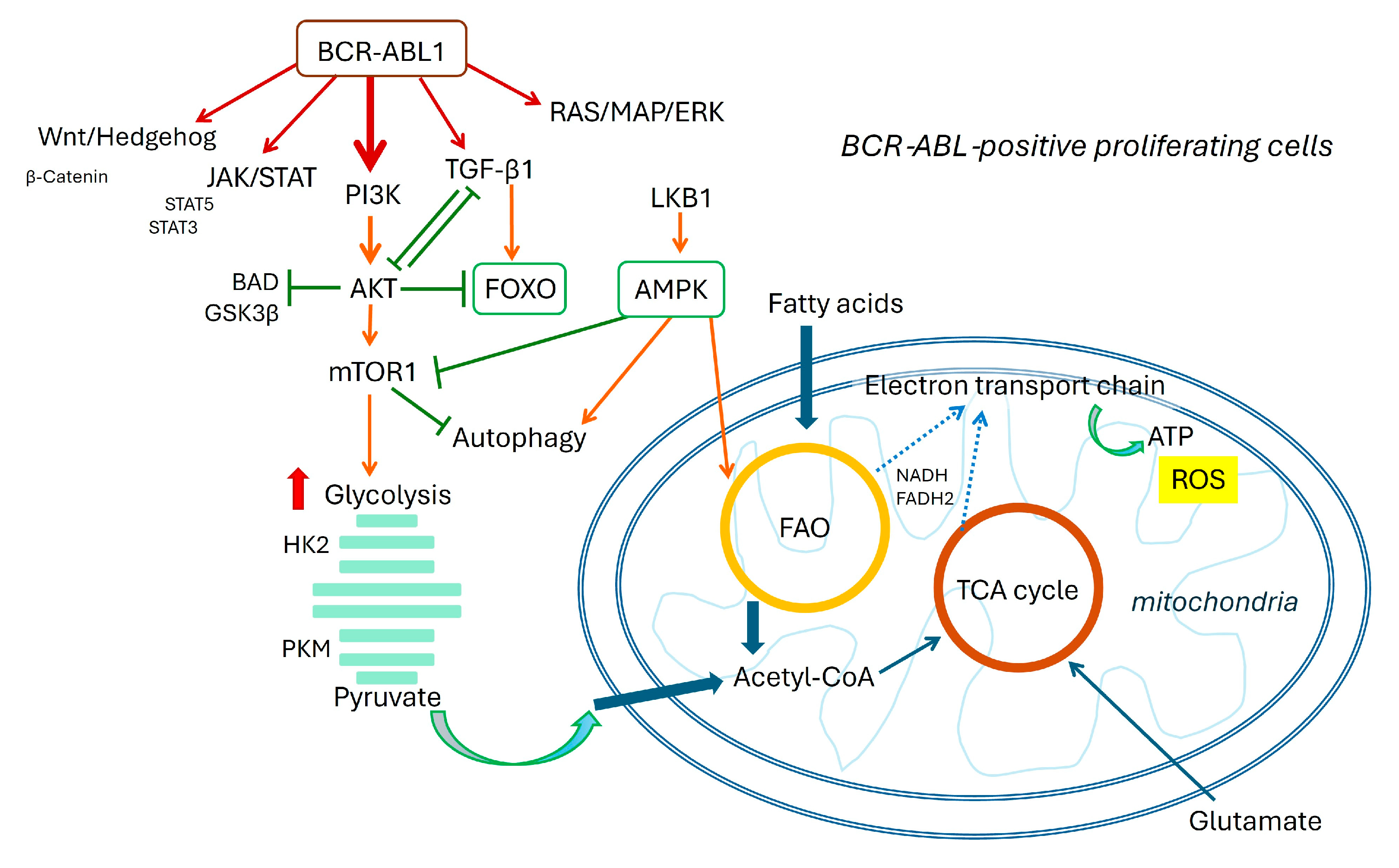
Figure 2.
Schematic overview of signaling networks in CML: LSC quiescent cells.
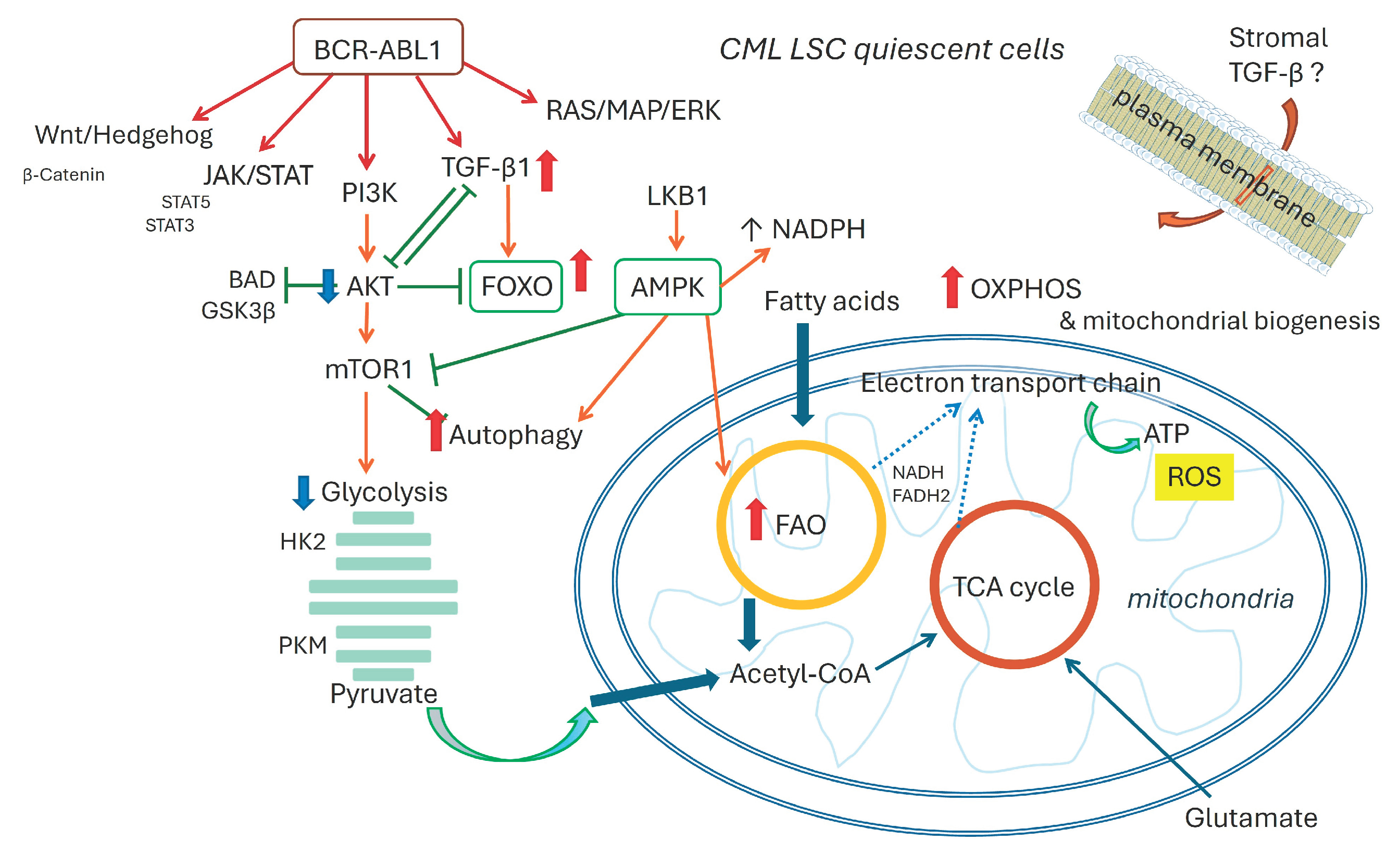
Disclaimer/Publisher’s Note: The statements, opinions and data contained in all publications are solely those of the individual author(s) and contributor(s) and not of MDPI and/or the editor(s). MDPI and/or the editor(s) disclaim responsibility for any injury to people or property resulting from any ideas, methods, instructions or products referred to in the content. |
© 2026 by the authors. Licensee MDPI, Basel, Switzerland. This article is an open access article distributed under the terms and conditions of the Creative Commons Attribution (CC BY) license (http://creativecommons.org/licenses/by/4.0/).
Copyright: This open access article is published under a Creative Commons CC BY 4.0 license, which permit the free download, distribution, and reuse, provided that the author and preprint are cited in any reuse.


