

Submitted:
18 June 2026
Posted:
22 June 2026
You are already at the latest version
Abstract
Potentially toxic elements (PTEs) are persistent contaminants that can bioaccumulate and transfer through food webs, making wildlife valuable sentinels of ecosystem health. This study provides the first characterization of metal and metalloid concentrations in whole blood from free-ranging Iberian lynxes (Lynx pardinus) and evaluates biological and environmental factors influencing their variability, including comparisons with captive individuals. A total of 229 blood samples collected from Iberian lynxes in Extremadura (SW Spain) between 2018 and 2024 were analyzed for Cr, Mn, Cu, Zn, As, Se, Cd, Hg, Fe, and Pb. Concentrations were generally within physiological ranges reported for other mammals, and no clinically detectable adverse effects were observed during routine health examinations. Significant correlations were detected among several elements, particularly between Zn and Fe, with moderate associations between Mn and Zn and between Fe and Cu, suggesting shared environmental sources and interconnected physiological regulation mechanisms. Element concentrations were significantly influenced by endogenous factors (age and sex) and exogenous variables (geographical area and sampling period), especially for As, Zn, and Se, reflecting differences in bioaccumulation, metabolism, and environmental exposure. Principal Component Analysis revealed a largely common multielemental profile across individuals, with no clear segregation among populations or biological groups, indicating relatively homogeneous exposure patterns despite subtle spatial and individual variability. These results establish the first reference values for inorganic contaminant biomonitoring in Iberian lynx whole blood and provide a baseline for ecotoxicological assessment and conservation management of this endangered felid.
Keywords:
metals
; blood
; Iberian lynx
; exposure
; biomonitoring
1. Introduction
Potentially toxic elements (PTEs), including certain heavy metals and metalloids, are of increasing environmental concern due to their persistence, capacity for biomagnification and/or bioaccumulation, and associated health risks [1,2,3,4,5]. Essential elements such as Cu, Fe, Mn, Se, and Zn play crucial roles in metabolic, enzymatic, and hormonal processes, whereas non-essential elements, including Cd, As, Hg, and Pb, lack known physiological functions and may induce toxic effects even at low concentrations [6,7,8,9]. PTEs reach wildlife through both natural processes [10] and anthropogenic activities such as mining, agriculture, and industrial discharges [11,12,13,14]. At smaller scales, practices such as hunting and fishing may also contribute to exposure, particularly to Pb [15]. Toxic effects are element-specific and include hematological alterations, nephrotoxicity, neurotoxicity, genotoxicity, oxidative stress, and carcinogenesis [16,17,18].
Research on PTEs exposure in wildlife has employed a wide range of biological matrices, including liver, kidney, brain, blood, hair, feathers, and bone, across diverse taxa ranging from aquatic organisms (fish, amphibians, reptiles) to terrestrial vertebrates (birds and mammals) [19,20,21,22,23,24,25,26]. Such studies have also enabled the identification of specific species as bioindicators of environmental contamination [27,28,29,30], thereby facilitating the early detection of ecosystem disturbances [11].
The endangered Iberian lynx (Lynx pardinus), an apex predator endemic to the Iberian Peninsula, is of particular relevance for biomonitoring studies because of its trophic position, marked dietary specialization largely based on the European rabbit (Oryctolagus cuniculus), and strict habitat requirements [31,32]. Historical population declines of the species, as well as of other carnivores, were exacerbated by disease outbreaks affecting their principal prey species [33,34,35]. Current conservation measures, including the four LIFE projects (Lince-Andalucía, Conservación y reintroducción del lince ibérico en Andalucía, Iberlince, and Lynxconnect, the latter being the only ongoing initiative), have substantially contributed to population recovery, allowing the species’ conservation status to improve to “Vulnerable” in December 2024 [36,37,38].
From an ecotoxicological perspective, information regarding PTEs exposure in this species remains scarce. Previous studies have focused exclusively on destructive matrices such as bone and liver [39], whereas no studies to date have evaluated non-destructive matrices such as whole blood. This gap also exists internationally for other lynx species, including the Eurasian lynx (Lynx lynx) and the bobcat (Lynx rufus), for which recent studies have similarly relied on liver samples [40,41,42]. However, destructive sampling approaches raise important ethical and ecological concerns, whereas non-destructive techniques provide an innovative alternative despite limitations regarding the representation of concentrations in target tissues. Their application enables continuous and ethically sustainable biomonitoring while preserving ecosystem integrity, facilitating the assessment of contaminant exposure in wildlife (particularly in elusive or protected species) and supporting biodiversity conservation efforts [43]. In addition, non-destructive sampling offers practical advantages for long-term temporal monitoring [44,45,46].
The present study provides the first assessment of metal and metalloid concentrations in whole blood from free-ranging Iberian lynxes. Concentrations of As, Cd, Cr, Cu, Fe, Hg, Mn, Pb, Se, and Zn were quantified. Furthermore, correlations among PTEs concentrations, individual characteristics or endogenous factors (age and sex), exogenous factors (habitat or geographical area), and temporal patterns were investigated following approaches previously applied in wildlife ecotoxicology studies [47,48,49,50]. Comparisons between free-ranging and captive individuals were also conducted to evaluate habitat contamination status. Ultimately, this study aims to establish baseline reference values for PTEs in the Iberian lynx and to contribute to the development of evidence-based conservation strategies for this emblematic apex predator.
2. Materials and Methods
2.1. Study Area
The region of Extremadura is located in the western Iberian Peninsula and covers an area of 41,634 km2. Despite its large territorial extent, the region has a population of 1,053,774 inhabitants, making it the least densely populated autonomous community in Spain. This low population density has resulted in the preservation of extensive areas with minimal human disturbance, providing optimal habitats for numerous animal and plant species. Consequently, Extremadura harbors a high level of biodiversity and constitutes an important refuge for emblematic species such as the Iberian imperial eagle (Aquila adalberti) and the endangered Iberian lynx, while also supporting the largest vulture population in the country.
The region includes several protected natural areas of high ecological value, including Monfragüe National Park, Tajo International Natural Park, and La Siberia, all of which are recognized as Biosphere Reserves. In addition, Extremadura contains 69 Special Protection Areas for Birds (ZEPA), representing 26.15% of the regional territory, and is crossed by two major river basins, the Guadiana and Tajo rivers. Collectively, these environmental characteristics make Extremadura one of the most suitable regions for the Iberian lynx. The areas currently occupied by this felid are mainly distributed across Matachel, Valdecigüeñas, the surroundings of the Ortiga River, and the areas surrounding the Valdecañas reservoir in the Ibores region [36] (Figure 1).
- Matachel (38.4238, -5.7820): Located in Southeast Extremadura, this valley is crossed by the Matachel River and bordered by the Sierra Grande de Hornachos. It hosts the largest Iberian lynx population in the region and is recognized for high biodiversity. The area features low-relief terrain with volcanic and clay-rich soils, predominantly used for cereal and vineyard cultivation. Three abandoned mines are present: Mina de Santa Marta (Zn, Pb), Mina Garandina (V), and Mina Peñas Blancas (Cu, Ni). The A-66 highway crosses the area, constituting a roadkill hotspot despite mitigation measures.
- Valdecigüeñas (38.0464, -5.9498): Covering 37.34 km2, is a Site of Community Importance (SCI), bounded by the Pintado reservoir and the Guadacanal and Hinojales ranges. The terrain is rugged with quartzitic, slate, and granite soils. Mining activity historically included Mina de la Jayona (Fe), now a Natural Monument, while industrial activity includes a galvanizing facility. The area supports established Iberian lynx populations and rich wildlife.
- Ortiga (38.9693, -5.9578): This region along the Ortiga River, including its main tributaries, is part of the Natura 2000 network. The landscape consists of holm oak woodlands (dehesa in Spanish), and low quartzitic hills. Abandoned mines include Mina San Nicolás (W), Mina Las Tejoneras (Cu), and Mina El Pedregal (calcite), along with the active Quintana de la Serena granite quarry. Sparse human settlements and large open areas support agricultural, livestock, and hunting activities.
- Valdecañas (39.7591, -5.6199): The Valdecañas reservoir is included in Natura 2000 and ZEPA, with active wildlife management programs. The area lies in the Tajo River basin, featuring sedimentary and granite outcrops, and is rich in Fe, currently under exploration for future mining. The A-5 highway crosses the region, creating roadkill risk for lynxes.
2.2. Animals and Sample Collection
Between 2018 and 2024, a total of 172 Iberian lynx individuals were captured across the different study areas in Extremadura. Some individuals were recaptured during consecutive years, resulting in a total of 229 blood samples collected (n=229). Captures were distributed among the study areas previously described and included individuals of different age classes.
Fieldwork was conducted within the framework of the LIFE Lynxconnect Project between October and January, with the aim of performing health assessments (vital parameters, body measurements, and body condition evaluation), radiotracking procedures (GPS collar deployment), and the collection of blood, hair, and fecal samples for toxicological, genetic, parasitological, and infectious disease studies. In this context, the age determination of free-ranging individuals was made possible through the long-term monitoring and surveillance activities carried out within the aforementioned project. Veterinary staff were responsible for the treatment of any injuries and supervised all handling procedures.
Standard live-capture techniques using box traps were employed, followed by chemical immobilization with dexmedetomidine (0.015 mg/kg), midazolam (0.3 mg/kg), and ketamine (2.5 mg/kg). Specifically, 4 mL of heparinized blood were collected for toxicological analyses. Samples were transported under refrigerated conditions and stored at −80 °C until processing. A marked imbalance in sample size among study areas was observed, with Matachel representing the area with the highest number of sampled individuals (n=144), followed by Ortiga (n=24), Valdecañas (n=20), and Valdecigüeñas (n=11).
The study was complemented with 30 additional samples obtained from captive individuals housed at the Iberian Lynx Breeding Centre as part of the ex situ conservation breeding program. Some of these individuals were subsequently reintroduced into the wild, where they continued to be monitored and provided additional samples during subsequent capture periods.
2.3. Chemical Analyses
Ten metals and metalloids were analyzed: Cr, Mn, Cu, Zn, As, Se, Cd, Hg, Fe, and Pb. Whole-blood samples were processed at the Elemental and Molecular Analysis Laboratory of the University of Extremadura, accredited according to ISO 9001:2008 standards, following an in-house validated protocol. Approximately 400 µL of each sample were transferred into glass digestion tubes and subjected to acid digestion using 3 mL of a 3:1 mixture of 69% HNO3 and H2O2 in a Milestone Ultrawave microwave digestion system. Following digestion, samples were diluted to a final volume of 25 mL with ultrapure water.
Calibration curves were prepared using certified multielement standard solutions. Internal standards (Y and Rh, 400 µg/L each) were continuously introduced by means of a peristaltic pump. Elemental analyses were performed using an Agilent 7900 inductively coupled plasma mass spectrometer (ICP-MS) equipped with a fourth-generation octopole reaction system, operating either in standard mode or in KED mode with He as collision gas. Limits of quantification (LOQ) were established at 5 µg/L for Cr, Mn, Cu, As, Se, Cd, and Pb; 10 µg/L for Fe; 25 µg/L for Hg; and 50 µg/L for Zn. Instrumental performance was monitored daily (CeO/Ce < 2.5%, Ce2+/Ce < 3%, background < 1 cps). Method accuracy was validated using the certified reference material Seronorm® Trace Elements Whole Blood, yielding recovery rates between 92% and 107% and coefficients of variation below 6.5%.
Fe was not analyzed during the 2020/21 sampling period, whereas Cr was only incorporated into the analytical protocol from that period onwards. Consequently, both elements were excluded from comparative analyses among sampling periods.
2.4. Statistical Analyses
All statistical analyses were performed using GraphPad Prism 9.0.2 software (GraphPad Software Inc., La Jolla, CA, USA). Descriptive statistics for PTEs concentrations were expressed as mean ± standard deviation (SD), minimum (Min.), 25th percentile (25% P.), median, 75th percentile (75% P.), maximum (Max.), and range. Data normality was assessed using the Anderson–Darling, D’Agostino and Pearson, Shapiro–Wilk, and Kolmogorov–Smirnov tests.
Values below the limit of quantification (LOQ) were replaced by one-half of the limit of detection (1/2 LOD). However, Cr, Cd, and Hg were excluded from correlation analyses because more than 60% of their values were below the LOQ. Likewise, Fe and Cr were excluded from comparisons among sampling periods due to incomplete datasets. The 2018/19 sampling period was excluded from analyses by individual period because of the limited sample size (n=4).
As the data did not meet normality assumptions, inferential statistical analyses were conducted using non-parametric methods. Correlations between PTEs concentrations were evaluated using Spearman’s rank correlation coefficient. Correlation strength was interpreted according to the criteria established by Kalisińska et al. (2023) [51], with the following categories: 0.8–1.0, very strong; 0.6–0.79, strong; 0.4–0.59, moderate; 0.2–0.39, weak; and 0–0.19, very weak. Statistical significance was set at p < 0.05. Differences among groups for each intrinsic and extrinsic factor were assessed using the Mann–Whitney U test (for two groups) or the Kruskal–Wallis test (for three or more groups).
The combined effects of sex, age, area, and sampling period on PTEs concentrations were evaluated using General Linear Models (GLMs) implemented in Jamovi 2.6 software (GAMLj3 module). Mineral concentrations were considered dependent variables, whereas sex (male/female), age class (kitten, juvenile, and breeding adult), area (Matachel, Ortiga, Valdecañas, Valdecigüeñas, and Captivity), and sampling periods (annual distribution between 2018/19 and 2023/24) were treated as explanatory factors. Finally, a Principal Component Analysis (PCA) was conducted to explore general patterns among variables using Jamovi 2.6 software (vijPlots module).
3. Results & Discussion
3.1. Metal and Metalloid Concentrations in Whole Blood
Descriptive statistical data for the individual elements are summarized in Table 1. As shown, all PTEs were detected in blood samples, except for Pb, Cr, Hg, and Cd, which were not detected in 34.0%, 66.0%, 63.70%, and 69.86% of the samples, respectively. Essential elements such as Zn, Cu, Fe, Mn, and Se, which are interrelated due to their physicochemical similarities and involvement in different physiological pathways [28], were detected in all samples, together with the toxic metalloid As.
In addition, a comparative table was compiled including the mean concentrations obtained for these metals and those previously reported in the blood of other species, such as the caracal (Caracal caracal) [52], the Angora cat (Felis catus) [53], and the domestic dog (Canis lupus familiaris) [54] (Table 2).
Overall, mean concentrations followed the order: Zn >> Cu = Fe > Se > As > Mn > Pb = Cr = Hg >> Cd. As expected, essential elements exhibited the highest concentrations, whereas toxic elements, with the exception of As, were present at comparatively lower levels. The overall concentrations obtained for each element, together with their toxicological relevance, are described below.
Although essential elements are required for normal physiological function, concentrations exceeding certain thresholds may pose health risks to animals.
Zn was the most abundant element, with concentrations of 2729 ± 375.6 µg/L, remaining below the toxicity thresholds reported in horses (6000–15000 µg/L), which have been associated with hemolytic anemia, renal failure, and gastrointestinal disorders [55,56]. The maximum recorded concentration (4962 µg/L) also remained below this toxicity range.
Mean Cu concentrations were 499.0 ± 109.0 µg/L, below the toxicity thresholds described in ruminants (1300 µg/L), in which chronic exposure may cause anemia, anorexia, and weakness, whereas acute exposure may induce diarrhea, dehydration, and hypovolemic shock [57,58]. A single juvenile male from Matachel sampled in 2019/20 reached a Cu concentration of 1684 µg/L without exhibiting clinical signs.
Fe concentrations were 459.4 ± 102.6 µg/L and did not exceed the toxicity thresholds (>3000 µg/L) established for mammals such as the mule deer (Odocoileus hemionus) and the black rhinoceros (Diceros bicornis minor) [59,60], above which clinical signs of intoxication such as lethargy, vomiting, and profuse diarrhea may occur [61]. The maximum concentration recorded (926.8 µg/L) likewise remained below this threshold.
Se concentrations averaged 354.7 ± 89.69 µg/L, remaining below toxicity thresholds reported for the mule deer (545 µg/L) and below concentrations associated with sublethal reproductive effects or overt toxicity in other taxa such as fish and birds [62,63,64,65,66]. A juvenile female from Matachel sampled in 2023/24 exhibited a Se concentration of 596.2 µg/L, slightly exceeding the proposed threshold, although no clinical signs of toxicity were observed.
Mean As concentrations were 146.3 ± 174.5 µg/L, exceeding the average values reported in healthy domestic cats (85 µg/L) [67]. The main symptoms associated with As intoxication include genetic alterations (DNA damage), behavioral changes, endocrine disruption, weakness, and diarrhea [67,68,69]. A juvenile female from Ortiga sampled in 2019/20 reached a maximum concentration of 1041 µg/L, markedly higher than reference values; nevertheless, the individual remained clinically healthy. The absence of species-specific toxicity thresholds for the Iberian lynx highlights the need for further investigation.
For Mn, the mean ± SD concentration was 27.82 ± 19.94 µg/L, considerably lower than the toxicity thresholds described in mammals such as the West Indian manatee (Trichechus manatus, 40000–75000 µg/L), in which anemia, serum biochemical alterations, renal insufficiency, and arrhythmias may occur above these levels [55,60]. The maximum concentration recorded was 295.6 µg/L.
For Pb, the mean ± SD concentration was 8.490 ± 14.86 µg/L, well below the toxicity thresholds established in mammals such as brown rat (Rattus norvegicus) and Japanese macaque (Macaca fuscata) (150–200 µg/L), above which hematological, neurological, and gastrointestinal effects may occur [6,70,71,72,73]. The maximum recorded value (169.4 µg/L) did not exceed this reference threshold, which was adopted in the absence of species-specific data. Under this criterion, several individuals fell within the proposed threshold range, although it is acknowledged that such thresholds are not universally applied in ecotoxicological studies. In approximately one-third of the samples, blood Pb concentrations could not be quantified.
Mean Cr concentrations were 6.075 ± 9.527 µg/L, remaining below the toxicity thresholds (10 µg/L) described in the small Indian mongoose (Herpestes javanicus) and macaques (Macaca radiata), in which sublethal reproductive and endocrine effects or overt toxicity may occur [74,75,76]. However, one reproductive adult male from Matachel sampled in 2022/23 exhibited a concentration of 104.2 µg/L, far exceeding reference values, although no clinical signs were observed.
Hg concentrations were 5.795 ± 8.342 µg/L, markedly lower than toxicity thresholds reported in felids such as the Florida panther (Felis concolor coryi, 500 µg/L), above which reproductive impairment has been described [77,78]. The maximum concentration recorded (88.87 µg/L) remained well below this threshold.
Cd concentrations were 0.722 ± 0.326 µg/L, substantially lower than toxicity thresholds described in birds and mammals such as the wood mouse (Apodemus sylvaticus, 26 µg/L), in which hematological alterations may occur [66,79]. The maximum concentration recorded (1.560 µg/L) also remained far below this threshold. Cd was the least frequently detected element, being quantifiable in only 30% of the samples.
Regarding the comparative data presented in Table 2, Iberian lynx individuals (n=229) exhibited lower concentrations of all PTEs (with the exception of Pb) than caracal individuals (n=67) from South Africa, with the greatest differences observed for Mn, Zn, Cr, Hg, and Cd. This pattern was not observed in the other felid species considered, the Angora cat (n=30) from Turkey, which showed lower concentrations of both Zn and Se. Fe concentrations reported for this species (2440 ± 364.0 µg/L) were notably higher than those observed in the Iberian lynx (459.4 ± 102.6 µg/L). In the case of a non-felid facultative carnivore, the domestic dog (n=140) sampled in Turkey, As (21.02 ± 0.580 µg/L) and Hg (0.740 ± 0.220 µg/L) concentrations were considerably lower than those detected in the Iberian lynx (146.0 ± 174.0 µg/L and 5.790 ± 8.340 µg/L, respectively). No marked interspecific differences were detected for the remaining PTEs.
3.2. Correlation Between Metal and Metalloid Concentrations
Potential relationships among the different PTEs were also evaluated. Spearman correlation analyses revealed multiple statistically significant associations between metals and metalloids (Figure 2). As shown, a strong positive correlation was observed between Zn and Fe (rho = 0.72). Moderate correlations were detected between Mn and Zn (rho = 0.42) and between Fe and Cu (rho = 0.43). In addition, weak correlations were identified between As and Se (rho = 0.34), Mn and Fe (rho = 0.28), and Se and Fe (rho = 0.26). The remaining correlations were classified as very weak according to the criteria established by Kalisińska et al. (2023) [51]. Collectively, these findings suggest that the concentrations of these elements tend to covary, possibly as a consequence of shared environmental sources or common physiological interactions.
3.3. Influence of Endogenous (Age, Sex) and Exogenous (Sampling Period and Geographical Zone) Factors on Metal and Metalloid Concentrations
Regarding endogenous factors, age and sex were considered, whereas body condition was not included despite its recognized relevance in this type of study. This decision was based on the fact that 98% of the individuals included in the present work exhibited an optimal body condition, ranging between 2.5 and 3.5 (with 3/5 considered the ideal value on a five-point scale). In felids, particularly domestic cats, a nine-point scale (1/9) is more commonly used, with an ideal body condition of 5/9 [80], which, when extrapolated to the scale used in the present study, corresponds to 3/5, the mean value observed in nearly 100% of the sampled individuals. In other international studies assessing metal concentrations in more invasive matrices such as liver, body condition has been identified as an important covariate to be considered [40]. However, contrary to previous assumptions, low body condition does not necessarily represent a direct consequence of toxic effects of inorganic elements but may instead facilitate their accumulation [40].
The effect of age on blood concentrations of metals and metalloids was evaluated using generalized linear models (GLMs) (Table 3), classifying individuals into three age categories: kittens (<1 year), juveniles (1–2 years), and breeding adults (>2 years). For As, significant differences were detected between juveniles and kittens in Matachel (p = 0.037) and Ortiga (p = 0.023), with higher concentrations observed in juveniles. Zn concentrations also showed significant differences between juveniles and kittens (p = 0.025), with higher values in the former. Se exhibited similar age-related trends, with higher concentrations in juveniles compared with kittens in Valdecigüeñas (p = 0.033), and in reproductive adults compared with kittens across all regions (p = 0.030).
These results indicate clear age-associated differences in blood concentrations of As, Zn, and Se, likely reflecting age-dependent bioaccumulation processes, differences in dietary intake across age classes, or metabolic changes [39]. Such metabolic changes may include increased intestinal absorption efficiency, variations in the homeostatic regulation of essential elements, and the progressive development of hepatic detoxification systems [81,82]. In particular, juveniles exhibit higher metabolic demands and growth rates, which may promote the uptake and circulation of essential elements such as Zn and Se, whereas increased As levels may be associated with higher trophic exposure and the maturation of biotransformation pathways [83].
In the descriptive statistical analysis of age-class–stratified data, a slight increasing trend in mean concentrations was observed for Pb and Se. Kittens exhibited mean Pb and Se concentrations of 6.502 ± 12.67 µg/L and 332.3 ± 82.26 µg/L, respectively, followed closely by juveniles (8.272 ± 12.49 µg/L and 356.7 ± 101.0 µg/L) and, subsequently, by adult individuals (8.655 ± 16.51 µg/L and 361.0 ± 89.35 µg/L). For the remaining metals, mean concentrations remained relatively stable across age classes (Table 4). In the case of Pb, these results are more likely attributable to the bioaccumulation and biomagnification processes previously described, particularly in apex predators such as the Iberian lynx. Regarding Se, the observed pattern may also be related to age-dependent variations in the homeostatic regulation of essential elements, as discussed above.
Sex was also evaluated as a potentially influential factor affecting metal levels. However, only As showed a significant sex-related effect (p < 0.05; Table 3), with significantly higher concentrations observed in females than in males (p = 0.037). This difference may be attributable to sex-specific metabolic pathways, including the potential influence of estrogens on As methylation [84]. Given the limited number of studies focused on As compared with other metals such as Pb, additional research is required to elucidate other mechanisms underlying sex-related differences in blood As concentrations.
In the descriptive statistical analysis stratified by sex, Pb concentrations in male individuals were almost twice as high as those observed in females (8.500 ± 12.36 and 4.857 ± 4.560 µg/L, respectively). For the remaining metals, mean concentrations remained relatively stable between sexes (Table 4). These findings may also be associated with the aforementioned sex-specific metabolic pathways.
Temporal trends were evaluated by compiling descriptive statistical data according to sampling periods (Table 4). Likewise, the effect of sampling period on metal concentrations was assessed. Although the number of animals analyzed during the 2020/21 period (n = 31) decreased compared with 2019/20 (n = 45), a progressive increase was subsequently observed, reaching 61 individuals in 2023/24. This trend may reflect a slight but continuous increase in Iberian lynx populations within the study region.
Regarding metal concentrations, no significant variations were observed among the analyzed periods, with the exception of As, whose concentrations decreased by approximately half during 2020/21 and 2021/22 (88.67 ± 107.4 µg/L and 95.17 ± 159.7 µg/L, respectively) compared with those recorded in 2019/20 (178.5 ± 232.2 µg/L), subsequently approaching the latter values again during the two most recent sampling periods. In the case of Pb, mean ± SD concentrations (16.45 ± 13.28 µg/L) recorded in 2019/20 progressively decreased during the following two sampling periods before stabilizing at values close to one-quarter of the aforementioned concentration.
Only As and Se exhibited statistically significant differences among sampling periods (p < 0.05; Table 3). As concentrations were significantly higher in 2019/20 (p = 0.023), 2022/23 (p = 0.038), and 2023/24 (p < 0.001) compared with 2020/21. Se concentrations were significantly higher in 2019/20 than in 2021/22 (p = 0.002). Although these findings could suggest potential temporal bioaccumulation, longitudinal analyses of recaptured individuals monitored over three or more sampling periods did not reveal consistent or significant increases in metal concentrations for most individuals (Figure 3). Whole-blood concentrations of Se and As showed marked interannual variability together with extremely low coefficients of determination (R2 = 0.0045 and 0.0216, respectively), indicating that temporal variation alone does not explain the differences observed among sampling periods. Instead, factors other than bioaccumulation, such as the aforementioned metabolic variability or dietary differences, are likely to contribute substantially to the observed patterns.
When 0. Table 3). As concentrations were higher in Matachel (p = 0.037) and Ortiga (p = 0.023) compared with captive individuals. Se concentrations were higher in all free-ranging areas compared with captivity: Matachel (p = 0.01), Valdecañas (p = 0.002), Ortiga (p = 0.022), and Valdecigüeñas (p = 0.033). These differences are likely associated with environmental exposure and dietary factors. Captive lynxes receive controlled diets and are not exposed to the same environmental sources of metals as free-ranging individuals, which frequently display wandering and territorial behaviors [85]. Indeed, although other metals and metalloids did not show significant area-related differences, the overall trend suggested slightly lower concentrations in captive individuals due to their more limited environmental exposure.
In a study conducted in the United States by Thomason et al. (2016) [42], in which metal concentrations were measured in liver samples from the bobcat, geographic area was found to exert less influence on metal levels than dietary ecology or behavior, as the authors compared results obtained in this apex predator with those of more diurnal and omnivorous mesocarnivores. It is noteworthy that, when all factors were considered jointly, only sampling periods and age exhibited a significant interaction, with reproductive adults showing significantly higher As concentrations than cubs during the 2021/22 (p < 0.001) and 2022/23 (p = 0.038) periods.
In the descriptive statistical analysis of data classified according to study area, Pb concentrations in the blood of individuals from Valdecigüeñas and Captivity were higher than those observed in the remaining areas. This pattern is more likely associated with dietary factors than with environmental exposure, given that captive animals are maintained under controlled conditions and do not exhibit wandering or territorial behaviors.
Regarding As, the mean blood concentration detected in individuals from Ortiga was markedly higher than those recorded in the other study areas, suggesting the possible existence of specific exposure sources unique to this region. For the remaining metals, mean concentrations remained relatively stable across study areas (Table 4).
3.4. Principal Component Analysis (PCA)
PCA revealed that the first two principal components explained 52.4% of the total variance. The first component (PC1, 31.3%) represented a general gradient of metal accumulation, whereas the second component (PC2, 21.1%) distinguished between essential and non-essential elements (Figure 4).
The projection of individual Iberian lynx specimens onto the PC1–PC2 plane did not reveal a clear separation according to study area (Figure 5), indicating a broadly similar multielement profile among the sampled areas. Nevertheless, subtle spatial trends were observed, which may reflect local differences in environmental exposure or diet, potentially associated with the distinct mining, agricultural, and livestock practices characteristic of each study area.
Likewise, no clear segregation was detected when individuals were classified according to sex, sampling periods, or age class (Figure 5), suggesting that these factors do not exert a marked influence on the overall multielement pattern. However, kittens exhibited a slight tendency toward lower values along the general exposure axis (PC1), consistent with a lower degree of metal accumulation compared with breeding adults. This observation is in agreement with the expected dynamics of bioaccumulation and persistence of inorganic elements along the trophic chain and within the organism, as previously discussed.
4. Conclusions
The present study provides the first comprehensive assessment of ten metals and metalloids in whole blood from Iberian lynx populations in Extremadura, based on a substantial sample size (n = 229). Overall, blood concentrations of Pb, Mn, Cu, Zn, As, Se, Fe, Cr, Cd, and Hg remained within ranges considered physiological and/or representative of background exposure levels, and no evident signs of toxicity were observed. These findings indicate that, under current environmental conditions, Iberian lynxes inhabiting the studied wild areas are not experiencing concerning levels of exposure to these elements. This conclusion is further supported by comparisons with previous studies and by the good health status exhibited by the animals at the time of sampling and veterinary examination.
Among the analyzed elements, As, Zn, and Se were significantly influenced by both biological and environmental factors, including age, sex, area, and sampling periods. The differences observed among groups are likely associated with specific bioaccumulation processes, metabolic activity, and dietary variation. Notably, lynxes from the four natural areas exhibited higher concentrations of As and Se than captive individuals, reflecting greater exposure under free-ranging environmental conditions. Although the detected environmental levels do not appear to represent an immediate health risk, continued monitoring remains necessary, particularly in view of potential future industrial development within these areas.
The continuation of research efforts is essential to support conservation strategies aimed at ensuring the persistence and recovery of this emblematic species, in a manner comparable to initiatives developed for other lynx species such as the Eurasian lynx [40,86,87]. Furthermore, the present study is expected to serve as a valuable reference for the development of future biomonitoring and risk-assessment studies in this species, one of the most iconic representatives of Iberian wildlife.
Author Contributions
Conceptualization and data curation, Férnandez-Casado, David; Soler-Rodríguez, Francisco; formal analysis, Férnandez-Casado, David; Pérez-López, Marcos; investigation, Férnandez-Casado, David; Soler-Rodríguez, Francisco; methodology, Férnandez-Casado, David; visualization, Férnandez-Casado, David; Soler-Rodríguez, Francisco; Pérez-López, Marcos; writing - original draft, Férnandez-Casado, David; supervision and validation, Rodríguez-Somoza, Elsa; Portillo-Moreno, Ángel; Carrillo-Heredero, Alicia María; Sánchez-Cuerda, Susana; Galán-Carrillo, María; Guerrero, Álvaro; Martínez-Morcillo, María Salomé; Míguez-Santiyán, María Prado; Bertini, Simone; Pérez-López, Marcos; Palacios, María Jesús; Soler-Rodríguez, Francisco; visualization, Rodríguez-Somoza, Elsa; Portillo-Moreno, Ángel; Carrillo-Heredero, Alicia María; Sánchez-Cuerda, Susana; Galán-Carrillo, María; Guerrero, Álvaro; writing - review editing, Martínez-Morcillo, María Salomé; Míguez-Santiyán, María Prado; Bertini, Simone; Pérez-López, Marcos; Palacios, María Jesús; Soler-Rodríguez, Francisco.
Funding
This work was made possible through the support and funding provided by the Junta de Extremadura under the following collaboration agreements: 1. Collaboration agreement between the Regional Ministry for Ecological Transition and Sustainability of the Regional Government of Extremadura and the University of Extremadura for the assessment of toxicological processes in wildlife. 1.1. Year 2019: File No. CMSERSO19029. 1.2. Years 2020–2022: File No. 2051999FR002. 2. Collaboration agreement between the Regional Ministry for Ecological Transition and Sustainability of the Regional Government of Extremadura and the University of Extremadura for the study of interactions between toxic chemical substances and wildlife from Extremadura, resulting from exposure through poisoned baits or environmental contamination, as a basis for species and habitat conservation management. Years 2023–2025. File No. 2251999FR004.
Acknowledgments
The authors also wish to thank all members of the LIFE projects developed for the Iberian lynx in Extremadura (LIFE IBERLINCE and LIFE LYNXCONNECT), as well as FOTEX and the staff of the “Los Hornos” Wildlife Recovery and Environmental Education Centre of the Regional Government of Extremadura, together with the NGO AMUS (Acción por el Mundo Salvaje), for their invaluable assistance in sample collection and collaboration throughout the study. The authors also acknowledge their collaboration with the journal Animals.
Conflicts of Interest
The authors declare no conflicts of interest.
References
- García-Fernández, A.J.; Calvo, J.F.; Martínez-López, E.; María-Mojica, P.; Martínez, J.E. Raptor ecotoxicology in Spain. A review on persistent environmental contaminants. AMBIO A J. Hum. Environ. 2008, 37, 432–439. [Google Scholar] [CrossRef]
- Lazarus, M.; Orct, T.; Sergiel, A.; Vranković, L.; Marijić, V.F.; Rašić, D.; Reljić, S.; Aladrović, J.; Zwijacz-Kozica, T.; Zięba, F.; Jurasović, J.; Erk, M.; Maślak, R.; Selva, N.; Huber, D.; Huber, Đ. Metal (loid) exposure assessment and biomarker responses in captive and free-ranging European brown bear (Ursus arctos). Environ. Res. 2020, 183, 109166. [Google Scholar] [CrossRef] [PubMed]
- Pérez-López, M.; de Mendoza, M.H.; Beceiro, A.L.; Soler-Rodríguez, F. Heavy metal (Cd, Pb, Zn) and metalloid (As) content in raptor species from Galicia (NW Spain). Ecotoxicol. Environ. Saf. 2008, 70(1), 154–162. [Google Scholar] [CrossRef] [PubMed]
- Rashed, M.N.; Soltan, M.E. Animal hair as biological indicator for heavy metal pollution in urban and rural areas. Environ. Monit. Assess. 2005, 110, 41–53. [Google Scholar] [CrossRef] [PubMed]
- Tchounwou, P.B.; Yedjou, C.G.; Patlolla, A.K.; Sutton, D.J. Heavy Metal Toxicity and the Environment. In Molecular, clinical and environmental toxicology 3: Environmental toxicology; Luch, A., Ed.; Springer Science & Business Media: Heidelberg, 2012; pp. 133–164. [Google Scholar] [CrossRef] [PubMed]
- Buekers, J.; Redeker, E.S.; Smolders, E. Lead toxicity to wildlife: derivation of a critical blood concentration for wildlife monitoring based on literature data. Sci. Total Environ. 2009, 407(11), 3431–3438. [Google Scholar] [CrossRef] [PubMed]
- Kalisińska, E. Endothermic animals as biomonitors of terrestrial environments. In Mammals and birds as bioindicators of trace element contaminations in terrestrial environments, an Ecotoxicological Assessment of the Northern Hemisphere; Kalisińska, E., Ed.; Springer Nature Switzerland, AG: Berlin, 2019; pp. 21–53. [Google Scholar] [CrossRef]
- Lazarus, M.; Sekovanić, A.; Reljić, S.; Kusak, J.; Ferenčaković, M.; Sindičić, M.; Gomerčić, T.; Huber, Đ. Lead and other trace element levels in brains of Croatian large terrestrial carnivores: influence of biological and ecological factors. Toxics 2022, 11(1), 4. [Google Scholar] [CrossRef] [PubMed]
- NRC. Mineral Tolerance of Animals; National Research Council, National Academy Press: Washington, DC, 2005. [Google Scholar] [CrossRef] [PubMed]
- Pirrone, N.; Cinnirella, S.; Feng, X.; Finkelman, R.B.; Friedli, H.R.; Leaner, J.; Telmer, K. Global mercury emissions to the atmosphere from anthropogenic and natural sources. Atmos. Chem. Phys. 2010, 10(13), 5951–5964. [Google Scholar] [CrossRef]
- Di Marzio, A.; Lambertucci, S.A.; Fernández, A.G.; Martínez-López, E. From Mexico to the Beagle Channel: A review of metal and metalloid pollution studies on wildlife species in Latin America. Environ. Res. 2019, 176, 108462. [Google Scholar] [CrossRef] [PubMed]
- Gall, J.E.; Boyd, R.S.; Rajakaruna, N. Transfer of heavy metals through terrestrial food webs: a review. Environ. Monit. Assess. 2015, 187, 1–21. [Google Scholar] [CrossRef] [PubMed]
- Jiménez-Ballesta, R.; García-Navarro, F.J.; Bravo, S.; Amorós, J.A.; Pérez-de-los-Reyes, C.; Mejías, M. Environmental assessment of potential toxic trace element contents in the inundated floodplain area of Tablas de Daimiel wetland (Spain). Environ. Geochem. Health 2016, 39(5), 1159–1177. [Google Scholar] [CrossRef] [PubMed]
- Nakayama, S.M.; Ikenaka, Y.; Hamada, K.; Muzandu, K.; Choongo, K.; Teraoka, H.; Mizuno, N.; Ishizuka, M. Metal and metalloid contamination in roadside soil and wild rats around a Pb–Zn mine in Kabwe, Zambia. Environ. Pollut. 2011, 159(1), 175–181. [Google Scholar] [CrossRef] [PubMed]
- Ferreyra, H.; Romano, M.; Beldomenico, P.; Caselli, A.; Correa, A.; Uhart, M. Lead gunshot pellet ingestion and tissue lead levels in wild ducks from Argentine hunting hotspots. Ecotoxicol. Environ. Saf. 2014, 103, 74–81. [Google Scholar] [CrossRef] [PubMed]
- Ercal, N.; Gurer-Orhan, H.; Aykin-Burns, N. Toxic metals and oxidative stress part I: mechanisms involved in metal-induced oxidative damage. Curr. Top. Med. Chem. 2001, 1(6), 529–539. [Google Scholar] [CrossRef] [PubMed]
- Espín, S.; Martinez-Lopez, E.; Jimenez, P.; Maria-Mojica, P.; Garcia-Fernandez, A.J. Effects of heavy metals on biomarkers for oxidative stress in Griffon vulture (Gyps fulvus). Environ. Res. 2014, 129, 59–68. [Google Scholar] [CrossRef] [PubMed]
- Wu, X.; Cobbina, S.J.; Mao, G.; Xu, H.; Zhang, Z.; Yang, L. A review of toxicity and mechanisms of individual and mixtures of heavy metals in the environment. Environ. Sci. Pollut. Res. 2016, 23(9), 8244–8259. [Google Scholar] [CrossRef] [PubMed]
- Burger, J.; Jeitner, C.; Schneider, L.; Vogt, R.; Gochfeld, M. Arsenic, cadmium, chromium, lead, mercury, and selenium levels in blood of four species of turtles from the Amazon in Brazil. J. Toxicol. Environ. Health Part A 2009, 73(1), 33–40. [Google Scholar] [CrossRef] [PubMed]
- Casas, J.M.; Rosas, H.; Solé, M.; Lao, C. Heavy metals and metalloids in sediments from the Llobregat basin, Spain. Environ. Geol. 2003, 44, 325–332. [Google Scholar] [CrossRef]
- Cruz-Santiago, O.; Pérez-Maldonado, I. N.; González-Mille, D. J.; Espinosa-Reyes, G.; Martínez-Toledo, Á.; Ilizaliturri-Hernández, C. A. Nondestructive biomarkers in giant toad (Rhinella marina) to assess the effect of complex mixture of pollutants in Coatzacoalcos River, Mexico. Environ. Toxicol. Pharmacol. 2021, 82, 103558. [Google Scholar] [CrossRef] [PubMed]
- Da Rocha, M.P.; Dourado, P.L.R.; de Souza Rodrigues, M.; Raposo, J.L.; Grisolia, A.B.; de Oliveira, K.M.P. The influence of industrial and agricultural waste on wáter quality in the Agua Boa stream (Dourados, Mato Grosso do Sul, Brazil). Environ. Monit. Assess. 2015, 187(7), 442. [Google Scholar] [CrossRef] [PubMed]
- Ilizaliturri-Hernández, C. A.; González-Mille, D. J.; Mejía-Saavedra, J.; Espinosa-Reyes, G.; Torres-Dosal, A.; Pérez-Maldonado, I. Blood lead levels, δ-ALAD inhibition, and hemoglobin content in blood of giant toad (Rhinella marina) to asses lead exposure in three areas surrounding an industrial complex in Coatzacoalcos, Veracruz, Mexico. Environ. Monit. Assess. 2013, 185, 1685–1698. [Google Scholar] [CrossRef] [PubMed]
- Lorenzo-Márquez, H.; Torres-Dosal, A.; Barba Macías, E.; Ilizaliturri Hernández, C.A.; Martínez-Salinas, R.I.; Morales López, J.J.; Sánchez Moreno, I. Estimación de riesgo de exposición a metales pesados por consumo de plecos (Pterygoplichthys spp.) en infantes de comunidades ribereñas de los ríos Grijalva y Usumacinta, México. Rev. Int. Contam. Ambient. 2016, 32(2), 153–164. [Google Scholar] [CrossRef]
- Nakayama, S.M.; Ikenaka, Y.; Muzandu, K.; Choongo, K.; M’kandawire, E.; Yasuda, J.; Ishizuka, M. Metal and metalloid levels and bio-accumulation characteristics in soil, sediment, land plants and hippopotami (Hippopotamus amphibius L) from the South Luangwa National Park, Zambia. Ecotoxicol. Environ. Saf. 2012, 80, 333–338. [Google Scholar] [CrossRef] [PubMed]
- Siegal-Willott, J.L.; Harr, K.E.; Hall, J.O.; Hayek, L.A.C.; Auil-Gomez, N.; Powell, J.A.; Bonde, R.K.; Heard, D. Blood mineral concentrations in manatees (Trichechus manatus latirostris and Trichechus manatus manatus). J. Zoo. Wildl. Med. 2013, 44(2), 285–294. [Google Scholar] [CrossRef] [PubMed]
- Chiari, M.; Cortinovis, C.; Bertoletti, M.; Alborali, L.; Zanoni, M.; Ferretti, E.; Caloni, F. Lead, Cadmium and Organochlorine Pesticide Residues in Hunted Red Deer and Wild Boar from Northern Italy. Food Addit. Contam. Part A 2015, 32(11), 1867–1874. [Google Scholar] [CrossRef] [PubMed]
- García-Muñoz, J.; Cacciola, N. A.; Plazzi, F.; Prado Míguez-Santiyán, M.; Rodríguez, F. S.; López-Beceiro, A.; Fidalgo, L.E.; Martínez-Morcillo, S.; Pérez-López, M. Metal and metalloid concentrations in wild mammals from SW Europe: European hedgehog (Erinaceus europaeus) and badger (Meles meles). Environ. Sci. Pollut. Res. 2023, 1–16. [Google Scholar] [CrossRef]
- Morales, J.S.; Rojas, R.M.; Perez-Rodriguez, F.; Casas, A.A.; López, M.A. Risk assessment of the lead intake by consumption of red deer and wild boar meat in Southern Spain. Food Addit. Contam. Part A 2011, 28(8), 1021–1033. [Google Scholar] [CrossRef] [PubMed]
- Squadrone, S.; Robetto, S.; Orusa, R.; Griglione, A.; Falsetti, S.; Paola, B.; Abete, M.C. Wildlife hair as bioindicators of metal exposure. Biol. Trace Elem. Res. 2022, 200(12), 5073–5080. [Google Scholar] [CrossRef] [PubMed]
- Ferreras, P.; Travaini, A.; Zapata, S.C.; Delibes, M. Short-term responses of mammalian carnivores to a sudden collapse of rabbits in Mediterranean Spain. Basic appl. ecol. 2011, 12(2), 116–124. [Google Scholar] [CrossRef]
- Illanas, S.; González, A.G.; Cabrera, S.B.; Simón, M.A.; de Toda, S.S.M. Selección del hábitat y permeabilidad del territorio para el lince ibérico (“Lynx pardinus”) en Andalucía: influencia del estado de comportamiento y de la resolución cartográfica. Cuad. Soc. Esp. Cienc. For. 2017, 43, 193–208. [Google Scholar] [CrossRef]
- Carvalho, J.C.; Gomes, P. Feeding resource partitioning among four sympatric carnivores in the Peneda-Gerês National Park (Portugal). J. Zool. 2004, 263, 275–283. [Google Scholar] [CrossRef]
- Fedriani, J.; Palomares, F.; Delibes, M. Niche relations among three sympatric Mediterranean carnivores. Oecologia 1999, 121, 138–148. [Google Scholar] [CrossRef] [PubMed]
- Palacín, C.; Calleja, I. Importancia de los paisajes agrícolas tradicionales para la conservación del turón (Mustela putorius). XII Congreso de la SECEM (Burgos), 2015; p. 105. [Google Scholar]
- LIFE Lynxconnect (2025) Proyecto LIFE Lynxconnect. Available online: https://lifelynxconnect.eu/proyecto/ (accessed on 21/10/2025).
- MITECO. Proyecto LIFE Iberlince. 2018. Available online: https://www.miteco.gob.es/es/parques-nacionales-oapn/centros-fincas/lince/iberlince.html (accessed on 21/10/2025).
- Real Decreto 139/2011 for the development of the List of Wild Species under Special Protection and the Spanish Catalog of Endangered Species.
- Millán, J.; Mateo, R.; Taggart, M.A.; López-Bao, J.V.; Viota, M.; Monsalve, L.; Camarero, P.R.; Blázquez, E.; Jiménez, B. Levels of heavy metals and metalloids in critically endangered Iberian lynx and other wild carnivores from Southern Spain. Sci. Total Environ. 2008, 399(1-3), 193–201. [Google Scholar] [CrossRef] [PubMed]
- Bedouet, L.; Ryser-Degiorgis, M.P.; Borel, S.; Amiot, C.; Afonso, E.; Coeurdassier, M. Evaluating trace elements as a conservation concern for Eurasian lynx (Lynx lynx) in Switzerland. Ecotoxicol. Environ. Saf. 2025, 298, 118300. [Google Scholar] [CrossRef] [PubMed]
- Coeurdassier, M.; Bedouet, L.; Amiot, C.; Berny, P.; Decors, A. Imprégnation du Lynx boréal Lynx lynx par les éléments traces métalliques en France. Naturae 2026, 2026(6), 31–35. [Google Scholar] [CrossRef]
- Thomason, R.K.; Lockhart, J.M.; Loughry, W.J.; Bielmyer-Fraser, G.K. Metal accumulation in bobcats in the Southeastern EE.UU. Environ. Monit. Assess. 2016, 188(10), 565. [Google Scholar] [CrossRef] [PubMed]
- García-Muñoz, J.; Fernández-Casado, D.; Portillo-Moreno, A.; Míguez-Santiyán, M.P.; Soler, F.; López-Beceiro, A.; Fidalgo, L.E.; Martínez-Morcillo, S.; Pérez-López, M. First study of heavy metals analysis in hair and oxidative status of European otters (Lutra lutra) from Southwestern Europe. Ecotox 2025, 34, 1351–1367. [Google Scholar] [CrossRef] [PubMed]
- Ortiz-Santaliestra, M.E.; Tauler-Ametller, H.; Lacorte, S.; Hernández-Matías, A.; Real, J.; Mateo, R. Accumulation of pollutants in nestlings of an endangered avian scavenger related to territory urbanization and physiological biomarkers. Environ. Pollut. 2019, 252, 1801–1809. [Google Scholar] [CrossRef] [PubMed]
- Powolny, T.; Scheifler, R.; Raoul, F.; Fritsch, C. Is blood a reliable indicator of trace metal concentrations in organs of small mammals? Chemosphere 2019, 217, 320–328. [Google Scholar] [CrossRef] [PubMed]
- Powolny, T.; Scheifler, R.; Raoul, F.; Coeurdassier, M.; Fritsch, C. Effects of chronic exposure to toxic metals on haematological parameters in free-ranging small mammals. Environ. Pollut. 2023, 317, 120675. [Google Scholar] [CrossRef] [PubMed]
- López-Perea, J.J.; Laguna, C.; Jiménez-Moreno, M.; Martín-Doimeadios, R.C.R.; Feliu, J.; Mateo, R. Metals and metalloids in blood and feathers of common moorhens (Gallinula chloropus) from wetlands that receive treated wastewater. Sci. Total Environ. 2019, 646, 84–92. [Google Scholar] [CrossRef] [PubMed]
- Rzymski, P.; Niedzielski, P.; Poniedziałek, B.; Rzymski, P.; Pacyńska, J.; Kozak, L.; Dąbrowski, P. Free-ranging domestic cats are characterized by increased metal content in reproductive tissues. Reprod. Toxicol. 2015, 58, 54–60. [Google Scholar] [CrossRef] [PubMed]
- Sánchez-Chardi, A.; López-Fuster, M.J. Metal and metalloid accumulation in shrews (Soricomorpha, Mammalia) from two protected Mediterranean coastal sites. Environ. Pollut. 2009, 157(4), 1243–1248. [Google Scholar] [CrossRef] [PubMed]
- Tajchman, K.; Ukalska-Jaruga, A.; Bogdaszewski, M.; Pecio, M.; Dziki-Michalska, K. Accumulation of toxic elements in bone and bone marrow of deer living in various ecosystems. A case study of farmed and wild-living deer. Animals 2020, 10(11), 2151. [Google Scholar] [CrossRef] [PubMed]
- Kalisińska, E.; Kot, K.; Łanocha-Arendarczyk, N. Red fox as a potential bioindicator of metal contamination in a European environment. Chemosphere 2023, 319, 138037. [Google Scholar] [CrossRef] [PubMed]
- Parker, K.H.; Bishop, J.M.; Serieys, L.E.; Mateo, R.; Camarero, P.R.; Leighton, G.R. A heavy burden: Metal exposure across the land-ocean continuum in an adaptable carnivore. Environ. Pollut. 2023, 327, 121585. [Google Scholar] [CrossRef] [PubMed]
- Kabakci, R.; Yiğit, A.A.; Ekici, H.; Aluç, Y.; Kara, E. Determination of blood heavy metal concentrations and oxidant-antioxidant capacities in Angora cats at different age and gender. Ank. Univ. Vet. Fak. Derg. 2023, 1–20. [Google Scholar] [CrossRef]
- Altinok-Yipel, F.; Yipel, M.; Altuğ, N.; Özdemir, N. Blood concentrations of potentially toxic trace elements (PTEs) and correlation with biochemical and hematological parameters in dogs from thrace region, Turkey. Chemosphere 2022, 293, 133649. [Google Scholar] [CrossRef] [PubMed]
- Manatus, M. Blood mineral concentrations in manatees. J. Zoo. Wildl. Med. 2013, 44(2), 285–294. [Google Scholar] [CrossRef] [PubMed]
- Sileo, L.; Beyer, W.N.; Mateo, R. Pancreatitis in wild zinc-poisoned waterfowl. Avian Pathol. 2003, 32, 655–660. [Google Scholar] [CrossRef] [PubMed]
- Mendel, M.; Chłopecka, M.; Dziekan, N. Haemolytic crisis in sheep as a result of chronic exposure to copper. Pol. J. Vet. Sci. 2007, 10(1), 51–56. [Google Scholar] [PubMed]
- Merck Copper poisoning in animals at. 2025. Available online: https://www.merckvetmanual.com/ (accessed on 05/10/2025).
- Mylniczenko, N.D.; Sullivan, K.E.; Corcoran, M.E.; Fleming, G.J.; Valdes, E.V. Management strategies of iron accumulation in a captive population of black rhinoceroses (Diceros bicornis minor). J. Zoo Wildl. Med. 2012, 43, 3. [Google Scholar] [CrossRef] [PubMed]
- Roug, A.; Swift, P.K.; Gerstenberg, G.; Woods, L.W.; Kreuder-Johnson, C.; Torres, S.G.; Puschner, B. Comparison of trace mineral concentrations in tail hair, body hair, blood, and liver of mule deer (Odocoileus hemionus) in California. J. Vet. Diagn. Invest. 2015, 27(3), 295–305. [Google Scholar] [CrossRef] [PubMed]
- Albretsen, J. The toxicity of iron, an essential element. Vet. Med.-Bonn. Springs Then Edwardsville- 2006, 101(2), 82. [Google Scholar]
- DeForest, D.K.; Adams, W.J. Selenium Accumulation and Toxicity in Freshwater Fishes. In Environmental contaminants in biota: interpreting tissue concentrations; Beyer, W.N., Meador, J.P., Eds.; CRC Press: Boca Raton, Florida, 2011; Volume 5, pp. 193–230. [Google Scholar] [CrossRef]
- Ohlendorf, H.M. Bioaccumulation and effects of selenium in wildlife. Selenium Agric. Environ. 1989, 23, 133–177. [Google Scholar] [CrossRef]
- Ohlendorf, H.M.; Heinz, G.H. Selenium in Birds. In Environmental contaminants in biota: interpreting tissue concentrations; Beyer, W.N., Meador, J.P., Eds.; CRC Press: Boca Raton, Florida, 2011; Volume 21, pp. 669–702. [Google Scholar] [CrossRef]
- Stout, J.D.; Brinker, D.F.; Driscoll, C.P.; Davison, S.; Murphy, L.A. Serum biochemistry values, plasma mineral levels, and whole blood heavy metal measurements in wild northern goshawks (Accipiter gentilis). J. Zoo Wildl. Med. 2010, 649–655. [Google Scholar] [CrossRef] [PubMed]
- Wayland, M.; Garcia-Fernandez, A.J.; Neugebauer, E.; Gilchrist, H.G. Concentrations of cadmium, mercury and selenium in blood, liver and kidney of common eider ducks from the Canadian Arctic. Environ. Monit. Assess. 2001, 71(3), 255–267. [Google Scholar] [CrossRef] [PubMed]
- Buchweitz, J.P.; Drankhan, H.R.; Lehner, A.F. Blood arsenic concentrations in felids. Vet. Rec. 2019, 185(7), 207–207. [Google Scholar] [CrossRef] [PubMed]
- Mehta, M.; Hundal, S.S. Assessment of genotoxic potential of arsenic in female albino rats at permissible dose levels. Toxicol. Int. 2014, 21(1), 24. [Google Scholar] [CrossRef] [PubMed]
- Patlolla, A.K.; Todorov, T.I.; Tchounwou, P.B.; Van der Voet, G.; Centeno, J.A. Arsenic-induced biochemical and genotoxic effects and distribution in tissues of Sprague–Dawley rats. Microchem. J. 2012, 105, 101–107. [Google Scholar] [CrossRef] [PubMed]
- Abadin, H.; Ashizawa, A.; Llados, F.; Stevens, Y.W. Toxicological Profile for Lead; Agency for Toxic Substances and Disease Registry (ATSDR), 2007. [Google Scholar]
- Bellinger, D.C.; Burger, J.; Cade, T.J.; Cory-Slechta, D.A.; Finkelstein, M.; Hu, H.; Kosnett, M.; Landrigan, P.J.; Lanphear, B.; Pokras, M.A.; Redig, P.T.; Rideout, B.A.; Silbergeld, E.; Wright, R.; Smith, D.R. Health risks from lead-based ammunition in the environment. Environ. Health Perspect. 2013, 121(6), 178–179. [Google Scholar] [CrossRef] [PubMed]
- Fuchs, B.; Thiel, A.; Zedrosser, A.; Brown, L.; Hydeskov, H.B.; Rodushkin, I.; Evans, A.L.; Boesen, A.H.; Graesli, A.R.; Kindberg, J.; Arnemo, J.M. High concentrations of lead (Pb) in blood and milk of free-ranging brown bears (Ursus arctos) in Scandinavia. Environ. Pollut. 2021, 287, 117595. [Google Scholar] [CrossRef] [PubMed]
- Wei-Chun, M. Lead in Mammals. In Environmental contaminants in biota: interpreting tissue concentrations; Beyer, W.N., Meador, J.P., Eds.; CRC Press: Boca Raton, Florida, 2011; Volume 17, pp. 563–594. [Google Scholar] [CrossRef]
- Aruldhas, M.M.; Subramanian, S.; Sekar, P.; Vengatesh, G.; Chandrahasan, G.; Govindarajulu, P.; Akbarsha, M.A. Chronic chromium exposure-induced changes in testicular histoarchitecture are associated with oxidative stress: study in a non-human primate (Macaca radiata Geoffroy). Hum. Reprod. 2005, 20(10), 2801–2813. [Google Scholar] [CrossRef] [PubMed]
- Shaista-Andleeb, S.A.; Tariq Mahmood, T.M.; Azeem Khalid, A.K.; Faraz Akrim, F.A.; Hira Fatima, H.F. Hexavalent chromium induces testicular dysfunction in small Indian mongoose (Herpestes javanicus) inhabiting tanneries area of Kasur district, Pakistan. Ecotoxicol. Environ. Saf. 2018, 148, 1001–1009. [Google Scholar] [CrossRef]
- Subramanian, S.; Rajendiran, G.; Sekhar, P.; Gowri, C.; Govindarajulu, P.; Aruldhas, M.M. Reproductive toxicity of chromium in adult bonnet monkeys (Macaca radiata Geoffrey). Reversible oxidative stress in the semen. Toxicol. Appl. Pharmacol. 2006, 215(3), 237–249. [Google Scholar] [CrossRef] [PubMed]
- Roelke, M.E.; Schutz, C.F.; Sundolf, S. Mercury contamination in the free-ranging endangered Florida panther (Felis concolor coryi). Proc. Am. Assoc. Zoo. Vet. 1991, 20, 277–283. [Google Scholar]
- Shore, R.F.; Glória-Pereira, M.; Walker, L.A.; Thompson, D.R. Mercury in Nonmarine Birds and Mammals. In Environmental contaminants in biota: interpreting tissue concentrations; Beyer, W.N., Meador, J.P., Eds.; CRC Press: Boca Raton, Florida, 2011; Volume 18, pp. 609–626. [Google Scholar] [CrossRef]
- Tête, N.; Afonso, E.; Bouguerra, G.; Scheifler, R. Blood parameters as biomarkers of cadmium and lead exposure and effects in wild wood mice (Apodemus sylvaticus) living along a pollution gradient. Chemosphere 2015, 138, 940–946. [Google Scholar] [CrossRef] [PubMed]
- WSAVA. 2020. Available online: https://wsava.org/wp-content/uploads/2020/10/Body-Condition-Score-Chart-Cat-Spanish-updated-August-2020.pdf (accessed on 27/04/2026).
- ATSDR (Agency for Toxic Substances and Disease Registry). Toxicological profile for manganese. 2012. Available online: https://www.atsdr.cdc.gov/toxprofiles/tp151.pdf (accessed on 25-01-2026).
- Nordberg, G.F.; Nogawa, K.; Nordberg, M. Chapter 32 - Cadmium. In Handbook on the Toxicology of Metals, 4th ed.; Nordberg, G.F., Fowler, B.A., Nordberg, M., Eds.; Academic Press: Cambridge, Reino Unido., 2015; Vol. 1, pp. 667–716. [Google Scholar]
- Jooste, A.; Marr, S.M.; Addo-Bediako, A.; Luus-Powell, W.J. Metal bioaccumulation in the fish of the Olifants River, Limpopo province, South Africa, and the associated human health risk: a case study of rednose labeo Labeo rosae from two impoundments. Afr. J. Aquat. Sci. 2014, 39(3), 271–277. [Google Scholar] [CrossRef]
- Vahter, M.; Åkesson, A.; Lidén, C.; Ceccatelli, S.; Berglund, M. Gender differences in the disposition and toxicity of metals. Environ. Res. 2007, 104(1), 85–95. [Google Scholar] [CrossRef] [PubMed]
- MITECO Centros de Cría en Cautividad gestionados por el OAPN. 2025. Available online: https://www.miteco.gob.es/es/parques-nacionales-oapn/centros-fincas/lince/lince-centros-cria.html (accessed on 21/10/2025).
- Arlettaz, R.; Chapron, G.; Kéry, M.; Klaus, E.; Mettaz, S.; Roder, S.; Vignali, S.; Zimmermann, F.; Braunisch, V. Illegal hunting threatens the establishment of a lynx population and highlights the need of a centralized judiciary approach. BioRxiv 2020, 2020–08. [Google Scholar] [CrossRef]
- Rodríguez-Estival, J.; Mateo, R. Exposure to anthropogenic chemicals in wild carnivores: a silent conservation threat demanding long-term surveillance. COESH 2019, 11, 21–25. [Google Scholar] [CrossRef]
Figure 1.
Distribution of the Iberian lynx in Extremadura.
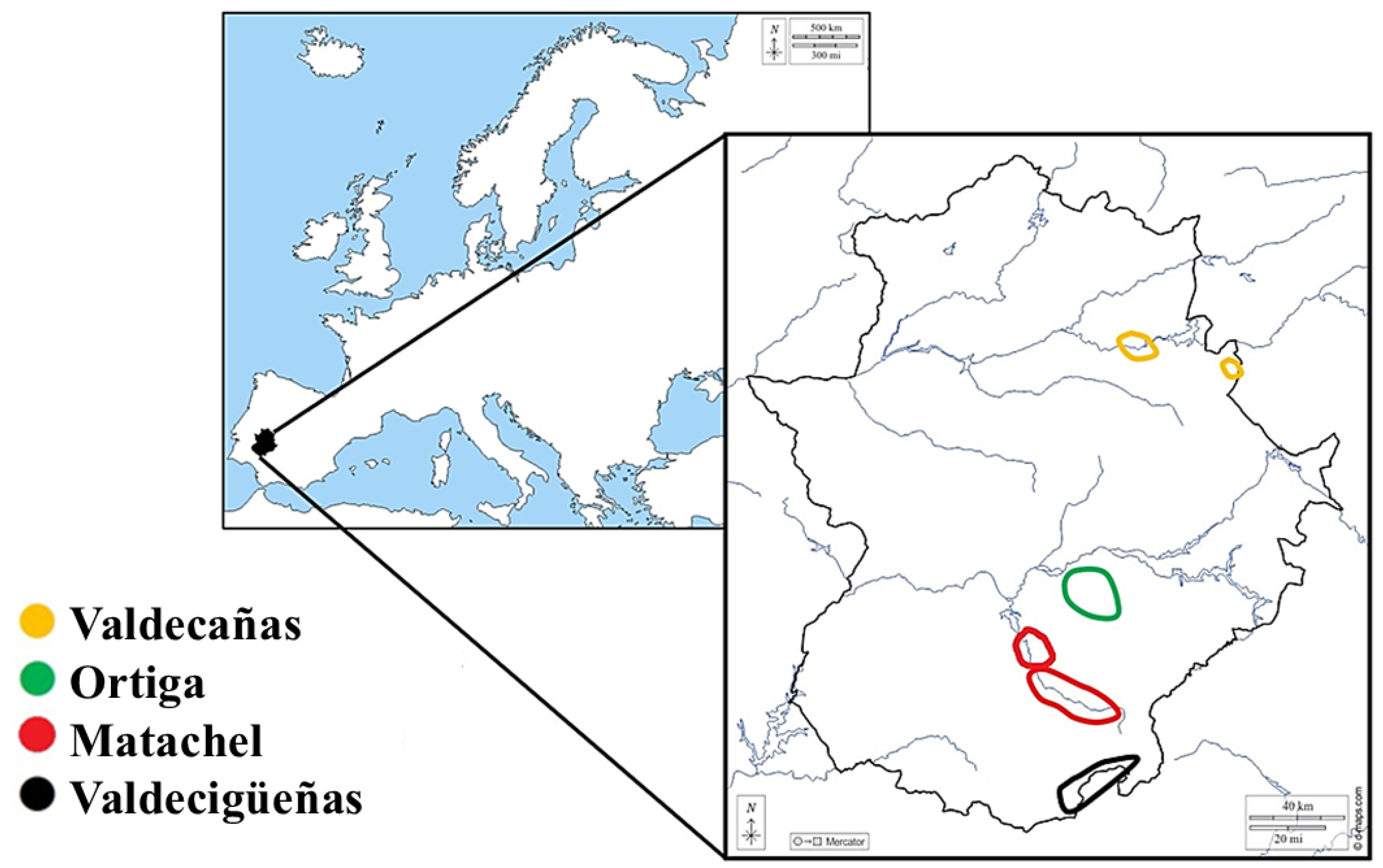
Figure 2.
Spearman’s rank correlation matrix of metal concentrations in whole blood of the Iberian lynx. Correlation coefficients (rho) are shown in the upper triangle, while corresponding p-values are displayed in the lower triangle. Statistical significance was set at p < 0.05.
Figure 2.
Spearman’s rank correlation matrix of metal concentrations in whole blood of the Iberian lynx. Correlation coefficients (rho) are shown in the upper triangle, while corresponding p-values are displayed in the lower triangle. Statistical significance was set at p < 0.05.
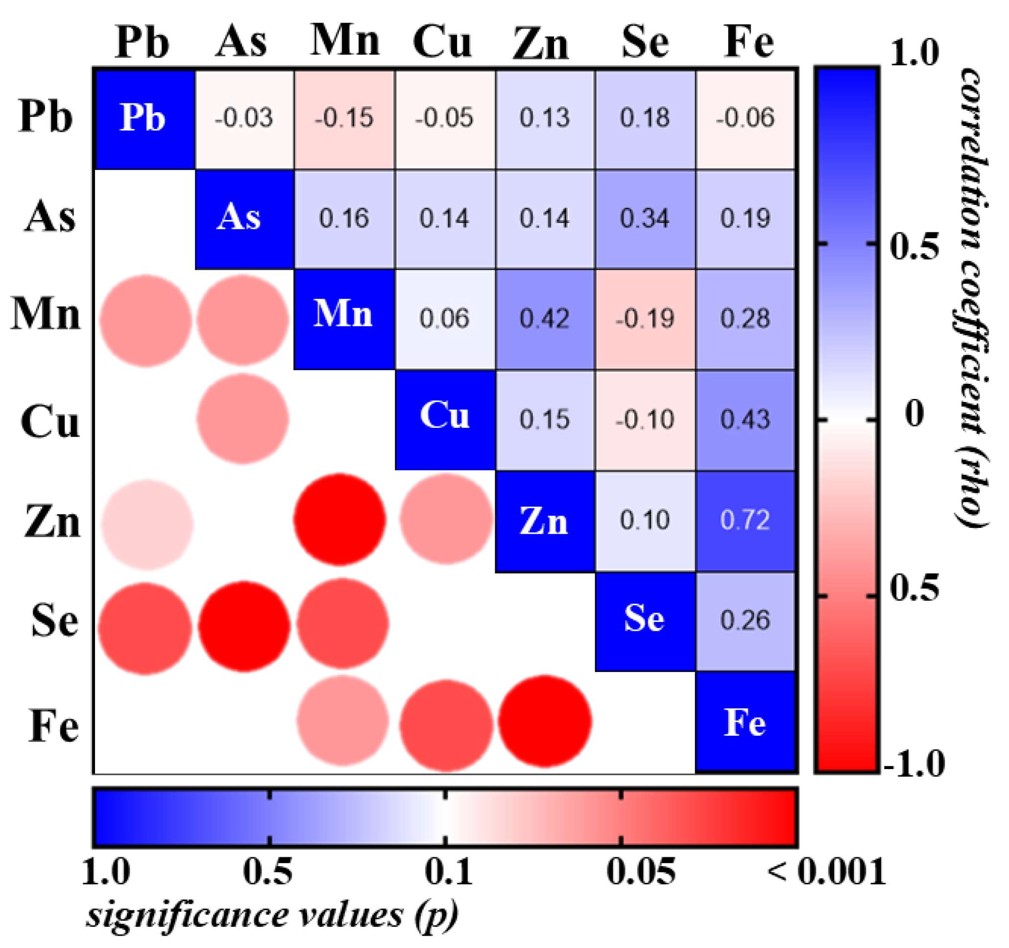
Figure 3.
Temporal trends in whole blood As and Se concentrations of recaptured Iberian lynxes during successive sampling periods.
Figure 3.
Temporal trends in whole blood As and Se concentrations of recaptured Iberian lynxes during successive sampling periods.
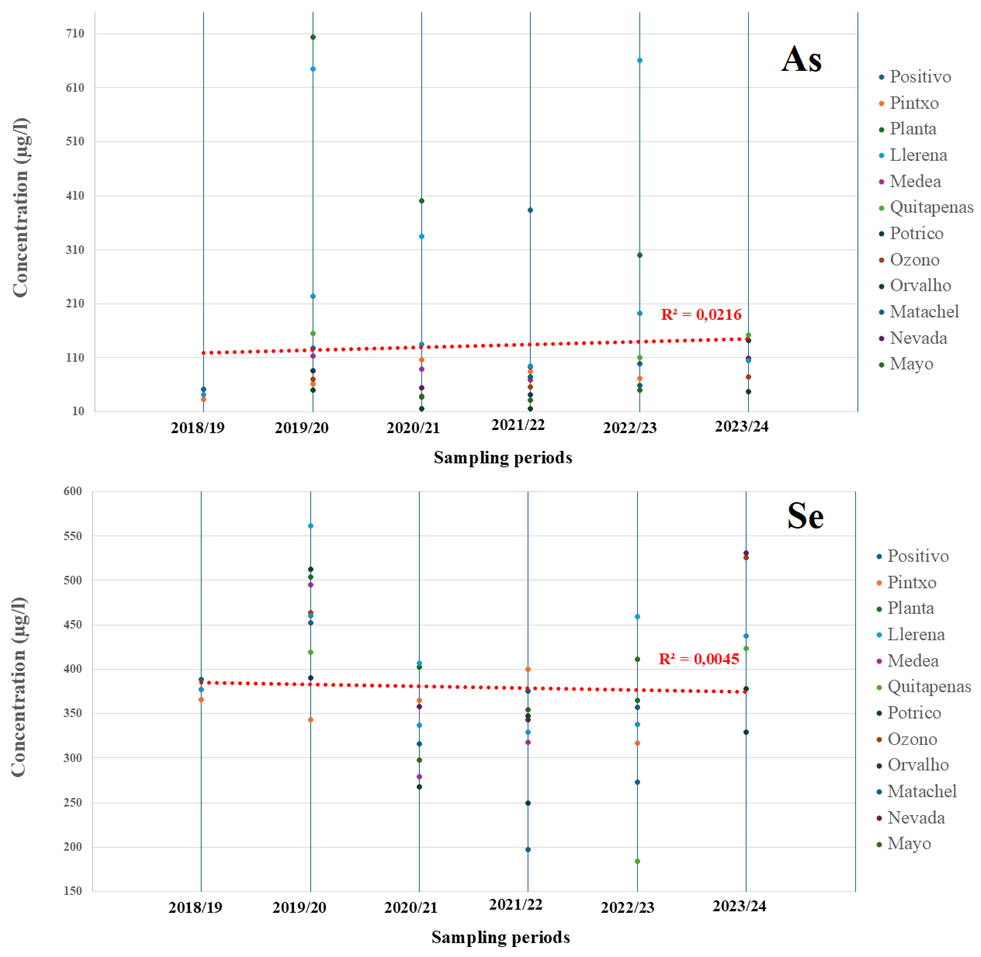
Figure 4.
Distribution of metals and metalloids in the different components generated by Principal Component Analysis (PCA).
Figure 4.
Distribution of metals and metalloids in the different components generated by Principal Component Analysis (PCA).
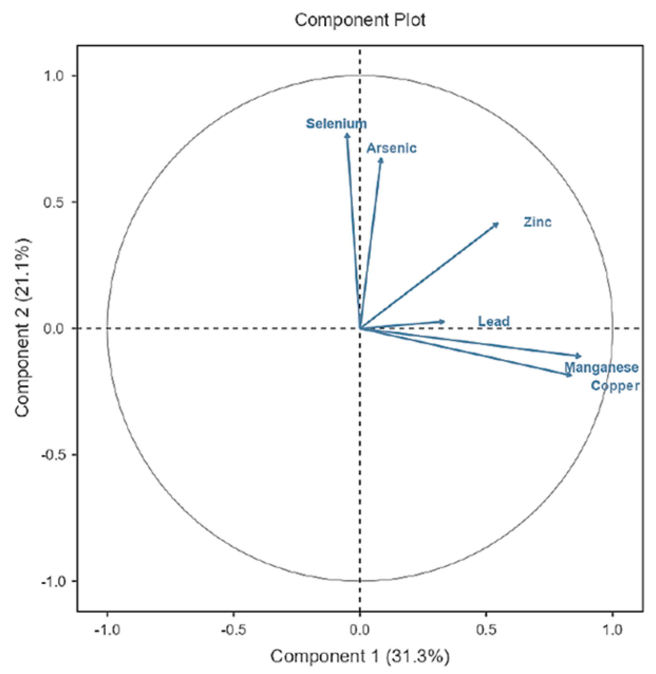
Figure 5.
Distribution plots of samples according to sex, zone, sampling period and age based on the Principal Component Analysis (PCA).
Figure 5.
Distribution plots of samples according to sex, zone, sampling period and age based on the Principal Component Analysis (PCA).
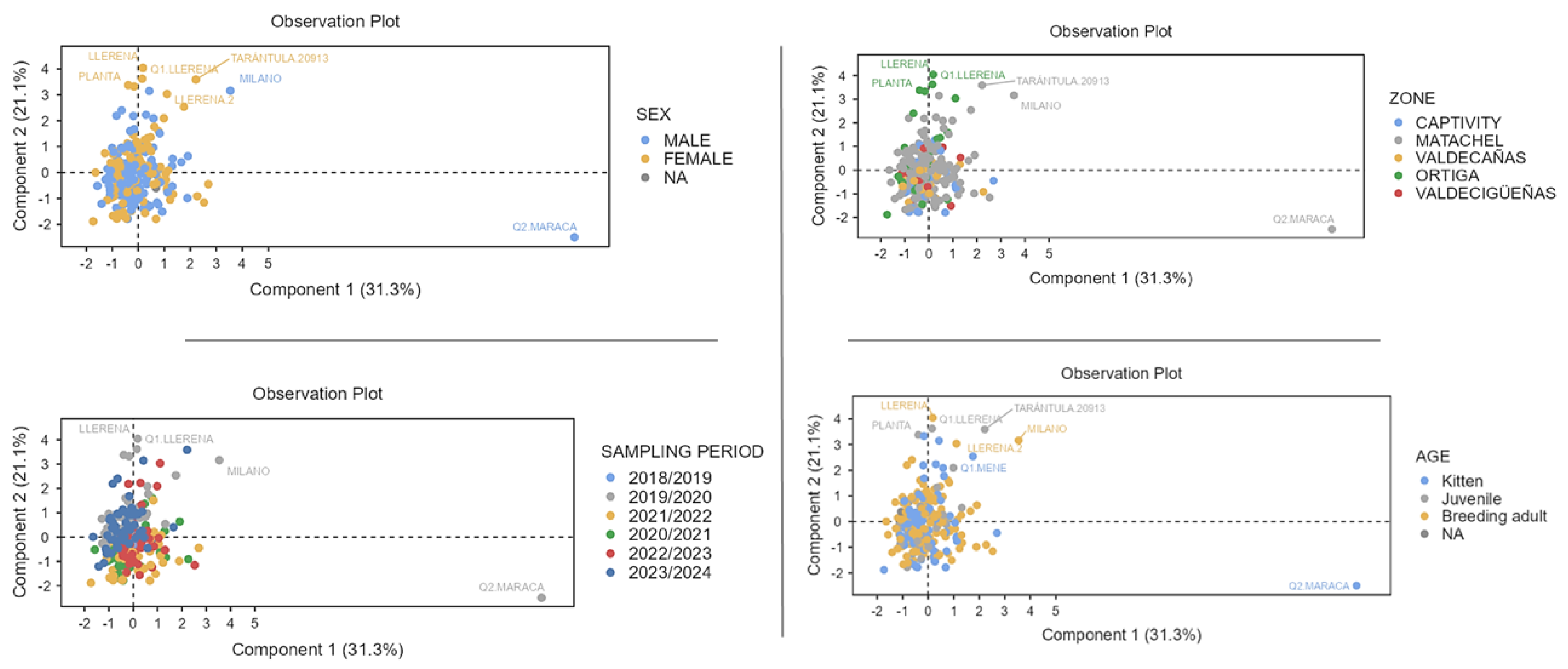

Table 1.
Descriptive statistics of the overall results obtained from the analysis of metals and metalloids in whole blood of the Iberian lynx. Results are expressed in µg/L.
Table 1.
Descriptive statistics of the overall results obtained from the analysis of metals and metalloids in whole blood of the Iberian lynx. Results are expressed in µg/L.
| Metal and metalloids | Mean±SD | Min. | 25% P. | Median | 75% P. | Max. | Range | % <LOQ* | n |
|---|---|---|---|---|---|---|---|---|---|
| Pb | 8.490±14.86 | 0.800 | 1.000 | 6.200 | 9.355 | 169.4 | 168.6 | 34.0 | 229 |
| As | 146.3±174.5 | 12.90 | 53.70 | 89.90 | 153.0 | 1041 | 1028 | 0 | 229 |
| Mn | 27.82±19.94 | 11.30 | 20.45 | 25.80 | 31.70 | 295.6 | 284.3 | 0 | 229 |
| Cu | 499.0±109.0 | 338.9 | 446.2 | 483.4 | 526.9 | 1684 | 1346 | 0 | 229 |
| Zn | 2729±375.6 | 1813 | 2503 | 2679 | 2889 | 4962 | 3149 | 0 | 229 |
| Se | 354.7±89.69 | 176.2 | 282.8 | 359.1 | 410.3 | 596.2 | 420.0 | 0 | 229 |
| Cr | 6.075±9.527 | 0.000 | 1.000 | 3.900 | 8.100 | 104.2 | 104.2 | 66.0 | 179 |
| Hg | 5.795±8.342 | 0.400 | 4.160 | 4.200 | 5.000 | 88.87 | 88.47 | 63.70 | 229 |
| Fe | 459.4±102.6 | 331.4 | 408.8 | 437.4 | 476.2 | 926.8 | 595.4 | 0 | 50 |
| Cd | 0.722±0.326 | 0.050 | 0.420 | 0.830 | 1.000 | 1.560 | 1.510 | 69.90 | 229 |
* Percentage of total samples with concentrations below the LOQ.
Table 2.
Comparative summary of mean metals and metalloids concentrations (mean ± SD; µg/L) measured in the blood of three different species.
Table 2.
Comparative summary of mean metals and metalloids concentrations (mean ± SD; µg/L) measured in the blood of three different species.
| Caracal (Caracal caracal) |
Angora domestic cat (Felis catus) |
Domestic dog (Canis lupus familiaris) |
|
|---|---|---|---|
| Pb | 3.000±3.000 | 18.00±1.700 | 12.37±1.170 |
| As | 374.0±838.0 | - | 21.02±0.580 |
| Mn | 384.0±179.0 | 34.00±4.000 | - |
| Cu | 588.0±566.0 | 593.0±54.50 | 509.0±6.730 |
| Zn | 5655±5418 | 732.0±71.50 | 3633±74.45 |
| Se | 369.0±155.0 | 270.0±133.5 | - |
| Cr | 29.00±76.00 | 27.00±2.600 | 57.78±1.110 |
| Hg | 45.00±74.00 | - | 0.740±0.220 |
| Fe | - | 2440±364.0 | 763.7±17.15 |
| Cd | 6.000±14.00 | - | 0.190±0.020 |
| Sample size (n) | 67 | 30 | 140 |
| Location | South Africa | Turkey | Turkey |
| Reference | Parker et al. (2023) | Kabakci et al. (2023) | Altinok-Yipel et al. (2022) |
Table 3.
Summary of GLM results for six representative metals. Each model includes the following factors: sex, age, study area, and sampling periods. Only factors with p < 0.05 were considered significant contributors.
Table 3.
Summary of GLM results for six representative metals. Each model includes the following factors: sex, age, study area, and sampling periods. Only factors with p < 0.05 were considered significant contributors.
| PTEs | R2 | p-value (model) |
n | Contributor factors |
|---|---|---|---|---|
| Pb | 0.284 | 0.882 | 227 | - |
| As | 0.514 | <0.001 | 227 | Sex, Age, Area, Sampling periods |
| Mn | 0.269 | 0.941 | 227 | - |
| Cu | 0.286 | 0.871 | 227 | - |
| Zn | 0.412 | 0.048 | 227 | Age |
| Se | 0.709 | <0.001 | 227 | Age, Area, Sampling periods |
Table 4.
Descriptive analysis of the results obtained from the analysis of metals and metalloids in whole blood samples from Iberian lynx classified by sex, age, area and sampling periods. Results are expressed in µg/L.
Table 4.
Descriptive analysis of the results obtained from the analysis of metals and metalloids in whole blood samples from Iberian lynx classified by sex, age, area and sampling periods. Results are expressed in µg/L.
| Mean±SD | |||||||
|---|---|---|---|---|---|---|---|
| Pb | As | Mn | Cu | Zn | Se | ||
| Sex | Males | 8.500±12.36 | 135.1±154.4 | 28.26±25.26 | 502.0±125.6 | 2692±346.6 | 351.1±82.28 |
| Females | 4.857±4.560 | 121.6±132.4 | 27.39±7.471 | 508.8±83.30 | 2756±415.2 | 338.8±94.50 | |
| Age | Kittens | 6.502±12.67 | 137.7±185.2 | 25.51±7.336 | 500.2±76.15 | 2704±325.3 | 332.3±82.26 |
| Juveniles | 8.272±12.49 | 198.8±238.0 | 27.73±11.70 | 469.2±47.25 | 2783±408.5 | 356.7±101.0 | |
| Adults | 8.655±16.51 | 138.2±137.8 | 27.38±9.506 | 499.3±78.76 | 2716±388.1 | 361.0±89.35 | |
| Area | Matachel | 8.211±15.56 | 142.7±157.2 | 29.10±24.31 | 507.1±122.5 | 2717±404.6 | 360.9±91.22 |
| Valdecañas | 6.618±7.985 | 115.8±59.01 | 26.35±8.002 | 508.9±72.06 | 2763±345.9 | 328.7±78.69 | |
| Ortiga | 7.068±6.530 | 334.1±304.6 | 26.18±9.822 | 465.0±34.70 | 2686±374.8 | 368.0±89.41 | |
| Valdecigüeñas | 11.13±18.31 | 73.67±26.81 | 26.12±6.225 | 482.7±67.45 | 2768±247.8 | 364.2±89.53 | |
| Captivity | 11.25±18.33 | 60.39±43.98 | 24.62±7.341 | 487.1±102.1 | 2784±290.8 | 328.5±86.61 | |
| Sampling period | 2019/20 | 16.45±13.28 | 178.5±232.2 | 28.68±41.15 | 484.9±191.6 | 2895±480.2 | 426.6±66.60 |
| 2020/21 | 10.13±15.87 | 88.67±107.4 | 25.16±8.590 | 494.6±72.15 | 2703±341.4 | 292.0±66.66 | |
| 2021/22 | 5.720±3.020 | 95.17±159.7 | 26.58±8.710 | 538.2±107.2 | 2683±315.0 | 282.7±63.44 | |
| 2022/23 | 6.950±25.20 | 175.7±190.4 | 33.44±8.750 | 488.7±56.38 | 2813±233.7 | 314.0±66.52 | |
| 2023/24 | 4.530±5.740 | 172.4±140.2 | 25.26±8.970 | 494.0±50.36 | 2582±369.2 | 415.2±64.45 | |
Disclaimer/Publisher’s Note: The statements, opinions and data contained in all publications are solely those of the individual author(s) and contributor(s) and not of MDPI and/or the editor(s). MDPI and/or the editor(s) disclaim responsibility for any injury to people or property resulting from any ideas, methods, instructions or products referred to in the content. |
© 2026 by the authors. Licensee MDPI, Basel, Switzerland. This article is an open access article distributed under the terms and conditions of the Creative Commons Attribution (CC BY) license (http://creativecommons.org/licenses/by/4.0/).
Copyright: This open access article is published under a Creative Commons CC BY 4.0 license, which permit the free download, distribution, and reuse, provided that the author and preprint are cited in any reuse.


