

Submitted:
21 July 2026
Posted:
23 July 2026
You are already at the latest version
Abstract
My companion paper reported that stochastic genome loading constrains the authentic single-genome fraction of recombinant adeno-associated virus (rAAV) to approximately 36.8%. Here, I evaluate an independent structural constraint: occlusion of the fivefold genome-entry channels by intraluminal N-terminal extensions of VP1 and VP2, which are absent from VP3. Each capsid contains twelve such channels. I hypothesize that any penton containing one or more VP1 or VP2 subunits occludes its channel, so only all-VP3 pentons are packaging-competent. Under stochastic incorporation of VP1, VP2, and VP3, as demonstrated by mass spectrometry, the probability that a penton is composed entirely of VP3 is fVP35, and the number of open channels per capsid follows B(12, fVP35). At the canonical 1:1:10 ratio (fVP3 ≈ 0.83), approximately 40% of channels are open, and approximately 99.8% of capsids retain at least one. Because a single open channel suffices for genome entry, obstruction has little effect at the canonical ratio and does not reduce the stochastic packaging ceiling. At matched genome supply, obstruction instead decreases the multi-occupancy (Head-Full) fraction while increasing the empty-capsid fraction, indicating that the two mechanisms partially oppose rather than compound one another. Channel obstruction becomes the dominant source of empty capsids only below a sharp threshold near fVP3 ≈ 0.75; at a 1:1:2 ratio, only approximately 32% of capsids are predicted to be packaging-competent. These findings identify VP1:VP2:VP3 stoichiometry as a critical quality attribute with threshold-dependent behavior and recast a VP3-only, surface-engineered capsid to improve particle homogeneity, robustness to stoichiometric drift, and uniform infectivity-factor dosage.
Keywords:
adeno-associated virus
; VP1
; VP2
; VP3
; capsid stoichiometry
; genome-entry channel
; fivefold pore
; phospholipase A2
; nuclear localization signal
; empty capsids
; gene therapy manufacturing
1. Introduction
The AAV capsid is a ~25 nm icosahedral shell composed of 60 subunits derived from three co-terminal proteins (VP1, VP2, and VP3) in an approximate 1:1:10 molar ratio (Penaud-Budloo et al. 2018). All three share the VP3 core, which forms the shell and binds the receptor; VP2 adds an N-terminal extension, and VP1 adds a longer one that carries a phospholipase A2 (PLA2) domain essential for endosomal escape and basic-region nuclear-localization signals (NLS) required for nuclear import (Bleker et al. 2005, Girod et al. 2002). In the assembled particle, these N-terminal extensions are sequestered inside the capsid and externalized only upon activation, threading out through the fivefold pore during cell entry (Bennett et al. 2017).
The companion paper (Davis 2026) identified a ceiling effect in productive packaging during stochastic genome loading. Here we ask whether the capsid’s architecture imposes a second, structural constraint upstream of loading. If the VP1/VP2 N-terminal peptides occupy the fivefold channels through which DNA must pass, then a capsid’s packaging-competent channels are limited to those pentons assembled entirely from VP3. I call VP1 and VP2 “capsid killers” in this narrow manufacturing sense, not because they harm the particles that package a genome (they are, of course, essential for infectivity), but because, by occupying channels, they can prevent packaging and increase the empty fraction.
At the outset, it is important to distinguish the model’s prediction from a simpler interpretation of the obstruction hypothesis. At the canonical VP ratio, channel obstruction has little effect on whether a capsid can be packaged and does not raise the stochastic ceiling for authentic single-genome capsids. Its principal signature is threshold behavior: when VP1 and VP2 are over-incorporated, as can occur in recombinant systems, the open-channel fraction declines nonlinearly, and empty capsids become predominant. The central result is therefore not a universal reduction in packaging capacity, but a stoichiometry-dependent transition. At fixed genome supply, obstruction also produces a counterintuitive tradeoff, reducing multi-occupancy defectives while increasing empty capsids rather than compounding the defect. This behavior is illustrated in the accompanying figures.
1.1. The Fivefold Pore is the Packaging Portal
Genome packaging in AAV, as in related parvoviruses, proceeds via Rep-motor-driven translocation of single-stranded DNA through a pore at a fivefold vertex into a preformed capsid (Bleker et al. 2006, Bleker et al. 2005, King et al. 2001). Mutations lining the AAV2 fivefold pore impair packaging through steric and Rep-interaction effects (Bleker et al. 2006, Bleker et al. 2005). A similar result in minute virus of mice, where a pore-occluding mutation abolishes packaging, confirms the fivefold channel as the portal (Plevka et al. 2011). The capsid has twelve such channels.
1.2. VP1, VP2, VP3: Shared Core, Distinct N-Termini
The three proteins arise from a single cap ORF via alternative splicing and start-codon usage; all share the VP3 core, with VP2 carrying ~65 additional N-terminal residues and VP1 ~135 more beyond VP2, including the PLA2 domain and the basic regions BR1, BR2, and BR3, which function in nuclear localization and import (Girod et al. 2002, Grieger et al. 2007). VP3 has no N-terminal extension and thus contributes no intraluminal peptide. Only the VP3 core is required for capsid assembly; particles can be assembled from VP3 alone, and VP-reduced particles can package the genome (Grieger et al. 2007, Warrington et al. 2004).
1.3. The N-Termini Are Internal and Must Reach the Interior Through the Pore
Immunological, proteolytic, and cryo-EM studies predominantly place the VP1/VP2 N-termini inside the native capsid, externalizing only upon acidification during entry (Bennett et al. 2017, Kronenberg et al. 2005). The question this paper addresses is how they reach the interior. The most parsimonious path for an N-terminal peptide synthesized at the capsid exterior is through the fivefold pore at the penton, where its subunit sits. Notably, recent structural work indicates that the fivefold pore is largely empty before packaging and becomes occupied by capsid-protein N-terminal density only after packaging (Kaelber et al. 2025). This timing is discussed in Section 1.5 because it is both the central objection to the hypothesis and, I argue, an unsettled question worth raising.
1.4. VP Stoichiometry Is Stochastic and Variable
Native mass spectrometry has shown that AAV capsids are not uniform: they assemble from 60 random draws from the expressed VP pool, yielding a broad distribution of per-capsid VP compositions centered on the 5:5:50 mean (Wörner et al. 2021). This stochastic assembly model underpins my channel availability calculation. Critically, the VP1:VP2:VP3 ratio is not fixed: it varies by serotype, platform, and process, and recombinant systems under heterologous promoters can deviate substantially from 1:1:10, sometimes with elevated VP1/VP2 [Wörner et al., 2021]. The threshold behavior I describe makes this variability critically consequential.
1.5. The Channel-Obstruction Hypothesis
I propose that a penton containing one or more VP1 or VP2 subunits is non-permissive for ssDNA insertion because the intraluminal N-terminal peptide of the corresponding subunit occludes the fivefold channel. In this model, only pentons composed entirely of VP3 present packaging-competent channels. This steric argument is consistent with reported AAV fivefold-channel constrictions of approximately 9–20 Å (Bleker et al. 2006, Bleker et al. 2005, Kronenberg et al. 2005). A translocating single strand of DNA has an effective transverse dimension of approximately 11–13 Å, about half the double-stranded dimensions reported by Mandelkern et al. (1981), whereas an extended, denatured, unhydrated polypeptide chain exceeds approximately 4–6 Å (Buscaglia et al. 2006). Given these dimensions, a resident peptide and a translocating DNA strand are unlikely to occupy the same constriction simultaneously. I therefore propose that even a single peptide chain resident in the channel would obstruct ssDNA passage during packaging. This interpretation is plausible but not established: the capsid channel is conformationally dynamic, and the identity of the resident pore density observed by cryo-EM remains unresolved.
A recent cryo-electron microscopy study of AAV5 offers an important refinement rather than a direct contradiction of this model. Gliwa and colleagues (Gliwa et al. 2025) resolved a rod-like density within the fivefold channel and most plausibly assigned it to the VP3 N-terminus. They also raised the possibility that the twelve pores are not equivalent: most may contain VP3 N-terminal density, whereas a minority of specialized pores may mediate genome traffic. However, the authors explicitly note that this specialized-pore interpretation remains speculative (Gliwa et al. 2025). These observations are relevant because they broaden the structural interpretation of channel occupancy. However, they do not eliminate the central question addressed here: whether longer VP1/VP2 N-terminal extensions, when present in or near a packaging channel, impose a greater and more persistent obstruction to ssDNA entry than the shorter VP3 N-terminus. Two qualifications therefore follow.
First, if the shared VP3-core N-terminus contributes density within the fivefold channel, an all-VP3 penton should not be assumed to be necessarily empty. Rather, the relevant distinction is the magnitude, persistence, and functional consequence of channel occupancy. In that framework, the longer VP1 and VP2 extensions are still expected to pose a greater steric barrier to ssDNA passage than the shorter VP3 N-terminus. Second, pore-to-pore heterogeneity may provide an additional mechanism by which a subset of channels remains competent for genome traffic, without requiring all pores to be structurally equivalent. Because the new evidence comes from AAV5, whereas several pore-dimension estimates cited here derive from AAV2, the cross-serotype inference should be regarded as provisional. These qualifications refine the structural interpretation but do not weaken the model’s central quantitative prediction: VP1/VP2 over-incorporation should reduce the fraction of packaging-competent capsids in a threshold-dependent manner and thereby increase the empty-capsid fraction. This prediction is directly testable by VP-ratio titration, independent of a complete structural resolution of the packaging intermediate.
This model differs from the prevailing structural interpretation, which holds that the fivefold pore remains unobstructed during packaging and that N-terminal occupancy occurs only after genome encapsidation. The claim advanced here is therefore deliberately stronger and correspondingly falsifiable. My position is that the timing of N-terminal occupancy during active packaging has not yet been directly observed: mature-particle structures describe the post-packaging state, whereas packaging-intermediate structures that could resolve the timing question are not yet available. Thus, if the N-terminal peptide of a VP1 or VP2 subunit is present in or near its penton channel when Rep attempts to load that channel, obstruction should occur; whether that peptide is present at that moment remains the key unresolved variable. The model therefore predicts that capsids captured during packaging will show N-terminal density in channels associated with VP1/VP2-containing pentons, thereby partitioning channels into competent all-VP3 and obstructed VP1/VP2-containing classes. It further predicts that experimentally increasing the VP1/VP2 fraction will increase the empty-capsid fraction in the threshold-dependent manner derived below, independently of genome supply. Conversely, a demonstration that VP1/VP2 N-termini are consistently excluded or retracted from the pore throughout packaging would refute the proposed mechanism and itself provide important insight into encapsidation and manufacturing control.
2. Materials and Methods
2.1. The Channel-Availability Model
The capsid presents twelve fivefold channels, one per vertex, each formed by a pentamer of five subunits. Under the stochastic-assembly model of VP composition [Wörner et al., 2021], each of the 60 subunits is an independent draw, with VP3 occurring with probability equal to the VP3 mole fraction f_VP3. The twelve pentons occupy spatially distinct sets of five positions and are therefore independent. A penton is packaging-competent (“open”) only when all five of its subunits are VP3. The probability that a given penton is open is P(open) = f_VP3⁵, and the number of open channels per capsid is binomially distributed as B(12, f_VP3⁵). To couple channel availability to genome loading, each open channel is modeled as independently loading a DNA molecule with probability π, set by genome supply and shared across the population, so that inserts per capsid follow Binomial(c, π) with bulk MOE = E[c]·π. Both models were evaluated across the full ranges of f_VP3 and π; the binomial derivation is given in the Appendix.
2.2. Model Development and Computational Methods
The channel-obstruction hypothesis and its quantitative consequences were developed by the author, building on the established stochastic-assembly model of VP composition. During manuscript preparation, the generative AI assistant Claude Opus 4.8 (Anthropic) was used under the author’s direction and verification to: (i) express the channel-availability and coupling models in compact symbolic form; (ii) evaluate the binomial and coupled distributions across the full range of VP3 mole fraction and per-channel loading probability; and (iii) compute and plot the resulting parameter-space behavior. This analysis made explicit the sharp stoichiometric threshold, or “knee,” near a VP3 mole fraction of 0.75, which had not been fully resolved in the original hand-derived discrete examples. It also confirmed that, at fixed genome supply, channel obstruction partially opposes rather than compounds the companion paper’s stochastic multi-occupancy mechanism. The author reviewed all AI-assisted calculations, figures, and interpretations for consistency with the hand analysis and with the assumptions and claims stated in the text. The full derivation is provided in the Appendix.
3. Results
3.1. Channel Availability at the Canonical Ratio
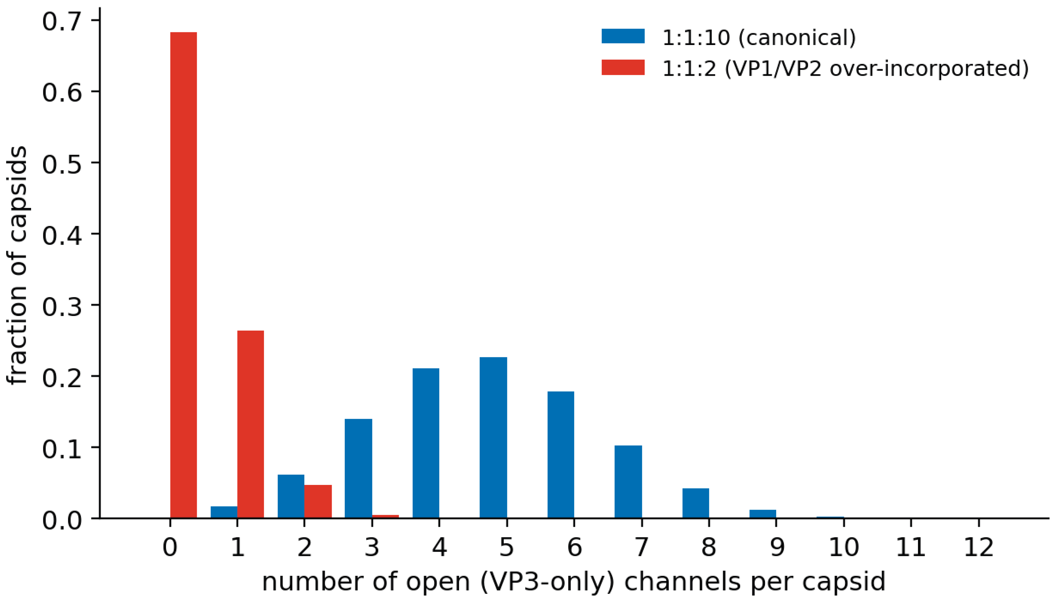
At the canonical ratio f_VP3 = 50/60 ≈ 0.833, P(open) = 0.833⁵ ≈ 0.40. The mean number of open channels per capsid is 12 × 0.40 ≈ 4.8, and the probability that a capsid has at least one open channel is 1 − (1 − 0.40)¹² ≈ 0.998. Almost every capsid can package; the probability of the all-twelve-open particle is 0.40¹² ≈ 2 × 10⁻⁵.
Figure 1.
Distribution of the number of open (all-VP3) channels per capsid, B(12, f_VP3⁵), at the canonical 1:1:10 ratio (blue) versus a skewed 1:1:2 ratio (Lecomte et al.). At 1:1:2, the distribution collapses toward zero open channels.
Figure 1.
Distribution of the number of open (all-VP3) channels per capsid, B(12, f_VP3⁵), at the canonical 1:1:10 ratio (blue) versus a skewed 1:1:2 ratio (Lecomte et al.). At 1:1:2, the distribution collapses toward zero open channels.
3.2. Coupling to the Stochastic Ceiling
The two constraints might be expected to be multiplied (open-channel fraction times the single-genome probability); they are not, because the open-channel fraction (0.40) is the proportion of open channels, not the proportion of capsids that can package, and a capsid needs only one open channel. The correct capsid-level availability factor at the canonical 1:1 ratio is ~0.998.
At the canonical ratio, the combined ceiling for genome supply is ~38.4% authentic single-genome capsids (essentially identical to the all-channels-open case), because a higher genome supply (π ≈ 0.21 versus 0.08) fully compensates for the fewer open channels. Channel obstruction at the canonical ratio can be seen to cost almost nothing.
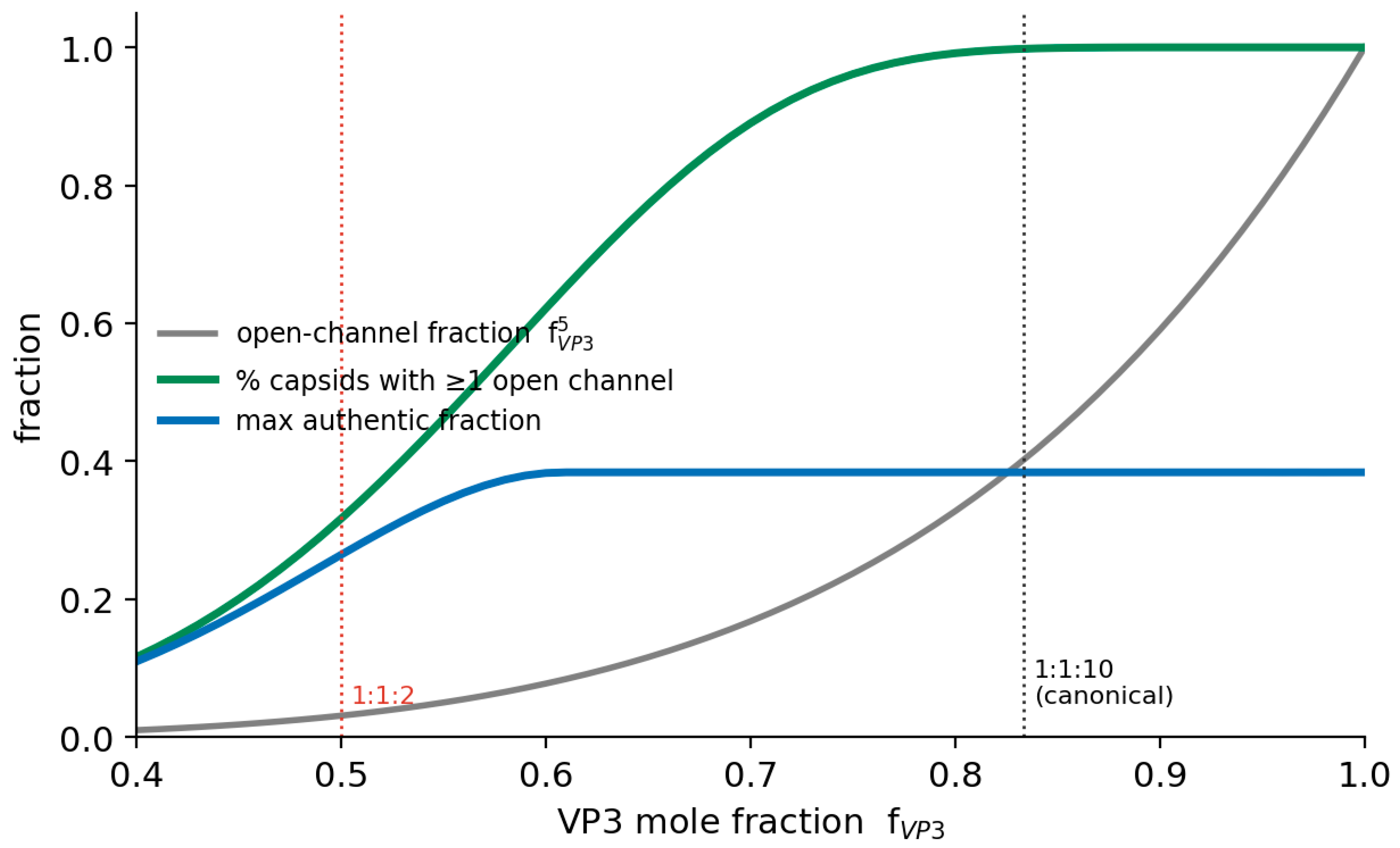
The obstruction effect becomes most noticeable not at the canonical ratio but below a threshold. Because P(open) = f_VP3⁵ falls steeply as the VP3 mole fraction drops, the fraction of capsids able to package collapses once VP1/VP2 are over-incorporated. The knee is near f_VP3 ≈ 0.75: above it, essentially all capsids can package, and the maximum authentic fraction holds at ~38%; below it, empties dominate. At a 1:1:2 ratio (f_VP3 = 0.50), the mean open-channel count is 0.4. About 68% of capsids have no open channel; only ~32% can package, and the maximum authentic fraction falls to ~26%. This matches the ~70% empty behavior observed for the skewed-ratio case in the original poster.
Figure 2.
Open-channel fraction (f_VP3⁵), the percentage of capsids with at least one open channel, and the maximum achievable authentic fraction, as functions of the VP3 mole fraction. The threshold near f_VP3 ≈ 0.75 separates a safe plateau from steep packaging failure; canonical (1:1:10) and skewed (1:1:2) ratios are indicated.
Figure 2.
Open-channel fraction (f_VP3⁵), the percentage of capsids with at least one open channel, and the maximum achievable authentic fraction, as functions of the VP3 mole fraction. The threshold near f_VP3 ≈ 0.75 separates a safe plateau from steep packaging failure; canonical (1:1:10) and skewed (1:1:2) ratios are indicated.
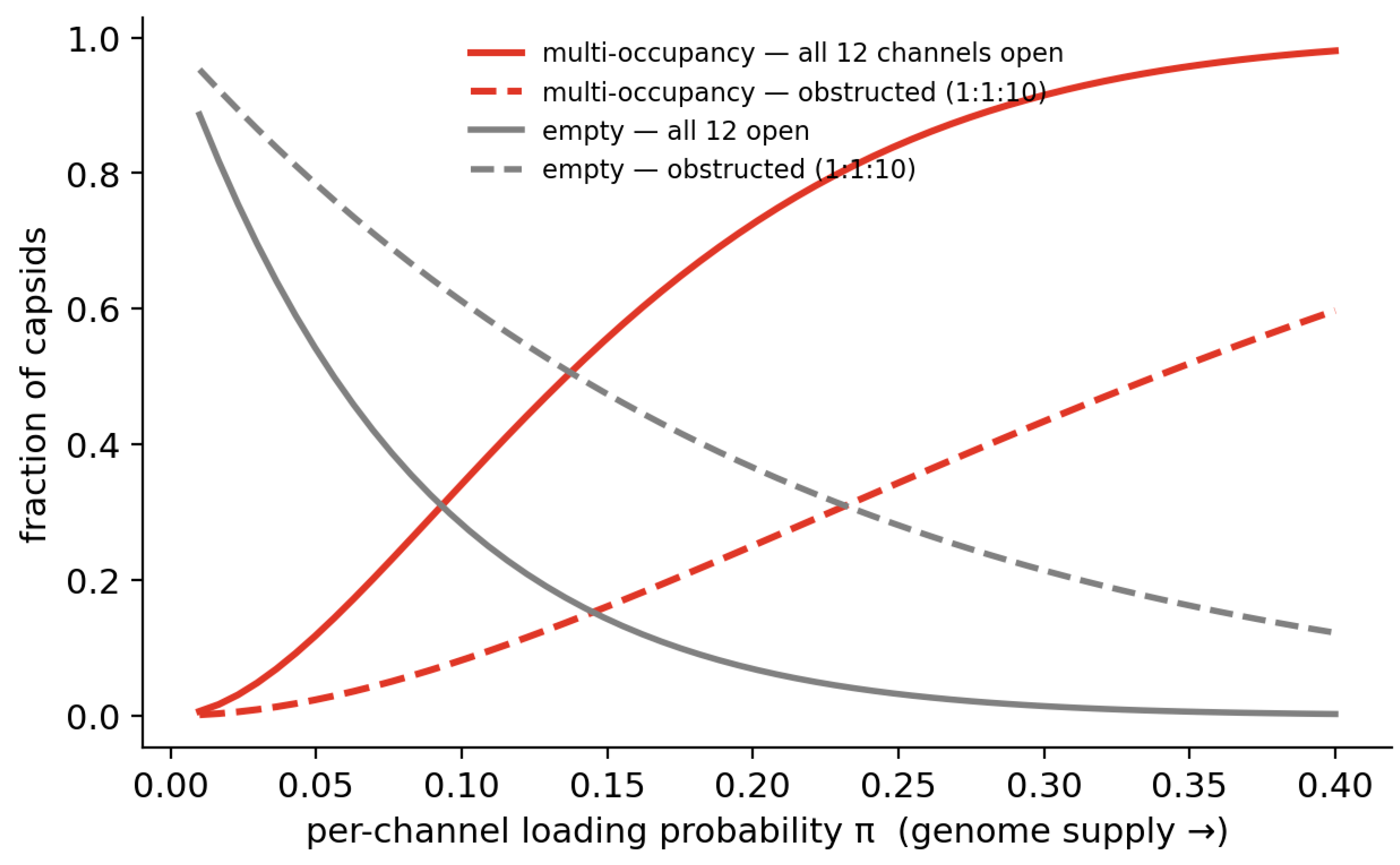
The direction of this coupling is more important than its absolute magnitude. At a fixed genome supply, a population with fewer open channels produces a lower fraction of multi-occupancy (Head-Full) defectives than a fully open population, but at the cost of a higher empty-capsid fraction. This result follows directly from the reduced number of entry routes: fewer open channels reduce the probability that multiple genomes enter the same capsid. Thus, channel obstruction does not add to the stochastic multi-occupancy mechanism described in the companion paper in a simple multiplicative manner. Instead, the two effects partially counterbalance one another. In practical terms, obstruction can reduce stealth-defective particles only by shifting part of the defect burden toward empty capsids.
Figure 3.
At matched genome supply (per-channel loading probability π), channel obstruction at the canonical ratio lowers the multi-occupancy (Head-Full) fraction. It increases the empty fraction relative to that of a fully open capsid. Note that the two mechanisms oppose each other rather than compound.
Figure 3.
At matched genome supply (per-channel loading probability π), channel obstruction at the canonical ratio lowers the multi-occupancy (Head-Full) fraction. It increases the empty fraction relative to that of a fully open capsid. Note that the two mechanisms oppose each other rather than compound.
The truest combined statement is therefore: VP1/VP2 channel obstruction is a stoichiometry threshold, not a second universal encapsidation ceiling. At wild-type-like ratios, it is nearly silent and even protective against multi-occupancy; over-incorporation of VP1/VP2 makes it a dominant, independent source of empty capsids.
4. Discussion
This paper and its companion identify two distinct contributors to AAV encapsidation failure: the companion paper defines a stochastic ceiling on authentic single-genome packaging, whereas the present work proposes a structural, stoichiometry-dependent obstruction of genome-entry channels. The main advance is to consider how these mechanisms interact under dynamic packaging conditions. They do not combine to impose a lower multiplicative ceiling. At canonical VP stoichiometry, channel obstruction is predicted to be nearly silent, and at fixed genome supply, it partially opposes stochastic multi-occupancy by reducing the number of available entry routes. The obstruction mechanism becomes consequential only beyond a stoichiometric threshold, when VP1/VP2 over-incorporation drives a large fraction of capsids toward zero open channels.
Current structural consensus primarily describes mature, post-packaging particles, whereas the packaging-intermediate state remains incompletely resolved. The hypothesis advanced here addresses that unresolved interval. Its associated quantitative prediction—that the empty-capsid fraction should depend sharply on the VP3 mole fraction—is testable without first resolving the precise structural timing of N-terminal occupancy. A direct experimental test would titrate VP1/VP2 levels and measure the resulting empty-capsid fraction.
This proposal is intended to extend, rather than displace, prior work. The stochastic-assembly model of VP composition is derived from Wörner et al. (2021); the channel-availability binomial developed here applies that model to the fivefold-pore problem. Likewise, the effect of VP stoichiometry on potency is well established, whereas the contribution proposed here concerns packaging efficiency and empty-capsid formation. VP3-only assembly, VP-reduced genome packaging, and surface rescue of infectivity have also been demonstrated (Grieger et al. 2007, Warrington et al. 2004). The present contribution is to integrate those observations into a quantitative threshold model and to reconsider the engineering rationale for a single-capsid-protein vector.
4.1. Evidence
VP3-only and VP-reduced particles provide the key experimental foundation for the remedy considered below. Particles assembled without VP1, or from VP3 alone, can form authentic capsids, package genomes, and, when VP1-associated functions are supplied from the capsid surface, restore infectivity (Warrington et al. 2004). More directly, Grieger et al. (2007) showed that a surface-exposed VP1-NLS fusion rescues the infectivity of otherwise noninfectious VP2/VP3 and VP3-only capsids but does not rescue fivefold-pore mutants. These findings establish two points central to the proposed design strategy: VP1/VP2 N-terminal extensions are not required for genome packaging itself, and their essential post-packaging functions can, in principle, be repositioned to an external capsid site rather than retained as intraluminal peptides threaded through the penton pore.
VP stoichiometry is a quality attribute that affects potency. Deviations from 1:1:10 alter transduction and are increasingly treated as a critical “quality attribute” (Maruno et al. 2025, Wörner et al. 2021). My model makes a specific, separable prediction about packaging (empty fraction): the threshold dependence of the empty fraction on the VP3 mole fraction.
The pervasive, serotype-independent excess of empty capsids (Penaud-Budloo et al. 2018) is conventionally attributed to kinetic uncoupling of assembly and replication. The threshold model described here offers a complementary, stoichiometry-dependent contribution that is additive to the kinetic one and can be tested by VP-ratio titration.
Structural localization of the N-termini. Cryo-electron microscopy places VP1/VP2 N-terminal density inside the capsid and, in mature particles, within the fivefold pore (Kaelber et al. 2025, Kronenberg et al. 2005). A recent AAV5 study resolves a rod-like density in the channel but, most plausibly, assigns it to the VP3 N-terminus and reports possible pore-to-pore heterogeneity, tentatively offering a specialized genome pore (Gliwa et al. 2025). The unresolved questions (Section 1.5) are therefore two, viz., the timing of N-terminal occupancy relative to packaging and the identity of the peptide resident in any given pore.
4.2. Implications
If the threshold model is correct, several implications follow. First, the empty-capsid burden includes a structural, stoichiometry-dependent component that is independent of genome supply and therefore cannot be eliminated by genome-side optimization alone. Second, the VP1:VP2:VP3 ratio should be treated as a critical quality attribute because its effect on empty-capsid formation is nonlinear: above f_VP3 ≈ 0.75, the system remains on a relatively safe plateau, whereas below that threshold, empty-capsid production increases steeply. Third, production conditions or vector designs that shift the capsid pool toward higher VP1/VP2 content should disproportionately increase empty-capsid production. Fourth, VP-ratio analytics, including mass spectrometry and CE-SDS, have predictive value rather than merely descriptive value. Fifth, natural differences in VP stoichiometry among serotypes may contribute to serotype-specific differences in packaging efficiency. Importantly, mild channel obstruction is not necessarily harmful at fixed genome supply because it can reduce multi-occupancy defectives even as it increases empty capsids; in practical terms, this shifts part of the defect burden from stealth-defective particles to empty particles, which are more readily separated by density-based purification.
4.3. A Reconsidered Remedy: The VP3-Only, Surface-Engineered Capsid
The hypothesis suggests a potential design remedy: remove VP1 and VP2 from the assembling capsid to keep all twelve fivefold channels unobstructed, while restoring their essential post-packaging functions by displaying PLA2 and NLS activities at an external capsid site, such as the VRIV loop, away from the genome-entry channel (Tseng and Agbandje-McKenna 2014). Two lines of precedent support this strategy. First, the VRIV loop tolerates large insertions: the approximately 236-residue fluorescent protein mCherry has been inserted into VRIV without loss of particle production, infectivity, or fluorescence (Judd et al. 2012). Similarly, comparably large enzymes, including β-lactamase, have been displayed on the capsid surface while preserving assembly and titer. Thus, insert size is unlikely to be the primary constraint. Second, and more directly, Grieger et al. (2007) showed that a surface-displayed VP1-NLS fusion restores infectivity to VP3-only capsids, addressing the central feasibility issue for this design.
This corrected analysis also revises the rationale proposed in the earlier foundational poster (Davis 2022). In the present model, a VP3-only capsid does not increase the authentic-yield ceiling: at the canonical ratio, that ceiling is already approximately 38%, and opening all channels does not raise it. Moreover, at a fixed genome supply, a fully open capsid is predicted to produce more Head-Full multi-occupancy defectives than an obstructed capsid (Section 3.2). Therefore, a VP3-only design must be paired with the supply-control strategy described in the companion paper, particularly low instantaneous MOE; otherwise, it would be expected to exchange empty particles for stealth-defective particles.
The principal advantages of a VP3-only, surface-engineered capsid therefore lie elsewhere. Such a design would be insensitive to VP-ratio drift because there would be no stoichiometric threshold to cross; it would produce a homogeneous single-protein particle, simplifying quality control; it would provide uniform PLA2/NLS dosage, potentially up to 60 copies of the engineered functions per particle rather than approximately five; and it would remove VP stoichiometry as a variable in process development. These are substantive benefits, but they differ from the yield-ceiling rationale advanced in the earlier poster presentation.
Designing the engineered cassette. Faithful reconstitution of infectivity from the loop requires more than relocating the PLA2 and an NLS, as the VP1-unique (VP1u) region shows. Beyond its contiguous PLA2 domain (the AAV PLA2 is a single, contiguous catalytic module, not a split one) and three clusters of basic residues, the VP1u carries additional conserved, infection-obligatory motifs: a YXXQ endosomal-sorting/trafficking motif (mapped to approximately residues 79–82 of VP1u), additional tyrosine-based motifs, and PDZ-binding motifs at the extreme N-terminus implicated in nuclear uptake (Popa-Wagner et al. 2012). An engineered cassette intended to restore infectivity must therefore port this set of elements onto the loop (at minimum, the endosomal-targeting tyrosine motif(s) together with the phospholipase and a nuclear-localization function) rather than the PLA2 alone. Because VRIV tolerates large insertions (above), assembling such a multi-motif cassette is feasible in principle; the design question is which elements are individually necessary, not whether there is room for them.
Two specific substitutions are worth proposing. First, the three native basic regions are individually weak NLSs; replacing them with a single canonical strong NLS (for example, an SV40-type signal) could improve nuclear delivery per particle, though the resulting potency changes are a feature to test rather than a foregone benefit. Second, the native PLA2 could be replaced by a robust heterologous secreted PLA2. The rationale here is robustness and defined activity, not size, since the AAV VP1u PLA2 and a bee-venom-type sPLA2 are comparable in size. The AAV domain is itself a structural homolog of the small canonical secreted phospholipases, and parvoviral PLA2 domains are functionally interchangeable in an AAV The AAV domain is itself a structural homolog of the small canonical secreted phospholipases, and parvoviral PLA2 domains are functionally interchangeable in an AAV capsid (Girod et al. 2002, Hull et al. 2025, Popa-Wagner et al. 2012, Zadori et al. 2001).
Three caveats govern any PLA2 substitution: the role of PLA2 in AAV endosomal escape is itself contested, as recent work suggests AAVR-dependent trans-Golgi routing and questions the necessity of lytic escape, and one trafficking study found intracellular trafficking to be independent of PLA2 activity (Cabanes-Creus et al. 2025). Therefore, the engineered enzyme’s contribution must be validated functionally rather than assumed. A heterologous enzyme, particularly a venom-derived one, raises questions about immunogenicity and toxicity that must be assessed directly. The calcium dependence and pH-activity profile of the chosen sPLA2 must match the compartment in which it must act.
The open experimental questions are otherwise tractable: whether the VP3-VRIV cassette subunit assembles efficiently; which of the ported VP1u trafficking motifs are individually necessary; whether the displayed phospholipase retains activity in the relevant compartment; whether the displayed NLS is import-competent; and whether the immunological profile differs meaningfully from wild type.
5. Conclusions
I propose that VP1 and VP2 N-terminal extensions can obstruct fivefold genome-entry channels, leaving only all-VP3 pentons packaging-competent and causing the number of open channels per capsid to follow B(12, f_VP3⁵). This mechanism does not impose a second multiplicative packaging ceiling. At the canonical 1:1:10 ratio, nearly all capsids retain at least one open channel. The combined authentic-yield ceiling remains close to that predicted in the companion paper (~38%), and, at fixed genome supply, obstruction suppresses rather than compounds Head-Full multi-occupancy. Its principal effect is threshold-dependent: below f_VP3 ≈ 0.75, empty capsids become predominant, and at a 1:1:2 ratio, only ~32% of capsids are predicted to be packaging-competent. These results identify the VP1:VP2:VP3 ratio as a critical quality attribute with nonlinear threshold behavior. They also refine the rationale for a VP3-only, surface-engineered capsid. Such a particle, displaying a reconstituted infectivity cassette—comprising an endosomal-targeting motif, a contiguous phospholipase, and a strong nuclear-localization signal—on the insertion-tolerant VRIV loop, would be expected to improve particle homogeneity, robustness to VP-ratio drift, and uniform infectivity-factor dosage, but not to increase the packaging ceiling. Finally, the proposed channel-occlusion mechanism remains a falsifiable claim centered on the unresolved packaging-intermediate state. A VP-ratio titration experiment should therefore test the central quantitative prediction—threshold-dependent empty-capsid formation—regardless of how the underlying structural timing is ultimately resolved.
Author Contributions
G.L.D.: Conceptualization, theory development, mathematical analysis, manuscript writing.
Funding
This research received no external funding.
Data Availability Statement
This is a theoretical manuscript. No new experimental data were generated or analyzed. All mathematical derivations underlying the results are presented in full in the Appendix. Additional intermediate derivation steps, symbolic-computation output, and the parameter-space calculations underlying Figure 1 through 3 are available from the author upon reasonable request.
Acknowledgments
I am grateful to the doubters, dismissers, and the indifferent who provoked me to write these papers.
Conflicts of Interest
The author declares no competing interests.
Declaration of Generative AI and AI-Assisted Technologies in the Manuscript Preparation Process:
During the preparation of this work, the author used Claude Opus 4.8 (Anthropic) to convert hand-derived mathematical expressions into compact symbolic notation, to assist (under the author's direction) in extending and solving the model, to generate the computational figures, and to improve the readability of the text. After using this tool, the author reviewed and edited the content as needed and takes full responsibility for the content of the published article.
Appendix A. Channel Availability: the Binomial Premises
Each capsid presents twelve fivefold channels, each formed by a pentamer of five subunits.
Subunits are incorporated independently of the expressed VP pool; a given subunit is VP3 with probability (the VP3 mole fraction) and VP1 or VP2 otherwise (the stochastic-assembly model).
A channel is packaging-competent (“open”) if and only if all five of its subunits are VP3.
The twelve pentons occupy disjoint sets of positions and are therefore independent.
Derivation. By premises 2 and 3, for a single penton
By premise 4, the number of open channels per capsid is a sum of twelve independent Bernoulli trials, i.e. binomially distributed:
The coefficients are exactly those of the polynomial expansion of with
Canonical ratio :
Almost every capsid retains at least one open channel, and a capsid needs only one to package.
Skewed ratio :
so, only of capsids can be packaged. Because falls steeply (for example ), the open-channel fraction collapses nonlinearly below a threshold near , separating a safe plateau from steep packaging failure.
Coupling to the stochastic ceiling. Let each open channel independently load a genome with probability set by genome supply, so inserts per capsid follow with and bulk MOE . Optimizing , the maximal single-genome fraction at the canonical ratio is — essentially the all-channels-open value of Appendix B, because a higher genome supply ( versus ) compensates for fewer open channels. Channel obstruction at the canonical ratio therefore costs almost nothing, and at matched supply it lowers multi-occupancy while raising empties, so the two mechanisms partly oppose rather than compound.
Note on derivational lineage. The derivations in these appendices correspond directly to the longhand polynomial expansions developed for the original poster presentations and the companion paper. The Poisson factorial coefficients in Appendix B, the geometric-sum coefficients in Appendix C, and the binomial coefficients in Appendix A are identical to those produced by standard symbolic expansion. As in the companion paper, the only newly defined construct is π (see the analogous construct in Davis, submitted, The 37% Problem, Appendix C), which is introduced as a definition rather than derived as an independent quantity.
References
- Bennett, A.; Mietzsch, M.; Agbandje-McKenna, M. Understanding capsid assembly and genome packaging for adeno-associated viruses. Future Virol 2017, 12, 283–297. [Google Scholar] [CrossRef] [PubMed]
- Bleker, S.; Pawlita, M.; Kleinschmidt, J.A. Impact of capsid conformation and Rep-capsid interactions on adeno-associated virus type 2 genome packaging. J Virol 2006, 80, 810–20. [Google Scholar] [CrossRef] [PubMed]
- Bleker, S.; Sonntag, F.; Kleinschmidt, J.A. Mutational analysis of narrow pores at the fivefold symmetry axes of adeno-associated virus type 2 capsids reveals a dual role in genome packaging and activation of phospholipase A2 activity. J Virol 2005, 79, 2528–40. [Google Scholar] [CrossRef] [PubMed]
- Buscaglia, M.; Lapidus, L.J.; Eaton, W.A.; Hofrichter, J. Effects of denaturants on the dynamics of loop formation in polypeptides. Biophys J 2006, 91, 276–88. [Google Scholar] [CrossRef] [PubMed]
- Cabanes-Creus, M.; Liao, S.H.Y.; Pardo-Piñón, M.; Rojas, A.L.; Kelich, J.; Coyne, J. Lisowski L (2025) AAV2 Bypasses Direct Endosomal Escape by Using AAVR to Access the Trans-Golgi Network en Route to the Nucleus. bioRxiv. [CrossRef]
- Davis, G.L. Capsid Killers: How VP1 and VP2 Throttle AAV Genome Packaging. preprints.org 2026061522. 2026. [Google Scholar] [CrossRef]
- Girod, A.; Wobus, C.E.; Zadori, Z.; Ried, M.; Leike, K.; Tijssen, P.; Hallek, M. The VP1 capsid protein of adeno-associated virus type 2 is carrying a phospholipase A2 domain required for virus infectivity. J Gen Virol 2002, 83, 973–978. [Google Scholar] [CrossRef] [PubMed]
- Gliwa, K.; Hull, J.; Kansol, A.; Zembruski, V.; Lakshmanan, R.; Mietzsch, M. McKenna R (2025) Biophysical and structural insights into AAV genome ejection. J Virol 99, e0089924. [CrossRef] [PubMed]
- Grieger, J.C.; Johnson, J.S.; Gurda-Whitaker, B.; Agbandje-McKenna, M.; Samulski, R.J. Surface-exposed adeno-associated virus Vp1-NLS capsid fusion protein rescues infectivity of noninfectious wild-type Vp2/Vp3 and Vp3-only capsids but not that of fivefold pore mutant virions. J Virol 2007, 81, 7833–43. [Google Scholar] [CrossRef] [PubMed]
- Judd, J.; Wei, F.; Nguyen, P.Q.; Tartaglia, L.J.; Agbandje-McKenna, M.; Silberg, J.J.; Suh, J. Random Insertion of mCherry Into VP3 Domain of Adeno-associated Virus Yields Fluorescent Capsids With no Loss of Infectivity. Mol Ther Nucleic Acids 2012, 1, e54. [Google Scholar] [CrossRef] [PubMed]
- Kaelber, J.T.; Barnakov, V.; Shen, J.; Hernandez, K.; Tarbox, H.J.; Khan, A.; Escalante, C.R. Insights into the AAV packaging mechanism: Cryo-EM Structure of the AAV2 Rep-Capsid Packaging Complex. bioRxiv 2025. [Google Scholar] [CrossRef] [PubMed]
- King, J.A.; Dubielzig, R.; Grimm, D.; Kleinschmidt, J.A. DNA helicase-mediated packaging of adeno-associated virus type 2 genomes into preformed capsids. EMBO J 2001, 20, 3282–91. [Google Scholar] [CrossRef] [PubMed]
- Kronenberg, S.; Bottcher, B.; von der Lieth, C.W.; Bleker, S.; Kleinschmidt, J.A. A conformational change in the adeno-associated virus type 2 capsid leads to the exposure of hidden VP1 N termini. J Virol 2005, 79, 5296–303. [Google Scholar] [CrossRef] [PubMed]
- Lecomte, E.; Tournaire, B.; Cogne, B.; Dupont, J.B.; Lindenbaum, P.; Martin-Fontaine, M. Leger A (2015) Advanced Characterization of DNA Molecules in rAAV Vector Preparations by Single-stranded Virus Next-generation Sequencing. Mol Ther Nucleic Acids 4, e260. [CrossRef] [PubMed]
- Maruno, T.; Fukuhara, M.; Tsunaka, Y.; Matsushita, A.; Hirohata, K.; Bandoh, K. Uchiyama S (2025) Variation of VP2 stoichiometry and deamidation of VP1 during production and their impacts on the transduction efficiency of AAV vectors. Mol Ther Methods Clin Dev 33, 101581. [CrossRef] [PubMed]
- Penaud-Budloo, M.; Francois, A.; Clement, N.; Ayuso, E. Pharmacology of Recombinant Adeno-associated Virus Production. Mol Ther Methods Clin Dev 2018, 8, 166–180. [Google Scholar] [CrossRef] [PubMed]
- Plevka, P.; Hafenstein, S.; Li, L.; D'Abrgamo, A., Jr.; Cotmore, S.F.; Rossmann, M.G.; Tattersall, P. Structure of a packaging-defective mutant of minute virus of mice indicates that the genome is packaged via a pore at a 5-fold axis. J Virol 2011, 85, 4822–7. [Google Scholar] [CrossRef] [PubMed]
- Popa-Wagner, R.; Sonntag, F.; Schmidt, K.; King, J.; Kleinschmidt, J.A. Nuclear translocation of adeno-associated virus type 2 capsid proteins for virion assembly. J Gen Virol 2012, 93, 1887–1898. [Google Scholar] [CrossRef] [PubMed]
- Tseng, Y.S.; Agbandje-McKenna, M. Mapping the AAV Capsid Host Antibody Response toward the Development of Second Generation Gene Delivery Vectors. Front Immunol 2014, 5, 9. [Google Scholar] [CrossRef] [PubMed]
- Warrington, K.H., Jr.; Gorbatyuk, O.S.; Harrison, J.K.; Opie, S.R.; Zolotukhin, S.; Muzyczka, N. Adeno-associated virus type 2 VP2 capsid protein is nonessential and can tolerate large peptide insertions at its N terminus. J Virol 2004, 78, 6595–609. [Google Scholar] [CrossRef] [PubMed]
- Wörner, T.P.; Bennett, A.; Habka, S.; Snijder, J.; Friese, O.; Powers, T. Heck AJR (2021) Adeno-associated virus capsid assembly is divergent and stochastic. Nat Commun 12, 1642. [CrossRef] [PubMed]
Disclaimer/Publisher’s Note: The statements, opinions and data contained in all publications are solely those of the individual author(s) and contributor(s) and not of MDPI and/or the editor(s). MDPI and/or the editor(s) disclaim responsibility for any injury to people or property resulting from any ideas, methods, instructions or products referred to in the content. |
© 2026 by the authors. Licensee MDPI, Basel, Switzerland. This article is an open access article distributed under the terms and conditions of the Creative Commons Attribution (CC BY) license (http://creativecommons.org/licenses/by/4.0/).
Copyright: This open access article is published under a Creative Commons CC BY 4.0 license, which permit the free download, distribution, and reuse, provided that the author and preprint are cited in any reuse.


