

Submitted:
17 June 2026
Posted:
18 June 2026
You are already at the latest version
Abstract
Cardiac senescence is not an isolated organ decline but a systemic consequence driven by pathological crosstalk between the heart and its peripheral metabolic organs. In this review, we discard the traditional organ–centric perspective and construct an integrated framework around multi‑organ crosstalk axes, including the epicardial adipose tissue–heart axis, the skeletal muscle–heart axis, the gut–heart axis, and the kidney–heart axis. For each axis, we dissect the local molecular mediators–inflammatory cytokines, lipotoxic metabolites, microbiota‑derived compounds such as trimethylamine N‑oxide (TMAO), renin‑angiotensin‑aldosterone system (RAAS) effectors, and extracellular vesicle (EV) cargoes–and illustrate how they converge onto common pathways of oxidative stress, impaired autophagy, and cellular senescence. Importantly, we emphasize that these signals do not operate in isolation; they act synergistically through the circulation, converting local organ dysfunction into systemic cardiac aging. By redefining aging as a treatable multi‑organ crosstalk, we propose actionable nodal points–senolytics, myokine mimetics, gut microbiota modulation, RAAS/sodium–glucose cotransporter 2 (SGLT2) inhibitors, and integrated lifestyle strategies–to block pathological crosstalk and delay cardiovascular aging. This framework shifts the research focus from isolated organs to systemic multi–organ crosstalk, providing new insights into cardiometabolic aging.
Keywords:
multi-organ
; crosstalk
; cardiometabolic aging
; systemic senescence
; therapeutic opportunities
1. Introduction
With advances in medical technology, public health, and socioeconomic development, the global aging population is growing rapidly. By 2050, people aged 65 years and older are expected to account for more than 20% of the US population and more than 29% of the European population [1]. The cardiovascular metabolic aging is inevitably triggered. According to statistics, cardiovascular disease (CVD) has become the leading cause of global disease burden and the main cause of mortality worldwide[2]. CVDs, including hypertension, will affect more than 184 million adults, and the prevalence of hypercholesterolemia, obesity, and diabetes will also increase significantly[3]. Traditional perspectives view cardiovascular aging as an isolated process confined to the cardiovascular system, yet this perspective struggles to explain the increasingly common coexistence of multiple metabolic diseases in clinical settings. Against this backdrop, the American Heart Association (AHA) officially proposed the concept of cardiovascular-renal-metabolic (CKM) syndrome in 2023[4], The CKM syndrome integrates metabolic risk, chronic kidney disease, and cardiovascular diseases into a unified management framework[5], suggesting the pivotal role of multi-organ crosstalk in cardiometabolic aging.
Aging itself is a critical driver of accelerated inter-organ interactions[6]. Aging itself is a key driver of accelerated inter-organ interactions[6]. Recent biological evidence has increasingly supported this notion.The bidirectional dialogue between cardiac and skeletal muscle has been confirmed: skeletal muscle synthesizes and releases abundant extracellular vesicles (EVs), myostatin(e.g., follistatin-like protein 1 (FSTL1), fibronectin type III domain-containing protein 5 (FNDC5)/irisin), and microRNAs (e.g., miR-126) mediating communication between skeletal muscle, the heart, and blood vessels, while heart failure (HF) patients often suffer from skeletal muscle atrophy and exercise intolerance, forming a vicious cycle[7]. Epicardial adipose tissue (EAT), as a metabolically active fat depot surrounding the heart, induces myocardial lipotoxicity, fibrosis, and microvascular dysfunction by secreting inflammatory cytokines and free fatty acids, becoming a central node linking obesity to heart failure with preserved ejection fraction (HFpEF) [8,9]. The gut microbiota establishes a bidirectional gut-heart axis through its metabolites, including pro-inflammatory TMAO, phenylacetylglutamine (PAGln), lipopolysaccharide (LPS), as well as protective short-chain fatty acids (SCFAs), hydrogen sulfide (H₂S), etc., accelerating HF progression[10,11].
Based on the above research clues, this review proposes a core argument: cardiometabolic aging is not a passive, organ-autonomous decline, but a cascade reaction driven by multi-organ metabolic crosstalk; pathological changes in any organ can transmit signals to other organs via humoral factors, metabolites, and EVs, ultimately triggering cardiac aging. Therefore, we construct an integrated framework around organ crosstalk axes and further propose actionable intervention nodes, aiming to shift the research focus of cardiac aging from repairing the aged heart to repairing the aged multi-organ crosstalk network.
2. Multi-Organ Crosstalk Axes Driving Cardiometabolic Aging
2.1. Adipose Tissue -Heart Axis
The bidirectional interaction between the heart and adipose tissue is one of the key organ crosstalk axes driving cardiometabolic aging. In this interaction network, EAT, due to its anatomical location adjacent to the myocardium without fascial separation, has been widely regarded as a key link connecting obesity to aging-related CVD, especially HFpEF[12,13]. EAT not only serves as an energy storage depot but also functions as a powerful endocrine organ, regulating adjacent myocardium through paracrine and endocrine pathways[14], triggering inflammation, atrial fibrillation, coronary artery disease, cardiac hypertrophy, HF, and myocardial infarction (MI)[15]. The traditional obesity assessment index body mass index (BMI) cannot distinguish the distribution difference between subcutaneous and visceral fat, and the latter plays a decisive role in driving cardiometabolic aging. Zhou et al. found that EAT volume showed stronger independent predictive value for diastolic dysfunction and adverse clinical outcomes[8], highlighting the clinical significance of epicardial fat distribution in the assessment of cardiometabolic aging. A recent SCOT-HEART trial showed that in 1770 patients with cardiometabolic disease, EAT volume independently predicted the risk of future MI, independent of traditional cardiovascular risk factors, but this association was not present in patients without cardiometabolic disease[16].
The acceleration of cardiovascular metabolic aging by EAT involves multiple mechanisms, initially characterized by an increase in mechanical pressure load. EAT covers 80% of the heart surface and constitutes 20% of the total heart weight, with the EAT of the right ventricle being three to four times that of the left ventricle[17]. When EAT excessively accumulates and hypertrophies, the external compressive force impedes right ventricular filling and venous return, leading to right ventricular dysfunction and exacerbating cardiac mechanical load at the hemodynamic level[13,18,19]. Second is paracrine inflammation and lipotoxicity. Hypertrophic EAT exhibits a pro-inflammatory secretory profile, releasing interleukin-6 (IL-6), tumor necrosis factor-α (TNF-α), monocyte chemoattractant protein-1 (MCP-1), hypoxia-inducible factor-1 (HIF-1), plasminogen activator inhibitor-1, and various pro-fibrotic factors, inducing myocardial fibrosis[20,21]. Concurrently, excessive EAT accumulation leads to massive release of free fatty acids that infiltrate cardiomyocytes[22], triggering endoplasmic reticulum (ER) stress and intracellular calcium homeostasis disruption, and inducing insulin resistance, accelerating the development of metabolic cardiomyopathy[23,24]. Third is EV-mediated fat-heart communication, adding a new molecular dimension to the understanding of cardiometabolic aging. Adipose tissue-derived EVs carry various bioactive molecules, including nucleic acids, lipids, and proteins, and act on the heart and vessel walls via the circulation, thereby regulating the functional status and aging process of target cells[25]. Wang et al. found that EV-derived miR-141-3p directly targets PTEN to promote phosphatidylinositol 3-kinase/protein kinase B (PI3K/Akt) signaling, thereby mediating hepatic glucose homeostasis by regulating hepatic neogenesis and glycogen[26]. Notably, specific knockdown of Rab27b GTPase, which is essential for EV secretion, significantly improves the obesity-related HFpEF phenotype, establishing a central role for EV-mediated fat-heart direct communication in cardiometabolic aging[27], Modulating the adipose tissue secretome, including EVs, protein factors, lipid mediators, and metabolites, has been considered a new direction for treating HF[28].
In summary, the cardiovascular-adipose tissue crosstalk axis encompasses mechanical pressure, paracrine inflammation, lipotoxic injury, and EV-mediated signal transmission, constituting one of the core hubs driving cardiometabolic aging. Future efforts should standardize EAT imaging measurements, deeply analyze the EAT immune-metabolic microenvironment, and conduct large-scale clinical trials directly targeting pathological EAT-myocardial crosstalk, thereby providing precise prevention and treatment strategies for cardiometabolic aging (Figure 1).
2.2. Skeletal Muscle -Heart Axis
Emerging evidence emphasizes that skeletal muscle is not only the largest motor organ but also an extremely active endocrine and signal-integrating organ. Skeletal muscle-derived EVs, myokines, and microRNAs mediate inter-organ communication between skeletal muscle and the heart and vessels[7,29], forming the core physiological basis for the protective effects of exercise on the cardiovascular system. Among these, irisin and FSTL1 are the two most intensively studied representative myokines. Exercise upregulates the expression of peroxisome proliferator-activated receptor γ coactivator-1α (PGC-1α) in skeletal muscle, promoting the secretion and release of irisin, a cleaved product of FNDC5[30]. Irisin further activates AMP-activated protein kinase (AMPK), PI3K/Akt and other signaling pathways in cardiomyocytes and endothelial cells, exerting protective effects such as improving metabolism, inhibiting inflammation, alleviating myocardial injury and fibrosis[31,32]. FSTL1 is released in response to resistance exercise after MI and reduces myocardial fibrosis and promotes cardiac angiogenesis via the transforming growth factor-β (TGFβ)-Smad2/3 signaling pathway[33,34]. Notably, exerkines are a class of peptides and nucleic acids secreted by myokines and other organs under physical exercise, mediating molecular dialogue between skeletal muscle and the heart by regulating myocardial oxidative stress, calcium homeostasis imbalance, abnormal activation of the RAAS, ER stress, inflammatory signaling pathways, and mitochondrial dysfunction[35]. In recent years, Metrnl (Meteorin-like protein), as an emerging exerkine, has received increasing attention for its role in mediating exercise-related cardiovascular benefits[36,37], indicating that skeletal muscle status has a peripheral-central regulatory effect on cardiovascular function.
Likewise, the heart exerts reverse regulation on skeletal muscle, forming a true bidirectional dialogue. In HFpEF models, hyperphosphorylation of cardiac titin is significantly associated with muscle fiber atrophy and reduced muscle strength, accompanied by upregulation of muscle-specific RING finger protein-1 (MuRF1) and abnormal calcium regulatory proteins[38]. Studies have shown that MuRF1 inhibitors can reduce ubiquitin-mediated myofibrillar protein degradation and alleviate HF-related sarcopenia in animal models[39,40]. Moreover, obese HFpEF rats exhibit muscle atrophy, reduced skeletal muscle blood flow, microvascular remodeling, and elevated titin phosphorylation, whereas such changes are not observed in hypertensive Dahl salt-sensitive (DSS) rats, suggesting that the mechanism of HF-induced skeletal muscle injury may be etiology-specific[41,42]. The clinical consequences of HF-related sarcopenia have been substantiated by ample evidence. Elderly HF patients show decreased skeletal muscle oxidative capacity, accumulation of damaged mitochondria, and excessive mitochondrial reactive oxygen species (mtROS) production, inducing sarcopenia[43,44,45,46], while mitochondria-targeted antioxidants (e.g., MitoQ, SS-31) can clear accumulated mtROS in skeletal muscle and restore mitochondrial bioenergetics[47]. A meta-analysis including 43 studies with 38,768 HF patients showed that low short physical performance battery (SPPB) score was the strongest predictor of poor prognosis in HF, followed by sarcopenia and low muscle strength[48]. Another meta-analysis of 15 studies with 5,713 HF patients further confirmed that sarcopenia significantly increased the risk of adverse clinical outcomes by 62%, with all-cause mortality increased by 89% and major adverse cardiovascular events by 37%[49]. These data clearly indicate that HF-driven skeletal muscle injury is an important driver accelerating cardiac disease progression and worsening patient prognosis.
In summary, the cardiovascular-skeletal muscle crosstalk is a bidirectional, dynamically balanced signaling network. Its forward component mediates the protective effects of exercise on the cardiovascular system via myokines and EVs, while its reverse component mediates skeletal muscle atrophy during HF progression. Maintaining the homeostasis of this axis is crucial for delaying cardiometabolic aging.
2.3. Kidney -Heart Axis
There is a deeply coupled bidirectional interaction between the heart and kidneys, with dysfunction of each organ causing and amplifying the other, forming a vicious cycle that accelerates aging and disease progression. The AHA formally proposed the CKM syndrome in 2023. The CKM syndrome integrates metabolic risk, CKD, and CVD into a unified staging management system. Approximately one-third of American adults have at least one CKM risk factor[4,5].
As renal function declines, a large amount of metabolic waste accumulates in the body, forming a pool of uremic toxins such as indoxyl sulfate (IS) and p-cresyl sulfate (PCS)[50]. These toxins activate the aryl hydrocarbon receptor (AhR) and nuclear factor-κB (NF-κB) signaling pathways, inducing inflammatory responses, oxidative stress, mitochondrial dysfunction, and tissue fibrosis in cardiomyocytes, endothelial cells, and fibroblasts, directly impairing myocardial systolic and diastolic function[51]. In CKD patients, characteristic manifestations of cardiac remodeling include left ventricular hypertrophy, myocardial fibrosis, myocardial electrical activity disorders, and capillary rarefaction[52,53]. Notably, a cardiac magnetic resonance imaging study confirmed that imaging and serum biomarkers of myocardial fibrosis (high-sensitivity troponin T, N-terminal pro-B-type natriuretic peptide, procollagen type I N-terminal propeptide (P1NP) and procollagen type III N-terminal propeptide (P3NP)) increase with CKD progression, leading to mutual acceleration of renal and cardiac aging processes[54], providing a new dimension beyond hemodynamics for understanding heart-kidney crosstalk.
The long-term consequences of cardiac dysfunction also feedback to damage the kidneys. At the macro-hemodynamic level, reduced cardiac output directly lowers renal perfusion pressure, while systemic venous congestion elevates renal venous pressure, further exacerbating renal interstitial edema and reducing glomerular filtration rate (GFR). At the microcirculatory level, renal medullary hypoperfusion and hypoxia are considered key pathogenic links in HF-related CKD[55]. Indeed, in HF patients, the severity of venous congestion, rather than cardiac dysfunction itself, is the major determinant of worsening renal function. Venous excess ultrasound (VExUS) evidence further reveals that persistent venous congestion impairs glomerular filtration by increasing renal interstitial pressure and reducing trans-renal perfusion pressure[56]. The above hemodynamic disturbances are further compounded by persistently elevated RAAS activity, sympathetic nervous system (SNS) overactivation, and systemic inflammatory responses in HF patients, delivering a multidimensional impact on renal function. Angiotensin II (Ang II) is a potent vasoconstrictor that not only directly impairs glomerular mechanical integrity by elevating intraglomerular pressure and filtration fraction, but also drives tubulointerstitial fibrosis and glomerulosclerosis by stimulating the secretion of pro-fibrotic factors such as transforming growth factor-β1 (TGF-β1), accelerating CKD progression[57,58]. Mitochondrial dysfunction is particularly critical in HF-driven renal injury. The proximal tubule, with its extremely high mitochondrial density, is exquisitely sensitive to mitochondrial DNA (mtDNA) leakage, which further triggers local and systemic inflammatory cascades via activation of cyclic GMP-AMP synthase (cGAS)-stimulator of interferon genes (STING), Toll-like receptor 9 (TLR9), and NOD-like receptor family pyrin domain containing 3 (NLRP3) pathways[59], while excessive mtROS production further aggravates the senescence phenotype and DNA damage of renal tubular cells[60].
In conclusion, the cardiovascular-kidney crosstalk is a complex bidirectional interaction network driven by hemodynamic factors, RAAS activation, uremic toxins, systemic inflammation, oxidative stress, and other multidimensional factors. A deep understanding of the contribution weight and temporal relationship of each molecular node in this interaction network not only facilitates early identification of high-risk individuals but also provides a theoretical basis for developing novel intervention strategies targeting heart-kidney crosstalk (Figure 2).
2.4. Gut - Heart Axis
There is a bidirectional dynamic interaction between the gut microbiota and the cardiovascular system. Pathological gut-heart crosstalk depends not only on the types of metabolites produced by the gut microbiota but also on whether these metabolites can enter the circulation. Under healthy conditions, the intestinal epithelial barrier and gut-vascular barrier effectively restrict bacteria and endotoxins within the gut. However, multiple factors such as aging, high-fat/high-sugar diet, and sedentary lifestyle can lead to downregulation of tight junction protein expression, increased epithelial permeability, and elevation of the gut-vascular barrier endothelial marker plasmalemma vesicle-associated protein-1 (PV-1), disrupting the gut barrier and creating a hyperpermeable state [61,62]. This barrier disruption allows large amounts of bacterial endotoxins such as LPS to enter the circulation, becoming a key driver of systemic low-grade chronic inflammation. LPS binds to TLR4 receptors on monocytes and tissue macrophages, activating NF-κB, TLR4, and NLRP3 inflammasome signaling pathways, releasing a cascade of pro-inflammatory cytokines including IL-1β, IL-6, and TNF-α[63], and reducing reverse cholesterol transport (RCT), thereby exacerbating atherosclerosis[64]. TMAO is another representative harmful metabolite during aging. TMAO is produced from dietary choline, carnitine, and other substrates via gut microbiota metabolism[65], and directly promotes oxidative stress, inflammatory responses, and vascular endothelial dysfunction by activating the NLRP3 inflammasome and inhibiting the sirtuin-3 (SIRT3)-superoxide dismutase 2 (SOD2) antioxidant pathway[66]. Moreover, gut dysbiosis leads to remodeling of microbiota-derived metabolites, with increased levels of pro-inflammatory, pro-oxidant, pro-fibrotic metabolites, and decreased levels of anti-inflammatory, antioxidant, and cardioprotective metabolites (e.g., SCFAs, H₂S, indole derivatives) [10]. This shift in metabolite profile constitutes the core molecular mechanism underlying the transition of gut-heart axis function from homeostasis to pathological imbalance.
More importantly, during aging, the gut microbiota not only exhibits decreased diversity and compositional imbalance but also establishes novel molecular links with the aging phenotype of cardiovascular cells. Recent studies show that the conversion of dietary phenylalanine to phenylacetic acid (PAA) by the gut microbiota is critical for the production of PAGln, a metabolite associated with atherosclerotic CVD[67]. PAGln drives aortic endothelial cell senescence and impairs angiogenesis via modulation of the senescence-associated secretory phenotype (SASP)[68,69]. PAA directly activates the expression of senescence-associated genes in cardiomyocytes via the novel epigenetic modification of histone phenylacetylation, driving cardiac aging and contractile dysfunction; fecal microbiota transplantation (FMT) experiments confirm that transplanting young gut microbiota reverses this process[70]. Nemet et al., using large-scale untargeted metabolomics, further discovered that PAGln promotes thrombosis and increases cardiovascular event risk by directly activating α2A, α2B, and β2-adrenergic receptors[71]. Thus, PAGln provides a novel pathogenic mechanism for the gut-heart axis that is independent of TMAO – direct regulation of neuroendocrine signaling by metabolites to affect cardiac function.
In summary, pathological gut-heart crosstalk is a cascade process involving the synergistic action of four levels: accumulation of harmful metabolites, reduction of protective metabolites, loss of barrier integrity, and spread of aging signals. Understanding the multidimensional drivers of this axis provides important theoretical support for developing novel intervention strategies targeting the microbiota, inhibiting harmful metabolite production, repairing the gut barrier, and targeting aging-related pathways.
3. Therapeutic Strategies Based on Multi -Organ Crosstalk
3.1. Adipose -Heart Axis: From Metabolic Inflammation to Targeted Intervention
Given the central role of EAT in driving cardiometabolic aging, targeting EAT has become an important strategy to delay cardiovascular aging. Current interventions targeting the adipose-heart axis mainly include the following categories.
3.1.1. GLP-1 Receptor Agonists
Glucagon-like peptide-1 (GLP-1) is an incretin hormone secreted by intestinal L cells that promotes insulin secretion in a glucose-dependent manner and inhibits glucagon release, and exerts direct cardiovascular protective effects by binding to GLP-1 receptors (GLP-1R) expressed in cardiac and vascular tissues[72]. Common GLP-1R agonists, such as liraglutide[73], and semaglutide[74], reduce EAT volume and improve its inflammatory status, thereby ameliorating CVD risk in overweight or obese individuals. For example, luseogliflozin has been shown to reduce EAT accumulation in patients with type 2 diabetes mellitus (T2DM)[75]. Exenatide enhances adiponectin (APN) production and promotes fatty acid catabolism, thereby slowing vascular aging and atherosclerotic plaque progression[76]. Moreover, tirzepatide, a dual glucose-dependent insulinotropic polypeptide (GIP)/GLP-1 receptor agonist, not only suppresses appetite and significantly reduces body weight in T2DM patients[77], but also reduces total cholesterol, low-density lipoprotein, and triglycerides, decreases EAT, and exerts anti-atherosclerotic and anti-HF effects[78,79]. Dipeptidyl peptidase-4 (DPP-4) is a ubiquitous multifunctional glycoprotease that rapidly degrades GLP-1. DPP-4 inhibitors (e.g., sitagliptin, vildagliptin) delay the degradation of endogenous GLP-1 by inhibiting DPP-4 activity, thereby indirectly enhancing GLP-1 signaling[80], emerging as another promising therapeutic candidate targeting the adipose-heart axis.
3.1.2. Sodium-Glucose Cotransporter 2 Inhibitors (SGLT -2i)
SGLT-2 inhibitors are a class of novel antidiabetic drugs that reduce glucose reabsorption and promote urinary glucose excretion by inhibiting SGLT-2 expression in the renal proximal tubule, thus improving glycemic control. More importantly, SGLT-2i have demonstrated cardiovascular protective effects beyond glucose lowering in cardiovascular outcome trials[81]. Mechanistic studies indicate that SGLT-2i reduce inflammatory infiltration and ferroptosis of EAT in HF patients, and improve myocardial energy metabolism and diastolic function[82]. Clinical studies likewise show that empagliflozin[83]and dapagliflozin[84]reduce epicardial fat thickness and myocardial glucose uptake in T2DM patients, significantly increase myocardial flow reserve, in part due to their regulatory effects on EAT. Therefore, SGLT-2i are regarded as important drugs targeting the fat-heart axis. In addition to the above drugs, other strategies targeting EAT are also being explored. For example, modulating the secretion and function of adipose tissue-derived EVs and their carried microRNAs (e.g., miR-141-3p) may become a new direction to interrupt pathological adipose - heart crosstalk[85]. Future large-scale randomized controlled trials are required to further validate the long-term efficacy and safety of these strategies in delaying cardiometabolic aging.
3.2. Skeletal Muscle -Heart Axis: From Exercise Protection to Nutritional Support
3.2.1. Physical Exercise
Physical exercise is the most well-established modulator of the skeletal muscle-heart axis, exerting pleiotropic protective effects on the heart. Numerous studies have shown that aerobic exercises such as running and swimming significantly enhance skeletal muscle antioxidant capacity, upregulate ubiquitin-proteasome pathway-related proteins MuRF-1, Atrogin-1, and cathepsin L, prevent chronic heart failure (CHF)-induced oxidative damage, and increase cardiac index[86,87]. In post-MI HF models, exercise training restores mitochondrial oxygen consumption, reduces H₂O₂ release, alleviates myocardial fibrosis and apoptosis, and regulates mitochondrial dynamics[88,89,90]. Regarding the optimal exercise intensity, current evidence is not fully consistent. Moreira et al. suggested that moderate-intensity continuous training (CMT) and high-intensity interval training (HIT) have similar effects on improving cardiac contractile function, skeletal muscle redox balance, and preventing muscle atrophy, but HIT is superior to CMT in improving aerobic capacity[91]. Lu et al. further confirmed that HIT induces higher concentrations of glutathione peroxidase (GPx), phosphofructokinase-1 (PFK-1), and carnitine palmitoyltransferase-1 (CPT-1) than CMT, and also increases the proportion of ATP synthesis, thus HIT may have greater advantages in improving cardiac function and reducing oxidative stress[92]. Meng et al. confirmed that resistance training, CMT, and HIT all improve post-MI cardiac function, reduce cardiac fibrosis, and regulate the expression of insulin-like growth factor-1 (IGF-1), mechano-growth factor (MGF), neuregulin-1 (NRG1), and myostatin (MSTN), activating Akt and extracellular signal-regulated kinase 1/2 (Erk1/2) signaling pathways in the soleus muscle, thereby increasing muscle weight and myofiber cross-sectional area[93]. Therefore, in clinical practice, exercise intensity and mode should be individualized according to the patient’s exercise tolerance and cardiac function classification.
3.2.2. Nutritional Support
Nutritional support and metabolic intervention play a key role in restoring skeletal muscle anabolism. Supplementation with essential amino acids (EAAs) has been shown to be an effective therapeutic strategy. In 60 elderly patients with heart failure with reduced ejection fraction (HFrEF) and sarcopenia, EAA supplementation significantly improved sarcopenia indicators, markedly reduced oxidative stress biomarkers such as Nox-2 and 8-iso-prostaglandin F2α, and significantly increased left ventricular ejection fraction (LVEF) and global longitudinal strain (GLS)[94]. Intravenous iron therapy focuses on improving skeletal muscle mitochondrial energy metabolism. Evangelista et al. conducted a systematic review of 10 randomized controlled trials of high-protein dietary interventions in HF patients, further confirming that high-protein diets increased walking distance, improved muscle strength, and reduced HF rehospitalization rates by 18%[95]. Omega-3 polyunsaturated fatty acids have been widely recognized for their cardiovascular protective value, and their role in promoting skeletal muscle health has received increasing attention. Karimi et al. pointed out that omega-3 fatty acids not only improve muscle strength in healthy individuals but also show positive effects on enhancing large-vessel endothelial function, reducing arterial stiffness, and increasing nitric oxide bioavailability[96]. Creatine, a naturally occurring nitrogen-containing organic acid in skeletal muscle and myocardium, serves as an energy buffer through the phosphocreatine system, facilitating rapid ATP resynthesis, and plays a key supportive role in high-energy-demand tissues such as skeletal muscle and myocardium[97]. The FERRIC-HF II study showed that 2 weeks of ferric derisomaltose treatment significantly enhanced skeletal muscle mitochondrial complex I-dependent respiration in HF patients, providing the first human evidence that iron supplementation improves skeletal muscle function by enhancing mitochondrial electron transport chain activity [98]. These positive findings regarding metabolic and nutritional support provide important clinical translation evidence for breaking HF-driven muscle atrophy.
3.3. Kidney-Heart Axis: From Hemodynamics to Multi-Target Drugs
The core of therapeutic strategies targeting kidney-heart crosstalk lies in interrupting the vicious cycle between RAAS overactivation and progressive loss of renal function, while also addressing cardiac structural repair and metabolic homeostasis re-establishment. A shift from traditional “blood pressure lowering-diuresis” to “anti-aging-multi-organ protection” is required.
3.3.1. RAAS Inhibitors
RAAS inhibitors, including angiotensin-converting enzyme inhibitors (ACEIs), angiotensin receptor blockers (ARBs), and mineralocorticoid receptor antagonists (MRAs), are first-line drugs for the treatment of HF and CKD. Patel et al., in a cohort study of 168,860 HF patients, showed that those receiving high-dose RAAS inhibitors had a 15% lower risk of kidney failure within 5 years[99]. Cox proportional hazards model analysis indicated that for CKD patients with HFrEF, RAS inhibitors also reduced the risk of adverse cardiovascular events and all-cause mortality[100]. MRAs provide irreplaceable cardiorenal protection by blocking aldosterone-dependent and -independent pro-fibrotic signaling pathways. The FIDELIO-DKD and FIGARO-DKD phase III trials have demonstrated that the non-steroidal MRA finerenone significantly reduces renal and cardiovascular event risks in patients with CKD and T2DM[101,102]. Angiotensin receptor-neprilysin inhibitors (ARNIs) are a novel class of drugs that simultaneously act on the functional regulatory mechanisms of the heart and kidneys. Data from the STRATS-HF-ARNI registry showed that in 1074 HF patients receiving sacubitril/valsartan, LVEF improved, and cardiovascular outcomes were superior to those with traditional RAAS inhibitors[103].
3.3.2. SGLT2 Inhibitors
SGLT-2i, originally developed as antidiabetic drugs, including empagliflozin, dapagliflozin, and canagliflozin, are now widely recognized as foundational drugs for cardiorenal protection, with their protective effects extended from diabetic populations to non-diabetic HF patients and CKD patients[104]. At the kidney level, these drugs significantly reduce HF hospitalizations and CKD progression by ameliorating glomerular hyperfiltration, reducing oxidative stress and inflammation, and optimizing cardiac energy metabolism[105], importantly, their cardiorenal protective effects are significant regardless of the patient’s estimated GFR (eGFR) level[106]. At the cardiac level, the DAPA-HF and EMPEROR-Reduced trials showed that SGLT-2i significantly reduce HF hospitalizations and cardiovascular death in HFrEF patients, regardless of diabetes status[107,108], while the EMPEROR-Preserved and DELIVER trials extended these benefits to HFpEF patients[109,110]. Moreover, SGLT-2i reduce venous congestion via osmotic diuresis and improved vascular compliance, thereby reducing cardiac preload and afterload without activating the neurohormonal system, avoiding the prerenal injury caused by traditional diuretics[111,112], In addition, SGLT-2i inhibit sympathetic overactivity by modulating neurohumoral activation and renal afferent signaling pathways, which is a key mechanism of cardiorenal protection[113]. These strategies collectively constitute a multi-dimensional intervention network beyond RAAS inhibitors.
3.4. Gut-Heart Axis: From Dysbiosis to Microecological Reconstruction
3.4.1. Probiotics and Fecal Microbiota Transplantation
Based on the upstream driver of pathological gut-heart crosstalk – gut dysbiosis, restoring microbial homeostasis through exogenous probiotics or prebiotics constitutes the most direct and fundamental intervention strategy for gut-heart crosstalk. A meta-analysis published in 2025, including 11 studies, confirmed that probiotic intervention mildly improves cardiac function indices (LVEF, left ventricular end-systolic volume), reduces levels of inflammatory factors such as high-sensitivity C-reactive protein (hs-CRP), IL-6, and TNF-α, modulates the proportion of dominant bacterial taxa, and reduces HF readmission rates[114]. Another meta-analysis of HF patients similarly confirmed that probiotic supplementation can serve as an anti-inflammatory, antioxidant, and gut microbiota-modulating agent for HF or MI patients, effectively improving HF-related inflammatory markers and clinical outcomes[115].
FMT is an innovative intervention strategy that transplants the entire gut microbiota from a healthy donor into the patient’s body to reconstruct a functional micro-ecosystem. Compared with selective interventions such as probiotics or prebiotics, FMT can restore the recipient’s full microbial metabolic network in a single intervention, showing greater potential for severe dysbiosis[116,117]. In recent years, multiple animal studies have confirmed that FMT improves HF-related cardiac function and metabolic phenotypes. In a mouse model of MI-reperfusion-induced HF, FMT from mice treated with the Chinese herbal preparation Xin-Ji-Er-Kang (XJEK) partially improved cardiac function in recipient mice[118], Moreover, transplanting young mouse fecal microbiota into aged mice significantly improved vascular endothelial-dependent diastolic dysfunction and metabolic disorders in aged mice by activating the AMPK/SIRT1 signaling pathway, restoring telomere function, and reducing vascular inflammation[119]. FMT research in CVD is moving from exploratory studies to early clinical trials. An AHA science advisory has listed gut microbiome modulation as an emerging direction for hypertension treatment[120]. A multicenter, randomized, double-blind, placebo-controlled trial evaluating FMT for hypertension found that oral healthy microbiota capsules significantly reduced systolic blood pressure compared to placebo after 1 week of intervention, without safety concerns; however, the long-term effect on blood pressure control remains to be clarified[121]. Future efforts should explore multiple FMT courses and engraftment of specific functional bacterial strains to overcome the current bottleneck of transient FMT efficacy, opening new avenues for precision therapy of the gut -heart axis.
3.4.2. Dietary Intervention
Dietary intervention and strategies directly targeting harmful metabolites occupy an important position in gut-heart axis therapy. Dietary fiber is fermented by the gut microbiota to release acidic metabolites known as SCFAs[110]. Studies have found that SCFAs exert cardioprotective effects by lowering blood pressure, inhibiting cardiac hypertrophy and fibrosis, and ameliorating ischemia-reperfusion injury; the number of SCFA-producing bacteria in the gut of HFpEF patients is significantly reduced, exacerbating HF progression[122,123]. A randomized, double-blind trial demonstrated that SCFA supplementation (acetate and butyrate) significantly lowered systolic blood pressure in hypertensive patients[124]. Another study found that dietary fiber supplementation (inulin) reduced traditional cardiovascular risk factors such as cholesterol, triglycerides, and blood pressure, possibly due to increased SCFA abundance[125]. This may be related to the inhibition of harmful metabolites such as TMAO by plant -based dietary patterns[126]. Another animal study found that SCFAs produced by the gut microbiota are crucial for immune function and cardiac repair after MI[127]. Furthermore, food -and -medicine homologous components such as berberine have been found to reduce intestinal TMA production by binding and inhibiting CutC/D enzyme activity, thereby lowering circulating TMAO levels[128]. Resveratrol (RSV), a natural plant polyphenol with anti-atherosclerotic effects, increases the abundance of Lactobacillus and Bifidobacterium and promotes hepatic bile acid production to alleviate TMAO induced atherosclerosis[129]. The above studies have accumulated substantial evidence for dietary intervention in reducing cardiovascular event risk, but their clinical efficacy still requires confirmation by large-scale randomized trials (Figure 3).
4. Conclusions
Cardiometabolic aging is not a passive decline of the heart as a single organ, but a systemic self-accelerating process driven by pathological crosstalk between the heart and multiple peripheral metabolic organs, including epicardial adipose tissue, skeletal muscle, gut, and kidneys. This review has focused on four core axes: the adipose -heart axis, skeletal muscle -heart axis, gut-heart axis, and kidney -heart axis, and has outlined the specific molecular mediators of each axis, including inflammatory cytokines, lipotoxic metabolites, TMAO and PAGln, RAAS effectors, and EVs, illustrating how these signals converge on common pathways of oxidative stress, impaired autophagy, and cellular senescence, converting local organ dysfunction into systemic cardiac aging.
Based on the above mechanistic analysis, we propose axis -specific intervention strategies. For the adipose -heart axis, GLP-1 receptor agonists, SGLT2 inhibitors, and weight loss interventions can reduce EAT volume and improve its inflammatory profile. For the skeletal muscle -heart axis, regular exercise exerts cardiovascular protection by upregulating myokines such as irisin, FSTL1, and Metrnl, with exercise intensity needing individualization; concurrently, nutritional support such as EAAs, intravenous iron, and MuRF1 inhibitors can counteract HF-driven skeletal muscle atrophy. For the gut -heart axis, probiotics, dietary fiber, and FMT can modulate microbiota-derived metabolites, reduce TMAO, and repair the gut barrier. For the kidney -heart axis, RAAS inhibitors, SGLT2 inhibitors, and finerenone constitute a dual-pillar cardiorenal protection strategy. In addition, senolytics and lifestyle interventions can simultaneously suppress multiple axes of crosstalk.
Future research directions should focus on several key areas. First, conduct large-scale longitudinal cohort studies based on multi-omics to clarify the interaction weights and temporal relationships among different axes and identify early aging warning biomarkers. Second, explore the synergistic effects of axis-specific interventions with broad-spectrum anti-aging drugs, for example head-to-head comparison trials of SGLT2 inhibitors combined with finerenone or senolytics in CKM syndrome. Third, promote the clinical translation of novel therapies such as FMT, engineered EVs, and myokine mimetics, focusing on bottlenecks such as engraftment efficiency, targeted delivery, and long-term safety. Fourth, integrate the multi-organ crosstalk framework into the current precision staging and management pathways of cardiometabolic diseases, achieving a paradigm shift from single-organ treatment to systemic network intervention. Through these efforts, cardiometabolic aging may be transformed from an irreversible physiological process into a targetable, intervenable, and delayable systemic syndrome.
Author Contributions
Conceptualization, S.Z. and X.Z.; writing—original draft preparation, S.Z.; writing—review and editing, Y.S.; visualization, C.X.; supervision, X.Z.; funding acquisition, Y.S. All authors have read and agreed to the published version of the manuscript.
Funding
This research was funded by This work was supported by the Yunnan Key Laboratory of Integrated Traditional Chinese and Western Medicine for Chronic Disease in Prevention and Treatment.
Data Availability Statement
No new data were created or analyzed in this study.
Conflicts of Interest
The authors declare no conflicts of interest. The funders had no role in the writing of the manuscript and the decision to publish the results.
Abbreviations
The following abbreviations are used in this manuscript:
| AHA | American Heart Association |
| AKT | protein kinase B (PKB) |
| AMPK | AMP-activated protein kinase |
| Ang II | angiotensin II |
| APN | adiponectin |
| ARB | angiotensin receptor blocker |
| ARNI | angiotensin receptor-neprilysin inhibitor |
| BMI | body mass index |
| CHF | chronic heart failure |
| CKD | chronic kidney disease |
| CKM | cardiovascular-renal-metabolic |
| CMT | continuous moderate-intensity training |
| CPT-1 | carnitine palmitoyltransferase-1 |
| CVD | cardiovascular disease |
| DKD | diabetic kidney disease |
| DPP-4 | dipeptidyl peptidase-4 |
| DSS | Dahl salt-sensitive |
| EAA | essential amino acid |
| EAT | epicardial adipose tissue |
| eGFR | estimated glomerular filtration rate |
| ERK1/2 | extracellular signal-regulated kinase 1/2 |
| EV | extracellular vesicle |
| FMT | fecal microbiota transplantation |
| FNDC5 | fibronectin type III domain-containing protein 5 |
| FSTL1 | follistatin-like protein 1 |
| GFR | glomerular filtration rate |
| GIP | glucose-dependent insulinotropic polypeptide |
| GLP-1 | glucagon-like peptide-1 |
| GLP-1R | GLP-1 receptor |
| GLS | global longitudinal strain |
| GPx | glutathione peroxidase |
| HF | heart failure |
| HFpEF | heart failure with preserved ejection fraction |
| HFrEF | heart failure with reduced ejection fraction |
| HIT | high-intensity interval training |
| H₂S | hydrogen sulfide |
| IGF-1 | insulin-like growth factor-1 |
| IL-6 | interleukin-6 |
| IS | indoxyl sulfate |
| LPS | lipopolysaccharide |
| LVEF | left ventricular ejection fraction |
| MCP-1 | monocyte chemoattractant protein-1 |
| MGF | mechano-growth factor |
| MI | myocardial infarction |
| MRA | mineralocorticoid receptor antagonist |
| MSTN | myostatin |
| mtDNA | mitochondrial DNA |
| mtROS | mitochondrial reactive oxygen species |
| MuRF1 | muscle-specific RING finger protein-1 |
| NF-κB | nuclear factor-κB |
| PAA | phenylacetic acid |
| PAGln | phenylacetylglutamine |
| PCS | p-cresyl sulfate |
| P1NP | procollagen type I N-terminal propeptide |
| P3NP | procollagen type III N-terminal propeptide |
| PFK-1 | phosphofructokinase-1 |
| PGC-1α | peroxisome proliferator-activated receptor γ coactivator-1α |
| PI3K | phosphatidylinositol 3-kinase |
| PV-1 | plasmalemma vesicle-associated protein-1 |
| RAAS | renin-angiotensin-aldosterone system |
| RCT | reverse cholesterol transport |
| RSV | resveratrol |
| SASP | senescence-associated secretory phenotype |
| SCFA | short-chain fatty acid |
| SGLT-2i | sodium-glucose cotransporter 2 inhibitor |
| SIRT3 | sirtuin-3 |
| SNS | sympathetic nervous system |
| SOD2 | superoxide dismutase 2 |
| SPPB | short physical performance battery |
| T2DM | type 2 diabetes mellitus |
| TGF-β | transforming growth factor-β |
| TLR4 | Toll-like receptor 4 |
| TMAO | trimethylamine N-oxide |
| TNF-α | tumor necrosis factor-α |
| VExUS | venous excess ultrasound |
| XJEK | Xin-Ji-Er-Kang |
References
- Schmanske, N.; Ngo, J.M.; Kalra, K.; Nanna, M.G.; Damluji, A.A. Healthy ageing in older adults with cardiovascular disease. Eur. Heart J. 2025, 46, 2536–2551. [Google Scholar] [CrossRef] [PubMed]
- Li, J.; Pandian, V.; Davidson, P.M.; Song, Y.; Chen, N.; Fong, D.Y.T. Burden and attributable risk factors of non-communicable diseases and subtypes in 204 countries and territories, 1990-2021: a systematic analysis for the global burden of disease study 2021. Int. J. Surg. 2025, 111, 2385–2397. [Google Scholar] [CrossRef] [PubMed]
- Joynt Maddox, K.E.; Elkind, M.S.V.; Aparicio, H.J.; Commodore-Mensah, Y.; de Ferranti, S.D.; Dowd, W.N.; Hernandez, A.F.; Khavjou, O.; Michos, E.D.; Palaniappan, L.; et al. Forecasting the Burden of Cardiovascular Disease and Stroke in the United States Through 2050-Prevalence of Risk Factors and Disease: A Presidential Advisory From the American Heart Association. Circulation 2024, 150, e65–e88. [Google Scholar] [PubMed]
- Ndumele, C.E.; Rangaswami, J.; Chow, S.L.; Neeland, I.J.; Tuttle, K.R.; Khan, S.S.; Coresh, J.; Mathew, R.O.; Baker-Smith, C.M.; Carnethon, M.R.; et al. Cardiovascular-Kidney-Metabolic Health: A Presidential Advisory From the American Heart Association. Circulation 2023, 148, 1606–1635. [Google Scholar] [CrossRef] [PubMed]
- Khan, S.S.; Coresh, J.; Pencina, M.J.; Ndumele, C.E.; Rangaswami, J.; Chow, S.L.; Palaniappan, L.P.; Sperling, L.S.; Virani, S.S.; Ho, J.E.; et al. Novel Prediction Equations for Absolute Risk Assessment of Total Cardiovascular Disease Incorporating Cardiovascular-Kidney-Metabolic Health: A Scientific Statement From the American Heart Association. Circulation 2023, 148, 1982–2004. [Google Scholar] [CrossRef] [PubMed]
- Sen, I.; Trzaskalski, N.A.; Hsiao, Y.; Liu, P.P.; Shimizu, I.; Derumeaux, G.A. Aging at the Crossroads of Organ Interactions: Implications for the Heart. Circ. Res. 2025, 136, 1286–1305. [Google Scholar] [CrossRef] [PubMed]
- Chen, J.; Zong, J.; Su, S.; Ji, X.; Wang, L.; Han, X.; Zhao, M. Cardiovascular System is Influenced by Skeletal Muscle-derived Extracellular Vesicles, Myokines and MicroRNAs Based on Interorgan Communication: A Systematic Review. Int. J. Med. Sci. 2025, 22, 2382–2397. [Google Scholar] [CrossRef] [PubMed]
- Zhou, Y.; Yan, H.; Zhu, Y.; Jiang, W.; Zhang, S. Multifactorial mechanisms of obesity-related HFpEF: the central role of epicardial adipose tissue and therapeutic perspectives. Front. Cardiovasc. Med. 2025, 12, 1701459. [Google Scholar] [CrossRef] [PubMed]
- Mihăilă, M.; Caloian, B.; Frîngu, F.I.; Irimie, D.A.; Minciună, I.A.; Pop, D. Beyond Traditional Risk Factors: Integrating Epicardial Adipose Tissue into the Comorbidity Landscape of HFpEF. J. Clin. Med. 2025, 14, 6139. [Google Scholar] [CrossRef] [PubMed]
- Guo, S.; Zhang, W.; Cui, X.; Yin, B. The bidirectional regulatory mechanism of gut microbiota metabolites on myocardial injury in heart failure from the perspective of the gut-heart axis: a review. Front. Microbiol. 2025, 16, 1710051. [Google Scholar] [CrossRef] [PubMed]
- Cuervo, L.; McAlpine, P.L.; Olano, C.; Fernández, J.; Lombó, F. Low-Molecular-Weight Compounds Produced by the Intestinal Microbiota and Cardiovascular Disease. Int. J. Mol. Sci. 2024, 25, 10397. [Google Scholar] [CrossRef] [PubMed]
- van Woerden, G.; van Veldhuisen, D.J.; Westenbrink, B.D.; de Boer, R.A.; Rienstra, M.; Gorter, T.M. Connecting epicardial adipose tissue and heart failure with preserved ejection fraction: mechanisms, management and modern perspectives. Eur. J. Heart Fail. 2022, 24, 2238–2250. [Google Scholar] [CrossRef] [PubMed]
- Koepp, K.E.; Obokata, M.; Reddy, Y.N.V.; Olson, T.P.; Borlaug, B.A. Hemodynamic and Functional Impact of Epicardial Adipose Tissue in Heart Failure With Preserved Ejection Fraction. JACC. Heart Fail. 2020, 8, 657–666. [Google Scholar] [CrossRef] [PubMed]
- Lodewijks, F.; McKinsey, T.A.; Robinson, E.L. Fat-to-heart crosstalk in health and disease. Front. Genet. 2023, 14, 990155. [Google Scholar] [CrossRef] [PubMed]
- Bermúdez, V.; Durán, P.; Rojas, E.; Díaz, M.P.; Rivas, J.; Nava, M.; Chacín, M.; Cabrera De Bravo, M.; Carrasquero, R.; Ponce, C.C.; et al. The Sick Adipose Tissue: New Insights Into Defective Signaling and Crosstalk With the Myocardium. Front. Endocrinol. 2021, 12, 735070. [Google Scholar] [CrossRef] [PubMed]
- Geers, J.; Manral, N.; Park, C.; Tomasino, G.F.; Grodecki, K.; Lenell, J.; Buchwald, M.; Razipour, A.; Kwiecinski, J.; Matsumoto, H.; et al. AI-quantified epicardial adipose tissue and prediction of future myocardial infarction in patients with cardiometabolic disease: a post-hoc analysis from the SCOT-HEART trial. Cardiovasc. Diabetol. 2025, 24, 403. [Google Scholar] [CrossRef] [PubMed]
- Rabkin, S.W. Epicardial fat: properties, function and relationship to obesity. Obesity reviews: an official journal of the International Association for the Study of Obesity 2007, 8, 253–261. [Google Scholar] [CrossRef] [PubMed]
- Gorter, T.M.; van Woerden, G.; Rienstra, M.; Dickinson, M.G.; Hummel, Y.M.; Voors, A.A.; Hoendermis, E.S.; van Veldhuisen, D.J. Epicardial Adipose Tissue and Invasive Hemodynamics in Heart Failure With Preserved Ejection Fraction. JACC. Heart Fail. 2020, 8, 667–676. [Google Scholar] [CrossRef] [PubMed]
- Obokata, M.; Reddy, Y.N.V.; Pislaru, S.V.; Melenovsky, V.; Borlaug, B.A. Evidence Supporting the Existence of a Distinct Obese Phenotype of Heart Failure With Preserved Ejection Fraction. Circulation 2017, 136, 6–19. [Google Scholar] [CrossRef] [PubMed]
- Vyas, V.; Blythe, H.; Wood, E.G.; Sandhar, B.; Sarker, S.; Balmforth, D.; Ambekar, S.G.; Yap, J.; Edmondson, S.J.; Di Salvo, C.; et al. Obesity and diabetes are major risk factors for epicardial adipose tissue inflammation. JCI Insight 2021, 6, e145495. [Google Scholar] [CrossRef] [PubMed]
- Song, Y.; Tan, Y.; Deng, M.; Shan, W.; Zheng, W.; Zhang, B.; Cui, J.; Feng, L.; Shi, L.; Zhang, M.; et al. Epicardial adipose tissue, metabolic disorders, and cardiovascular diseases: recent advances classified by research methodologies. MedComm 2023, 4, e413. [Google Scholar] [CrossRef] [PubMed]
- Suffee, N.; Moore-Morris, T.; Jagla, B.; Mougenot, N.; Dilanian, G.; Berthet, M.; Proukhnitzky, J.; Le Prince, P.; Tregouet, D.A.; Pucéat, M.; et al. Reactivation of the Epicardium at the Origin of Myocardial Fibro-Fatty Infiltration During the Atrial Cardiomyopathy. Circ. Res. 2020, 126, 1330–1342. [Google Scholar] [CrossRef] [PubMed]
- Mirabelli, M.; Misiti, R.; Sicilia, L.; Brunetti, F.S.; Chiefari, E.; Brunetti, A.; Foti, D.P. Hypoxia in Human Obesity: New Insights from Inflammation towards Insulin Resistance-A Narrative Review. Int. J. Mol. Sci. 2024, 25, 9802. [Google Scholar] [CrossRef] [PubMed]
- Brown, M.; Dainty, S.; Strudwick, N.; Mihai, A.D.; Watson, J.N.; Dendooven, R.; Paton, A.W.; Paton, J.C.; Schröder, M. Endoplasmic reticulum stress causes insulin resistance by inhibiting delivery of newly synthesized insulin receptors to the cell surface. Mol. Biol. Cell 2020, 31, 2597–2629. [Google Scholar] [CrossRef] [PubMed]
- Michel, L.Y.M. Extracellular Vesicles in Adipose Tissue Communication with the Healthy and Pathological Heart. Int. J. Mol. Sci. 2023, 24, 7745. [Google Scholar] [CrossRef] [PubMed]
- Wang, Z.; Ma, T.; Bai, G.; Fang, Q.; Ou, B.; Chen, M.; Xu, P.; Tian, M.; Xu, A.; Ma, Y. Adipose Tissue-Derived Extracellular Vesicles Loaded with miR-141-3p Regulate Obesity-Induced Insulin Resistance by Targeting Glycogen Synthesis and Gluconeogenesis. Int. J. Nanomed. 2025, 20, 5709–5726. [Google Scholar] [CrossRef] [PubMed]
- Packer, M. Adipoexosomal MicroRNAs as Adipose Tissue-Derived Messengers in Heart Failure With a Preserved Ejection Fraction. JACC. Heart Fail. 2025, 13, 102656. [Google Scholar] [CrossRef] [PubMed]
- Salyer, L.G.; Wang, Y.; Ma, X.; Foryst-Ludwig, A.; Kintscher, U.; Chennappan, S.; Kontaridis, M.I.; McKinsey, T.A. Modulating the Secretome of Fat to Treat Heart Failure. Circ. Res. 2025, 136, 1363–1381. [Google Scholar] [CrossRef] [PubMed]
- Shero, J.A.; Lindholm, M.E.; Sandri, M.; Stanford, K.I. Skeletal Muscle as a Mediator of Interorgan Crosstalk During Exercise: Implications for Aging and Obesity. Circ. Res. 2025, 136, 1407–1432. [Google Scholar] [CrossRef] [PubMed]
- Lan, T.; Yan, X.; Li, J.; Gu, H.; Hou, Q.; He, E.; Yang, Q. Irisin, the Myokine: Guardian and Mediator in Cardiovascular System. Endocrinol. Diabetes Metab. 2025, 8, e70097. [Google Scholar] [CrossRef] [PubMed]
- Laurindo, L.F.; Rodrigues, V.D.; Laurindo, L.F.; Cherain, L.M.A.; de Lima, E.P.; Boaro, B.L.; Da Silva Camarinha Oliveira, J.; Chagas, E.F.B.; Catharin, V.C.S.; Dos Santos Haber, J.F.; et al. Targeting AMPK with Irisin: Implications for metabolic disorders, cardiovascular health, and inflammatory conditions - A systematic review. Life Sci. 2025, 360, 123230. [Google Scholar] [CrossRef] [PubMed]
- Li, D.; Li, Y.; Yuan, H.; Qu, L.; Wang, P. The novel exercise-induced hormone irisin protects against neuronal injury via activation of the Akt and ERK1/2 signaling pathways and contributes to the neuroprotection of physical exercise in cerebral ischemia. Metab. Clin. Exp. 2017, 68, 31–42. [Google Scholar] [CrossRef] [PubMed]
- Xi, Y.; Gong, D.; Tian, Z. FSTL1 as a Potential Mediator of Exercise-Induced Cardioprotection in Post-Myocardial Infarction Rats. Sci. Rep. 2016, 6, 32424. [Google Scholar] [CrossRef] [PubMed]
- Xi, Y.; Hao, M.; Liang, Q.; Li, Y.; Gong, D.; Tian, Z. Dynamic resistance exercise increases skeletal muscle-derived FSTL1 inducing cardiac angiogenesis via DIP2A-Smad2/3 in rats following myocardial infarction. J. Sport Health Sci. 2021, 10, 594–603. [Google Scholar] [CrossRef] [PubMed]
- Yu, F.; Zong, B.; Tian, Z.; Jia, D.; Wang, R. Exerkines: Potential regulators of diabetic cardiomyopathy. Ageing Res. Rev. 2025, 110, 102816. [Google Scholar] [CrossRef] [PubMed]
- Alizadeh, H.; Parsaeifar, A.; Mohammadi Mirzaei, R. Meteorin-like protein (Metrnl): a key exerkine in exercise-mediated cardiovascular health. Arch. Physiol. Biochem. 2025, 131, 713–727. [Google Scholar] [CrossRef] [PubMed]
- Zhou, L.; Long, H.; Alizadeh, H. Meteorin-Like Protein (Metrnl): An Exercise-Induced Myokine With Therapeutic Potential in Metabolic and Inflammatory Disorders. Obesity reviews: an official journal of the International Association for the Study of Obesity 2026, 27, e70046. [Google Scholar] [PubMed]
- Vahle, B.; Schauer, A.; Augstein, A.; Jarabo, M.P.; Friedrich, J.; Barthel, P.; M盲nnel, A.; Mangner, N.; Labeit, S.; Bowen, T.S.; et al. MyoMed205 Counteracts Titin Hyperphosphorylation and the Expression of Contraction-Regulating Proteins in a Rat Model of HFpEF. J. Cachexia Sarcopenia Muscle 2025, 16, e13843. [Google Scholar] [CrossRef] [PubMed]
- Bowen, T.S.; Adams, V.; Werner, S.; Fischer, T.; Vinke, P.; Brogger, M.N.; Mangner, N.; Linke, A.; Sehr, P.; Lewis, J.; et al. Small-molecule inhibition of MuRF1 attenuates skeletal muscle atrophy and dysfunction in cardiac cachexia. J. Cachexia Sarcopenia Muscle 2017, 8, 939–953. [Google Scholar] [CrossRef] [PubMed]
- Liu, X.; Wen, Y.; Lu, Y. Targeting MuRF1 to Combat Skeletal Muscle Wasting in Cardiac Cachexia: Mechanisms and Therapeutic Prospects. Med. Sci. Monit. Int. Med. J. Exp. Clin. Res. 2024, 30, e945211. [Google Scholar] [CrossRef] [PubMed]
- Espino-Gonzalez, E.P.G.B. Abnormal skeletal muscle blood flow, contractile mechanics and fibre morphology in a rat model of obese-HFpEF. J. Physiol. 2021, 599. [Google Scholar]
- Vahle, B.; Kldtke, R.; Schauer, A.; Bowen, T.S.; Wislff, U.; Linke, A.; Adams, V. Skeletal Muscle Alterations in Different Phenotypes of Heart Failure with Preserved Ejection Fraction. Int. J. Mol. Sci. 2025, 26. [Google Scholar] [CrossRef] [PubMed]
- Molina, A.J.A.; Bharadwaj, M.S.; Van Horn, C.; Nicklas, B.J.; Lyles, M.F.; Eggebeen, J.; Haykowsky, M.J.; Brubaker, P.H.; Kitzman, D.W. Skeletal Muscle Mitochondrial Content, Oxidative Capacity, and Mfn2 Expression Are Reduced in Older Patients With Heart Failure and Preserved Ejection Fraction and Are Related to Exercise Intolerance. JACC. Heart Fail. 2016, 4, 636–645. [Google Scholar] [CrossRef] [PubMed]
- Liu, S.Z.; Marcinek, D.J. Skeletal muscle bioenergetics in aging and heart failure. Heart Fail. Rev. 2017, 22, 167–178. [Google Scholar] [PubMed]
- Johnson, M.L.; Robinson, M.M.; Nair, K.S. Skeletal muscle aging and the mitochondrion. Trends Endocrinol. Metab. TEM 2013, 24, 247–256. [Google Scholar] [CrossRef] [PubMed]
- Ferri, E.; Marzetti, E.; Calvani, R.; Picca, A.; Cesari, M.; Arosio, B. Role of Age-Related Mitochondrial Dysfunction in Sarcopenia. Int. J. Mol. Sci. 2020, 21, 5236. [Google Scholar] [CrossRef] [PubMed]
- Dai, D.; Hsieh, E.J.; Chen, T.; Menendez, L.G.; Basisty, N.B.; Tsai, L.; Beyer, R.P.; Crispin, D.A.; Shulman, N.J.; Szeto, H.H.; et al. Global proteomics and pathway analysis of pressure-overload-induced heart failure and its attenuation by mitochondrial-targeted peptides. Circ. Heart Fail. 2013, 6, 1067–1076. [Google Scholar] [CrossRef] [PubMed]
- Tran, T.T.Q.; Huang, S.; Ho, H.Q.; Hsu, C.; Hou, W.; Kang, J. The impacts of sarcopenia and its components on heart failure patients: A systematic review and network meta-analysis. Medicine 2025, 104, e46096. [Google Scholar] [CrossRef] [PubMed]
- Lu, J.; Gao, Y.; Zhou, L.; Peng, X.; Feng, H.; Zheng, Z. Sarcopenia defined by multidimensional factors and its prognostic role in heart failure: a systematic review and meta-analysis. Front. Med. 2025, 12–2025. [Google Scholar]
- Curaj, A.; Vanholder, R.; Loscalzo, J.; Quach, K.; Wu, Z.; Jankowski, V.; Jankowski, J. Cardiovascular Consequences of Uremic Metabolites: an Overview of the Involved Signaling Pathways. Circ. Res. 2024, 134, 592–613. [Google Scholar] [CrossRef] [PubMed]
- Noels, H.; van der Vorst, E.P.C.; Rubin, S.; Emmett, A.; Marx, N.; Tomaszewski, M.; Jankowski, J. Renal-Cardiac Crosstalk in the Pathogenesis and Progression of Heart Failure. Circ. Res. 2025, 136, 1306–1334. [Google Scholar] [CrossRef] [PubMed]
- Kaesler, N.; Babler, A.; Floege, J.; Kramann, R. Cardiac Remodeling in Chronic Kidney Disease. Toxins 2020, 12, 161. [Google Scholar] [CrossRef] [PubMed]
- Jankowski, J.; Floege, J.; Fliser, D.; Böhm, M.; Marx, N. Cardiovascular Disease in Chronic Kidney Disease: Pathophysiological Insights and Therapeutic Options. Circulation 2021, 143, 1157–1172. [Google Scholar] [CrossRef] [PubMed]
- Hayer, M.K.; Radhakrishnan, A.; Price, A.M.; Liu, B.; Baig, S.; Weston, C.J.; Biasiolli, L.; Ferro, C.J.; Townend, J.N.; Steeds, R.P.; et al. Defining Myocardial Abnormalities Across the Stages of Chronic Kidney Disease: A Cardiac Magnetic Resonance Imaging Study. JACC. Cardiovasc. Imaging 2020, 13, 2357–2367. [Google Scholar] [CrossRef] [PubMed]
- Trask-Marino, A.L.; Marino, B.; Lancefield, T.F.; See, E.J.; May, C.N.; Booth, L.C.; Raman, J.; Lankadeva, Y.R. Renal macro- and microcirculatory perturbations in acute kidney injury and chronic kidney disease associated with heart failure and cardiac surgery. Am. J. Physiol. Ren. Physiol. 2025, 328, F452–F469. [Google Scholar] [CrossRef] [PubMed]
- Bitar, Z.I.; Maadarani, O.S.; Bitar, M. Ultrasound indicators of organ venous congestion: a narrative review. Ann. Intensive Care 2025, 15, 184. [Google Scholar] [CrossRef] [PubMed]
- Wolf, G. Renal injury due to renin-angiotensin-aldosterone system activation of the transforming growth factor-beta pathway. Kidney Int. 2006, 70, 1914–1919. [Google Scholar] [CrossRef] [PubMed]
- Ketteler, M.; Noble, N.A.; Border, W.A. Transforming growth factor-beta and angiotensin II: the missing link from glomerular hyperfiltration to glomerulosclerosis? Annu. Rev. Physiol. 1995, 57, 279–295. [Google Scholar] [CrossRef]
- Liu, J.; Jia, Z.; Gong, W. Circulating Mitochondrial DNA Stimulates Innate Immune Signaling Pathways to Mediate Acute Kidney Injury. Front. Immunol. 2021, 12, 680648. [Google Scholar] [CrossRef] [PubMed]
- Kuo, Y.; Chen, L.; Chen, Y.; Chen, P.; Lin, H.Y. Mitochondrial Dysfunction at the Intersection of CKM Syndrome: Molecular Mechanisms and Path-to-Target Therapies. Int. J. Mol. Sci. 2026, 27, 4120. [Google Scholar] [CrossRef] [PubMed]
- Quirk, A.R.; Schifferer, J.K.; Maki, K.A.; Robinson, A.T.; Keirns, B.H. Biomarkers of intestinal permeability are linked to incident cardiovascular diseases and cardiovascular events: a review of prospective studies. Am. J. Physiol. Gastrointest. Liver Physiol. 2025, 329, G79–G87. [Google Scholar] [CrossRef] [PubMed]
- Lewis, C.V.; Taylor, W.R. Intestinal barrier dysfunction as a therapeutic target for cardiovascular disease. Am. J. Physiol. Heart Circ. Physiol. 2020, 319, H1227–H1233. [Google Scholar] [CrossRef] [PubMed]
- González-Hermosillo, L.M.; Ávila-Soto, K.I.; Méndez-García, L.A.; Cérbulo-Vázquez, A.; Esquivel-Velázquez, M.; Bueno-Hernández, N.; Fonseca-Sánchez, M.Á.; Escobedo, G. The molecular interplay among gut dysbiosis, adipose tissue, and metabolite-derived damage-associated molecular patterns in metainflammation and atherogenesis. Front. Immunol. 2025, 16, 1694061. [Google Scholar] [CrossRef] [PubMed]
- Anto, L.; Blesso, C.N. Interplay between diet, the gut microbiome, and atherosclerosis: Role of dysbiosis and microbial metabolites on inflammation and disordered lipid metabolism. J. Nutr. Biochem. 2022, 105, 108991. [Google Scholar] [CrossRef] [PubMed]
- Alsulami, M.; Alamri, H.; Barhoumi, T.; Munawar, N.; Alghanem, B. The effect of TMAO on aging-associated cardiovascular and metabolic pathways and emerging therapies. Mol. Cell. Biochem. 2025, 480, 5659–5669. [Google Scholar] [CrossRef] [PubMed]
- Calvillo, L.; Vanoli, E.; Ferrara, F.; Caradonna, E. Interplay Among Gut Microbiota-Derived TMAO, Autonomic Nervous System Dysfunction, and Heart Failure Progression. Int. J. Mol. Sci. 2025, 27, 203. [Google Scholar] [CrossRef] [PubMed]
- Zhu, Y.; Dwidar, M.; Nemet, I.; Buffa, J.A.; Sangwan, N.; Li, X.S.; Anderson, J.T.; Romano, K.A.; Fu, X.; Funabashi, M.; et al. Two distinct gut microbial pathways contribute to meta-organismal production of phenylacetylglutamine with links to cardiovascular disease. Cell Host Microbe 2023, 31, 18–32. [Google Scholar] [CrossRef] [PubMed]
- Saeedi Saravi, S.S.; Pugin, B.; Constancias, F.; Shabanian, K.; Spalinger, M.; Thomas, A.; Le Gludic, S.; Shabanian, T.; Karsai, G.; Colucci, M.; et al. Gut microbiota-dependent increase in phenylacetic acid induces endothelial cell senescence during aging. Nat. Aging 2025, 5, 1025–1045. [Google Scholar] [CrossRef] [PubMed]
- Chen, W.; Li, M.; Zeng, G.; Xu, X.; Yin, S.; Xu, C.; Li, L.; Wen, K.; Yu, X.; Wang, G. Gut microbiota-derived metabolite phenylacetylglutamine in cardiovascular and metabolic diseases. Pharmacol. Res. 2025, 217, 107794. [Google Scholar] [CrossRef] [PubMed]
- Xie, Y.; Qiao, X. The gut microbiota metabolite phenylacetic acid regulates cardiac senescence through phenylacetylation modification of histones. Eur. Heart J. 2025, 46 (Supplement_1), ehaf784–ehaf930. [Google Scholar] [CrossRef]
- Nemet, I.; Saha, P.P.; Gupta, N.; Zhu, W.; Romano, K.A.; Skye, S.M.; Cajka, T.; Mohan, M.L.; Li, L.; Wu, Y.; et al. A Cardiovascular Disease-Linked Gut Microbial Metabolite Acts via Adrenergic Receptors. Cell 2020, 180, 862–877. [Google Scholar] [CrossRef] [PubMed]
- Douros, J.D.; Novikoff, A.; DuBois, B.; Rohlfs, R.; Mokrosinski, J.; Hogendorf, W.F.J.; Augustin, R.; Merkestein, M.; Egaa Martini, L.B.; Linderoth, L.; et al. A GLP-1 analogue optimized for cAMP-biased signaling improves weight loss in obese mice. Mol. Metab. 2025, 100, 102124. [Google Scholar] [CrossRef] [PubMed]
- Neeland, I.J.; Marso, S.P.; Ayers, C.R.; Lewis, B.; Oslica, R.; Francis, W.; Rodder, S.; Pandey, A.; Joshi, P.H. Effects of liraglutide on visceral and ectopic fat in adults with overweight and obesity at high cardiovascular risk: a randomised, double-blind, placebo-controlled, clinical trial. Lancet. Diabetes Endocrinol. 2021, 9, 595–605. [Google Scholar] [CrossRef] [PubMed]
- Iacobellis, G.; Villasante Fricke, A.C. Effects of Semaglutide Versus Dulaglutide on Epicardial Fat Thickness in Subjects with Type 2 Diabetes and Obesity. J. Endocr. Soc. 2020, 4, bvz42. [Google Scholar] [CrossRef] [PubMed]
- Bouchi, R.; Terashima, M.; Sasahara, Y.; Asakawa, M.; Fukuda, T.; Takeuchi, T.; Nakano, Y.; Murakami, M.; Minami, I.; Izumiyama, H.; et al. Luseogliflozin reduces epicardial fat accumulation in patients with type 2 diabetes: a pilot study. Cardiovasc. Diabetol. 2017, 16, 32. [Google Scholar] [CrossRef] [PubMed]
- Yang, G.; Lei, Y.; Inoue, A.; Piao, L.; Hu, L.; Jiang, H.; Sasaki, T.; Wu, H.; Xu, W.; Yu, C.; et al. Exenatide mitigated diet-induced vascular aging and atherosclerotic plaque growth in ApoE-deficient mice under chronic stress. Atherosclerosis 2017, 264, 1–10. [Google Scholar] [CrossRef] [PubMed]
- Nauck, M.A.; D'Alessio, D.A. Tirzepatide, a dual GIP/GLP-1 receptor co-agonist for the treatment of type 2 diabetes with unmatched effectiveness regrading glycaemic control and body weight reduction. Cardiovasc. Diabetol. 2022, 21, 169. [Google Scholar] [CrossRef] [PubMed]
- Rzepiński, Ł.; Tywoniuk, A.; Jaraczewska, J.; Al-Shaer, A.; Wiciński, M. Beyond Diabetes: The Vasculoprotective Effects and Anti-Atherosclerotic Potential of Tirzepatide. Int. J. Mol. Sci. 2025, 26, 12028. [Google Scholar] [CrossRef] [PubMed]
- Kramer, C.M.; Borlaug, B.A.; Zile, M.R.; Ruff, D.; DiMaria, J.M.; Menon, V.; Ou, Y.; Zarante, A.M.; Hurt, K.C.; Murakami, M.; et al. Tirzepatide Reduces LV Mass and Paracardiac Adipose Tissue in Obesity-Related Heart Failure: SUMMIT CMR Substudy. J. Am. Coll. Cardiol. 2025, 85, 699–706. [Google Scholar] [CrossRef] [PubMed]
- Fadini, G.P.; Avogaro, A. Cardiovascular effects of DPP-4 inhibition: beyond GLP-1. Vasc. Pharmacol. 2011, 55, 10–16. [Google Scholar] [CrossRef] [PubMed]
- Morciano, C.; Gugliandolo, S.; Capece, U.; Di Giuseppe, G.; Mezza, T.; Ciccarelli, G.; Soldovieri, L.; Brunetti, M.; Avolio, A.; Splendore, A.; et al. SGLT2 inhibition and adipose tissue metabolism: current outlook and perspectives. Cardiovasc. Diabetol. 2024, 23, 449. [Google Scholar] [CrossRef] [PubMed]
- Kasperova, B.J.; Mraz, M.; Svoboda, P.; Hlavacek, D.; Kratochvilova, H.; Modos, I.; Vrzackova, N.; Ivak, P.; Janovska, P.; Kobets, T.; et al. Sodium-glucose cotransporter 2 inhibitors induce anti-inflammatory and anti-ferroptotic shift in epicardial adipose tissue of subjects with severe heart failure. Cardiovasc. Diabetol. 2024, 23, 223. [Google Scholar] [CrossRef] [PubMed]
- Hiruma, S.; Shigiyama, F.; Hisatake, S.; Mizumura, S.; Shiraga, N.; Hori, M.; Ikeda, T.; Hirose, T.; Kumashiro, N. A prospective randomized study comparing effects of empagliflozin to sitagliptin on cardiac fat accumulation, cardiac function, and cardiac metabolism in patients with early-stage type 2 diabetes: the ASSET study. Cardiovasc. Diabetol. 2021, 20, 32. [Google Scholar] [CrossRef] [PubMed]
- Cinti, F.; Leccisotti, L.; Sorice, G.P.; Capece, U.; D'Amario, D.; Lorusso, M.; Gugliandolo, S.; Morciano, C.; Guarneri, A.; Guzzardi, M.A.; et al. Dapagliflozin treatment is associated with a reduction of epicardial adipose tissue thickness and epicardial glucose uptake in human type 2 diabetes. Cardiovasc. Diabetol. 2023, 22, 349. [Google Scholar] [CrossRef] [PubMed]
- Soci, U.P.R.; Cavalcante, B.R.R.; Improta-Caria, A.C.; Roever, L. The Epigenetic Role of MiRNAs in Endocrine Crosstalk Between the Cardiovascular System and Adipose Tissue: A Bidirectional View. Front. Cell Dev. Biol. 2022, 10, 910884. [Google Scholar] [CrossRef] [PubMed]
- Bardi, E.; Majerczak, J.; Zoladz, J.A.; Tyrankiewicz, U.; Skorka, T.; Chlopicki, S.; Jablonska, M.; Bar, A.; Jasinski, K.; Buso, A.; et al. Voluntary physical activity counteracts Chronic Heart Failure progression affecting both cardiac function and skeletal muscle in the transgenic Tgαq*44 mouse model. Physiol. Rep. 2019, 7, e14161. [Google Scholar] [CrossRef] [PubMed]
- Zhao, D.; Sun, Y.; Tan, Y.; Zhang, Z.; Hou, Z.; Gao, C.; Feng, P.; Zhang, X.; Yi, W.; Gao, F. Short-Duration Swimming Exercise after Myocardial Infarction Attenuates Cardiac Dysfunction and Regulates Mitochondrial Quality Control in Aged Mice. Oxidative Med. Cell. Longev. 2018, 2018, 4079041. [Google Scholar] [CrossRef] [PubMed]
- Tucker, W.J.; Haykowsky, M.J.; Seo, Y.; Stehling, E.; Forman, D.E. Impaired Exercise Tolerance in Heart Failure: Role of Skeletal Muscle Morphology and Function. Curr. Heart Fail. Rep. 2018, 15, 323–331. [Google Scholar] [CrossRef] [PubMed]
- Campos, J.C.; Queliconi, B.B.; Dourado, P.M.M.; Cunha, T.F.; Zambelli, V.O.; Bechara, L.R.G.; Kowaltowski, A.J.; Brum, P.C.; Mochly-Rosen, D.; Ferreira, J.C.B. Exercise training restores cardiac protein quality control in heart failure. PLoS ONE 2012, 7, e52764. [Google Scholar] [CrossRef] [PubMed]
- Jia, D.; Hou, L.; Lv, Y.; Xi, L.; Tian, Z. Postinfarction exercise training alleviates cardiac dysfunction and adverse remodeling via mitochondrial biogenesis and SIRT1/PGC-1α/PI3K/Akt signaling. J. Cell. Physiol. 2019, 234, 23705–23718. [Google Scholar] [PubMed]
- Moreira, J.B.N.; Bechara, L.R.G.; Bozi, L.H.M.; Jannig, P.R.; Monteiro, A.W.A.; Dourado, P.M.; Wisløff, U.; Brum, P.C. High- versus moderate-intensity aerobic exercise training effects on skeletal muscle of infarcted rats. J. Appl. Physiol. 2013, 114, 1029–1041. [Google Scholar] [CrossRef] [PubMed]
- Lu, K.; Wang, L.; Wang, C.; Yang, Y.; Hu, D.; Ding, R. Effects of high-intensity interval versus continuous moderate-intensity aerobic exercise on apoptosis, oxidative stress and metabolism of the infarcted myocardium in a rat model. Mol. Med. Rep. 2015, 12, 2374–2382. [Google Scholar] [CrossRef] [PubMed]
- Cai, M.; Wang, Q.; Liu, Z.; Jia, D.; Feng, R.; Tian, Z. Effects of different types of exercise on skeletal muscle atrophy, antioxidant capacity and growth factors expression following myocardial infarction. Life Sci. 2018, 213, 40–49. [Google Scholar] [CrossRef] [PubMed]
- Armentaro, G.; Cassano, V.; Loiacono, P.; Fuoco, C.; Severini, G.; Pastura, C.A.; Panza, A.; Panza, M.; Mazza, E.; Miceli, S.; et al. Early Effect of Supplementation with Essential Amino Acids on Cardiac Performance in Elderly Patients with Heart Failure and Sarcopenia. Int. J. Mol. Sci. 2025, 26, 7533. [Google Scholar] [CrossRef] [PubMed]
- Evangelista, L.S.; Meraz, R.; Wierenga, K.L.; Nguyen, A.P.; Angosta, A.D.; Kawi, J. High-Protein Dietary Interventions in Heart Failure: A Systematic Review of Clinical and Functional Outcomes. Nutrients 2025, 17, 2361. [Google Scholar] [CrossRef] [PubMed]
- Karimi, E.; Keske, M.A.; Beba, M.; Kaur, G. Effects of omega-3 fatty acids on skeletal muscle vascular health and metabolism. Curr. Opin. Clin. Nutr. Metab. Care 2025, 28, 496–500. [Google Scholar] [CrossRef] [PubMed]
- Keceli, G.; Gupta, A.; Sourdon, J.; Gabr, R.; Schär, M.; Dey, S.; Tocchetti, C.G.; Stuber, A.; Agrimi, J.; Zhang, Y.; et al. Mitochondrial Creatine Kinase Attenuates Pathologic Remodeling in Heart Failure. Circ. Res. 2022, 130, 741–759. [Google Scholar] [CrossRef] [PubMed]
- Barakat, M.F.; Amaral, N.; Brayson, D.; Amin-Youssef, G.; Abu-Own, H.; Ayis, S.; Papalia, F.; Jouhra, F.; Nabeebaccus, A.; Monaghan, M.; et al. Ferric derisomaltose augments intrinsic skeletal muscle electron transport chain activity in heart failure: A FERRIC-HF II molecular substudy. Eur. J. Heart Fail. 2025, 27, 2343–2351. [Google Scholar] [CrossRef] [PubMed]
- Patel, S.S.; Raman, V.K.; Zhang, S.; Sheriff, H.M.; Fonarow, G.C.; Heidenreich, P.A.; Faselis, C.; Lam, P.H.; Morgan, C.J.; Moore, H.; et al. Renin Angiotensin Inhibition and Lower Risk of Kidney Failure in Patients with Heart Failure. Am. J. Med. 2025, 138, 1384–1393. [Google Scholar] [CrossRef] [PubMed]
- Tsujimoto, T.; Kajio, H. Efficacy of renin-angiotensin system inhibitors for patients with heart failure with preserved ejection fraction and mild to moderate chronic kidney disease. Eur. J. Prev. Cardiol. 2018, 25, 1268–1277. [Google Scholar] [PubMed]
- Taub, P.R.; Greene, S.J.; Fudim, M. The role of finerenone in the concomitant management of chronic kidney disease-type 2 diabetes and the implication for heart failure prevention and treatment. Heart Fail. Rev. 2025, 30, 971–984. [Google Scholar] [CrossRef] [PubMed]
- Filippatos, G.; Anker, S.D.; Agarwal, R.; Ruilope, L.M.; Rossing, P.; Bakris, G.L.; Tasto, C.; Joseph, A.; Kolkhof, P.; Lage, A.; et al. Finerenone Reduces Risk of Incident Heart Failure in Patients With Chronic Kidney Disease and Type 2 Diabetes: Analyses From the FIGARO-DKD Trial. Circulation 2022, 145, 437–447. [Google Scholar] [CrossRef] [PubMed]
- Park, C.S.; Park, J.; Bae, N.Y.; Kwak, S.; Choi, H.; Yoon, Y.E.; Lee, S.; Kim, Y.; Hwang, I.; Kim, H. Characteristics, Predictors, and Clinical Outcomes in Heart Failure With Reduced Ejection Fraction According to a 1-Year Left Ventricular Ejection Fraction Following Sacubitril/Valsartan Treatment. J. Am. Heart Assoc. 2024, 13, e36763. [Google Scholar]
- Heerspink, H.J.L.; Perkins, B.A.; Fitchett, D.H.; Husain, M.; Cherney, D.Z.I. Sodium Glucose Cotransporter 2 Inhibitors in the Treatment of Diabetes Mellitus: Cardiovascular and Kidney Effects, Potential Mechanisms, and Clinical Applications. Circulation 2016, 134, 752–772. [Google Scholar] [CrossRef] [PubMed]
- Grigoriou, K.; Karakasis, P.; Nasoufidou, A.; Stachteas, P.; Klisic, A.; Karagiannidis, E.; Fyntanidou, B.; Popovic, D.S.; Patoulias, D.; Antoniadis, A.P.; et al. SGLT2 Inhibitors in the Management of Cardio-Renal-Metabolic Syndrome: A New Therapeutic Era. Medicina 2025, 61, 1903. [Google Scholar] [CrossRef] [PubMed]
- Brown, E.; Wilding, J.P.H.; Alam, U.; Barber, T.M.; Karalliedde, J.; Cuthbertson, D.J. The expanding role of SGLT2 inhibitors beyond glucose-lowering to cardiorenal protection. Ann. Med. 2021, 53, 2072–2089. [Google Scholar] [CrossRef] [PubMed]
- Mcmurray, J.J.V.; Solomon, S.D.; Inzucchi, S.E.; Kber, L.; Langkilde, A.M. Dapagliflozin in Patients with Heart Failure and Reduced Ejection Fraction. N. Engl. J. Med. 2019, 381. [Google Scholar]
- Packer, M.; Anker, S.D.; Butler, J.; Filippatos, G.; Birkenfeld, A. Cardiovascular and Renal Outcomes with Empagliflozin in Heart Failure. N. Engl. J. Med. 2020, 383. [Google Scholar]
- Solomon, S.; Mcmurray, J.; Claggett, B.; Boer, R.D.D.; Demets, D.; Hernandez, A.F.; Inzucchi, S.; Kosiborod, M.; Lam, C.; Martinez, F. Dapagliflozin in Heart Failure with Mildly Reduced or Preserved Ejection Fraction. N. Engl. J. Med. 2022. [Google Scholar] [CrossRef] [PubMed]
- Vaduganathan, M.; Docherty, K.; Claggett, B.; Jhund, P.; Boer, R.A.; Hernandez, A.F.; Inzucchi, S.; Kosiborod, M.; Lam, C.; Martinez, F. SGLT-2 inhibitors in patients with heart failure: a comprehensive meta-analysis of five randomised controlled trials. The Lancet 2022, 400, 757–767. [Google Scholar] [CrossRef] [PubMed]
- Lee, Y.H.; Lim, S.; Davies, M.J. Cardiometabolic and renal benefits of sodium-glucose cotransporter 2 inhibitors. Nat. Rev. 2025, 21. [Google Scholar] [CrossRef] [PubMed]
- Vahid, S.S.; Jahromi, M.S.; Moukarbel, G.V.; Hill, J.W. Mechanistic insights and clinical horizons of SGLT2 inhibitors in heart failure management. Trends Cardiovasc Med. 2026, 36, 193–201. [Google Scholar] [CrossRef] [PubMed]
- Katsurada, K. Interaction between SGLT2 and the sympathetic nervous system in normal and various cardiovascular metabolic disease states. Hypertens. Res. [CrossRef] [PubMed]
- Zhu, Z.; Chen, A.; Wang, M.; Zhang, H.; Dai, S.; Liu, R.; Liu, J. Effects of probiotics on heart failure: a systematic review and meta-analysis. Front. Nutr. 2025. [Google Scholar] [CrossRef] [PubMed]
- Taslim, N.A.; Yusuf, M.; Ambari, A.M.; Del Rosario Puling, I.M.; Ibrahim, F.Z.; Hardinsyah, H.; Kurniawan, R.; Gunawan, W.B.; Mayulu, N.; Joseph, V.F.F.; et al. Anti-Inflammatory, Antioxidant, Metabolic and Gut Microbiota Modulation Activities of Probiotic in Cardiac Remodeling Condition: Evidence from Systematic Study and Meta-Analysis of Randomized Controlled Trials. Probiotics Antimicrob. Proteins 2023, 15, 1049–1061. [Google Scholar] [CrossRef] [PubMed]
- Scisciola, L.; Basilicata, M.G.; Belmonte, M.; Pesapane, A.; Fontanella, R.A.; Balzano, N.; Palazzo, A.M.M.; Joshi, R.; Zia, A.; Tortorella, G.; et al. Dietary Nutrients, Gut Microbiota, and Cardiac Function: From Metabolic Mechanisms to Clinical Applications. Nutrients 2026, 18. [Google Scholar] [CrossRef] [PubMed]
- Zhang, S.; Li, J.; Li, L.; Yuan, X. Gut microbiota on cardiovascular diseases-a mini review on current evidence. Front Microbiol. 2025, 16, 1690411. [Google Scholar] [CrossRef] [PubMed]
- Gong, Z.; Xia, Y.; Jiang, Y.; Zhang, Y.; Xu, C.; Zhao, L.; Zhang, R.; Cai, W.; Wen, Y.; Ma, J.; et al. Xin-Ji-Er-Kang alleviates heart failure induced by myocardial ischemia-reperfusion injury through reshaping gut microbiota and metabolites. Phytomed. Int. J. Phyther. Phytopharm. 2026, 151, 157800. [Google Scholar] [CrossRef] [PubMed]
- Cheng, C.K.; Gao, J.; Kang, L.; Huang, Y. Fecal Microbiota Transfer from Young Mice Reverts Vascular Aging Hallmarks and Metabolic Impairments in Aged Mice. Aging Dis. 2024, 16, 1576–1585. [Google Scholar] [CrossRef] [PubMed]
- Yang, T.; Maki, K.A.; Marques, F.Z.; Cai, J.; Joe, B.; Pepine, C.J.; Pluznick, J.L.; Meyer, K.A.; Kirabo, A.; Bennett, B.J.; et al. Hypertension and the Gut Microbiome: A Science Advisory From the American Heart Association; Hypertension: Dallas, Tex., 1979; Volume 2025, pp. 82–e170. [Google Scholar]
- Fan, L.; Chen, J.; Zhang, Q.; Ren, J.; Chen, Y.; Yang, J.; Wang, L.; Guo, Z.; Bu, P.; Zhu, B.; et al. Fecal microbiota transplantation for hypertension: an exploratory, multicenter, randomized, blinded, placebo-controlled trial. Microbiome 2025, 13, 133. [Google Scholar] [CrossRef] [PubMed]
- Bartolomaeus, H.; Balogh, A.; Yakoub, M.; Homann, S.; Markó, L.; Höges, S.; Tsvetkov, D.; Krannich, A.; Wundersitz, S.; Avery, E.G.; et al. Short-Chain Fatty Acid Propionate Protects From Hypertensive Cardiovascular Damage. Circulation 2019, 139, 1407–1421. [Google Scholar] [CrossRef] [PubMed]
- Hu, T.; Wu, Q.; Yao, Q.; Jiang, K.; Yu, J.; Tang, Q. Short-chain fatty acid metabolism and multiple effects on cardiovascular diseases. Ageing Res. Rev. 2022, 81, 101706. [Google Scholar] [CrossRef] [PubMed]
- Jama, H.A.; Rhys-Jones, D.; Nakai, M.; Yao, C.K.; Climie, R.E.; Sata, Y.; Anderson, D.; Creek, D.J.; Head, G.A.; Kaye, D.M.; et al. Prebiotic intervention with HAMSAB in untreated essential hypertensive patients assessed in a phase II randomized trial. Nat. Cardiovasc Res. 2023, 2, 35–43. [Google Scholar] [CrossRef] [PubMed]
- Vijay, A.; Astbury, S.; Panayiotis, L.; Marques, F.Z.; Spector, T.D.; Menni, C.; Valdes, A.M. Dietary Interventions Reduce Traditional and Novel Cardiovascular Risk Markers by Altering the Gut Microbiome and Their Metabolites. Front Cardiovasc Med. 2021, 8, 691564. [Google Scholar] [CrossRef] [PubMed]
- Wang, M.; Wang, Z.; Lee, Y.; Lai, H.T.M.; de Oliveira Otto, M.C.; Lemaitre, R.N.; Fretts, A.; Sotoodehnia, N.; Budoff, M.; DiDonato, J.A.; et al. Dietary Meat, Trimethylamine N-Oxide-Related Metabolites, and Incident Cardiovascular Disease Among Older Adults: The Cardiovascular Health Study. Arterioscler. Thromb. Vasc. Biol. 2022, 42, e273–e288. [Google Scholar] [CrossRef] [PubMed]
- Tang, T.W.H.; Chen, H.; Chen, C.; Yen, C.Y.T.; Lin, C.; Prajnamitra, R.P.; Chen, L.; Ruan, S.; Lin, J.; Lin, P.; et al. Loss of Gut Microbiota Alters Immune System Composition and Cripples Postinfarction Cardiac Repair. Circulation 2019, 139, 647–659. [Google Scholar] [CrossRef] [PubMed]
- Wang, Z.; Shao, Y.; Wu, F.; Luo, D.; He, G.; Liang, J.; Quan, X.; Chen, X.; Xia, W.; Chen, Y.; et al. Berberine ameliorates vascular dysfunction by downregulating TMAO-endoplasmic reticulum stress pathway via gut microbiota in hypertension. Microbiol. Res. 2024, 287, 127824. [Google Scholar] [CrossRef] [PubMed]
- Chen, M.; Yi, L.; Zhang, Y.; Zhou, X.; Ran, L.; Yang, J.; Zhu, J.; Zhang, Q.; Mi, M. Resveratrol Attenuates Trimethylamine-N-Oxide (TMAO)-Induced Atherosclerosis by Regulating TMAO Synthesis and Bile Acid Metabolism via Remodeling of the Gut Microbiota. mBio 2016, 7, e2210–e2215. [Google Scholar] [CrossRef] [PubMed]
Figure 1.
Mechanisms of epicardial adipose tissue in promoting cardiovascular aging. Excessive accumulation and hypertrophy of epicardial adipose tissue lead to cardiovascular aging through multiple levels. A. Excessive accumulation of epicardial adipose tissue increases mechanical pressure on the heart, causing hemodynamic disturbances and cardiac dysfunction. B. On one hand, hypertrophic epicardial adipose tissue secretes a large number of inflammatory cytokines and pro-fibrotic factors, inducing myocardial fibrosis; on the other hand, it releases free fatty acids that infiltrate cardiomyocytes, leading to insulin resistance, endoplasmic reticulum stress, and calcium homeostasis imbalance, thereby exacerbating the development of metabolic cardiomyopathy. C. Adipose tissue-derived exosomal vesicles act on the heart and vessel walls through the circulation and target relevant cells, resulting in cardiac aging.
Figure 1.
Mechanisms of epicardial adipose tissue in promoting cardiovascular aging. Excessive accumulation and hypertrophy of epicardial adipose tissue lead to cardiovascular aging through multiple levels. A. Excessive accumulation of epicardial adipose tissue increases mechanical pressure on the heart, causing hemodynamic disturbances and cardiac dysfunction. B. On one hand, hypertrophic epicardial adipose tissue secretes a large number of inflammatory cytokines and pro-fibrotic factors, inducing myocardial fibrosis; on the other hand, it releases free fatty acids that infiltrate cardiomyocytes, leading to insulin resistance, endoplasmic reticulum stress, and calcium homeostasis imbalance, thereby exacerbating the development of metabolic cardiomyopathy. C. Adipose tissue-derived exosomal vesicles act on the heart and vessel walls through the circulation and target relevant cells, resulting in cardiac aging.
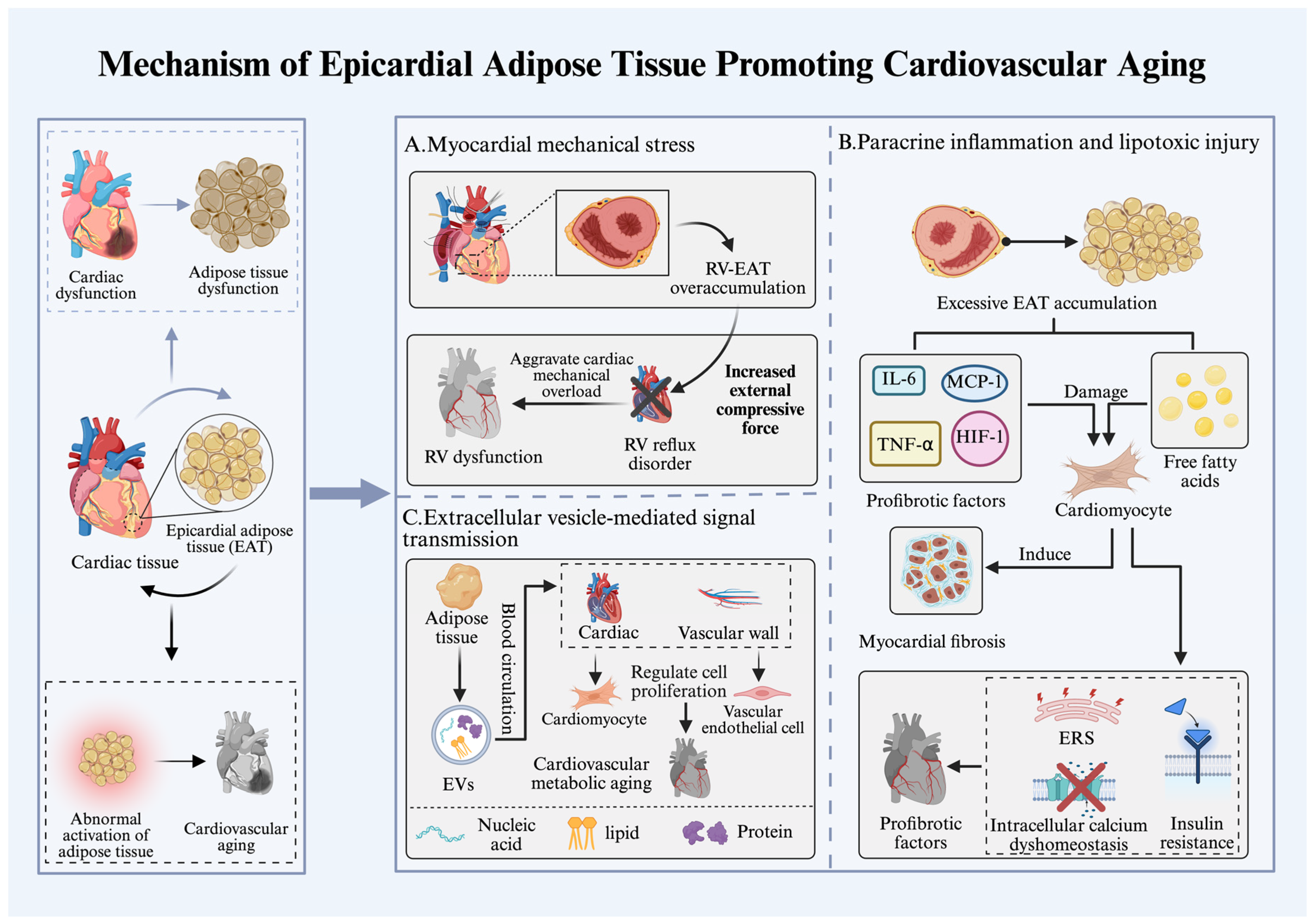
Figure 2.
Interplay between the heart and kidneys. The heart and kidneys mutually influence each other: on one hand, renal dysfunction drives cardiac aging; on the other hand, heart failure exacerbates functional impairment in both organs. A. When renal function declines, a large amount of metabolic waste accumulates in the body, forming a pool of uremic toxins. These accumulated toxins activate the aryl hydrocarbon receptor and NF-κB signaling pathways, inducing inflammation, oxidative stress, mitochondrial dysfunction, and tissue fibrosis, ultimately leading to cardiac dysfunction. B. After heart failure, hemodynamic disturbances occur, with reduced cardiac output decreasing renal perfusion pressure. Meanwhile, right-heart venous congestion exacerbates renal interstitial edema, leading to renal dysfunction. In heart failure patients, excessive activation of the RAAS and SNS systems disrupts the mechanical integrity of the glomerulus and stimulates the secretion of pro-fibrotic factors, resulting in glomerulosclerosis. Furthermore, due to the high sensitivity of proximal tubular cells to mitochondria, heart failure-induced mitochondrial dysfunction activates inflammation-related pathways, further aggravating renal dysfunction.
Figure 2.
Interplay between the heart and kidneys. The heart and kidneys mutually influence each other: on one hand, renal dysfunction drives cardiac aging; on the other hand, heart failure exacerbates functional impairment in both organs. A. When renal function declines, a large amount of metabolic waste accumulates in the body, forming a pool of uremic toxins. These accumulated toxins activate the aryl hydrocarbon receptor and NF-κB signaling pathways, inducing inflammation, oxidative stress, mitochondrial dysfunction, and tissue fibrosis, ultimately leading to cardiac dysfunction. B. After heart failure, hemodynamic disturbances occur, with reduced cardiac output decreasing renal perfusion pressure. Meanwhile, right-heart venous congestion exacerbates renal interstitial edema, leading to renal dysfunction. In heart failure patients, excessive activation of the RAAS and SNS systems disrupts the mechanical integrity of the glomerulus and stimulates the secretion of pro-fibrotic factors, resulting in glomerulosclerosis. Furthermore, due to the high sensitivity of proximal tubular cells to mitochondria, heart failure-induced mitochondrial dysfunction activates inflammation-related pathways, further aggravating renal dysfunction.
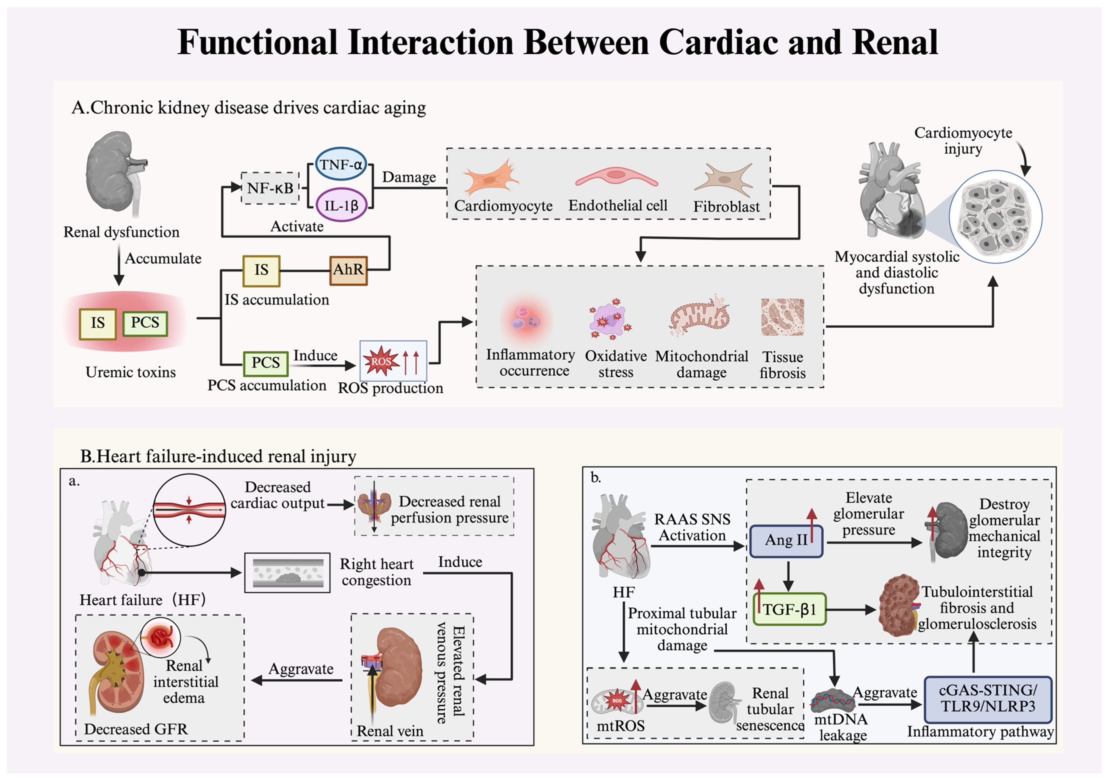
Figure 3.
Multi-organ crosstalk-based targeted therapeutic strategies. In the setting of heart failure, pathological crosstalk among adipose tissue, skeletal muscle, kidneys, and gut constitutes the core driver of disease progression. Targeting this network is a key direction for comprehensive heart failure management. Adipose tissue: Pharmacological agents such as SGLT2 inhibitors and GLP-1 receptor agonists reduce visceral fat accumulation, alleviate systemic inflammation and lipotoxicity, and block adiposity-induced myocardial damage signals. Skeletal muscle: Aerobic exercise strengthens atrophied skeletal muscle, while nutritional support with amino acids and other supplements ameliorates sarcopenia, restores the secretion of protective myokines, thereby reducing myocardial fibrosis and exerting cardioprotective effects. Kidneys: Targeting the kidney system affected by heart failure with mineralocorticoid receptor antagonists and SGLT2 inhibitors improves renal dysfunction and reduces the incidence of heart failure. Gut: After heart failure, gut microbiota dysbiosis occurs, which can be corrected by probiotic supplementation, fecal microbiota transplantation, or dietary intervention to modulate microbial composition, repair the intestinal barrier, and improve heart failure.
Figure 3.
Multi-organ crosstalk-based targeted therapeutic strategies. In the setting of heart failure, pathological crosstalk among adipose tissue, skeletal muscle, kidneys, and gut constitutes the core driver of disease progression. Targeting this network is a key direction for comprehensive heart failure management. Adipose tissue: Pharmacological agents such as SGLT2 inhibitors and GLP-1 receptor agonists reduce visceral fat accumulation, alleviate systemic inflammation and lipotoxicity, and block adiposity-induced myocardial damage signals. Skeletal muscle: Aerobic exercise strengthens atrophied skeletal muscle, while nutritional support with amino acids and other supplements ameliorates sarcopenia, restores the secretion of protective myokines, thereby reducing myocardial fibrosis and exerting cardioprotective effects. Kidneys: Targeting the kidney system affected by heart failure with mineralocorticoid receptor antagonists and SGLT2 inhibitors improves renal dysfunction and reduces the incidence of heart failure. Gut: After heart failure, gut microbiota dysbiosis occurs, which can be corrected by probiotic supplementation, fecal microbiota transplantation, or dietary intervention to modulate microbial composition, repair the intestinal barrier, and improve heart failure.
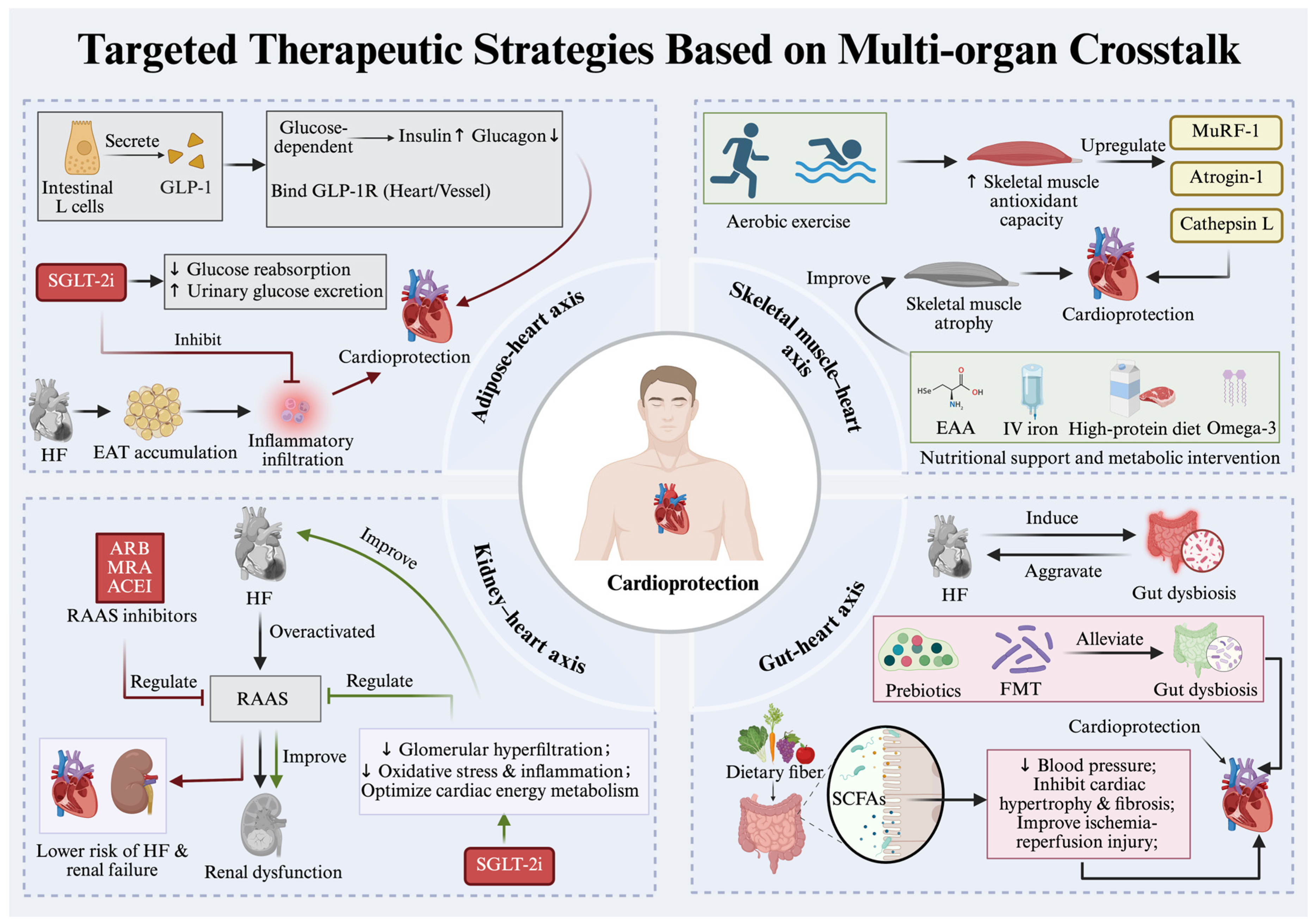
Disclaimer/Publisher’s Note: The statements, opinions and data contained in all publications are solely those of the individual author(s) and contributor(s) and not of MDPI and/or the editor(s). MDPI and/or the editor(s) disclaim responsibility for any injury to people or property resulting from any ideas, methods, instructions or products referred to in the content. |
© 2026 by the authors. Licensee MDPI, Basel, Switzerland. This article is an open access article distributed under the terms and conditions of the Creative Commons Attribution (CC BY) license (http://creativecommons.org/licenses/by/4.0/).
Copyright: This open access article is published under a Creative Commons CC BY 4.0 license, which permit the free download, distribution, and reuse, provided that the author and preprint are cited in any reuse.


