
Submitted:
16 June 2026
Posted:
17 June 2026
You are already at the latest version
Abstract
Recent technological advancements have significantly transformed the diagnosis and management of cardiovascular disease. Traditional reliance on two-dimensional (2D) and three-dimensional (3D) imaging has been enhanced by emerging 3D visualization technologies, particularly 3D printing and extended reality (XR). 3D printing enables the creation of patient-specific physical models that accurately replicate cardiovascular anatomy and pathology. These models play a crucial role in surgical plan-ning, simulation of interventional procedures, medical education, and patient commu-nication, offering tangible insights into complex cardiovascular structures. XR, on the other hand, provides an immersive 3D environment for exploring volumetric imaging data with interaction with the physical world. This enhances the understanding of in-tricate cardiovascular anatomy and pathology, supporting more informed clinical deci-sion-making and pre-surgical planning. This review explores the current applications of 3D printing and XR in cardiovascular surgery planning and intervention, emphasizing their potential to address challenges in planning complex procedures. Limitations and future directions for research and clinical integration are also discussed.
Keywords:
3D printing
; cardiovascular disease
; extended reality
; intervention
; surgery
; planning
; simulation
1. Introduction
Imaging diagnosis of cardiovascular disease has undergone rapid developments over the last decades with advancements of cardiac imaging modalities, mainly the increasing use of cardiac computed tomography (CT), cardiac magnetic resonance (MRI) and echocardiography imaging. These imaging techniques allow for depiction of cardiac anatomy and physiology/pathology with excellent spatial and temporal resolution through generation of two-dimensional (2D) and three-dimensional (3D) visualizations, thus playing an important role in understanding the complexity of cardiovascular disease and assisting pre-surgical planning or simulation of cardiovascular procedures. Translation of 3D imaging data to patient-specific or personalised 3D printed models represents a new direction in this respect that offers personalized treatment to cardiovascular disease through the use of realistic physical models to provide comprehensive evaluation of cardiovascular anatomy and pathology which cannot be obtained by standard cardiac imaging methods [1,2,3].
Three-dimensional printing technology is increasingly used in the medical field with contributions in many areas from the original applications in orthopaedics and maxillofacial surgeries to more recent applications in cardiovascular disease [4,5,6,7,8,9,10]. Patient-specific 3D printed models deriving from cardiac imaging data accurately replicate anatomical structures and pathological changes, hence enhance medical education through learning cardiovascular anatomy and pathology more effectively by providing the tactile experience of using 3D printed models when compared to the traditional teaching methods [11,12]. 3D printed models can also enhance patient education and communication [13,14,15,16]. Another superior advantage of using 3D printed models lies in its clinical value serving as a useful tool to simulate cardiovascular interventional procedures with increasing evidence confirming the value in this aspect. Due to lack of realistic simulators or limited accessibility to expensive simulators, 3D printed models meet the requirements to train and guide junior or inexperienced clinicians when performing cardiovascular interventional procedures.
In addition to 3D printing technology, extended reality (XR) is showing potential value in the care of cardiovascular surgery by generating 3D immersive environment, such as 3D virtual models by use of virtual reality (VR), and interaction of virtual objects with physical world achieved by augmented reality (AR) and mixed reality (MR). XR encompasses all of these three techniques (VR, AR and MR) (Figure 1) [17,18,19,20]. The XR-guided pre-surgical planning and simulation is gaining interest in the cardiovascular surgery, although most of the current applications focus on the use of VR modality in the field of cardiothoracic surgery, while use of AR and MR is still at its infancy stage despite some promising reports [17].
This review article provides an overview of both 3D printing and XR technologies in the cardiovascular disease, with a focus on their clinical value in surgical planning and simulation of cardiovascular procedures. Limitations associated with these technologies are emphasized, with future research directions highlighted to guide appropriate use of 3D printing and XR technologies in cardiovascular interventions.
2. 3D Printed Models in Surgical Planning of Cardiovascular Disease
The following sections will focus on a summary of using 3D printed models in the cardiovascular disease, in particular, describing the application of these 3D models in pre-surgical planning and simulation of congenital heart disease, aortic aneurysmal disease, valvular heart disease and left atrial appendage treatment. The rationale for focusing on these areas is due to the high volume of published studies demonstrating the utility and value of using 3D printed models in surgical planning and simulation of cardiovascular procedures.
2.1. 3D Printing in Surgical Planning and Simulation of Congenital Heart Disease
Three-dimensional printed models have gained acceptance in congenital cardiac surgery for pre-operative planning or decision making of congenital heart disease (CHD) [21,22,23,24,25]. The variable spectrum of CHD conditions, in particular, the concomitant presence of more morphological defects in the presence of complex CHD scenarios often require complex surgical procedures which present challenges to understand the CHD conditions with use of standard 2D or 3D image visualizations. 3D printed models overcome the limitations by delineating complex spatial relationship in these cardiovascular diseases (Figure 2). Bernhard et al conducted a systematic review and meta-analysis of 3D printed models in cardiovascular interventions among studies published between 2005 and 2021 [3]. Of 107 studies eligible in their analysis, 38 studies were published on the use of 3D printed models to guide cardiovascular intervention in patients with CHD, of which only 6 studies compared 3D printed models to standard therapy with inclusion of control group without using 3D printed models. Most of the remaining studies were case or case series reports.
Valverde et al in their multi-center study reported the clinical impact of 3D printed heart models on surgical planning and treatment in complex CHD cases [26]. Of 40 cases with complex CHD recruited from 10 international centers, 48% of the surgical decisions were modified or redefined with aid of 3D printed models when compared to decisions made only based on imaging data. Gomez-Ciriza and Ryan et al documented their long-term follow-up experiences with over 100 and 79 3D printed heart models included in the studies, respectively [27,28]. Similar findings were reported in Gomez-Ciriza’s study as the surgical planning of 83 patients with CHD was modified in 47.5% of the cases with use of 3D printed models when compared to traditional surgical planning based on medical imaging and clinical information [27]. Ryan et al reported their single-site three-year experience of incorporating 3D printed models into clinical workflow for surgical planning of CHD [28]. They produced 79 3D printed models of different CHD types which were specially used for surgical planning. Use of 3D printed models reduced the readmission rate when compared to the standard of care (60% vs. 71.6%, p=0.16). No significant difference was also noted in the 30-day mortality between 3D printing group and standard of care group (p=0.59).
Kiraly et al reported their multidisciplinary team experience about the impact of 3D printed models on surgical planning of complex congenital cardiac surgery [29]. Of 15 CHD cases undergoing complex intracardiac repairs, 3D printed models provided new information in 9 cases and refined the diagnosis in 13 cases. All complex operative procedures were rehearsed on the 3D printed models prior to operation, with 3D printed models contributing to modifying/improving operative plan in 13 cases. Another uniqueness of their study is the production of both hollow and volume 3D printed models (Figure 3 and Figure 4) which allowed for hands-on simulation of surgical procedures.
Due to wide variation of CHD types and rarity of pathology associated with congenital heart defects, there is a strong demand on developing training programs for surgical trainees to develop their practical skills prior to performing complex cardiac surgeries on patients. 3D printed models serve as a useful tool for hands-on surgical training course enabling participants to practice or simulate surgical or interventional cardiac procedures on the realistic physical models (Figure 5 and Figure 6) [27,30,31,32,33,34,35]. Incorporation of hands-on training program based on 3D printed models into medical curricular represents another potential application for training cardiac professionals and enhancing interdisciplinary communication [36,37].
2.2. 3D Printing in Aortic Disease
Endovascular aortic repair (EVAR) has become a widely used procedure to treat patients with abdominal aortic aneurysm (AAA) and aortic dissection due to its less invasiveness and fewer complications when compared to open surgery. The current practice of preoperative planning EVAR relies on 2D and 3D CT imaging visualisations, which meet most of the requirements for aiding vascular surgeons to plan the EVAR. However, they do not provide a realistic 3D representation of complex vascular diseases in relation to anatomical structures [38]. The use of 3D printing technology has overcome the limitations by producing realistic 3D models with high accuracy of replicating anatomical structures that cannot be obtained by current imaging modalities. Although VR training platforms showed promising outcomes with reduction of procedural time and post-procedural complications such as endoleaks, physical phantoms are more important tools for surgery trainees to develop practical skills required for EVAR procedures [39]. Commercially available phantoms are not only expensive, but also lack patient-specific details, which limit their clinical value for surgical training [40]. Use of 3D printed personalised vascular models has addressed the research gap in this area.
Of 107 studies included in Bernhard et al review, the use of 3D printed models in aortic disease is the 3rd most common applications in cardiovascular interventions, with 10 studies retrieved from the literature [3]. After reviewing the 3D printed models, the decision making in managing EVAR planning was changed in 20% of cases in patients with aortic aneurysm when compared to reviewing standard CT images [41]; the fluoroscopy and intervention times was reduced by 30% and 29%, respectively; and the contrast medium was reduced by 25% in the trainee training in EVAR [42].
3D printed templates have been shown to assist with development of physician-modified stent grafts (PMEG) in preoperative planning of EVAR, especially when dealing with complex aortic aneurysms, leading to decreased surgical time and procedure-related complications [43]. Furthermore, 3D printed aortic templates address the limitation of PMEG due to lack of standardization and interobserver variability [44]. Coles-Black et al reviewed 9 studies (5 case reports, 2 prospective trials, 1 technical report and 1 letter to the editor) published before 2020 documenting the feasibility of 3D printed-assisted PMEG for fenestrated stent grafting [45]. Their critical analysis of the literature shows that 3D-printed templates represent a promising technique to assist with fenestrations in PMEG with the potential to reduce cost and time savings, as well as increase the accuracy of fenestration alignment, however, further evidence is needed to validate its workflow and optimize manufacturing logistics.
Recent studies published in the last five years with inclusion of more cases and clinical follow-up outcomes have suggested the integration of 3D printed models into daily practice of stent graft modification [46,47,48,49,50]. Patel et al in their recent review identified 7 studies reporting the use of 3D printed models in complex aortic aneurysm repair [43]. Of these 7 studies, four focused on the 3D printed models in PMEG construction for complex aortic aneurysm and aortic dissection, with inclusion of cases ranging from 12 to 43, involving up to 162 fenestrations [46,47,48,49]. It is generally agreed that the use of 3D printed models offer several advantages, such as enhancing trainee education, reducing surgical time and complications, providing more precise fenestrated placement/or alignment. Figure 7 is an example showing the use of 3D printed template to aim accurate design of a fenestrated stent graft, while Figure 8 shows the clinical outcomes following 3D printed models-assisted fenestrated procedures with a mean follow-up of 14 months [48].
Majority of the current used 3D printed models are produced with only delineating anatomical and pathological details without presenting hemodynamic condition as observed in a realistic cardiovascular system. Karkkainen et al addressed this limitation by proving the feasibility of connecting a 3D-printed aortic aneurysm model to a fluid pump. A group of vascular surgical trainees performed 22 EVAR simulation procedures through this approach with high fidelity achieved [39]. Daring and Sun recently developed 3D printed models based on common vascular diseases (carotid artery stenosis, coronary artery stenosis, renal artery stenosis and abdominal aortic aneurysm) instead of including complex aortic disease scenarios [51]. They presented their 3D models to 21 specialists (15 vascular surgeons and 6 radiologists) who practiced the EVAR/interventional radiology procedures on these 3D printed models (Figure 9). Seventy-five percent of the participants gave a ranking of 7 or above out of 10 regarding the recommendation of the use of the 3D printed vascular models. The abdominal aorta model was ranked the highest for realism (4.10 ± 0.89, p = 0.002), the planning of interventions and simulations (3.90 ± 1.12 and 4.05 ± 0.95), the development of haptic skills (3.56 ± 0.98), reducing the procedure time (3.47 ± 1.12), and clarifying the pathology to patients (4.33 ± 0.69, p all >0.05). This study further validates the usefulness of 3D printed models for pre-surgical simulation of EVAR procedures.
2.3. 3D Printing in Transcatheter Aortic Valve Replacement
Transcatheter aortic valve replacement (TAVR) has been established as an effective alternative to traditional thoracotomy in the treatment of severe aortic stenosis or regurgitation owing to its less invasive nature, reliable safety and efficacy, with resultant significant reduction in risks of stroke or other complications [52]. Despite these advantages, TAVR is associated with several complications including paravalvlular leak (PVL), rupture of the aortic annulus and coronary artery obstruction. Additionally, understanding the patient’s anatomy plays an essential role in choosing the aortic valve that best fits the individual patient’s condition. Preoperative imaging provides detailed information about the aortic root anatomy required for procedural planning, however, it is limited with 2D views lacking accurate assessment of internal anatomical structures, thus presenting challenges for surgeons to predict how the prosthetic valve adapts in situ or anticipate complications that could be avoided. Use of 3D printed personalised models have made it possible to develop optimal strategy to tailor individual patient’s unique anatomy with improved outcomes (Figure 10) [3,53,54,55,56,57,58].
Hornstein et al reviewed the application of 3D printed models in cardiac structural disease with studies published before 2021 [55]. They retrieved 37 studies for inclusion in their qualitative analysis, of which 13 studies reported the use of 3D printed models in TAVR, and this represents the most common application in their analysis. All 13 studies documented the use of 3D printed models in operative planning, 7 in complication prevention, and 1 on the use of 3D printed models in medical education. Five studies reported the prevention of PVL (71.4%) with two mentioning the prevention of acute valve dysfunction (28.6%). Another recent systematic review analysed 13 articles focusing on the 3D printed models in preoperative planning for TAVR [56]. Of 13 studies reviewed, 7 were case reports or case series, and 6 were retrospective studies recruiting patients between 6 and 30 cases who underwent TAVR. All of the 3D models were developed based on preoperative CT images. Their analysis shows that 3D printed models seem to be an accurate approach for predicting post-procedural complication of PVL, which was reported in 6 studies. The review also supports the safe performance of pre-procedural rehearsal of TAVR on 3D printed models, with reduction of the risk of procedural complications, as well as prediction of the adverse event and potential guidance on the valve implantation. Authors raised concerns about the small sample size and retrospective nature of the studies in the existing literature, and they suggested that prospective assessment is needed to provide further insight into the diagnostic value of TAVR simulation before 3D printed models can be recommended for use in routine clinical practice [56].
Two recent articles published by the same research team from China provide further evidence about the clinical application of 3D printed models in simulation and planning of TAVR [57,58]. Ma et al in their review of the applications of 3D printed models in TAVR providing a comprehensive overview of details from methodology of generating 3D printed models to clinical applications, with coverage of preprocedural assessment and reduction of complications, as well as TAVR simulation (Figurer 11) [57]. Their research team has so far completed 682 cases of TAVR over the last four years with use of 3D printed models to guide planning, with significant reduction in postprocedural complications. The 3D models assist clinical decision making, reduce radiation dose associated with X-ray exposure time and blood loss. Mao et al enrolled 48 patients with aortic regurgitation with 24 patients allocated to the group with 3D printed model simulation, and another 24 to the control group without having 3D printed models [58]. Twelve experts were invited with 6 of them performing the procedures before TAVR on the 3D printed models, and another 6 performing the TAVR procedures using the conventional approach. Another 10 experts and 10 young proceduralists participated in completing the TAVR simulations (Figure 12). Significant reductions were found in crossing-valve time and total operation time (p<0.001) in the 3D printing group as opposed to the non-3D printing group. Furthermore, radiation exposure and complications were significantly reduced with aid of 3D printed models. Zhang et al in their recent case report further advanced the application of 3D printed models in TAVR when managing a rare case of aortic valvular disease, namely quadricuspid aortic valve [59]. A combination of 3D printed model and computational fluid dynamics enables analysis of hemodynamic parameters and provides a better foundation for presurgical planning to develop optimized strategy for treating patients with complex aortic valve disease (Figure 13 and Figure 14).
2.4. 3D Printing in Left Atrial Appendage Occlusion
Left atrial appendage (LAA) is an outpouching structure attending to the left atrium and it contributes to thrombus formation in patients with atrial fibrillation. Percutaneous occlusion of LAA has been shown to successfully prevent embolization of LAA thrombus, however, placement of the LAA occlusion devices could be a technically challenging procedure as it requires accurate device sizing prior to the procedure, and accurate device positioning at the ostium of LAA to avoid peri-device leaks [60]. Comprehensive assessment of the LAA anatomy is critical for ensuring the procedural success and treatment outcomes, and this is routinely performed by 2D and 3D transesophageal echocardiography or cardiac CT imaging examinations to decide the diameter of the landing zone, the number of additional lobes needed for determining the LAA occlude device size and type [61,62]. Due to complexity and variation of LAA structure, the current imaging-based preprocedural planning lacks accurate device sizing and fails to provide overall 3D understanding of the LAA structure [63,64]. 3D printed models based on patient’s imaging data are increasingly used to solve these limitations.
Use of 3D printed models in LAA occlusion is the 2nd most commonly applied area in cardiovascular interventions as indicated by a systematic review and other studies [3,65,66]. There are four articles published between 2022 and 2025 focusing on a systematic review and meta-analysis of the literature about usefulness of 3D printed models in LAA closure or device sizing [67,68,69,70]. Tarababanis et al analysed 19 studies with last search of studies published until October 2021 [67]. Most of the studies recorded the application of 3D printed models in accurately sizing the LAA occluder devices prior to procedures. The intraprocedural impact of 3D printed models lies in significant decrease of devices per procedure, thus contributing to improvement in intraprocedural outcomes. Fluoroscopic times were also decreased by 30-45% with use of 3D printed models. However, the postprocedural impact of 3D printed models on LAA occlusion procedures is limited due to lack of studies documenting the outcomes or postprocedural complications [67]. Similarly, DeCampos et al in their systematic review and meta-analysis analysed eight studies which were published by September 2021 [68]. They performed a quantitative analysis of these studies by using a fixed-effect model, with 3D printed models associated with significant reduction in the PDL when compared to imaging assessment only (p<0.001). Use of 3D printed models significantly reduced the risk of adverse events (p<0.001), and 3D printed models showed superiority for guiding LAA occlusion procedures. Meta-analysis of secondary endpoints including procedural time, fluoroscopy time and radiation dose could not be performed due to variations in study design, however, all endpoints were significantly reduced with aid of 3D printed models.
The remaining two review articles included studies published until June 2024 which included more recent studies [69,70]. Santos et al in their meta-analysis only included three cohort studies with a total of 204 patients with a mean follow-up of 25 months [66,71,72]. Similar to the previous reports, their meta-analysis showed significant lower rates of PDL in the 3D printing-guided group as opposed to the conventional imaging group (p<0.001). The incidence of device mismatch was also significantly lower in the 3D printing-guided group. Although the prevalence of device-related thromboembolism was 49% less in the 3D printing group, the difference was not significant (p=0.58). Procedural time was significantly shorter with use of 3D printed models than that in the conventional imaging group (p<0.001). While this analysis further confirms the promising findings, further research based on large randomized controlled trials is needed to generate robust findings.
Kahalm et al included 10 studies in their analysis with last search of studies published until June 2024, and this review represents the most recent review of these studies on 3D printing in LAA occlusion procedures [70]. Of 10 studies, nine were prospective studies with one being case-control design. Similar findings were noted in these areas as summarised in the previous three reviews, such as accurate device sizing, reducing post-procedural leakages, decreasing the number of devices per procedure and reducing complications (Figure 15 and Figure 16) [70,73,74]. The limited sample sizes and lack of long-term follow-up outcomes and multi-center studies are still required before 3D printed models can be incorporated into standard LAA occlusion planning.
3. Extended Reality Technologies
The use of stereoscopic head-mounted displays (HMDs) has emerged as an innovative new frontier in the visualisation of three-dimensional anatomy for planning and executing surgeries for the treatment of cardiovascular disease. After an initial wave of enthusiasm in the 1990s and a subsequent period of disillusionment in the 2000s, the near-simultaneous release of multiple consumer-grade headsets in 2016 produced a second wave of enthusiasm which shows little sign of slowing [75]. The affordability and accessibility of these new headsets, many of which are compatible with user-friendly game engines like Unity or Unreal, have accelerated the adoption of extended reality technologies in medicine by dramatically lowering the technical barriers to creating bespoke visualisation tools for research and commercialisation [76].
HMDs are usually classified according to their position on Milgram and Kishino’s reality–virtuality continuum [77]. HMDs are typically classified as a device which generates either a virtual reality (VR), which is a completely immersive synthetic experience, or an augmented reality (AR), which blends real and synthetic content. A third category, mixed reality (MR), is sometimes used, but the distinction between AR and MR is inconsistent and the two categories are often conflated [78]. Although early second-wave devices like the Oculus Rift or Microsoft HoloLens were hardware-locked to a single modality, modern devices like the Meta Quest 3 and Apple Vision Pro have the capacity to adjust the balance of real and synthetic content on demand. This versatility means that it is increasingly productive to refer to HMDs as devices on the extended reality (XR) spectrum, capable of operating in either VR or AR mode as required.
The stereoscopic optics of modern XR HMDs give users true depth perception, which is crucial for accurately interpreting complex cardiovascular anatomy. As a result, these headsets are frequently compared with patient-specific 3D printed heart models, which offer a similar degree of depth perception. While both XR visualisations and patient-specific 3D printed models outperform conventional flat-screen formats such as DICOM (digital imaging and communications in medicine) viewers or 3D portable document formats (PDFs), studies usually report only marginal differences between the VR and 3D printed heart models or 3D PDFs approaches [79,80,81,82,83,84]. In contrast, MR was ranked as the preferred modality when compared to DICOM and 3D printed models in delineating complex CHD lesions, portraying spatial relationship between cardiac structures and facilitating pre-operative planning of complex CHD surgeries [82]. The main limitation of XR visualisation as compared to 3D printing is the prevailing absence of tactile cues. This is also confirmed by previous studies that 3D printed models were considered the best tool in patient communication as opposed to other visualisation approaches [80,82,83]. Most XR systems offer little to no haptic feedback, whereas a physical print can be touched, manipulated, and even incised, giving users the kinaesthetic information needed for tasks that demand fine motor control. Consequently, when practising hands-on procedural skills, such as vessel handling or drain placement, printed models often outperform purely virtual simulations [85].
4. Extended Reality for Surgical Planning and Simulation of Cardiovascular Disease
XR HMDs are increasingly being incorporated into pre-operative planning workflows. During this stage, surgeons focus on understanding a patient’s unique anatomical constraints, mapping operative corridors, and anticipating potential hazards. Multiple studies suggest that XR increases preoperative awareness of rare anatomical variants, sharpens interpretation of complex three-dimensional structures, and clarifies spatial relationships between structures [86,87,88,89,90]. XR visualisations lets surgeons inspect patient-specific cardiac reconstructions from precisely the viewpoint they will occupy in theatre, revealing spatial constraints far more clearly than conventional 2D imaging (Figure 17). Deng et al reported that the immersive perspective prompted 60% of surgeons to revise operative plans that had been drafted using 2D images alone [91].
Surface mesh files have a long history in medical XR visualisation because they are computationally simple to render and portray anatomical features as crisp, unambiguous surfaces [91]. These binary shapes cut through the visual clutter of the operating theatre, delivering anatomical cues that remain clear even under challenging lighting or motion. Some systems go further by tracking surgical instruments in real time and projecting their positions intraoperatively onto the segmented mesh, giving the surgeon a map-like overview of the patient’s internal “terrain” as the procedure unfolds (Figure 18) [92]. In addition, many planning workflows—such as finite-element analysis (FEM) and computational fluid dynamics (CFD)—require watertight surface meshes, typically supplied as standard tessellation language (STL) or OBJ files. When these high-fidelity anatomical models are coupled with physics-based simulations, they form the backbone of emerging digital twin frameworks, opening the door to patient-specific virtual testing and optimisation of surgical strategies [93]. The primary drawback to the use of surface-rendered meshes is that generating the models is labour intensive and time-consuming task requiring high degrees of manual intervention [94]. Deep learning techniques show considerable promise in automating this process, but have yet to be integrated into routine clinical workflows [95].
An increasing number of XR visualisation platforms now employ volume rendering to sidestep this labour-intensive mesh-generation step. Developed in the 1980s, volume rendering produces images directly from the voxel data of CT or MRI scans, eliminating the need to first extract an explicit surface model [96]. Volume rendering is far more computationally demanding than drawing a lightweight polygon mesh, especially for stereoscopic rendering, and in its early years was regarded an interesting but ultimately less practical alternative to surface rendering [97]. Thanks to modern GPUs and optimized ray-casting algorithms, the performance gap has largely closed, and researchers have started to push the envelope of visual fidelity with cinematic rendering techniques that incorporate advanced lighting models to produce photorealistic images [98].
Most of the clinical applications of XR in cardiothoracic surgery is dominated by the VR visualization with only a few studies reporting the use of AR and MR, according to a review in this aspect [17]. This is also confirmed by a recent systematic review and meta-analysis of XR interventions in patients undergoing elective cardiac surgical and interventional procedures [99]. Authors analysed 22 studies with all of them using VR interventions, of which 10 were included in the meta-analysis. Their review focused on the patient benefits such as how VR reduced anxiety and improved patient’s pre-procedural knowledge and postoperative physical function, but did not address the benefits of XR-guided cardiovascular procedures. Arjomandi et al analyzed 21 studies published between 2007 and 2019 with regard to the application of XR in thoracic surgery training [100]. Although authors summarised the applications of XR in preoperative planning (11 studies), thoracic surgery training (7 studies) and intraoperative assistance (9 studies) showing potential in thoracic surgery training and planning, their XR only included VR and AR without including MR visualization in any of these studies they reviewed. The same limitation is observed in another recent systematic review by Nanchahal et al [101]. Their review focused on the use of XR (VR and AR) in assisting mitral valve surgery. Fifty studies were included in their analysis with VR/AR applications covering two main areas: preoperative planning and predicting postoperative outcomes. XR (VR/AR) showed promising applications in enhancing outcomes and patient-cared care, but its feasibility and clinical value remains to be determined.
Recent case reports demonstrated the promising applications of MR-guided preoperative planning and intraoperative guidance of complex cardiovascular surgical procedures [102,103,104,105]. Aye et al presented their experience of using MR-guided minimally invasive cardiac interventions in three cases [105]. Figure 19 shows the use of MR (Hololens 2) to guide minimally invasive procedure for aortic valve replacement by superimposing 3D volume rendering model onto the real patient’s anatomy, while Figure 20 is another example of MR-guided procedure for pulmonary valve replacement. With aid of MR-guided approach, the mean operative time in these three cardiac surgical procedures was reduced by 34.3 minutes. Authors concluded that the real clinical value will need to be validated by larger cohort of studies with inclusion of more patients, but their first experience proves the feasibility of using novel MR visualization to guide cardiac interventional procedures.
5. Summary and Future Directions
3D printing and XR technologies have revolutionized the current practice in diagnosing and preprocedural planning of patients with cardiovascular disease, with a paradigm shift from traditional reliance on standard imaging modalities to potential incorporation of advanced visualization tools into clinical practice. 3D printing and XR provide more realistic 3D information which is not achievable with these standard imaging modalities. The increasing use of 3D printing and XR has created great opportunities to change the current practice by delivering personalized medicine.
Use of 3D printed models in cardiovascular system, including medical education, and clinical training as well as simulation and preoperative planning has been extensively studied, with research proving its value in medical education of learning cardiac anatomy or congenital heart disease when compared to the current teaching methods as shown in some randomized controlled trials (RCTs) [106,107,108,109]. Despite promising reports available in the literature, there is a lack of solid evidence and robust study design to determine the clinical value of 3D printed models in cardiovascular surgical interventions. While 3D printed physical models serve as a useful tool for clinical cardiovascular surgical planning and simulation, very few studies based on RCTs are available in the existing literature. Xie et al analysed RCT studies about their applications in medical education. Of 33 studies included in their review and meta-analysis, only three studies were focused on the comparison of cardiovascular models with conventional methods in terms of medical education and practical skill improvement [110]. Figure 21 shows their meta-analysis of these studies. Thus, improvements in study design with consideration of above-mentioned limitation are essential to provide further evidence of the impact of 3D printed models on clinical outcomes of cardiovascular surgery, in particular in the subspecialty areas including congenital heart disease, aortic disease, valvular disease and left atrial appendage occlusion.
XR is an emerging technology showing great potential in cardiology or cardiovascular disease [111,112,113]. In a more recent systematic review, Kanschik and colleagues performed a comprehensive review of the literature reporting the use of XR in cardiology and this review included all types of studies (from RCTs to case reports, with language published in English or German) on XR in cardiology published until 31 July 2024 [113]. They identified 164 studies for inclusion in their analysis comprising the use of XR in peri-procedural setting (78 studies), for training cardiovascular procedures (36 studies), for educational purpose (31 studies) and for cardiac rehabilitation (19 studies) (Figure 22). Of these applications, XR is increasingly used in the two most common areas: congenital heart diseases and cardiac catheterization with review of 28 and 35 studies, respectively. Authors summarised the reported findings of XR in these applications but did highlight the fact that the use of XR in the field of cardiovascular medicine, especially in patient management and outcome assessment is still in its early stage. Future research should focus on post-procedural patient care and clinical outcomes of XR-guided cardiovascular surgical procedures, especially based on larger cohort or randomized controlled studies. Another aspect to consider in the future research is to develop immersive, patients-specific 3D models and real-time navigation systems. Artificial intelligence (AI)-assisted image processing and automatic segmentation and AI-assisted decision tools will fill the current research gaps and further improve the accuracy, safety, and efficiency of cardiovascular interventional procedures [114,115,116,117,118,119]. Implementation of 3D printing and XR technologies into cardiovascular practice requires a multi-disciplinary team collaboration as these innovative technologies require the knowledge and skills from different disciplines. A systematic approach/framework will also need to be developed by expert groups to consider the barriers and challenges so that both 3D printing and XR meet clinical requirements when managing cardiovascular interventions.
Author Contributions
Conceptualization, Z.S. and M.O.; writing-original draft and preparation, Z.S. and M.O.; wiring-review and editing, Z.S., M.O. and Y.W. All authors have read and agreed to the final version of the manuscript.
Funding
This research received no external funding.
Institutional Review Board Statement
Not applicable.
Informed Consent Statement
Not applicable.
Conflicts of Interest
The authors declare no conflicts of interest.
References
- Sun, Z. Insights into 3D printing in medical applications. Quant. Imaging. Med. Surg. 2019, 9(1), 1-5. [CrossRef]
- Witowski, J.; Sitkowski, M.; Zuzak, T.; Coles-Black, J.; Chuen, J.; Major, P.; Pdziwiatr, M. From ideas to long-term studies: 3D printing clinical trials review. Int. J. Comput. Assist. Radiol. Surg. 2018, 13(9), 1473-1478. [CrossRef]
- Bernhard B, Illo J, Gloeckler M, et al. Imaging-based, patient-specific three-dimensional printing to plan, train, and guider cardiovascular interventions: A systematic review and meta-analysis. Heart. Lung. Circ. 2022; 31:1203-1218. [CrossRef]
- Perica, E.; Sun Z. Patient-specific three-dimensional printing for pre-surgical planning in hepatocellular carcinoma treatment. Quant. Med. Imaging. Surg. 2017, 7(6), 668-677.
- Bagaria, V.; Bhansali, R.; Pawar, P. 3D printing-creating a blueprint for the future of orthopedics: Current concept review and the road ahead! J. Clin. Ortho.Trauma. 2018, 9, 207. [CrossRef]
- Skelley, N.W.; Hagerty, M.P.; Stannard, J.T.; Feltz, K.P.; Ma, R. Sterility of 3D-Printed Orthopedic Implants Using Fused Deposition Modeling. Orthopedics. 2020, 43, 46–51. [CrossRef]
- Gomes, E.N.; Dias, R.R.; Rocha, B.A.; Santiago, J.A.D.; Dinato, F.J.S.; Saadi, E.K.; Gomes, W.J.; Jatene, F.B. Use of 3D printing in preoperative planning and training for aortic endovascular repair and aortic valve disease. Braz. Cardiovasc. J. 2018, 33, 490-495. [CrossRef]
- Gallo, M.; D'Onofrio, A.; Tarantini, G.; Nocerino, E.; Remondino, F.; Gerosa, G. 3D-printing model for complex aortic transcatheter valve treatment. Int. J. Cardiol. 2016, 210, 139-140. [CrossRef]
- Schmauss, D.; Haeberle, S.; Hagl, C.; Sodian, R. Three-dimensional printing in cardiac surgery and interventional cardiology: a single-centre experience. Eur. J. CardioThorac. Surg. 2015, 47, 1044-1052. [CrossRef]
- Giannopoulos, A.A.; Steigner, M.L.; George, E.; Barlie, M.; Hunsaker, A.R.; Rybicki, F.J.; Mitsouras, D. Cardiothoracic applications of 3-dimensional printing. J. Thorac. Imaging. 2016, 31, 253–272. [CrossRef]
- White, S.C.; Sedler, J.; Jones, T.W.; Seckeler, M. Utility of three-dimensional models in resident education on simple and complex intracardiac congenital heart defects. Congenit. Heart. Dis. 2018, 13, 1045-1049. [CrossRef]
- Lim, K.H.; Loo, Z.Y.; Goldie, S.; Adams, J.; McMenamin, P. Use of 3D printed models in medical education: A randomized control trial comparing 3D prints versus cadaveric materials for learning external cardiac anatomy. Anat. Sci. Educ. 2016, 9, 213-221. [CrossRef]
- Biglino, G.; Koniordou, D.; Gasparini, M.; Capelli., C.; Leaver, L.K.; Khambadkone, S.; Schievano, S.; Taylor, A.M.; Wray, J. Piloting the use of patient-specific cardiac models as a novel tool to facilitate communication during cinical consultations. Pediatr. Cardiol. 2017, 38, 813-818. [CrossRef]
- Illmann, C.F.; Hosking, M.; Harris, K.C. Utility and Access to 3-Dimensional Printing in the Context of Congenital Heart Disease: An International Physician Survey Study. CJC. Open. 2020; 2, 207–213. [CrossRef]
- Guo, H.; Wang, Y.; Dai, J.; Ren, C.; Li, J.; Lai, Y. Application of 3D printing in the surgical planning of hypertrophic obstructive cardiomyopathy and physician-patient communication: A preliminary study. J. Thorac. Dis. 2018; 10, 867–873. [CrossRef]
- Traynor, G.; Shearn, A.I.U.; Milano, E.G.; Ordonez, M.V.; Velasco Forte, M.N.; Caputo, M.; Schievano, S.; Mustard, H.; Wray, J.; Biglino, G. The use of 3D-printed models in patient communication: A scoping review. J .3D Print. Med. 2022, 6, 13–23. [CrossRef]
- Sadeghi, A.; el Mathari, S.; Abjigitova, D.; Maat, A.P.W.M.; Taverne, Y.J.H.J.; Bogers, A.J.J.C.; Mahtab, E.A.F. Current and future applications of virtual, augmented, and mixed reality in cardiothoracic surgery. Ann. Thorac. Surg. 2022, 113, 681-691. [CrossRef]
- Grine, M.; Guerreriro, C.; Costa, F.M.; Menezes, M.N.; Ladeiras-Lopes, R.; Ferreira, D.; Oliveira-Santos, M. Digital health in cardiovascular medicine: An oveview of key appliations and clinical impact by the Portuguese Society of Cardiology Study Group on Digital Health. Rev. Port. Cardiol. 2025, 44, 107-119. [CrossRef]
- Mahtab EAF, Max SA, Braun J, et al. Developing a systematic approach for the implementation of medical extended reality learning modules in cardiothoracic health care: Recommendations from an international expert group. JACC. Adv. 2025, 4, 101633.
- Sharieff, I.I.; Ravikumar, D.B.; Joshi, S.; Regeer, M.V.; Kaufman, B.; Dunning, J.; Bileraaj, B.; Andreas, M.; Lecoq, R.R.; Klinceva, M.; et al. Applications of augmented reality in cardiology till 2024: a comprehensive review of innovations and clinical impacts. Front. Virtual. Real. 2024, 6, 1580619.
- Sun, Z.; Lau, I.; Wong, Y.H.; Yeong, C.H. Personlized three-dimensional printed models in congenital heart disease. J. Clin. Med. 2019, 8(4), 522. [CrossRef]
- Sun, Z. Patient-specific 3D-printed models in pediatric congenital heart disease. Children. 2023, 10, 319. [CrossRef]
- Martelli, N.; Serrano, C.; van den Brink, H.; Pineau, J.; Prognon, P.; Borget, I.; El Batti, S. Advantages and disadvantages of 3-dimensoional printingin surgery: A systematic review. Surgery. 2016, 159, 1485-1500.
- Kappanayil, M.; Koneti, N.R.; Kannan, R.R.; Kottayil, B.P.; Kumar, K. Three-dimensional-printed cardiac prototypes aid surgical decision-making and preoperative planning in selected cases of complex congenital heart diseases: Eearly experience and proof of concept in a resource-limited environment. Ann. Pediatr. Cardiol. 2017, 10, 117-125. [CrossRef]
- Winlaw, D.S.; Ayer, J.G. Commentary: Three-dimensional printing for preoperative planning-Beyong illustrating the obvious. JTCVS .Tech. 2020, 2, 141-142. [CrossRef]
- Valverde, I.; Gomez-Ciriza, G.; Hussain, T.; Suarez-Mejias, C.; Velasco-Forte, M.N.; Byrne, N.; Ordonez, A.; Gonzalez-Calle, A.; Anderson, D.; Hazekamp, M.G.; et al. Three-dimensional printed models for surgical planning of complex congenital heart defects: an international multicenter study. Eur. J. Cardiothorac. Surg. 2017, 52, 1139-1148. [CrossRef]
- Gomez-Ciriza, G.; Gomez-Cia, T.; Rivas-Gonzalez, J.A.; Velasco Forte, M.N.; Valverde, I. Affordable three-dimensional printed heart models. Front. Cardiovasc. Med. 2021, 8, 642011. [CrossRef]
- Ryan, J.; Plasencia, J.; Richardson, R.; Velez, D.; Nigro, J.J.; Pophal, S.; Frakes, D. 3D printing for congenital heart disease: A single site’s initial three-year experience. 3D. Print. Med. 2018, 4, 10. [CrossRef]
- Kiraly, L.; Shah, N.C.; Abdullah, O.; Al-Ketan, O.; Rowshan, R. Three-dimensional virtual and printed protypes in complex congenital and pedaitric cardiac surgery-A multidisciplinary team-learning experience. Biomolecules. 2021, 11, 1703. [CrossRef]
- Yoo, S.J.; Hussein, N.; Peel, B.; Coles, J.; van Arsdell, G.S.; Honjo, O.; Haller, C.; Lam, C.Z.; Seed, M.; Barron, D. 3D modeling and printing in congenital heart surgery: Entering the stage of maturation. Front. Pediatr. 2021, 9, 621672. [CrossRef]
- Wang; H., Song, H.; Yang, Y; Wu, Z.; Hu, R.; Chen, J.; Guo, J.; Wang, Y.; Jia, D.; Cao, S., et al. Morphology display and hemodynamic testing using 3D printing may aid in the prediction of LVOT obstruction after mitral valve replacement. Int. J. Cardiol. 2021, 331, 296-306. [CrossRef]
- Kiraly, L.; Tofeig, M.; Jha, N.K.; Talo, H. Three-dimensional prototypes refine the anatomy of post-modified Norwood-I complex aortic arch obstruction and allow presurgical simulation of the repair. Interact. Cardiovasc. Thorac. Surg. 2016, 22, 238-240. [CrossRef]
- Hussein, N.; Honji, O.; Barron, D.J.; Haller, C.; Coles, J.G.; Yoo, S.J. The incorporation of hands-on surgical training in a congenital heart surgery training curriculum. Ann. Thorac. Surg. 2021, 112, 1672-1680. [CrossRef]
- Brunner, B.S.; Thierij, A.; Jakob, A.; Tenglar, A.; Grab, M.; Thierfelder, N.; Leuner, N.A.; Haas, N.A.; Hopfner, C. 3D-printed heart models for hands-on training in pediatric cardiology-the future of modern learning and teaching? GMS. J. Med. Educ. 2022, 39, Doc23. [CrossRef]
- Hoashi, T.; Ichikawa, H.; Nakata, T.; Shimada, M.; Ozawa, H.; Higashida, A.; Kurosaki, K.; Kanzaki, S.; Shiraishi, I. Utility of a super-flexible three-dimensional printed heart model in congenital heart surgery. Interact. Cardiovasc. Thorac. Surg. 2018, 7, 749-755.
- Hon, N.W.L.; Hussein, N.; Honjo, O.; Yoo, S.J. Evaluating the impact of medical student inclusion into hands-on surgical simulation in congenital heart surgery. J. Surg. Ed. 2020, 78, 207-213. [CrossRef]
- Olivieri, L.; Su, L.; Hynes, C.F.; Krieger, A.; Alfares, F.A.; Ramakrishnan, K.; Zurakoswski, D.; Marshall, M.B.; Kim, P.C.W.; Joans, R.A.; et al. “Just-in-time” simulation training using 3-D printed cardiac models after congenital cardiac surgery. World. J. Pediatr. Congenit. Heart. Surg. 2016, 7, 164-168. [CrossRef]
- Bonvini, S.; Raunig, I.; Demi, L.; Spadoni, N.; Tasselli, S. Unsuspected limitations of 3D printed model in planning of complex aortic aneurysm endovascular treatment. Vasc. Endovasc. Surg. 2024, 58, 645–650. [CrossRef]
- Karkkainen, J.M.; Sandri, G.; Tenorio, E.R.; Alexander, A.; Bjellum, K.; Matsumoto, J.; Morris, J.; Mendes, B.C.; De Martino, R.R.; Oderich, G.S. Simulation of endovascular aortic repair using 3D printed abdominal aortic aneurysm model and fluid pump. Cardiovasc. Intervent. Radiol. 2019, 42, 1627-1634. [CrossRef]
- Little, C.D.; Mackle, E.C.; Maneas, E.; Chong, D.; Nikitichev, D.; Constantinou, J.; Tsui, J.; Hamilton, G.; Rakhit, R.D.; Mastracci, T.M.;, et al. A patient-specific multi-modality abdominal aortic aneurysm imaging phantom. Int. J. Comput. Assist. Radiol. Surg. 2022, 17, 1611-1617. [CrossRef]
- Tam, M.D.; Latham, .T.R.; Lewis, M.; Khanna, K.; Zaman, A.; Parker, M.; Grunwald, I.Q. A pilot study assessing the impact of 3-D printed models of aortic aneurysms on management decisions in EVAR planning. Vasc. Endovascular. Surg. 2016, 50, 4–9.
- Torres, I.O.; Luccia, N.D. A simulator for training in endovascular aneurysm repair: The use of three-dimensional printers. Eur. J. Vasc. Endovasc. Surg. 2017, 54, 247–253. [CrossRef]
- Patel, H.; Choi, P.; Ku, J.; Vergara, R.; Malgor, R.; Patel, D.; Li, Y. Application of three-dimensional printing in the planning and execution of aortic aneurysm repair. Front. Cardiovasc. Med. 2025, 11, 1485267. [CrossRef]
- Canonge, J.; Jayet, J.; Heim, F.; Chakfe, N.; Coggia, M.; Coscas, R.; Cochennec, F. Comprehensive review of physician modified aortic stent grafts: Technical and clinical outcomes. Eur. J. Vasc. Endovasc. Surg. 2021, 61, 560-569. [CrossRef]
- Coles-Black, J.; Barber, T.; Bolton, D.; Chuen, J. A systematic review of three-dimensional printed template-assisted physician-modified stent grafts for fenestrated endovascular aneurysm repair. J. Vasc. Surg. 2021, 74, 296–306. [CrossRef]
- Tong, Y.H.; Yu, T.; Zhou, M.J.; Liu, C.; Zhou, M.; Jiang, Q.; Liu, C.J.; Li, J.Q.; Liu, Z. Use of 3D printing to guide creation of fenestrations in physician-modified stent-grafts for treatment of thoracoabdominal aortic disease. J. Endovasc. Ther. 2020, 27(3), 385–93. [CrossRef]
- Branzan, D.; Geisler, A.; Grunert, R.; Steiner, S.; Bausback, Y.; Gockel, I.; Scheinert, D.; Schmidt, A. The influence of 3D printed aortic models on the evolution of physician modified stent grafts for the urgent treatment of thoraco-abdominal and pararenal aortic pathologies. Eur. J. Vasc. Endovasc. Surg. 2021, 61(3), 407–12. [CrossRef]
- Rynio, P.; Jedrzejczak, T.; Rybicka, A.; Milner, R.; Gutowski, P.; Kazimierczak, A. Initial experience with fenestrated physician-modified stent grafts using 3D aortic templates. J. Clin. Med. 2022, 11(8), 2180. [CrossRef]
- Tong, Y.; Qin, Y.; Yu, T.; Zhou, M.; Liu, C.; Liu, C.; Li, X.; Liu, Z. Three-dimensional printing to guide the application of modified prefenestrated stent grafts to treat aortic arch disease. Ann. Vasc. Surg. 2020, 66, 152–9. [CrossRef]
- Rhee, Y.; Park, S.J.; Kim, T.; Kim, N.; Yang, D.H.; Kim, J.B. Pre-sewn multi-branched aortic graft and 3D-printing guidance for Crawford extent II or III thoracoabdominal aortic aneurysm repair. Semin. Thorac. Cardiovasc. Surg. 2021, 34(3), 816–22. [CrossRef]
- Daring, D.L.; Sun, Z. Investigation of the clinical value of three-dimensional-printed personalised vascular models for the education and training of clinicians when performing interventional endovascular procedures. Appl. Sci. 2025, 15, 5695. [CrossRef]
- Swift, S.L.; Puehler, T.; Misso, K.; Lang, S.H.; Forbes, C.; Kleijnen, J.; Danner, M.; Kuhn, C.; Haneya, A.; Seoudy, H.; et al. Transcatheter aortic valve implantation versus surgical aortic valve replacement in patients with severe aortic stenosis: a systematic review and meta-analysis. BMJ. Open. 2021, 11, e054222. [CrossRef]
- Haghiashtiani, G.; Qiu, K.; Sanchez, J.D.Z.; Fuenning, Z.J.; Nair, P.; Ahlberg, S.E.; Iaizzo, P.A.; McAlpine, M.C. 3D printed patient-specific aortic root models with internal sensors for minimally invasive applications. Sci. Adv. 2020, 6, eabb4641. [CrossRef]
- Ripley, B.; Kelil, T.; Cheezum, K.; Goncalves, A.; Di Carli, M.D.; Rybicki, F.J.; Steigner, M.; Mitsouras, D.; Blankstein, R. 3D printing based on cardiac CT assists anatomic visualization prior to transcatheter aortic valve replacement. J. Cardiovasc. Comput. Tomogr. 2016, 10, 28-36. [CrossRef]
- Hornstein, G.; Diep, C.; Masson, J.B.; Potvin, J.; Gobeil. J.F.; Noiseux, N.; Forcillo, J. The use of three-dimensional printing in cardiac structural disease: A review. Innovations. 2023, 18, 132-143. [CrossRef]
- Xenofontos, P.; Zamani, R.; Akrami, M. The application of 3D printing in preoperative planning for transcatheter aortic valve replacement: a systematic review. Biomed. Eng. Online. 2022, 21, 59. [CrossRef]
- Ma, Y.; Mao, Y.; Zhu, G.; Yang, J. Application of cardiovascular 3-dimensional printing in transcatheter aortic valve replacement. Cell. Regeneration. 2022, 11, 35. [CrossRef]
- Mao, Y.; Liu, Y.; Ma, Y.; Zhai, M.; Li, L.; Jin, P.; Yang, J. Feasibility of 3-dimensional printed models in simulated training and teaching of transcatheter aortic valve replacement. Open. Med. 2024, 19, 20240909. [CrossRef]
- Zhang, Y.; Cai, D.; Wang, W.; Du, H.; Gu, J.; Li, X. Case report: Patient-specific three-dimensional printing and computational fluid dynamics in the planning of transcatheter aortic valve replacement for quadricuspid aortic valve. Front. Cardiovasc. Med. 2025, 12, 1555718. [CrossRef]
- Wang, D.D.; Eng, M.; Kupsky, D.; Myers, E.; Forbes, M.; Rahman, M.; Zaidan, M.; Parikh, S.; Wyman, J.; Pantelic, M.; et al. Application of 3-dimensional computed tomographic imaging guidance to WATACHMAN implantation and impact on early operator learning curve: single-center experience. JACC. Cardiovasc. Interv. 2016, 9, 2329-2340.
- Wunderlich, N.C.; Beigel, R.; Swaans, M.J.; Ho, S.Y.; Siegel, R.J. Percutaneous interventions for left atrial appendage exclusion: options, assessment, and imaging using 2D and 3D echocardiography. JACC. Cardiovasc. Imaging. 2015, 8, 472-488. [CrossRef]
- Pison, I.; Potpara, T.S.; Chen, J.; Larsen, T.B.; Bongiorni, M.G.; Blomstrom-Lundqvist, C. Left atrial appendage closure-indications, techniques, and outcomes: results of the European Heart Rhythm Association Survey. Europace. 2015, 17, 642-646. [CrossRef]
- Clemente, A.; Avogliero, F.; Berti, S.; Paradossi, U.; Jamagidze, G.; Rezzaghi, M.; Della Latta, D.; Chiappino, D. Multimodality imaging in preoperative assessment of left atrial appendage transcatheter occlusion with the Amplatzer Cardiac Plug. Eur. Heart. J. Cardiovasc. Imaging. 2015, 16, 1276-1287. [CrossRef]
- Viles-Gonzalez, J.F.; Kar, S.; Douglas, P.; Dukkipati, S.; Feldman, T.; Horton, R.; Holmes, D.; Reddy, V.Y. The clinical impact of incomplete left atrial appendage closure with the Watchman Device in patients with atrial fibrillation: a PROSPECT AF (Percutaneous Closure of the Left Atrial Appendage Versus Warfarin Therapy for Prevention of Stroke in Patients with Atrial Fibrillation) substudy. J. Am. Coll. Cardiol. 2012, 59, 923-929.
- Hell, M.M.; Achenbach, S.; Yoo, I.S.; Franke, J.; Blachutzik, F.; Roether, J.; Graf, V.; Raaz-Schrauder, D.; Marwan, M.; Schlundt, C. 3D printing for sizing left atrial appendage closure device: head-to-head comparison with computed tomography and transesophageal echocardiography. EuroIntervention. 2017, 13, 1234-1241. [CrossRef]
- Obasare, E.; Mainigi, S.K.; Morris, D.L.; Slipczuk, L.; Goykhman, I.; Friend, E.; Ziccardi, M.R.; Pressman, G.S. CT based 3D printing is superior to transesophageal echocardiography for preoperative planning in left atrial appendage device closure. Int. J. Cardiovasc. Imaging. 2018, 34, 821-831. [CrossRef]
- Tarabanis, C.; Klapholz, J.; Zahid, S.; Jankelson, L. A systematic review of the use of 3D printing in left atrial appendage occlusion procedures. J. Cardiovasc. Electrophysiol. 2022, 33, 2367-2374. [CrossRef]
- DeCampos, D.; Teixeira, R.; Saleiro, C.; Oliveira-Santos, M.; Paiva, L.; Costa, M.; Botelho, A.; Goncalves, L. 3D printing for left atrial appendage closure: A meta-analysis and systematic review. Int. J. Cardiol. 2022, 356, 38-43. [CrossRef]
- Santos, J.D.; Beloy, F.J.; Sulague, R.M.; Okudzeto, H.; Medina, J.R.; Cartojano, T.D.; Cruz, N.; Mortalla, E.D.; Kpodonu, J. Three-dimensional-printed models reduce adverse events of left atrial appendage occlusion: A systematic review and meta-analysis. Catherter. Cardiovasc. Interv. 2025, 105, 1688-1694. [CrossRef]
- Kahlam, J.; Savinova, O.V.; Shamilov, D.D.; Tran, J.; Borgmann, J.; Tran, T.; Jean, N.; Lo, D.F. Three-dimensional (3D) printing for left atrial appendage occlusion device sizing: A systematic review and comprehensive analysis. Cureus. 2025, 17, e82043. [CrossRef]
- Ciobotaru, V.; Combes, N.; Martin, C.A.; Marijon, E.; Maupas, E.; Bortone, A.; Bruguiere, E.; Thambo, J.B.; Teiger, E.; Pujadas-Berthault, P, et al. left atrial appendage occlusion simulation based on three-dimensional printing: New insights into outcome and technique. Eurointervention. 2018, 14, 176-184. [CrossRef]
- Fan, Y.; Yang, F.; Cheung, G.S.H.; Chan, A.K.Y.; Wang, D.D.; Lam, Y.Y.; Chow, M.C.K.; Leong, M.C.W.; et al Device sizing guided by echocardiography-based three-dimensional printing is associated with superior outcome after percutaneous left atrial appendage occlusion. J. Am. Soc. Ecocardiogr. 2019, 32, 708-719. [CrossRef]
- Kim, W.D.; Cho, I.; Kim, Y.D.; Cha, M.J.; Kim, S.W.; Choi, Y.; Shin, S.Y. Improving left atrial appendage occlusion device size determination by three-dimensional printing-based procedural simulation. Front. Cardiovasc. Med. 2022, 9, 830062. [CrossRef]
- Valvez, S,; Oliveira-Santos, M.; Goncalves, L.; Amaro, A.M.; Piedade, A.P. Preprocedural planning of left atrial appendage occlusion: A review of the use of additive manufacturing. 3D. Print. Addit. Manuf. 2024, 11, 333-346. [CrossRef]
- Venkatesan, M.; Mohan, H.; Ryan, J.R.; Schurch, C.M.; Nolan, G.P.; Frakes, D.H.; Coskun, A.F. Virtual and augmented reality for biomedical applications. Cell. Rep. Med. 2021, 2(7), 100348. [CrossRef]
- Zuo G, Wang R, Wan C, Zhang Z, Zhang S, Yang W. Unveiling the Evolution of Virtual Reality in Medicine: A Bibliometric Analysis of Research Hotspots and Trends over the Past 12 Years. Healthcare. 2024, 12(13), 1266. [CrossRef]
- Milgram P, Takemura H, Utsumi A, Kishino F. Augmented reality: a class of displays on the reality-virtuality continuum. In: Das H, editor. Boston, MA; 1995;282–92.
- Vervoorn, M.T.; Wulfse, M.; Van Doormaal, T.P.C.; Ruurda, J.P.; Van Der Kaaij, N.P.; De Heer, L.M. Mixed Reality in Modern Surgical and Interventional Practice: Narrative Review of the Literature. JMIR. Serious. Games. 2023, 11, e41297.
- Ong, C.S.; Krishnan, A.; Huang, C.Y.; Spevak, P.; Vricella, L.; Hibino, N.; Garcia, J.R.; Gaur, L. Role of virtual reality in congenital heart disease. Congenit. Heart. Dis. 2018, 13(3), 357–61. [CrossRef]
- Lau, I.; Gupta, A.; Sun, Z. Clinical Value of Virtual Reality versus 3D Printing in Congenital Heart Disease. Biomolecules. 2021, 11(6), 884. [CrossRef]
- Raimondi, F.; Vida, V.; Godard, C.; Bertelli, F.; Reffo, E.; Boddaert, N.; El Beheiry, M.; Masson, J.B. Fast-track virtual reality for cardiac imaging in congenital heart disease. J. Card. Surg. 2021, 36(7), 2598–602. [CrossRef]
- Lau, I.; Gupta, A.; Ihdayhid, A.; Sun, Z. Clinical Applications of Mixed Reality and 3D Printing in Congenital Heart Disease. Biomolecules. 2022, 12(11), 1548. [CrossRef]
- Lee, S.Y.; Squelch, A.; Sun, Z. Investigation of the Clinical Value of Four Visualization Modalities for Congenital Heart Disease. J. Cardiovasc. Dev. Dis. 2024, 11(9), 278. [CrossRef]
- Awori, J.; Friedman, S.D.; Howard, C.; Kronmal, R.; Buddhe, S. Comparative effectiveness of virtual reality (VR) vs 3D printed models of congenital heart disease in resident and nurse practitioner educational experience. 3D. Print. Med. 2023, 9(1), 2. [CrossRef]
- Rubio-López, A.; García-Carmona, R.; Zarandieta-Román, L.; Rubio-Navas, A.; González-Pinto, Á, Cardinal-Fernández P. Innovative approaches to pericardiocentesis training: a comparative study of 3D-printed and virtual reality simulation models. Adv. Simul. 2025, 10(1), 19. [CrossRef]
- Ender, J.; Končar-Zeh, J.; Mukherjee, C.; Jacobs, S.; Borger, M.A.; Viola, C.; Gessat, M.; Fassi, J.; Mohr, F.W.; Falk, V. Value of Augmented Reality-Enhanced Transesophageal Echocardiography (TEE) for Determining Optimal Annuloplasty Ring Size During Mitral Valve Repair. Ann. Thorac. Surg. 2008, 86(5), 1473–8. [CrossRef]
- Farooqi, K.M.; Uppu, S.C.; Nguyen, K.; Srivastava, S.; Ko, H.H.; Choueiter, N.; Wollstein, A.; Parness, I.A.; Narula, J.; Sanz, J.; et al. Application of Virtual Three-Dimensional Models for Simultaneous Visualization of Intracardiac Anatomic Relationships in Double Outlet Right Ventricle. Pediatr. Cardiol. 2016, 37(1), 90–8. [CrossRef]
- Frajhof, L.; Borges, J.; Hoffmann, E.; Lopes, J.; Haddad, R. Virtual reality, mixed reality and augmented reality in surgical planning for video or robotically assisted thoracoscopic anatomic resections for treatment of lung cancer. J. Vis. Surg. 2018, 4, 143–143. [CrossRef]
- Kanschik, D.; Haschemi, J.; Klein, K.; Maier, O.; Binneboessel, S.; Tokhi, U.; Afzai, S.; Serruys, P.W.; Tsai, T.Y.; Antoch, G.et al. Virtual reality for pre-procedural planning of valve-in-valve transcatheter aortic valve implantation. Eur. Heart. J. Digit. Health. 2025, 6, 372-381. [CrossRef]
- Rad, A.A.; Vardanyan, R.; Lopuszko, A.; Alt, C.; Stoffels, I.; Schmach, B.; Ruhparwar, A.; Zhigalvo, K.; Zabarevich, A.; Weymann, A. Virtual and Augmented Reality in Cardiac Surgery. Braz. J. Cardiovasc. Surg. 2022, 37, 123-127. [CrossRef]
- Deng, S.; Wheeler, G.; Toussaint, N.; Munroe, L.; Bhattacharya, S.; Sajith, G.; Lin, E.; Singh, E.; Chu, K.Y.K.; Kabir, S.; et al. A Virtual Reality System for Improved Image-Based Planning of Complex Cardiac Procedures. J. Imaging. 2021, 7(8), 151. [CrossRef]
- Liu, J.; Al’Aref, S.J.; Singh, G.; Caprio, A.; Moghadam, A,A,A,; Jang, S.J.; Wong, S.C.; Min, J.K.; Dunham, S.; Mosadegh, B. An augmented reality system for image guidance of transcatheter procedures for structural heart disease. PLOS. ONE. 2019, 14(7), e0219174. [CrossRef]
- Costagliola, E.; Musumeci, F.; Gandolfo, C.; Pilato, M.; Pasta, S. Merging mixed reality and computational modeling for enhanced visualization of cardiac biomechanics. Med. Eng. Phys. 2024, 134, 104258. [CrossRef]
- Byrne, N.; Velasco Forte, M.; Tandon, A.; Valverde, I.; Hussain, T. A systematic review of image segmentation methodology, used in the additive manufacture of patient-specific 3D printed models of the cardiovascular system. JRSM. Cardiovasc. Dis. 2016, 5, 2048004016645467. [CrossRef]
- Herten, R van. Data-efficient Deep Learning For Automated 3d Cardiac Structure Segmentation And Mesh Generation In Ct Angiography. J. Cardiovasc. Comput. Tomogr. 2025, 19(1), S11–2. [CrossRef]
- Levoy, M. Display of surfaces from volume data. IEEE. Comput. Graph. Appl. 1988, 8(3), 29–37.
- Udupa, J.K.; Hung, H.M.; Chuang, K.S. Surface and volume rendering in three-dimensional imaging: A comparison. J. Digit. Imaging. 1991, 4(3), 159–68. [CrossRef]
- Rowe, S.P.; Johnson, P.T.; Fishman, E.K. Cinematic rendering of cardiac CT volumetric data: Principles and initial observations. J. Cardiovasc. Comput. Tomogr. 2018, 12(1), 56–9. [CrossRef]
- Harris, E.; Fenton, S.; Stephenson, J.; Ewart, F.; Goharinezhad, S.; Lee, H.; Astin, F. Do extended reality interventions benefit patients undergoing elective cardiac surgical and interventional procedures? A systematic review and meta-analysis. J. Clin. Nurs. 2025, 34, 1465-1492. [CrossRef]
- Arjomandi Rad, A.; Vardanyan, R.; Thavarajasingam, S.G.; Zubarevich, A.; den Eynde, J.V.; Sa, M.P.B.; Zhigalov, K.; Nia, P.S.; Ruhparwar, A.; Weymann, A. Extended, virtual and augmented reality in thoracic surgery: a systematic review. Interact. Cardiovasc. Thorac. Surg. 2022, 34, 201-211. [CrossRef]
- Nanchahal, S.; Arjomandi Rad, A.; Naruka, V.; Chacko, J.; Liu, G.; Afoke, J.; Miller, G.; Malawana, J.; Punjabi, P. Mitral valve surgery assisted by virtual and augmented reality: cardiac surgery at the front of innovation. Perfusion. 2024, 39, 244-255. [CrossRef]
- Salavitabar, A,; Zampi, J.D.; Thomas, C.; Zanaboni, D.; Les, A.; Lowery, R.; Yu, S.; Whiteside, W. Augmented reality visualization of 3D rotational angiography in congenital heart disease: A comparative study to standard computer visualization. Pediatr. Cardiol. 2024, 45(8), 1759–66. [CrossRef]
- Garcia-Vazquez, V.; von Haxthausen, F.; Jackle, S.; Schumann, C.; Khhlemann, I.; Bouchagiar, J.; Hofer, A.C.; Matysiak, F.; Huttmann, G.; Goltz, J.P.; et al. Navigation and visualisation with Hololens in endovascular aortic repair. Innov. Surg. Sci. 2018, 3, 167-177. [CrossRef]
- Ponzoni, M.; Bertelli, F.; Yoo, S.J.; Peel, B.; Seatle. H.; Honjo, O.; Haller, C.; Barron, D.J.; Seed, M.; Lam, C.Z.; et al. Mixed reality for preoperative planning and intraoperative planning and intraoperative assistance of surgical correction of complex congenital heart defects. J. Thorac. Cardiovasc. Surg. 2025, 170, 327-335.e1. [CrossRef]
- Aye, W.M.M.; Kiraly, L.; Kumar, S.S.; Kasivishavanaath, A.; Goa, Y.; Kofidis, T. Mixed reality (holography)-guided minimally invasive cardiac surgery-A novel comparative feasibility study. J. Cardiovasc. Dev. Dis. 2025, 12, 49. [CrossRef]
- Loke, Y.H.; Harahsheh, A.S.; Krieger, A.; Olivieri, L.J. Usage of 3D models of tetralogy of Fallot for medical education: impact on learning congenital heart disease. BMC. Med. Educ. 2017, 17, 54. [CrossRef]
- Su, W.; Xiao, Y.; He, S.; Huang, P.; Deng, X. Three-dimensional printing models in congenital heart disease education for medical students: a controlled comparative study. BMC. Med. Educ. 2018; 18, 178. [CrossRef]
- White, S.C.; Sedler, J.; Jones, T.W.; Seckeler, M. Utility of three-dimensional models in resident education on simple and complex intracardiac congenital heart defects. Congenit. Heart. Dis. 2018, 13, 1045-1049. [CrossRef]
- Lim, K.H.A.; Loo, Z.Y.; Goldie, S.J.; Adams, J.W.; McMenamin, P.G Use of 3D printed models in medical education: A randomized control trial comparing 3D prints versus cadaveric materials for learning external cardiac anatomy. Anat. Sci. Educ. 2016, 9, 213-221. [CrossRef]
- Xie, G.; Wang, T.; Fu, H.; Liu, D.; Deng, L.; Zheng, X.; Li, L.; Liao, J. The role of three-dimensional printing models in medical education: a systematic review and meta-analysis of randomized controlled trials. BMC. Med. Educ. 2025, 25, 826. [CrossRef]
- Siddiqui, M.F.; Jabeen, S.; Alwazzan, A.; Vacca, S.; Dalal, L.; Al-Haddad, B.; Jaber, A.; Ballout, F.F.; Abou Zeid, H.K.; Haydamous, J.; et al. Integration of augmented reality, virtual reality, and extended reality in healthcare and medical education: A glimpse into the emerging horizon in LMICs-A systematic review. J. Med. Educ. Curric .Dev. 2025; 12, 23821205251342315. [CrossRef]
- Mondal, H.; Mondal. S. Adopting augmented reality and virtual reality in medical education in resource-limited settings: constraints and the way forward. Adv. Physiol. Educ. 2025, 49, 503-507. [CrossRef]
- Kanschik, D.; Bruno, R.R.; van Genderen, M.E.; Serruys, P.W.; Tsai, T.Y.; Klem, M.; Jung, C. Extended reality in cardiovascular care: a systematic review. Eur. Heart. J. Digit. Health. 2025, 6, 878-887. [CrossRef]
- Saitta, S.; Sturla, F.; Caimi, A.; Riva, A.; Palumbo, M.C.; Nano, G.; Votta, E.; Corte, A.D.; Glauber, M.; Chiappino, D.; et al. A deep learning-based and fully automated pipeline for thoracic aorta geometric analysis and planning for endovascular repair from computed tomography. J. Digit. Imaging. 2022, 35(2), 226–39. [CrossRef]
- Fantazzini, A.; Esposito, M.; Finotello, A.; Auticchio, F.; Pane, B.; Basso, C.; Spinella, G.; Conti, M. 3D automatic segmentation of aortic computed tomography angiography combining multi-view 2D convolutional neural networks. Cardiovasc. Eng. Technol. 2020, 11(5), 576–86. [CrossRef]
- Raffort, J.; Adam, C.; Carrier, M.; Ballaith, A.; Coscas, R.; Jean-Baptiste, E.; Hassen-Khodja, R.; Chakfe, N.; Lareyre, F. Artificial intelligence in abdominal aortic aneurysm. J. Vasc. Surg. 2020, 72(1), 321–33.e1. [CrossRef]
- Raimondi, F.; Ortiz-Garrido, A.; Voges, I. Artificial intelligence, extended reality and computational modelling in cross-sectional cardiovascular imaging in congenital heart disease: a narrative review. Cardiovasc. Diagn. Ther. 2026, 16, 29. [CrossRef]
- Kumr, P.; Siddarthan, I.; Keim, C.K.; Cho, D.K.; Rubin, J.E.; White, R.S.; Jotwani, R. Integrating AI segmentation, simulated digital twins, and extended reality into medical education: A narrative technical review and proof-of-concept case study. J. Pers. Med. 2026, 16, 202. [CrossRef]
- Tene, T.; Vique Lopez, D.F.; Garcia Veloz, M.J.; Rojas Oviedo, B.S.; Tene-Fernandez, R. Artificial intelligence, extended reality, and emerging AI-XR integrations in medical education. Front. Digit. Health. 2026, 7, 1740557. [CrossRef]
Figure 1.
Virtual, augmented, and mixed reality. Clinical and surgical applications of extended reality (XR) modalities in cardiothoracic surgery. Practical examples are displayed for the applicability of XR in preoperative planning (eg, virtual reality [VR], bottom left), guided preoperative planning of lung cancer surgery and intraoperative (eg, mixed [bottom right]) and augmented reality–guided (bottom center) navigation during thoracotomy/VATS for lung cancer surgery. (3D, 3-dimensional; VATS, video-assisted thoracoscopic surgery). Reprinted with permission under the open access from Sadeghi et al [17].
Figure 1.
Virtual, augmented, and mixed reality. Clinical and surgical applications of extended reality (XR) modalities in cardiothoracic surgery. Practical examples are displayed for the applicability of XR in preoperative planning (eg, virtual reality [VR], bottom left), guided preoperative planning of lung cancer surgery and intraoperative (eg, mixed [bottom right]) and augmented reality–guided (bottom center) navigation during thoracotomy/VATS for lung cancer surgery. (3D, 3-dimensional; VATS, video-assisted thoracoscopic surgery). Reprinted with permission under the open access from Sadeghi et al [17].
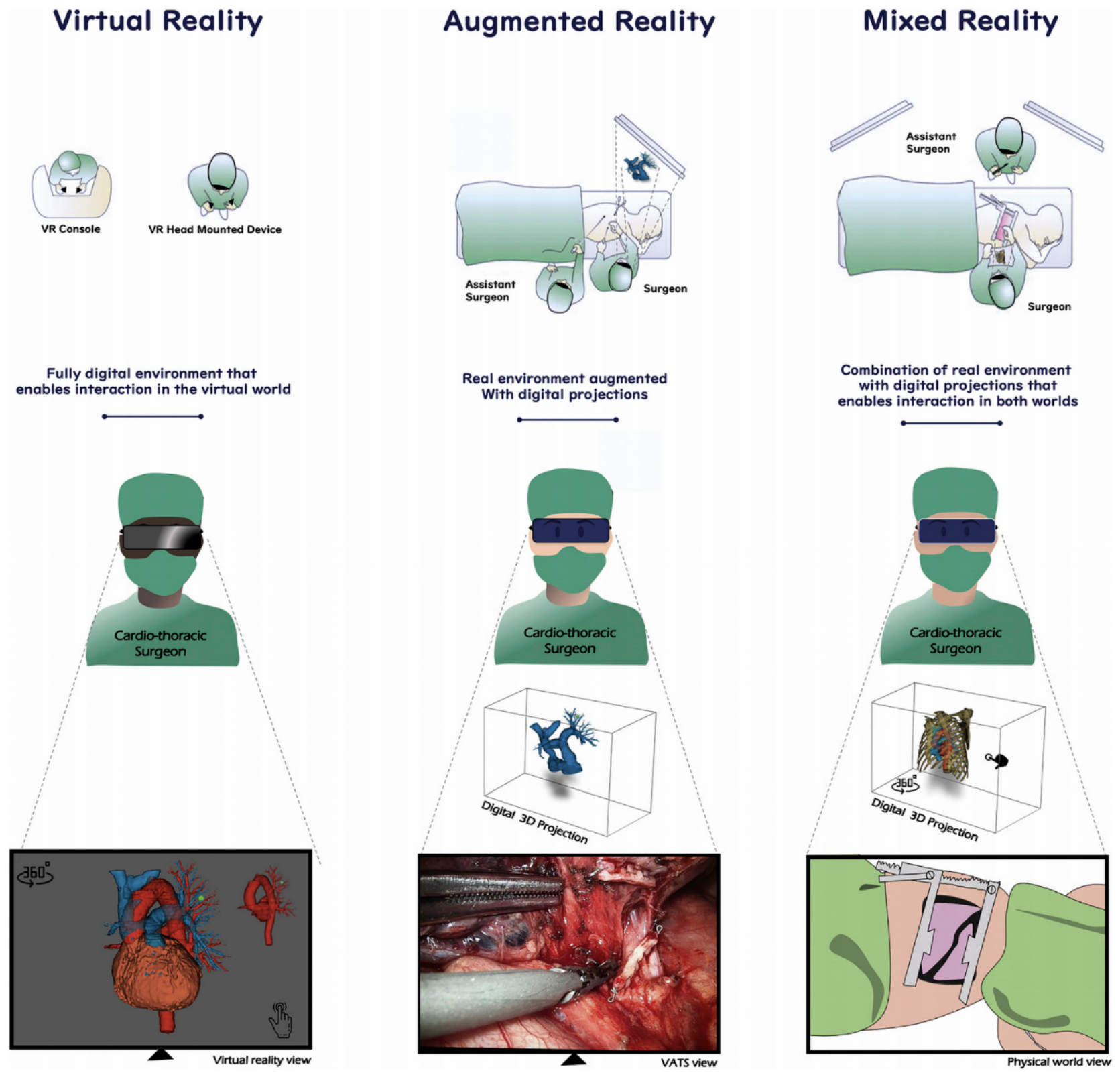
Figure 2.
3D printed heart model of a case with Tetralogy of Fallot (ToF). A: The model was printed in one piece. B: The model was printed in two halves (with the same material Agilus30 but different colours) showing the internal cardiac chambers and vascular abnormalities. Arrows indicate the pulmonary artery stenosis. AO-aorta (overriding aorta); PA-pulmonary artery; RV-right ventricle. Reprinted with permission under the open access from Sun et al [22].
Figure 2.
3D printed heart model of a case with Tetralogy of Fallot (ToF). A: The model was printed in one piece. B: The model was printed in two halves (with the same material Agilus30 but different colours) showing the internal cardiac chambers and vascular abnormalities. Arrows indicate the pulmonary artery stenosis. AO-aorta (overriding aorta); PA-pulmonary artery; RV-right ventricle. Reprinted with permission under the open access from Sun et al [22].
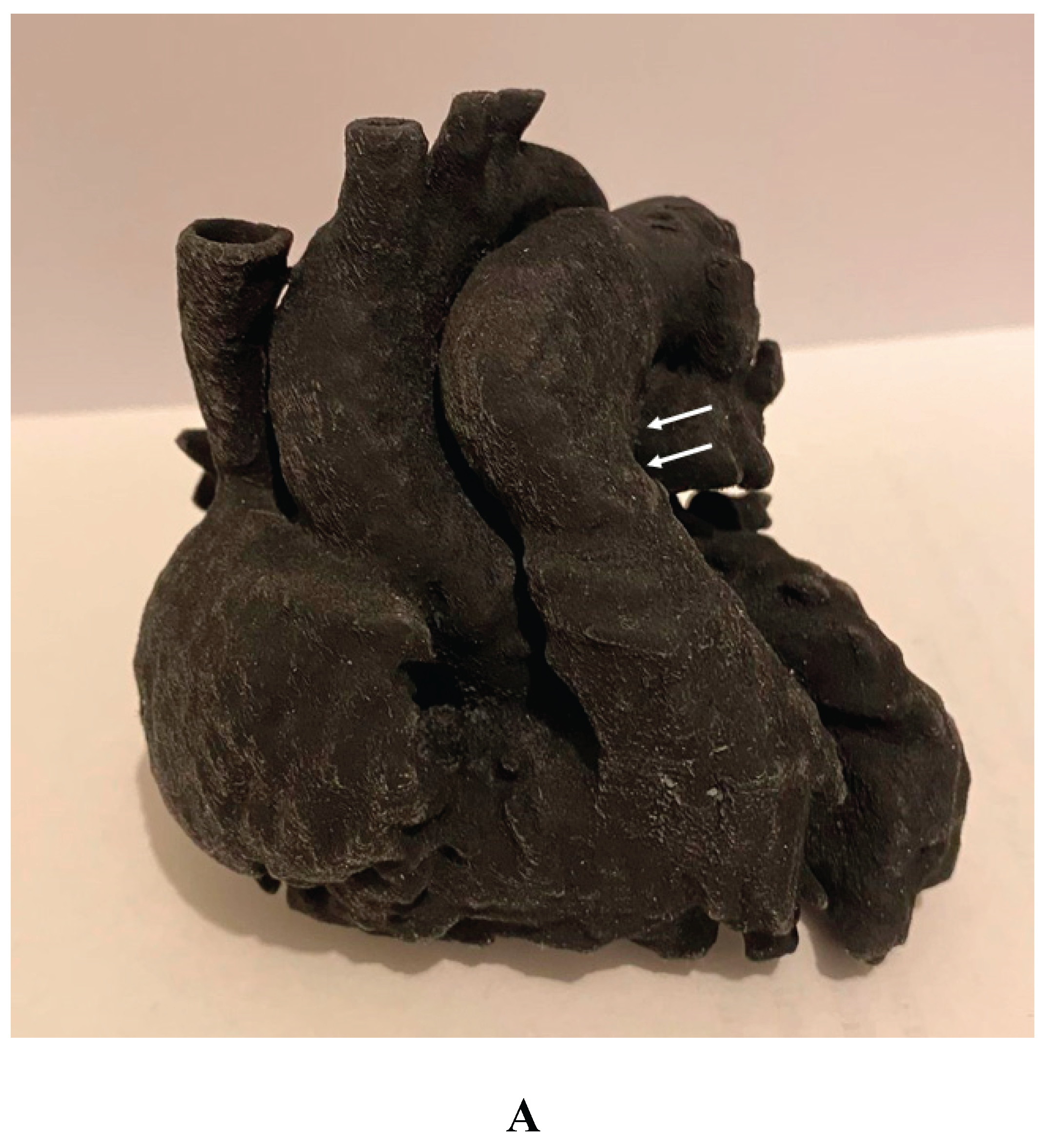
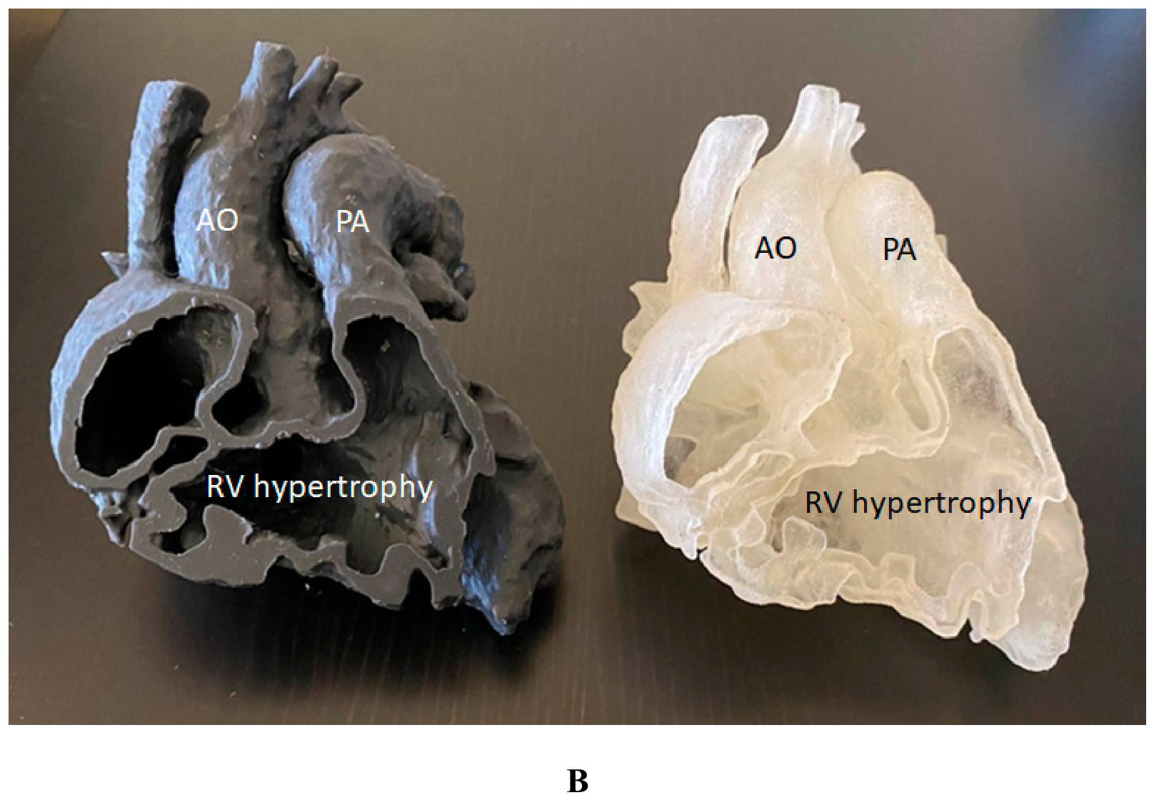
Figure 3.
3D-printed hollow model of right atrial isomerism, visceral heterotaxy, dextrocardia, common atrium, and hemiazygos continuity to left superior vena cava. (A) Right anterior oblique view with the ventricular apex removed. (B) Posterior view. Abbreviations: AAo: ascending aorta, DAo: descending aorta, Haz: hemiazygos vein, HV: hepatic vein, innom art/vein: innominate artery and vein, IVS: interventricular septum, L-AA: left-sided morphologically right atrial appendage, LCCA: left common carotid artery, LPA: left pulmonary artery, LPV: left pulmonary vein, LSCA: left subclavian artery, L-SVC: left superior vena cava, LV: left ventricle, PT: pulmonary trunk, R-AA: right-sided morphologically right atrial appendage, RPA: right pulmonary artery, RPV: right pulmonary vein, RV: right ventricle. Reprinted with permission under the open access from Kiraly et al [29].
Figure 3.
3D-printed hollow model of right atrial isomerism, visceral heterotaxy, dextrocardia, common atrium, and hemiazygos continuity to left superior vena cava. (A) Right anterior oblique view with the ventricular apex removed. (B) Posterior view. Abbreviations: AAo: ascending aorta, DAo: descending aorta, Haz: hemiazygos vein, HV: hepatic vein, innom art/vein: innominate artery and vein, IVS: interventricular septum, L-AA: left-sided morphologically right atrial appendage, LCCA: left common carotid artery, LPA: left pulmonary artery, LPV: left pulmonary vein, LSCA: left subclavian artery, L-SVC: left superior vena cava, LV: left ventricle, PT: pulmonary trunk, R-AA: right-sided morphologically right atrial appendage, RPA: right pulmonary artery, RPV: right pulmonary vein, RV: right ventricle. Reprinted with permission under the open access from Kiraly et al [29].
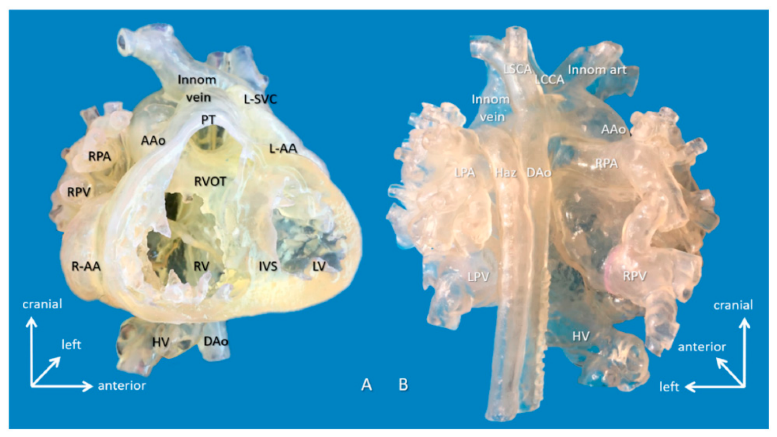
Figure 4.
3D-printed blood volume model of mesocardia, common atrium, criss-cross heart (supero-inferior ventricles), transposition of the great arteries, and pulmonary atresia, restrictive VSD and thrombus formation in the left ventricle; operated bidirectional superior cavopulmonary (Glenn) anastomosis. Modeling was indicated to assess the extent of the left ventricle thrombus and suitability for biventricular repair. Abbreviations: IV: innominate vein, IVC: inferior vena cava, LAD: left anterior descending coronary artery, RA: right atrium, RAA: right atrial appendage, RCA: right coronary artery, RIJV: right internal jugular vein, other abbreviations are the same as shown in Figure 3. Reprinted with permission under the open access from Kiraly et al [29].
Figure 4.
3D-printed blood volume model of mesocardia, common atrium, criss-cross heart (supero-inferior ventricles), transposition of the great arteries, and pulmonary atresia, restrictive VSD and thrombus formation in the left ventricle; operated bidirectional superior cavopulmonary (Glenn) anastomosis. Modeling was indicated to assess the extent of the left ventricle thrombus and suitability for biventricular repair. Abbreviations: IV: innominate vein, IVC: inferior vena cava, LAD: left anterior descending coronary artery, RA: right atrium, RAA: right atrial appendage, RCA: right coronary artery, RIJV: right internal jugular vein, other abbreviations are the same as shown in Figure 3. Reprinted with permission under the open access from Kiraly et al [29].
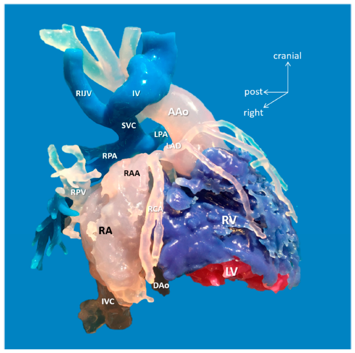
Figure 5.
Surgical and interventional planning on 3D-printed heart models. DORV case, internal vision from the left ventricle (left). DORV (another case), external view (right). DORV-double outlet right ventricle. Reprinted with permission under the open access from Gomez-Ciriza et al [27].
Figure 5.
Surgical and interventional planning on 3D-printed heart models. DORV case, internal vision from the left ventricle (left). DORV (another case), external view (right). DORV-double outlet right ventricle. Reprinted with permission under the open access from Gomez-Ciriza et al [27].
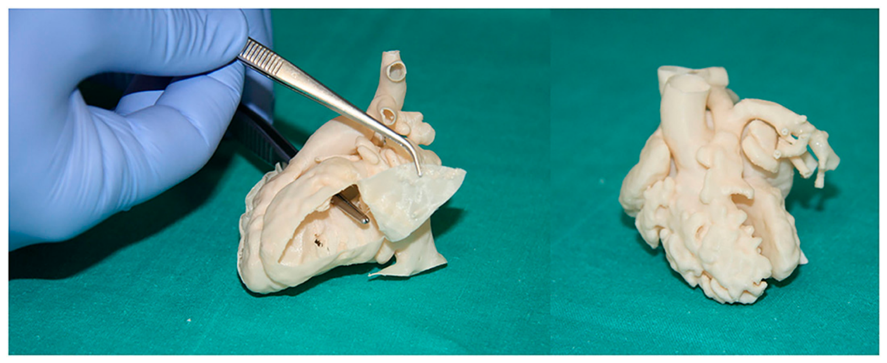
Figure 6.
Catheterization applications. Left image shows the fluid inlets and outlets to superior and inferior vena cava and aortic arch/branches, respectively. Interventional planning with 3D-printed model (up right) and real intervention in the patient (down right). Reprinted with permission under the open access from Gomez-Ciriza et al [27].
Figure 6.
Catheterization applications. Left image shows the fluid inlets and outlets to superior and inferior vena cava and aortic arch/branches, respectively. Interventional planning with 3D-printed model (up right) and real intervention in the patient (down right). Reprinted with permission under the open access from Gomez-Ciriza et al [27].
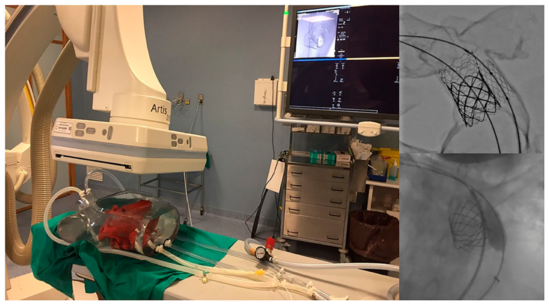
Figure 7.
Stages of stent graft modification. Stent graft deployed in the 3D aortic template (A). Stent graft with created fenestrations (B). A step of reducing-tie creation (C). Reduced-diameter stent graft (D). A preloaded guidewire to the superior mesenteric artery (E). Stent graft resheathing using the aortic valve crimper (F). Reprinted with permission under the open access from Rynio et al [48].
Figure 7.
Stages of stent graft modification. Stent graft deployed in the 3D aortic template (A). Stent graft with created fenestrations (B). A step of reducing-tie creation (C). Reduced-diameter stent graft (D). A preloaded guidewire to the superior mesenteric artery (E). Stent graft resheathing using the aortic valve crimper (F). Reprinted with permission under the open access from Rynio et al [48].
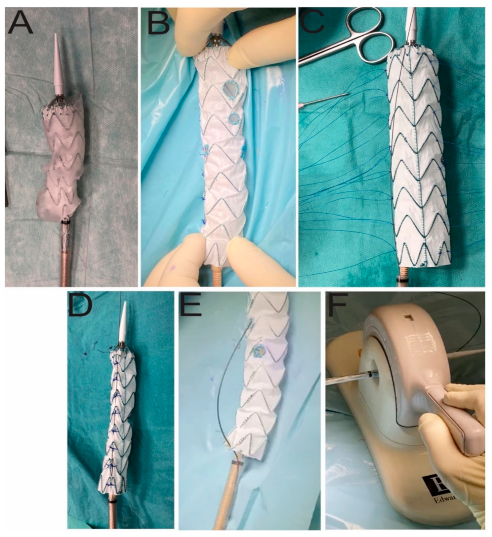
Figure 8.
The target vessel patency rates are presented on Kaplan–Meier curves. Right renal patency (A). Left renal artery patency (B). Superior mesenteric patency (C). Celiac trunk patency (D). Reprinted with permission under the open access from Rynio et al [48].
Figure 8.
The target vessel patency rates are presented on Kaplan–Meier curves. Right renal patency (A). Left renal artery patency (B). Superior mesenteric patency (C). Celiac trunk patency (D). Reprinted with permission under the open access from Rynio et al [48].
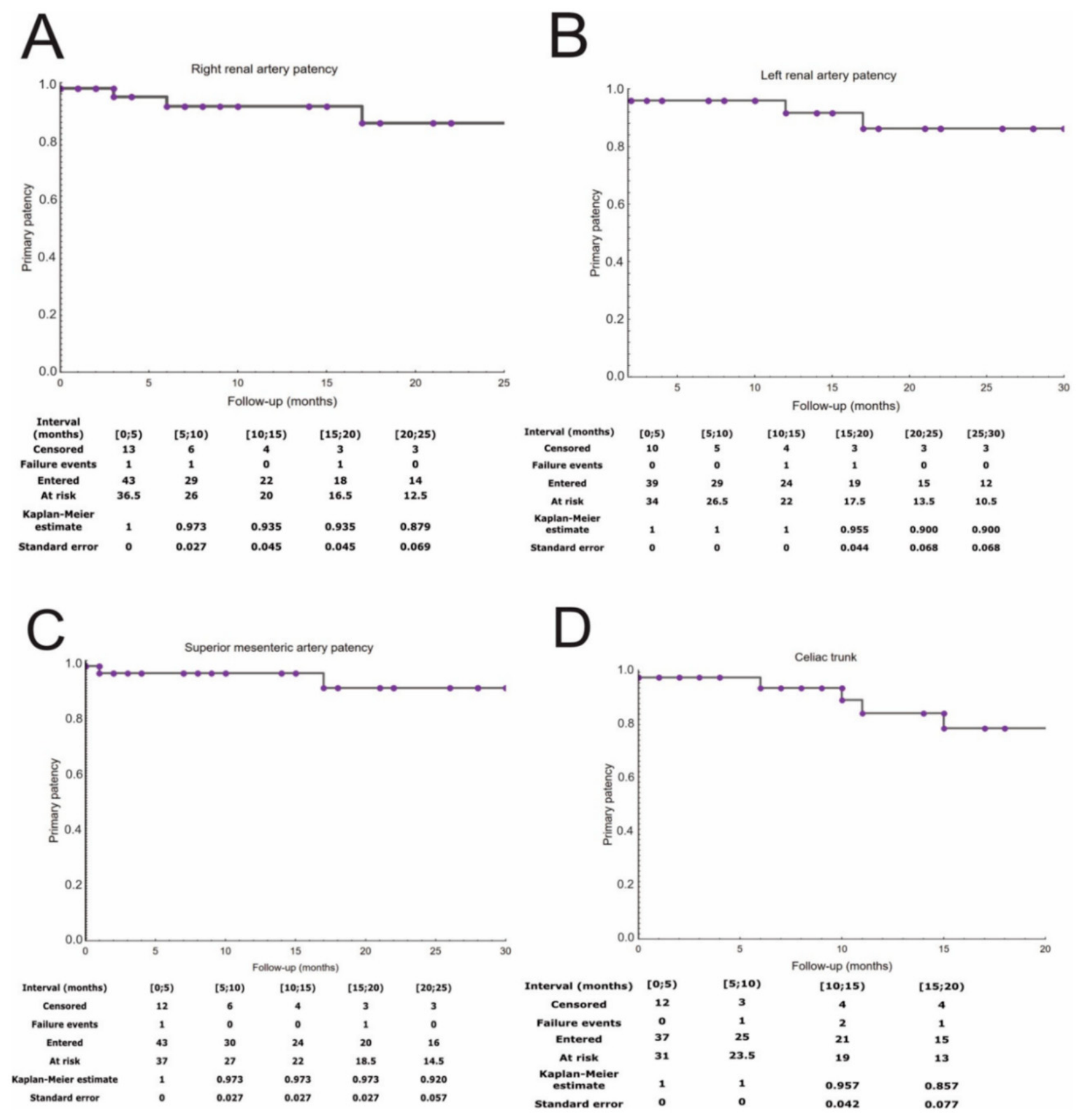
Figure 9.
Different 3D printing materials used for initial material selection. From left to right: V2 resin—draft; PETG—polyethylene terephthalate glycol; PLA—polylactic acid; HIPS—high-impact polystyrene; PMMA—polymethacrylate; TPU—thermoplastic polyurethane; 60C resin—tough white; TPU—polyurethane and rubber, soft and very soft. Reprinted with permission under the open access from Daring and Sun [51].
Figure 9.
Different 3D printing materials used for initial material selection. From left to right: V2 resin—draft; PETG—polyethylene terephthalate glycol; PLA—polylactic acid; HIPS—high-impact polystyrene; PMMA—polymethacrylate; TPU—thermoplastic polyurethane; 60C resin—tough white; TPU—polyurethane and rubber, soft and very soft. Reprinted with permission under the open access from Daring and Sun [51].
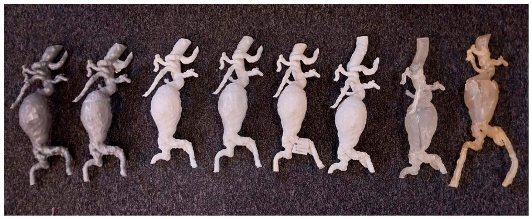
Figure 10.
Selected 3DPSP used for the planning of cardiovascular interventions. (A) CCT images of a patient with coronary anomaly translated into a 3D printed phantom (compliant PolyJet 3DPSP) for planning of complex percutaneous coronary artery intervention. (B) Silicone casted aortic root model made from a fused deposition modelling negative used for simulated transcatheter aortic valve implantation and postprocedural testing of paravalvular leakage with dye injection. (C) Compliant multi-material printed PolyJet 3DPSP aortic root translated from CCT imaging for planning transcatheter aortic valve implantation. 3DPSP, three-dimensional printed patient-specific phantom; CCT, cardiac computed tomography. Reprinted with permission under the open access from Bernhard et al [3].
Figure 10.
Selected 3DPSP used for the planning of cardiovascular interventions. (A) CCT images of a patient with coronary anomaly translated into a 3D printed phantom (compliant PolyJet 3DPSP) for planning of complex percutaneous coronary artery intervention. (B) Silicone casted aortic root model made from a fused deposition modelling negative used for simulated transcatheter aortic valve implantation and postprocedural testing of paravalvular leakage with dye injection. (C) Compliant multi-material printed PolyJet 3DPSP aortic root translated from CCT imaging for planning transcatheter aortic valve implantation. 3DPSP, three-dimensional printed patient-specific phantom; CCT, cardiac computed tomography. Reprinted with permission under the open access from Bernhard et al [3].
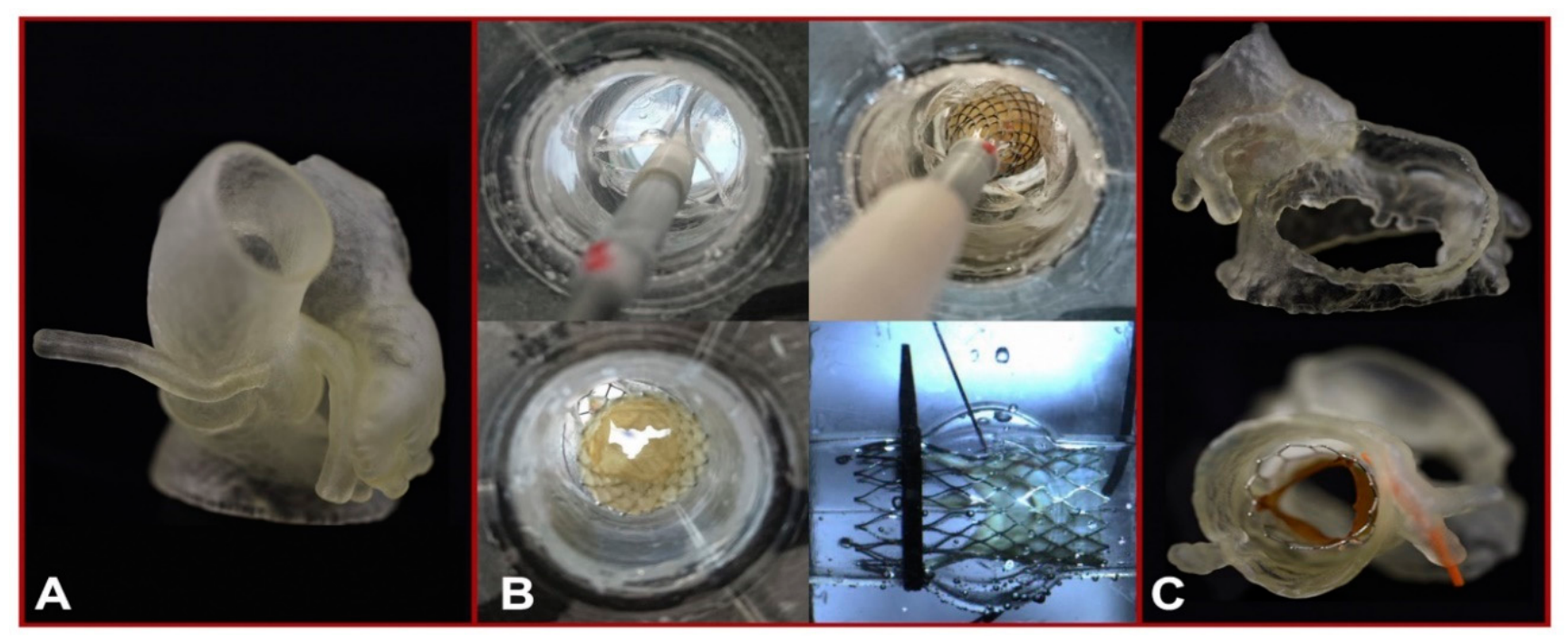
Figure 11.
The release of different valves was simulated in the 3-dimensional printing models. A-C The simulated release of a Venus A valve (Qiming Company, Hangzhou, Jiangsu Province, China). D-F The simulated release of a J-valve (Jiecheng Company, Suzhou, Jiangsu Province, China). G-I The simulated release of a Prizvalve® (Newmed Company, Shanghai, China). Reprinted with permission under the open access from Ma et al [57].
Figure 11.
The release of different valves was simulated in the 3-dimensional printing models. A-C The simulated release of a Venus A valve (Qiming Company, Hangzhou, Jiangsu Province, China). D-F The simulated release of a J-valve (Jiecheng Company, Suzhou, Jiangsu Province, China). G-I The simulated release of a Prizvalve® (Newmed Company, Shanghai, China). Reprinted with permission under the open access from Ma et al [57].
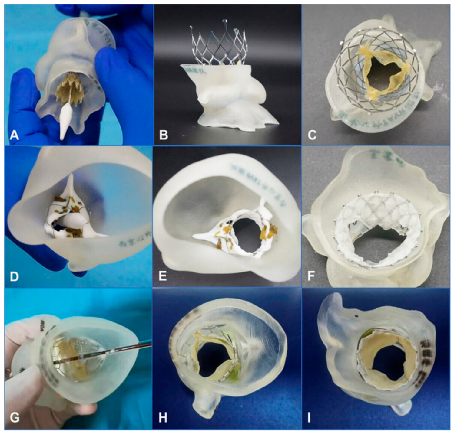
Figure 12.
Simulation training using 3D-printed aortic root models and pulsatile simulators. (a and b) The simulation using different balloon sizes to observe the changes in leaflets and calcification. (c) Preprocedural simulations using the 3D-printed aortic root model to predict paravalvular leakage. (d) Preprocedural simulations using the 3D-printed aortic root model to predict conduct blockage. (e) Trainees simulated TAVR using the pulsatile simulator. (f) Balloon expansion was simulated using a pulsatile simulator via a peripheral artery approach. 3D, 3-dimensional; TAVR, transcatheter aortic valve replacement. Reprinted with permission under the open access from Mao et al [58].
Figure 12.
Simulation training using 3D-printed aortic root models and pulsatile simulators. (a and b) The simulation using different balloon sizes to observe the changes in leaflets and calcification. (c) Preprocedural simulations using the 3D-printed aortic root model to predict paravalvular leakage. (d) Preprocedural simulations using the 3D-printed aortic root model to predict conduct blockage. (e) Trainees simulated TAVR using the pulsatile simulator. (f) Balloon expansion was simulated using a pulsatile simulator via a peripheral artery approach. 3D, 3-dimensional; TAVR, transcatheter aortic valve replacement. Reprinted with permission under the open access from Mao et al [58].
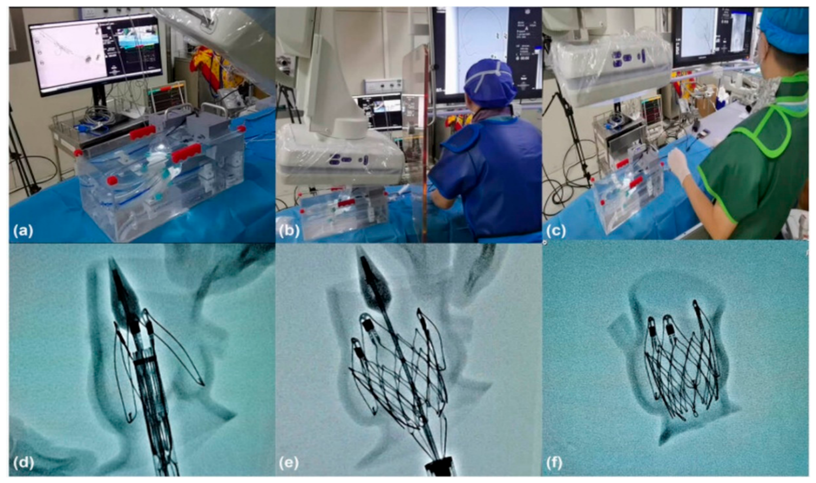
Figure 13.
Preoperative patient-specific three-dimensional printing and computational fluid dynamics simulation. (A) Patient-specific three-dimensional printed aortic root model. (B, C) The preoperative simulation of implanting the 29-mm J-valve bioprosthesis valve. (D,E) Preoperative and postoperative computational fluid dynamics conceptual models. Red area indicates aortic valve; green area indicates bioprosthetic stent. Reprinted with permission under the open access from Zhang et al [59].
Figure 13.
Preoperative patient-specific three-dimensional printing and computational fluid dynamics simulation. (A) Patient-specific three-dimensional printed aortic root model. (B, C) The preoperative simulation of implanting the 29-mm J-valve bioprosthesis valve. (D,E) Preoperative and postoperative computational fluid dynamics conceptual models. Red area indicates aortic valve; green area indicates bioprosthetic stent. Reprinted with permission under the open access from Zhang et al [59].
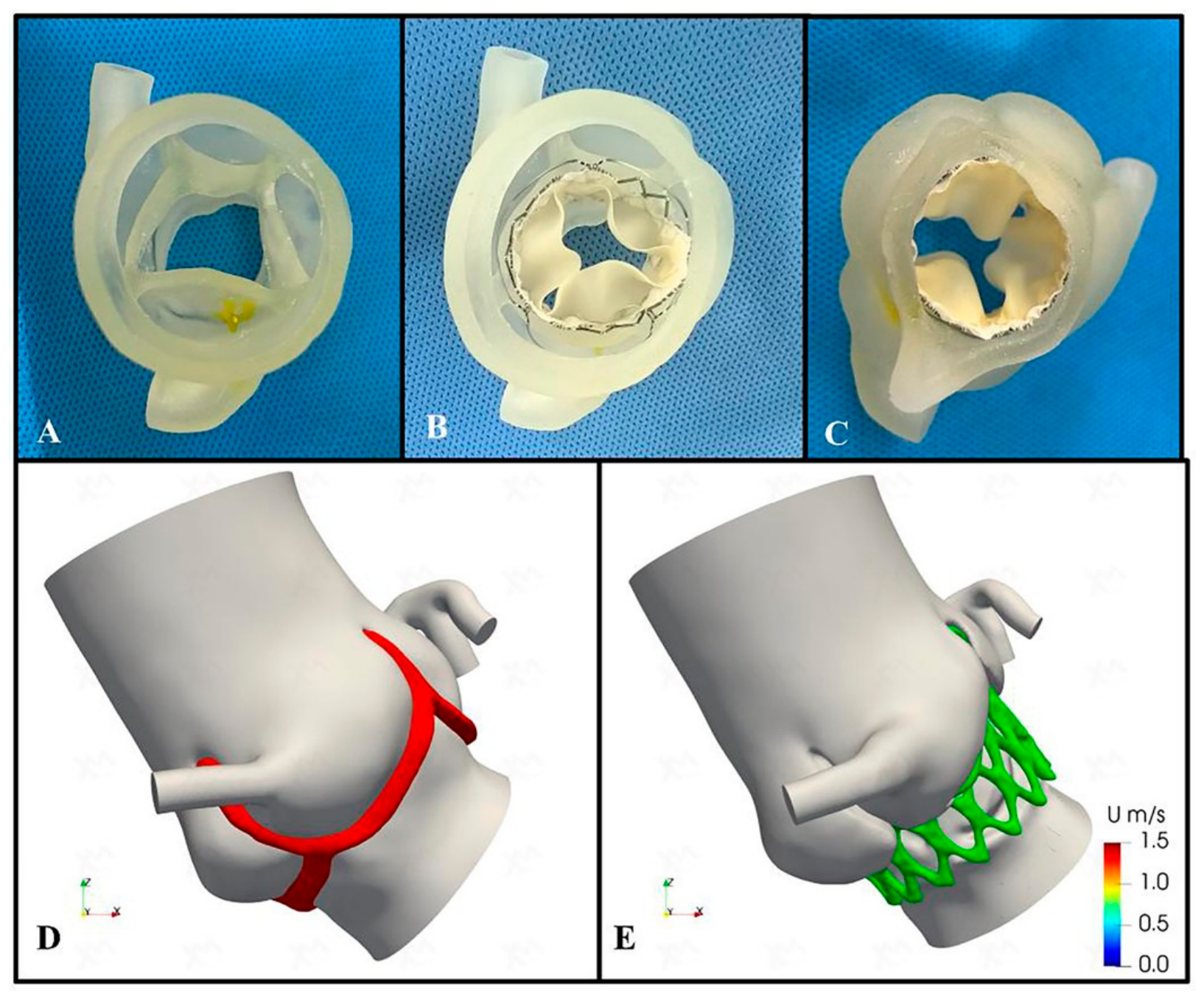
Figure 14.
The change of pressure, velocity, and wall shear stress by procedural simulation. The trans-aortic valve pressure decreases from 4.7 mmHg to 3.5 mmHg; the velocity decrease from 1.02 m/s to 0.89 m/s; the low wall shear stress area increased from 18.92 cm2 to 19.15 cm2. WSS, wall shear stress. Reprinted with permission under the open access from Zhang et al [59].
Figure 14.
The change of pressure, velocity, and wall shear stress by procedural simulation. The trans-aortic valve pressure decreases from 4.7 mmHg to 3.5 mmHg; the velocity decrease from 1.02 m/s to 0.89 m/s; the low wall shear stress area increased from 18.92 cm2 to 19.15 cm2. WSS, wall shear stress. Reprinted with permission under the open access from Zhang et al [59].
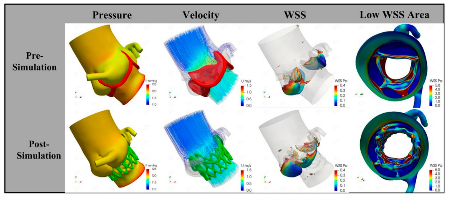
Figure 15.
3D-printed anatomical model of the left atrium and LAA, printed using elastic resin on a Formlabs Form3B printer with a 2 mm-thick wall. (b) Same model with a Legacy Watchman™ device placed in the appendage to simulate occlusion. Reprinted with permission under the open access from Kahlam et al [70].
Figure 15.
3D-printed anatomical model of the left atrium and LAA, printed using elastic resin on a Formlabs Form3B printer with a 2 mm-thick wall. (b) Same model with a Legacy Watchman™ device placed in the appendage to simulate occlusion. Reprinted with permission under the open access from Kahlam et al [70].
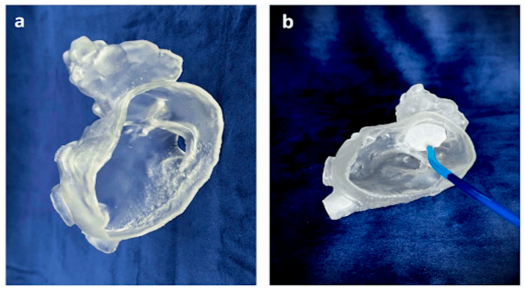
Figure 16.
Accuracy of device size prediction based on 3D printing simulation. (A) Correlation between 3D-printed simulation-based predicted size and actually implanted lobe size. (B) The Bland–Altman plot comparing the predicted size by 3D printing simulation and the actual device size. Reprinted with permission under the open access from Kim et al [73].
Figure 16.
Accuracy of device size prediction based on 3D printing simulation. (A) Correlation between 3D-printed simulation-based predicted size and actually implanted lobe size. (B) The Bland–Altman plot comparing the predicted size by 3D printing simulation and the actual device size. Reprinted with permission under the open access from Kim et al [73].
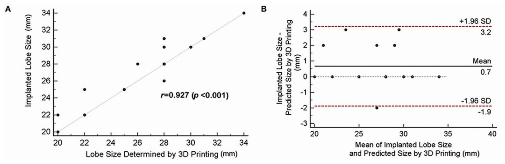
Figure 17.
Microsoft HoloLens being used in the surgical operating room. Reprinted with permission under the open access from Rad et al [90].
Figure 17.
Microsoft HoloLens being used in the surgical operating room. Reprinted with permission under the open access from Rad et al [90].
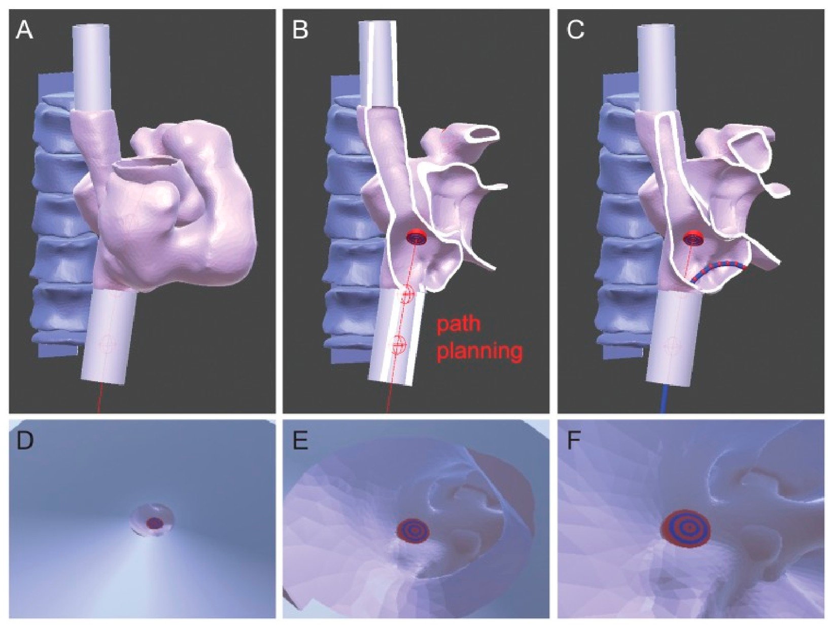
Figure 18.
3D rendering of patient heart and spine together with surgical path and tracked catheter. Reprinted with permission under the open access from Liu et al [92].
Figure 18.
3D rendering of patient heart and spine together with surgical path and tracked catheter. Reprinted with permission under the open access from Liu et al [92].
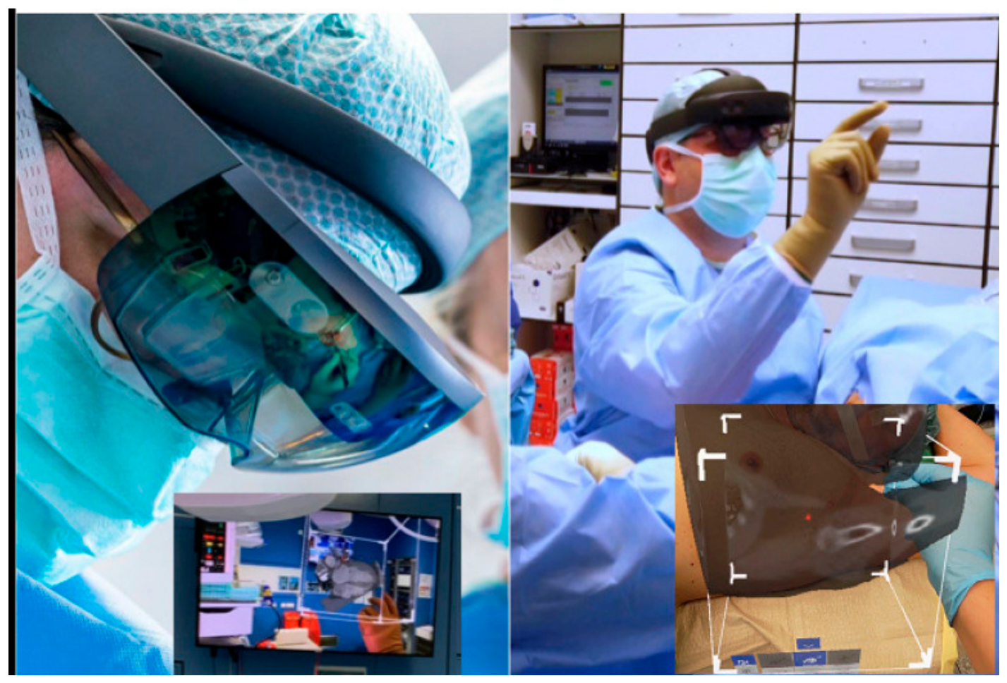
Figure 19.
HoloLens® 2-guided minimally invasive surgery for aortic valve replacement. Reprinted with permission under the open access from Aye et al [105].
Figure 19.
HoloLens® 2-guided minimally invasive surgery for aortic valve replacement. Reprinted with permission under the open access from Aye et al [105].
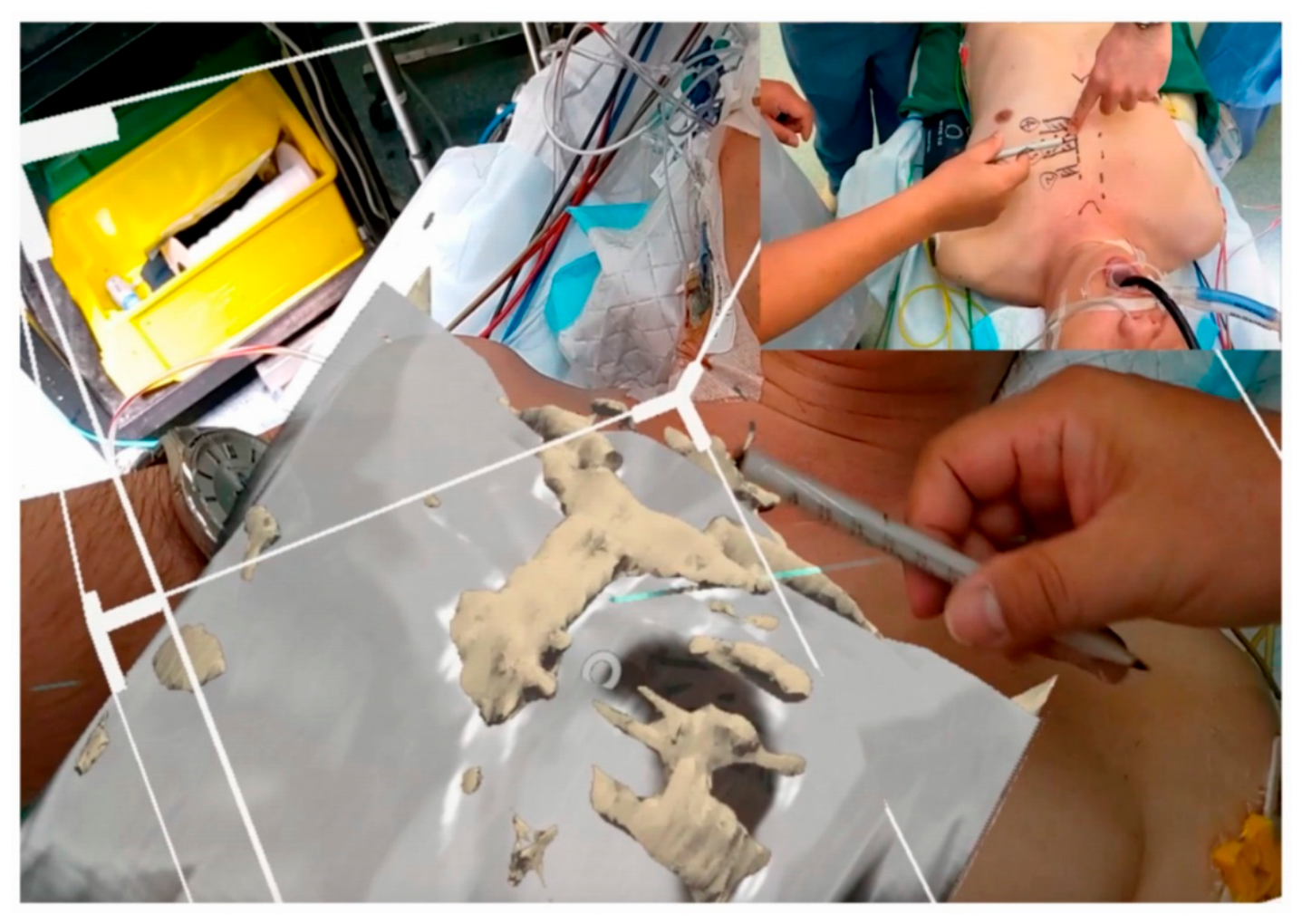
Figure 20.
HoloLens 2 guided minimally invasive surgery for pulmonary valve replacement. Reprinted with permission under the open access from Aye et al [105].
Figure 20.
HoloLens 2 guided minimally invasive surgery for pulmonary valve replacement. Reprinted with permission under the open access from Aye et al [105].
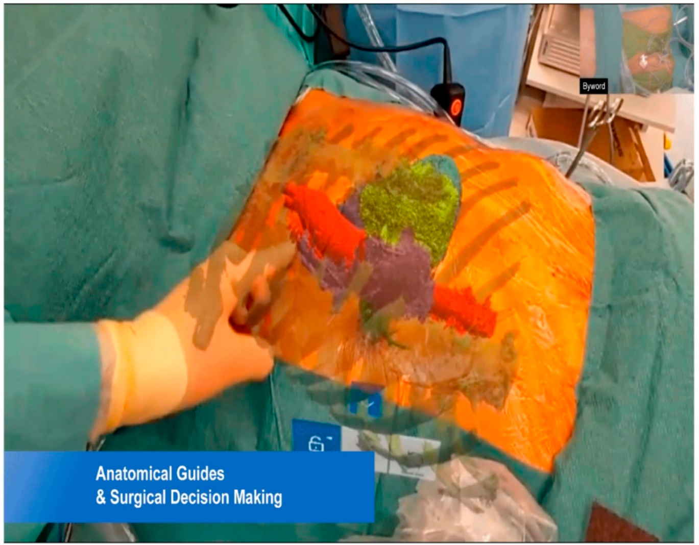
Figure 21.
A Meta-analysis of lab test of 3DPMs compared with control and B Sensitivity analysis of lab test of 3DPMs compared with control. Reprinted with permission under the open access from Xie et al [110].
Figure 21.
A Meta-analysis of lab test of 3DPMs compared with control and B Sensitivity analysis of lab test of 3DPMs compared with control. Reprinted with permission under the open access from Xie et al [110].
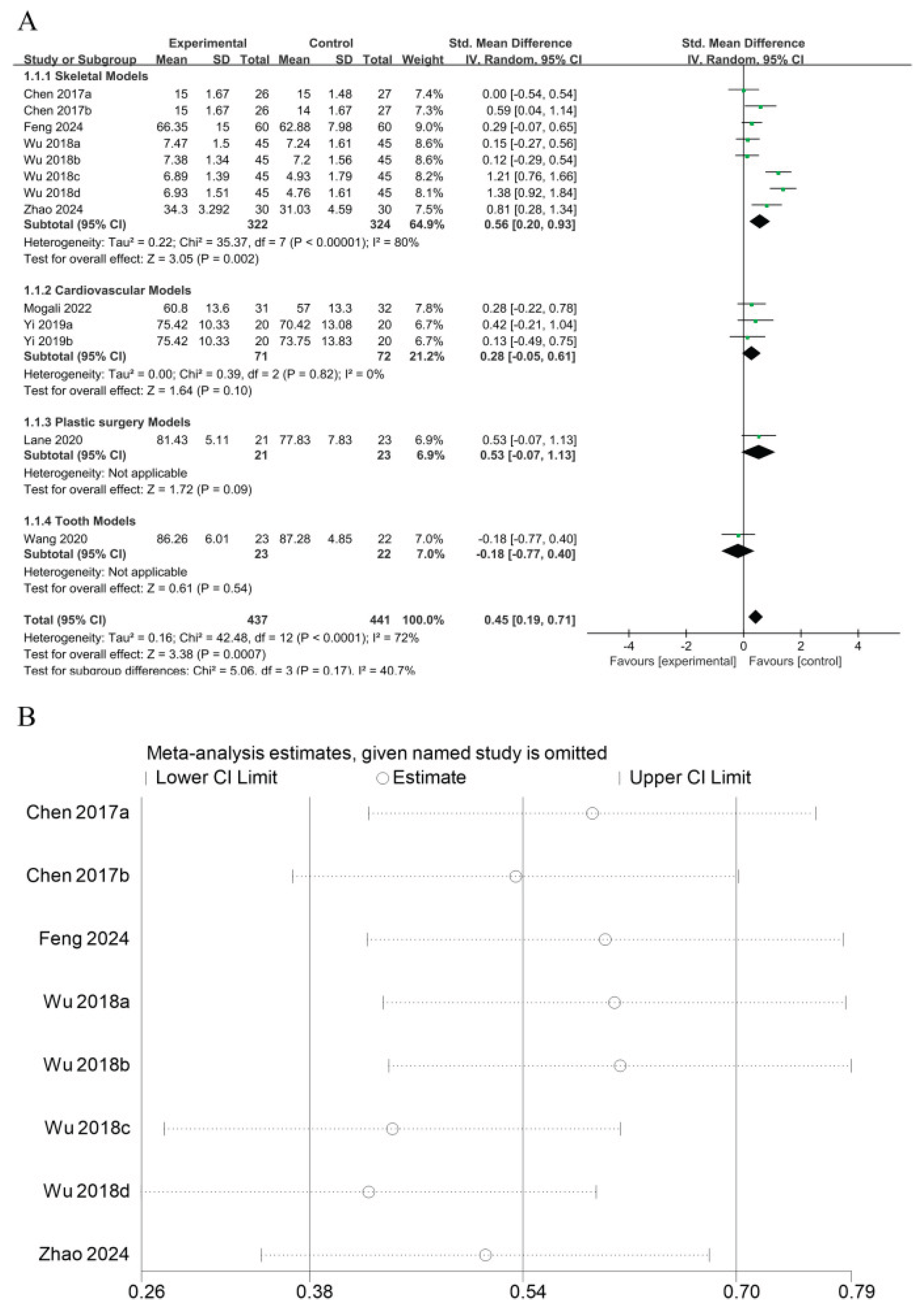
Figure 22.
Number of studies reported the use of XR in cardiovascular case. Reprinted with permission under the open access from Kanschik et al [113].
Figure 22.
Number of studies reported the use of XR in cardiovascular case. Reprinted with permission under the open access from Kanschik et al [113].
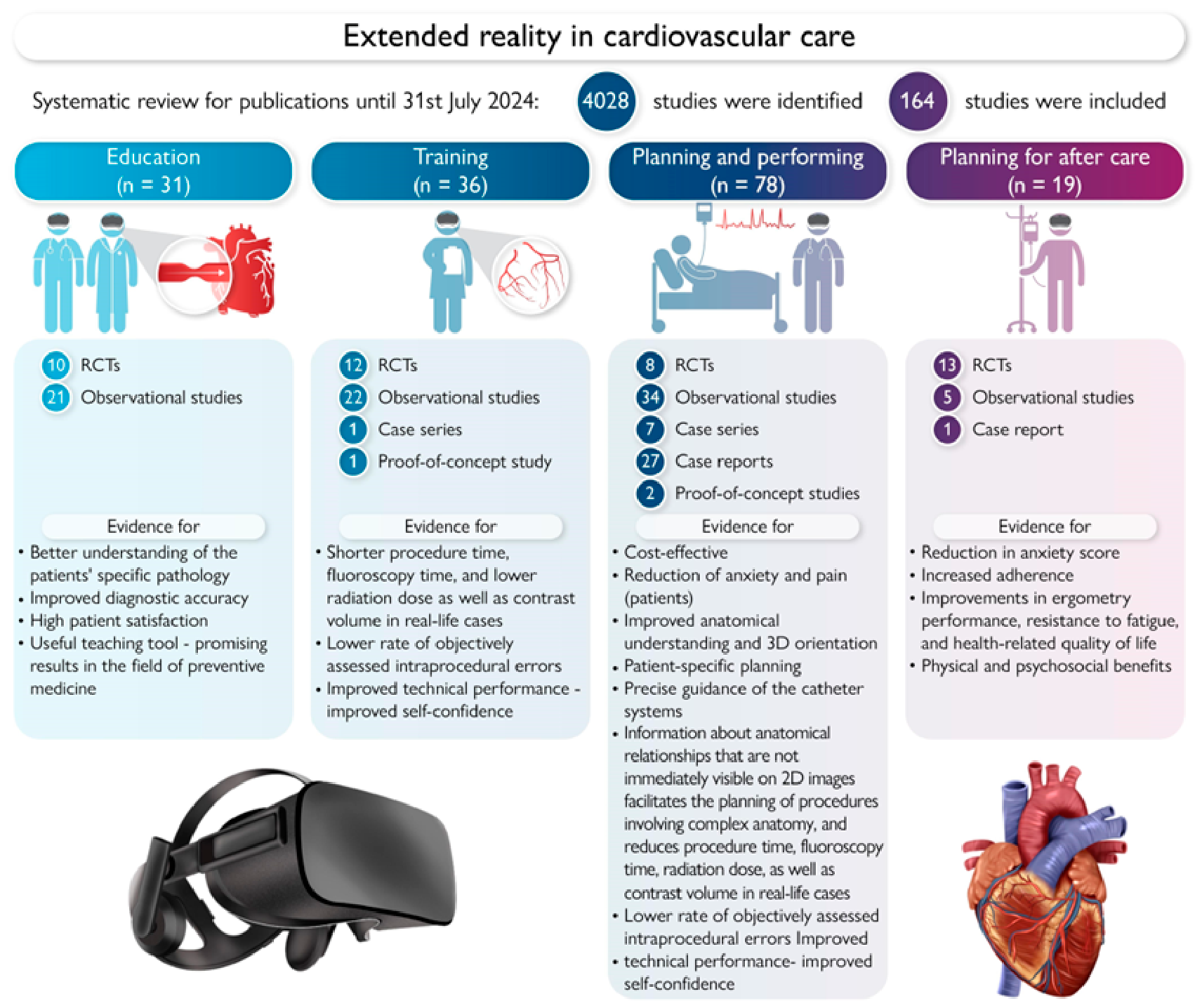
Disclaimer/Publisher’s Note: The statements, opinions and data contained in all publications are solely those of the individual author(s) and contributor(s) and not of MDPI and/or the editor(s). MDPI and/or the editor(s) disclaim responsibility for any injury to people or property resulting from any ideas, methods, instructions or products referred to in the content. |
© 2026 by the authors. Licensee MDPI, Basel, Switzerland. This article is an open access article distributed under the terms and conditions of the Creative Commons Attribution (CC BY) license (http://creativecommons.org/licenses/by/4.0/).
Copyright: This open access article is published under a Creative Commons CC BY 4.0 license, which permit the free download, distribution, and reuse, provided that the author and preprint are cited in any reuse.


