

Submitted:
15 June 2026
Posted:
16 June 2026
You are already at the latest version
Abstract
A steady increase in the elderly demographic, particularly those 85+ in age, has given rise to an elevation in global prevalence of neurodegenerative disorders, such as Alzheimer's and Parkinson's diseases, and underscores an urgent need for novel, preventive, and disease-modifying strategies. Marine ecosystems represent a prolific source of bioactive compounds, among which xanthophyll carotenoids—oxygenated derivatives including astaxanthin, fucoxanthin, and canthaxanthin—have emerged as compelling candidates for neuroprotection. These compounds exhibit superior bioavailability and blood-brain barrier permeability compared to hydrocarbon carotenoids, attributed to their polar functional groups. Their potent bioactivity stems from a multifaceted ability to modulate core pathological processes: directly quenching reactive oxygen species, activating the Nrf2-mediated antioxidant response, inhibiting NF-κB-driven neuroinflammation, and interfering with the aggregation of toxic proteins like amyloid-β and α-synuclein. This review evaluates the preclinical and clinical evidence supporting the neuroprotective potential of these three marine xanthophylls. Astaxanthin demonstrates the most robust and extensive evidence base, from cellular models to human cognitive trials. Fucoxanthin shows parallel promise with unique effects on mitochondrial biogenesis, while canthaxanthin remains significantly understudied despite its strong antioxidant profile. We further discuss the translational challenges of bioavailability and formulation, highlighting innovative delivery systems, and propose future research directions to harness these marine-derived compounds for promoting brain health and mitigating age-related cognitive decline.
Keywords:
astaxanthin
; fucoxanthin
; canthaxanthin
; neuroprotection
; oxidative stress
; neuroinflammation
; Alzheimer’s disease
; Parkinson’s disease
; Nrf2
; blood-brain barrier
1. Introduction
The global demographic shift towards an elderly, post-retirement age, population has precipitated a steep increase in the incidence of age-related neurodegenerative diseases, most notably Alzheimer’s disease (AD) and Parkinson’s disease (PD). These conditions are characterized by progressive, irreversible neuronal loss, leading to debilitating cognitive and motor deficits [1]. The underlying pathogenesis is multifactorial, involving a complex interplay of chronic oxidative stress, mitochondrial dysfunction, persistent neuroinflammation, and the aberrant aggregation of proteins such as amyloid-β (Aβ) in AD and α-synuclein in PD [2,3]. Current pharmacological interventions are predominantly symptomatic and fail to halt or reverse disease progression, highlighting a critical unmet need for therapeutic agents that target these fundamental pathological mechanisms [4].
In this context, natural products, particularly those derived from marine environments, have gained considerable attention as reservoirs of structurally unique and biologically active compounds [5]. Carotenoids, a class of C40 tetraterpenoid pigments, are among the most promising. Within this class, xanthophylls—carotenoids containing oxygen functional groups (e.g., hydroxyl, keto, epoxy)—demonstrate enhanced biological properties relevant to neuroprotection compared to their hydrocarbon counterparts (e.g., β-carotene) [6]. The polarity conferred by these groups improves their integration into cellular membranes, influences their pharmacokinetics, and, crucially, facilitates their transit across the blood-brain barrier (BBB) [7,8].
This review focuses on three marine-derived xanthophyll carotenoids with emerging neuroprotective credentials: astaxanthin, fucoxanthin, and canthaxanthin. While they share a common conjugated polyene backbone responsible for potent antioxidant activity, their distinct structural features dictate unique biological effects and mechanistic nuances. Astaxanthin, from the microalgae Haematococcus pluvialis, is renowned as one of nature’s most powerful antioxidants [9]. Fucoxanthin, the principal carotenoid in brown algae, possesses a unique allenic bond and epoxide group. Canthaxanthin, a diketo-carotenoid found in crustaceans and some microorganisms, is a potent antioxidant but remains relatively unexplored in neurology.
Here, we provide a comprehensive and comparative analysis of the neuroprotective mechanisms of these compounds. We systematically evaluate evidence from in vitro studies, animal models, and human clinical trials, focusing on their roles in modulating oxidative stress, inflammation, and proteinopathy in AD and PD. Furthermore, we address the key translational challenges of bioavailability and delivery, explore advanced formulation strategies, and delineate a roadmap for future research to validate these marine xanthophylls as viable agents for preserving brain health and combating neurodegeneration.
2. Structural Foundations and Bioavailability of Marine Xanthophylls
The neuroprotective efficacy of carotenoids is intrinsically linked to their chemical architecture. The foundational element is an extended system of conjugated carbon-carbon double bonds, which forms a resonant electron cloud capable of effectively quenching reactive oxygen species (ROS), thereby terminating destructive oxidative chain reactions [10]. Xanthophyll carotenoids are distinguished from the more hydrophobic carotenes by the presence of oxygen-containing functional groups, which dramatically alter their physicochemical and biological behavior (Figure 1).
Astaxanthin (3,3′-dihydroxy-β,β-carotene-4,4′-dione) features hydroxyl and keto groups on both ionone rings (Figure 1). This amphiphilic structure enables it to span lipid bilayers, with the polar end groups anchored at the membrane surface and the nonpolar polyene chain embedded within [7,11]. This unique orientation is ideal for intercepting free radicals within the membrane interior, providing superior protection against lipid peroxidation—a key process in neuronal damage [12].
Fucoxanthin possesses a more complex structure, containing an allenic bond (C-7′=C-8′), a 5,6-monoepoxide, and a conjugated carbonyl group (Figure 1). This configuration is highly polar and influences its metabolic fate. Dietary fucoxanthin is deacetylated in the gut to fucoxanthinol and further metabolized in the liver to amarouciaxanthin A, which are considered the primary bioactive forms in systemic circulation [13]. The allenic bond is particularly reactive and contributes to its distinct biological activities.
Canthaxanthin (β,β-carotene-4,4′-dione) is a symmetrical diketo-carotenoid, lacking hydroxyl groups (Figure 1). Its two keto groups confer significant antioxidant potential but result in different solubility and membrane interaction profiles compared to other xanthophyll carotenoids [14].
Bioavailability and Blood-Brain Barrier Penetration: The oxygenated groups of xanthophylls enhance their aqueous solubility relative to pure hydrocarbons like β-carotene (Figure 1), improving their absorption in the gastrointestinal tract, especially when co-consumed with dietary lipids [15]. Critically, this polarity also facilitates passive diffusion across the BBB. In vivo studies confirm that astaxanthin and its metabolites accumulate in brain tissue, whereas β-carotene shows minimal penetration [16,17]. While direct evidence for fucoxanthin and canthaxanthin is sparser, their structural similarities suggest a favorable profile. Their transport is further aided by association with plasma lipoproteins such as LDL and HDL, which act as carriers to the brain endothelium [18].
3. Astaxanthin: A Multifaceted Neuroprotectant
3.1. Sources and Physicochemical Properties
Astaxanthin is a red ketocarotenoid predominantly biosynthesized by the freshwater microalgae Haematococcus pluvialis. Astaxanthin is rapidly synthesized and accumulates in H. pluvialis under stress conditions. It is also found in yeast (Phaffia rhodozyma) and marine animals like krill and salmon, which obtain it through their diet. The reddish-pink color of these organisms is directly attributed to astaxanthin. Its molecular structure, featuring hydroxyl and keto groups at the ionone rings, allows it to span lipid bilayers, with polar ends anchored at the membrane surface [7,11]. This unique orientation is critical for its exceptional capacity to inhibit lipid peroxidation [12].
3.2. General Biological Activities and Mechanisms
Beyond neuroprotection, astaxanthin exhibits broad-spectrum bioactivity, including anti-inflammatory, immunomodulatory, and cardiometabolic benefits, primarily mediated through two core pathways:
- Antioxidant/Nrf2 Activation: Astaxanthin is a potent direct scavenger of reactive oxygen and nitrogen species. In addition, it activates the nuclear factor erythroid 2–related factor 2 (Nrf2) pathway, leading to the transcriptional upregulation of a suite of cytoprotective enzymes, including heme oxygenase-1 (HO-1), NAD(P)H quinone dehydrogenase 1, and glutathione S-transferase (GST) [19].
- Anti-inflammatory/NF-κB Inhibition: Astaxanthin suppresses the nuclear factor kappa-light-chain-enhancer of activated B cells (NF-κB) pathway by inhibiting IκB kinase activity and subsequent IκBα degradation [20]. This prevents nuclear translocation of NF-κB, thereby downregulating the expression of pro-inflammatory cytokines such as tumor necrosis factor-alpha (TNF-α), interleukin-1β (IL-1β), and IL-6 [20].
3.3. Neuroprotective Evidence in Alzheimer’s Disease Pathogenesis
In vitro studies demonstrate that astaxanthin protects neuronal cultures, such SH-SY5Y cells and PC12 cells, against Aβ-induced cytotoxicity by reducing agents of oxidative stress, for example ROS and malondialdehyde, attenuating caspase-3 activation, and preserving mitochondrial membrane potential [21]. It also directly inhibits Aβ1-42 aggregation and enhances microglial-mediated Aβ clearance [22].
In vivo neuroprotective evidence is robust. In transgenic AD mouse models, in particular mice engineered to over-express mutant forms of amyloid precursor protein (APP) and Presenilin-1 (PS1), sustained astaxanthin supplementation improves spatial learning and memory in the Morris water maze, correlating with reduced hippocampal Aβ plaque load, decreased phosphorylated tau levels, and diminished glial activation [23,24]. Mechanistically, these benefits are linked to the upregulation of the PI3K/Akt/GSK-3β signaling axis and enhanced expression of brain-derived neurotrophic factor [25].
Clinical Translation: Several randomized controlled trials in older adults with mild cognitive impairment or age-related forgetfulness report that astaxanthin supplementation (8-12 mg/day) improves cognitive processing speed, working memory, and reduces mental fatigue [26,27,28]. A pilot clinical trial in AD patients is underway (NCT05015374), marking a critical step toward targeted clinical evaluation.
3.4. Neuroprotective Evidence in Parkinson’s Disease Pathogenesis
In cellular models of PD, in particular neurons treated with cellular toxins, astaxanthin protects dopaminergic neurons by inhibiting the activation of pro-apoptotic c-Jun N-terminal kinase and p38 mitogen-activated protein kinase (MAPK) pathways, while promoting pro-survival PI3K/Akt signaling [29]. It also mitigates mitochondrial dysfunction.In vivo, in 1-methyl-4-phenyl-1,2,3,6-tetrahydropyridine (MPTP)-induced mouse models of PD, astaxanthin pretreatment preserves tyrosine hydroxylase-positive neurons in the substantia nigra pars compacta, maintains striatal dopamine levels, and ameliorates motor deficits [30]. These effects are associated with suppressed microglial activation and reduced expression of inflammatory mediators in the nigrostriatal pathway.
3.5. Bioavailability Enhancement Strategies
Acknowledging its lipophilic nature, research has focused on improving astaxanthin’s delivery. Innovative approaches include encapsulation in nanostructured lipid carriers for intranasal administration, which provides sustained release and direct nose-to-brain delivery, bypassing first-pass metabolism [31]. Liposomal and phospholipid complex formulations have also been developed to enhance oral bioavailability and tissue distribution of astaxanthin with demonstrable neuroprotective effects [32].
4. Fucoxanthin: A Unique Carotenoid with Metabolic and Neural Benefits
4.1. Sources, Metabolism, and General Properties
Fucoxanthin is the predominant carotenoid in brown seaweeds (Phaeophyceae), such as wakame (Undaria pinnatifida) and kombu (Laminaria japonica). As noted, it is metabolized to fucoxanthinol and amarouciaxanthin A, which are the primary circulating metabolites responsible for its systemic effects [13]. Beyond neuroprotection, it is widely studied for its anti-obesity effects, mediated through the uncoupling protein 1 activation in white adipose tissue, and for its anti-diabetic and anti-cancer properties [33,34].
4.2. Neuroprotective Mechanisms and Evidence
Fucoxanthin and its metabolites share several neuroprotective pathways with astaxanthin but also exhibit distinct characteristics.
- Antioxidant and Anti-inflammatory Actions: Fucoxanthinol potently activates the Nrf2/HO-1 pathway and inhibits NF-κB signaling in activated microglial cells, reducing the production of nitric oxide, PGE2, TNF-α, and IL-6 [35].
- Modulation of AD Pathology: In neuronal models, fucoxanthin attenuates Aβ-induced toxicity and inhibits Aβ1-42 fibrillation [36]. It also modulates the PI3K/Akt/GSK-3β pathway, leading to reduced tau phosphorylation [37]. In vivo, fucoxanthin supplementation in APP/PS1 mice improves cognitive performance, reduces Aβ burden, and alleviates oxidative stress and inflammation in the hippocampus [38].
- Mitochondrial Biogenesis: A key feature and distinctive mechanism of action for fucoxanthin is its potent activation of peroxisome proliferator-activated receptor gamma coactivator 1-alpha (PGC-1α), a master regulator of mitochondrial biogenesis and function [39]. This is particularly relevant for PD and other disorders with strong mitochondrial etiology.
- Effects in PD Models: In MPTP-induced murine PD models, fucoxanthin treatment protects dopaminergic neurons, improves motor coordination, and reduces striatal dopamine depletion [40]. These effects correlate with increased mitochondrial biogenesis, reduced oxidative stress, and inhibition of neuroinflammatory responses [40]. Interestingly, fucoxanthin has also been identified as a dopamine D2/D3 receptor agonist, suggesting a potential dual mechanism in PD therapy [41].
4.3. Bioavailability Challenges and Nanotechnological Solutions
The bioavailability of oral fucoxanthin is low, relative to astaxanthin, due to poor stability in the GI tract and extensive metabolism [42]. To overcome this, advanced delivery systems are under development. For instance, fucoxanthin-loaded poly(lactic-co-glycolic acid) (PLGA) nanoparticles have demonstrated enhanced cellular uptake, superior antioxidant activity in vitro, and improved cognitive outcomes in AD mouse models following intravenous administration [43]. These strategies will be crucial for future translational development of fucoxanthin.
5. Canthaxanthin: An Underexplored Antioxidant with Neuroprotective Potential
5.1. Sources, Synthesis, and General Bioactivity
Canthaxanthin is found in nature in crustaceans, certain fish, and some algae and bacteria. Commercially, it is often produced synthetically or via microbial fermentation using organisms like Blakeslea trispora or engineered Escherichia coli [44]. It is a potent antioxidant, with studies showing its capacity to inhibit lipid peroxidation as effectively as α-tocopherol [14]. It also exhibits immunomodulatory and anti-carcinogenic properties [45].
5.2. Neuroprotective Evidence and Research Gaps
The neuroprotective research on canthaxanthin is limited but promising.
- In vitro Evidence: In neuronal PC12 cells, canthaxanthin pretreatment significantly attenuates hydrogen peroxide-induced cytotoxicity, apoptosis, and loss of mitochondrial membrane potential [46]. In cell culture experiments, canthaxanthin demonstrates a protective effect against Aβ(25-35)-induced toxicity, improving cell viability and reducing intracellular ROS and calcium influx [47]. The presence of its keto groups is thought to be crucial for this activity.
- Mechanistic Insights and Limitations: The protective effects appear to be mediated primarily through direct antioxidant scavenging and membrane stabilization [48]. However, investigations into its influence on key neuroprotective signaling pathways (Nrf2, NF-κB, PI3K/Akt) are conspicuously absent. There is a profound lack of in vivo studies of canthaxanthin in AD or PD animal models, and no clinical trials have investigated its effects on cognition or neurodegeneration.
- Safety Note: Historical concerns regarding canthaxanthin relate to the association of high-dose synthetic supplementation with crystalline retinopathy [49]. However, these effects are dose-dependent and not associated with dietary or potential therapeutic neuroprotective doses, which would likely be significantly lower [49].
5.3. A Case for Targeted Investigation
Given its structural similarity to astaxanthin (both are ketocarotenoids) and its demonstrated in vitro efficacy, canthaxanthin represents a significant knowledge gap. Future research is needed to evaluate its bioavailability, BBB penetration, efficacy in relevant animal models, and its full mechanistic profile.
6. Comparative Analysis and Mechanistic Insights
A side-by-side comparison reveals the relative strengths and translational readiness of each compound (See Table 1).
7. Translational Challenges and Future Perspectives
Despite the promising preclinical data, several hurdles must be overcome to translate these marine xanthophylls into clinically effective neurotherapeutics.
7.1. Overcoming Bioavailability and Delivery Barriers
The lipophilic nature of these compounds limits their aqueous solubility, gastrointestinal absorption, and brain delivery. Future success hinges on advanced formulation science:
- Nano-Delivery Systems: Lipid-based nanoparticles (Nanostructured lipid carriers, liposomes), polymeric nanoparticles, and nanoemulsions can encapsulate carotenoids, protecting them from degradation, enhancing absorption, and enabling targeted delivery. Functionalizing nanoparticles with ligands, for example transferrin, could further facilitate receptor-mediated transcytosis across the BBB [50].
- Alternative Administration Routes: Intranasal delivery offers a direct nose-to-brain pathway, bypassing the BBB and first-pass metabolism, and is a promising route for compounds like astaxanthin [31].
- Prodrug Strategies: Synthesizing water-soluble prodrugs (e.g., phosphate esters) that are cleaved by brain enzymes to release the active carotenoid could improve pharmacokinetics.
7.2. Addressing Critical Knowledge Gaps
- For Canthaxanthin: A dedicated research program is imperative. Priorities include: (i) Establishing its pharmacokinetics and brain uptake in animal models; (ii) Evaluating its efficacy in transgenic AD and toxin-induced PD models; (iii) Elucidating its effects on Nrf2, NF-κB, and other key signaling pathways.
- For All Compounds: More detailed mechanistic studies are needed to identify precise molecular targets and interactions. Long-term toxicology and safety studies in relevant animal models are required, particularly for canthaxanthin, to define therapeutic windows.
- Biomarker Development: Identifying robust biomarkers (e.g., via neuroimaging, cerebral spinal fluid (CSF) analysis, or peripheral oxidative stress markers) will be crucial for demonstrating target engagement and efficacy in clinical trials.
7.3. Designing Definitive Clinical Trials
Future clinical trials must be well-designed to provide conclusive evidence:
- Population: Focus on prodromal or early-stage AD/PD patients, where neuroprotection may have the greatest impact.
- Intervention: Use advanced bioavailable formulations- lipid-based, nanoparticles, or other. Consider combination therapies (e.g., carotenoid + omega-3 fatty acids) to assess synergistic potential.
- Outcomes: Include both clinical cognitive/motor scales and validated biomarker endpoints, for example amyloid PET, MRI volumetry, cerebrospinal fluid, p-tau, inflammatory cytokines.
- Duration: Trials must be of sufficient duration, typically18-24 months, to detect disease-modifying effects.
7.4. Exploring Synergy and Novel Applications
The multi-target nature of these carotenoids makes them ideal candidates for combination therapy with other disease-modifying agents (e.g., anti-amyloid antibodies, GSK-3β inhibitors) to achieve broader neuroprotection. Furthermore, their strong antioxidant/anti-inflammatory profiles warrant investigation in other neurological conditions, such as traumatic brain injury, stroke, and multiple sclerosis.
8. Conclusions
Marine-derived xanthophyll carotenoids—astaxanthin, fucoxanthin, and canthaxanthin—represent a promising class of natural, multi-target agents with significant potential to counteract the complex pathophysiology of neurodegenerative diseases. Indeed, in a recent study by the NIH mice fed astaxanthin were observed to have a substantial 12% increase in average lifespan compared to control mice [51]. The shared ability of these compounds to mitigate oxidative stress and neuroinflammation, coupled with their capacity to interfere with pathogenic protein aggregation, aligns perfectly with the multifactorial nature of AD and PD.
- Astaxanthin stands as the current vanguard, supported by a robust evidence chain from molecule to man, and is poised for pivotal clinical trials in neurodegenerative populations.
- Fucoxanthin holds comparable promise, with the intriguing added mechanism of promoting mitochondrial health and biogenesis, but its clinical translation is contingent upon solving significant delivery challenges.
- Canthaxanthin remains an untapped resource; its strong foundational bioactivity underscores a clear mandate for focused research to determine its true neuroprotective value.
The journey from marine organism to human medicine requires a concerted interdisciplinary effort, bridging marine biology, pharmaceutical chemistry, neuroscience, and clinical neurology. By addressing the key challenges of bioavailability, conducting rigorous mechanistic and clinical research, and exploring innovative therapeutic paradigms, these vibrant compounds from the sea may illuminate new paths to preserving brain health and combating cognitive decline.
Author Contributions
All authors contributed to the conceptualization of this article.
Funding
This research received no external funding.
Data Availability
No new datasets were generated or analyzed during the preparation of this manuscript. Data sharing is not applicable.
Conflicts: of Interest
The authors declare no conflicts of interest.
Abbreviations
| AD | Alzheimer’s disease |
| PD | Parkinson’s disease |
| Aβ | Amyloid-β |
| BBB | Blood brain barrier |
| ROS | Reactive oxygen species |
| LDL | Low density lipoprotein |
| HDL | High density lipoprotein |
| Nrf2 | Nuclear factor erythroid 2–related factor 2 |
| HO-1 | Heme oxygenase-1 |
| GST | Glutathione S-transferase |
| NF-κB | Nuclear factor kappa-light-chain-enhancer of activated B cells |
| TNF-α | Tumor necrosis factor-alpha |
| IL-1β | Interleukin-1β |
| APP | Amyloid precursor protein |
| PS1 | Presenilin-1 |
| MAPK | Mitogen-activated protein kinase |
| MPTP | 1-methyl-4-phenyl-1,2,3,6-tetrahydropyridine |
| PGE2 | Prostaglandin E2 |
| PGC-1α | Peroxisome proliferator-activated receptor gamma coactivator 1-alpha |
| PLGA | Poly(lactic-co-glycolic acid) |
| GSK-3β | Glycogen synthase kinase-3 beta |
References
- GBD 2016 Neurology Collaborators. Global, regional, and national burden of neurological disorders, 1990-2016. Lancet Neurol. 2019;18(5):459-480.
- Wang, X. et al. Oxidative stress and mitochondrial dysfunction in Alzheimer’s disease. Biochim. Biophys. Acta. 2014. 1842(8):1240-1247.
- Poewe, W. et al. Parkinson disease. Nat Rev Dis Primers. 2017. 3:17013.
- Cummings, J. et al. Alzheimer’s disease drug development pipeline: Alzheimers Dement. 2023. 9(2):e12385.
- Mayer, A.M.S. et al. Marine pharmacology in 2014-2015. 2017. Mar Drugs. 15(9):273.
- Milani, A. et al. Carotenoids: biochemistry, pharmacology and treatment. Br J Pharmacol. 2017. 174(11):1290-1324.
- Gammone, M.A. et al. Marine carotenoids against oxidative stress. Mar Drugs. 2015. 13(10):6226-6246.
- Manabe, Y. et al. Dietary astaxanthin accumulates in the brain and protects against oxidative stress in rats. Biosci Biotechnol Biochem. 2018. 82(8):1333-1338.
- Medoro A. et al. Dietary Astaxanthin: A Promising Antioxidant and Anti-Inflammatory Agent for Brain Aging and Adult Neurogenesis. Mar Drugs. 2023. Vol 21.
- Britton, G. Structure and properties of carotenoids. Prog Lipid Res. 1995. 34(2):109-134.
- McNulty HP. et al. Differential effects of carotenoids on lipid peroxidation due to membrane interactions: X-ray diffraction analysis. Biochim Biophys Acta. 2007. 1768(1):167-74.
- Hussein, G. et al. Astaxanthin, a carotenoid with potential. J Nat Prod. 2006. 69(3):443-449.
- Asai, A. et al. Biotransformation of fucoxanthin. 2004. J Nutr. 134(1):1-5.
- Palozza, P. and Krinsky, N.I. Antioxidant effects of carotenoids. 1992. Methods Enzymol. 213:403-420.
- Yonekura, L. and Nagao, A. Intestinal absorption of dietary carotenoids. 2007. Mol Nutr Food Res. 51(1):107-115.
- Suganuma, K. et al. Astaxanthin uptake in domestic dogs and cats. 2020. Nutrients. 12(2):556.
- Craft, N.E. et al. Carotenoid, tocopherol, and retinol concentrations in elderly human brain. 2004. J Nutr Health Aging. 8(3):156-162.
- During, A. et al. Carotenoid transport is decreased and expression of the lipid transporters SR-BI, NPC1L1, and ABCA1 is downregulated in Caco-2 cells treated with ezetimibe. 2005. J Nutr. 135(10):2305-2312.
- Li, Z.; et al. Protective roles of astaxanthin against oxidative stress via Nrf2 signaling pathway. 2019. Oxid Med Cell Longev. 2019:9527295.
- Lee, S.J. et al. Astaxanthin inhibits NF-κB. Biochem Pharmacol. 2003. 65(8):1381-1390.
- Chang, C.H. et al. Astaxanthin protects neuronal cells. 2010. J Agric Food Chem. 58(22):11601-11607.
- Grimmig, B. et al. Astaxanthin reduces Aβ. J Neurosci. 2017. 37(30):7288-7301.
- Wang, L. et al. Astaxanthin improves cognitive function and reduces amyloid burden in APP/PS1 mice. J Nutr Biochem. 2020. 85:108491.
- Zhang, X. et al. Astaxanthin ameliorates oxidative stress and inflammation in the hippocampus of APP/PS1 mice. Neurochem Int. 2021. 147:105028.
- Zhang, X.S. et al. Astaxanthin alleviates oxidative stress and apoptosis via the PI3K/Akt pathway. Mol Neurobiol. 2014. 49(2):685-700.
- Ito, N. et al. Effects of astaxanthin supplementation on cognitive function in elderly subjects. J Clin Biochem Nutr. 2022. 71(2):123-130.
- Seki, S. et al. Effects of astaxanthin on cognitive performance and oxidative stress in middle-aged and older adults. Nutrients. 2021. 13(9):3045.
- Katagiri M, et al. Effects of astaxanthin-rich Haematococcus pluvialis extract on cognitive function: a randomised, double-blind, placebo-controlled study. J Clin Biochem and Nutrition. 2012. 51(2):102–7.
- Liu, X. and Osawa, T. Astaxanthin protects neurons. Brain Res. 2009. 1251:18-26.
- Wu, Q. et al. Astaxanthin and PD model. Mol Neurobiol. 2015. 51(2):555-567.
- Pardeshi, C.V. et al. Recent advances in nose-to-brain drug delivery. J Pharmacol Exp Ther. 2022. 381(1):86-99.
- Gu Z. et al. PEGylated-liposomal astaxanthin ameliorates Aβ neurotoxicity and Alzheimer-related phenotypes by scavenging formaldehyde. J Control Release. 2024. 366:783–97.
- Maeda, H. et al. Fucoxanthin and metabolic syndrome. Biochem Biophys Res Commun. 2005. 332(2):392-397.
- Peng, J. et al. Fucoxanthin, a marine carotenoid. Mar Drugs. 2011. 9(10):1806-1828.
- Heo, S.J. and Jeon, Y.J. Antioxidant effect of fucoxanthin. 2009. Eur Food Res Technol. 228(2):207-213.
- Xiang, J. et al. Fucoxanthin inhibits Aβ42 fibrillation and protects SH-SY5Y cells from Aβ-induced toxicity. Mar Drugs. 2017. 15(7):43.
- Wang, J. et al. Fucoxanthin improves cognitive deficits and attenuates tau pathology in APP/PS1 transgenic mice. J Agric Food Chem. 2020. 68(47):13058-13067.
- Chen, S. et al. Fucoxanthin ameliorates Alzheimer’s disease-like pathology in APP/PS1 mice. Mol Nutr Food Res. 2021. 65(13):e2000866.
- Wang, J. et al. Fucoxanthin regulates mitochondrial function via the PGC-1α pathway in cellular models. Mar Drugs. 2022. 20(8):500.
- Zhang, L. and Wang, H. Fucoxanthin in PD model. Neurosci Lett. 2018. 682:123-130.
- Nakamura, S. et al. Fucoxanthin stimulates dopamine release. Biosci Biotechnol Biochem. 2018. 82(6):958-964.
- Din N. et al. Brown Algae as Functional Food Source of Fucoxanthin: A Review. Foods. 2022. 11(15):2235.
- Sun, Y. et al. PLGA nanoparticle-based delivery of fucoxanthin for improved bioavailability and neuroprotective effects. Int J Pharm. 2022. 615:121492.
- Mata-Gómez, L.C. et al. Biotechnological production of carotenoids by yeasts. Appl Microbiol Biotechnol. 2014. 98(26):1351-1370.
- Clark, R.M. et al. Canthaxanthin supplementation and immune response. J Nutr. 2000. 130(10):2514-2517.
- Liu, C. et al. Canthaxanthin protects against oxidative stress in neuronal cells. J Nutr Biochem. 2006. 17(12):815-822.
- Ono, K. and Yamada, M. Antioxidant compounds inhibit amyloid β-protein aggregation. Neurobiol Aging. 2006. 27(8):1125-1134.
- Rebelo B. et al. Canthaxanthin, a Red-Hot Carotenoid: Applications, Synthesis, and Biosynthetic Evolution. Plants (Basel). 2020. 15;9(8):1039.
- Haught, J.M. et al. Canthaxanthin retinopathy. Arch Ophthalmol. 2007. 125(5):706-707.
- Saraiva C. et al. Nanoparticle-mediated brain drug delivery: Overcoming blood-brain barrier to treat neurodegenerative diseases. J Control Release. 2016. 235:34-47.
- Harrison DE et al. Astaxanthin and meclizine extend lifespan in UM-HET3 male mice; fisetin, SG1002 (hydrogen sulfide donor), dimethyl fumarate, mycophenolic acid, and 4-phenylbutyrate do not significantly affect lifespan in either sex at the doses and schedules used. Geroscience. 2023. 46(1):795-816.
Figure 1.
Chemical structures and key bioactive features of marine xanthophylls. Structures of Astaxanthin, Fucoxanthin, and Canthaxanthin, highlighting their distinctive functional groups relative to the more hydrophobic beta-carotene.
Figure 1.
Chemical structures and key bioactive features of marine xanthophylls. Structures of Astaxanthin, Fucoxanthin, and Canthaxanthin, highlighting their distinctive functional groups relative to the more hydrophobic beta-carotene.
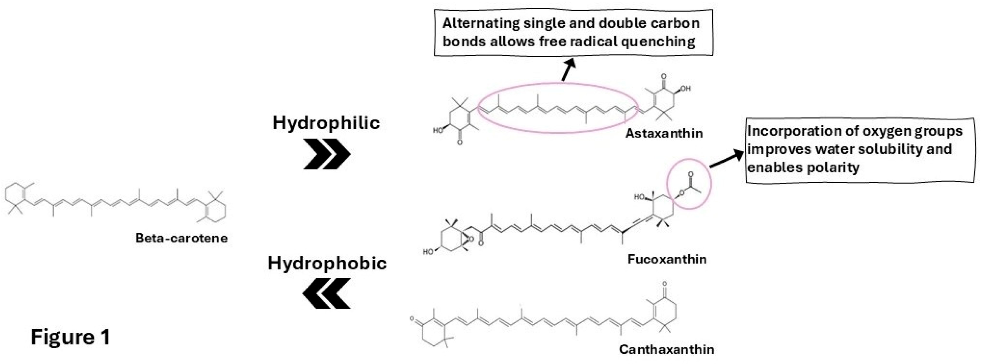

Table 1.
Comparative Analysis of Marine Xanthophylls Carotenoids as Neuroprotective Agents.
| Feature | Astaxanthin | Fucoxanthin | Canthaxanthin |
| Primary Source | Microalgae (H. pluvialis) | Brown Seaweeds (Undaria, Laminaria) | Crustaceans, Algae, Bacteria |
| Key Structural Moieties | 3,3′-dihydroxy, 4,4′-diketo | Allenic bond, 5,6-epoxide, conjugated carbonyl | 4,4′-diketo (symmetrical) |
| Primary Bioactive Form | Parent compound | Metabolites (Fucoxanthinol, Amarouciaxanthin A) | Parent compound |
| BBB Permeability | High (Confirmed in vivo) | Moderate/Probable (Metabolites) | Unknown/Predicted Moderate |
| Core Neuroprotective Mechanisms | 1. Potent Nrf2 activator 2. Strong NF-κB inhibitor 3. PI3K/Akt/GSK-3β modulator 4. Direct anti-aggregation (Aβ) | 1. Nrf2 activator 2. NF-κB inhibitor 3. PI3K/Akt/GSK-3β modulator 4. PGC-1α activator (Mitochondrial biogenesis) 5. D2/D3 receptor agonist | 1. Potent direct antioxidant 2. Anti-aggregation (Aβ in vitro) (Other pathways unexplored) |
| Evidence in AD Models | Extensive (in vitro, multiple transgenic mouse models) | Strong (in vitro, APP/PS1 mouse models) | Very Limited (in vitro only) |
| Evidence in PD Models | Extensive (in vitro, MPTP/6-OHDA models) | Strong (in vitro, MPTP models) | None |
| Human Clinical Evidence (for cognition) | Promising (Multiple RCTs in aging/MCI) | None (Trials for metabolic health only) | None |
| Major Translational Hurdle | Formulation for optimal bioavailability | Low oral bioavailability; requires advanced delivery | Lack of basic in vivo and mechanistic research |
| Translational Readiness | High (Ready for Phase II/III neuro trials) | Medium (Requires formulation optimization and pilot neuro trials) | Low (Requires foundational in vivo research) |
The key highlights from these studies are: 1) Astaxanthin is the most comprehensively studied and clinically advanced, acting as a broad-spectrum neuroprotectant. 2) Fucoxanthin offers a comparable profile with the added, potentially unique, benefit of enhancing mitochondrial biogenesis—a target of great interest in neurodegeneration. 3) Canthaxanthin, while bioactive, remains a prototype requiring extensive validation.
Disclaimer/Publisher’s Note: The statements, opinions and data contained in all publications are solely those of the individual author(s) and contributor(s) and not of MDPI and/or the editor(s). MDPI and/or the editor(s) disclaim responsibility for any injury to people or property resulting from any ideas, methods, instructions or products referred to in the content. |
© 2026 by the authors. Licensee MDPI, Basel, Switzerland. This article is an open access article distributed under the terms and conditions of the Creative Commons Attribution (CC BY) license (http://creativecommons.org/licenses/by/4.0/).
Copyright: This open access article is published under a Creative Commons CC BY 4.0 license, which permit the free download, distribution, and reuse, provided that the author and preprint are cited in any reuse.


