Submitted:
13 June 2026
Posted:
15 June 2026
You are already at the latest version
Abstract
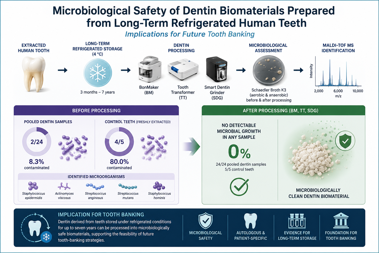
Dentin derived from extracted human teeth has emerged as a promising autogenous biomaterial for bone regeneration. The growing interest in tooth-derived graft materials has also stimulated the development of tooth-banking concepts, in which extracted teeth are preserved for future regenerative applications. However, microbial contamination remains a potential concern, particularly when teeth are stored for prolonged periods before processing and clinical use. This in vitro study evaluated the microbiological status of dentin biomaterials prepared from human teeth stored under long-term refrigerated conditions and subsequently processed using three commercially available dentin-processing systems: BonMaker (BM), Tooth Transformer (TT), and Smart Dentin Grinder (SDG). A total of 72 extracted teeth collected between 2018 and 2025 were allocated to experimental groups and processed into 24 pooled dentin samples. Additionally, five freshly extracted teeth served as controls. Microbiological assessment was performed before and after processing using Schaedler Broth K3 culture media under aerobic and anaerobic conditions. Microorganisms isolated from positive cultures were identified using MALDI-TOF mass spectrometry. Baseline microbiological assessment detected bacterial contamination in 2 of 24 pooled dentin samples (8.3%) and in 4 of 5 control teeth (80.0%). The identified microorganisms included Staphylococcus epidermidis, Actinomyces viscosus, Streptococcus anginosus, Streptococcus mutans, and Staphylococcus hominis. No detectable microbial growth was observed in any sample following processing with BM, TT, or SDG, irrespective of storage duration, including teeth stored under refrigerated conditions for up to seven years. These findings suggest that the investigated dentin-processing protocols effectively eliminated microbiologically detectable contamination in dentin derived from long-term refrigerated human teeth. The results support the microbiological safety of dentin-derived biomaterials and provide preliminary microbiological evidence supporting the feasibility of future tooth-banking strategies. Further studies employing molecular microbiological techniques and larger sample sizes are required to confirm these findings.
Keywords:
autogenous dentin
; dentin-derived biomaterials
; tooth-derived graft
; long-term refrigerated storage
; oral microbiota
; dentin processing devices
; bone regeneration
1. Introduction
The search for biomaterials with high biocompatibility and regenerative potential remains a major focus in contemporary regenerative medicine and dentistry. In oral surgery and implantology, bone augmentation procedures are frequently required to restore alveolar bone defects and facilitate implant placement. Among the available grafting materials, autogenous tissues remain the gold standard due to their excellent biological integration, absence of immunogenic reactions, and favorable remodeling characteristics [1,2,3].
In recent years, increasing attention has been directed toward dentin derived from extracted human teeth as a potential autogenous biomaterial for bone regeneration. Human dentin exhibits substantial chemical and structural similarity to bone tissue and consists primarily of hydroxyapatite, type I collagen, and non-collagenous proteins involved in tissue remodeling. These include bone morphogenetic proteins (BMPs), transforming growth factor-β (TGF-β), and insulin-like growth factors (IGFs), which may contribute to the osteoconductive and osteoinductive properties of dentin-derived biomaterials [4,5,6,7,8]. Numerous experimental and clinical studies have demonstrated that dentin particles obtained from extracted teeth can support new bone formation and serve as a scaffold for tissue regeneration [9,10,11].
The increasing clinical use of tooth-derived graft materials has also stimulated interest in the concept of tooth banking, in which extracted teeth are preserved and subsequently processed into autogenous biomaterials when regenerative procedures become necessary. Such an approach could increase the availability of patient-specific graft materials while reducing dependence on alternative bone substitutes [4,5,6,7,8,9,10,11]. However, the long-term biological and microbiological safety of stored teeth remains an important prerequisite for the successful implementation of such strategies.
The concept of tooth banking has also gained attention as a broader strategy for preserving extracted teeth as a source of biologically valuable tissues, including stem cells and regenerative biomaterials for future therapeutic applications. Early studies demonstrated the feasibility of cryopreserving extracted teeth for future banking purposes [12], whereas subsequent investigations proposed organized tooth-banking systems designed to facilitate the future clinical use of extracted teeth [13]. More recently, extracted teeth have also been recognized as valuable sources of stem cells and regenerative biomaterials, further expanding the potential applications of dental biobanking [10].
Despite the promising regenerative potential of dentin-derived biomaterials, microbial contamination remains a significant concern. The oral cavity harbors a highly diverse microbiome composed of hundreds of bacterial species capable of colonizing both soft and hard tissues [14,15,16]. Importantly, microorganisms can penetrate dentinal tubules and persist within the dentin matrix, where they may remain protected from environmental conditions and conventional surface decontamination procedures [17,18,19]. Consequently, dentin may act as a microbial reservoir, potentially compromising the microbiological safety of tooth-derived graft materials.
This issue becomes particularly relevant when extracted teeth are stored for extended periods before processing. Previous studies have demonstrated that microorganisms may survive within dentinal tubules for prolonged periods and remain viable under unfavorable environmental conditions [17,18,19]. Therefore, effective decontamination protocols are essential to ensure the safe clinical application of dentin-derived biomaterials.
To address this challenge, several chairside systems have been developed to process extracted teeth into particulate dentin graft material. These systems combine mechanical grinding with chemical treatment protocols designed to eliminate microbial contamination while simultaneously modifying the mineral–organic structure of dentin. Among the most widely used systems are BonMaker (BM), Tooth Transformer (TT), and Smart Dentin Grinder (SDG), which utilize different combinations of acids, oxidizing agents, alcohols, and alkaline solutions for decontamination and partial demineralization [20,21,22,23,24,25].
In clinical practice, however, immediate processing of extracted teeth is not always feasible. Teeth may be stored for variable periods before being used as biomaterials, and refrigerated storage at approximately 4 °C is commonly employed to slow microbial proliferation. Nevertheless, such conditions do not ensure sterility, and microorganisms retained within dental tissues may potentially survive prolonged storage [13,22,26,27,28]. Despite the growing interest in dentin-derived biomaterials and tooth banking strategies, limited information is available regarding the microbiological status of dentin obtained from teeth stored under long-term refrigerated conditions and subsequently processed into grafting biomaterials.
Therefore, the aim of this in vitro study was to evaluate the microbiological status of dentin biomaterials prepared from human teeth stored under long-term refrigerated conditions and subsequently processed using BM, TT, and SDG. We hypothesized that the investigated processing protocols would effectively eliminate microbiologically detectable contamination irrespective of storage duration.
2. Materials and Methods
2.1. Study Design
This in vitro study evaluated the microbiological status of dentin-derived biomaterials prepared from human teeth stored under long-term refrigerated conditions. The presence of aerobic and anaerobic microorganisms was assessed before and after dentin processing using three commercially available dentin-processing systems.
The study was conducted in 2025 at two centers. Sample preparation and dentin processing were performed at Dłucik Dental Clinic (private dental practice), Katowice, Poland, whereas microbiological analyses were conducted at the Department of Microbiology and Immunology, Medical University of Silesia in Katowice, Poland. The study workflow is presented in Figure 1.
2.2. Tooth Collection and Storage
A total of 77 extracted human teeth were included in the study. Teeth were obtained from patients undergoing routine dental extractions between 2018 and 2025.
Eligible teeth included permanent human teeth extracted for routine dental indications. Teeth with previous endodontic treatment were excluded from the study. Immediately after extraction, teeth were immersed in 3% hydrogen peroxide for 3–5 min and mechanically cleaned under magnification to remove soft tissue remnants, calculus deposits, carious lesions, and restorative materials. The cleaning procedure was repeated, followed by drying with sterile gauze and compressed air.
Each tooth was individually sealed in sterilization pouches compliant with ISO 11607 standards, labeled with the extraction date, and stored at 4 °C until further processing. The storage period ranged from 3 months to 7 years, depending on the year of extraction. Additionally, five freshly extracted teeth collected in 2025 served as controls.
Figure 2.
Cleaning of extracted teeth (A) and representative samples prepared for long-term refrigerated storage (B).
Figure 2.
Cleaning of extracted teeth (A) and representative samples prepared for long-term refrigerated storage (B).

2.3. Experimental Groups
The experimental design consisted of eight storage groups corresponding to the year of tooth extraction (2018–2025). Each storage group contained three subgroups according to the dentin-processing protocol applied: BM, TT, and SDG.
Each subgroup consisted of three teeth. Consequently, the experimental material comprised 24 subgroups and 72 teeth in total.
For microbiological analysis, dentin obtained from the three teeth within each subgroup was pooled and evaluated as a single microbiological sample. Therefore, the experimental design included 24 pooled dentin samples representing eight storage periods and three dentin-processing systems.
The control group consisted of five freshly extracted teeth that were analyzed individually.
The distribution of samples across the experimental groups is presented in Table 1.
2.4. Preparation of Dentin Samples for Baseline Microbiological Analysis
Prior to dentin processing, teeth were removed from refrigeration and transferred to a surgical environment under aseptic conditions.
All procedures were performed by two operators (a dentist and a surgical assistant) using sterile gloves, surgical gowns, caps, FFP2 masks, protective shields, and protective eyewear.
The teeth were subsequently dried using sterile gauze and compressed air. Mechanical fragmentation was performed using a sterile surgical mortar and hammer to obtain dentin particles.
Dentin fragments obtained from the three teeth assigned to each subgroup were pooled and mixed thoroughly. A representative portion of each pooled dentin sample was transferred into Schaedler Broth K3 culture medium supplemented with vitamin K3 (bioMérieux, Marcy-l’Étoile, France) using a sterile biomaterial spoon. This procedure was performed to evaluate the presence of microorganisms before chemical processing.
Strict aseptic technique was maintained throughout sample handling. Culture tubes were opened only briefly, dentin fragments were inserted, and the tube caps and upper internal surfaces were flame sterilized immediately before closure to minimize environmental contamination.
Figure 3.
The Schaedler Broth K3 culture media with dentin fragments immediately before incubation at 37oC.
Figure 3.
The Schaedler Broth K3 culture media with dentin fragments immediately before incubation at 37oC.

Figure 4.
Schaedler Broth K3 culture medium containing control teeth following incubation at 37 °C under microbiological culture conditions.
Figure 4.
Schaedler Broth K3 culture medium containing control teeth following incubation at 37 °C under microbiological culture conditions.

All culture tubes were incubated at 37 °C for up to 10 days. Samples exhibiting visible sediment formation were considered positive for microbial growth and were subjected to further microbiological analysis. Samples showing no visible growth after 10 days of incubation were classified as microbiologically negative.
2.5. Dentin Processing Procedures
Dentin processing was performed according to the manufacturers’ recommendations and as previously described by Dłucik et al.
In the BM protocol, tooth fragments were mechanically ground using a dedicated milling device. The obtained dentin particles were sieved and subjected to chemical treatment using 3.5–5% hydrochloric acid (HCl), 70% ethanol, and 5% hydrogen peroxide. The total processing time was approximately 20 min.
In the TT protocol, tooth fragments were placed into the automated grinding chamber. The system performed grinding, decontamination, and partial demineralization using 25–50% hydrochloric acid (HCl), 10% hydrogen peroxide, and demineralized water. The complete automated cycle lasted approximately 25 min.
In the SDG protocol, tooth fragments were mechanically pulverized in a disposable grinding chamber. The dentin particles were subsequently sieved and chemically treated using 0.5 M sodium hydroxide (NaOH), 20% ethanol, and phosphate-buffered saline (PBS). The total processing time ranged from 15 to 20 min.
The chemical reagents used in each dentin-processing system are summarized in Table 2.
Following processing, dentin samples were transferred into fresh Schaedler Broth K3 culture medium and incubated under identical conditions to those used during baseline microbiological assessment in order to evaluate post-processing microbial contamination.
2.6. UV Sterilization
A total of six pooled dentin samples corresponding to the BM, TT, and SDG subgroups from storage groups 1 and 2 were additionally subjected to ultraviolet (UV) irradiation following dentin processing. UV sterilization was performed using a Revolution UV sterilization unit (model 281239; Revolution, Poland), a commercial device commonly used for microbiological decontamination in the food-processing industry. The device operates at a power of 32 W. Samples were exposed to UV irradiation for approximately 2 min 30 s according to the standard operating cycle of the device.
The inclusion of UV irradiation was planned as an additional precautionary decontamination step in the event that post-processing microbiological cultures revealed persistent bacterial contamination. Because no detectable microbial growth was observed in any processed dentin sample, no separate comparative analysis of UV-treated and non-UV-treated samples was performed.
2.7. Microbiological Analysis
Microbiological examinations were performed at the Department of Microbiology and Immunology, Medical University of Silesia in Katowice, Poland. Dentin samples and control teeth placed in Schaedler Broth K3 culture medium were incubated under conditions suitable for the growth of aerobic and anaerobic microorganisms.
When visible microbial growth was observed, or after completion of the 10-day incubation period, 20 μL of culture medium was inoculated onto solid culture media. Columbia agar supplemented with 5% sheep blood was used for aerobic cultivation, whereas Schaedler K3 agar was used for anaerobic cultivation.
The inoculated media were incubated at 37 °C for 24–48 h under appropriate aerobic and anaerobic conditions. Microorganisms isolated from positive cultures were identified using MALDI-TOF MS (matrix-assisted laser desorption/ionization time-of-flight mass spectrometry) with the VITEK® MS PRIME system (bioMérieux SA, Marcy-l’Étoile, France).
2.8. Statistical Analysis
Due to the descriptive nature of the study and the limited number of positive microbiological findings, no formal statistical analysis was performed. Results are presented descriptively as frequencies and percentages.
3. Results
Microbiological Assessment
A total of 77 extracted human teeth were evaluated, including 72 teeth allocated to the experimental groups and 5 teeth included in the control group. For microbiological assessment, the experimental material was analyzed as 24 pooled dentin samples representing eight storage periods and three dentin-processing systems.
In contrast, microbial growth was detected in 4 of 5 control teeth (80.0%). The identified microorganisms included Streptococcus anginosus (two samples), Streptococcus mutans (one sample), and Staphylococcus hominis (one sample).
Baseline microbiological assessment revealed bacterial contamination in 2 of 24 pooled dentin samples (8.3%). Positive cultures were observed in pooled samples representing the 2019 and 2023 storage groups. The identified microorganisms were Staphylococcus epidermidis and Actinomyces viscosus. Because dentin obtained from three teeth within each subgroup was pooled before microbiological analysis, it was not possible to determine which individual tooth was the source of contamination.
Following dentin processing, no detectable microbial growth was observed in any pooled dentin sample processed using the BonMaker (BM), Tooth Transformer (TT), or Smart Dentin Grinder (SDG) systems. Negative microbiological findings were observed under both aerobic and anaerobic culture conditions regardless of the dentin-processing protocol applied or the duration of tooth storage.
The absence of detectable bacterial growth was observed in all processed samples, including dentin derived from teeth stored under refrigerated conditions for up to seven years. Furthermore, no microbial growth was detected in pooled dentin samples that underwent additional UV irradiation following dentin processing (Table 1).
4. Discussion
Autogenous dentin derived from extracted human teeth has emerged as a promising biomaterial for bone regeneration in oral surgery and implantology. Due to its structural and biochemical similarity to bone tissue, dentin contains both an inorganic mineral phase and an organic matrix rich in collagen and growth factors that may support tissue regeneration. Consequently, increasing attention has been directed toward the clinical use of tooth-derived biomaterials as an alternative to conventional bone grafting materials [1,2,3,4,5,6,7,8,9,10,11].
The growing popularity of dentin grafts has stimulated interest in the concept of tooth banking, in which extracted teeth are preserved and stored for potential future regenerative procedures. Such an approach would allow patients to retain their own biological material and subsequently use it as an autogenous graft when bone augmentation becomes necessary. Long-term preservation of extracted teeth has attracted increasing attention not only in regenerative dentistry but also in dental research. Standardized protocols for the storage, preservation, and microbiological safety of extracted human teeth have been emphasized as essential prerequisites for future clinical applications [22]. The concept has subsequently evolved toward organized tooth-banking systems and dental biobanking initiatives designed to preserve extracted teeth for future therapeutic applications. Early studies demonstrated the feasibility of cryopreserving extracted teeth for future banking purposes [12], whereas subsequent investigations proposed dedicated tooth-banking systems intended to ensure the safe preservation and future clinical use of extracted teeth [13]. More recently, extracted teeth have also been recognized as valuable sources of stem cells and regenerative biomaterials, further expanding the scope of dental biobanking [10].
The present study specifically addressed one of the most important challenges associated with tooth banking: the microbiological safety of teeth stored for prolonged periods before clinical use. To the best of our knowledge, limited information is available regarding the microbiological status of dentin-derived biomaterials prepared from teeth stored under refrigerated conditions for periods extending up to several years. Therefore, the present findings provide data directly relevant to the future implementation of tooth-banking strategies.
Baseline microbiological assessment demonstrated the presence of viable microorganisms in two pooled dentin samples and in four of five freshly extracted control teeth. These findings confirm that bacterial contamination may persist within dental tissues despite initial cleaning procedures and prolonged refrigerated storage. Importantly, the positive cultures observed in the control group validate the microbiological methodology applied in the present investigation and demonstrate that viable microorganisms could be successfully detected and identified when present.
The microorganisms identified in the present study, including Staphylococcus epidermidis, Actinomyces viscosus, Streptococcus mutans, Streptococcus anginosus, and Staphylococcus hominis, are commonly associated with the oral microbiota and have previously been isolated from dental tissues and oral biofilms [14,15,16,17,18]. Their detection confirms that dentin may serve as a reservoir for viable microorganisms and highlights the importance of effective decontamination procedures before clinical use of tooth-derived biomaterials.
Following dentin processing, no detectable microbial growth was observed in any sample processed using the BM, TT, or SDG systems. Complete absence of bacterial growth was observed irrespective of storage duration, including teeth that had been preserved under refrigerated conditions for up to seven years. These findings suggest that the investigated processing protocols are capable of effectively eliminating microbiologically detectable contamination even after prolonged storage.
The present results are consistent with previous studies evaluating the microbiological safety and clinical application of dentin-derived graft materials. Recent reviews have highlighted the increasing use of autogenous dentin grafts in regenerative dentistry and emphasized the importance of standardized processing and decontamination protocols [23,29]. Chemical disinfection protocols have previously been shown to effectively eliminate pathogenic microorganisms from particulate tooth graft material while preserving its suitability for regenerative applications [30]. Complete microbiological decontamination of autogenous dentin processed using alkaline ethanol-based protocols has also been reported [31]. Favorable microbiological outcomes have additionally been observed following the use of autogenous dentin matrix in alveolar bone regeneration procedures [26]. These observations support the concept that appropriate chemical processing protocols can reliably eliminate viable bacterial contamination from tooth-derived biomaterials and are in agreement with the absence of detectable microbial growth observed in the present study.
Because no microbial growth was detected after standard dentin processing, a separate evaluation of the additional UV irradiation step was not possible and therefore was not performed.
The effectiveness of the investigated protocols is likely related to the combined effects of mechanical fragmentation and chemical decontamination. Hydrochloric acid, hydrogen peroxide, ethanol, sodium hydroxide, and other reagents used by the investigated systems possess well-documented antimicrobial activity and may contribute to substantial reduction of bacterial viability. Simultaneously, partial demineralization of dentin exposes the collagen matrix and growth factors embedded within the tissue, potentially enhancing the biological performance of the resulting graft material [20,24,25].
The microbiological findings observed in the present study should also be considered together with previous investigations evaluating the biological characteristics of dentin-derived biomaterials. Histological analyses have demonstrated preservation of the dentin matrix architecture after processing and confirmed favorable structural characteristics of tooth-derived graft materials prepared using different devices and clinical protocols [8,32]. Furthermore, studies evaluating sticky tooth formulations have reported intimate integration of dentin particles within fibrin-rich matrices, supporting their potential role as biologically active scaffolds for regenerative procedures [8]. The present microbiological results complement these observations by demonstrating that the investigated processing protocols not only produce biologically acceptable graft materials but also effectively eliminate microbiologically detectable contamination. Together, these findings further support the clinical use of dentin-derived biomaterials prepared using different chairside processing systems.
From a clinical perspective, the present findings are particularly important because immediate processing of extracted teeth is often impractical. Patients frequently undergo tooth extraction years before requiring implant placement, sinus floor elevation, ridge augmentation, or other regenerative procedures. Previous clinical studies have demonstrated favorable regenerative outcomes following the use of autogenous tooth grafts in bone defects and implant-related procedures [33,34,35]. The possibility of preserving extracted teeth under simple refrigerated conditions and subsequently converting them into microbiologically safe graft materials could substantially increase the clinical feasibility of tooth banking.
Tooth banking may provide several advantages over conventional grafting strategies. In contrast to allogenic, xenogenic, or synthetic biomaterials, tooth-derived grafts are entirely autogenous and therefore eliminate concerns related to immunogenicity, donor compatibility, disease transmission, and ethical considerations. Furthermore, tooth banking transforms extracted teeth from biological waste into a valuable patient-specific regenerative resource, aligning with contemporary concepts of personalized and sustainable medicine [10,12,13,22].
The successful implementation of future tooth-banking systems requires not only microbiological safety but also preservation of the physicochemical and biological properties of stored dental tissues. Recently, dentin-derived biomaterials have been shown to maintain favorable physicochemical characteristics during long-term storage, with no clinically relevant deterioration of their structural composition or surface properties [27]. These findings suggest that prolonged storage may preserve the biological value of extracted teeth and support their future use as regenerative biomaterials. Moreover, recommendations regarding preservation and sterilization procedures for extracted human teeth have emphasized the importance of standardized storage protocols to ensure their safe use in research and clinical applications [22,28].
When considered together with the present microbiological results, the available evidence indicates that long-term storage of extracted teeth may satisfy three fundamental prerequisites for future tooth banking: biological suitability, physicochemical stability, and microbiological safety. Histological studies have demonstrated preservation of favorable dentin architecture and biological characteristics after processing [8,32], physicochemical investigations have confirmed structural stability during prolonged storage [27], and the present study demonstrates effective elimination of microbiologically detectable contamination following processing. Collectively, these findings provide a growing body of evidence supporting the feasibility of long-term preservation strategies and the future development of standardized tooth-banking protocols.
The results of the present study may therefore have direct implications for the development of evidence-based tooth-banking systems. The absence of detectable microbial contamination in dentin biomaterials prepared from teeth stored for up to seven years suggests that long-term preservation of extracted teeth may be feasible when appropriate processing and decontamination protocols are subsequently applied. Together with emerging evidence regarding histological integrity, clinical effectiveness, and physicochemical stability, these findings strengthen the rationale for broader clinical implementation of tooth-derived biomaterials and may contribute to the future establishment of organized tooth-banking networks in regenerative dentistry.
5. Limitations
This study has several limitations. First, microbiological analyses were performed using pooled dentin samples obtained from three teeth within each subgroup, which prevented assessment of contamination at the level of individual teeth. Pooling may have reduced the sensitivity for detecting contamination present in single specimens and could potentially have masked low-level microbial colonization. Second, the microbiological evaluation was based exclusively on culture-dependent methods. Consequently, microorganisms that were non-cultivable, present in very low abundance, or unable to grow under the applied culture conditions may not have been detected. Third, the sample size was limited, and all teeth were stored under refrigerated conditions at a single center, which may restrict the generalizability of the findings. Finally, molecular microbiological techniques, such as 16S rRNA gene sequencing or metagenomic analyses, were not employed. Future studies incorporating larger sample sizes, individual-tooth analyses, culture-independent molecular methods, and comparisons between refrigerated and cryopreserved tooth storage protocols are warranted to provide a more comprehensive characterization of the microbiological status and long-term preservation potential of dentin-derived biomaterials intended for future tooth-banking applications.
6. Conclusions
Within the limitations of this in vitro study, dentin biomaterials prepared from human teeth stored under long-term refrigerated conditions demonstrated no microbiologically detectable contamination following processing with the investigated dentin-processing systems. The low prevalence of microbiological contamination observed before dentin processing suggests that appropriate cleaning, decontamination, packaging, and refrigerated storage procedures may substantially reduce the microbial burden of extracted teeth during long-term preservation. The evaluated processing protocols effectively eliminated microbiologically detectable microorganisms regardless of storage duration, including teeth stored for up to seven years. These findings support the microbiological safety of dentin-derived biomaterials prepared from long-term stored teeth and suggest that prolonged refrigerated storage may be compatible with future tooth-banking strategies. Further studies incorporating larger sample sizes and molecular microbiological techniques are required to confirm these findings and establish standardized protocols for long-term tooth preservation and clinical application.
Author Contributions
Conceptualization, R.D.; methodology, R.D., A.M. and Z.P.Cz.; investigation, R.D., K.Z. and Z.P.Cz.; microbiological analysis, A.M. and Z.P.Cz.; resources, R.D. and K.Z.; data curation, R.D.; writing—original draft preparation, R.D.; writing—review and editing, R.D., A.M. and B.O.-W.; supervision, B.O.-W. and Z.P.Cz. All authors have read and agreed to the published version of the manuscript.
Conflicts of Interest
The authors declare that they have no conflicts of interest related to this work. The authors have no financial or personal relationships that could have inappropriately influenced the research reported in this study.
Funding
This research received institutional support from the Medical University of Silesia in Katowice, Poland.
Data Availability Statement
The datasets generated and analyzed during the current study are available from the corresponding author upon reasonable request.
Informed Consent Statement
The teeth analyzed in this study were obtained during routine dental extraction procedures. All samples were anonymized before laboratory analysis. Patients provided informed consent allowing the use of extracted teeth for research purposes. This study was approved by the Bioethics Committee of the Medical University of Silesia in Katowice, Poland (protocol code no. KNW/0022/KBI/18/18 SUM and date of approval 15 May 2018).
Acknowledgments
This research was conducted using the research infrastructure provided by Silesia LabMed—Research and Implementation Center, Medical University of Silesia in Katowice.
References
- Sapoznikov, L.; Humphrey, M. Progress in Dentin-Derived Bone Graft Materials: A New Xenogeneic Dentin-Derived Material with Retained Organic Component Allows for Broader and Easier Application. Cells 2024, 13, 1806. [CrossRef]
- Minetti, E.; Taschieri, S.; Berardini, M.; Corbella, S. New Classification of Autologous Tooth-Derived Grafting Materials: Fundamental Concepts. Int. J. Dent. 2025, 2025, 6646405. [CrossRef]
- Kim, Y.K.; Lee, J.; Um, I.W.; Kim, K.W.; Murata, M.; Akazawa, T.; Mitsugi, M. Tooth-Derived Bone Graft Material. J. Korean Assoc. Oral Maxillofac. Surg. 2013, 39, 103–111. [CrossRef]
- Janjua, O.S.; Qureshi, S.M.; Shaikh, M.S.; Alnazzawi, A.; Rodriguez-Lozano, F.J.; Pecci-Lloret, M.P.; Zafar, M.S. Autogenous Tooth Bone Grafts for Repair and Regeneration of Maxillofacial Defects: A Narrative Review. Int. J. Environ. Res. Public Health 2022, 19, 3690. [CrossRef]
- Sun, H.; Yin, X.; Yang, C.; Kuang, H.; Luo, W. Advances in Autogenous Dentin Matrix Graft as a Promising Biomaterial for Guided Bone Regeneration in Maxillofacial Region: A Review. Medicine 2024, 103, e39422. [CrossRef]
- Dixit, D.S.; Mundada, B.P.; Bhola, N.; Agarwal, A. Dentin Grafts: Navigating the Paradigm Shift in Regenerative Dentistry. Cureus 2024, 16, e70760. [CrossRef]
- Picone, A.; Castro, F.; Falcão, A.; Medina, J.G.; Minetti, E.; Fernandes, J.C.H.; Fernandes, G.V.O. Autogenous Tooth Graft Biomaterial in Guided Bone Regeneration: A Comprehensive Review. Surgeries 2024, 5, 929–947. [CrossRef]
- Dłucik, R.; Firlej, M.; Bogus, K.; Dłucik, D.; Orzechowska-Wylęgała, B. Histological Analysis of Sticky Tooth and Sticky Bone. J. Funct. Biomater. 2025, 16, 233. [CrossRef]
- Dłucik, R.; Orzechowska-Wylęgała, B.; Dłucik, D.; Puzzolo, D.; Santoro, G.; Micali, A.; Testagrossa, B.; Acri, G. Comparison of Clinical Efficacy of Three Different Dentin Matrix Biomaterials Obtained from Different Devices. Expert Rev. Med. Devices 2023, 20, 313–327. [CrossRef]
- Zeitlin, B.D. Banking on Teeth—Stem Cells and the Dental Office. Biomed. J. 2020, 43, 124–133. [CrossRef]
- Gual-Vaqués, P.; Polis-Yanes, C.; Estrugo-Devesa, A.; Ayuso-Montero, R.; Mari-Roig, A.; López-López, J. Autogenous Teeth Used for Bone Grafting: A Systematic Review. Med. Oral Patol. Oral Cir. Bucal 2018, 23, e112–e119. [CrossRef]
- Oh, Y.H.; Che, Z.M.; Hong, J.C.; Lee, E.J.; Lee, S.J.; Kim, J. Cryopreservation of Human Teeth for Future Organization of a Tooth Bank—A Preliminary Study. Cryobiology 2005, 51, 322–329. [CrossRef]
- Kim, Y.K.; Um, I.W.; Murata, M. Tooth Bank System for Bone Regeneration—Safety Report. J. Hard Tissue Biol. 2014, 23, 371–376. [CrossRef]
- Zhang, J.S.; Chu, C.H.; Yu, O.Y. Oral Microbiome and Dental Caries Development. Dent. J. 2022, 10, 184. [CrossRef]
- Krishnan, K.; Chen, T.; Paster, B.J. A Practical Guide to the Oral Microbiome and Its Relation to Health and Disease. Oral Dis. 2017, 23, 276–286. [CrossRef]
- Dewhirst, F.E.; Chen, T.; Izard, J.; Paster, B.J.; Tanner, A.C.R.; Yu, W.H.; Lakshmanan, A.; Wade, W.G. The Human Oral Microbiome. J. Bacteriol. 2010, 192, 5002–5017.
- Love, R.M.; Jenkinson, H.F. Invasion of Dentinal Tubules by Oral Bacteria. Crit. Rev. Oral Biol. Med. 2002, 13, 171–183. [CrossRef]
- Peters, L.B.; Wesselink, P.R.; Buijs, J.F.; van Winkelhoff, A.J. Viable Bacteria in Root Dentinal Tubules of Teeth with Apical Periodontitis. Int. Endod. J. 2001, 34, 505–509.
- Pashley, D.H. Dentin Permeability and Dentin Sensitivity. Proc. Finn. Dent. Soc. 1992, 88, 31–37.
- Khurshid, Z.; Adanir, N.; Ratnayake, J.; Dias, G.; Cooper, P.R. Demineralized Dentin Matrix for Bone Regeneration in Dentistry: A Critical Update. Saudi Dent. J. 2024, 36, 443–450. [CrossRef]
- Calvo-Guirado, J.L.; Ballester-Montilla, A.; de Aza, P.N.; Fernández-Domínguez, M.; Gehrke, S.A.; Cegarra-Del Pino, P.; Mahesh, L.; Pelegrine, A.A.; Aragoneses, J.M.; Maté-Sánchez de Val, J. Particulated Extracted Human Teeth Characterization by SEM-EDX Evaluation as a Biomaterial for Socket Preservation: An In Vitro Study. Materials 2019, 12, 380. [CrossRef]
- Nawrocka, A.; Łukomska-Szymańska, M. Extracted Human Teeth and Their Utility in Dental Research. Recommendations on Proper Preservation: A Literature Review. Dent. Med. Probl. 2019, 56, 185–190. [CrossRef]
- Olchowy, A.; Olchowy, C.; Zawiślak, I.; Matys, J.; Dobrzyński, M. Revolutionizing Bone Regeneration with Grinder-Based Dentin Biomaterial: A Systematic Review. Int. J. Mol. Sci. 2024, 25, 9583. [CrossRef]
- Feng, Y.; Zhao, R.; Li, J.; Yuan, Z.; Xu, X.; Gong, J. Efficacy of Autogenous Particulated Dentin Graft for Alveolar Ridge Preservation: A Systematic Review and Meta-Analysis of Randomized Controlled Trials. Medicine 2023, 102, e36391. [CrossRef]
- Grawish, M.E.; Grawish, L.M.; Grawish, H.M.; Grawish, M.M.; Holiel, A.A.; Sultan, N.; El-Negoly, S.A. Demineralized Dentin Matrix for Dental and Alveolar Bone Tissues Regeneration: An Innovative Scope Review. Tissue Eng. Regen. Med. 2022, 19, 687–701. [CrossRef]
- Kubaszek, B.; Morawiec, T.; Mertas, A.; Wachoł, K.; Nowak-Wachoł, A.; Śmieszek-Wilczewska, J.; Łopaciński, M.; Cholewka, A. Radiological and Microbiological Evaluation of the Efficacy of Alveolar Bone Repair Using Autogenous Dentin Matrix—Preliminary Study. Coatings 2022, 12, 909. [CrossRef]
- Dłucik, R.; Scoglio, A.; Puzzolo, D.; Testagrossa, B.; Alibrandi, A.; Toscano, A.; Orzechowska-Wylęgała, B.; Acri, G. Physicochemical Stability of Dentin-Derived Biomaterials During Long-Term Storage. J. Funct. Biomater. 2026, 17, 284. [CrossRef]
- Sandhu, S.V.; Tiwari, R.; Bhullar, R.K.; Bansal, H.; Bhandari, R.; Kakkar, T.; Bhusri, R. Sterilization of Extracted Human Teeth: A Comparative Analysis. J. Oral Biol. Craniofac. Res. 2012, 2, 170–175. [CrossRef]
- Braga, Y.F.; Maferano, E.F.; Lopes, T.S.; Neto, M.R.; Macas, L.E.; Costa, F.W. Autogenous Dentin Grafts in Implant Dentistry: A Scoping Review of Clinical Applications and Processing Protocols. Med. Oral Patol. Oral Cir. Bucal 2026, 31, e251–e258. [CrossRef]
- Calvo-Guirado, J.L.; Garcés-Villalá, M.A.; Mahesh, L.; De Carlos-Villafranca, F.A. Effectiveness of Chemical Disinfection in Discarding Pathogenic Bacteria of Human Particulate Tooth Graft: An In Vitro Study. Indian J. Dent. Sci. 2021, 13, 277–282. [CrossRef]
- Jaworski, A.; Zawiślak, I.; Pajączkowska, M.; Nowicka, J.; Kosior, P.; Watras, A.; Dobrzyński, M.; Wiglusz, R.J. Microbiological Purity of Autogenous Dental Augmentative Material After Processing with an Alkaline Ethanol Solution—In Vitro Study. Appl. Sci. 2026, 16, 238. [CrossRef]
- Dłucik, R.; Orzechowska-Wylęgała, B.; Dłucik, D.; Bogus, K. Histological Examination of Tooth-Derived Biomaterials Obtained from Different Devices. Expert Rev. Med. Devices 2023, 20, 979–988. [CrossRef]
- Cervera-Maillo, J.M.; Morales-Schwarz, D.; Morales-Melendez, H.; Mahesh, L.; Calvo-Guirado, J.L. Autologous Tooth Dentin Graft: A Retrospective Study in Humans. Medicina 2022, 58, 56. [CrossRef]
- Minetti, E.; Inchingolo, A.M.; Ferrante, L.; Marinelli, G.; Inchingolo, F.; Inchingolo, A.D.; Palermo, A.; Dipalma, G. Six-Year Implants Follow-Up After Guided Bone Regeneration Using Autologous Tooth Graft: Innovative Biomaterial for Bone Regeneration Tooth Transformer®. J. Funct. Biomater. 2025, 16, 172. [CrossRef]
- Sánchez-Labrador, L.; Martín-Ares, M.; Cortés-Bretón Brinkmann, J.; López-Quiles, J.; Martínez-González, J.M. Assessment of Changes in the Outcome of Autogenous Tooth Grafts Over Time: A Clinical Study Evaluating Periodontal Healing in Bone Defects After Lower Third Molar Removal. J. Oral Maxillofac. Surg. 2024, 82, 1121–1128. [CrossRef]
Figure 1.
Study workflow.


Table 1.
Distribution of Teeth Across Experimental Groups.
| Group | Year of Extraction | BM (n) | TT (n) | SDG (n) | Control Teeth (n) | UV Irradiation |
|---|---|---|---|---|---|---|
| 1 | 2018 | 3 | 3 | 3 | — | Yes |
| 2 | 2019 | 3 | 3 | 3 | — | Yes |
| 3 | 2020 | 3 | 3 | 3 | — | No |
| 4 | 2021 | 3 | 3 | 3 | — | No |
| 5 | 2022 | 3 | 3 | 3 | — | No |
| 6 | 2023 | 3 | 3 | 3 | — | No |
| 7 | 2024 | 3 | 3 | 3 | — | No |
| 8 | 2025 | 3 | 3 | 3 | — | No |
| 9 (Control Group) | 2025 | — | — | — | 5 | No |
| Total | — | 24 | 24 | 24 | 5 | — |
BM – BonMaker; TT – Tooth Transformer; SDG – Smart Dentin Grinder.
Table 2.
Dentin processing systems and corresponding chemical reagents used for dentin biomaterial preparation.
Table 2.
Dentin processing systems and corresponding chemical reagents used for dentin biomaterial preparation.
| Device | Main Demineralizing Reagent | Additional Reagents | Processing Time |
|---|---|---|---|
| BonMaker (BM) | Hydrochloric acid (HCl), 3.5–5% | 70% ethanol; 5% hydrogen peroxide | ~20 min |
| Tooth Transformer (TT) | Hydrochloric acid (HCl), 25–50% | 10% hydrogen peroxide; demineralized water | ~25 min |
| Smart Dentin Grinder (SDG) | Sodium hydroxide (NaOH) | 20% ethanol; phosphate-buffered saline (PBS) | 15–20 min |
Abbreviations: Abbreviations: HCl, hydrochloric acid; NaOH, sodium hydroxide; PBS, phosphate-buffered saline.
Table 3.
Microbiological findings before and after dentin processing. Data are presented as number of samples and percentages.
Table 3.
Microbiological findings before and after dentin processing. Data are presented as number of samples and percentages.
| Sample category | Positive cultures, n (%) | Negative cultures, n (%) |
|---|---|---|
| Baseline pooled dentin samples (n = 24) | 2 (8.3) | 22 (91.7) |
| Control teeth (n = 5) | 4 (80.0) | 1 (20.0) |
| BonMaker (BM) after processing (n = 8) | 0 (0) | 8 (100) |
| Tooth Transformer (TT) after processing (n = 8) | 0 (0) | 8 (100) |
| Smart Dentin Grinder (SDG) after processing (n = 8) | 0 (0) | 8 (100) |
Table 4.
Microorganisms identified during microbiological assessment.
| Sample source | Identified microorganism |
|---|---|
| Pooled dentin sample representing the 2019 storage group | Staphylococcus epidermidis |
| Pooled dentin sample representing the 2023 storage group | Actinomyces viscosus |
| Control tooth 1 | Streptococcus anginosus |
| Control tooth 3 | Staphylococcus hominis |
| Control tooth 4 | Streptococcus mutans |
| Control tooth 5 | Streptococcus anginosus |
Disclaimer/Publisher’s Note: The statements, opinions and data contained in all publications are solely those of the individual author(s) and contributor(s) and not of MDPI and/or the editor(s). MDPI and/or the editor(s) disclaim responsibility for any injury to people or property resulting from any ideas, methods, instructions or products referred to in the content. |
© 2026 by the authors. Licensee MDPI, Basel, Switzerland. This article is an open access article distributed under the terms and conditions of the Creative Commons Attribution (CC BY) license (http://creativecommons.org/licenses/by/4.0/).
Copyright: This open access article is published under a Creative Commons CC BY 4.0 license, which permit the free download, distribution, and reuse, provided that the author and preprint are cited in any reuse.


