

Submitted:
12 June 2026
Posted:
15 June 2026
You are already at the latest version
Abstract
Background and Objectives: Long-standing contact between oral mucosa and restorative materials may be associated with adaptive epithelial and stromal remodeling. This study evaluated non-dysplastic epithelial plasticity and cumulative remodeling burden in oral mucosa adjacent to different long-term restorative materials. Materials and Methods: This retrospective study included 150 formalin-fixed, paraffin-embedded oral mucosal specimens divided into five equal groups: control, dental amalgam, resin composite, ceramic, and metallic/metal–ceramic restorations (n = 30 each). Exposed cases had documented mucosal adjacency or repetitive contact with a dominant restorative material for at least 5 years. Histopathological parameters, CK19, Ki67, p53, and COX-2 expression were assessed. A Material-Associated Epithelial Remodeling Score (MAERS) was calculated, and intergroup, correlation, and age- and sex-adjusted regression analyses were performed. Results: Histopathological remodeling differed significantly among groups, with the highest inflammatory and epithelial remodeling burden in metallic/metal–ceramic and dental amalgam-associated mucosa. Ceramic specimens remained close to controls, while resin composite specimens showed an intermediate profile. CK19 redistribution, Ki67 expression, and COX-2 immunoreactivity showed a coordinated material-associated pattern, whereas p53 expression was less structured and showed no dominant strong diffuse overexpression. MAERS differed significantly among groups (p < 0.001), with the highest values in metallic/metal–ceramic and amalgam groups. Restorative material category remained independently associated with remodeling burden after adjustment for age and sex. Conclusions: Long-term mucosal adjacency to restorative materials is associated with distinct, non-dysplastic epithelial and stromal remodeling profiles, with the highest burden observed near metallic/metal–ceramic and dental amalgam restorations.
Keywords:
restorative materials
; oral mucosa
; epithelial remodeling
; epithelial plasticity
; im-munohistochemistry
; CK19
; Ki67
; COX-2
1. Introduction
Dental restorative materials remain in prolonged contact with oral tissues and therefore represent a clinically relevant interface between biomaterials and the stratified squamous epithelium of the gingival and alveolar mucosa. Although contemporary restorative dentistry prioritizes mechanical durability, marginal integrity, esthetics, and biocompatibility, the long-term biological response of adjacent mucosa is not uniform across material classes. Metallic alloys, metal–ceramic systems, dental amalgam, resin composites, and ceramic restorations differ in surface chemistry, corrosion behavior, ion release, roughness, plaque-retentive potential, and potential to favor local inflammatory microenvironments [1,2,3]. These features may influence epithelial homeostasis, not through a direct deterministic pathway, but through chronic low-grade stimulation at the material–mucosa interface [1,2,3,4].
The oral mucosa is a dynamic barrier tissue in which epithelial maturation, basal cell renewal, stromal remodeling, and immune surveillance are tightly coordinated [5,6]. Persistent mechanical irritation, microbial biofilm accumulation, and exposure to degradation products or released ions can be associated with adaptive epithelial and stromal responses, including basal/parabasal compartment expansion, epithelial thickening, inflammatory infiltration, and subepithelial fibrosis [5,6,7]. In the context of long-standing contact with dental restorative materials, such responses may overlap with reactive or lichenoid mucosal patterns rather than necessarily indicating epithelial dysplasia [8,9,10]. However, their biological interpretation becomes more complex when structural changes are accompanied by shifts in epithelial immunophenotype, proliferative activity, and inflammatory signaling [6,8,9,10]. For this reason, histopathological assessment alone may underestimate the extent of material-associated mucosal adaptation.
Immunohistochemical markers can provide additional insight into inflammation-associated epithelial plasticity [11,12,13]. Cytokeratin 19 (CK19) is generally limited in architecturally preserved oral epithelium, whereas suprabasal redistribution may indicate altered epithelial differentiation or expansion of a less mature epithelial compartment [11]. Ki67 reflects proliferative activity and helps identify enlargement of the cycling basal/parabasal cell population [11,12,13,14]. Cyclooxygenase-2 (COX-2) is involved in inflammatory signaling and may link stromal inflammation to epithelial response patterns [12,13]. By contrast, p53 expression must be interpreted cautiously: in non-dysplastic mucosa, limited or heterogeneous nuclear staining is more appropriately considered a marker of cellular stress response rather than evidence of malignant transformation or TP53 mutation [11,13,15].
Despite the extensive use of restorative materials, comparative human studies evaluating material-associated oral mucosal changes across multiple restorative categories remain limited [16,17,18,19]. Most available evidence addresses isolated material reactions, contact lesions, hypersensitivity phenomena, lichenoid mucosal conditions, or associations between dental restorations/prostheses and oral mucosal disorders [16,17,18,19]. In addition, the histopathological distinction between reactive, lichenoid, and potentially dysplastic epithelial alterations may be challenging and often requires careful clinicopathological correlation [20,21]. This gap is relevant because material-related mucosal changes may be subtle, multifactorial, and dependent on the interaction between biomaterial properties, local inflammation, epithelial turnover, and stromal remodeling.
Therefore, this retrospective histopathological and immunohistochemical study evaluated oral mucosal specimens adjacent to long-standing restorative materials, including dental amalgam, resin composite, ceramic, and metallic/metal–ceramic restorations, compared with control mucosa. The study aimed to characterize material-associated epithelial and stromal remodeling patterns using conventional histopathological parameters and CK19, Ki67, p53, and COX-2 expression. A composite Material-Associated Epithelial Remodeling Score (MAERS) was additionally used to quantify cumulative remodeling burden. We hypothesized that long-term mucosal adjacency to different restorative material classes would be associated with distinct, non-dysplastic patterns of inflammation-driven epithelial plasticity.
2. Materials and Methods
2.1. Study Design and Ethical Approval
The study was conducted in accordance with the Declaration of Helsinki and was approved by the Ethics Committee of the University Center of Dental Medicine of Galati, “Dunărea de Jos” University of Galati, Romania (approval no. 16/23 April 2026). The clinical procedures were performed in the affiliated dental clinical setting of the Faculty of Medicine and Pharmacy, “Dunărea de Jos” University of Galati, Romania, and histopathological and immunohistochemical analyses were carried out at the Department of Oral Pathology of the same institution.
2.2. Study Cohort and Group Allocation
The study cohort included 150 unique patients, corresponding to 150 formalin-fixed, paraffin-embedded oral mucosal specimens retrieved from the archives of the Department of Oral Pathology. The cohort was designed as a balanced comparative sample, with five study groups of 30 specimens each, according to the dominant restorative material located in direct contact with, or in close anatomical proximity to, the sampled mucosa.
Each included patient contributed only one specimen, and each specimen was assigned to a single exposure category. The study groups were defined as follows: control group, without direct mucosal contact with restorative materials at the sampled site (n = 30); dental amalgam group (n = 30); resin composite group (n = 30); ceramic restoration group (n = 30); and metallic/metal-ceramic restoration group (n = 30). The control group included oral mucosal specimens obtained from comparable alveoloplastic extraction procedures in patients without direct mucosal contact with restorative materials at the sampled site.
Cases were assigned to an exposure group only when clinical records confirmed long-standing mucosal adjacency, repetitive contact, or direct contact between the oral mucosa and a single dominant restorative material for at least 5 years. Cases with more than one restorative material adjacent to the sampled mucosal site were excluded to minimize exposure misclassification. Whenever possible, groups were balanced by age, sex, anatomical sampling site, and duration of material contact.
2.3. Inclusion and Exclusion Criteria
Inclusion criteria comprised oral mucosal tissue obtained during alveoloplastic extraction procedures, histologically preserved non-neoplastic stratified squamous epithelium, documented long-term contact between the oral mucosa and the restorative material, and sufficient tissue available for complete histopathological and immunohistochemical assessment.
Exclusion criteria included previously diagnosed oral epithelial dysplasia or oral squamous cell carcinoma, autoimmune mucosal disease, active periodontal disease, extensive ulceration or severe tissue fragmentation, inadequate tissue preservation, incomplete clinical documentation, and cases with multiple adjacent restorative materials preventing reliable exposure classification.
2.4. Histopathological Evaluation
For histopathological assessment, 4 μm sections were cut from paraffin blocks and stained with hematoxylin–eosin (H&E; Bio-Optica, Milan, Italy). Slides were independently evaluated by two experienced oral pathologists who were blinded to the restorative material category. Discordant assessments were resolved by joint reevaluation and consensus review.
A predefined semi-quantitative scoring system was applied to assess epithelial and stromal remodeling parameters. Basal hyperplasia was graded as 0, absent; 1, mild; 2, moderate; and 3, marked, according to the degree of basal layer expansion and elongation of epithelial ridges. Acanthosis was scored on a similar four-point scale from 0 to 3 according to spinous layer thickening. Parakeratosis was recorded as 0, absent, or 1, present. Spongiosis was graded from 0 to 3 according to the extent of intercellular edema. Subepithelial fibrosis was evaluated on a four-point scale from 0 to 3, reflecting the density and extent of collagen deposition beneath the epithelial compartment. Inflammatory infiltrate intensity was graded as 1, mild and focal; 2, moderate and diffuse; and 3, intense.
During microscopic evaluation, particular attention was given to epithelial architectural preservation, maturation pattern, basal/parabasal compartment expansion, epithelial–stromal interface changes, and the presence or absence of epithelial dysplasia. Cases showing architectural or cytological features compatible with oral epithelial dysplasia were excluded from the comparative analysis.
2.5. Immunohistochemistry
Immunohistochemical staining was performed on 4 μm sections mounted on positively charged slides (Thermo Fisher Scientific, Waltham, MA, USA). After deparaffinization in xylene (Merck KGaA, Darmstadt, Germany) and rehydration through graded alcohol solutions (Merck KGaA, Darmstadt, Germany), antigen retrieval was performed using heat-induced epitope retrieval. Citrate buffer (pH 6.0; Agilent Technologies, Santa Clara, CA, USA) was used for CK19, Ki67, and COX-2, whereas EDTA buffer (pH 9.0; Agilent Technologies, Santa Clara, CA, USA) was used for p53.
Endogenous peroxidase activity was blocked by incubation with 3% hydrogen peroxide (Sigma-Aldrich, St. Louis, MO, USA). Sections were incubated with the following primary antibodies: CK19, mouse monoclonal antibody, clone RCK108 (Agilent Technologies, Santa Clara, CA, USA); Ki67, mouse monoclonal antibody, clone MIB-1 (Dako, Glostrup, Denmark); p53, mouse monoclonal antibody, clone DO-7 (Dako, Glostrup, Denmark); and COX-2, rabbit monoclonal antibody, clone SP21 (Thermo Fisher Scientific, Waltham, MA, USA). Primary antibodies were applied according to manufacturer-recommended dilutions and incubation times.
Immunodetection was performed using a polymer-based peroxidase detection system (EnVision FLEX Detection System; Dako, Glostrup, Denmark), with 3,3′-diaminobenzidine (DAB; Dako, Glostrup, Denmark) as chromogen. Slides were counterstained with hematoxylin (Merck KGaA, Darmstadt, Germany), dehydrated, cleared, and mounted using DPX mounting medium (Sigma-Aldrich, St. Louis, MO, USA). Appropriate positive and negative tissue controls were included for each immunohistochemical run.
2.6. Immunohistochemical Scoring
Immunohistochemical expression was evaluated semi-quantitatively by two independent observers who were blinded to the restorative material category. Discordant scores were resolved by joint review and consensus.
CK19 expression was assessed according to epithelial distribution and staining intensity, as follows: score 0, absence of staining; score 1, weak staining limited to the basal/parabasal layers; score 2, moderate staining extending into suprabasal epithelial layers; and score 3, diffuse suprabasal or near full-thickness epithelial staining.
The Ki67 labeling index was determined by counting at least 300 epithelial cells in areas showing the highest proliferative activity, defined as hot spots. Ki67 expression was categorized as score 0, ≤5% positive nuclei; score 1, 6–10% positive nuclei; and score 2, ≥11% positive nuclei.
For p53, only nuclear staining was considered positive. Expression was classified as score 0, absent staining or <5% positive epithelial cells; score 1, weak nuclear staining in 6–10% of epithelial cells; and score 2, moderate or strong nuclear staining in ≥11% of epithelial cells. In the absence of morphological features of epithelial dysplasia or carcinoma, p53 expression was interpreted as a marker of cellular stress response rather than as evidence of TP53 mutational status.
COX-2 expression was evaluated according to epithelial and subepithelial staining intensity and distribution, as follows: score 0, absent staining; score 1, weak focal staining; score 2, moderate multifocal staining; and score 3, strong diffuse staining.
2.7. Material-Associated Epithelial Remodeling Score (MAERS)
To evaluate the cumulative burden of structural, inflammatory, and immunophenotypic remodeling at the individual case level, a composite Material-Associated Epithelial Remodeling Score (MAERS) was constructed. The score was calculated as the sum of five parameters: basal hyperplasia, inflammatory infiltrate intensity, CK19 redistribution, categorized Ki67 proliferative activity, and COX-2 expression.
The theoretical MAERS range extended from 1 to 14, with higher values indicating greater epithelial remodeling intensity and inflammation-associated epithelial plasticity. p53 expression was not included in the composite score because it was considered a marker of heterogeneous cellular stress response rather than a primary component of the coordinated remodeling phenotype.
MAERS was analyzed as a continuous variable and was used to compare the cumulative remodeling burden among the control, dental amalgam, resin composite, ceramic, and metallic/metal-ceramic groups.
2.8. Statistical Analysis
Statistical analysis was performed using IBM SPSS Statistics version 26.0 (IBM Corp., Armonk, NY, USA) and GraphPad Prism version 9.0 (GraphPad Software, San Diego, CA, USA), as appropriate.
Continuous variables were expressed as mean ± standard deviation, whereas ordinal variables were summarized using medians and interquartile ranges. Normality was assessed for continuous variables using the Shapiro–Wilk test and visual inspection of distribution plots. Because histopathological and immunohistochemical scores were ordinal by design, they were analyzed using non-parametric methods irrespective of distributional assumptions.
Intergroup comparisons of continuous variables were performed using one-way analysis of variance or the Kruskal–Wallis test, depending on distributional characteristics. Ordinal histopathological and immunohistochemical scores were compared among restorative material groups using the Kruskal–Wallis test. When significant intergroup differences were detected, post hoc pairwise comparisons were performed using Dunn’s test with Bonferroni correction. Categorical variables were compared using the chi-square test or Fisher’s exact test, as appropriate.
Associations among age, sex, restorative material category, histopathological parameters, immunohistochemical markers, and MAERS were evaluated using Spearman’s rank correlation coefficient.
To address potential confounding by age and sex, multivariable regression analyses were performed. The association between restorative material category and MAERS was assessed using multivariable linear regression adjusted for age and sex. For the main high-grade remodeling outcomes, binary logistic regression analyses were performed using clinically relevant thresholds, including inflammatory infiltrate ≥2, basal hyperplasia ≥2, suprabasal CK19 expression ≥2, Ki67 score 2, and COX-2 score ≥2. Restorative material category, age, and sex were entered simultaneously as predictors in each model. A two-tailed p-value <0.05 was considered statistically significant.
3. Results
3.1. Study Cohort and Clinicodemographic Characteristics
A total of 150 unique patients were included in the analysis, corresponding to 150 formalin-fixed, paraffin-embedded oral mucosal specimens. The cohort was equally distributed into five groups according to the dominant restorative material adjacent to the sampled mucosa: control, dental amalgam, resin composite, ceramic, and metallic/metal–ceramic groups, with 30 specimens in each group.
All specimens consisted of gingival or alveolar mucosal tissue obtained during alveoloplastic extraction procedures. No statistically significant intergroup differences were observed for age (p = 0.412), sex distribution (p = 0.962), or anatomical sampling site (p = 0.973). In the exposed groups, mean contact duration ranged from 10.7 ± 4.2 years in the ceramic group to 13.4 ± 5.7 years in the metallic/metal–ceramic group, without statistically significant intergroup differences (p = 0.286). All exposed specimens had documented contact duration of at least 5 years. The clinicodemographic characteristics of the study cohort are summarized in Table 1.
3.2. Histopathological Remodeling Patterns across Restorative Material Groups
Comparative histopathological evaluation showed significant intergroup differences for all assessed epithelial and stromal parameters. No architectural or cytological features compatible with oral epithelial dysplasia were identified in any study group.
Basal hyperplasia differed significantly among groups (p < 0.001). Scores 2–3 were more frequent in the dental amalgam and metallic/metal–ceramic groups than in the control and ceramic groups. Acanthosis also showed significant intergroup variation (p = 0.002), with higher scores observed more frequently in the dental amalgam and metallic/metal–ceramic groups.
Parakeratosis was significantly more frequent in the dental amalgam and metallic/metal–ceramic groups than in the control and ceramic groups (p = 0.004). Spongiosis also differed significantly among restorative material categories (p = 0.006), with moderate-to-marked scores occurring more often in the dental amalgam and metallic/metal–ceramic groups.
Subepithelial fibrosis showed significant intergroup variation (p < 0.001). Scores 2–3 were most frequent in the dental amalgam and metallic/metal–ceramic groups, whereas lower fibrosis scores predominated in the control and ceramic groups. Inflammatory infiltrate intensity also differed significantly among groups (p < 0.001), with moderate-to-intense inflammatory infiltration occurring most frequently in the dental amalgam and metallic/metal–ceramic groups.
Overall, the control and ceramic groups showed lower histopathological scores across most parameters, resin composite specimens showed intermediate values, and dental amalgam and metallic/metal–ceramic specimens showed the highest frequencies of high-grade remodeling parameters. The distribution of histopathological parameters according to restorative material category is summarized in Table 2.
The percentage of cases showing high-grade histopathological remodeling is illustrated in Figure 1. High-grade basal hyperplasia, acanthosis, spongiosis, subepithelial fibrosis, and inflammatory infiltrate were more frequent in the metallic/metal–ceramic and dental amalgam groups, intermediate in the resin composite group, and lower in the ceramic and control groups.
3.3. Immunohistochemical Expression Patterns
Immunohistochemical evaluation demonstrated significant intergroup differences for CK19, Ki67, p53, and COX-2 expression across restorative material categories.
CK19 expression differed significantly among groups (p < 0.001). Suprabasal CK19 redistribution, defined as scores 2–3, was observed in 16.7% of control specimens and 16.7% of ceramic-associated specimens, compared with 40.0% in the resin composite group, 66.7% in the dental amalgam group, and 73.3% in the metallic/metal–ceramic group.
Ki67 proliferative activity also showed significant intergroup variation (p < 0.001). Elevated Ki67 expression, defined as score 2, was identified in 13.3% of control specimens and 13.3% of ceramic-associated specimens, compared with 36.7% in the resin composite group, 63.3% in the dental amalgam group, and 73.3% in the metallic/metal–ceramic group.
COX-2 expression differed significantly among groups (p < 0.001). Moderate-to-strong COX-2 expression, defined as scores 2–3, was observed in 13.3% of control specimens and 13.3% of ceramic-associated specimens, compared with 33.3% in the resin composite group, 63.3% in the dental amalgam group, and 70.0% in the metallic/metal–ceramic group.
p53 expression showed significant intergroup variation (p = 0.031). Score 2 p53 expression was observed in 6.7% of control specimens, 6.7% of ceramic-associated specimens, 13.3% of resin composite-associated specimens, 16.7% of dental amalgam-associated specimens, and 23.3% of metallic/metal–ceramic-associated specimens. Strong diffuse p53 overexpression was not observed as a dominant pattern in any group.
Overall, CK19 ≥2, Ki67 score 2, and COX-2 ≥2 were most frequent in the metallic/metal–ceramic and dental amalgam groups, intermediate in the resin composite group, and lowest in the ceramic and control groups. p53 score 2 was less frequent and showed a less pronounced intergroup gradient. The full distribution of immunohistochemical scores according to restorative material category is presented in Table 3.
The percentage of cases showing high-grade immunohistochemical expression is illustrated in Figure 2. CK19 ≥2, Ki67 score 2, and COX-2 ≥2 showed a parallel increase across restorative material groups, with the highest percentages in the metallic/metal–ceramic and dental amalgam groups, intermediate percentages in the resin composite group, and the lowest percentages in the ceramic and control groups. p53 score 2 was less frequent and showed a less pronounced intergroup gradient.
3.4. Comparative Analysis of MAERS across Restorative Materials
Comparative analysis of the Material-Associated Epithelial Remodeling Score (MAERS) demonstrated significant intergroup variation according to restorative material category (p < 0.001). The highest MAERS values were observed in the metallic/metal-ceramic and dental amalgam groups, whereas the ceramic and control groups showed the lowest cumulative remodeling burden. Resin composite-associated specimens displayed intermediate MAERS values between the metallic/amalgam groups and the ceramic/control groups.
Mean MAERS values were 4.8 ± 1.3 in the control group, 9.7 ± 2.1 in the dental amalgam group, 7.1 ± 1.8 in the resin composite group, 5.0 ± 1.4 in the ceramic group, and 10.3 ± 2.4 in the metallic/metal-ceramic group. Pairwise post hoc analysis demonstrated significantly higher MAERS values in the metallic/metal-ceramic and amalgam groups compared with the ceramic and control groups (adjusted p < 0.01). No statistically significant difference was identified between the control and ceramic groups after Bonferroni correction.
The distribution of MAERS values also demonstrated narrower interquartile ranges in the control and ceramic groups and wider score dispersion in the metallic/metal-ceramic and amalgam groups, indicating greater interindividual variability in high-grade remodeling phenotypes associated with chronic exposure to metallic restorative materials.
Ninety-five percent confidence intervals confirmed progressive score escalation across restorative material categories. The metallic/metal-ceramic group demonstrated the highest cumulative remodeling burden (95% CI: 9.4–11.2), followed by the dental amalgam group (95% CI: 8.9–10.5), resin composite group (95% CI: 6.5–7.7), ceramic group (95% CI: 4.5–5.5), and control group (95% CI: 4.3–5.3).
Overall, MAERS analysis demonstrated a structured material-dependent remodeling gradient characterized by minimal cumulative remodeling in ceramic-associated mucosa, intermediate remodeling in resin composite-associated specimens, and the highest remodeling burden in mucosa adjacent to metallic and dental amalgam restorations. The distribution of MAERS values according to restorative material category is illustrated in Figure 3. Boxplots illustrate the distribution of MAERS values across study groups. Central lines represent median values, boxes indicate interquartile ranges, whiskers represent minimum and maximum values, and dots correspond to individual cases. Colored dots and error bars indicate group mean values with 95% confidence intervals. Colors were used exclusively for visual differentiation between restorative material groups and do not represent additional biological or statistical categories.
3.5. Correlation Network of Histopathological and Immunohistochemical Parameters
Spearman correlation analysis demonstrated a structured interaction network linking inflammatory remodeling, epithelial proliferative activity, and immunophenotypic redistribution patterns across the study cohort. The strongest positive correlations were identified between CK19 redistribution and Ki67 proliferative activity (ρ = 0.71, p < 0.001), as well as between COX-2 expression and inflammatory infiltrate intensity (ρ = 0.76, p < 0.001). These findings support the presence of a coordinated inflammation-associated epithelial remodeling phenotype.
Moderate positive correlations were also observed between basal hyperplasia and Ki67 expression (ρ = 0.63, p < 0.001), basal hyperplasia and CK19 redistribution (ρ = 0.59, p < 0.001), and inflammatory infiltrate intensity and subepithelial fibrosis (ρ = 0.68, p < 0.001). Together, these associations indicate coupling between chronic inflammatory stimulation, epithelial compartment expansion, and stromal remodeling.
MAERS demonstrated strong positive correlations with CK19 redistribution (ρ = 0.82, p < 0.001), Ki67 proliferative activity (ρ = 0.79, p < 0.001), inflammatory infiltrate intensity (ρ = 0.84, p < 0.001), and COX-2 expression (ρ = 0.81, p < 0.001), confirming its ability to reflect cumulative epithelial and stromal remodeling burden across restorative material groups.
In contrast, p53 expression showed weaker and more heterogeneous correlations with the remaining histopathological and immunohistochemical parameters, including weak-to-moderate associations with CK19 redistribution (ρ = 0.31, p = 0.004) and Ki67 proliferative activity (ρ = 0.28, p = 0.011). These findings support the interpretation of p53 as a variable stress-associated marker rather than a central component of the coordinated remodeling network.
Overall, the correlation analysis identified a consistent CK19–Ki67–COX-2 remodeling axis associated with inflammatory epithelial plasticity and cumulative remodeling burden. The complete Spearman correlation matrix is illustrated in Figure 4. The heatmap illustrates the strength of pairwise Spearman correlation coefficients (ρ) among histopathological variables, immunohistochemical markers, and MAERS. Warmer colors indicate stronger positive correlations, while the numerical values within each cell represent individual Spearman correlation coefficients. The strongest associations were observed between inflammatory infiltrate intensity, CK19 redistribution, COX-2 expression, Ki67 proliferative activity, and MAERS, supporting the presence of a coordinated inflammation-associated epithelial remodeling network.
3.6. Multivariable Analysis
To evaluate whether restorative material category remained independently associated with epithelial remodeling after adjustment for potential demographic confounders, multivariable regression analyses were performed using age and sex as covariates.
In multivariable linear regression analysis, restorative material category remained independently associated with MAERS after adjustment for age and sex (p < 0.001). Metallic/metal-ceramic restorations demonstrated the strongest independent association with increased cumulative remodeling burden (β = 0.48, p < 0.001), followed by dental amalgam restorations (β = 0.44, p < 0.001). Resin composite-associated mucosa demonstrated a weaker but still statistically significant association with elevated MAERS values (β = 0.21, p = 0.018). In contrast, ceramic restorations did not show an independent association with increased remodeling burden compared with the control group after adjustment.
Binary logistic regression analyses additionally demonstrated significant independent associations between restorative material category and several high-grade remodeling outcomes. Metallic/metal-ceramic restorations were independently associated with high-grade inflammatory infiltrate (odds ratio [OR]: 5.84, 95% confidence interval [CI]: 2.11–16.17, p < 0.001), suprabasal CK19 redistribution (OR: 6.32, 95% CI: 2.28–17.49, p < 0.001), elevated Ki67 proliferative activity (OR: 5.97, 95% CI: 2.16–16.52, p < 0.001), and increased COX-2 expression (OR: 5.41, 95% CI: 1.98–14.78, p = 0.001).
Dental amalgam restorations demonstrated a similar independent remodeling profile, including significant associations with high-grade inflammatory infiltrate (OR: 5.12, 95% CI: 1.94–13.54, p = 0.001), suprabasal CK19 redistribution (OR: 5.76, 95% CI: 2.11–15.74, p < 0.001), and elevated Ki67 proliferative activity (OR: 4.88, 95% CI: 1.86–12.81, p = 0.002). Resin composite-associated mucosa demonstrated intermediate odds ratios across most remodeling outcomes, whereas ceramic-associated specimens showed no statistically significant increase in remodeling risk after adjustment.
Age demonstrated weak positive associations with cumulative remodeling burden, whereas sex did not independently predict major histopathological or immunohistochemical remodeling outcomes in adjusted models.
Overall, multivariable analysis confirmed that restorative material category remained independently associated with cumulative epithelial remodeling burden and inflammation-associated epithelial plasticity after adjustment for demographic covariates. The strongest independent associations were consistently observed for metallic/metal-ceramic and dental amalgam restorations.

Table 4.
Age- and Sex-Adjusted Regression Analysis of Remodeling Outcomes.
| Outcome variable | Predictor | Adjusted β / OR | 95% CI | p-value |
|---|---|---|---|---|
| MAERS (linear regression) | Metallic/metal-ceramic restorations | β = 0.48 | 0.31–0.65 | <0.001 |
| Dental amalgam restorations | β = 0.44 | 0.27–0.61 | <0.001 | |
| Resin composite restorations | β = 0.21 | 0.04–0.38 | 0.018 | |
| Ceramic restorations | β = 0.06 | −0.09–0.21 | 0.412 | |
| High-grade inflammatory infiltrate (≥2) | Metallic/metal-ceramic restorations | OR = 5.84 | 2.11–16.17 | <0.001 |
| Dental amalgam restorations | OR = 5.12 | 1.94–13.54 | 0.001 | |
| Resin composite restorations | OR = 2.47 | 0.98–6.23 | 0.056 | |
| Ceramic restorations | OR = 1.18 | 0.41–3.36 | 0.761 | |
| High-grade CK19 redistribution (≥2) | Metallic/metal-ceramic restorations | OR = 6.32 | 2.28–17.49 | <0.001 |
| Dental amalgam restorations | OR = 5.76 | 2.11–15.74 | <0.001 | |
| Resin composite restorations | OR = 2.94 | 1.12–7.69 | 0.031 | |
| Ceramic restorations | OR = 1.07 | 0.36–3.12 | 0.903 | |
| Elevated Ki67 proliferative activity (score 2) | Metallic/metal-ceramic restorations | OR = 5.97 | 2.16–16.52 | <0.001 |
| Dental amalgam restorations | OR = 4.88 | 1.86–12.81 | 0.002 | |
| Resin composite restorations | OR = 2.51 | 0.97–6.48 | 0.059 | |
| Ceramic restorations | OR = 1.01 | 0.34–2.98 | 0.981 | |
| Increased COX-2 expression (≥2) | Metallic/metal-ceramic restorations | OR = 5.41 | 1.98–14.78 | 0.001 |
| Dental amalgam restorations | OR = 4.63 | 1.75–12.24 | 0.002 | |
| Resin composite restorations | OR = 2.29 | 0.88–5.95 | 0.089 | |
| Ceramic restorations | OR = 0.96 | 0.31–2.83 | 0.944 |
Note: Multivariable linear regression was performed for continuous MAERS analysis, whereas binary logistic regression models were used for high-grade remodeling outcomes. All models were adjusted for age and sex; these covariates were included in the models but are not displayed as separate predictors in the table to maintain readability. Control specimens served as the reference category for restorative material comparisons. β = standardized regression coefficient; OR = odds ratio; CI = confidence interval.
4. Discussion
4.1. Principal Findings
This retrospective comparative study demonstrated that oral mucosa adjacent to long-standing restorative materials exhibits distinct, material-associated epithelial and stromal remodeling profiles. The highest cumulative remodeling burden was observed in the metallic/metal–ceramic and dental amalgam groups, whereas ceramic-associated mucosa showed a profile close to control specimens. Resin composite-associated mucosa displayed an intermediate pattern. Importantly, no epithelial dysplasia was identified in any group, indicating that the observed changes should be interpreted as non-dysplastic reactive remodeling rather than premalignant transformation.
4.2. Material-Associated Histopathological Remodeling
The metallic/metal–ceramic and dental amalgam groups showed the most pronounced histopathological remodeling, characterized by higher frequencies of basal hyperplasia, acanthosis, spongiosis, inflammatory infiltration, parakeratosis, and subepithelial fibrosis. In contrast, ceramic-associated mucosa showed the lowest remodeling burden and remained close to the control group, while resin composite-associated specimens displayed an intermediate pattern. This distribution suggests that long-term mucosal adjacency to different restorative material categories is associated with unequal tissue adaptation rather than a uniform response to the mere presence of a restoration.
These findings are biologically plausible because restorative materials differ substantially in surface chemistry, corrosion behavior, ion release, roughness, plaque-retentive potential, and interaction with oral biofilms [1,2,3,22,23]. Metallic and metal–ceramic restorations may be particularly relevant in this context because cobalt–chromium and other dental alloys can release metal ions under oral conditions, especially when affected by surface modification, welding, corrosion, or local environmental changes [22]. Such processes should not be interpreted as directly causing epithelial remodeling; rather, they may contribute to a local microenvironment in which material degradation products, plaque accumulation, and inflammatory mediators coexist at the material–mucosa interface [17,22,24].
The higher inflammatory infiltrate and subepithelial fibrosis observed in the metallic/metal–ceramic and dental amalgam groups are consistent with previous clinical evidence on oral lichenoid contact lesions, dental metal allergy, type IV hypersensitivity reactions, and soft-tissue responses associated with restorative materials [4,16,17,18,24]. However, the present study differs from most previous reports because it did not focus only on clinically evident contact lesions or hypersensitivity reactions. Instead, it compared histologically preserved non-neoplastic oral mucosa across several restorative material categories and quantified remodeling even in the absence of epithelial dysplasia. This approach supports the interpretation that material-associated mucosal changes may exist along a spectrum ranging from subtle reactive remodeling to more clinically recognizable lichenoid or inflammatory lesions [8,9,10,20,21].
The comparatively low remodeling burden in ceramic-associated mucosa is also relevant. In the present cohort, ceramic specimens demonstrated lower inflammatory scores, less basal epithelial expansion, and reduced subepithelial fibrosis, with a profile close to control mucosa. This does not prove biological inertness, because biofilm formation on ceramic and other tooth-colored restorative surfaces may still be influenced by roughness, polishing, surface energy, aging, and oral environmental conditions [1,3,23,25]. Nevertheless, under the conditions evaluated in this study, ceramic-associated mucosa showed the most stable histopathological profile among the exposed groups.
Resin composite-associated mucosa occupied an intermediate position between ceramic/control specimens and metallic/amalgam-associated specimens. This pattern may reflect the combined influence of surface roughness, plaque accumulation, resin matrix degradation, and local biofilm dynamics, all of which can affect the biological behavior of restorative surfaces in the oral cavity [1,2,3,23]. The intermediate histopathological scores suggest that resin composite restorations were associated with more remodeling than ceramic restorations, but less than metallic/metal–ceramic and dental amalgam restorations.
4.3. Immunohistochemical Evidence of Epithelial Plasticity
The coordinated increase in CK19 redistribution, Ki67 expression, and COX-2 immunoreactivity supports the presence of an inflammation-associated epithelial plasticity phenotype rather than isolated marker variation [11,12,13,26]. In the present cohort, this pattern was most evident in metallic/metal–ceramic and dental amalgam-associated mucosa, less pronounced in resin composite specimens, and minimal in ceramic and control groups, paralleling the histopathological remodeling gradient observed across material categories.
Suprabasal CK19 expression may indicate altered epithelial differentiation or expansion of a less mature epithelial compartment [11,12]. Increased Ki67 expression reflects enlargement of the proliferative basal/parabasal cell population and supports the interpretation of epithelial compartment expansion in reactive or inflammatory oral mucosal contexts [11,12,13,14]. COX-2 expression is closely related to inflammatory signaling and may connect stromal inflammatory activity with epithelial response patterns [12,13]. In the present study, the parallel increase in CK19, Ki67, and COX-2 suggests that mucosal remodeling adjacent to metallic/metal–ceramic and dental amalgam restorations involved both epithelial proliferative adaptation and inflammatory pathway activation.
These findings are consistent with previous evidence indicating that long-term contact with dental amalgam and other restorative materials may be associated with histopathological and immunohistochemical epithelial responses, particularly in inflammatory, reactive, or contact-related mucosal settings [11,13,16,17,18,26]. However, the present study extends this perspective by comparing multiple restorative material categories within the same histopathological and immunohistochemical framework. The lower CK19, Ki67, and COX-2 scores in ceramic-associated mucosa support the comparatively stable histopathological profile observed in this group, whereas resin composite specimens showed an intermediate immunophenotypic pattern.
4.4. Interpretation of p53 Expression
p53 expression requires careful interpretation in the context of the present study. Although p53 showed statistically significant intergroup variation, its distribution was less structured than CK19, Ki67, and COX-2, and strong diffuse overexpression was not observed as a dominant pattern in any restorative material group. This finding suggests that p53 was not part of the coordinated CK19–Ki67–COX-2 remodeling axis identified in this cohort.
In non-dysplastic oral mucosa, limited or heterogeneous p53 nuclear staining should not be interpreted as evidence of malignant transformation or TP53 mutational status. Instead, it may reflect a variable cellular stress response occurring in reactive or inflammation-associated epithelial settings [11,13,15,27]. This interpretation is supported by the absence of architectural or cytological epithelial dysplasia in all study groups and by the weaker correlations between p53 expression and the main remodeling parameters.
This distinction is clinically relevant because reactive, lichenoid, and dysplastic epithelial changes may overlap morphologically and require careful clinicopathological correlation [8,9,10,20,21]. Therefore, in this study, p53 expression should be considered a secondary stress-related marker rather than a primary indicator of material-associated epithelial plasticity or malignant potential.
4.5. Relevance of MAERS and Adjusted Analysis
The Material-Associated Epithelial Remodeling Score (MAERS) was designed to integrate histopathological inflammation, basal epithelial expansion, CK19 redistribution, Ki67 proliferative activity, and COX-2 expression into a single cumulative measure of epithelial and stromal remodeling. The inclusion of these parameters is biologically justified because inflammatory infiltrate, epithelial compartment expansion, altered CK19 distribution, increased Ki67 activity, and COX-2 expression reflect interconnected components of inflammation-associated epithelial plasticity [5,6,7,11,12,13].
In the present study, MAERS showed a clear material-associated gradient, with the highest values in metallic/metal–ceramic and dental amalgam groups, intermediate values in resin composite specimens, and the lowest values in ceramic and control groups. This distribution supports the interpretation that cumulative remodeling burden differs across restorative material categories rather than being uniformly related to the presence of a restoration.
The strong correlations between MAERS and inflammatory infiltrate, CK19 redistribution, Ki67 proliferative activity, and COX-2 expression indicate that the score captured a coordinated remodeling phenotype rather than isolated histological or immunohistochemical variation. This is consistent with the concept that epithelial turnover, immunophenotypic redistribution, stromal inflammation, and inflammatory signaling are biologically linked in reactive oral mucosal adaptation [5,6,11,12,13].
Multivariable analysis further supported an independent association between restorative material category and remodeling burden after adjustment for age and sex. Metallic/metal–ceramic and dental amalgam restorations showed the strongest adjusted associations with increased MAERS and high-grade remodeling outcomes, whereas ceramic restorations did not show an independent association with increased remodeling burden compared with control mucosa.
Therefore, MAERS may be useful as an exploratory composite measure for summarizing material-associated epithelial remodeling in retrospective histopathological cohorts. However, because MAERS was developed for this study, it should be considered a research tool rather than a validated clinical index until external validation is performed in independent cohorts.
4.6. Limitations and Future Perspectives
This study has several limitations that should be acknowledged. First, its retrospective design prevents causal inference; therefore, the observed differences should be interpreted as associations between restorative material category and mucosal remodeling, not as evidence that specific materials directly cause epithelial or stromal changes. Although the groups were balanced for major clinicodemographic variables and cases with multiple adjacent restorative materials were excluded to reduce exposure misclassification, residual confounding cannot be fully eliminated. Oral hygiene status, plaque accumulation, occlusal trauma, restoration age, surface degradation, polishing quality, local microbiota, smoking status, systemic inflammatory background, medication use, and individual immune susceptibility may have influenced the histopathological and immunohistochemical findings.
Second, material exposure was defined according to clinical documentation and long-standing mucosal adjacency, but no direct physicochemical analysis of the restorative surfaces was performed. Therefore, surface roughness, corrosion, ion release, resin degradation, and biofilm composition could not be directly correlated with tissue-level remodeling. Third, the study included archived formalin-fixed, paraffin-embedded specimens obtained during alveoloplastic extraction procedures; consequently, the results may not be fully generalizable to all oral mucosal sites or to clinically symptomatic contact lesions. Fourth, MAERS is a study-specific composite score designed to summarize cumulative epithelial remodeling burden. Although it showed strong internal correlations with inflammatory infiltrate, CK19 redistribution, Ki67 proliferative activity, and COX-2 expression, external validation in independent cohorts is required before broader clinical or research application.
Future prospective studies should integrate standardized clinical exposure assessment, detailed restoration characterization, surface analysis, microbiological profiling, patient-level inflammatory and behavioral risk factors, and longer follow-up. Such studies may clarify whether the material-associated remodeling patterns observed here remain stable, regress after removal or replacement of the adjacent restoration, or progress toward clinically recognizable inflammatory or lichenoid mucosal conditions.
6. Conclusions
This retrospective histopathological and immunohistochemical study showed that long-standing mucosal adjacency to different restorative material classes is associated with distinct, non-dysplastic epithelial and stromal remodeling profiles. Metallic/metal–ceramic and dental amalgam restorations were associated with the highest remodeling burden, resin composite restorations showed an intermediate profile, and ceramic-associated mucosa remained close to control specimens.
The coordinated increase in inflammatory infiltrate, CK19 redistribution, Ki67 proliferative activity, and COX-2 expression supports an inflammation-associated epithelial plasticity phenotype, particularly in metallic/metal–ceramic and amalgam-associated mucosa. In contrast, p53 expression should be interpreted cautiously as a heterogeneous cellular stress-response marker rather than evidence of malignant transformation in the absence of epithelial dysplasia.
MAERS may serve as an exploratory composite measure of material-associated mucosal remodeling, but external validation is required before broader clinical or research use.
Author Contributions
Conceptualization, R.-C.M., K.E. and A.S.; methodology, R.-C.M., K.E., A.S. and D.T.; software, R.-C.M. and M.N.M.; validation, K.E., A.S., C.-M.P. and D.T.; formal analysis, R.-C.M., M.N.M. and G.V.P.; investigation, R.-C.M., M.N.M., C.P., G.V.P. and D.T.; resources, R.-C.M., K.E., A.S., C.P. and D.T.; data curation, R.-C.M., M.N.M. and G.V.P.; writing—original draft preparation, R.-C.M., K.E. and A.S.; writing—review and editing, R.-C.M., K.E., A.S., C.-M.P., M.N.M., C.P., G.V.P. and D.T.; visualization, R.-C.M., M.N.M. and G.V.P.; supervision, K.E., A.S. and D.T.; project administration, R.-C.M. and D.T. All authors have read and agreed to the published version of the manuscript.
Funding
This research received no external funding.
Institutional Review Board Statement
The study was conducted in accordance with the principles of the Declaration of Helsinki. Ethical approval of the study was granted by the University Center of Dental Medicine of Galati (“Dunarea de Jos” University of Galati, Romania) (approval no. 16/23 April 2026).
Informed Consent Statement
Written informed consent for the diagnostic procedures and for the use of biological material for research purposes was obtained from all subjects involved in the study.
Data Availability Statement
Data supporting the reported results are available from the corresponding authors upon reasonable request.
Acknowledgments
During the preparation of this manuscript/study, the authors used ChatGPT (GPT-5.5 Thinking; OpenAI, San Francisco, CA, USA) for minor English Language refinements. The authors have reviewed and edited the output and take full responsibility for the content of this publication.
Conflicts of Interest
The authors declare no conflicts of interest.
References
- Engel, A.S.; Kranz, H.T.; Schneider, M.; Tietze, J.P.; Piwowarczyk, A.; Kuzius, T.; Arnold, W.H.; Naumova, E.A. Biofilm formation on different dental restorative materials in the oral cavity. BMC Oral Health 2020, 20, 162. [Google Scholar] [CrossRef] [PubMed]
- Giti, R.; Dabiri, S.; Motamedifar, M.; Derafshi, R. Surface roughness, plaque accumulation, and cytotoxicity of provisional restorative materials fabricated by different methods. PLoS ONE 2021, 16, e0249551. [Google Scholar] [CrossRef] [PubMed]
- Tu, Y.; Ren, H.; He, Y.; Ying, J.; Chen, Y. Interaction between microorganisms and dental material surfaces: general concepts and research progress. J. Oral Microbiol. 2023, 15, 2196897. [Google Scholar] [CrossRef] [PubMed]
- Kothari, H.; Pawar, A.M.; Gupta, P.; Luke, A.M.; Hamad Ingafou, M.S.; Banga, P.; Karobari, M.I.; Wahjuningrum, D.A. Clinical resolution of oral lichenoid lesions after amalgam replacement: A systematic review and meta-analysis of observational studies. J. Oral Biol. Craniofac. Res. 2026, 16, 101404. [Google Scholar] [CrossRef] [PubMed]
- Şenel, S. An Overview of Physical, Microbiological and Immune Barriers of Oral Mucosa. Int. J. Mol. Sci. 2021, 22, 7821. [Google Scholar] [CrossRef] [PubMed]
- Gaffen, S.L.; Moutsopoulos, N.M. Regulation of host-microbe interactions at oral mucosal barriers by type 17 immunity. Sci. Immunol. 2020, 5, eaau4594. [Google Scholar] [CrossRef] [PubMed]
- Griffin, M.F.; Fahy, E.J.; King, M.; Guardino, N.; Chen, K.; Abbas, D.B.; Lavin, C.V.; Diaz Deleon, N.M.; Lorenz, H.P.; Longaker, M.T.; Wan, D.C. Understanding Scarring in the Oral Mucosa. Adv. Wound Care 2022, 11, 537–547. [Google Scholar] [CrossRef] [PubMed]
- Rotaru, D.I.; Sofineti, D.; Bolboacă, S.D.; Bulboacă, A.E. Diagnostic Criteria of Oral Lichen Planus: A Narrative Review. Acta Clin. Croat. 2020, 59, 513–522. [Google Scholar] [CrossRef] [PubMed]
- Odell, E.; Kujan, O.; Warnakulasuriya, S.; Sloan, P. Oral epithelial dysplasia: Recognition, grading and clinical significance. Oral Dis. 2021, 27, 1947–1976. [Google Scholar] [CrossRef] [PubMed]
- Raj, A.T.; Behura, S.S.; Sarode, S.C.; Sarode, G.S.; Awan, K.H.; Patil, S. The natural history of oral mucosal lesions with both lichenoid and epithelial dysplastic features: a systematic review. Transl. Cancer Res. 2020, 9, 3076–3083. [Google Scholar] [CrossRef] [PubMed]
- Mehedinti, R.-C.; Cocoș, D.I.; Stefanescu, A.; Matei, M.N.; Popa, G.V.; Tutunaru, D. Oral Epithelial Remodeling Associated with Long-Term Contact with Conventional Coronal Dental Amalgam Restorations: A Retrospective Histopathological and Immunohistochemical Study. Medicina 2026, 62, 963. [Google Scholar] [CrossRef] [PubMed]
- Zdrojewski, J.; Nowak, M.; Nijakowski, K.; Jankowski, J.; Scribante, A.; Gallo, S.; Pascadopoli, M.; Surdacka, A. Potential Immunohistochemical Biomarkers for Grading Oral Dysplasia: A Literature Review. Biomedicines 2024, 12, 577. [Google Scholar] [CrossRef] [PubMed]
- Aziz, S.; Hamad, S.; Qasim, Y. The expression of p53, ki67 and COX-2 in erosive-type oral lichen planus. Cell. Mol. Biol. 2023, 69, 242–247. [Google Scholar] [CrossRef] [PubMed]
- Sundberg, J.; Pandey, S.; Giglio, D.; Holmberg, E.; Kjeller, G.; Kovács, A.; Sand, L.P.; Tokozlu, B.; Öhman, J.; Sapkota, D.; Hasséus, B. Expression of p53, p63, podoplanin and Ki-67 in recurring versus non-recurring oral leukoplakia. Sci. Rep. 2021, 11, 20781. [Google Scholar] [CrossRef] [PubMed]
- Sawada, K.; Momose, S.; Kawano, R.; Kohda, M.; Irié, T.; Mishima, K.; Kaneko, T.; Horie, N.; Okazaki, Y.; Higashi, M.; Tamaru, J.-I. Immunohistochemical Staining Patterns of p53 Predict the Mutational Status of TP53 in Oral Epithelial Dysplasia. Mod. Pathol. 2022, 35, 177–185. [Google Scholar] [CrossRef] [PubMed]
- Almeida, T.F.A.; Oliveira, S.R.; Noronha, M.S.; Moreno, A.; Mesquita, R.A.; Abreu, L.G.; Silva, T.A. Type IV Hypersensitivity Associated with Restorative Materials: Clinical Report and Systematic Literature Review. J. Prosthet. Dent. 2022, 128, 1201–1210. [Google Scholar] [CrossRef] [PubMed]
- Tsushima, F.; Sakurai, J.; Shimizu, R.; Kayamori, K.; Harada, H. Oral Lichenoid Contact Lesions Related to Dental Metal Allergy May Resolve after Allergen Removal. J. Dent. Sci. 2022, 17, 1300–1306. [Google Scholar] [CrossRef] [PubMed]
- Daume, L.; Kreis, C.; Bohner, L.; Jung, S.; Kleinheinz, J. Clinical characteristics of oral lichen planus and its causal context with dental restorative materials and oral health-related quality of life. BMC Oral Health 2021, 21, 262. [Google Scholar] [CrossRef] [PubMed]
- Kindler, S.; Seebauer, C.; Mksoud, M.; Samietz, S.; Kocher, T.; Holtfreter, B.; Lucas, C.; Völzke, H.; Metelmann, H.-R.; Rau, A.; Ittermann, T. Impact of dental restorations and removable prostheses on potentially malignant oral mucosal disorders in the general population. J. Prosthet. Dent. 2023, 129, 89–95. [Google Scholar] [CrossRef] [PubMed]
- Lodolo, M.; Gobbo, M.; Bussani, R.; Torelli, L.; Rupel, K.; Ottaviani, G.; Poropat, A.; Biasotto, M. Histopathology of oral lichen planus and oral lichenoid lesions: An exploratory cross-sectional study. Oral Dis. 2023, 29, 1259–1268. [Google Scholar] [CrossRef] [PubMed]
- Korkitpoonpol, N.; Kanjanabuch, P. Direct immunofluorescence cannot be used solely to differentiate among oral lichen planus, oral lichenoid lesion, and oral epithelial dysplasia. J. Dent. Sci. 2023, 18, 1669–1676. [Google Scholar] [CrossRef] [PubMed]
- Carek, A.; Slokar Benić, L.; Bubalo, V. Metal Ions Release from Welded Co—Cr Dental Alloys. Materials 2023, 16, 3398. [Google Scholar] [CrossRef] [PubMed]
- Komalsingsakul, A.; Srisatjaluk, R.L.; Senawongse, P. Effect of Brushing on Surface Roughness, Fluoride Release, and Biofilm Formation with Different Tooth-Colored Materials. J. Dent. Sci. 2022, 17, 389–398. [Google Scholar] [CrossRef] [PubMed]
- Ju, H.M.; Yu, S.N.; Ahn, Y.W.; Ok, S.M.; Ahn, S.C.; Jeong, S.H. Correlation between Metal Ions and Cytokines in the Saliva of Patients with Oral Lichenoid Lesions. Yonsei Med. J. 2021, 62, 767–775. [Google Scholar] [CrossRef] [PubMed]
- Boanca, C.; Earar, K.; Focsaneanu, S.C.; Cocoș, D.I.; Budacu, C.C. Evaluation of the Surface Properties of Three CAD/CAM Ceramics: A Comparative In Vitro Study. Dent. J. 2025, 13, 550. [Google Scholar] [CrossRef] [PubMed]
- Mehedinti, R.-C.; Satala, C.-B.; Earar, K.; Matei, M.N.; Popa, G.V.; Stefanescu, A.; Covaci, A.M.; Petrescu, R.A.B.; Petcu, C.; Tutunaru, D. Dental Amalgam and Oral Biological Responses: A Narrative Review of Current Evidence. Dent. J. 2026, 14, 188. [Google Scholar] [CrossRef] [PubMed]
- Keim-del Pino, C.; Ramos-García, P.; Pimenta-Barros, L.A.; González-Moles, M.Á. Implications of p53 Protein Upregulation in Oral Lichen Planus: A Systematic Review and Meta-Analysis. Med. Oral Patol. Oral Cir. Bucal 2024, 29, e832–e842. [Google Scholar] [CrossRef] [PubMed]
Figure 1.
Distribution of high-grade histopathological remodeling parameters according to restorative material category.
Figure 1.
Distribution of high-grade histopathological remodeling parameters according to restorative material category.
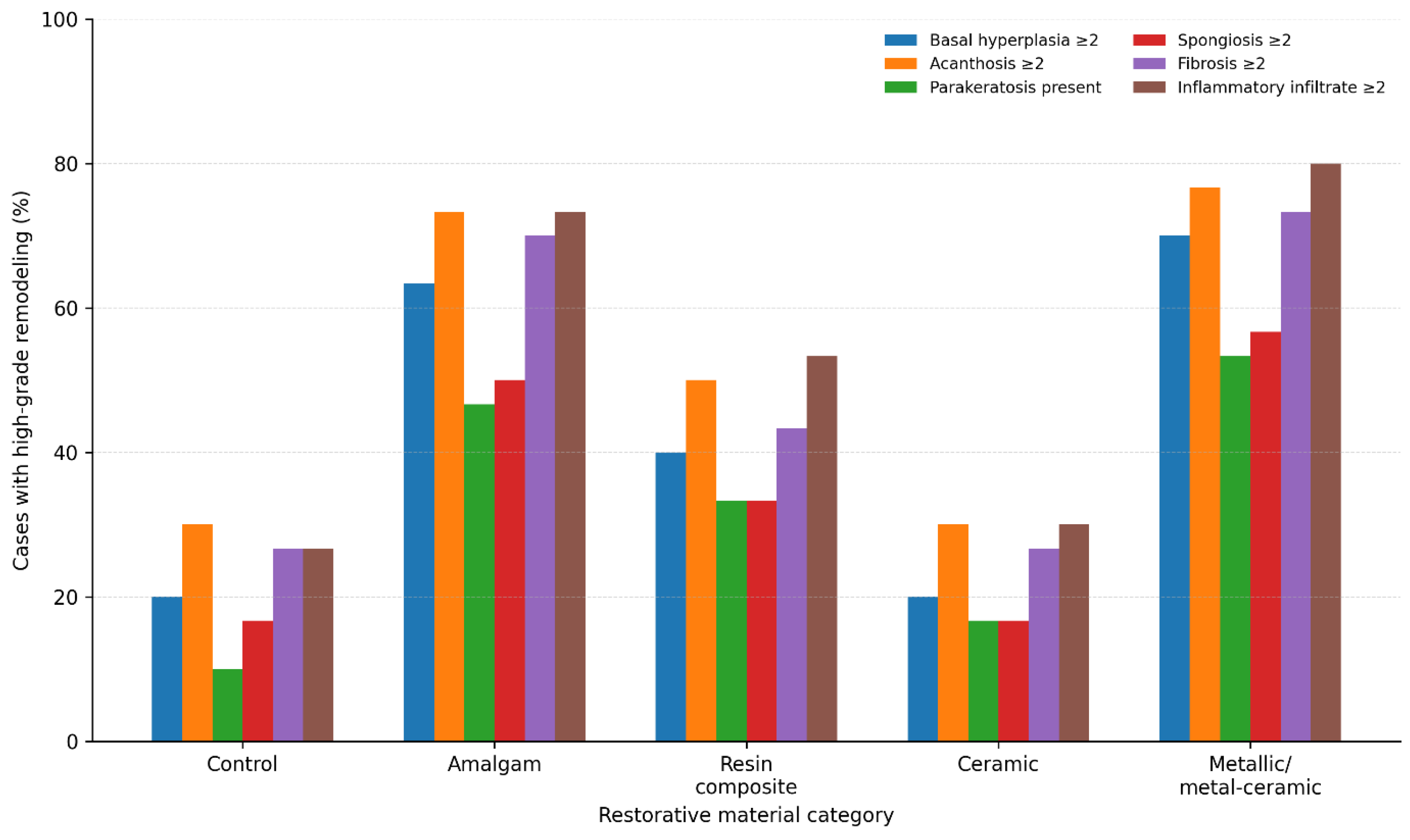
Figure 2.
Representative immunohistochemical expression patterns of CK19, Ki67, p53, and COX-2 across restorative material groups.
Figure 2.
Representative immunohistochemical expression patterns of CK19, Ki67, p53, and COX-2 across restorative material groups.
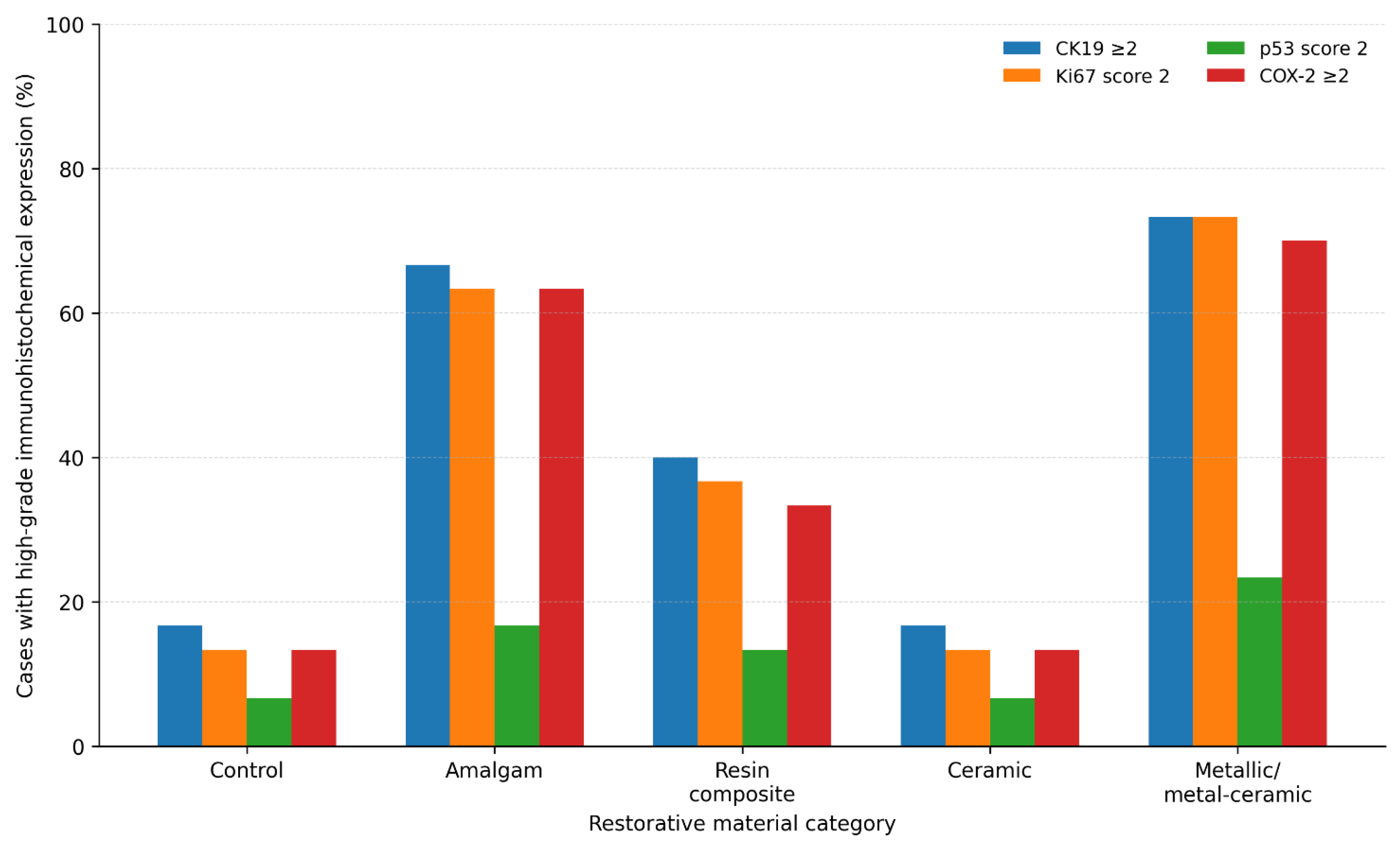
Figure 3.
Distribution of MAERS according to restorative material category.
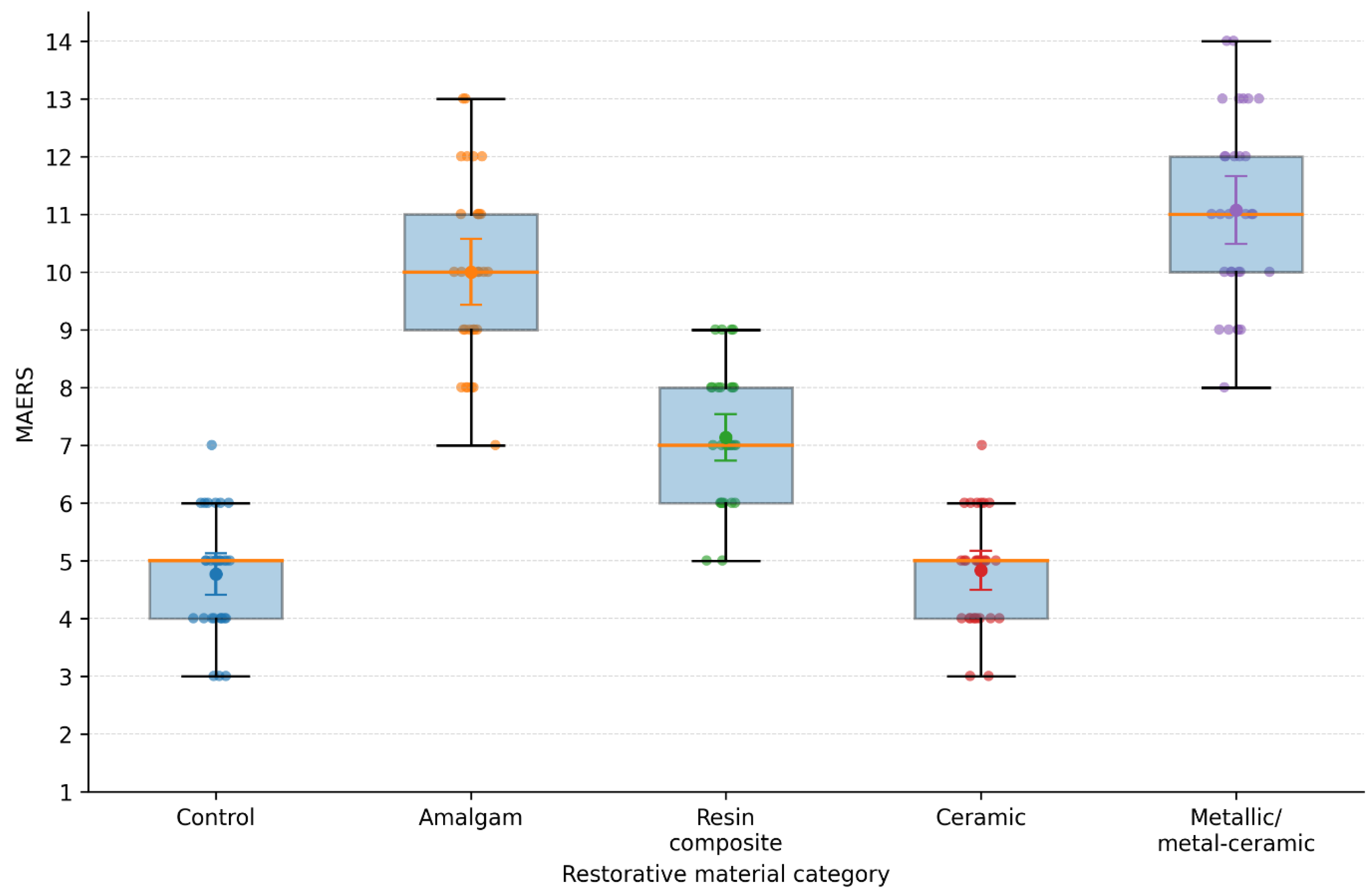
Figure 4.
Spearman correlation matrix of histopathological and immunohistochemical remodeling parameters.
Figure 4.
Spearman correlation matrix of histopathological and immunohistochemical remodeling parameters.
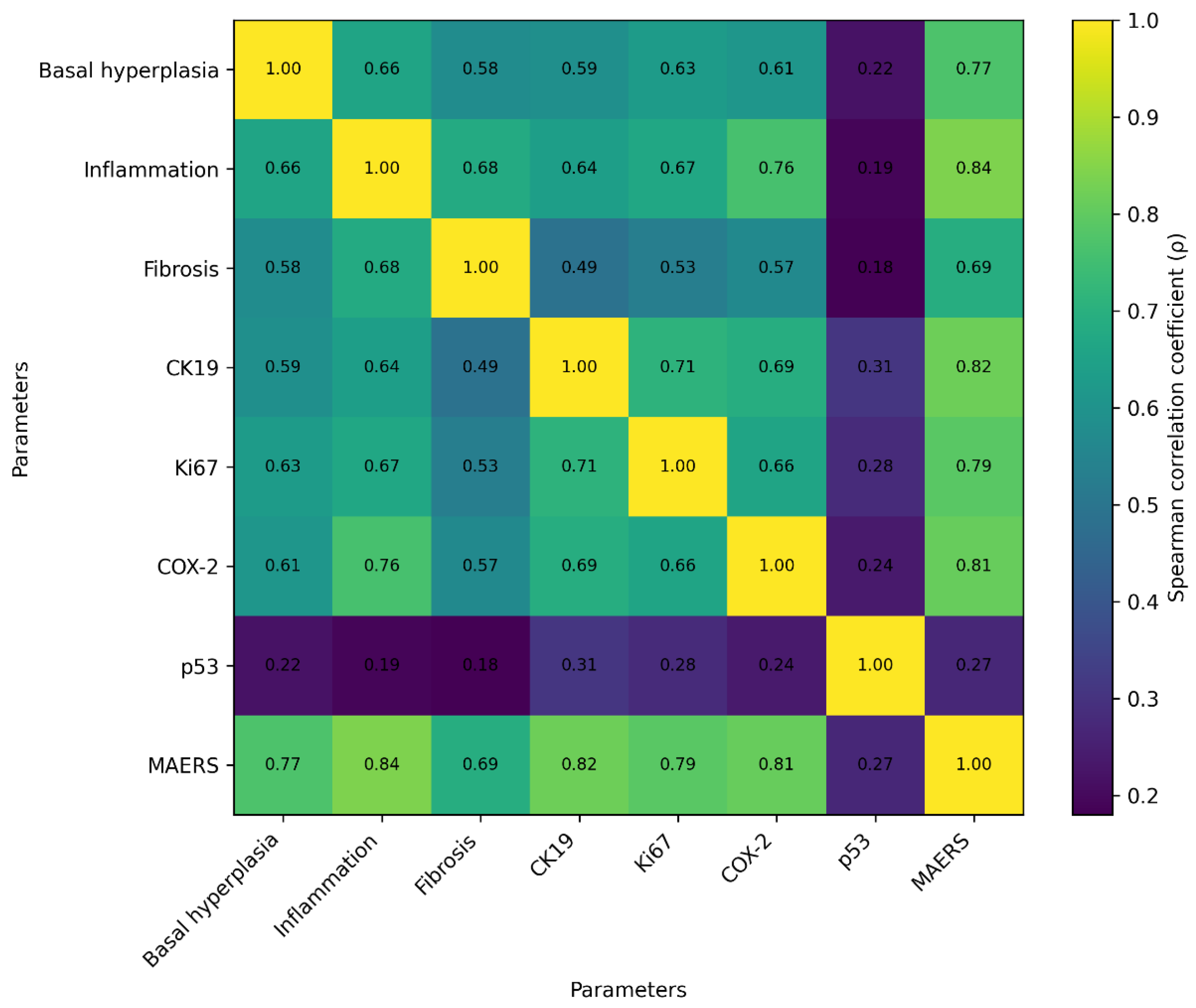
Table 1.
Clinicodemographic Characteristics of the Study Cohort.
| Parameter | Control (n=30) | Amalgam (n=30) | Resin composite (n=30) | Ceramic (n=30) | Metallic/metal-ceramic (n=30) | p-value |
|---|---|---|---|---|---|---|
| Age, years, mean ± SD | 49.2 ± 8.7 | 51.6 ± 9.1 | 48.8 ± 8.4 | 50.3 ± 7.9 | 52.1 ± 8.8 | 0.412 |
| Female, n (%) | 17 (56.7) | 18 (60.0) | 16 (53.3) | 17 (56.7) | 18 (60.0) | 0.962 |
| Male, n (%) | 13 (43.3) | 12 (40.0) | 14 (46.7) | 13 (43.3) | 12 (40.0) | 0.962 |
| Gingival mucosa, n (%) | 19 (63.3) | 20 (66.7) | 18 (60.0) | 19 (63.3) | 20 (66.7) | 0.973 |
| Alveolar mucosa, n (%) | 11 (36.7) | 10 (33.3) | 12 (40.0) | 11 (36.7) | 10 (33.3) | 0.973 |
| Contact duration, years, mean ± SD | NA | 12.8 ± 5.4 | 11.9 ± 4.8 | 10.7 ± 4.2 | 13.4 ± 5.7 | 0.286 |
| Contact duration ≥5 years, n (%) | NA | 30 (100.0) | 30 (100.0) | 30 (100.0) | 30 (100.0) | NA |
Note: SD = standard deviation; NA = not applicable. All specimens were obtained during alveoloplastic extraction procedures. Contact duration refers only to exposed groups.
Table 2.
Distribution of Histopathological Parameters According to Restorative Material Category.
| Histopathological parameter | Score | Control (n=30) | Amalgam (n=30) | Resin composite (n=30) | Ceramic (n=30) | Metallic/metal-ceramic (n=30) | p-value |
|---|---|---|---|---|---|---|---|
| Basal hyperplasia | 0 | 6 | 2 | 4 | 7 | 1 | <0.001 |
| 1 | 18 | 9 | 14 | 17 | 8 | ||
| 2 | 6 | 14 | 10 | 5 | 14 | ||
| 3 | 0 | 5 | 2 | 1 | 7 | ||
| Acanthosis | 0 | 1 | 0 | 1 | 2 | 0 | 0.002 |
| 1 | 20 | 8 | 14 | 19 | 7 | ||
| 2 | 9 | 16 | 12 | 8 | 15 | ||
| 3 | 0 | 6 | 3 | 1 | 8 | ||
| Parakeratosis | 0 | 27 | 16 | 20 | 25 | 14 | 0.004 |
| 1 | 3 | 14 | 10 | 5 | 16 | ||
| Spongiosis | 0 | 14 | 5 | 8 | 13 | 4 | 0.006 |
| 1 | 11 | 10 | 12 | 12 | 9 | ||
| 2 | 5 | 11 | 8 | 4 | 11 | ||
| 3 | 0 | 4 | 2 | 1 | 6 | ||
| Subepithelial fibrosis | 0 | 4 | 1 | 3 | 5 | 1 | <0.001 |
| 1 | 18 | 8 | 14 | 17 | 7 | ||
| 2 | 8 | 15 | 11 | 7 | 14 | ||
| 3 | 0 | 6 | 2 | 1 | 8 | ||
| Inflammatory infiltrate | 1 | 22 | 8 | 14 | 21 | 6 | <0.001 |
| 2 | 8 | 15 | 12 | 8 | 14 | ||
| 3 | 0 | 7 | 4 | 1 | 10 |
Note: Histopathological scores were compared among groups using the Kruskal–Wallis test. The p-value refers to the overall intergroup comparison for each histopathological parameter, not to each individual score category. Basal hyperplasia, acanthosis, spongiosis, and subepithelial fibrosis were graded from 0 to 3. Parakeratosis was recorded as absent (0) or present (1). Inflammatory infiltrate was graded as 1, mild and focal; 2, moderate and diffuse; and 3, intense. No epithelial dysplasia was identified in any study group.
Table 3.
Distribution of Immunohistochemical Markers According to Restorative Material Category.
| Immunohistochemical marker | Score | Control (n=30) | Amalgam (n=30) | Resin composite (n=30) | Ceramic (n=30) | Metallic/metal-ceramic (n=30) | p-value |
|---|---|---|---|---|---|---|---|
| CK19 | 0 | 9 | 2 | 5 | 8 | 1 | <0.001 |
| 1 | 16 | 8 | 13 | 17 | 7 | ||
| 2 | 4 | 13 | 9 | 4 | 13 | ||
| 3 | 1 | 7 | 3 | 1 | 9 | ||
| Ki67 | 0 | 8 | 2 | 5 | 9 | 1 | <0.001 |
| 1 | 18 | 9 | 14 | 17 | 7 | ||
| 2 | 4 | 19 | 11 | 4 | 22 | ||
| p53 | 0 | 21 | 15 | 18 | 22 | 13 | 0.031 |
| 1 | 7 | 10 | 8 | 6 | 10 | ||
| 2 | 2 | 5 | 4 | 2 | 7 | ||
| COX-2 | 0 | 12 | 3 | 7 | 13 | 2 | <0.001 |
| 1 | 14 | 8 | 13 | 13 | 7 | ||
| 2 | 4 | 12 | 8 | 3 | 12 | ||
| 3 | 0 | 7 | 2 | 1 | 9 |
Note: Immunohistochemical scores were compared among groups using the Kruskal–Wallis test. The p-value refers to the overall intergroup comparison for each immunohistochemical marker, not to each individual score category. CK19 was scored from 0 to 3 according to epithelial distribution and staining intensity. Ki67 was scored from 0 to 2 according to the percentage of positive epithelial nuclei. p53 was scored from 0 to 2 based on nuclear staining extent and intensity. COX-2 was scored from 0 to 3 according to epithelial and subepithelial staining intensity and distribution. For each marker and group, values represent the number of cases assigned to each score category. Strong diffuse p53 overexpression was not observed as a dominant pattern in any group.
Disclaimer/Publisher’s Note: The statements, opinions and data contained in all publications are solely those of the individual author(s) and contributor(s) and not of MDPI and/or the editor(s). MDPI and/or the editor(s) disclaim responsibility for any injury to people or property resulting from any ideas, methods, instructions or products referred to in the content. |
© 2026 by the authors. Licensee MDPI, Basel, Switzerland. This article is an open access article distributed under the terms and conditions of the Creative Commons Attribution (CC BY) license (http://creativecommons.org/licenses/by/4.0/).
Copyright: This open access article is published under a Creative Commons CC BY 4.0 license, which permit the free download, distribution, and reuse, provided that the author and preprint are cited in any reuse.


