

Submitted:
12 June 2026
Posted:
15 June 2026
You are already at the latest version
Abstract
Background: The incidence of placenta accreta spectrum (PAS) with placenta previa has been previously reported. However, the incidence varies across reports, suggesting that unknown risk factors may be involved. This study aimed to reevaluate the risk of PAS in patients with placenta previa. Methods: This multicenter retrospective study was conducted from 2015 to 2024 in patients with placenta previa who underwent cesarean section (CS). PAS was defined based on pathological or clinical findings, such as manual removal of the placenta or obvious retention of the placenta if a hysterectomy was not performed. The incidence of PAS and associated risk factors were analyzed using multivariable logistic regression. Results: PAS was observed in 26% of women with placenta previa. The incidence of PAS increased significantly with the number of prior CSs: 13.2% in women with no prior CS, 41.9% in those with one prior CS, and 66.7% in those with two or more prior cesarean sections. Multivariate analysis identified major placenta previa (aOR 2.69, 95% CI 1.11–6.54), including complete and partial placenta previa, and number of prior CSs (one prior: aOR 4.35, 95% CI 1.94-9.73; two or more prior: aOR 9.48, 95% CI 2.55-35.2) as independent risk factors. Conclusions: The incidence of PAS with placenta previa was higher than that previously reported, and major placenta previa was shown to be an independent risk factor, regardless of prior CS history. Comprehensive evaluation, including prior CS and placenta previa classification, is crucial for accurate risk stratification and perinatal management.
Keywords:
cesarean section
; placenta accreta
; placenta previa
; postpartum hemorrhage
; risk factors
1. Introduction
Placenta accreta spectrum (PAS) is a disease in which chorionic villi adhere to and invade the myometrium beyond the decidua propria. PAS is associated with increased maternal perinatal morbidity and mortality due to increased blood loss during cesarean section (CS) [1,2]. Risk factors for PAS include previous CS, assisted reproductive technology (ART) pregnancy, prior uterine artery embolization, advanced maternal age, and placenta previa [3,4,5,6]. A preoperative diagnosis of PAS has not yet been established, although ultrasonography and MRI have been reported, and a confirmed diagnosis is difficult to make [7,8]. Given the high risk of massive hemorrhage in PAS, obstetricians should ensure a multidisciplinary approach, including readiness for blood transfusion and interventional radiology, at the time of delivery.
The reported incidence of PAS in placenta previa varies widely depending on population characteristics, PAS definitions, and inclusion criteria [9,10]. Notably, a recent large-scale population-based study combined placenta previa and low-lying placenta and focused primarily on women with prior cesarean scars [11]. While such studies provide valuable epidemiological insights, they cannot be directly applied in clinical practice, because the extent and location of the placenta may influence the likelihood of abnormal trophoblastic invasion. In addition, Asian populations, including those in Japan, exhibit characteristics such as an aging maternal population, increased ART use, and a concentration of high-risk deliveries, which may create a distinct risk environment [12].
In the present study, we present the incidence of PAS in pregnant Japanese women with placenta previa and investigate the risk factors for PAS by considering the classification of placenta previa and placental location. This study aimed to provide practical knowledge that will contribute to the optimization of safety management systems for pregnant women with PAS.
2. Materials and Methods
Study Design and Participants
This multi-center observational study included patients who delivered at the Perinatal Center of Gunma University Hospital (Maebashi, Japan) and Japanese Red Cross Maebashi Hospital (Maebashi, Japan) between April 2015 and December 2024 and January 2017 and December 2023, respectively. The study protocol was approved by the Institutional Review Board of the Gunma University Hospital on May 8th, 2024 (approval number: HS2023-110). All patients diagnosed with placenta previa who delivered via CS during the study period were enrolled.
Collected Variables
To evaluate maternal characteristics, data on maternal age, ART rate, pre-pregnancy body mass index (BMI), incidence of endometriosis, number of prior CS, placental location (anterior or posterior), type of previa (minor or major), incidence of emergent CS, and gestational age at delivery were collected. Anterior placental location was defined as cases in which the placenta covered the uterine incision at the CS. Major placenta previa was defined as a placental edge covering the internal os on transvaginal ultrasonography immediately before delivery. Minor placenta previa was defined as a placental edge that did not cover the internal cervical os [13].
The maternal outcomes included intraoperative blood loss, intensive care unit (ICU) admission, use of intrauterine balloon tamponade, incidence of cesarean hysterectomy, incidence of PAS, incidence of blood transfusion, and hospitalization period. PAS was defined based on pathological or clinical findings, such as manual removal of the placenta or obvious retention of the placenta if a hysterectomy was not performed.
Statistical Analysis
The Mann–Whitney U test was used for the analysis of continuous values in the 2 groups and the χ2 test for categorical values. We evaluated the annual temporal trends in baseline characteristics and outcomes according to the number of prior CS using the Cochran-Armitage trend and Jonckheere-Terpstra tests. All statistical tests were two-tailed, and a P value < 0.05 was considered statistically significant. To examine whether the number of prior CSs was a risk factor for PAS, univariate logistic regression was performed and reported as odds ratios (OR) with corresponding 95% confidence intervals (CI) and p-values. Multivariate analysis was performed using multivariate logistic regression with stepwise elimination of variables including placental location, type of placenta, number of prior CS, ART pregnancy, and incidence of endometriosis. Statistical analyses were performed using EZR version 1.52 (Saitama Medical Center, Jichi Medical University, Saitama, Japan), a graphical user interface for R version 4.02 (R Foundation for Statistical Computing, Vienna, Austria).
3. Results
3.1. Baseline Characteristics and Maternal Outcomes
Data from 213 patients diagnosed with placenta previa who delivered via CS during the study period were retrospectively analyzed. The baseline characteristics and outcomes of each participant (control and PAS groups) are summarized in Table 1. There were no significant differences between the PAS and control groups in terms of maternal age, ART pregnancy, pre-pregnancy BMI, or incidence of endometriosis. However, the incidence of prior CS, placental anterior location, and major placenta previa was significantly higher in the PAS group than in the control group (60.0% versus [vs.] 19.0% [p < 0.001], 44.0% vs. 19.6% [p = 0.001], and 84.0% vs. 65.0% [p = 0.013], respectively). The PAS group had a significantly earlier gestational age at delivery compared to the control group (36.3 vs. 37.1 weeks [p = 0.001]). Maternal outcomes such as intraoperative blood loss, ICU admission rate, blood transfusion rate, and hysterectomy rate were significantly worse in the PAS group than in the control group (2819 g vs. 1295 g [p < 0.001], 26.0% vs. 3.7% [p < 0.001], 36.0% vs. 9.8% [p < 0.001], and 52.0% vs. 2.5% [p < 0.001], respectively). No significant differences in hospitalization period, incidence of emergent CS, or use of intrauterine balloon tamponade were found between the groups.
3.2. Clinical Characteristics According to the Number of Prior Cesarean Sections
Consistent with previous findings, the association between prior CS and PAS was clear. Therefore, we investigated patient characteristics and outcomes based on the number of prior CS (Table 2). There were no significant differences between the groups in terms of ART pregnancy or major placenta previa across CS strata. The incidence of endometriosis and gestational age at delivery showed significant decreasing trends (p-trend = 0.032 and p-trend < 0.001, respectively). Maternal age, pre-pregnancy BMI, and incidence of anterior placental location increased significantly with an increasing number of prior CS (p-trend = 0.040, p-trend = 0.010, and p-trend < 0.001, respectively). Furthermore, the incidence of PAS increased significantly with an increasing number of prior CS (p-trend < 0.001; Figure 1), occurring in 13.2% (20/152) of women with no prior CS, 41.9% (18/43) with one prior CS, and 66.7% (12/18) with two or more prior CS. As expected, maternal outcomes such as intraoperative blood loss, ICU admission rate, blood transfusion rate, and hysterectomy rate were significantly worse with increasing numbers of prior CS (Table 2).
This figure shows the incidence of placenta accreta spectrum (PAS) among women with placenta previa stratified by the number of prior cesarean sections (CSs). PAS occurred in 13.2% (20/152) of women with no prior CS, 41.9% (18/43) with one prior CS, and 66.7% (12/18) with two or more prior CSs. A significant increasing trend in PAS incidence was observed with an increasing number of prior CSs (P for trend < 0.001, Cochran–Armitage trend test).
3.3. Independent Risk Factors for Placenta Accreta Spectrum
In the univariate analysis, anterior placental location, major placenta previa, and the number of prior CS were associated with PAS. After adjusting for the aforementioned covariates, major placenta previa and an increased number of prior CS remained significantly associated with an increased risk of PAS. Specifically, major placenta previa was associated with an adjusted odds ratio (aOR) of 2.69 (95% CI, 1.11–6.54). Compared to women with no prior CS, those with one prior CS had an aOR of 4.35 (95% CI, 1.94–9.73), and those with two or more prior CS had an aOR of 9.48 (95% CI, 2.55–35.2) (Table 3).
4. Discussion
In the present study, the incidence of PAS increased with the number of prior CS and was higher than that previously reported. Furthermore, in addition to the conventional risk factors, major placenta previa, encompassing both complete and partial placenta previa, has been identified as a new independent risk factor for PAS.
In this study, the risk increased progressively with the number of prior CS, which is consistent with previous reports; however, the incidence of PAS was higher than previously reported [14,15,16]. The increasing maternal risk profile due to delayed childbearing and ART pregnancies, combined with the tertiary referral nature of our facilities, results in a concentration of high-risk cases [12,17]. In Japan, placenta previa and low-lying placenta are clearly distinguished; all cases in this study were placenta previa, and the low-lying placenta was not included. Furthermore, the definition of PAS includes both pathological and clinical diagnoses. According to FIGO guidelines, PAS is diagnosed based on pathological or clinical findings; however, pathological diagnosis is often unattainable, and clinical diagnosis is frequently relied upon in clinical practice [7]. Therefore, in this study, both clinical and pathological diagnoses were included in the PAS definition in accordance with these guidelines. These factors may have contributed to the increased incidence of PAS.
This study classified placenta previa, which had previously been treated as a single category, into major previa (including complete and partial previa) and minor previa (including marginal previa), and demonstrated that major placenta previa independently increased the risk of PAS. In major placenta previa, the placenta extensively covers the lower uterine segment, including the internal os, making it difficult for the normal decidua basalis to form and allowing the placental villi to readily approach and invade the uterine muscle layer [18]. Even in the absence of a previous cesarean scar, specific structural features, such as thinning of the myometrium in the lower uterine segment or altered blood flow combined with a large placental attachment area, can potentially cause PAS [19]. Defects in the decidua are considered the fundamental cause of PAS, and major placenta previa may provide the anatomical conditions that promote this defect. Given that major previa alone increases the risk of PAS, it may be necessary to consider PAS and implement careful management even in patients without a history of CS.
Based on previous studies, we included ART pregnancy and a history of endometriosis as adjustment factors in the analysis model. The pathophysiology by which an ART pregnancy increases the risk of PAS is postulated to involve abnormal endometrial maturation during embryo transfer and abnormalities in the uterine environment [20,21,22]. The mechanisms linking PAS to endometriosis include impaired decidualization associated with chronic inflammation and fibrosis as well as an abnormal endometrial environment due to coexistence with ART pregnancies [23,24]. However, in this study, ART pregnancy and endometriosis did not emerge as independent risk factors, and their risk independence may have been obscured by other strong structural uterine factors. Therefore, the onset of PAS should be understood as a multifactorial disease resulting from the cumulative effects of multiple factors, including placenta previa classification, method of conception, and history of uterine disorders, rather than being caused by a single factor.
This study had several limitations. First, this was a retrospective observational study; therefore, there may be variability in the accuracy of medical record documentation and the consistency of chart entries. Furthermore, it cannot be ruled out that differences in PAS diagnosis and management policies between facilities may have influenced the analysis. Second, due to the limited number of PAS cases overall, detailed stratification analyses, such as placenta previa classification, number of prior CS, and placental location, may not have had sufficient statistical power. While previous studies have reported a higher incidence of PAS in patients with placenta previa, in whom the placenta is located in the anterior uterine wall, this finding did not emerge as an independent risk factor in this study [11,25]. This discrepancy may be explained by the strong correlation between anterior placentation and previous cesarean scars, leading to confounding factors in the multivariable models. Third, we could not include clinically important factors, such as surgeon experience and detailed placental ultrasound findings, in the analysis. These unadjusted confounding factors may have influenced the risk assessment. Fourth, because this study was based on data collected at tertiary facilities, the incidence and risk factors of PAS identified in this study may not be directly applicable to the general population.
5. Conclusions
In conclusion, this study demonstrated that the risk of PAS significantly increased in patients with major placenta previa, even in those with a history of one or fewer CS. A detailed evaluation of placental location is essential to enable earlier risk stratification and appropriate referral to tertiary care facilities.
Author Contributions
A.M. contributed to the conception and design of the work, data analysis, interpretation of the data, and manuscript writing; A.M., Y.O., and S.W. collected and analyzed data; K.T., R.U., A.T., D.H. and T.S. searched and interpreted the data and critically revised the manuscript; A.I. contributed to the conception and design of the work, acquisition and interpretation of the data, and critical revision of the manuscript.
Funding
This research received no external funding.
Institutional Review Board Statement
The study was conducted in accordance with the Declaration of Helsinki, and approved by the Institutional Review Board of the Gunma University Hospital on May 8th, 2024 (approval number: HS2023-110).
Informed Consent Statement
Informed consent was obtained from all subjects involved in the study.
Data Availability Statement
The raw data supporting the conclusions of this article will be made available by the authors on request.
Acknowledgments
We would like to thank the members of department of Obstetrics and Gynecology, Gunma University Hospital. We would like to thank Editage (www.editage.jp) for the English language editing.
Conflicts of Interest
The authors declare no conflicts of interest.
Abbreviations
The following abbreviations are used in this manuscript:
| ART | Assisted Reproductive Technology |
| aOR | Adjusted Odds Ratio |
| BMI | Body Mass Index |
| CI | Confidence Interval |
| CS | Cesarean Section |
| ICU | Intensive Care Unit |
| MRI | Magnetic Resonance Imaging |
| OR | Odds Ratio |
| PAS | Placenta Accreta Spectrum |
References
- Collins, S.L.; Alemdar, B.; van Beekhuizen, H.J.; Bertholdt, C.; Braun, T.; Calda, P.; Delorme, P.; Duvekot, J.J.; Gronbeck, L.; Kayem, G.; Langhoff-Roos, J.; Marcellin, L.; Martinelli, P.; Morel, O.; Mhallem, M.; Morlando, M.; Noergaard, L.N.; Nonnenmacher, A.; Pateisky, P.; Petit, P.; Rijken, M.J.; Ropacka-Lesiak, M.; Schlembach, D.; Sentilhes, L.; Stefanovic, V.; Strindfors, G.; Tutschek, B.; Vangen, S.; Weichert, A.; Weizsäcker, K.; Chantraine, F.; International Society for Abnormally Invasive Placenta (IS-AIP). Evidence-based guidelines for the management of abnormally invasive placenta: recommendations from the International Society for Abnormally Invasive Placenta. Am. J. Obstet. Gynecol. 2019, 220, 511–26. [Google Scholar] [CrossRef] [PubMed]
- Marcellin, L.; Delorme, P.; Bonnet, M.P.; Grange, G.; Kayem, G.; Tsatsaris, V.; Goffinet, F. Placenta percreta is associated with more frequent severe maternal morbidity than placenta accreta. Am. J. Obstet. Gynecol. 2018, 219, 193.e1–e9. [Google Scholar] [CrossRef] [PubMed]
- Belfort, M.A. Placenta accreta. Am. J. Obstet. Gynecol. 2010, 203, 430–9. [Google Scholar] [CrossRef] [PubMed]
- Matsuzaki, S.; Lee, M.; Nagase, Y.; Jitsumori, M.; Matsuzaki, S.; Maeda, M.; Takiuchi, T.; Kakigano, A.; Mimura, K.; Ueda, Y.; Tomimatsu, T.; Endo, M.; Kimura, T. A systematic review and meta-analysis of obstetric and maternal outcomes after prior uterine artery embolization. Sci. Rep. 2021, 11, 16914. [Google Scholar] [CrossRef] [PubMed]
- Matsuzaki, S.; Nagase, Y.; Takiuchi, T.; Kakigano, A.; Mimura, K.; Lee, M.; Matsuzaki, S.; Ueda, Y.; Tomimatsu, T.; Endo, M.; Kimura, T. Antenatal diagnosis of placenta accreta spectrum after in vitro fertilization-embryo transfer: a systematic review and meta-analysis. Sci. Rep. 2021, 11, 9205. [Google Scholar] [CrossRef] [PubMed]
- Miller, D.A.; Chollet, J.A.; Goodwin, T.M. Clinical risk factors for placenta previa-placenta accreta. Am. J. Obstet. Gynecol. 1997, 177, 210–4. [Google Scholar] [CrossRef] [PubMed]
- Jauniaux, E.; Bhide, A.; Kennedy, A.; Woodward, P.; Hubinont, C.; Collins, S. FIGO Placenta Accreta Diagnosis and Management Expert Consensus Panel. FIGO consensus guidelines on placenta accreta spectrum disorders: Prenatal diagnosis and screening. Int. J. Gynaecol. Obstet. 2018, 140, 274–80. [Google Scholar] [CrossRef] [PubMed]
- Tanimura, K.; Yamasaki, Y.; Ebina, Y.; Deguchi, M.; Ueno, Y.; Kitajima, K.; Yamada, H. Prediction of adherent placenta in pregnancy with placenta previa using ultrasonography and magnetic resonance imaging. Eur. J. Obstet. Gynecol. Reprod. Biol. 2015, 187, 41–4. [Google Scholar] [CrossRef] [PubMed]
- Silver, R.M.; Landon, M.B.; Rouse, D.J.; Leveno, K.J.; Spong, C.Y.; Thom, E.A.; et al. Maternal morbidity associated with multiple repeat cesarean deliveries. Obstet. Gynecol. 2006, 107, 1226–32. [Google Scholar] [CrossRef] [PubMed]
- Sumigama, S.; Itakura, A.; Ota, T.; Okada, M.; Kotani, T.; Hayakawa, H.; Yoshida, K.; Ishikawa, K.; Hayashi, K.; Kurauchi, O.; Yamada, S.; Nakamura, H.; Matsusawa, K.; Sakakibara, K.; Ito, M.; Kawai, M.; Kikkawa, F. Placenta previa increta/percreta in Japan: a retrospective study of ultrasound findings, management and clinical course. J. Obstet. Gynaecol. Res. 2007, 33, 606–11. [Google Scholar] [CrossRef] [PubMed]
- Kayem, G.; Seco, A.; Vendittelli, F.; Crenn Hebert, C.; Dupont, C.; Branger, B.; Huissoud, C.; Fresson, J.; Winer, N.; Langer, B.; Rozenberg, P.; Morel, O.; Bonnet, M.P.; Perrotin, F.; Azria, E.; Carbillon, L.; Chiesa, C.; Raynal, P.; Rudigoz, R.C.; Patrier, S.; Beucher, G.; Dreyfus, M.; Sentilhes, L.; Deneux-Tharaux, C. Risk factors for placenta accreta spectrum disorders in women with any prior cesarean and a placenta previa or low lying: a prospective population-based study. Sci. Rep. 2024, 14, 6564. [Google Scholar] [CrossRef] [PubMed]
- Katagiri, Y.; Jwa, S.C.; Kuwahara, A.; Iwasa, T.; Ono, M.; Kato, K.; Kishi, H.; Kuwabara, Y.; Taniguchi, F.; Harada, M.; Iwase, A.; Sugino, N. Assisted reproductive technology in Japan: A summary report for 2022 by the Ethics Committee of the Japan Society of Obstetrics and Gynecology. Reprod. Med. Biol. 2024, 23, e12620. [Google Scholar] [CrossRef] [PubMed]
- Calì, G.; Giambanco, L.; Puccio, G.; Forlani, F. Morbidly adherent placenta: evaluation of ultrasound diagnostic criteria and differentiation of placenta accreta from percreta. Ultrasound Obstet. Gynecol. 2013, 41, 406–12. [Google Scholar] [CrossRef] [PubMed]
- Bowman, Z.S.; Eller, A.G.; Bardsley, T.R.; Greene, T.; Varner, M.W.; Silver, R.M. Risk factors for placenta accreta: a large prospective cohort. Am. J. Perinatol. 2014, 31, 799–804. [Google Scholar] [CrossRef] [PubMed]
- Fitzpatrick, K.E.; Sellers, S.; Spark, P.; Kurinczuk, J.J.; Brocklehurst, P.; Knight, M. Incidence and risk factors for placenta accreta/increta/percreta in the UK: a national case-control study. PLoS ONE 2012, 7, e52893. [Google Scholar] [CrossRef] [PubMed]
- Ogawa, K.; Jwa, S.C.; Morisaki, N.; Sago, H. Risk factors and clinical outcomes for placenta accreta spectrum with or without placenta previa. Arch. Gynecol. Obstet. 2022, 305, 607–15. [Google Scholar] [CrossRef] [PubMed]
- Raymo, J.M.; Uchikoshi, F.; Yoda, S. Marriage intentions, desires, and pathways to later and less marriage in Japan. Demogr. Res. 2021, 44, 67–98. [Google Scholar] [CrossRef] [PubMed]
- Hecht, J.L.; Baergen, R.; Ernst, L.M.; Katzman, P.J.; Jacques, S.M.; Jauniaux, E.; Khong, T.Y.; Metlay, L.A.; Poder, L.; Qureshi, F.; Rabban, J.T., 3rd; Roberts, D.J.; Shainker, S.; Heller, D.S. Classification and reporting guidelines for the pathology diagnosis of placenta accreta spectrum (PAS) disorders: recommendations from an expert panel. Mod. Pathol. 2020, 33, 2382–96. [Google Scholar] [CrossRef] [PubMed]
- Arakaza, A.; Zou, L.; Zhu, J. Placenta Accreta Spectrum Diagnosis Challenges and Controversies in Current Obstetrics: A Review. Int. J. Womens Health 2023, 15, 635–54. [Google Scholar] [CrossRef] [PubMed]
- Jwa, S.C.; Tamaru, S.; Takamura, M.; Namba, A.; Kajihara, T.; Ishihara, O.; Kamei, Y. Assisted reproductive technology-associated risk factors for placenta accreta spectrum after vaginal delivery. Sci. Rep. 2024, 14, 7454. [Google Scholar] [CrossRef] [PubMed]
- Kyozuka, H.; Yamaguchi, A.; Suzuki, D.; Fujimori, K.; Hosoya, M.; Yasumura, S.; Yokoyama, T.; Sato, A.; Hashimoto, K. Japan Environment and Children’s Study (JECS) Group. Risk factors for placenta accreta spectrum: findings from the Japan environment and Children’s study. BMC Pregnancy Childbirth 2019, 19, 447. [Google Scholar] [CrossRef] [PubMed]
- Liu, X.; Li, W.; Wen, W.; Wang, T.; Wang, T.; Sun, T.; Zhang, N.; Pan, D.; Xie, J.; Liu, X.; Cai, H.; Li, X.; Shi, Z.; Wang, R.; Lu, N.; Bai, H.; Pan, R.; Tian, L.; Meng, B.; Mu, X.; Jia, H.; Zhou, H.; Cao, X.; Liu, T.; Qu, P.; Liu, D.; Mol, B.W.; Shi, J. Natural cycle versus hormone replacement therapy as endometrial preparation in ovulatory women undergoing frozen-thawed embryo transfer: The compete open-label randomized controlled trial. PLoS Med. 2025, 22, e1004630. [Google Scholar] [CrossRef] [PubMed]
- Matsuzaki, S.; Ueda, Y.; Nagase, Y.; Matsuzaki, S.; Kakuda, M.; Kakuda, S.; Sakaguchi, H.; Hisa, T.; Kamiura, S. Placenta Accreta Spectrum Disorder Complicated with Endometriosis: Systematic Review and Meta-Analysis. Biomedicines 2022, 10, 390. [Google Scholar] [CrossRef] [PubMed]
- Wang, F.; Wang, Q.; Song, Y.; Ding, J.; Li, H.; Meng, Q. Programmed frozen embryo transfer cycles are associated with a higher risk of abnormal placental development: a retrospective cohort study of singleton live births. Front Endocrinol. 2023, 14, 1202044. [Google Scholar] [CrossRef] [PubMed]
- White, A.; Malik, M.; Pruszynski, J.E.; Do, Q.N.; Spong, C.Y.; Herrera, C.L. Contemporary Placenta Accreta Spectrum Disorder Incidence and Risk Factors. Obstet. Gynecol. 2025, 145, 665–73. [Google Scholar] [CrossRef] [PubMed]
Figure 1.
Incidence of placenta accreta spectrum according to the number of prior cesarean sections.
Figure 1.
Incidence of placenta accreta spectrum according to the number of prior cesarean sections.
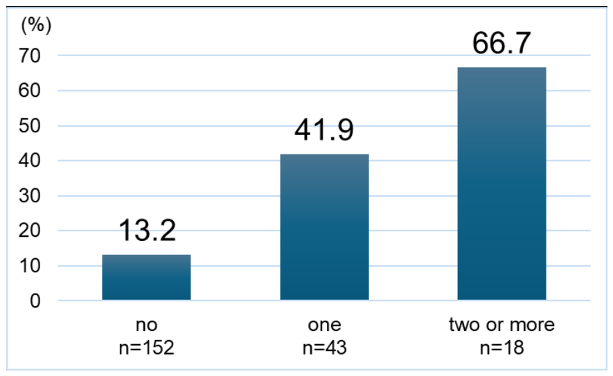

Table 1.
Maternal and obstetric characteristics of women with placenta previa.
| control (n=163) | PAS (n=50) | P value | |
| Maternal age (years) | 35.0 (32.0-38.0) | 35.0 (32.3-38.8) | 0.203 |
| ART pregnancy, n (%) | 35 (21.5) | 13 (26.0) | 0.562 |
| Pre-pregnancy BMI | 21.2 (19.5-23.9) | 21.2 (19.4-24.0) | 0.762 |
| Endometriosis, n (%) | 36 (22.1) | 7 (14.0) | 0.235 |
| Prior CS, n (%) | 31 (19.0) | 30 (60.0) | <0.001 |
| Anterior location, n (%) | 32 (19.6) | 22 (44.0) | 0.001 |
| Major planenta previa, n (%) | 106 (65.0) | 42 (84.0) | 0.013 |
| Gestational age at delivery (weeks) | 37.1 (36.4-37.4) | 36.3 (35.6-37.0) | 0.001 |
| Emergency CS, n (%) | 46 (28.2) | 13 (26.0) | 0.857 |
| Intraoperative blood loss, (g) | 1295 (979-1857) | 2819 (1775-4117) | <0.001 |
| Birth weight (g) | 2564 (2325-2794) | 2561 (2337-2727) | 0.651 |
| ICU admission, n (%) | 6 (3.7) | 13 (26.0) | <0.001 |
| Blood transfusion, n (%) | 16 (9.8) | 18 (36.0) | <0.001 |
| Intrauterine balloon tamponade, n (%) | 48 (29.4) | 19 (38.0) | 0.297 |
| Hospitalization period (days) | 13.0 (9.0-29.50) | 17.0 (10.0-30.8) | 0.181 |
| Cesarean hysterctomy, n (%) | 4 (2.5) | 26 (52.0) | <0.001 |
Values are mean (SD: standard deviation). Abbreviation: BMI, body mass index; CS, cesarean section; ART, assisted reproductive technology; ICU, intensive care unit.
Table 2.
Clinical characteristics stratified by the number of prior cesarean sections.
| No prior (n=152) |
one prior (n=43) |
two or more prior (n=18) |
p for trend | |
| Maternal age (years) | 35.0 (32.0-37.0) | 37.0 (32.0-39.0) | 35.5 (35.0-39.0) | 0.04 |
| ART pregnancy, n (%) | 36 (23.7) | 10 (23.3) | 2 (11.1) | 0.36 |
| Pre-pregnancy BMI | 20.7 (19.3-23.7) | 21.5 (20.5-25.6) | 22.4 (21.3-23.8) | 0.01 |
| Endometriosis, n (%) | 37 (24.3) | 4 (9.3) | 2 (11.1) | 0.032 |
| Anterior location, n (%) | 26 (17.1) | 16 (37.2) | 12 (66.7) | <0.001 |
| Major planenta previa, n (%) | 103 (67.8) | 31 (72.1) | 14 (77.8) | 0.627 |
| Gestational age at delivery (weeks) | 37.1 (36.6-37.4) | 36.1 (35.3-37.0) | 36.4 (35.6-36.7) | <0.001 |
| PAS, n (%) | 20 (13.2) | 18 (41.9) | 12 (66.7) | <0.001 |
| Emergency CS, n (%) | 41 (27.0) | 16 (37.2) | 2 (11.1) | 0.65 |
| Intraoperative blood loss, (g) | 1317 (982-1953) | 2031 (1310-3620) | 3092 (2202-6103) | <0.001 |
| Birth weight (g) | 2572 (2324-2794) | 2468 (2213-2665) | 2613 (2507-2793) | 0.517 |
| ICU admission, n (%) | 6 (3.9) | 8 (18.6) | 5 (27.8) | <0.001 |
| Blood transfusion, n (%) | 14 (9.2) | 12 (27.9) | 8 (44.4) | <0.001 |
| Intrauterine balloon tamponade, n (%) | 43 (28.3) | 20 (46.5) | 4 (22.2) | 0.464 |
| Hospitalization period (days) | 11.50 (9.0-27.0) | 21.0 (10.5-35.5) | 17.0 (13.0-32.5) | 0.012 |
| Cesarean hysterctomy, n (%) | 2 (1.3) | 14 (32.6) | 14 (77.8) | <0.001 |
Values are mean (SD: standard deviation). Abbreviation: BMI, body mass index; CS, cesarean section; PAS, placenta accreta spectrum; ART, assisted reproductive technology; ICU, intensive care unit.
Table 3.
Multivariable logistic regression analysis for placenta accreta spectrum.
| univariate odds ratio (95% CI) |
P value | adjusted odds ratio (95% CI) |
P value | ||
| Placental location | Posterior | ref | ref | ||
| Anterior | 3.22 (1.63-6.34) | <0.001 | 1.69 (0.76-3.76) | 0.200 | |
| Placenta previa type | Minor | ref | ref | ||
| Major | 2.82 (1.24-6.42) | 0.013 | 2.69 (1.11-6.54) | 0.029 | |
| Prior cesarean | No | ref | ref | ||
| One | 4.75 (2.21-10.2) | < 0.001 | 4.35 (1.94-9.73) | < 0.001 | |
| Two or more | 10.6 (3.14-35.5) | < 0.001 | 9.48 (2.55-35.2) | < 0.001 | |
| ART pregnancy | No | ref | ref | ||
| Yes | 0.99 (0.64-1.54) | 0.974 | 1.16 (0.72-1.87) | 0.544 | |
| Endometriosis | No | ref | ref | ||
| Yes | 0.57 (0.24-1.39) | 0.217 | 0.95 (0.36-2.48) | 0.913 |
Abbreviation: ART, assisted reproductive technology.
Disclaimer/Publisher’s Note: The statements, opinions and data contained in all publications are solely those of the individual author(s) and contributor(s) and not of MDPI and/or the editor(s). MDPI and/or the editor(s) disclaim responsibility for any injury to people or property resulting from any ideas, methods, instructions or products referred to in the content. |
© 2026 by the authors. Licensee MDPI, Basel, Switzerland. This article is an open access article distributed under the terms and conditions of the Creative Commons Attribution (CC BY) license.
Copyright: This open access article is published under a Creative Commons CC BY 4.0 license, which permit the free download, distribution, and reuse, provided that the author and preprint are cited in any reuse.


